Article

Multiscale Entropy Analysis of Surface Electromyographic Signals from the Urethral Sphincter as a Prognostic Indicator for Surgical Candidates with Primary Bladder Neck Obstruction

1
Department of Electrical Engineering, National Dong Hwa University, Hualien 97401, Taiwan
2
Department of Urology, Buddhist Tzu Chi General Hospital and Buddhist Tzu Chi University, Hualien 956, Taiwan
3
Department of Neurology, Buddhist Tzu Chi General Hospital and Buddhist Tzu Chi University, Hualien 956, Taiwan
4
Department of Emergency Medicine, E-Da Hospital, I-Shou University School of Medicine for International Students, Kaohsiung 82445, Taiwan
*
Author to whom correspondence should be addressed.
These authors contributed equally to this work.
Entropy 2015, 17(12), 8089-8098; https://0-doi-org.brum.beds.ac.uk/10.3390/e17127863
Submission received: 30 August 2015 / Revised: 22 November 2015 / Accepted: 30 November 2015 / Published: 8 December 2015
(This article belongs to the Special Issue Multiscale Entropy and Its Applications in Medicine and Biology)

Abstract

:
To explore information hidden in the electromyographic (EMG) signals of the urethral sphincter that may be of prognostic significance for patients with primary bladder neck obstruction (PBNO), 41 patients with voiding difficulty were divided into four groups: 1) patients with primary bladder neck obstruction (PBNO) with successful (Group 1, n = 14) and 2) unsuccessful (Group 2, n = 8) surgical outcomes, 3) patients with detrusor overactivity (Group 3, n = 7), and 4) patients with detrusor-external sphincter dyssynergia (Group 4, n = 12). All patients underwent baseline urodynamic studies (preoperative for Group 1 and Group 2) for comparison. The results demonstrated that, despite no significant difference in urodynamic parameters between Group 1 and Group 2, the large-scale multiscale entropy (MSE) of preoperative EMG (i.e., MSELS(EMG)) of Group 1 was significantly higher than that of Group 2 without notable difference between Group 1 and Group 3 (i.e., patients with normal sphincter function). Moreover, the MSELS(EMG) and small-scale MSE of preoperative EMG (i.e., MSESS(EMG)) of Group 2 were notably higher than those of Group 4 (i.e., patients with abnormal sphincter function), while both MSELS(EMG) and MSESS(EMG) of Group 3 were notably higher than those of Group 2. In conclusion, using MSE analysis for assessing preoperative urethral sphincter EMG signals successfully distinguished between PBNO patients with subsequent successful surgery from those with surgical failure possibly due to subtle functional impairment of the urethral sphincter that cannot be detected by routine urodynamic studies. The results, therefore, highlight the potential clinical significance of this analytical tool in guiding urologists regarding their choice of medical versus surgical treatment for this patient population.

Graphical Abstract

1. Introduction

Urodynamics, which examines the pressure-flow relationship between the bladder and the urethra, including the assessment of post-void residual volume, uroflowmetry, multichannel cystometry for evaluating the pressure in the rectum and in the bladder, fluoroscopy of the bladder and bladder neck during voiding, urethral pressure profilometry for evaluating the strength of sphincter contraction, and electromyography (EMG) measurement of electrical activity of external urethral sphincter (EUS), are routine physiological assessment tools for identifying the causes of urinary symptoms such as incontinence, frequency, or urgency in urination. However, considering its invasiveness, the indication for its application and its usefulness in differential diagnosis of certain urological disorders remain controversial [1,2].
Primary bladder neck obstruction (PBNO), which is a lower urinary tract dysfunction affecting men, women, and even children [3], is a major cause of voiding dysfunction in young men with lower urinary tract symptoms [4]. There have been several theories regarding the etiology of PBNO, including muscular and neurologic dysfunction and fibrosis [5]. While medical treatment of the condition includes the use of α-blockers and the injection of agent such as onabotulinumtoxin A [6], the most common treatment is the surgical approach of transurethral incision [7] which is the treatment of choice for patients refractory to medical approaches. Despite a success rate of the surgical treatment for the disorder as high as 87% [7], the outcome remains unsatisfactory for a significant proportion of patients for whom pre-operative urodynamic studies failed to provide useful guidance for treatment. Indeed, although uroflowmetry with [8] or without [3] simultaneous electromyography has been reported to be useful not only in diagnosis but also in assessing the response to medical treatment for patients with PBNO, there is still no reliable predictor for surgical outcome of the disease.
Multiscale entropy (MSE) analysis on surface EMG signals has been successfully applied in the discrimination of healthy subjects and those with neuromuscular disorders [9] as well as identifying muscle fatigue and determining contraction intensity [10]. With the knowledge that neuromuscular dysfunction plays a major role in the etiology of PBNO [5], the aim of the present study attempted to identify the subtle information hidden in EMG signals of the external urethral sphincter that are of prognostic significance for surgical candidates with PBNO using MSE analysis.

2. Methods

2.1. Study Population and Grouping

Between August 2014 and April 2015, 22 adult male patients diagnosed as having PBNO with video urodynamic study (VUDS) in the Department of Urology at Buddhist Hualien Tzu Chi General Hospital (a tertiary teaching hospital) were prospectively recruited. All patients were refractory to medical treatment for more than three months and had received transurethral incision of bladder neck (TUI-BN) as the standard operative procedure by a single surgeon (H.-C.K.). Exclusion criteria included age younger than 20 years or older than 80 years, patients diagnosed as having benign prostatic hyperplasia (i.e., total prostate volume more than 30 mL with voiding symptoms), urethral stricture, or neurogenic voiding dysfunction, and those with a history of urinary tract malignancy or previous lower urinary tract surgeries. Video urodynamic study was performed and EMG signals were collected for all testing subjects before surgery.
After postoperative follow-up for one month, the treatment outcome was assessed using global response assessment (GRA). Voiding symptoms were compared with baseline on a 7-point centered scale from markedly (+3), moderately (+2), and slightly improved (+1), no change (0), to slightly (−1), moderately (−2), and markedly worse (−3). Patients with GRA ≥ 2 (moderately and markedly improved results) were considered to have a successful surgical outcome. The 22 patients were divided into successful (Group 1, n = 14) and unsuccessful (Group 2, n = 8) groups by GRA. Besides, male patients diagnosed as having detrusor overactivity (DO) without bladder outlet obstruction (BOO) in VUDS were retrospectively collected as negative controls (Group 3, n = 7), while those with complete spinal cord injury and voiding dysfunction diagnosed as having DO with detrusor-external sphincter dyssynergia (DESD) were retrospectively enrolled as positive controls (Group 4, n = 12).
The protocol and procedures of the present study were approved by the Institutional Review Committee of Buddhist Hualien Tzu Chi General Hospital (Affidavit number: IRB103-91-A). Informed consent was obtained from each testing subject.

2.2. Study Protocol and Parameters

At the beginning of the study, all patients underwent VUDS including the measurements of intravesical pressure (Pves, cmH2O), abdominal pressure (Pabd, cmH2O), detrusor pressure (Pdet, cmH2O), maximal urinary flow rate (Qmax, mL/sec), EMG of EUS, voided volume (mL), and post-void residual urine volume (PVR, mL). Moreover, 1000 points from the EMG series of EUS were acquired before voiding (the point of detectable urinary flow) and 1000 points were obtained just after voiding. The standard deviations of the amplitudes of the EMG signals (before voiding and after voiding) were compared among the four groups. One thousand points were extracted before voiding from the EMG time series within 20 s, followed by detrending with ensemble empirical mode decomposition (EEMD) for MSE analysis.

2.3. Definitions

The diagnosis of different urological diseases was based on established criteria. The definition of PBNO in VUDS was based on relative high-pressure and low-flow voiding pattern with incomplete or delayed opening of bladder neck during voluntary detrusor contraction in cinefluoroscopy [4,5]. The definitions of DO, BOO, and DESD were based on International Continence Society definitions [11]. During the filling phase, involuntary detrusor contractions were defined as DO that may be spontaneous or provoked. BOO is characterized by increased Pdet and reduced urinary flow rate in pressure-flow studies [11]. Pressure-flow study is the gold standard for the diagnosis of BOO which is characterized by increased Pdet and reduced urinary flow rate [11,12]. A bladder outlet obstruction index (BOOI) >40 was considered obstructed (i.e., BOO), where BOOI = detrusor pressure at maximum flow rate (pdetQmax) – 2 × [maximum urine flow rate (Qmax)] [12]. DESD is defined as a detrusor contraction concurrent with an involuntary contraction of the urethral/or periurethral striated muscles, and increased EMG activity during detrusor contraction in the absence of Valsalva or Crede maneuvers were detected [13]. The patients diagnosed as DO without BOO considered to have normal and well-preserved EUS function served as negative control (Group 3). Patients with spinal cord injury diagnosed as having DO with DESD considered to have abnormal/spastic EUS during voiding served as positive control (Group 4).

2.4. Equipment for Videourodynamic Testing and Procedures

The Urovision Janus VI system by Life Tech (Urolab® System VI. Life Tech, Inc., Stafford, TX, USA) was used for acquiring videourodynamic parameters with a sampling rate of 50 Hz for all subjects in this study. The procedures of VUDS were performed as recommended by the International Continence Society [11]. Each testing subject was asked to empty their bladders before being placed in a supine position for five minutes in a quiet, temperature- and humidity-controlled room. Following disinfection of the perineal region, a 6-French double-lumen catheter was inserted into the bladder through the urethra to prepare for normal saline (mixed with radio-opaque contrast medium) infusion through one opening and intravesical pressure (Pves) recording through the other. Another 8-French balloon catheter was introduced into the rectum through the anus for abdomen pressure (Pabd) recording. Surface EMG electrodes were applied on the bilateral perineal skin region to acquire EMG signals from the external urethral sphincter. After appropriate placement of the catheters, the subject was moved to a sitting position. VUDS started after filling the bladder with radio-opaque contrast in normal saline at a filling rate of 20–30 mL/min. Multi-channel signals were recorded including Pves, Pabd, Pdet, infused volume, and EMG signals. The infusion stopped when the subject began to void, while the recording of above parameters continued in addition to the recordings of Qmax, voided volume, and real-time fluoroscopy to visualize the bladder and bladder outlet anatomy during voiding. The examination was repeated if needed.

2.5. Study Method

Multiscale entropy (MSE) was first proposed by Costa et al. as a method for analyzing the complexity of time series of nonlinear signals in 2002 [14]. It has been widely used in the analysis of physiological signals in different fields of biomedical sciences [15,16,17]. MSE consists of two main procedures, namely coarse-graining and calculation of sample entropy for each coarse-grained time series. First, the computation of sample entropy (SE) comprises:
(1)
Define the data series x(n) with length N and the two parameters of m and r (where m = Embedded dimension of the vector; r = tolerance).
(2)
Define N – m + 1 vectors, each of size m, composed as follows:
u m ( i ) = { x i , x i + 1 , , x i + m 1 } , 1 i N m + 1
(3)
Define d[um(i), um(j)] as the maximum value: d[um(i), um(j)] = max{|xi + k − xj + k|: 0 ≤ km − 1} (ij). Calculate the number of d[um(i), um(j)] within distance r and calculate the ratio of the number to the total N – m for each value of i ≤ N − m + 1 and an average to all points is defined as:
c m ( r ) = 1 N m + 1 i = 1 N m + 1 n i ( m ) N m + 1
(4)
Increase the embedded dimension to m + 1, gives:
c m + 1 ( r ) = 1 N m i = 1 N m n i ( m + 1 ) N m
(5)
Therefore, sample entropy (SE) is defined as:
S E ( m , r , N ) = ln c m ( r ) c m + 1 ( r )
Multiple coarse-grained time series are constructed by averaging the data points within non-overlapping windows of increasing length, τ (i.e., the scale factor), as follows:
y j ( τ ) = 1 τ i = ( j 1 ) τ + 1 j τ x i , 1 j N τ
Thus, the length of each coarse-grained time series is N/τ. Sample entropy is then computed for each new coarse-grained time series Equation (5), and plotted as a function of the scale factor [18]. On analyzing electrocardiographic signals using the MSE algorithm, Costa et al. reported the association of sample entropy of small time scale (τ = 1–5) with parasympathetic nervous activity and respiratory modulation [19]. Accordingly, the present study analyzed EMG signals by dividing the multiscale entropy index (MEI) into small scale (MEISS, scale 1–5) Equation (6) and large scale (MEILS, scale 6–10) Equation (7) using the MSE approach for comparison [17,19,20,21,22]:
ME I S S = τ = 1 5 MS E τ
ME I L S = τ = 6 10 MS E τ
Empirical mode decomposition (EEMD) for MSE analysis was performed as previously described [23,24,25]. The principle of EEMD is to add white noise, which populates the whole time-frequency space uniformly with the constituent components of different scales separated by a filter bank. Since the corresponding intrinsic mode functions (IMFs) of different series of noise do not correlate with each other, the means of the corresponding IMFs of different white noise series are likely to cancel each other. According to Huang [23], the steps for the EMD algorithm are as follows: (1) Identify all of the extrema (maxima and minima) of the signal x(t); (2) Generate the upper and lower envelopes by a cubic spline interpolation of the extrema points developed in step 1; (3) Calculate the mean function of the upper and lower envelopes m(t); (4) Calculate the difference signal h(t) = x(t) − m(t); (5) If h(t) becomes a zero-mean process, then the iteration stops, and h(t) is an IMF1, named as c1(t); otherwise, go to step 1, and replace x(t) with h(t); (6) Calculate the residue signal r(t) = x(t) − c1(t); (7) Repeat the procedure from steps 1–6 to obtain IMF2, named as c2(t). To obtain cn(t), continue steps 1–6 after n iterations. The process is stopped when the final residual signal r(t) is obtained as a monotonic function. At the end of the procedure, we have a residue r(t) and a collection of n IMF, named from c1(t) to cn(t). Now, the original signal can be represented as:
x ( t ) = i = 1 n c i ( t ) + r n = I M F 1 + I M F 2 + ... + I M F n + r n
According to Wu [24], the steps for the EEMD algorithm are as follows: Step 1. Add a white noise series to the recorded data; Step 2. Decompose the data with added white noise into IMFs (i.e., EMD as mentioned above); Step 3. Repeat steps 1 and 2 again and again, but with a different white noise series each time; Step 4. Obtain the (ensemble) means of corresponding IMFs of the decompositions as the final result. The result of EEMD is obtained when the number in the ensemble approaches infinity:
c i ( t ) = lim N 1 N k = 1 n [ c i ( t ) + a r k ( t ) ]
in which c i ( t ) + a r k ( t ) is the kth realization of the ith IMF in the noise-added signal, α is the standard deviation of the added noise, and rk(t) is the residual after extracting the first k IMF components. The trial number in the ensemble N has to be large. In our study, α is set as 0.2, and N is equal to 200 for quick computation.

2.6. Statistical Analysis

Data are expressed as mean ± standard deviation (SD). The SPSS software (Version 14.0, SPSS Inc., Chicago, IL, USA) was adopted for all statistical analyses. Significance of difference in urodynamic parameters and the SD of EMG amplitudes among the four groups were determined using Mann-Whitney U test. A p < 0.05 was considered statistically significant.

3. Results

3.1. Age and Urodynamic Parameters of Testing Subjects

Table 1 shows the mean age and urodynamic parameters among the four groups of testing subjects. In terms of functional integrity of EUS, Group 1 and 2 represent the study groups, and Group 3 and 4 served as negative controls (normal EUS function) and positive controls (abnormal EUS function), respectively.
The results demonstrate that there was no significant difference in age between Group 1 and Group 3, whereas subjects in Group 2 as a whole were significantly older than those in Group 4. While Vol was notably higher in Group 2 than that in Group 4, Pdet was significantly lower in Group 2 compared to that in Group 4. To this end, the findings underscore the characteristics of subjects in Group 4 (i.e., patients with detrusor-external sphincter dyssynergia) who had reduced voided volume on the one hand but with elevated bladder pressure on the other. Regarding urine flow, Qmax was markedly lower in Group 1 than that in Group 3. In terms of the SD of the amplitude of pre-operative EMG signals, there was no significant difference among all groups in their pre-void recordings, whereas SD of the EMG signals during voiding was substantially higher in Group 4 than that in Group 3. The results, therefore, highlight its role as an indicator of abnormal sphincter function. No difference in this parameter, however, was noted among Group 1, Group 2, and Group 3.
Table 1. Comparison of age and pre-operative urodynamic parameters among the four groups of testing subjects.
Group 1 (n = 14)Group 2 (n = 8)Group 3 (Negative Control) (n = 7)Group 4 (Positive Control) (n = 12)
Age (years)66.29 ± 6.6772.75 ± 7.31 ##63.86 ± 15.4038.25 ± 13.85
Pdet (cmH2O)46.69 ± 26.9734.13 ± 12.06 #36.14 ± 8.1857.25 ± 17.86
Qmax (mL/sec)6.50 ± 3.228.50 ± 3.9111.71 ± 4.71 *8.75 ± 9.22
Vol (mL)225.07 ± 74.65219.75 ± 109.86 #195.86 ± 86.6086.33 ± 49.60
PVR (mL)48.46 ± 54.4767.50 ± 96.5321.43 ± 19.5982.92 ± 81.76
SD of pre-void EMG (mV)2.94 ± 1.615.99 ± 5.823.04 ± 0.796.13 ± 5.26
SD of EMG during voiding (mV)2.18 ± 1.634.81 ± 3.603.16 ± 1.27 14.07 ± 13.86
Data are expressed as mean ± SD. Group 1: Patients with primary bladder neck obstruction (PBNO) with successful surgical outcomes; Group 2: Patients with PBNO with unsuccessful surgical outcomes; Group 3: Patients with detrusor overactivity; Group 4: Patients with detrusor-external sphincter dyssynergia; Pdet: Detrusor pressure; Qmax: Maximal flow rate; Vol: Voided volume; PVR: Post-void residual urine volume; SD of pre-void EMG: Standard deviation of EMG amplitudes from 1000 points before voiding in EMG series; SD of EMG during voiding: The standard deviation of EMG amplitudes from 1000 points just after voiding in EMG series. * p < 0.05: Group 3 vs. Group 1; # p < 0.05: Group 4 vs. Group 2; ## p < 0.001: Group 4 vs. Group 2; p < 0.05: Group 3 vs. Group 4 (Significance of difference determined by Mann-Whitney U test).

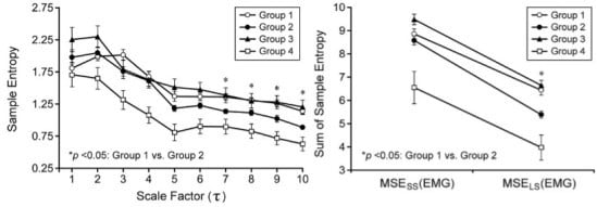
3.2. Multiscale Entropy (MSE) Analysis of Electromyographic (EMG) Signals

MSE analysis of EMG signals from different groups demonstrated the most notable difference in sample entropy between Group 3 (i.e., patients with detrusor overactivity but normal sphincter function) and Group 4 (i.e., patients with detrusor-external sphincter dyssynergia and overt sphincter dysfunction) compared to the differences among the other groups. The remarkably higher sample entropy in Group 3 than that in Group 4 at all scales (Figure 1a) suggests that suppressed sample entropy may be an indicator of urethral sphincter dysfunction. Therefore, with reference to sphincter function, the sample entropy of Group 1 (i.e., PBNO patients with successful surgical outcomes) was comparable to that of Group 3 at all scales except for scale 1. The results, therefore, reasonably suggest that patients with PBNO who underwent successful surgery had intact sphincter function. On the other hand, when compared with Group 4, the sample entropy of Group 2 (i.e., PBNO patients with unsatisfactory surgical outcomes) was significantly higher at scale 3, 4, 5, and 8 but the sample entropy was comparable at other scales between the two groups. Accordingly, the results imply certain degree of sphincter dysfunction in Group 2. In considering the difference between Group 1 and Group 2, significantly higher sample entropy was noted in the former from scale 7 onwards than that in the latter. The findings, therefore, implicate subtle impairment in sphincter function in Group 2 compared to that in Group 1 despite the lack of obvious difference in the fluctuations in EMG signals between the two groups (Table 1). To further support the contribution of a suppressed sample entropy to surgical failure, the sample entropy of Group 3 was found to be significantly higher than that in Group 2 from scale 5 onwards.
To investigate the overall changes in sample entropy among different groups, the sample entropies from scale 1 to 5 and those from scale 6 to 10 were summated to give “large-scale MSE of EMG” (i.e., MSESS(EMG)) and “small-scale MSE of EMG” (i.e., MSELS(EMG)), respectively. While both MSESS(EMG) and MSELS(EMG) were remarkably higher in Group 3 than those in Group 4 (both p = 0.002), no notable difference was noted in the two parameters between Group 3 and Group 1 (p = 0.304 and p = 0.506, respectively) (Figure 1b). On the other hand, both MSESS(EMG) and MSELS(EMG) were significantly higher in Group 2 compared to those in Group 4 (p = 0.039 and p = 0.033, respectively). Focusing on the difference between PBNO patients with and without satisfactory surgical outcomes, Group 1 (i.e., the successful group) had a significantly higher MSELS(EMG) than that of Group 2 (i.e., the surgical failure group) (p = 0.002). Also consistently with the results of changes in sample entropy with different scales (Figure 1a), both MSESS(EMG) and MSELS(EMG) were found to be significantly higher in Group 3 than those in Group 2 (p = 0.031 and p = 0.002, respectively) (Figure 1b).
Figure 1. (a) Changes in sample entropy with scale factors (1–10) among the four groups of patients with voiding difficulties; (b) Differences in “large-scale multiscale entropy (MSE) of electromyogram (EMG)” (i.e., MSELS(EMG)) and “small-scale MSE of EMG” (i.e., MSESS(EMG)) among the four groups of patients. Data are expressed as mean ± SD. Group 1: Patients with primary bladder neck obstruction (PBNO) with successful surgical outcomes; Group 2: Patients with PBNO with unsuccessful surgical outcomes; Group 3: Patients with detrusor overactivity; Group 4: Patients with detrusor-external sphincter dyssynergia. MSESS(EMG): Sum of sample entropy from scale 1 to 5; MSELS(EMG): Sum of sample entropy from scale 6 to 10. * p <0.05: Group 1 vs. Group 2.
Entropy 17 07863 g001

4. Discussion

The present study, which investigates the novel application of a computational method in the predication of prognostic outcomes of surgical candidates of a urological disease (i.e., PBNO) has several striking clinical implications. Firstly, in addition to assessing the postoperative progress after skeletomuscular surgeries in previous studies [10,26], the current study further highlights the use of MSE(EMG) in preoperative prediction of functional outcome after surgical intervention. Secondly, despite various theories put forward to explain the etiology of PBNO [5], the results of this study underscore the important role of impaired neuromuscular control of the external urethral sphincter in determining the surgical outcome. Finally, based on analysis of the subtle signal changes acquired through non-invasive surface EMG, this study introduced a simple means of predicting surgical outcome for patients with PBNO indicated for operation. The results, therefore, may provide a valuable preoperative reference for urologists to balance the risks and benefits of operation versus medical treatment for a specific individual afflicted with the disease in the absence of prognostic clues from routine urodynamic measurements.
PBNO is a common but easily neglected disorder without well-defined etiologies [27,28]. Although transurethral incision of the bladder neck (TUI-BN) has been accepted as a standard surgical treatment procedure for the disease [29], the factors that contribute to unsatisfactory surgical outcome remain unclear. Urodynamic testing, which includes the measurement of post-void residual volume, uroflowmetry, multichannel cystometry, urethral pressure profilometry, EMG for measurement of electrical activity of external urethral sphincter, and fluoroscopy of the bladder and bladder neck during voiding, is an important diagnostic tool for urologists for the differential diagnosis of patients with micturition problems. Nevertheless, the results of the tests cannot provide information on the prognosis after surgical treatment that the urologists are supposed to give to the patients before their operations. Since the exact etiology of PBNO is still controversial [5], there has been no single indicator targeting at predicting the surgical outcome for patients with the disease.
As a result, to test the hypothesis that the surgical outcome of patients with PBNO may depend on minor derangement in neuromuscular function of the urethral sphincter, the present study focused on the evaluation of subtle differences in EMG signals between PBNO patients with successful surgical outcome and those without by using MSE analysis. For comparison, patients with normal external urethral sphincter function (i.e., detrusor overactivity) and those with well-established sphincter dysfunction (i.e., detrusor-external sphincter dyssynergia) were recruited.
On comparing the demographic and urodynamic parameters among the four groups (Table 1), it is discernible that age was not a contributor to surgical outcomes for patients with PBNO in the present study. Besides, advanced age of subjects in Group 4 did not contribute to notable suppression of sample entropy compared to those in Group 2 who had diagnosed sphincter dysfunction (Table 1, Figure 1). Taken together, the findings indicate that age is not a factor significantly affecting sample entropy which appears to depend on the neuromuscular activity of urethral sphincter. This proposal is also supported by the results demonstrating significantly higher sample entropy in both MSESS(EMG) and MSELS(EMG) for subjects in Group 1 (i.e., intact sphincter function) compared to those in Group 4 as well as a higher sample entropy in MSELS(EMG) for Group 3 than that for Group 2 (Figure 1). The most noteworthy finding in the present study is the sensitivity of the MSE analytic approach that adopted signals acquired through non-invasive surface EMG from subjects in a resting state, taking into account the lack of significant difference in variations (i.e., SD) of EMG signals among the four groups under the same condition (Table 1). The ease with which this type of EMG signal analysis can be applied in clinical practice and study of the urodynamic system further enhances its value as an evaluation tool for patients with PBNO. Moreover, the therapeutic implications of the findings in this study may include potential medical interventions for preoperative correction of neuromuscular derangement of the urethral sphincter to augment surgical success. Nevertheless, the limitation of the present study is the relatively small patient populations in each group. On the other hand, the significant differences among different groups highlight the sensitivity and feasibility of the application of this diagnostic approach in the clinical setting.

5. Conclusions

The results of the current study not only support that subtle neuromuscular dysfunction unable to be detected by pre-operative urodynamic testing may be an important contributor to surgical failure for patients with PBNO, but also suggest the possibility of clinical use of MSE analysis for surface EMG signals as a simple non-invasive surgical prognostic indicator for patients with the disease.

Acknowledgments

This study was partly supported by a research grant from the Ministry of Science and Technology, Taiwan (Grant No. MOST 103-2221-E-259-011 and Grant No. MOST 104-2221-E-259-014).

Author Contributions

Hsien-Tsai Wu, Yuan-Hong Jiang, and Cheuk-Kwan Sun designed the study. An-Bang Liu, Yu-Nian Ou, and Chun-Wei Liu and Hann-Chorng Kuo were responsible for data collection and analysis. Yu-Nian Ou, Chun-Wei Liu, Yuan-Hong Jiang, and Cheuk-Kwan Sun reviewed relevant literature and interpreted the acquired data. Hsien-Tsai Wu and Cheuk-Kwan Sun drafted the manuscript. All authors have read and approved the final manuscript.

Conflicts of Interest

The authors declare no conflict of interest.

References

  1. Wein, A.J. Re: Urodynamics: Examining the Current Role of UDS Testing. What is the Role of Urodynamic Testing in Light of Recent AUA Urodynamics and Overactive Bladder Guidelines and the VALUE Study? J. Urol. 2015, 193, 1311–1334. [Google Scholar] [CrossRef] [PubMed]
  2. Danforth, T.L.; Ginsberg, D.A. Neurogenic lower urinary tract dysfunction: How, when, and with which patients do we use urodynamics? Urol. Clin. N. Am. 2014, 41, 445–452. [Google Scholar] [CrossRef] [PubMed]
  3. Padmanabhan, P.; Nitti, V.W. Primary bladder neck obstruction in men, women, and children. Curr. Urol. Rep. 2007, 8, 379–384. [Google Scholar] [CrossRef] [PubMed]
  4. Nitti, V.W.; Lefkowitz, G.; Ficazzola, M.; Dixon, C.M. Lower urinary tract symptoms in young men: Videourodynamic findings and correlation with noninvasive measures. J. Urol. 2002, 168, 135–138. [Google Scholar] [CrossRef]
  5. Nitti, V.W. Primary bladder neck obstruction in men and women. Rev. Urol. 2005, 7, S12–S17. [Google Scholar] [PubMed]
  6. Sacco, E.; Tienforti, D.; Bientinesi, R.; D’Addessi, A.; Racioppi, M.; Pinto, F.; Totaro, A.; Vittori, M.; D’Agostino, D.; Bassi, P. OnabotulinumtoxinA injection therapy in men with LUTS due to primary bladder-neck dysfunction: Objective and patient-reported outcomes. Neurourol. Urodyn. 2014, 33, 142–146. [Google Scholar] [CrossRef] [PubMed]
  7. Trockman, B.A.; Gerspach, J.; Dmochowski, R.; Haab, F.; Zimmern, P.E.; Leach, G.E. Primary bladder neck obstruction: Urodynamic findings and treatment results in 36 men. J. Urol. 1996, 156, 1418–1420. [Google Scholar] [CrossRef]
  8. Van Batavia, J.P.; Combs, A.J.; Fast, A.M.; Glassberg, K.I. Use of non-invasive uroflowmetry with simultaneous electromyography to monitor patient response to treatment for lower urinary tract conditions. J. Pediatr. Urol. 2014, 10, 532–537. [Google Scholar] [CrossRef] [PubMed]
  9. Istenic, R.; Kaplanis, P.A.; Pattichis, C.S.; Zazula, D. Multiscale entropy-based approach to automated surface EMG classification of neuromuscular disorders. Med. Biol. Eng. Comput. 2010, 48, 773–781. [Google Scholar] [CrossRef] [PubMed]
  10. Cashaback, J.G.; Cluff, T.; Potvin, J.R. Muscle fatigue and contraction intensity modulates the complexity of surface electromyography. J. Electromyogr. Kinesiol. 2013, 23, 78–83. [Google Scholar] [CrossRef] [PubMed]
  11. Abrams, P.; Cardozo, L.; Fall, M.; Griffiths, D.; Rosier, P.; Ulmsten, U.; van Kerrebroeck, P.; Victor, A.; Wein, A. The standardisation of terminology of lower urinary tract function: Report from the Standardisation Sub-committee of the International Continence Society. Neurourol. Urodyn. 2002, 21, 167–178. [Google Scholar] [CrossRef] [PubMed]
  12. Chapple, C.R.; Araño, P.; Bosch, J.L.; de Ridder, D.; Kramer, A.E.; Ridder, A.M. Solifenacin appears effective and well tolerated in patients with symptomatic idiopathic detrusor overactivity in aplacebo- and tolterodine-controlled phase 2 dose-finding study. BJU Int. 2004, 93, 71–77. [Google Scholar] [CrossRef] [PubMed]
  13. De, E.J.; Patel, C.Y.; Tharian, B.; Westney, O.L.; Graves, D.E.; Hairston, J.C. Diagnostic discordance of electromyography (EMG) versus voiding cystourethrogram (VCUG) for detrusor-external sphincter dyssynergy (DESD). Neurourol. Urodyn. 2005, 24, 616–621. [Google Scholar] [CrossRef] [PubMed]
  14. Costa, M.; Goldberger, A.L.; Peng, C.K. Multiscale entropy to distinguish physiologic and synthetic RR time series. Comput. Cardiol. 2002, 29, 137–140. [Google Scholar] [PubMed]
  15. Costa, M.; Goldberger, A.L.; Peng, C.K. Multiscale entropy analysis of complex physiologic time series. Phys. Rev. Lett. 2002, 89, 068102. [Google Scholar] [CrossRef] [PubMed]
  16. Costa, M.; Peng, C.K.; Goldberger, A.L.; Jeffrey, M.H. Multiscale entropy analysis of human gait dynamics. Physica A 2003, 330, 53–60. [Google Scholar] [CrossRef]
  17. Wu, H.T.; Hsu, P.C.; Lin, C.F.; Wang, H.J.; Sun, C.K.; Liu, A.B.; Lo, M.T.; Tang, C.J. Multiscale entropy analysis of pulse wave velocity for assessing atherosclerosis in the aged and diabetic. IEEE Trans. Biomed. Eng. 2011, 58, 2978–2981. [Google Scholar] [PubMed]
  18. Richman, J.S.; Moorman, J.R. Physiological time-series analysis using approximate entropy and sample entropy. Am. J. Phys. Heart Circ. Physiol. 2000, 278, H2039–H2049. [Google Scholar]
  19. Costa, M.; Goldberger, A.L.; Peng, C.K. Multiscale entropy analysis of biological signals. Phys. Rev. E 2005, 71, 021906. [Google Scholar] [CrossRef] [PubMed]
  20. Baumert, M.; Javorka, M.; Seeck, A.; Faber, R.; Sanders, P.; Voss, A. Multiscale entropy and detrended fluctuation analysis of QT interval and heart rate variability during normal pregnancy. Comput. Biol. Med. 2012, 42, 347–352. [Google Scholar] [CrossRef] [PubMed]
  21. Javorka, M.; Trunkvalterova, Z.; Tonhajzerova, I.; Javorkova, J.; Javorka, K.; Baumert, M. Short-term heart rate complexity is reduced in patients with type 1 diabetes mellitus. Clin. Neurophysiol. 2008, 119, 1071–1081. [Google Scholar] [CrossRef] [PubMed]
  22. Turianikova, Z.; Javorka, K.; Baumert, M.; Calkovska, A.; Javorka, M. The effect of orthostatic stress on multiscale entropy of heart rate and blood pressure. Physiol. Meas. 2011, 32, 1425–1437. [Google Scholar] [CrossRef] [PubMed]
  23. Huang, N.E.; Zheng, S.; Steven, R.L.; Wu, M.C.; Shih, H.H.; Zheng, Q.; Yen, N.C.; Tung, C.C.; Liu, H.H. The empirical mode decomposition and the Hilbert spectrum for nonlinear and non-stationary time series analysis. Proc. R. Soc. A Math. Phys. Eng. Sci. 1998, 454, 903–995. [Google Scholar] [CrossRef]
  24. Wu, Z.; Huang, N.E. Ensemble empirical mode decomposition: A noise-assisted data analysis method. Adv. Adapt. Data Anal. 2009, 1, 1–41. [Google Scholar] [CrossRef]
  25. Lo, M.T.; Tsai, P.H.; Lin, P.F.; Lin, C.; Hsin, Y.L. The nonlinear and nonstationary properties in EEG signals: Probing the complex fluctuations by Hilbert–Huang transform. Adv. Adapt. Data Anal. 2009, 1, 461–482. [Google Scholar] [CrossRef]
  26. Zhang, X.; Chen, X.; Barkhaus, P.E.; Zhou, P. Multiscale entropy analysis of different spontaneous motor unit discharge patterns. IEEE J. Biomed. Health Inform. 2013, 17, 470–476. [Google Scholar] [CrossRef] [PubMed]
  27. Roehrborn, C.G.; Oesterling, J.E.; Auerbach, S.; Kaplan, S.A.; Lloyd, L.K.; Milam, D.E.; Padley, R.J. The Hytrin Community Assessment Trial study: A one-year study of terazosin versus placebo in the treatment of men with symptomatic benign prostatic hyperplasia. Urology 1996, 2, 159–168. [Google Scholar] [CrossRef]
  28. Nitti, V.W.; Carlson, K.V.; Blaivas, J.G.; Dmochowski, R.R. Early results of pubovaginal sling lysis by midline sling incision. Urology 2002, 59, 47–51. [Google Scholar] [CrossRef]
  29. Kochakarn, W.; Lertsithichai, P. Unilateral transurethral incision for primary bladder neck obstruction: Symptom relief and fertility preservation. World J. Urol. 2003, 21, 159–162. [Google Scholar] [CrossRef] [PubMed]

