Article

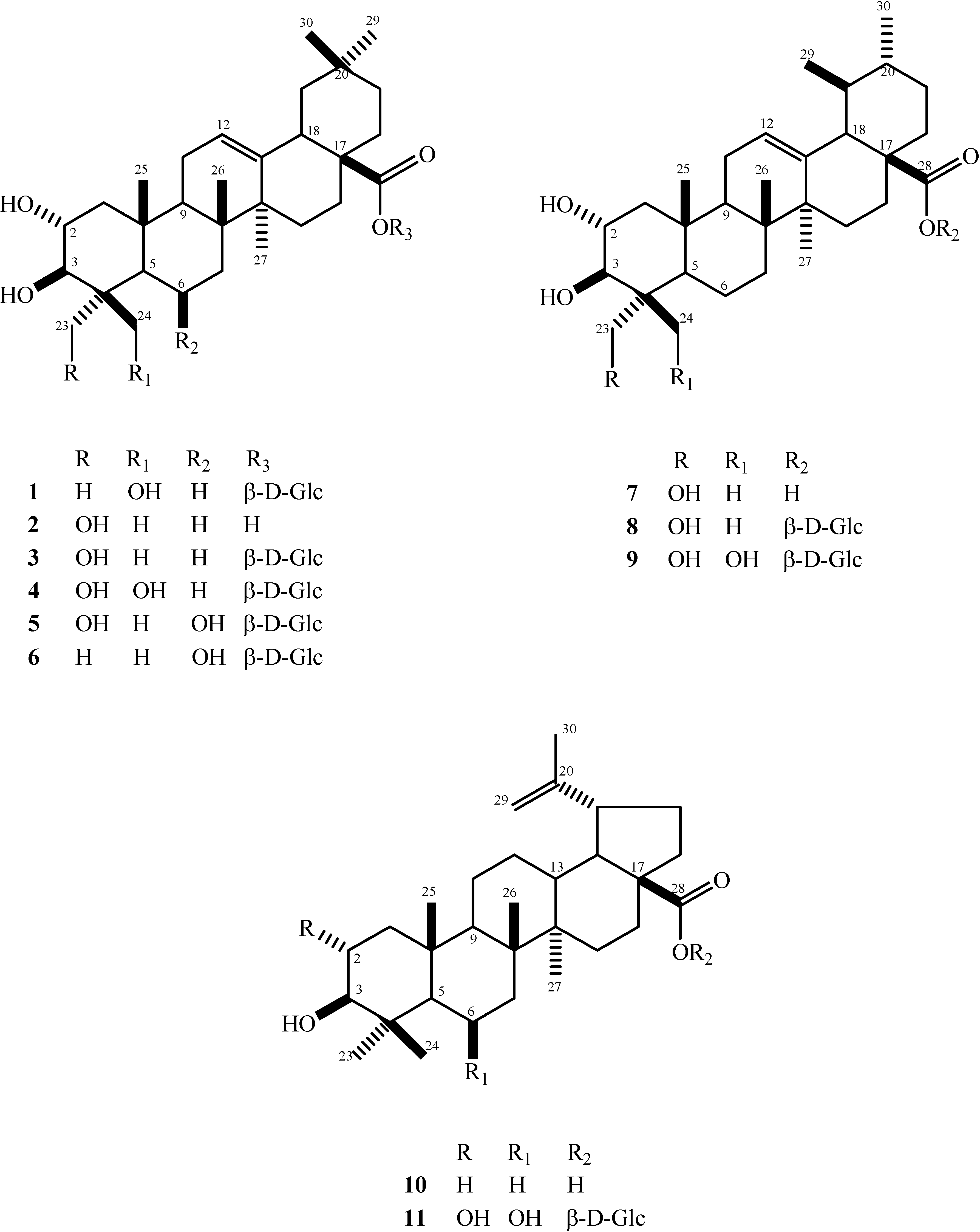
Bioactive Pentacyclic Triterpenes from the Stems of Combretum laxum

by
Eder Bisoli
,
Walmir Silva Garcez
,
Lidilhone Hamerski
,
Caroline Tieppo
and
Fernanda Rodrigues Garcez
*
Departamento de Química, Universidade Federal de Mato Grosso do Sul, Campo Grande, MS, Brasil, 79070-900
*
Author to whom correspondence should be addressed.
Submission received: 22 September 2008 / Revised: 16 October 2008 / Accepted: 20 October 2008 / Published: 1 November 2008
(This article belongs to the Special Issue Triterpenes and Triterpenoids 2013)

Abstract

:
Two new triterpene glucosides, β-d-glucopyranosyl 2α,3β,24-trihydroxyolean-12-en-28-oate and β-d-glucopyranosyl 2α,3β,23,24-tetrahydroxyurs-12-en-28-oate, in addition to nine known compounds belonging to three different triterpene classes (oleanane-, ursane- and lupane-type) have been isolated from the stems of a specimen of Combretum laxum growing in the “Pantanal” of the central-western region of Brazil. Among the known triterpenes, β-d-glucopyranosyl 2α,3β,6β-trihydroxyolean-12-en-28-oate is reported for the first time in the Combretaceae, while bellericoside and asiatic acid are described for the first time in the genus Combretum. The structures of the isolated compounds have been established on the basis of spectral techniques (1D-, 2D-NMR and MS). Their in vitro antifungal activities against standard strains of Candida albicans, C. krusei and Cryptococcus neoformans were also evaluated in this work.

Graphical Abstract

Introduction

The state of Mato Grosso do Sul, located in the central-western region of Brazil, is home of three major biomes, namely “Cerrado”, “Pantanal” and tropical forest. The “Pantanal”, the world’s largest wetland area, with a vast range of biodiversity, is found in Brazil in only two states: Mato Grosso and Mato Grosso do Sul, the latter comprising 2/3 of the total area of the Brazilian “Pantanal”, which is 150,355 km2 [1]. As part of our ongoing program to assess the chemical diversity of plants of the family Combretaceae which are distributed in the first two ecosystems [2,3,4], we have investigated the stems of Combretum laxum, a climbing shrub occurring in the “Pantanal” region and popularly known as “pombeiro-branco”. Amongst the 18 genera comprising the Combretaceae, the genus Combretum is the largest, with 250 species, which are mostly native to tropical and subtropical regions [5]. Plants of this genus are known as a rich source of secondary metabolites, in particular triterpenes and their glycosides, phenanthrenes, bibenzyls, stilbenes, flavonoids, in addition to other aromatic compounds, some of which display anticancer, antimicrobial and hepatoprotective activities, among others [6,7,8,9,10,11,12,13,14,15]. To date, only one chemical constituent, trans-4-hydroxyproline betaine, was reported to occur in a Venezuelan specimen of C. laxum [16]. Herein we describe the isolation and characterization of two new triterpene glucosides, β-d-glucopyranosyl 2α,3β,24-trihydroxyolean-12-en-28-oate and β-d-glucopyranosyl 2α,3β,23,24-tetrahydroxyurs-12-en-28-oate from the title plant, along with nine known oleanane-, ursane- and lupane-type triterpenoids, and the evaluation of their antifungal activities.

Results and Discussion

Compound 1 was obtained as a white, amorphous solid, and displayed a quasi-molecular ion at m/z 685.3755 [M+Cl]- in the HRESIMS, consistent with the molecular formula C36H58O10 (calcd. for C36H58O10-Cl 685.37204). The 1H-NMR spectrum of 1 showed six aliphatic methyl singlets at δ 0.85, 0.87, 0.97, 1.06, 1.18 and 1.54 and a broad singlet at δ 5.38 assignable to an olefinic hydrogen. The 13C and DEPT NMR spectra exhibited signals for six methyls, eleven methylenes (two oxymethylenes), eleven methines (one olefinic, seven oxymethines) and eight quaternary carbons (one olefinic and one ester carbonyl carbon), of which six were attributed to a sugar unit and 30 were consistent with an triterpene aglycon moiety. The presence of a Δ12-double bond, already inferred from the broad singlet at δ 5.38 ppm in the 1H-NMR spectrum was corroborated by resonances at 122.7 and 144.1 in the 13C-NMR spectrum, assigned to C-12 and C-13, respectively, of an olean-12-ene-type skeleton [17]. The presence of hydroxyl substituents at C-2 and C-3 was evident from both the chemical shifts and J values of the hydrogens ascribable to H-2 (δ 4.33, m) and H-3 (δ 3.54, d, J = 9.3 Hz), which showed connectivities in the HSQC spectrum with the oxymethine carbons at δ 68.6 and δ 85.7, respectively. The large coupling constant (9.3 Hz) between H-2 and H-3 pointed to their axial disposition, thereby indicating that the 2- and 3- hydroxyl groups were both equatorially (2α, 3β) oriented. Furthermore, the 1H-NMR spectrum also revealed two one-proton doublets [δ 3.69 and δ 4.42 (J = 11.0 Hz)] assignable to diastereotopic hydroxymethylene hydrogens, which in turn showed cross-peak correlations with a carbon signal at δ 65.7 in the HSQC spectrum. This information, along with an upfield shifted methyl carbon signal at δ 24.1 ascribable to C-23 and a downfield quaternary carbon resonance at δ 44.0 attributable to C-4 strongly suggested the location of this third hydroxyl substituent at C-24 [17]. This assumption could be confirmed by connectivities observed in the HMBC spectrum between this carbon signal and H-3 (δ 3.54) and methyl-23 (δ 1.54) resonances. The 13C-NMR data also allowed the assignment of the pyranose form of D-glucose [18], while the magnitude of the coupling constant of the 1H-NMR doublet at δ 6.29 (J = 7.9 Hz) indicated an α-orientation of the anomeric hydrogen of the glucosyl residue, thus establishing a β-configuration for the glucopyranosyl unity. The chemical shift value of its anomeric carbon (δ 95.8) supported the fact that compound 1 was an ester glucoside, since the anomeric carbons of O-glucosides are observed around 100 ppm, independently of the nature of the sugar residue [19]. This information, along with the ester carbonyl carbon resonance (δ 176.4) and the remaining carbon resonances of the triterpene skeleton indicated the likely location of the sugar attachment at C-28. The aforementioned data could be satisfactorily assembled to establish that 1 was the C-28 glucopyranosyl ester derivative of 2α,3β,24-trihydroxyolean-12-en-28-oic acid, whose aglycon, known as hyptatic acid-A, was previously isolated from Hyptis capitata (Labiatae) [17] and also obtained from chemical transformation of maslinic acid [20]. Indeed, the spectral data of 1 closely resembled those of the corresponding oleanane triterpene acid [17,20], except for the absence in the latter of the signals attributed to the glucosyl moiety in 1. The replacement of the signal for the C-28 carboxyl observed in the triterpene aglycon δ 181.8 [20] by an upfield shifted ester carbonyl carbon at δ 176.4, along with the long-range coupling discernible in the HMBC spectrum between this carbon and the anomeric hydrogen of the sugar (δ 6.29) further supported the linkage site of the glucopyranosyl moiety at C-28. Thus, the structure of 1 was determined to be β-d-glucopyranosyl 2α,3β,24-trihydroxyolean-12-en-28-oate.
The structures of the five additional oleanane-type triterpenes isolated from Combretum laxum were established on the basis of spectral analyses and by comparison with previously reported data or authentic samples as arjunolic acid (2) [3,4,17], arjunglucoside II (β-d-glucopyranosyl 2α,3β,23-tri- hydroxyolean-12-en-28-oate, 3) [4,21], bellericoside (β-d-glucopyranosyl 2α,3β,23,24-tetrahydroxy-olean-12-en-28-oate, 4) [22], chebuloside II (β-d-glucopyranosyl 2α,3β,6β,23-tetrahydroxyolean-12-en-28-oate, 5) [23] and β-d-glucopyranosyl 2α,3β,6β-trihydroxyolean-12-en-28-oate (6) [24]. Compound 6, which had previously only been isolated from Siphoneugena densiflora (Myrtaceae) [24], is therefore described for the first time in the Combretaceae. Also worthy of mention is the fact that although 4 has been shown to occur in other members of the Combretaceae, e.g. in Terminalia bellerica [22], it was hitherto unreported in the genus Combretum, while chebuloside II and arjunolic acid have been described in only one (C. quadrangulare) [12] and two species (C. quadrangulare and C. leprosum) [6,11] of this genus, respectively.
The structures of compounds 7 and 8 were readily discernible from both the 1H- and 13C-NMR spectra as those of the known ursane-type triterpenoids asiatic acid and β-d-glucopyranosyl 2α,3β,23-trihydroxy-urs-12-en-28-oate (quadranoside IV), respectively, whose spectral data were in accordance with those of an authentic sample [3] and those published in the literature [13]. In spite of the presence of asiatic acid either in members of the Combretaceae (e.g., in the genus Terminalia [3]) or in other plant families, this constitutes the first report of its isolation in the genus Combretum, whereas this is the second reported occurrence of quadranoside IV in this genus, where it was first described in C. quadrangulare [13]. Compound 9 was obtained as the minor component of an unresolved mixture with bellericoside (4). The comparison of the 13C-NMR spectrum of this mixture to that of pure compound 4 enabled assignment of its carbon resonances. The remaining ones were indicative that 9 was the urs-12-ene derivative analogue of bellericoside. This assumption was revealed by the typical resonance values of C-12 and C-13 of an ursane-type triterpene observed at δ 125.9 and 138.4, respectively [17] and the characteristic signals attributable to the ring E carbons, namely δ 53.2 (C-18), δ 39.3 (C-19), δ 39.1 (C-20), δ 30.7 (C-21) and δ 36.7 (C-22), as well as methyls 29 (δ 17.5) and 30 (δ 21.2) [17], which in turn were very similar to the corresponding signals observed in the spectrum of quadranoside IV (8). Resonance values of the remaining carbons in the structure of 9 were essentially the same as those of bellericoside (4). The foregoing account therefore allowed assignment of the structure of 9 as β-d-glucopyranosyl 2α,3β,23,24-tetrahydroxyurs-12-en-28-oate. This triterpene glucoside was previously described as being part of the structure of the castanopsinins A-H, galloyl and hexa-hydroxydiphenoyl-type ellagitannins possessing a mixture of olean-12–ene and ursan-12-ene triterpene glucoside cores in their molecules, which were isolated from Castanopsis cuspidate var. sieboldii (Fagaceae) [25].
The lupane-type triterpenes 10 and 11 obtained in the present work were found to be identical in their spectral properties with betulinic acid (data compared with those of an authentic sample) [3,4] and β-d-glucopyranosyl 2α,3β,6β-trihydroxy-lup-20(29)-en-28-oate (quadranoside I) [13], respectively. The latter has only been found in C. quadrangulare and Vochysia pacifica (Vochisiaceae) [26] and in spite of the wide distribution of betulinic acid in other plant genera, its previous occurrence in the genus Combretum has only been reported in C. quadrangulare [14]. The structures of all the isolated compounds are summarized in Figure 1.
Following isolation, compounds 1, 2, 5, 6, 10, 11 and the mixtures of 2 and 7, 3 and 8, 4 and 9 were further evaluated for their antifungal properties in vitro against Candida krusei, C. albicans and Cryptococcus neoformans, using the microbroth dilution assay. All samples displayed antifungal activities against the strains tested and their minimal inhibitory concentration (MIC) values are given in Table 1. Arjunolic acid (2) was the most active triterpenoid assayed, with MIC values = 50 μg/mL against the three fungal strains, while betulinic acid (10) showed strongest activities against Candida albicans and Cryptococcus neoformans (MIC = 100 μg/mL) and chebuloside II (5) against C. neoformans (MIC = 100 μg/mL). The crude ethanol extract, on the other hand, was shown to be inactive against these strains. Although crude extracts from a number of plants of the family Combretaceae, mostly occurring in the African continent, have shown antifungal properties against yeasts and/or filamentous fungi [27,28,29,30,31], only a few works have reported on the antifungal activities of pentacyclic triterpenes obtained from these species, most of them belonging to the genus Terminalia [32,33,34]. Recently, in an antifungal bioassay-guided fractionation of crude extracts of the leaves of Combretum nelsonii, a mixture of arjunolic and asiatic acids was reported to show strong activities against Candida albicans and Cryptococcus neoformans, being also active against filamentous fungi [35]. However, these triterpenes (2 and a mixture of 2 and 7) showed weak activities against the strains of Candida albicans and Crytococcus neoformans used in the present work. The results presented herein add new biological activity data of the secondary metabolites obtained from members of the genus Combretum.
Figure 1. Structures of the triterpenoids 111 isolated from C. laxum.
Molecules 13 02717 g001
Table 1. Minimal inhibitory concentration (MIC) values (μg/mL) of triterpenes isolated from C. laxum against Candida and Cryptococcus strains using the microbroth dilution assay.
CompoundsCandida albicansCandida kruseiCryptococcus neoformans
ATCC 90028ATCC 6258ATCC 32045
1200200200
2505050
5200200100
6200200200
10100200100
11200200200
2/7200200200
3/8200200200
4/9200200200
amphotericin B0.250.500.50

Experimental

General

Optical rotations were determined on a Perkin Elmer 341 polarimeter (Na filter, λ = 589 nm). 1H- and 13C- 1D and 2D NMR spectra were recorded in pyridine-d5 at 300 MHz (1H) and 75 MHz (13C) on a Bruker DPX-300 spectrometer. Standard pulse sequences were used for homo- and heteronuclear correlation experiments. HRESIMS spectrum was acquired in negative ion mode on an UltrOTOF-Q instrument (Bruker Daltonics). Column chromatography procedures were performed on silica gel 70-230 mesh and 230-400 mesh, RP-18 silica gel 230-400 mesh and Sephadex LH-20. Reversed phase semi-preparative HPLC separations were performed with a Shimadzu LC-6AD pump, using a RP-18, 21.6 x 250 mm, 5 μm particle size, Phenomenex Luna column, with a flow rate of 10 mL or 14 mL min-1 and monitoring at 210 nm.

Plant Material

Stems of Combretum laxum Jacq. were collected in Miranda (“Pantanal” of Mato Grosso do Sul), Brazil, in October 2004. The plant material was identified by MSc. Ubirazilda M. Resende, CGMS Herbarium, Universidade Federal de Mato Grosso do Sul, Brazil, where a voucher specimen (No. 18102) is deposited.

Extraction and Isolation

Air-dried and powdered stems of C. laxum (4,375 g) were extracted at room temperature with EtOH. After concentration in vacuo, the residue obtained from the EtOH extract was subsequently partitioned between MeOH/H2O 9:1 and hexane; MeOH/H2O 1:1 and CH2Cl2; MeOH/H2O 1:1 and EtOAc and MeOH/H2O 1:1 and n-BuOH to give the corresponding hexane, CH2Cl2, EtOAc and n-BuOH phases. Betulinic acid (10, 200.0 mg) was obtained from the CH2Cl2 phase (9.2 g), after a series of column chromatography procedures over silica gel (70-230 mesh) eluted with hexane, CH2Cl2, EtOAc and EtOAc/MeOH 20%. The EtOAc phase (7.86 g) was chromatographed on a silica gel (70-230 mesh) column eluted with hexane, CH2Cl2, EtOAc and EtOAc/MeOH 20%. Twenty-nine fractions of 250 mL each were collected and those exhibiting similar TLC profiles were combined to give, altogether, eight fractions (A-H). Fraction D (EtOAc, 1.31 g) was subjected to column chromatography on silica gel (230-400 mesh) eluted with CHCl3/MeOH 10% to yield twenty-eight fractions of 20 mL each. The second fraction (24.6 mg) consisted of a mixture of the isomeric triterpene acids 2 (major compound) and 7 and the sixth one (409.6 mg) was further separated on Sephadex LH-20 (MeOH) followed by semi-preparative HPLC (CH3CN/H2O 38:62) to afford 1 (16.3 mg), 6 (10.3 mg), 11 (8.0 mg) and a mixture of the isomeric triterpenes 3 (major compound) and 8 (25.0 mg). The n-BuOH phase (25.5 g) was subjected to column chromatography on RP-18 silica gel eluted with MeOH/H2O (6:4 to pure MeOH), to furnish seven fractions of 500 mL each. The fourth fraction (MeOH/H2O 8:2, 0.5 g) was rechromatographed on a Sephadex LH-20 column (MeOH) and further purified by semi-preparative HPLC (CH3CN/H2O 30:70) to yield 4 (5.8 mg), 5 (19.0 mg), additional amounts of a mixture of 3 and 8 (24.2 mg) and a mixture of 4 (major compound) and 9 (4.8 mg) which proved difficult to be separated in its individual components on subsequent chromatographic procedures.
β-d-Glucopyranosyl 2α,3β,24-trihydroxyolean-12-en-28-oate (1): colorless amorphous powder; Molecules 13 02717 i001: + 22.79o (c 0.17, MeOH); HRESIMS m/z 685.3755 [M+Cl]- (calcd. for C36H58O10Cl 685.3720); 1H-NMR: δ 4.33 (1H, m, H-2); 3.54 (1H, d, J = 9.3 Hz, H-3); 5.38 (1H, brs, H-12); 3.16 (1H, dd, J = 14.3, 3.8 Hz, H-18); 3.69 (1H, d, J = 11.0 Hz, H-24a); 4.42 (1H, d, J = 11.0 Hz, H-24b); 1.54 (3H, s, H-23); 0.97 (3H, s, H-25); 1.06 (3H, s, H-26); 1.18 (3H, s, H-27); 0.87 (3H, s, H-29); 0.85 (3H, s, H-30); sugar signals: δ 6.29 (1H, d, J = 7.9 Hz, H-1’); 4.00 (1H, m, H-5’); 4.18-4.45* (5H, m H-2’, H-3’, H-4’, H-6’) *overlapping signals; 13C-NMR: δ 47.8 (C-1); 68.6 (C-2); 85.7 (C-3); 44.0 (C-4); 56.5 (C-5); 19.3 (C-6); 33.4 (C-7); 40.0 (C-8); 48.3 (C-9); 38.3 (C-10); 24.1 (C-11); 122.7 (C-12); 144.1 (C-13); 42.1 (C-14); 28.2 (C-15); 23.4 (C-16); 47.0 (C-17); 41.7 (C-18); 46.2 (C-19); 30.7 (C-20); 34.0 (C-21); 32.5 (C-22); 24.1 (C-23); 65.7 (C-24); 17.3 (C-25); 17.4 (C-26); 26.0 (C-27); 176.4 (C-28); 33.1 (C-29); 23.6 (C-30); sugar signals: δ 95.8 (C-1’); 74.1 (C-2’); 78.9 (C-3’); 71.1 (C-4’); 79.3 (C-5’); 62.3 (C-6’).
Arjunolic acid (2) and Asiatic acid (7) as a mixture: colorless amorphous powder; 1H- and 13C-NMR data in accordance with those of an authentic sample and with the literature [3,4,17]. Compound 2: 13C-NMR: δ 47.6 (C-1); 68.8 (C-2); 78.2 (C-3); 43.6 (C-4); 47.9 (C-5); 18.5 (C-6); 32.8 (C-7); 39.8 (C-8); 48.1 (C-9); 38.4 (C-10); 23.6 (C-11); 122.4 (C-12); 144.8 (C-13); 42.2 (C-14); 28.2 (C-15); 23.9 (C-16); 46.6 (C-17); 41.9 (C-18); 46.3 (C-19); 30.9 (C-20); 34.1 (C-21); 33.2 (C-22); 66.4 (C-23); 14.3 (C-24); 17.3 (C-25); 17.5 (C-26); 26.1 (C-27); 180.2 (C-28); 33.2 (C-29); 23.7 (C-30). Compound 7: 13C-NMR: δ 47.6 (C-1); 68.8 (C-2); 78.2 (C-3); 43.6 (C-4); 48.2 (C-5); 18.5 (C-6); 32.8 (C-7); 40.0 (C-8); 47.6 (C-9); 38.3 (C-10); 23.6 (C-11); 125.5 (C-12); 139.2 (C-13); 42.2 (C-14); 28.6 (C-15); 24.8 (C-16); 48.0 (C-17); 53.5 (C-18); 39.4 (C-19); 39.3 (C-20); 31.0 (C-21); 37.4 (C-22); 66.4 (C-23); 14.3 (C-24); 17.4* (C-25); 17.4* (C-26); 23.8 (C-27); 180.6 (C-28); 17.3* (C-29); 21.3 (C-30) *interchangeable signals.
Arjunglucoside II (3) and Quadranoside IV (8) as a mixture: colorless amorphous powder; 1H- and 13C-NMR data in accordance with those of an authentic sample and/or with the literature [4,13,21]. Compound 3: 1H-NMR: δ 4.25* (1H, m, H-2); 4.28* (1H, m, H-3); 5.43 (1H, brs, H-12); 3.19 (1H, dd, J = 13.5, 4.2 Hz, H-18); 3.72 (1H, d, J = 10.6, H-23a); 4.22* (1H, d, J = 10.6, H-23b); 1.07 (3H, s, H-24); 1.10 (3H, s, H-25); 1.16 (6H, s, H-26 e H-27); 0.90 (3H, s, H-29); 0.88 (3H, s, H-30); sugar signals: δ 6.33 (1H, d, J = 9.0 Hz, H-1’); 4.21 (1H, m, H-5’); 4.22-4.46* (5H, m H-2’, H-3’, H-4’, H-6’) *overlapping signals; 13C-NMR: δ 47.6 (C-1); 68.8 (C-2); 78.1 (C-3); 43.5 (C-4); 48.0 (C-5); 18.4 (C-6); 32.7 (C-7); 39.9 (C-8); 47.7 (C-9); 38.3 (C-10); 23.8 (C-11); 122.7 (C-12); 144.0 (C-13); 42.1 (C-14); 28.1 (C-15); 23.2 (C-16); 46.9 (C-17); 41.6 (C-18); 46.0 (C-19); 30.6 (C-20); 33.8 (C-21); 32.4 (C-22); 66.3 (C-23); 14.2 (C-24); 17.5 (C-25); 17.6 (C-26); 25.9 (C-27); 176.4 (C-28); 32.9 (C-29); 23.5 (C-30); sugar signals: δ 95.6 (C-1’); 74.0 (C-2’); 78.8 (C-3’); 71.0 (C-4’); 79.2 (C-5’); 62.1 (C-6’). Compound 8: 1H-NMR: δ 4.25* (1H, m, H-2); 4.28* (1H, m, H-3); 5.43 (1H, brs, H-12); 2.52 (1H, d, J = 11.2 Hz, H-18); 3.72 (1H, d, J = 10.6, H-23a); 4.22* (1H, d, J = 10.6, H-23b); 1.07 (3H, s, H-24); 1.11 (3H, s, H-25); 1.20 (3H, s, H-26); 1.10 (3H, s, H-27); 0.92 (3H, d, J = 6.0 Hz, H-29); 0.88 (3H, brs, H-30); sugar signals: δ 6.27 (1H, d, J = 9.0 Hz, H-1’); 4.21 (1H, m, H-5’); 4.22-4.46* (5H, m H-2’, H-3’, H-4’, H-6’) *overlapping signals; 13C-NMR: δ 47.9 (C-1); 68.8 (C-2); 78.1 (C-3); 43.5 (C-4); 48.1 (C-5); 18.4 (C-6); 33.0 (C-7); 40.1 (C-8); 47.8 (C-9); 38.2 (C-10); 23.7 (C-11); 125.9 (C-12); 138.3 (C-13); 42.4 (C-14); 28.5 (C-15); 24.5 (C-16); 47.7 (C-17); 53.2 (C-18); 39.2 (C-19); 39.0 (C-20); 30.7 (C-21); 36.6 (C-22); 66.3 (C-23); 14.2 (C-24); 17.3 (C-25); 17.5 (C-26); 23.6 (C-27); 176.1 (C-28); 17.2 (C-29); 21.1 (C-30); sugar signals: δ 95.6 (C-1’); 73.9 (C-2’); 78.8 (C-3’); 71.1 (C-4’); 79.1 (C-5’); 62.1 (C-6’).
Bellericoside (4): colorless amorphous powder; 1H- and 13C-NMR data in accordance with the literature [22]. 1H-NMR: δ 4.07 (1H, m, H-2); 4.30* (1H, m, H-3); 5.42 (1H, brs, H-12); 3.18 (1H, dd, J = 13.1, 3.7 Hz, H-18); 4.24 (1H, d, J = 10.8 Hz, H-23a); 4.86 (1H, d, J = 10.8, H-23b); 3.97 (1H, d, J = 11.0 Hz, H-24a); 4.61 (1H, d, J = 11.0 Hz, H-24b); 1.13 (3H, s, H-25); 1.14 (3H, s, H-26); 1.16 (3H, s, H-27); 0.87 (6H, s, H-29, H-30); sugar signals: δ 6.32 (1H, d, J = 9.0 Hz, H-1’); 4.20 (1H, m, H-2’); 4.25-4.44* (4H, m, H-3’, H-4’, H-6’); 4.02 (1H, m, H-5’) *overlapping signals; 13C-NMR: δ 47.8 (C-1); 69.0 (C-2); 79.7 (C-3); 47.8 (C-4); 48.2 (C-5); 19.2 (C-6); 32.5 (C-7); 40.0 (C-8); 48.3 (C-9); 38.1 (C-10); 23.3 (C-11); 122.7 (C-12); 144.1 (C-13); 42.1 (C-14); 28.1 (C-15); 24.1 (C-16); 46.9 (C-17); 41.7 (C-18); 46.1 (C-19); 30.7 (C-20); 33.7 (C-21); 33.1 (C-22); 64.3 (C-23); 62.8 (C-24); 17.3 (C-25); 17.4 (C-26); 26.0 (C-27); 176.5 (C-28); 33.0 (C-29); 23.6 (C-30).
Chebuloside II (5): colorless amorphous powder; 1H- and 13C-NMR data in accordance with the literature [23].1H-NMR: 4.43 (1H, m, H-2); 4.25 (1H, d, J = 9.0 Hz, H-3); 5.10 (1H, brs, H-6), 3.17 (1H, dd, J = 13.8, 3.7, H-18); 4.04 (1H, d, J = 10.8 Hz, H-23a); 4.40 (1H, d, J = 10.8, H-23b); 1.74 (3H, s H-24); 1.78, 3H, s, H-25; 1.71 (3H, s, H-26); 1.16 (3H, s, H-27); 0.86 (3H, s, H-29); 0.85 (3H, s, H-30); sugar signals: δ 6.27 (1H, d, J = 9.0 Hz, H-1’); 4.20 (1H, m, H-2’); 4.28 (1H, m, H-3’); 4.30 (1H, m, H-4’); 4.00 (1H, m, H-5’); 4.40 and 4.45 (1H each, m, H-6’); 13C-NMR: δ 50.1 (C-1); 69.0 (C-2); 78.2 (C-3); 44.5 (C-4); 48.7 (C-5); 67.5 (C-6); 41.0 (C-7); 39.4 (C-8); 48.8 (C-9); 38.1 (C-10); 24.0 (C-11); 124.2 (C-12); 143.5 (C-13); 42.7 (C-14); 28.2 (C-15); 23.4 (C-16); 47.0 (C-17); 41.8 (C-18); 46.2 (C-19); 30.7 (C-20); 34.0 (C-21); 32.4 (C-22); 66.1 (C-23); 15.9 (C-24); 18.8 (C-25); 19.0 (C-26); 26.0 (C-27); 176.4 (C-28); 33.0 (C-29); 23.6 (C-30) .
β-d-glucopyranosyl 2α,3β,6β-trihydroxyolean-12-en-28-oate (6): colorless amorphous powder; 1H- and 13C-NMR data in accordance with the literature [24]. 1H-NMR: δ 4.30 (1H, m, H-2); 3.40 (1H, d, J = 9.0 Hz, H-3); 4.82 (1H, brs, H-6), 2.50 (2H, m, H-7); 5.50 (1H, brs, H-12); 3.19 (1H, dd, J = 13.5, 4.1 Hz, H-18); 5.54 (1H, brs, OH-6); 1.44 (3H, s, H-23); 1.76 (3H, s, H-24); 1.71 (3H, s, H-25); 1.68 (3H, s, H-26); 1.23 (3H, s, H-27); 0.92 (3H, s, H-29); 0.88 (3H, s, H-30); sugar signals: 6.27 (1H, d, J = 9.0 Hz, H-1’); 4.18-4.45 (5H, m, H-2’, H-3’, H-4’, H-6’); 4.00 (1H, m, H-5’); 13C-NMR: δ 50.0 (C-1); 68.7 (C-2); 84.0 (C-3); 39.5 (C-4); 57.0 (C-5); 67.5 (C-6); 41.2 (C-7); 40.6 (C-8); 48.6 (C-9); 38.3 (C-10); 23.9 (C-11); 123.2 (C-12); 143.5 (C-13); 42.7 (C-14); 28.1 (C-15); 23.4 (C-16); 46.9 (C-17); 41.7 (C-18); 46.2 (C-19); 30.6 (C-20); 34.0 (C-21); 32.4 (C-22); 29.1 (C-23); 18.5 (C-24); 18.7 (C-25); 19.1 (C-26); 26.0 (C-27); 176.3 (C-28); 33.0 (C-29); 23.5 (C-30); sugar signals: δ 95.7 (C-1’); 74.0 (C-2’); 78.6 (C-3’); 71.1 (C-4’); 79.1 (C-5’); 62.0 (C-6’).
Bellericoside (4) and β-d-glucopyranosyl 2α,3β,23,24-tetrahydroxyurs-12-en-28-oate (9) were obtained as a mixture: colorless amorphous powder. Compound (9): 1H-NMR (pyridine-d5): δ 5.45 (1H, brs, H-12); 3.18 (1H, dd, J = 13.0, 3.5 Hz, H-18); 4.05 (1H, m, H-2); 4.30* (1H, m, H-3); 4.24 (1H, d, J = 10.8 Hz, H-23a); 4.90 (1H, d, J = 10.8, H-23b); 3.95 (1H, d, J = 11.0 Hz, H-24a); 4.62 (1H, d, J = 11.0 Hz, H-24b); 1.15 (3H, s, H-25); 1.19 (3H, s, H-26); 1.10 (3H, s, H-27); 0.91 (3H, d, J = 6.0 Hz, H-29); 0.87 (3H, s, H-30); sugar signals: δ 6.30 (1H, d, J = 9.0 Hz, H-1’); 4.20 (1H, m, H-2’); 4.25-4.44* (4H, m, H-3’, H-4’, H-6’); 4.02 (1H, m, H-5’) *overlapping signals;. 13C-NMR (pyridine-d5): δ 47.4 (C-1), 69.0 (C-2); 79.7 (C-3); 47.8 (C-4); 48.0 (C-5); 19.2 (C-6); 33.5 (C-7); 40.2 (C-8); 48.2 (C-9); 38.0 (C-10); 23.9 (C-11); 125.9 (C-12); 138.4 (C-13); 42.4 (C-14); 28.6 (C-15); 24.6 (C-16); 47.8 (C-17); 53.2 (C-18); 39.3 (C-19); 39.1 (C-20); 30.7 (C-21); 36.7 (C-22); 64.3 (C-23); 62.9 (C-24); 17.3 (C-25); 17.4 (C-26); 23.7 (C-27); 176.2 (C-28); 17.5 (C-29); 21.2 (C-30); sugar signals: δ 95.7 (C-1’); 74.0 (C-2’); 78.8 (C-3’); 71.2 (C-4’); 79.1 (C-5’); 62.3 (C-6’).
Betulinic acid (10): colorless amorphous powder; 1H- and 13C-NMR data in accordance with those of an authentic sample and with literature [3,4,17]. 1H-NMR (pyridine-d5): δ 3.47 (1H, brt, J = 7.7 Hz, H-3); 3.55 (1H, m, H-18); 4.78 (1H, brs, H-29ª); 4.96 (1h, brs, H-29b); 1.24, (3H, s, H-23); 1.02 (3H, s, H-24); 0.83 (3H, s, H-25); 1.07*(3H, s, H-26); 1.08* (3H, s, H-27); 1.80 (3H, s, H-30) *interchangeable signals; 13C-NMR (pyridine-d5): δ 39.3 (C-1), 28.3 (C-2); 78.2 (C-3); 39.6 (C-4); 56.0 (C-5); 18.8 (C-6); 34.9 (C-7); 41.2 (C-8); 51.0 (C-9); 37.6 (C-10); 21.3 (C-11); 26.2 (C-12); 38.7 (C-13); 42.9 (C-14); 31.3 (C-15); 32.9 (C-16); 56.7 (C-17); 47.8 (C-18); 49.8 (C-19); 152.5 (C-20); 30.3 (C-21); 37.6 (C-22); 28.7 (C-23); 16.4 (C-24); 16.5 (C-25); 16.5 (C-26); 15.0 (C-27); 178.9 (C-28); 110.0 (C-29); 19.5 (C-30).
Quadranoside I (11): colorless amorphous powder; 1H- and 13C-NMR data in accordance with the literature [13]. 1H-NMR: δ 4.25 (1H, m, H-2); 3.43 (1H, d, J = 9.6 Hz, H-3); 4.81 (1H, brs, H-6); 5.50 (1H, d, J = 3.3 Hz, OH-6); 4.76 (1H, brs, H-29a); 4.90 (1H, brs, H-29b); 1.76 (6H, s, H-23, H-30); 1.45 (3H, s, H-24); 1.58 (3H, s, H-25); 1.78 (3H, s, H-26); 1.04 (3H, s, H-27); sugar signals: 6.42 (1H, d, J = 8.1 Hz, H-1’); 4.22 (1H, m, H-2’); 4.27 (1H, m, H-3’); 4.35 (1H, m, H-4’); 4.06 (1H, m, H-5’); 4.38-4.45 (2H, m, H-6’); 13C-NMR: δ 50.4 (C-1), 69.0 (C-2); 84.1 (C-3); 40.7 (C-4); 56.6 (C-5); 67.9 (C-6); 42.4 (C-7); 40.7 (C-8); 51.7 (C-9); 38.6 (C-10); 21.4 (C-11); 26.2 (C-12); 37.5 (C-13); 43.0 (C-14); 30.2 (C-15); 32.2 (C-16); 57.0 (C-17); 49.9 (C-18); 47.4 (C-19); 150.9 (C-20); 30.8 (C-21); 36.8 (C-22); 28.8 (C-23); 19.0 (C-24); 19.3 (C-25); 17.0 (C-26); 15.2 (C-27); 174.9 (C-28); 110.0 (C-29); 19.4 (C-30); sugar signals: δ 95.4 (C-1’); 74.3 (C-2’); 78.8 (C-3’); 70.9 (C-4’); 79.3 (C-5’); 62.2 (C-6’).

Antifungal assay

The following strains from the American Type Culture Collection (ATCC), Rockville, MD, USA were used for the antifungal evaluations: Candida albicans (ATCC 90028), C. krusei (ATCC 6258) and Cryptococcus neoformans (ATCC 32045). The antifungal activities were determined using microbroth dilution assays in 96-well microplates, in duplicate, following the guidelines of the Clinical and Laboratory Standards Institute (CLSI) [36]. The lowest concentration of compound at which no fungal growth was observed after incubation was recorded as the minimum inhibitory concentration (MIC). Amphotericin B was used as the reference antimycotic control.

Acknowledgements

The authors are grateful to FUNDECT-MS, CPq-PROPP-UFMS and PROAP-CAPES for their financial support and to CNPq for the scholarships awarded. Thanks are also given to MSc. Ubirazilda Maria Resende for her assistance in the identification of the plant material, to Dr. Marilene R. Chang (Department of Pharmacy and Biochemistry, Federal University of Mato Grosso do Sul, Campo Grande, MS) for providing the laboratory facilities for the antifungal assays and to Dr. Norberto P. Lopes and MSc. Denise Brentan da Silva (Faculdade de Ciências Farmacêuticas, USP, Ribeirão Preto, Brazil) for the HRESIMS.

References

  1. Available online: http://www.ibge.gov.br/home/presidencia/noticias/21052004biomashtml/shtm accessed in September 2008.
  2. Garcez, F.R.; Garcez, W.S.; Martins, M.M.; Lopes, F.A. Triterpenoids, lignan and flavans from Terminalia argentea (Combretaceae). Biochem Syst. Ecol. 2003, 31, 229–231. [Google Scholar] [CrossRef]
  3. Garcez, F.R.; Garcez, W.S.; Miguel, D.L.S.; Serea, A.A.T.; Prado, F.C. Chemical constituents from Terminalia glabrescens. J. Braz. Chem. Soc. 2003, 14, 461–465. [Google Scholar] [CrossRef]
  4. Garcez, F.R.; Garcez, W.S.; Santana, A.L.B.D.; Alves, M.M.; Matos, M.F.C.; Scaliante, A.M. Bioactive flavonoids and triterpenes from Terminalia fagifolia (Combretaceae). J. Braz. Chem. Soc. 2006, 17, 1223–1228. [Google Scholar]
  5. Pott, A.; Pott, V.J. Plantas do Pantanal; Empresa Brasileira de Pesquisa Agropecuária: Brasília, 1994; pp. 82–84. [Google Scholar] and references cited therein.
  6. Facundo, V.A.; Rios, K.A.; Medeiros, C.M.; Militão, J.S.L.T.; Miranda, A.L.P.; Epifanio, R.A.; Carvalho, M.P.; Andrade, A.T.; Pinto, A.C.; Rezende, C.M. Arjunolic acid in the ethanolic extract of Combretum leprosum root and its use as a potential multi-functional phytomedicine and drug for neurodegenerative disorders: anti-inflammatory and anticholinesterasic activities. J. Braz. Chem. Soc. 2005, 16, 1309–1312. [Google Scholar] [CrossRef]
  7. Martini, N.D.; Katerere, D.R.; Eloff, J.N. Seven flavonoids with antibacterial activity isolated from Combretum erythrophyllum. S. Afr. J. Bot. 2004, 70, 310–312. [Google Scholar]
  8. Cirla, A.; Mann, J. Combretastatins. From natural products to drug discovery. Nat. Prod. Rep. 2003, 20, 558–564. [Google Scholar] [CrossRef]
  9. Simon, G.; Dewelle, J.; Nacoulma, O.; Guissou, P.; Kiss, R.; Daloze, D.; Braekman, J.C. Cytotoxic triterpenes from Combretum nigricans. Fitoterapia 2003, 74, 339–344. [Google Scholar] [CrossRef]
  10. Adnyana, I.K.; Tezuka, Y.; Awale, S.; Banskota, A.H.; Tran, K.Q.; Kadota, S. 1-O-galloyl-6-O-(4-hydroxy-3,5-dimethoxy)benzoyl-β-d-glucose, a new hepatoprotective constituent from Combretum quadrangulare. Planta Med. 2001, 67, 370–371. [Google Scholar] [CrossRef]
  11. Adnyana, I.K.; Tezuka, Y.; Banskota, A.H; Tran, K.Q.; Kadota, S. Three new triterpenes from the seeds of Combretum quadrangulare and their hepatoprotective activity. J. Nat. Prod. 2001, 64, 360–363. [Google Scholar] [CrossRef]
  12. Adnyana, I.K.; Tezuka, Y.; Awale, S.; Banskota, A.H.; Tran, K.Q.; Kadota, S. Quadranosides VI-XI, six new triterpene glucosides from the seeds of Combretum quadrangulare. Chem. Pharm. Bull. 2000, 48, 1114–1120. [Google Scholar] [CrossRef]
  13. Adnyana, I.K.; Tezuka, Y.; Awale, S.; Banskota, A.H; Xiong, Q.B.; Tran, K.Q.; Kadota, S. Quadranosides I-V, new triterpene glucosides from the seeds of Combretum quadrangulare. J. Nat. Prod. 2000, 63, 496–500. [Google Scholar] [CrossRef]
  14. Banskota, A.H; Tezuka, Y.; Tran, K.Q.; Tanaka, K.; Saiki, I.; Kadota, S. Methyl quadrangularates A-D and related triterpenes from Combretum quadrangulare. Chem. Pharm. Bull. 2000, 48, 496–504. [Google Scholar] [CrossRef]
  15. Malan, E.; Swinny, E. Substituted bibenzyls, phenanthrenes and 9,10-dihydrophenanthrenes from the heartwood of Combretum apiculatum. Phytochemistry 1993, 34, 1139–1142. [Google Scholar] [CrossRef]
  16. Adrian-Romero, M.; Blunden, G.; Patel, A.V.; Armstrong, N.; Melendez, P.; Cuervo, A.C. Betaines and N-methylprolines from Venezuelan plants. Nat. Prod. Comm. 2007, 2, 863–868. [Google Scholar]
  17. Ahmad, V.U.; Rahman, A.U. Handbook of Natural Products Data. Volume 2. Pentacyclic Triterpenoids; Elsevier: Amsterdam, the Netherlands, 1994; pp. 1–1200. [Google Scholar]
  18. Agrawal, P.K. NMR spectroscopy in the structural elucidation of oligosaccharides and glycosides. Phytochemistry 1992, 31, 3307–3330. [Google Scholar] [CrossRef]
  19. Boyd, J.; Turvey, J.R. Identification by N.M.R.-C-13 spectroscopy of oligosaccharides derived from alginic acid. Carbohydr. Res. 1978, 61, 223–226. [Google Scholar] [CrossRef]
  20. García-Granados, A.; López, P.E.; Melguizo, E.; Parra, A.; Simeó, Y. Remote hydroxylation of methyl groups by regioselective cyclopalladation. Partial synthesis of hyptatic acid-A. J. Org. Chem. 2007, 72, 3500–3509. [Google Scholar] [CrossRef]
  21. Romussi, G.; De Tommasi, N. Arjunglucoside II from Quercus ilex L. and Quercus suber L. 16. Contents of Cupuliferae. Pharmazie 1992, 47, 877–878. [Google Scholar]
  22. Nandy, A.K.; Podder, G.; Sahu, N.P.; Mahato, S.B. Triterpenoids and their glucosides from Terminalia bellerica. Phytochemistry 1989, 28, 2769–2772. [Google Scholar] [CrossRef]
  23. Kundu, A.P.; Mahato, S.B. Triterpenoids and their glucosides from Terminalia chebula. Phytochemistry 1993, 32, 999–1002. [Google Scholar] [CrossRef]
  24. Gallo, M.B.C.; Silva, F.C.; Vieira, P.C.; Fernandes, J.B.; Silva, M.F.G.F. New natural products from Siphoneugena densiflora Berg (Myrtaceae) and their chemotaxonomic significance. J. Braz. Chem. Soc. 2006, 17, 279–288. [Google Scholar] [CrossRef]
  25. Ageta, M.; Nonaka, G.I.; Nishioka, I. Tannins and related compounds. LXVII. Isolation and characterization of castanopsinins A-H, novel ellagitannins containing a triterpenoid glycoside core, from Castanopsis cuspidate var. sieboldii Nakai. Chem. Pharm. Bull. 1988, 36, 1646–1663. [Google Scholar] [CrossRef]
  26. Weniger, B.; Lobstein, A.; Um, B.H.; Vonthron-Senechau, C.; Anton, R.; Usuga, N.J.; Basaran, H.; Lugnier, C. Bioactive triterpenoids from Vochisia pacifica interact with cyclic nucleotide phosphodiesterase isozyme PDE4. Phytother. Res. 2005, 19, 75–77. [Google Scholar]
  27. Liu, M.; Seidel, V.; Katerere, D.R.; Gray, A.I. Colorimetric broth microdilution method for the antifungal screening of plant extracts against yeasts. Methods 2007, 42, 325–329. [Google Scholar] [CrossRef]
  28. Masoko, P.; Picard, J.; Eloff, J.N. The antifungal activity of twenty-four southern African Combretum species (Combretaceae). South Afr. J. Bot. 2007, 73, 173–183. [Google Scholar] [CrossRef]
  29. Asres, K.; Mazumder, A.; Bucar, F. Antibacterial and antifungal activities of extracts of Combretum molle. Ethiop. Med. J. 2006, 44, 269–277. [Google Scholar]
  30. Batawila, K.; Kokou, K.; Koumaglo, K.; Gbéassor, M.; de Foucault, B.; Bouchet, Ph.; Akpagana, K. Antifungal activities of five Combretaceae used in Togolese traditional medicine. Fitoterapia 2005, 76, 264–268. [Google Scholar] [CrossRef]
  31. Fyhrquist, P.; Mwasumbi, L.; Hæggström, C.-A.; Vuorela, H.; Hiltunen, R.; Vuorela, P. Antifungal activity of selected species of Terminalia, Pteleopsis and Combretum (Combretaceae) collected in Tanzania. Pharm. Biol. 2004, 42, 308–317. [Google Scholar] [CrossRef]
  32. Srivastava, S.K.; Srivastava, S.D.; Chouksey, B.K. New antifungal constituents from Terminalia alata. Fitoterapia 2001, 72, 106–112. [Google Scholar] [CrossRef]
  33. Chouksey, B.K.; Srivastava, S.K. New constituent from the roots of Terminalia arjuna: antifungal agent. Indian J. Chem. 2001, 40B, 354–356. [Google Scholar]
  34. Nandy, A.K.; Chakraborty, A.; Podder, G. Antimicrobial activity of Terminalia bellerica triterpenoids. Fitoterapia 1997, 68, 178–180. [Google Scholar]
  35. Masoko, P.; Mdee, L.K.; Mampuru, L.J.; Eloff, J.N. Biological activity of two related triterpenes isolated from Combretum nelsonii (Combretaceae) leaves. Nat. Prod. Res. 2008, 22, 1074–1084. [Google Scholar] [CrossRef]
  36. National Committee for Clinical Laboratory Standards (NCCLS). Approved Standards M27-A2. Reference Method for Broth Dilution Antifungal Susceptibility Testing of Yeasts, 2nd edNCCLS: Wayne, PA, USA, 2002; Vol. 22, pp. 1–29. [Google Scholar]
  • Sample Availability: Contact the authors.

