Article

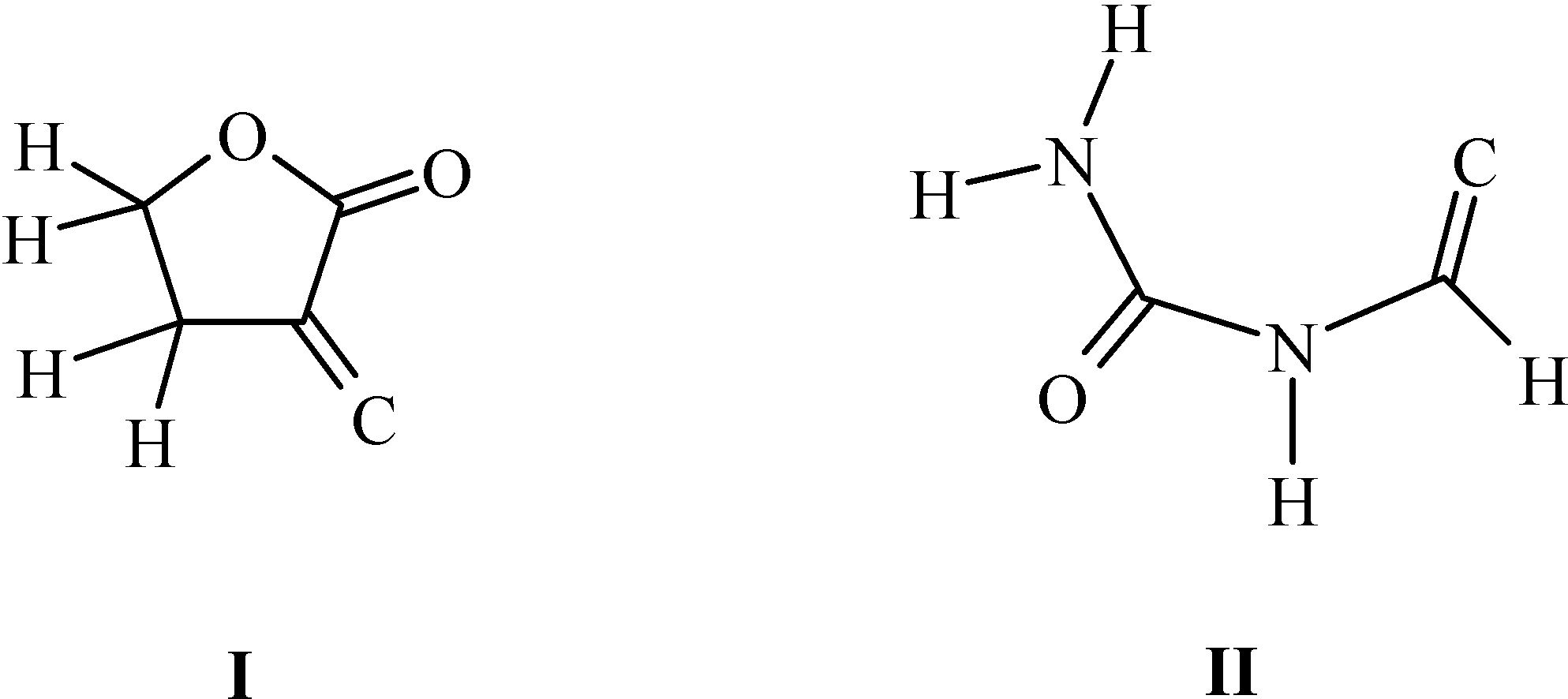
Synthesis and Structural Characterization of the 5-(2-Haloethyl)pyrimidines – Hydrogen-Bonded chains in α-(1-Carbamyliminomethylene)-γ-Butyrolactone

by
Tatjana Gazivoda
1,
Silvana Raić-Malić
1,
Antonija Hergold-Brundić
2 and
Mario Cetina
3,*
1
Department of Organic Chemistry, Faculty of Chemical Engineering and Technology, University of Zagreb, Marulićev trg 20, P.O. Box 177, HR-10000 Zagreb, Croatia
2
Laboratory of General and Inorganic Chemistry, Faculty of Science, University of Zagreb, Horvatovac 102a, HR-10000 Zagreb, Croatia
3
Department of Applied Chemistry, Faculty of Textile Technology, University of Zagreb, Prilaz baruna Filipovića 28a, HR-10000 Zagreb, Croatia
*
Author to whom correspondence should be addressed.
Submission received: 21 August 2008 / Revised: 26 September 2008 / Accepted: 13 October 2008 / Published: 6 November 2008

Abstract

:
Three novel 5-(2-haloethyl)pyrimidine derivatives were synthesized and characterized by 1H-NMR, 13C-NMR, MS, IR spectra and elemental analysis. Iodine and chlorine atoms in the C-5 side chain were introduced by reaction of 5-(2-hydroxyethyl)pyrimidine with hydroiodic acid and phosphoryl chloride, respectively. The structure of the intermediate α-(1-carbamyliminomethylene)-γ-butyrolactone was determined by X-ray crystal structure analysis. The molecule deviates very slightly from planarity. Three N−H···O hydrogen bonds link the molecules into one-dimensional chains of edge-fused rings.

Graphical Abstract

Introduction

A number of 5-substituted pyrimidine nucleoside analogues have been shown to have potent and selective cytotoxic activity in cells transfected with the HSV-1 TK gene [1,2,3]. The (E)-5-(2-halovinyl)-2’-deoxyuridines are among the most potent and selective agents that undergo selective phosphorylation by the viral-encoded enzyme [4]. Pyrimidine nucleoside analogues, e.g. 5-(2-fluoroethyl)-2’-deoxyuridine, labelled with radioisotopes have applications in positron emission tomography (PET) to monitor the successful transfection of gene products in vivo during cancer-prodrug therapy [5]. Recently, we have reported that some C-5 and/or C-6 substituted pyrimidine derivatives exhibited cytostatic activities against human tumor cell lines [6,7,8,9,10,11]. In view of the importance of 5-substituted uracil derivatives in cancer chemotherapy [12] and as antiviral agents [13,14,15] we became interested in the synthesis of the novel 5-substituted uracils that we describe in this paper (Scheme 1). In addition, we also present herein the X-ray crystal structure of α-(1-carbamyliminomethylene)-γ-butyrolactone, a precursor in the synthesis of these 5-substituted uracils. This compound, possessing two hydrogen-bonding donor groups and two acceptors, is very interesting from the supramolecular point of view, because it can form intricate hydrogen-bonded networks including chain motifs, rings, chains of rings and chains of edge-fused rings.
Scheme 1. Synthesis of the 5-(2-haloethyl)pyrimidines 4-6.
Molecules 13 02786 g005
Reagents and conditions: (i) NaOMe, ether, HCOOCH3, 24 h; (ii) urea, 3M HCl, 4 °C, 24 h; (iii) NaOEt, ethanol, reflux, 6 h; (iv) HI, -5 °C, 90 min; (v) POCl3, reflux, 1h; (vi) NaOMe, reflux, 6 h.

Results and Discussion
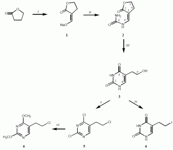
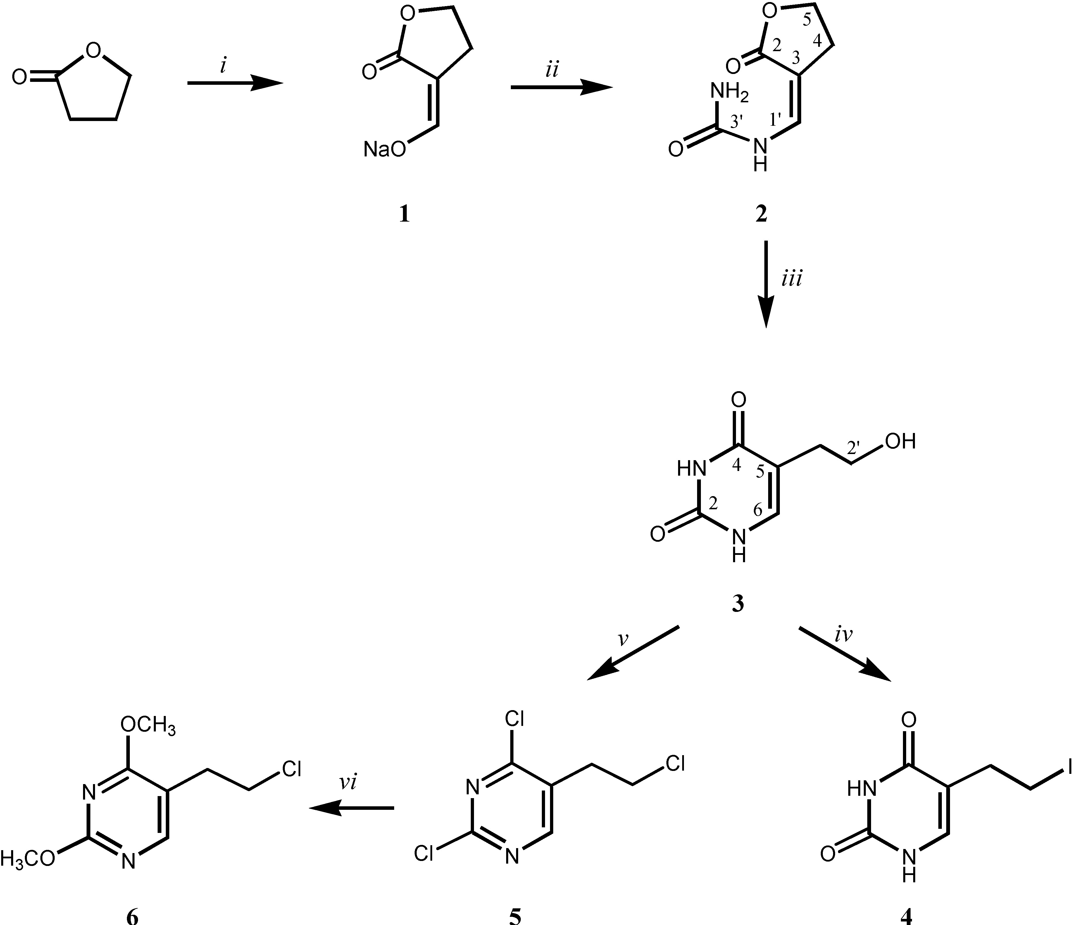
Synthesis

The sodium derivative 1, prepared by reaction of γ-butyrolactone with methylformate in dry ether with the presence of sodium methoxide, was subsequently reacted with urea to give α-(1-carbamyliminomethylene)-γ-butyrolactone (2) [16,17]. Reaction of 2 with sodium ethoxide afforded 5-(2-hydroxyethyl)uracil (3), which with 57% solution of hydroiodic acid gave 5-(2-iodoethyl)uracil (4). Chlorination of compound 3 with phosphoryl chloride afforded the chlorinated pyrimidine derivative 5, which was subsequently methoxylated giving 5-(2-chloroethyl)-2,4-dimethoxyuracil (6). The newly prepared compounds 4-6 were fully characterized by 1H- and 13C-NMR spectra, mass and IR spectra, as well as elemental analysis (see Experimental section).

X-ray crystal structure study
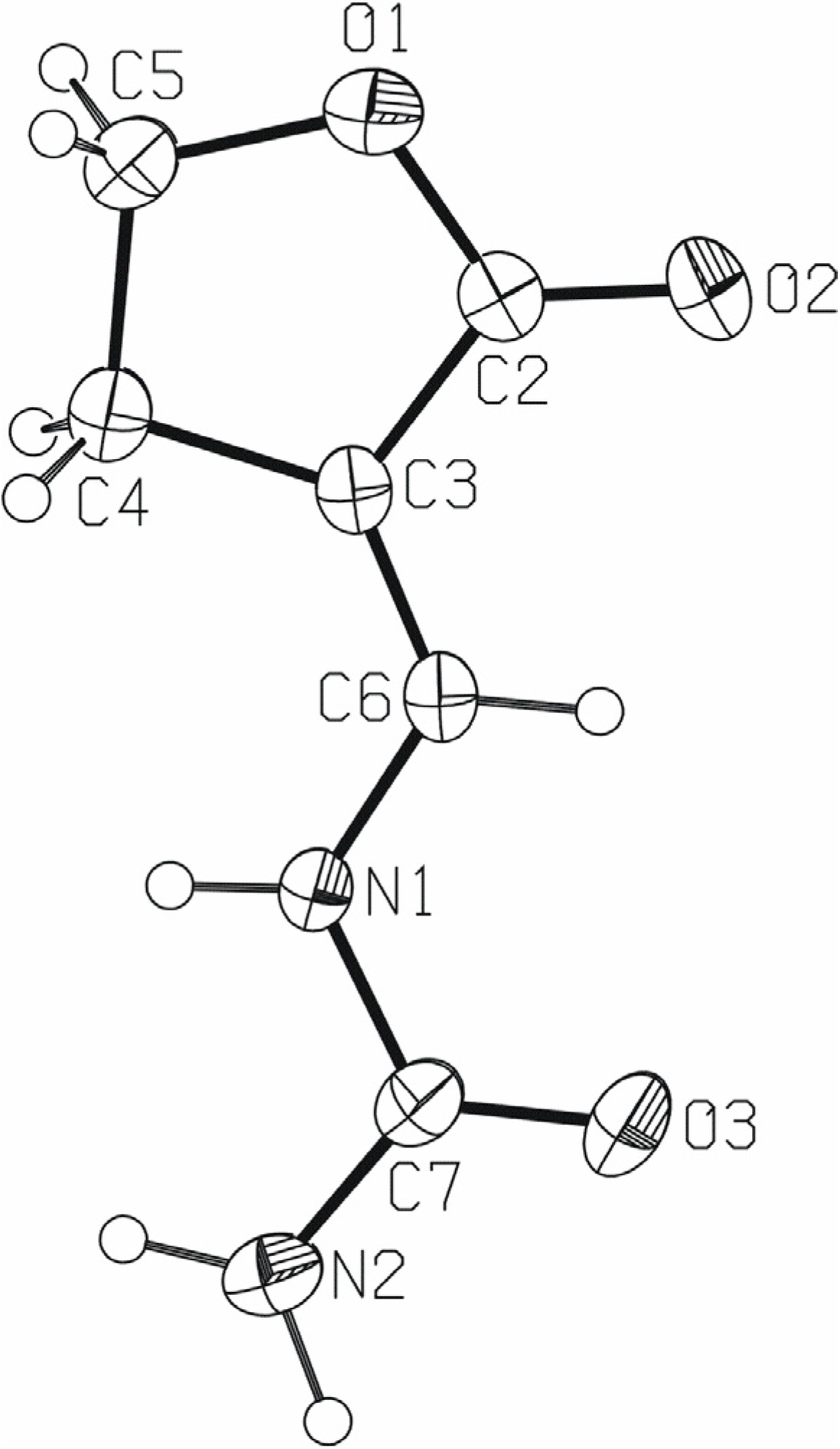
Compound 2 (Figure 1) consists of a carbamyliminomethylene moiety bonded to the butyrolactone ring. The whole molecule deviates very slightly from planarity, as the dihedral angle between the mean planes of the carbamyliminomethylene atoms N2/C7/O3/N1/C6 and butyrolactone ring atoms O1/O2/C2/C3/C4/C5 is 7.17(8)o.
Figure 1. A molecular structure of 2, with the atom-numbering scheme. Displacement ellipsoids for nonhydrogen atoms are drawn at the 40 % probability level.
Molecules 13 02786 g001
A survey of the Cambridge Structural Database [18] (CSD; error-free and disorder-free structures of organic compounds with R<0.075) revealed 16 structures in which the carbon atom is double bonded to the butyrolactone ring (Figure 2, fragment I) and only one structure possessing a carbamyliminomethylene moiety [19] (Figure 2, fragment II).
Figure 2. Fragments from a Cambridge Structural Database survey.
Molecules 13 02786 g002
The bond lengths and angles in 2 (Table 1) present no unexpected features and are within the ranges of equivalent ones in structures found in the CSD [18].
Table 1. Bond distances (Å) and bond angles (o) for 2.
N1−C61.363(2) O3−C71.2251(19)
N1−C71.3900(18) C2−C31.453(2)
N2−C71.339(2) C3−C61.333(2)
O1−C21.3417(17) C3−C41.495(2)
O1−C51.450(2) C4−C51.518(2)
O2−C21.2173(19)
C6−N1−C7121.43(13) C2−C3−C4108.73(12)
C2−O1−C5110.77(12) C3−C4−C5102.74(13)
O2−C2−O1120.86(14) O1−C5−C4107.73(12)
O2−C2−C3129.10(15) C3−C6−N1125.09(13)
O1−C2−C3110.02(12) O3−C7−N2124.88(15)
C6−C3−C2121.18(13) O3−C7−N1121.05(15)
C6−C3−C4130.00(14) N2−C7−N1114.07(13)
Table 2. Hydrogen-bonding geometry for 2.
D−H···AD−H
(Å)
H···A
(Å)
D···A
(Å)
D−H···
A
N1−H1···O2i0.85(2)2.06(2)2.869(2)158(2)
N2−H2A···O2i0.90(2)2.16(2)2.974(2)150(2)
N2−H2B···O3ii0.93(2)1.96(2)2.884(2)174(2)
Symmetry codes: (i) x, −1+y, z; (ii) 1−x, −1/2+y, 3/2−z.
The carbamyliminomethylene moiety nitrogen atoms N1 and N2 and carbonyl oxygen atoms O2 and O3 participate in the supramolecular assembly of 2. The N2 atom is a double proton donor and O2 atom is a double proton acceptor (Table 2).
The N1···O2i and N2···O2i intermolecular hydrogen bonds in 2 form a Molecules 13 02786 i001 
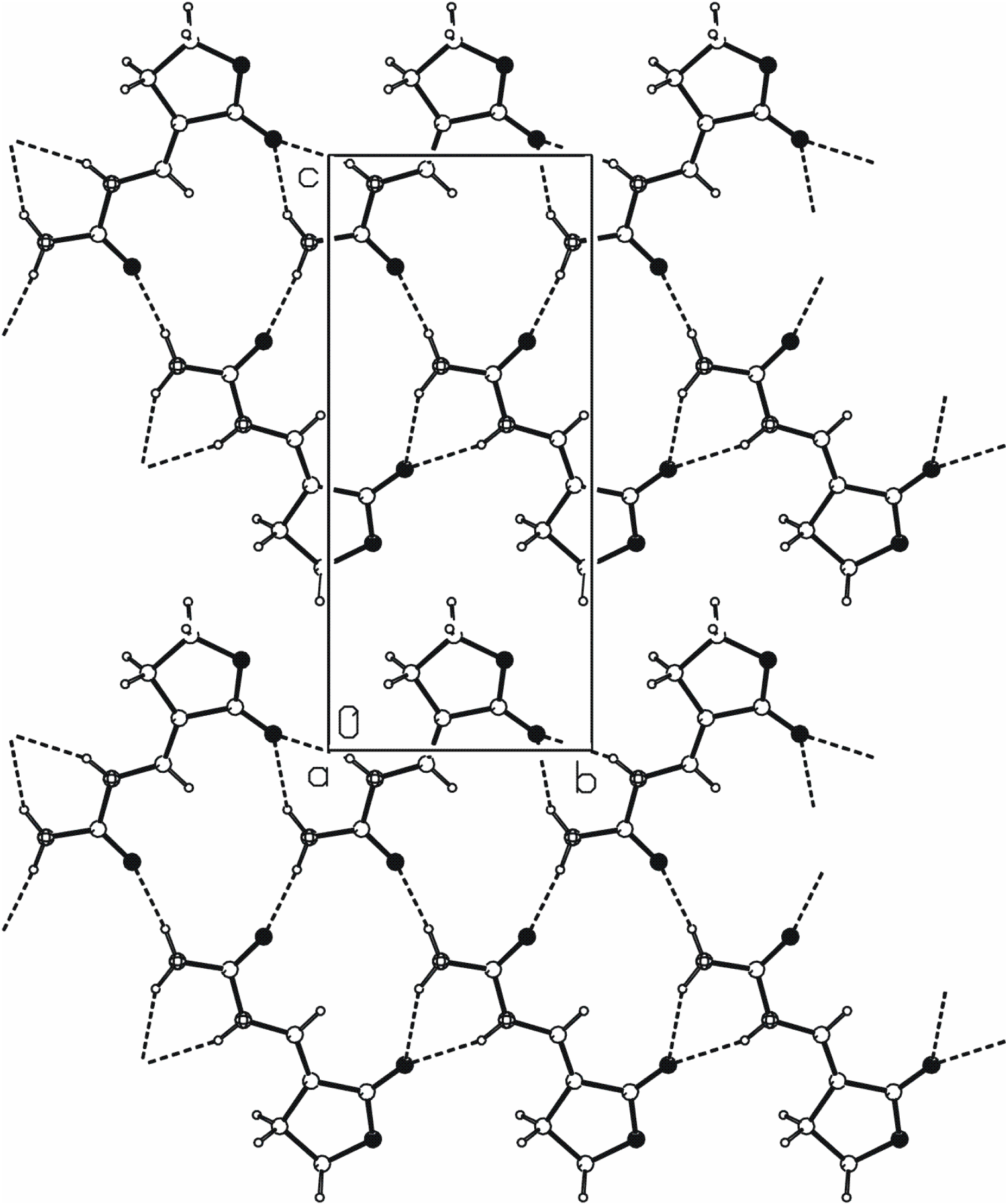
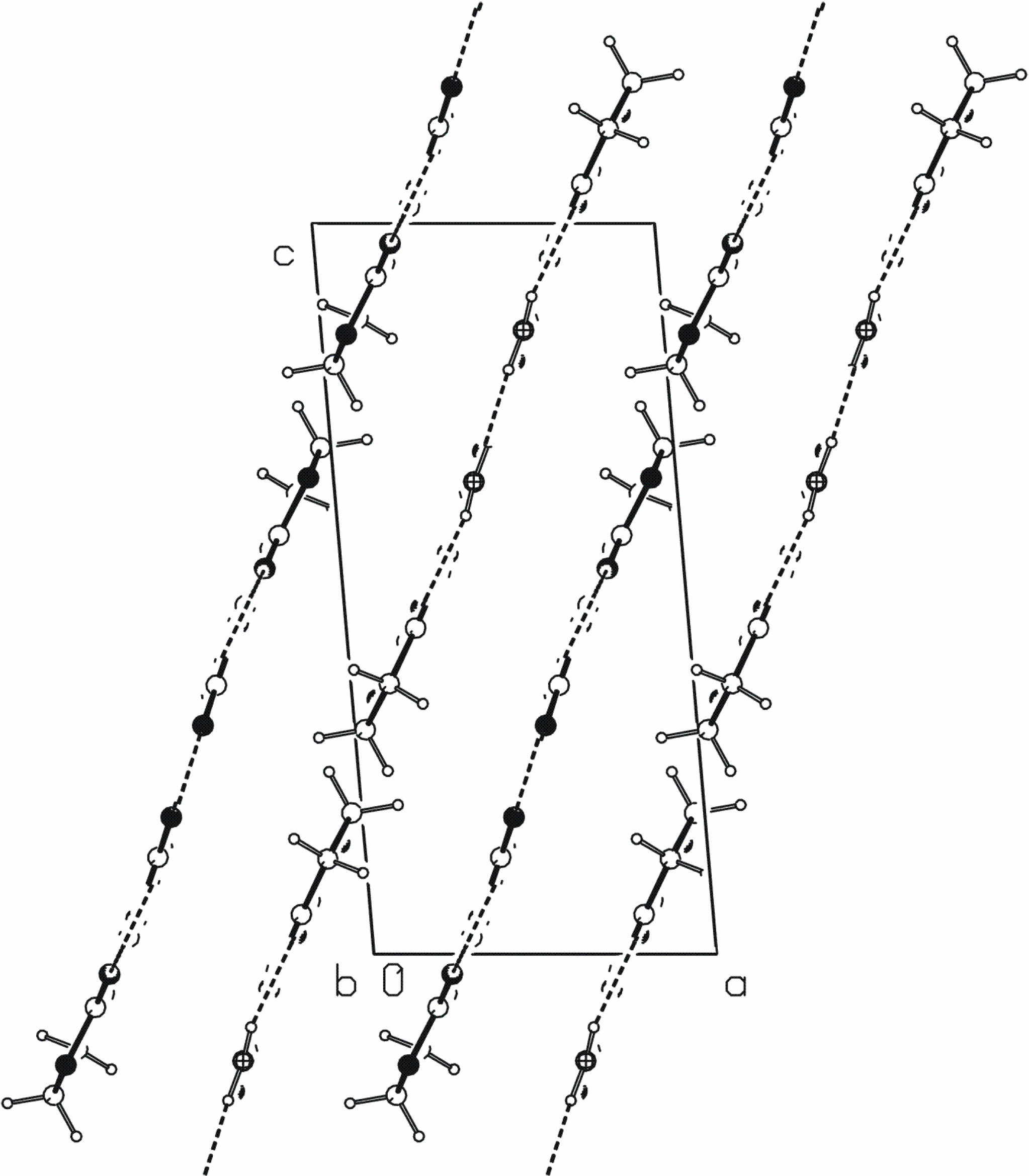
[20] chain of rings parallel to the b axis (Figure 3). The N2···O3ii hydrogen bond links the molecules into C(4) chains that are also parallel to the b axis. The combination of these two motifs forms new ring of Molecules 13 02786 i002 type. These three N−H···O hydrogen bonds generate mutually parallel one-dimensional chains of edge-fused rings (Figure 3).
Figure 3. A crystal packing diagram of 2, viewed along the a axis, showing chains of edge-fused rings formed by N−H···O hydrogen bonds.
Molecules 13 02786 g003
Figure 4. A crystal packing diagram of 2, viewed along the b axis, showing parallel chains of hydrogen-bonding molecules.
Molecules 13 02786 g004
The crystal packing diagram along b axis (Figure 4) reveals that the hydrogen-bonded chains are parallel to the (2, 0, 1). The distance between the planes passing through the center of the hydrogen-bonded molecules is approximately 3.05 Å. Very similar one-dimensional hydrogen-bonded chains generate N−H···O hydrogen bonds in 1-carbamyliminomethylenetetrahydropyrrol-2-one [19].

Experimental Section

General

Melting points were determined on a Kofler micro hot-stage apparatus (Reichert, Wien) and are uncorrected. The electron impact mass spectra were recorded with an EXTREL FT MS 2002 instrument with ionizing energy of 70 eV. 1H- and 13C-NMR spectra were recorded on a Bruker Avance 600 MHz spectrometer, operating at 600 MHz (1H) and 150.92 MHz (13C), respectively. The samples were dissolved in DMSO-d6 and measured in 5 mm NMR tubes. The 1H- and 13C-NMR chemical shift values (δ) are expressed in ppm referred to tetramethylsilane (TMS) and coupling constants (J) in Hz. The infrared spectra were recorded by a Fourier transform infrared (FTIR) spectrometer Bruker Vertex 70 equipped with an attenuated total reflection (ATR) accessory with a diamond crystal. Sixteen scans were collected for each measurement over the spectral range of 400-4000 cm-1 with resolution of 4 cm-1. Precoated Merck Silica Gel 60F-254 plates were used for thin layer chromatography (TLC) and the spots were detected under UV light (254 nm). Column chromatography (CLC) was performed using silica gel (0.063-0.2 mm) Fluka; glass column was slurry-packed under gravity. Compound purity was analyzed by HPLC with DAD detector.

Synthesis

Sodium α-hydroxymethylene-γ-butyrolactone (1) [16]. To a cooled suspension of NaOMe (35.5 g, 0.66 mol) in dry ether (650 mL), a solution of methylformate (52.6 mL, 0.85 mol) and γ-butyrolactone (50 mL, 0.66 mol) was added dropwise. The stirring was continued at room temperature overnight. After cooling of reaction mixture the precipitated product 1 was filtered off and washed with dry ether (37.5 g, 41.8 %, m.p. = 124 – 126 °C); MS m/z (EI) for C5H5NaO3 (M+) calc. 136.0812; found 136.1; 1H-NMR δ 8.46 (1H, s, H-1'), 4.24 (2H, t, J = 7.00 Hz, H-5), 2.44 (2H, dt, J = 9.97 Hz, 8 Hz, H-4); 13C-NMR δ 176.96 (C-2), 166.54 (C-1'), 132.60 (C-3), 68.37 (C-5), 27.50 (C-4).
α-(1-Carbamyliminomethylene)-γ-butyrolactone (2) [17]. To a solution of urea (30 g, 0.5 mol) in cold 3M HCl (200 mL) was added sodium α-hydroxymethylene-γ-butyrolactone (1, 34 g, 0.25 mol). After stirring at 4 °C overnight the precipitated product was filtered off and washed with cold water. Recrystallization from water – ethanol gave white crystals of 2 (2 g, 5.2 %, m.p. = 246 – 247 °C); MS m/z (EI) for C6H8N2O3 (M+) calc. 156.1394; found 156.1; 1H NMR δ 8.96 (1H, d, J = 11.71 Hz, NH), 7.60 (1H, d, J = 11.97 Hz, H-1'), 6.38 (2H, s, NH2), 4.28 (2H, t, J = 7.5 Hz, H-5), 2.75 (2H, dt, J = 7.5 Hz, 2.3 Hz, H-4); 13C-NMR δ 172.69 (C-3'), 154.35 (C-2), 132.60 (C-1'), 99.30 (C-3), 65.07 (C-5), 24.19 (C-4).
5-(2-Hydroxyethyl)uracil (3). To a solution of NaOEt (0.277 g, 4 mmol) in ethanol (20 mL) was added α-(1-carbamyliminomethylene)-γ-butyrolactone (2, 1.7 g, 0.01 mol). Reaction mixture was refluxed for 6 h, during which time product precipitated. The solvent was removed under reduced pressure and the residue was dissolved in the water and acidified to pH = 3. The precipitated product 3 was filtered off, washed with ethanol and dried in vaccuo (1.55 g, 91.2 %); MS m/z (EI) for C6H8N2O3 (M+) calc. 156.1394; found 156.1; 1H-NMR δ 11.41 (1H, s, NH), 11.13 (1H, s, NH), 7.21 (1H, s, H-6), 3.63 (1H, t, J = 6.68 Hz, OH), 3.41 (2H, t, J = 6.62 Hz, H-2'), 2.29 (2H, t, J = 6.51 Hz, H-1'); 13C-NMR δ 164.32 (C-4), 151.50 (C-2), 135.19 (C-6), 110.38 (C-5), 60.21 (C-2'), 29.06 (C-1').
5-(2-Iodoethyl)uracil (4). To a cooled solution of 3 (100 mg, 0.6 mmol) in H2O (10 mL), a solution of HI (57%, 1 mL) was added. The reaction mixture was stirred at -5 °C for 90 min, then solvent was evaporated and residue was purified by column chromatography (CH2Cl2:CH3OH = 60:1) to give oily product 4 (112 mg, 65 %); MS m/z (EI) for C6H7IN2O2 (M+) calc. 266.0365; found 266; Anal. for C6H7IN2O2 calc. C 27.09 %, H 2.65 %, N 10.53 %; found C 27.13 %, H 2.64 %, N 10.55 %; IR: 3110–2992 (ν N–H, CH2), 1720 (ν C=O), 1650–1420 (ν C=C and C–H ring), 1480 (δ CH2), 1022–770 (δ C=C–H, CH2), 940 (δ CH2–I), 520–480 (ν C–I) cm-1; 1H-NMR δ 10.97 (1H, s, NH), 10.62 (1H, s, NH), 7.20 (1H, s, H-6), 4.52 (2H, t, J = 5.4 Hz, H-2'), 2.28 (2H, t, J = 6.60 Hz, H-1'); 13C-NMR δ 165.14 (C-4), 151.82 (C-2), 139.27 (C-6), 109.61 (C-5), 59.86 (C-2'), 20.03 (C-1').
5-(2-Chloroethyl)-2,4-dichloropyrimidine (5). The reaction mixture of 3 (600 mg, 4 mmol) and POCl3 (17.7 mL, 0.19 mol) was refluxed for 1 h. Solvent was evaporated and ice was added in the residue which was then washed with ether and dried over Na2SO4. After drying the solvent was removed in vacuo and residue was purified by column chromatography (CH2Cl2:CH3OH = 30:1) which gave colourless oily product 5 (260 mg, 31%); MS m/z (EI) for C6H5Cl3N2 (M+) calc. 211.4575; found 211.5; Anal. for C6H5Cl3N2 calc. C 34.08 %, H 2.38 %, N 13.25 %; found C 34.01 %, H 2.39 %, N 13.22 %; IR: 1633–1320 (ν C=C and C–H ring), 1430 (δs CH2), 1090 (δ CH2–Cl), 980-760 (δ C=C–H, CH2), 580–510 (ν C–Cl) cm-1; 1H-NMR δ 8.82 (1H, s, H-6), 3.93 (2H, t, J = 6.78 Hz, H-2'), 3.19 (2H, t, J = 6.78, H-1'); 13C-NMR δ 172.46 (C-4), 162.01 (C-2), 156.87 (C-6), 112.70 (C-5), 43.15 (C-2'), 31.51 (C-1').
5-(2-Chloroethyl)-2,4-dimethoxypyrimidine (6). To a solution of NaOMe (120 mg, 2.2 mmol) in MeOH (15 mL) was added 5 (80 mg, 0.38 mmol), and the mixture was heated at reflux for 6 h. The solvent was evaporated, and H2O was added to dissolve NaCl. The oily layer was extracted with CH2Cl2, dried (Na2SO4), and concentrated under reduced pressure. The residue was purified by column chromatography (CH2Cl2:CH3OH = 30:1) which gave colourless oily product 6 (54 mg, 70.6 %); MS m/z (EI) for C8H11ClN2O2 (M+) calc. 202.6379; found 202.6; Anal. C 7.42 %, H 5.47 %, N 13.82 %; found C 47.31 %, H 5.47 %, N 13.84 %; IR: 2960 (ν CH3), 1640–1330 (ν C=C and C–H ring), 1420 (δ CH2), 1210 (νas C–O–C), 1100 (δ CH2–Cl), 1050 (νs C–O–C), 905–760 (δ C=C–H, CH2), 630–520 (ν C–Cl) cm-1; 1H-NMR δ 8.19 (1H, s, H-6), 3.92 (3H, s, OCH3), 3.87 (3H, s, OCH3), 3.77 (2H, t, J = 6.93 Hz, H-2'), 2.89 (2H, t, J = 6.93 Hz, H-1'); 13C-NMR δ 169.30 (C-4), 164.56 (C-2), 158.87 (C-6), 111.44 (C-5), 54.85 (OCH3), 54.33 (OCH3), 43.75 (C-2'), 29.76 (C-1').

Crystal structure determination of 2

A single crystal of 2 suitable for X-ray structure analysis was prepared by growth under slow evaporation at room temperature of a very dilute ethanol solution (v/v 96 %). The intensities were collected at 295 K on a Philips PW1100 diffractometer updated by Stoe and Cie [21,22] using Mo-Kα radiation (0.71073 Å). Details of crystal data, data collection and refinement parameters are given in Table 3.
Table 3. X-ray crystallographic data for 2.
FormulaC6H8N2O3
Formula weight156.14
Crystal systemmonoclinic
Space groupP 21/c
Unit cell dimensions
a (Å)7.0522(8)
b (Å)6.6691(6)
c (Å)15.1031(16)
β (o)94.869(9)
V3)707.76(13)
Z4
Dcalc. (g cm-3)1.465
Absorption coefficient μ (mm-1)0.119
F(000)328
θ range (°)2.71 - 27.99
Index ranges-9 ≤ h ≤ 9
-8 ≤ k ≤ 8
-11 ≤ l ≤ 19
Collected reflections No.2941
Independent reflections No. / Rint.1702 / 0.0231
Reflections No. I ≥ 2σ(I)1063
Refined parameters No.112
Goodness-of-fit on F2, S1.001
R [I ≥ 2σ(I)] / R [all data]0.0398 / 0.0783
wR [I ≥ 2σ(I)] / wR [all data]0.1061 / 0.1231
Max./min. electron density (e Å-3)0.189 / -0.141
The structure was solved by direct methods [23]. All non-hydrogen atoms were refined anisotropically by full-matrix least-squares calculations based on F2 [23]. The hydrogen atoms bonded to nitrogen atoms were found in a difference Fourier map and their coordinates and isotropic thermal parameters have been refined freely. All other hydrogen atoms were included in calculated positions as riding atoms, with SHELXL97 [23] defaults. PLATON [24] program was used for structure analysis and molecular and crystal structure drawings preparation. CCDC 695308 contains the supplementary crystallographic data for this paper. These data can be obtained free of charge via www.ccdc.cam.ac.uk/data_request/cif, by emailing [email protected], or by contacting The Cambridge Crystallographic Data Centre, 12, Union Road, Cambridge CB2 1EZ, UK; fax: +44 1223 336033.

Acknowledgements

Support for this study was provided by the Ministry of Science, Education and Sports of the Republic of Croatia (Projects #125-0982464-2925 and #119-1193079-3069).

References

  1. Balzarini, J.; Bohman, C.; De Clercq, E. Differential mechanism of cytostatic effect of (E)-5-(2-bromovinyl)-2'-deoxyuridine, 9-(1,3-dihydroxy-2-propoxymethyl)guanine, and other antiherpetic drugs on tumor cells transfected by the thymidine kinase gene of herpes simplex virus type 1 or type 2. J. Biol. Chem. 1993, 268, 6332–6337. [Google Scholar]
  2. Balzarini, J.; De Clercq, E.; Ayusawa, D.; Seno, T. Murine mammary FM3A carcinoma cells transformed with the herpes simplex type 1 thymidine kinase gene are highly sensitive to the growth-inhibitory properties of (E)-5-(2-bromovinyl)-2'-deoxyuridine and related compounds. FEBS Lett. 1985, 185, 95–100. [Google Scholar] [CrossRef]
  3. De Clercq, E. Biochemical aspects of the selective antiherpes activity of nucleoside analogues. Biochem. Pharmacol. 1984, 33, 2159–2169. [Google Scholar] [CrossRef]
  4. De Clercq, E.; Descamps, J.; Verhelst, G.; Walker, R.T.; Jones, A.S.; Torrence, P.F.; Shugar, D. Comparative efficacy of antiherpes drugs against different strains of herpes simplex virus. J. Infect. Dis. 1981, 41, 563–574. [Google Scholar]
  5. Yu, C.-S.; Eisenbarth, J.; Runz, A.; Weber, K.; Zeisler, S.; Oberdorfer, F. Synthesis of 5-(2-radiohaloethyl)- and 5-(2-radiohalovinyl)-2’-deoxyuridines. Novel types of radiotracer for monitoring cancer gene therapy with PET. J. Label. Compd. Radiopharm. 2003, 46, 421–439. [Google Scholar] [CrossRef]
  6. Prekupec, S.; Makuc, D.; Plavec, J.; Šuman, L.; Kralj, M.; Pavelić, K.; Balzarini, J.; De Clercq, E.; Mintas, M.; Raić-Malić, S. Novel C-6 fluorinated acyclic side chain pyrimidine derivatives: Synthesis, 1H and 13C NMR conformational studies, and antiviral and cytostatic evaluations. J. Med. Chem. 2007, 50, 3037–3045. [Google Scholar] [CrossRef]
  7. Raić-Malić, S.; Johayem, A.; Ametamey, S.M.; Batinac, S.; De Clercq, E.; Folkers, G.; Scapozza, L. Synthesis, 18F-radiolabelling and biological evaluations of C-6 alkylated pyrimidine nucleoside analogues. Nucleos. Nucleot. Nucleic Acids 2004, 23, 1707–1721. [Google Scholar] [CrossRef]
  8. Johayem, A.; Raić-Malić, S.; Lazzati, K.; Schubiger, P.A.; Scapozza, L.; Ametamey, S.M. Synthesis and characterization of a C(6) nucleoside analogue for the in vivo imaging of the gene expression of herpes simplex virus type-1 thymidine kinase (HSV1 TK). Chem. Biodiver. 2006, 3, 274–283. [Google Scholar] [CrossRef]
  9. Gazivoda, T.; Plevnik, M.; Plavec, J.; Kraljević, S.; Kralj, M.; Pavelić, K.; Balzarini, J.; De Clercq, E.; Mintas, M.; Raić-Malić, S. The novel pyrimidine and purine derivatives of l-ascorbic acid: Synthesis, one- and two-dimensional 1H and 13C NMR study, cytostatic and antiviral evaluation. Bioorg. Med. Chem. 2005, 13, 131–139. [Google Scholar] [CrossRef]
  10. Gazivoda, T.; Šokčević, M.; Kralj, M.; Šuman, L.; Pavelić, K.; De Clercq, E.; Andrei, G.; Snoeck, R.; Balzarini, J.; Mintas, M.; Raić-Malić, S. Synthesis and antiviral and cytostatic evaluations of the new C-5 substituted pyrimidine and furo[2,3-d]pyrimidine 4',5'-didehydro-l-ascorbic acid derivatives. J. Med. Chem. 2007, 50, 4105–4112. [Google Scholar] [CrossRef]
  11. Gazivoda, T.; Raić-Malić, S.; Marjanović, M.; Kralj, M.; Pavelić, K.; Balzarini, J.; De Clercq, E.; Mintas, M. The novel C-5 aryl, alkenyl and alkynyl substituted uracil derivatives of l-ascorbic acid: Synthesis, cytostatic and antiviral activity evaluations. Bioorg. Med. Chem. 2007, 15, 749–758. [Google Scholar] [CrossRef]
  12. Sigmond, J.; Peters, G.J. Pyrimidine and purine analogues, effects on cell cycle regulation and the role of cell cycle inhibitors to enhance their cytotoxicity. Nucleos. Nucleot. Nucleic Acids 2005, 24, 1997–2022. [Google Scholar] [CrossRef]
  13. De Clercq, E. Chemotherapeutic approaches to the treatment of the acquired immunodeficiency syndrome (AIDS). J. Med. Chem. 1986, 29, 1561–1569. [Google Scholar] [CrossRef]
  14. Herdewijn, P.; Van Aershot, A.; Balzarini, J.; De Clercq, E. 3’-Fluoro- and 3’-azido-substituted 2’,3’-dideoxynucleosides: Structure-activity relationship. Med. Chem. Res. 1991, 1, 9–19. [Google Scholar]
  15. De Clercq, E. Toward Improved Anti-HIV Chemotherapy: Therapeutic strategies for intervention with HIV infections. J. Med. Chem. 1995, 38, 2491–2517. [Google Scholar] [CrossRef]
  16. Fissekis, J.D.; Myles, A.; Brown, G.B. Synthesis of 5-hydroxyalkylpyrimidines from lactones. J. Org. Chem. 1964, 29, 2670–2673. [Google Scholar] [CrossRef]
  17. Fissekis, J.D.; Sweet, F. The chemistry of some 5-(2-hydroxyalkyl)uracil derivatives and a synthesis of 5-vinyluracil. J. Org. Chem. 1973, 38, 264–269. [Google Scholar] [CrossRef]
  18. Allen, F.H. The Cambridge Structural Database: a quarter of a million crystal structures and rising. Acta Crystallogr. 2002, B58, 380–388, (Version 5.29, January 2008). [Google Scholar] [CrossRef]
  19. Dinsmore, A.; Doyle, P.M.; Hitchcock, P.B.; Young, D.W. Extension of ring switching strategy to the glutamate antagonist 2-(pyrimidin-2,4-dione-5-yl-methyl)-(2S)-glycine and related compounds with two chiral centres. Tetrahedron Lett. 2000, 41, 10153–10158. [Google Scholar]
  20. Bernstein, J.; Davis, R.E.; Shimoni, L.; Chang, N.-L. Patterns in hydrogen bonding: functionality and graph set analysis in crystals. Angew. Chem. Int. Ed. Engl. 1995, 34, 1555–1573. [Google Scholar] [CrossRef]
  21. Stoe & Cie (1995). STADI4. Diffractometer control program, Version 1.05B; Darmstadt, Germany.
  22. Stoe & Cie (1995). X-RED. Diffractometer reduction program. Version 1.05B; Darmstadt, Germany.
  23. Sheldrick, G.M. A short history of SHELX. Acta Crystallogr. 2008, A64, 112–122. [Google Scholar] [CrossRef]
  24. Spek, A.L. Single-crystal structure validation with the program PLATON. J. Appl. Crystallogr. 2003, 36, 7–13. [Google Scholar] [CrossRef]
  • Sample Availability: Samples of the compounds are available from T. Gazivoda.

