
Decrystallization of Crystals Using Gold “Nano-Bullets” and the Metal-Assisted and Microwave-Accelerated Decrystallization Technique

Abstract
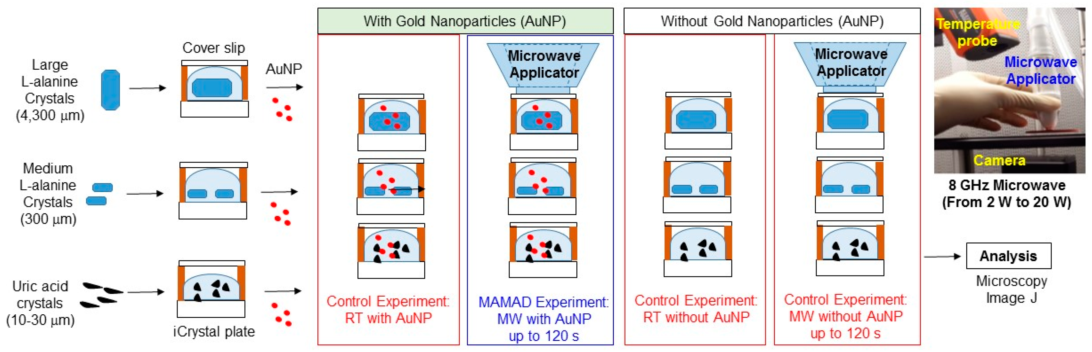
:
1. Introduction
2. Results
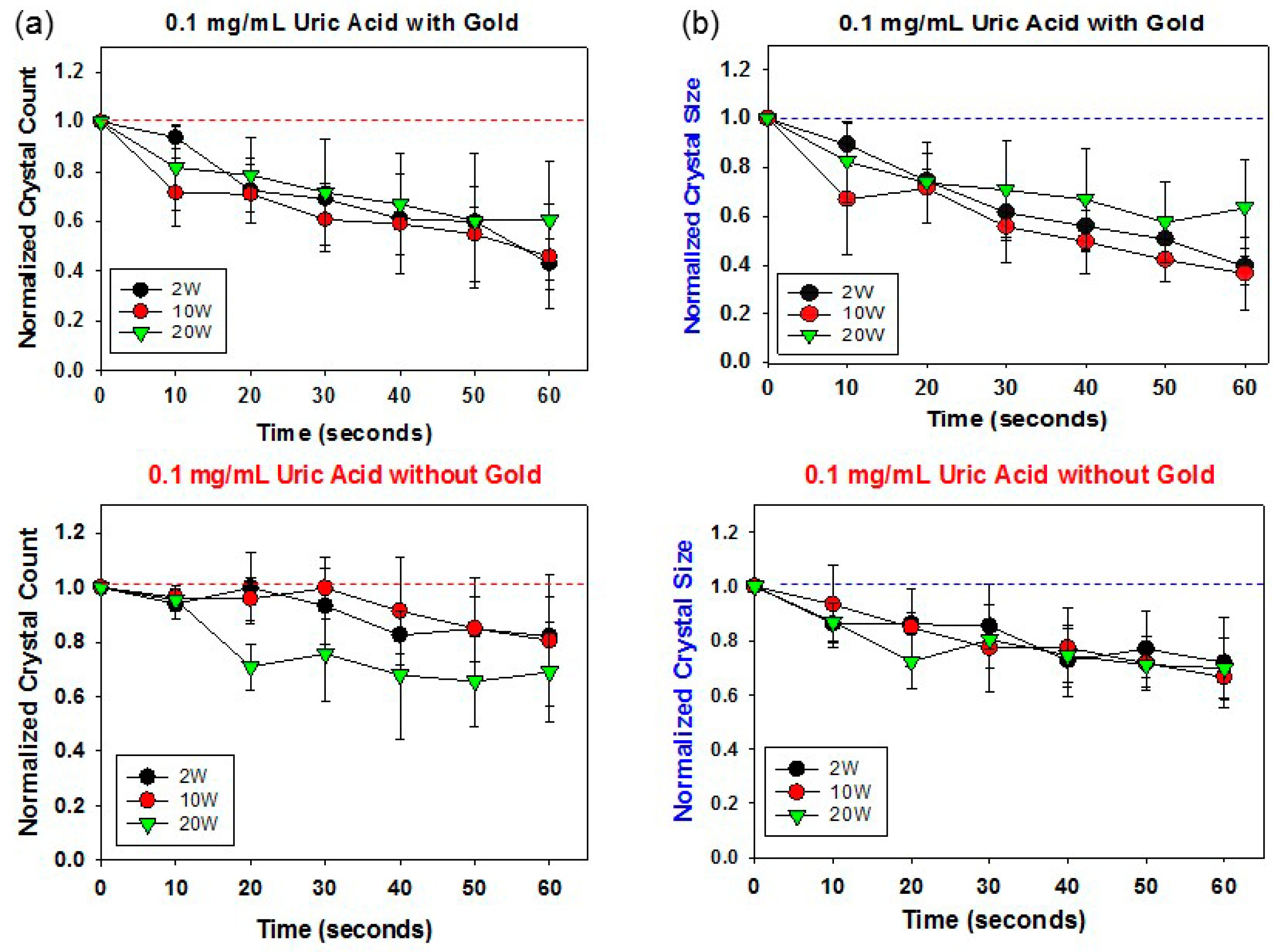
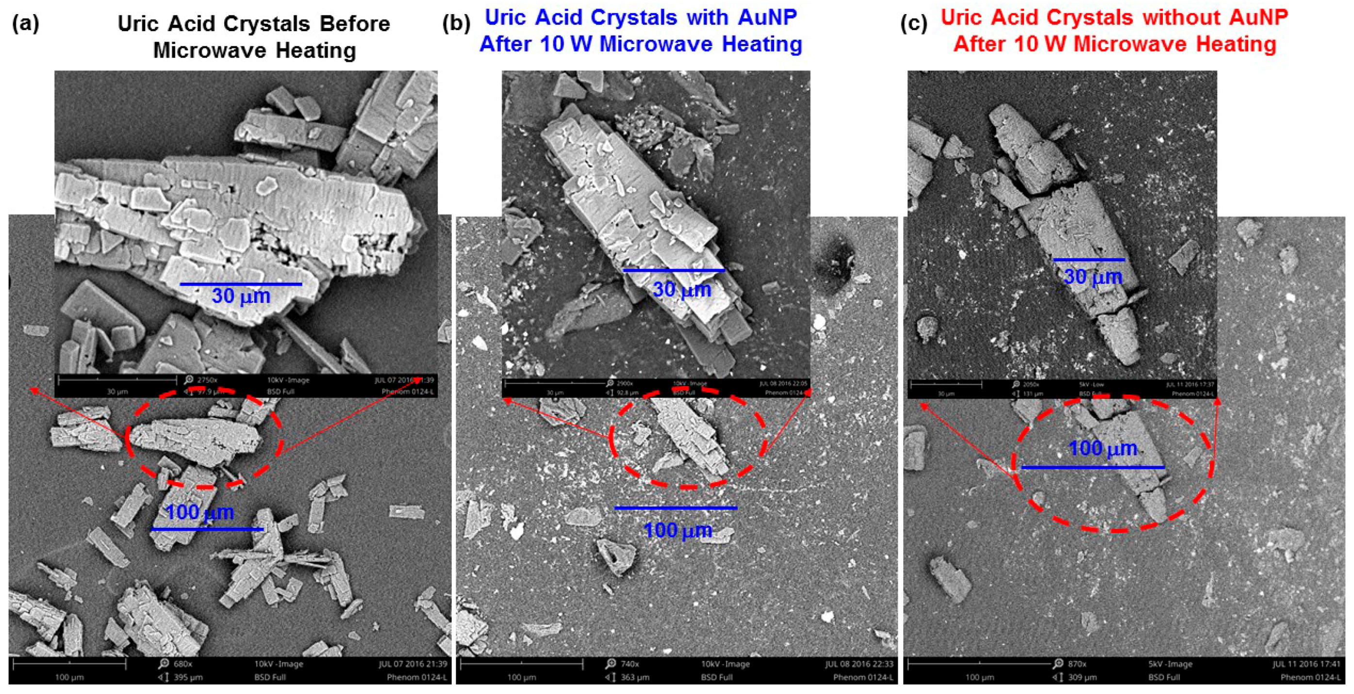
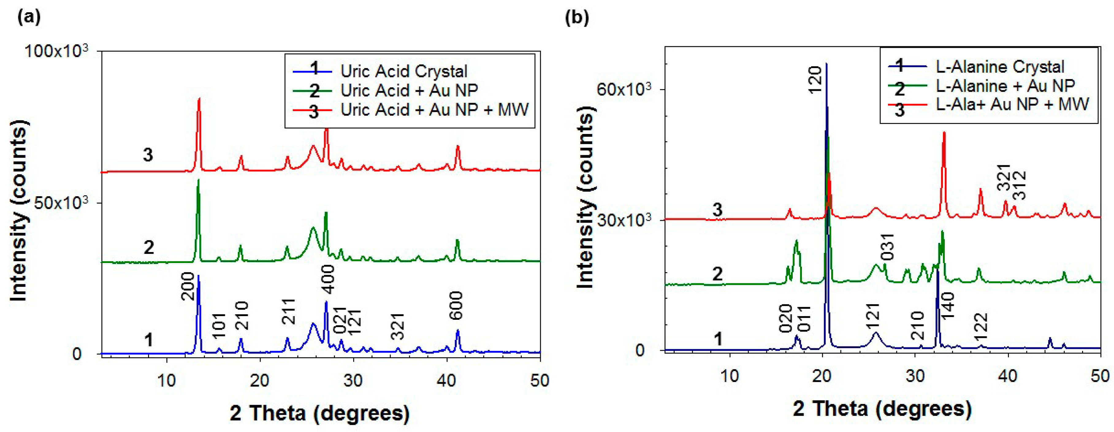
2.1. Decrystallization of Uric Acid Crystals
Image Analysis of Small Uric Acid Crystals
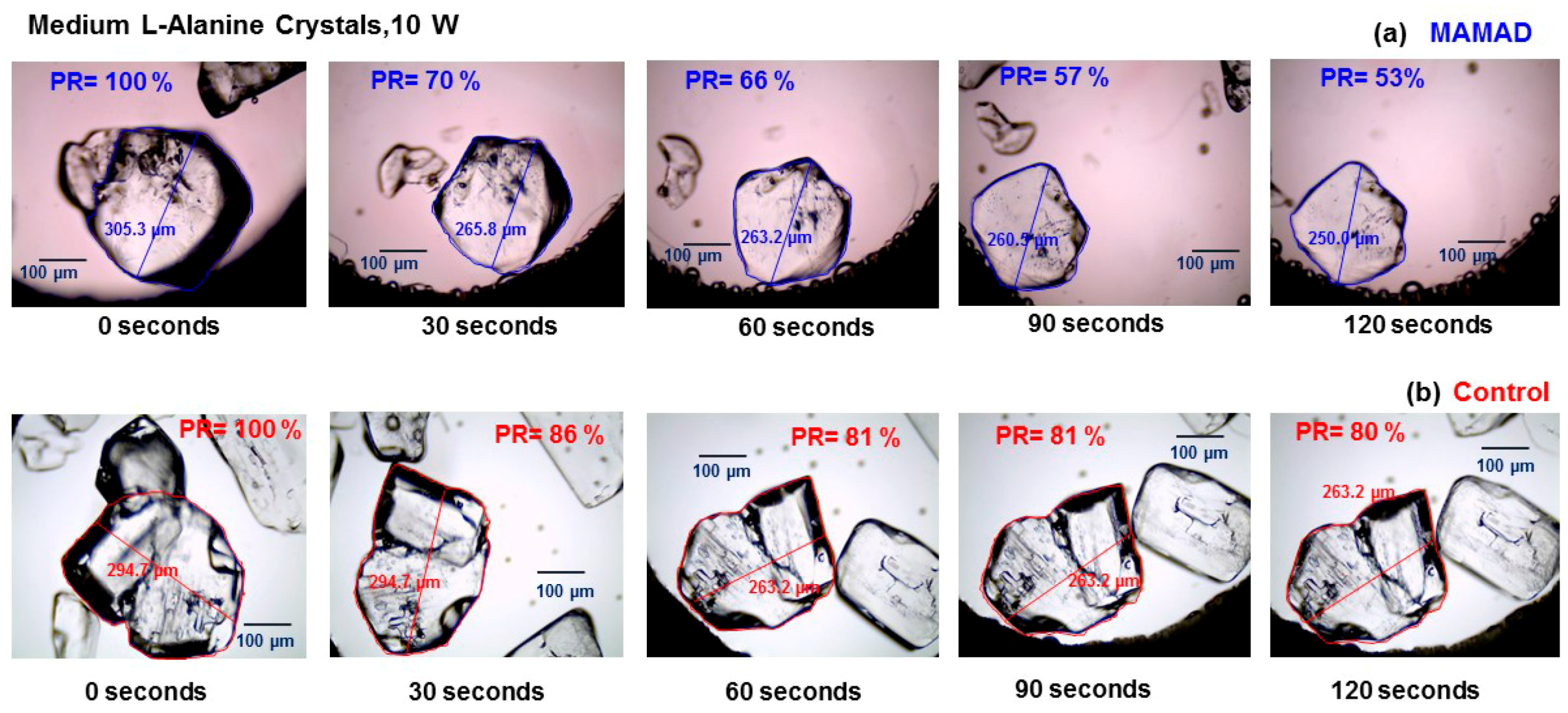
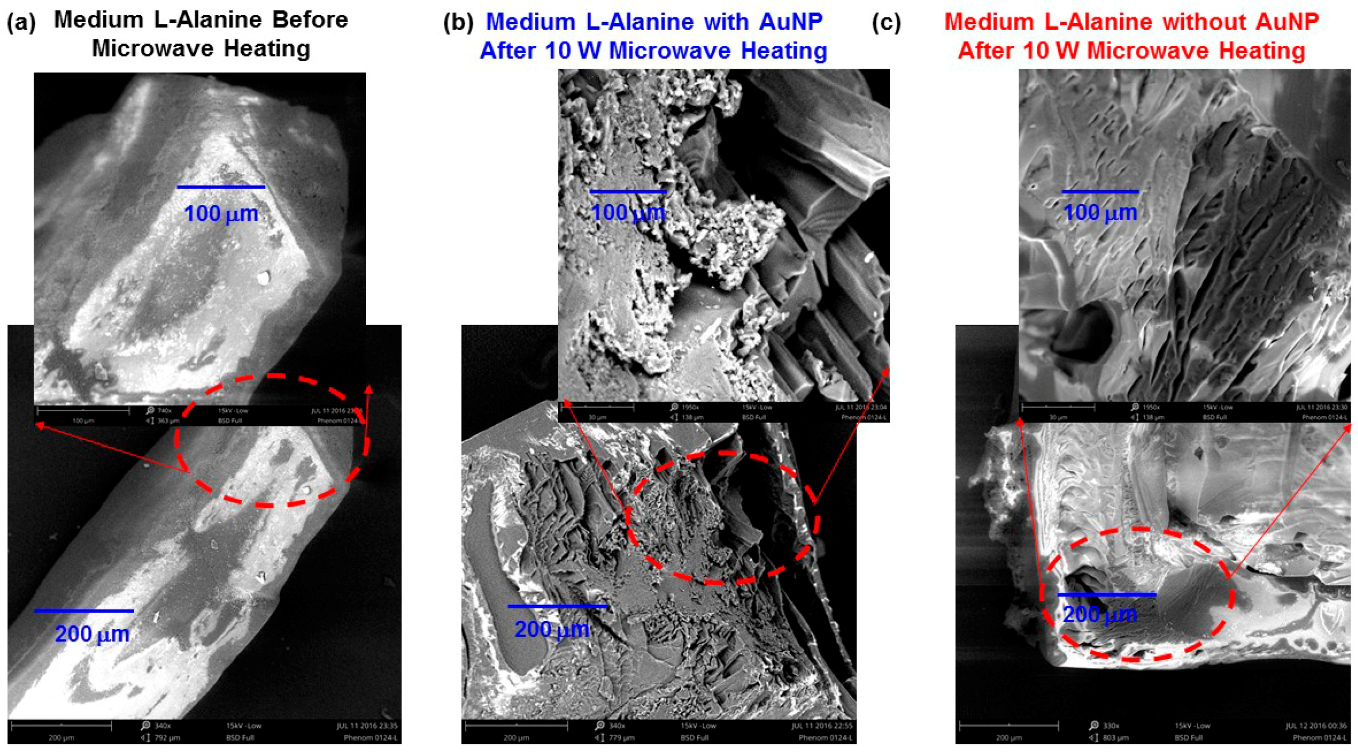
2.2. Decrystallization of Medium l-Alanine Crystals (~300 µm)
Image Analysis of Medium l-Alanine Crystals (~300 µm)
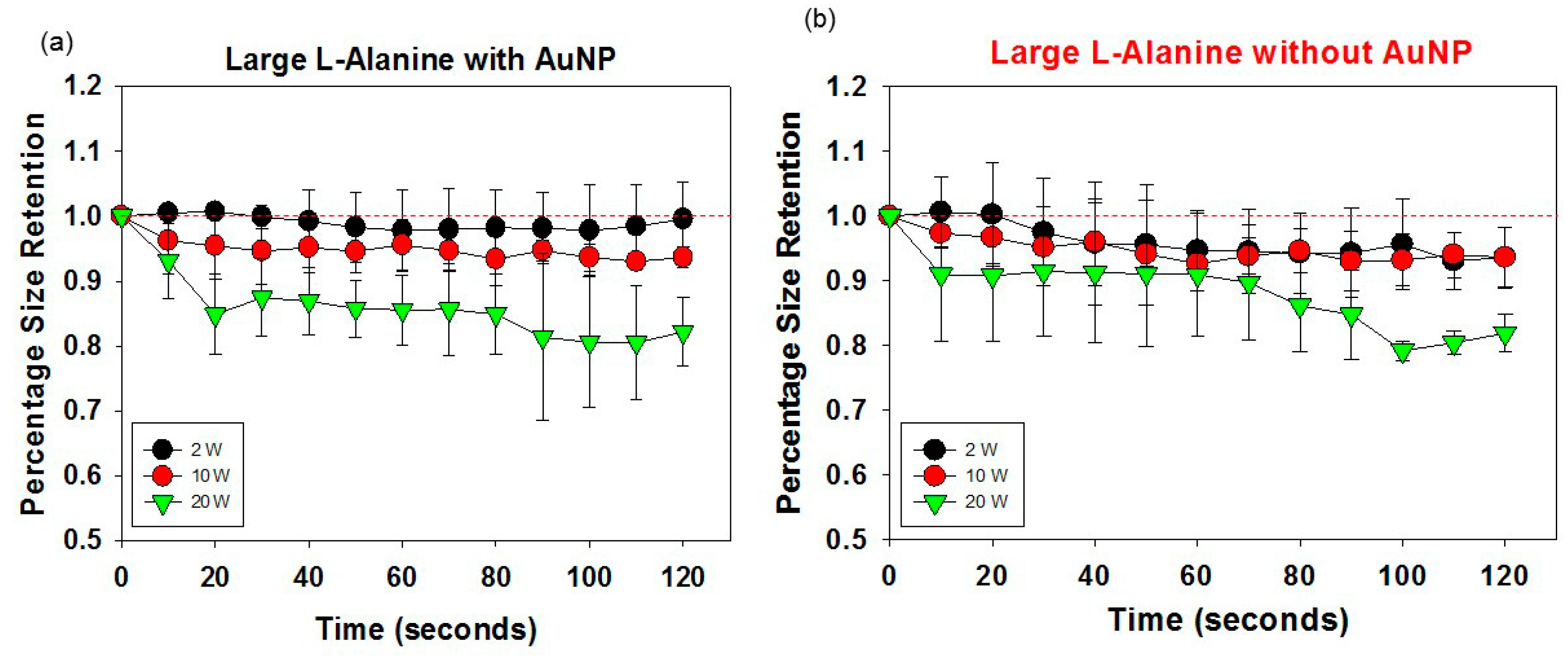
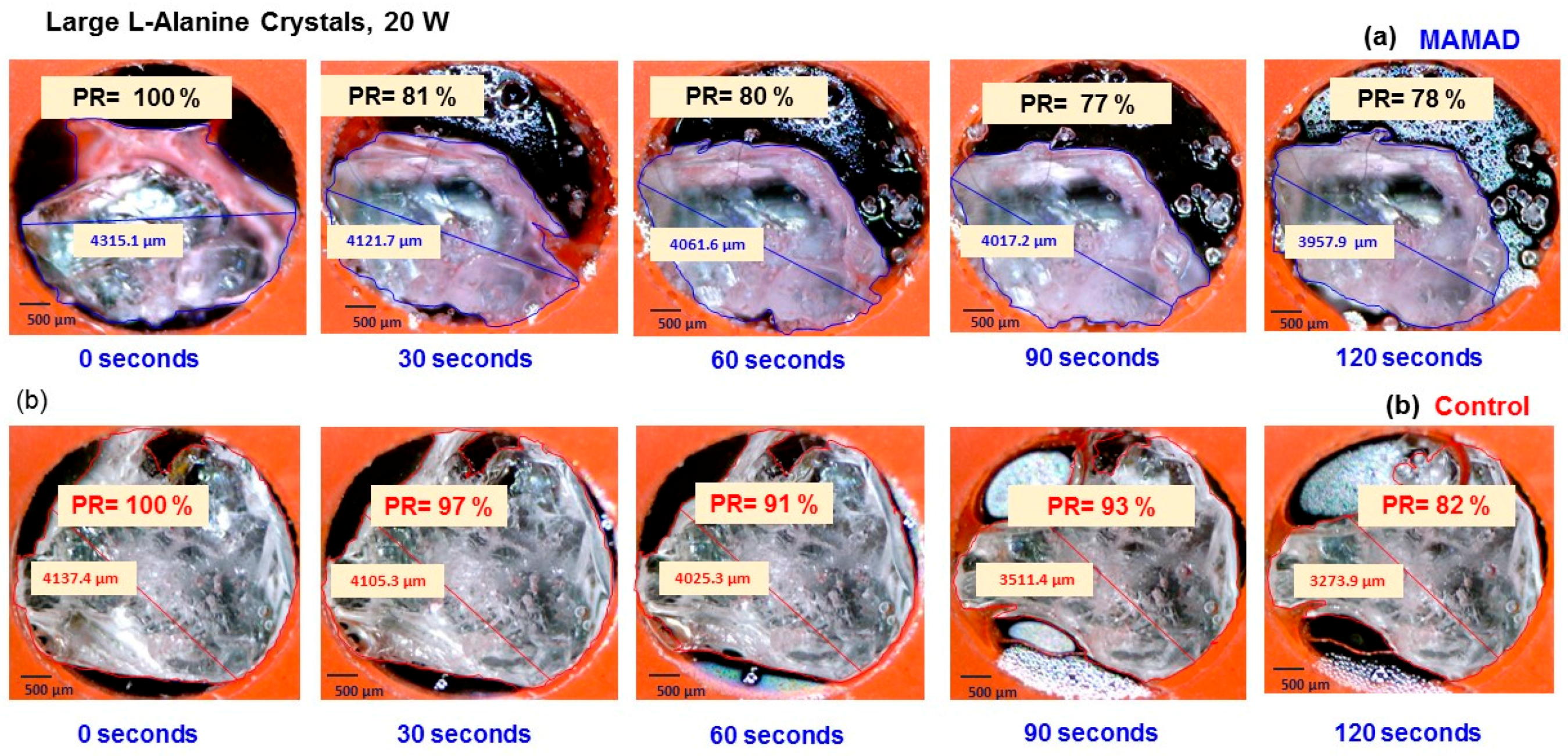
2.3. Decrystallization of Large l-Alanine Crystals (~4300 µm)
Optical Image Analysis of Large l-Alanine Crystals (~4300 µm)
3. Discussion
The Use of the MAMAD Technique for Decrystallization of Crystals
4. Materials and Methods
4.1. Materials
4.2. Methods
4.2.1. Preparation of Uric Acid Crystal Solution
4.2.2. Crystallization of Medium and Large l-Alanine Crystals
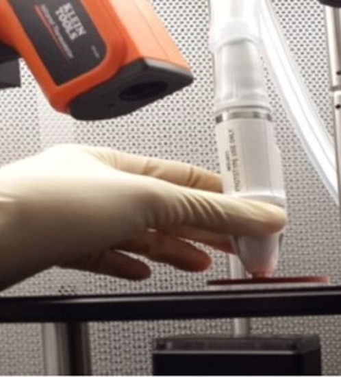
4.2.3. Decrystallization of the Small Uric Acid Crystals on the iCrystal Plates
4.2.4. Decrystallization of Medium and Large l-Alanine Crystals on the iCrystal Plates
4.2.5. X-ray Diffraction and ImageJ Analysis of Crystals
5. Conclusions
Supplementary Materials
Acknowledgments
Author Contributions
Conflicts of Interest
References
- Zhu, Y.; Pandya, B.J.; Choi, H.K. Prevalence of gout and hyperuricemia in the US general population: The national health and nutrition examination survey 2007–2008. Arthritis Rheum. 2011, 63, 3136–3141. [Google Scholar] [CrossRef] [PubMed]
- Qureshi, A.E.; Hameed, S.; Noeman, A. Relationship of serum uric acid level and angiographic severity of coronary artery disease in male patients with acute coronary syndrome. Pak. J. Med. Sci. 2013, 29, 1137–1141. [Google Scholar] [CrossRef]
- Choi, H.K.; Atkinson, K.; Karlson, E.W.; Willett, W.; Curhan, G. Purine-rich foods, dairy and protein intake, and the risk of gout in men. N. Engl. J. Med. 2004, 350, 1093–1103. [Google Scholar] [CrossRef] [PubMed]
- Seegmiller, J.; Grayzel, A.I.; Laster, L.; Liddle, L. Uric acid production in gout. J. Clin. Investig. 1961, 40, 1304. [Google Scholar] [CrossRef] [PubMed]
- Maiuolo, J.; Oppedisano, F.; Gratteri, S.; Muscoli, C.; Mollace, V. Regulation of uric acid metabolism and excretion. Int. J. Cardiol. 2016, 213, 8–14. [Google Scholar] [CrossRef] [PubMed]
- Perez-Ruiz, F. Treating to target: A strategy to cure gout. Rheumatol. 2009, 48, ii9–ii14. [Google Scholar] [CrossRef] [PubMed]
- Cronstein, B.N.; Terkeltaub, R. The inflammatory process of gout and its treatment. Arthritis Res. Ther. 2006, 8 (Suppl. 1), S3. [Google Scholar] [CrossRef] [PubMed]
- Kirchrath, J.; Schrr, K. Cyclooxygenase-2 inhibition and side-effects of non-steroidal anti-inflammatory drugs in the gastrointestinal tract. Curr. Med. Chem. 2000, 7, 1121–1129. [Google Scholar] [CrossRef]
- Mukherjee, D.; Nissen, S.E.; Topol, E.J. Risk of cardiovascular events associated with selective cox-2 inhibitors. JAMA 2001, 286, 954–959. [Google Scholar] [CrossRef] [PubMed]
- Ben-Chetrit, E.; Levy, M. Colchicine: 1998 update. Semin. Arthritis Rheum. 1998, 28, 48–59. [Google Scholar] [CrossRef]
- Moghadam-Kia, S.; Werth, V.P. Prevention and treatment of systemic glucocorticoid side effects. Int. J. Dermatol. 2010, 49, 239–248. [Google Scholar] [CrossRef] [PubMed]
- Simon, C.J.; Dupuy, D.E.; Mayo-Smith, W.W. Microwave ablation: Principles and applications. Radiographics 2005, 25, S69–S83. [Google Scholar] [CrossRef] [PubMed]
- Saldanha, D.F.; Khiatani, V.L.; Carrillo, T.C.; Yap, F.Y.; Bui, J.T.; Knuttinen, M.G.; Owens, C.A.; Gaba, R.C. Current tumor ablation technologies: Basic science and device review. Semin. Interv. Radiol. 2010, 27, 247–254. [Google Scholar] [CrossRef] [PubMed]
- Kioko, B.; Ogundolie, T.; Adebiyi, M.; Ettinoffe, Y.; Rhodes, C.; Gordon, B.; Thompson, N.; Mohammed, M.; Abel, B.; Aslan, K. De-crystallization of uric acid crystals in synovial fluid using gold colloids and microwave heating. Nano Biomed. Eng. 2014, 6, 104–110. [Google Scholar] [CrossRef] [PubMed]
- Ettinoffe, Y.S.; Kioko, B.M.; Gordon, B.I.; Thompson, N.A.; Adebiyi, M.; Mauge-Lewis, K.; Ogundolie, T.O.; Bonyi, E.; Mohammed, M.; Aslan, K. Metal-assisted and microwave-accelerated decrystallization. Nano Biomed. Eng. 2015, 7, 139–152. [Google Scholar] [CrossRef]
- Vrba, J.; Lapes, M. Medical applications of microwaves. In Proceedings of Microwave and Optical Technology 2003; SPIE Digital Library: Bellingham, WA, USA, 2004; Volume 5445. [Google Scholar]
- Wakim, K.G.; Herrick, J.F.; Martin, G.M.; Krusen, F.H. Therapeutic possibilities of microwaves; experimental and clinical investigation. J. Am. Med. Assoc. 1949, 139, 989–993. [Google Scholar] [CrossRef] [PubMed]
- Yadava, R.L. Rf/microwaves in bio-medical applications. In Proceedings of the 8th International Conference on Electromagnetic Interference and Compatibility, Chennai, India, 18–19 December 2003; IEEE: Piscataway, NJ, USA, 2003; pp. 81–85. [Google Scholar]
- Grant, J.P.; Clarke, R.N.; Symm, G.T.; Spyrou, N.M. In vivo dielectric properties of human skin from 50 MHz to 2.0 GHz. Phys. Med. Biol. 1988, 33, 607–612. [Google Scholar] [CrossRef] [PubMed]
- Tamyis, N.M.; Ghodgaonkar, D.K.; Taib, M.N.; Wui, W.T. Dielectric properties of human skin in vivo in the frequency range 20–38 GHz for 42 healthy volunteers. In Proceedings of the 28th URSI General Assembly, New Delhi, India, 23–29 October 2005.
- Aslan, K.; Geddes, C.D. Microwave-accelerated ultrafast nanoparticle aggregation assays using gold colloids. Anal. Chem. 2007, 79, 2131–2136. [Google Scholar] [CrossRef] [PubMed]
- Alabanza, A.M.; Aslan, K. Metal-assisted and microwave-accelerated evaporative crystallization: Application to l-alanine. Cryst. Growth Des. 2011, 11, 4300–4304. [Google Scholar] [CrossRef] [PubMed]
- Sample Availability: Not available.


© 2016 by the authors. Licensee MDPI, Basel, Switzerland. This article is an open access article distributed under the terms and conditions of the Creative Commons Attribution (CC-BY) license ( http://creativecommons.org/licenses/by/4.0/).
Share and Cite
Thompson, N.; Boone-Kukoyi, Z.; Shortt, R.; Lansiquot, C.; Kioko, B.; Bonyi, E.; Toker, S.; Ozturk, B.; Aslan, K. Decrystallization of Crystals Using Gold “Nano-Bullets” and the Metal-Assisted and Microwave-Accelerated Decrystallization Technique. Molecules 2016, 21, 1388. https://0-doi-org.brum.beds.ac.uk/10.3390/molecules21101388
Thompson N, Boone-Kukoyi Z, Shortt R, Lansiquot C, Kioko B, Bonyi E, Toker S, Ozturk B, Aslan K. Decrystallization of Crystals Using Gold “Nano-Bullets” and the Metal-Assisted and Microwave-Accelerated Decrystallization Technique. Molecules. 2016; 21(10):1388. https://0-doi-org.brum.beds.ac.uk/10.3390/molecules21101388
Chicago/Turabian StyleThompson, Nishone, Zainab Boone-Kukoyi, Raquel Shortt, Carisse Lansiquot, Bridgit Kioko, Enock Bonyi, Salih Toker, Birol Ozturk, and Kadir Aslan. 2016. "Decrystallization of Crystals Using Gold “Nano-Bullets” and the Metal-Assisted and Microwave-Accelerated Decrystallization Technique" Molecules 21, no. 10: 1388. https://0-doi-org.brum.beds.ac.uk/10.3390/molecules21101388