
Potential Use of Turkish Medicinal Plants in the Treatment of Various Diseases

 ,
,
Abstract
:1. Introduction
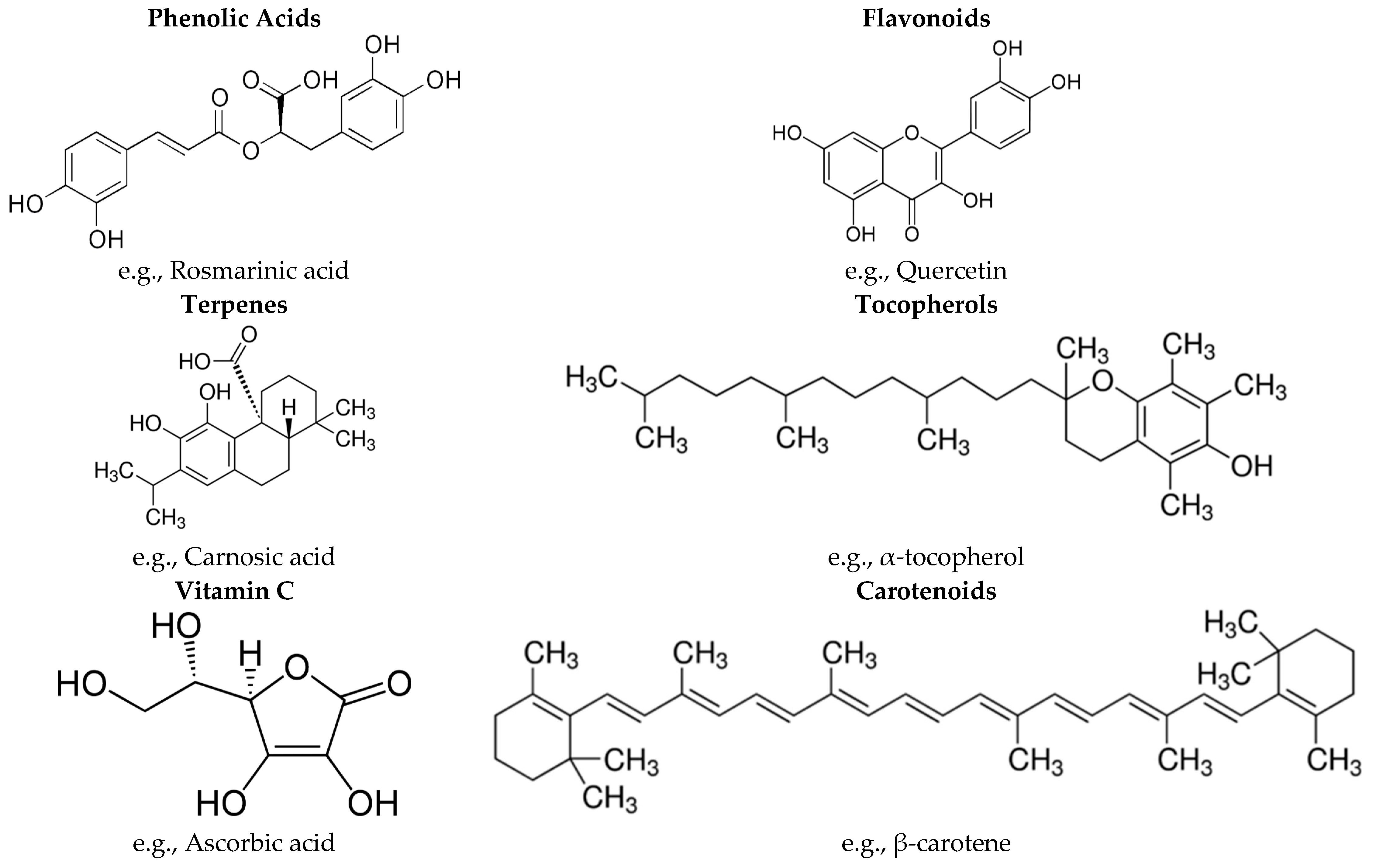
2. Antioxidant Properties of Medicinal Plants
3. Cancer
4. Cardiovascular Diseases
5. Diabetes
6. Infectious Diseases
7. Other Diseases
7.1. Wound Healing
7.2. Rheumatoid Arthritis
7.3. Neurological Disorders
7.4. Gastric Disorders
8. Conclusions
Acknowledgments
Author Contributions
Conflicts of Interest
References
- Zheng, W.; Wang, S.Y. Antioxidant activity and phenolic compounds in selected herbs. J. Agric. Food Chem. 2001, 49, 5165–5170. [Google Scholar] [CrossRef] [PubMed]
- Cai, Y.; Sun, M.; Corke, H. Antioxidant activity of betalains from plants of the Amaranthaceae. J. Agric. Food Chem. 2003, 51, 2288–2294. [Google Scholar] [CrossRef] [PubMed]
- Miliauskas, G.; Venskutonis, P.R.; Van Beek, T.A. Screening of radical scavenging activity of some medicinal and aromatic plant extracts. Food Chem. 2004, 85, 231–237. [Google Scholar] [CrossRef]
- Kamiloglu, S.; Capanoglu, E.; Yilmaz, O.; Duran, A.F.; Boyacioglu, D. Investigating the antioxidant potential of Turkish herbs and spices. Qual. Assur. Saf. Crop. Food. 2014, 6, 151–158. [Google Scholar] [CrossRef]
- Karadeniz, A.; Cinbilgel, I.; Gun, S.S.; Cetin, A. Antioxidant activity of some Turkish medicinal plants. Nat. Prod. Res. 2015, 29, 2308–2312. [Google Scholar] [CrossRef] [PubMed]
- Chivian, E. Biodiversity: Its Importance to Human Health. Master’s Thesis, Center for Health and the Global Environment, Harvard Medical School, Cambridge, UK, 2002. [Google Scholar]
- Willcox, J.K.; Ash, S.L.; Catignani, G.L. Antioxidants and prevention of chronic disease. Crit. Rev. Food Sci. Nutr. 2004, 44, 275–295. [Google Scholar] [CrossRef] [PubMed]
- Owen, R.W.; Giacosa, A.; Hull, W.E.; Haubner, R.; Spiegelhalder, B.; Bartsch, H. The antioxidant/anticancer potential of phenolic compounds isolated from olive oil. Eur. J. Cancer. 2000, 36, 1235–1247. [Google Scholar] [CrossRef]
- Sala, A.; Recio, M.D.C.; Giner, R.M.; Máñez, S.; Tournier, H.; Schinella, G.; Ríos, J.L. Anti-inflammatory and antioxidant properties of Helichrysum italicum. J. Pharm. Pharmacol. 2002, 54, 365–371. [Google Scholar] [CrossRef] [PubMed]
- Anderson, K.J.; Teuber, S.S.; Gobeille, A.; Cremin, P.; Waterhouse, A.L.; Steinberg, F.M. Walnut polyphenolics inhibit in vitro human plasma and LDL oxidation. J. Nutr. 2001, 131, 2837–2842. [Google Scholar] [PubMed]
- Yang, C.S.; Landau, J.M.; Huang, M.T.; Newmark, H.L. Inhibition of carcinogenesis by dietary polyphenolic compounds. Annu. Rev. Nutr. 2001, 21, 381–406. [Google Scholar] [CrossRef] [PubMed]
- Sun, J.; Chu, Y.F.; Wu, X.; Liu, R.H. Antioxidant and antiproliferative activities of common fruits. J. Agric. Food Chem. 2002, 50, 7449–7454. [Google Scholar] [CrossRef] [PubMed]
- Kültür, Ş. Medicinal plants used in Kırklareli province (Turkey). J. Ethnopharmacol. 2007, 111, 341–364. [Google Scholar]
- Tuzlacı, E.; Aymaz, P.E. Turkish folk medicinal plants, part IV: Gönen (Balıkesir). Fitoterapia 2001, 72, 323–343. [Google Scholar] [CrossRef]
- Kraisintu, K. Industrial exploitation of indigenous medicinal and aromatic plants: Formulation and industrial utilisation. In UNDP. 1997. [Google Scholar]
- Skrovankova, S.; Misurcova, L.; Machu, L. Antioxidant activity and protecting health effects of common medicinal plants. Adv. Food Nutr. Res. 2012, 67, 75–139. [Google Scholar] [PubMed]
- Kosar, M.; Dorman, H.J.D.; Hiltunen, R. Effect of an acid treatment on the phytochemical and antioxidant characteristics of extracts from selected Lamiaceae species. Food Chem. 2005, 91, 525–533. [Google Scholar] [CrossRef]
- Esiyok, D.; Otles, S.; Akcicek, E. Herbs as a food source in Turkey. Asian Pac. J. Cancer Prev. 2004, 5, 334–339. [Google Scholar] [PubMed]
- Demiray, S.; Pintado, M.E.; Castro, P.M.L. Evaluation of phenolic profiles and antioxidant activities of Turkish medicinal plants: Tilia argentea, Crataegi folium leaves and Polygonum bistorta roots. World Acad. Sci. Eng. Technol. 2009, 54, 312–317. [Google Scholar]
- Costa, R.M.; Vaz, A.F.; Xavier, H.S.; Correia, M.T.; Carneiro-da-Cunha, M.G. Phytochemical screening of Phthirusa pyrifolia leaf extracts: Free-radical scavenging activities and environmental toxicity. S. Afr. J. Bot. 2015, 99, 132–137. [Google Scholar] [CrossRef]
- Suzgec-Selcuk, S.; Birteksoz, A.S. Flavonoids of Helichrysum chasmolycicum and its antioxidant and antimicrobial activities. S. Afr. J. Bot. 2011, 77, 170–174. [Google Scholar] [CrossRef]
- Dincer, C.; Topuz, A.; Sahin-Nadeem, H.; Ozdemir, K.S.; Cam, I.B.; Tontul, I.; Gokturk, R.S.; Ay, S.T. A comparative study on phenolic composition, antioxidant activity and essential oil content of wild and cultivated sage (Salvia fruticosa Miller) as influenced by storage. Ind. Crop. Prod. 2012, 39, 170–176. [Google Scholar] [CrossRef]
- Bakkali, F.; Averbeck, S.; Averbeck, D.; Idaomar, M. Biological effects of essential oils—A review. Food Chem. Toxicol. 2008, 46, 446–475. [Google Scholar] [CrossRef] [PubMed]
- Ekren, S.; Yerlikaya, O.; Tokul, H.E.; Akpınar, A.; Acu, M. Chemical composition, antimicrobial activity and antioxidant capacity of some medicinal and aromatic plant extracts. Afr. J. Microbiol. Res. 2013, 7, 383–388. [Google Scholar]
- Vagi, E.; Rapavi, E.; Hadolin, M.; Vasarhelyine Peredi, K.; Balazs, A.; Blazovics, A.; Simandi, B. Phenolic and triterpenoid antioxidants from Origanum majorana L. herb and extracts obtained with different solvents. J. Agric. Food Chem. 2005, 53, 17–21. [Google Scholar] [CrossRef] [PubMed]
- Asensi-Fabado, M.A.; Munne-Bosch, S. Vitamins in plants: occurrence, biosynthesis and antioxidant function. Trends Plant. Sci. 2010, 15, 582–592. [Google Scholar] [CrossRef] [PubMed]
- Morales, P.; Carvalho, A.M.; Sanchez-Mata, M.C.; Camara, M.; Molina, M.; Ferreira, I.C. Tocopherol composition and antioxidant activity of Spanish wild vegetables. Genet. Resour. Crop. Evol. 2012, 59, 851–863. [Google Scholar] [CrossRef]
- Dua, A. A study of antioxidant properties and antioxidant compounds of cumin (Cuminum cyminum). Int. J. Pharm. Biol. Arch. 2012, 3, 1110–1116. [Google Scholar]
- Elgersma, A.; Soegaard, K.; Jensen, S.K. Fatty acids, α-tocopherol, β-carotene, and lutein contents in forage legumes, forbs, and a grass–clover mixture. J. Agric. Food Chem. 2013, 61, 11913–11920. [Google Scholar] [CrossRef] [PubMed]
- Capecka, E.; Mareczek, A.; Leja, M. Antioxidant activity of fresh and dry herbs of some Lamiaceae species. Food Chem. 2005, 93, 223–226. [Google Scholar] [CrossRef]
- Kamiloglu, S.; Toydemir, G.; Boyacioglu, D.; Beekwilder, J.; Hall, R.D.; Capanoglu, E. A review on the effect of drying on antioxidant potential of fruits and vegetables. Crit. Rev. Food Sci. Nutr. 2015. [Google Scholar] [CrossRef] [PubMed]
- Daly, T.; Jiwan, M.A.; O’Brien, N.M.; Aherne, S.A. Carotenoid content of commonly consumed herbs and assessment of their bioaccessibility using an in vitro digestion model. Plant. Foods Hum. Nutr. 2010, 65, 164–169. [Google Scholar] [CrossRef] [PubMed]
- Raposo, C.G.; Carpeno, J.D.; Baron, M.G. Causes of lung cancer: Smoking, environmental tobacco smoke exposure, occupational and environmental exposures, and genetic predisposition. Med. Clin. 2007, 128, 390–396. [Google Scholar]
- Lee, J.Y.; Li, J.W.; Yeung, E.S. Single-molecule detection of surfacehybridized human papillorna virus DNA for quantitative clinical screening. Anal. Chem. 2007, 79, 8083–8089. [Google Scholar] [CrossRef] [PubMed]
- Shiotani, A.; Iishi, H.; Uedo, N.; Ishiguro, S.; Tatsuta, M. Evidence that loss of sonic hedgehog is an indicator of Helicobater pylori-induced atrophic gastritis progressing to gastric cancer. Am. J. Gasteroenterol. 2005, 100, 581–587. [Google Scholar] [CrossRef] [PubMed]
- Vauhkonen, H.; Bohling, T.; Eissa, S.; Shoman, S.; Knuutila, S. Can bladder adenocarcinomas be distinguished from schistosomiasis-associated bladder cancers by using array comparative genomic hybridization analysis? Cancer Genet. Cytogen. 2007, 177, 153–157. [Google Scholar] [CrossRef] [PubMed]
- Groopman, J.D.; Wang, J.S.; Scholl, P. Molecular biomarkers for aflatoxins: from adducts to gene mutations to human liver cancer. Can. J. Physiol. Pharm. 1996, 74, 203–209. [Google Scholar] [CrossRef]
- Niki, E. Free radicals, antioxidants, and cancer. In Food Factors for Cancer Prevention; Ohigashi, H., Osawa, T., Terao, J., Watanabe, S., Yoshikawa, T., Eds.; Springer: Tokyo, Japan, 1997; pp. 55–57. [Google Scholar]
- Poulson, H.E.; Prieme, H.; Loft, S. Role of oxidative DNA damage in cancer initiation and promotion. Eur. J. Cancer Prev. 1998, 7, 9–16. [Google Scholar]
- Reddy, L.; Odhav, B.; Bhoola, K.D. Natural products for cancer prevention: A global perspective. Pharmacol. Ther. 2003, 99, 1–13. [Google Scholar] [CrossRef]
- Mathers, C.D.; Loncar, D. Projections of global mortality and burden of disease from 2002 to 2030. PLoS Med. 2006, 3, e442. [Google Scholar] [CrossRef] [PubMed]
- Cai, Y.Z.; Luo, Q.; Sun, M.; Corke, H. Antioxidant activity and phenolic compounds of 112 traditional Chinese medicinal plants associated with anticancer. Life Sci. 2004, 74, 2157–2184. [Google Scholar] [CrossRef] [PubMed]
- Mans, D.R.; da Rocha, A.B.; Schwartsmann, G. Anti-cancer drug discovery and development in Brazil: targeted plant collection as a rational strategy to acquire candidate anti-cancer compounds. Oncologist 2000, 5, 185–198. [Google Scholar] [CrossRef] [PubMed]
- Alonso-Castro, A.J.; Villarreal, M.L.; Salazar-Olivo, L.A.; Gomez-Sanchez, M.; Dominguez, F.; Garcia-Carranca, A. Mexican medicinal plants used for cancer treatment: pharmacological, phytochemical and ethnobotanical studies. J. Ethnopharmacol. 2011, 133, 945–972. [Google Scholar] [CrossRef] [PubMed]
- Shoeb, M. Cytotoxic Compounds from the Genus Centaurea. Ph.D. Thesis, The Robert Gordon University, Aberdeen, UK, 2005. [Google Scholar]
- Graham, J.G.; Quinn, M.L.; Fabricant, D.S.; Farnsworth, N.R. Plants used against cancer—An extension of the work of Jonathan Hartwell. J. Ethnopharmacol. 2000, 73, 347–377. [Google Scholar] [PubMed]
- Gordaliza, M. Natural products as leads to anticancer drugs. Clin. Transl. Oncol. 2007, 9, 767–776. [Google Scholar] [PubMed]
- Ozkan, A.; Erdogan, A. A comparative evaluation of antioxidant and anticancer activity of essential oil from Origanum onites (Lamiaceae) and its two major phenolic components. Turk. J. Biol. 2011, 35, 735–742. [Google Scholar]
- Yesilada, E.; Bedir, E.; Çalış, I.; Takaishi, Y.; Ohmoto, Y. Effects of triterpene saponins from Astragalus. species on in vitro cytokine release. J. Ethnopharmacol. 2005, 96, 71–77. [Google Scholar] [PubMed]
- Özmen, A.; Madlener, S.; Bauer, S.; Krasteva, S.; Vonach, C.; Giessrigl, B.; Michel, B. In vitro anti-leukemic activity of the ethno-pharmacological plant Scutellaria orientalis ssp. carica endemic to western Turkey. Phytomedicine 2010, 17, 55–62. [Google Scholar]
- Dalar, A.; Konczak, I. Cichorium intybus from Eastern Anatolia: Phenolic composition, antioxidant and enzyme inhibitory activities. Ind. Crop. Prod. 2014, 60, 79–85. [Google Scholar]
- Dalar, A.; Konczak, I. Phenolic contents, antioxidant capacities and inhibitory activities against key metabolic syndrome relevant enzymes of herbal teas from Eastern Anatolia. Ind. Crop. Prod. 2013, 44, 383–390. [Google Scholar] [CrossRef]
- Dalar, A.; Uzun, Y.; Mukemre, M.; Turker, M.; Konczak, I. Centaurea karduchorum Boiss from Eastern Anatolia: Phenolic composition, antioxidant and enzyme inhibitory activities. J. Herb. Med. 2015, 5, 211–216. [Google Scholar] [CrossRef]
- Aslan, M.; Orhan, N.; Orhan, D.D.; Ergun, F. Hypoglycemic activity and antioxidant potential of some medicinal plants traditionally used in Turkey for diabetes. J. Ethnopharmacol. 2010, 128, 384–389. [Google Scholar] [CrossRef] [PubMed]
- Orhan, N.; Hocbac, S.; Orhan, D.D.; Asian, M.; Ergun, F. Enzyme inhibitory and radical scavenging effects of some antidiabetic plants of Turkey. Iran. J. Basic Med. Sci. 2014, 17, 426–432. [Google Scholar] [PubMed]
- Sezik, M.; Aslan, M.; Orhan, D.D.; Erdemoglu, E.; Pekcan, M.; Mungan, T.; Sezik, E. Improved metabolic control and hepatic oxidative biomarkers with the periconception use of Helichrysum plicatum ssp. plicatum. J. Obstet. Gynaecol. 2010, 30, 127–131. [Google Scholar] [CrossRef] [PubMed]
- Ozkol, H.; Tuluce, Y.; Dilsiz, N.; Koyuncu, I. Therapeutic potential of some plant extracts used in Turkish traditional medicine on streptozocin-induced type 1 diabetes mellitus in rats. J. Memb. Biol. 2013, 246, 47–55. [Google Scholar] [CrossRef] [PubMed]
- Orhan, N.; Aslan, M.; Sukuroglu, M.; Orhan, D.D. In vivo and in vitro antidiabetic effect of Cistus laurifolius L. and detection of major phenolic compounds by UPLC–TOF-MS analysis. J. Ethnopharmacol. 2013, 146, 859–865. [Google Scholar] [CrossRef] [PubMed]
- Orhan, N.; Aslan, M.; Demirci, B.; Ergun, F. A bioactivity guided study on the antidiabetic activity of Juniperus oxycedrus subsp. oxycedrus L. leaves. J. Ethnopharmacol. 2012, 140, 409–415. [Google Scholar] [CrossRef] [PubMed]
- Dogan, A.; Celik, I.; Kaya, M.S. Antidiabetic properties of lyophilized extract of acorn (Quercus brantii Lindl.) on experimentally STZ-induced diabetic rats. J. Ethnopharmacol. 2015, 176, 243–251. [Google Scholar] [CrossRef] [PubMed]
- Bayramoglu, G.; Senturk, H.; Bayramoglu, A.; Uyanoglu, M.; Colak, S.; Ozmen, A.; Kolankaya, D. Carvacrol partially reverses symptoms of diabetes in STZ-induced diabetic rats. Cytotechnology 2014, 66, 251–257. [Google Scholar] [CrossRef] [PubMed]
- Sarikurkcu, C.; Zengin, G.; Oskay, M.; Uysal, S.; Ceylan, R.; Aktumsek, A. Composition, antioxidant, antimicrobial and enzyme inhibition activities of two Origanum vulgare subspecies (subsp. vulgare and subsp. hirtum) essential oils. Ind. Crop. Prod. 2015, 70, 178–184. [Google Scholar] [CrossRef]
- Zengin, G.; Sarikurkcu, C.; Aktumsek, A.; Ceylan, R. Antioxidant potential and inhibition of key enzymes linked to Alzheimer’s diseases and diabetes mellitus by monoterpene-rich essential oil from Sideritis galatica Bornm. endemic to Turkey. Rec. Nat. Prod. 2016, 10, 195–206. [Google Scholar]
- Basak, S.S.; Candan, F. Effect of Laurus nobilis L. essential oil and its main components on α-glucosidase and reactive oxygen species scavenging activity. Iran. J. Pharm. Res. 2013, 12, 367–379. [Google Scholar]
- Basak, S.S.; Candan, F. Chemical composition and in vitro antioxidant and antidiabetic activities of Eucalyptus camaldulensis Dehnh. essential oil. J. Iran. Chem. Soc. 2010, 7, 216–226. [Google Scholar] [CrossRef]
- Uysal, S.; Aktumsek, A. A phytochemical study on Potentilla anatolica: An endemic Turkish plant. Ind. Crop. Prod. 2015, 76, 1001–1007. [Google Scholar] [CrossRef]
- Zengin, G.; Sarikurkcu, C.; Aktumsek, A.; Ceylan, R.; Ceylan, O. A comprehensive study on phytochemical characterization of Haplophyllum myrtifolium Boiss. endemic to Turkey and its inhibitory potential against key enzymes involved in Alzheimer, skin diseases and type II diabetes. Ind. Crop. Prod. 2014, 53, 244–251. [Google Scholar] [CrossRef]
- Albayrak, S.; Aksoy, A. Evaluation of antioxidant and antimicrobial activities of two endemic anthemis species in Turkey. J. Food Biochem. 2013, 37, 639–645. [Google Scholar] [CrossRef]
- Özbilgin, A.; Durmuskahya, C.; Kayalar, H.; Ostan, I. Assessment of in vivo antimalarial activities of some selected medicinal plants from Turkey. Parasitol. Res. 2014, 113, 165–173. [Google Scholar] [CrossRef] [PubMed]
- Tekeli, Y.; Zengin, G.; Aktumsek, A.; Sezgin, M.; Torlak, E. Antibacterial activities of extracts from twelve Centaurea species from Turkey. Arch. Biol. Sci. 2011, 63, 685–690. [Google Scholar] [CrossRef]
- Askun, T.; Tekwu, E.M.; Satil, F.; Modanlioglu, S.; Aydeniz, H. Preliminary antimycobacterial study on selected Turkish plants (Lamiaceae.) against Mycobacterium tuberculosis and search for some phenolic constituents. BMC Complement. Altern. Med. 2013, 13, 365–376. [Google Scholar] [CrossRef] [PubMed]
- Kozan, E.; Çankaya, I.T.; Kahraman, C.; Akkol, E.K.; Akdemir, Z. The in vivo anthelmintic efficacy of some Verbascum species growing in Turkey. Exp. Parasitol. 2011, 129, 211–214. [Google Scholar] [CrossRef] [PubMed]
- Süntar, I.P.; Akkol, E.K.; Yalçın, F.N.; Koca, U.; Keleş, H.; Yesilada, E. Wound healing potential of Sambucus ebulus L. leaves and isolation of an active component, quercetin 3-O-glucoside. J. Ethnopharmacol. 2010, 129, 106–114. [Google Scholar] [CrossRef] [PubMed]
- Süntar, İ.; Baldemir, A.; Coşkun, M.; Keleş, H.; Akkol, E.K. Wound healing acceleration effect of endemic Ononis species growing in Turkey. J. Ethnopharmacol. 2011, 135, 63–70. [Google Scholar] [CrossRef] [PubMed]
- Süntar, I.P.; Akkol, E.K.; Yılmazer, D.; Baykal, T.; Kırmızıbekmez, H.; Alper, M.; Yeşilada, E. Investigations on the in vivo wound healing potential of Hypericum perforatum L. J. Ethnopharmacol. 2010, 127, 468–477. [Google Scholar] [CrossRef] [PubMed]
- Akdemir, Z.; Kahraman, Ç.; Tatlı, I.I.; Akkol, E.K.; Süntar, I.; Keles, H. Bioassay-guided isolation of anti-inflammatory, antinociceptive and wound healer glycosides from the flowers of Verbascum mucronatum Lam. J. Ethnopharmacol. 2011, 136, 436–443. [Google Scholar] [CrossRef] [PubMed]
- Çadirci, E.; Süleyman, H.; Gürbüz, P.; Uz, A.K.; Güvenalp, Z.; Demirezer, L.Ö. Anti-inflammatory effects of different extracts from three Salvia species. Turk. J. Biol. 2012, 36, 59–64. [Google Scholar]
- Koca, U.; Süntar, I.P.; Keles, H.; Yesilada, E.; Akkol, E.K. In vivo anti-inflammatory and wound healing activities of Centaurea iberica Trev. ex Spreng. J. Ethnopharmacol. 2009, 126, 551–556. [Google Scholar] [CrossRef] [PubMed]
- Türel, I.; Özbek, H.; Erten, R.; Öner, A.C.; Cengiz, N.; Yilmaz, O. Hepatoprotective and anti-inflammatory activities of Plantago major L. Indian J. Pharmacol. 2009, 41, 120–124. [Google Scholar] [PubMed]
- Yilmaz, B.S.; Altun, M.L.; Orhan, I.E.; Ergene, B.; Citoglu, G.S. Enzyme inhibitory and antioxidant activities of Viburnum tinus L. relevant to its neuroprotective potential. Food Chem. 2013, 141, 582–588. [Google Scholar] [CrossRef] [PubMed]
- Ercetin, T.; Senol, F.S.; Orhan, I.E.; Toker, G. Comparative assessment of antioxidant and cholinesterase inhibitory properties of the marigold extracts from Calendula arvensis L. and Calendula officinalis L. Ind. Crop. Prod. 2012, 36, 203–208. [Google Scholar] [CrossRef]
- Aktumsek, A.; Zengin, G.; Guler, G.O.; Cakmak, Y.S.; Duran, A. Antioxidant potentials and anticholinesterase activities of methanolic and aqueous extracts of three endemic Centaurea L. species. Food Chem. Toxicol. 2013, 55, 290–296. [Google Scholar] [CrossRef] [PubMed]
- Orhan, I.E.; Atasu, E.; Senol, F.S.; Ozturk, N.; Demirci, B.; Das, K.; Sekeroglu, N. Comparative studies on Turkish and Indian Centella asiatica (L.) Urban (gotu kola) samples for their enzyme inhibitory and antioxidant effects and phytochemical characterization. Ind. Crops Prod. 2013, 47, 316–322. [Google Scholar] [CrossRef]
- Zengin, G.; Sarikurkcu, C.; Uyar, P.; Aktumsek, A.; Uysal, S.; Kocak, M.S.; Ceylan, R. Crepis foetida L. subsp. rhoeadifolia (Bieb.) Celak. as a source of multifunctional agents: Cytotoxic and phytochemical evaluation. J. Funct. Foods. 2015, 17, 698–708. [Google Scholar]
- Orhan, I.; Senol, F.S.; Koca, U.; Ercetin, T.; Toker, G. Evaluation of the antioxidant and acetylcholinesterase inhibitory activities of Arnebia densiflora Ledeb. Turk. J. Biol. 2011, 35, 111–115. [Google Scholar]
- Senol, F.S.; Orhan, I.E.; Ozgen, U.; Renda, G.; Bulut, G.; Guven, L.; Karaoglan, E.S.; Sevindik, H.G.; Skalicka-Wozniak, K.; Caliskan, U.K.; et al. Memory-vitalizing effect of twenty-five medicinal and edible plants and their isolated compounds. S. Afr. J. Bot. 2015. [Google Scholar] [CrossRef]
- Akkol, E.K.; Orhan, I.E.; Yesilada, E. Anticholinesterase and antioxidant effects of the ethanol extract, ethanol fractions and isolated flavonoids from Cistus laurifolius L. leaves. Food Chem. 2012, 131, 626–631. [Google Scholar] [CrossRef]
- Ertas, A.; Boga, M.; Yesil, Y. Phytochemical profile and ABTS cation radical scavenging, cupric reducing antioxidant capacity and anticholinesterase activities of endemic. J. Coast. Life Med. 2014, 2, 555–559. [Google Scholar]
- Demirezer, L.O.; Gurbuz, P.; Ugur, E.P.K.; Bodur, M.; Ozenver, N.; Ayse, U.Z.; Guvenalp, Z. Molecular docking and ex vivo and in vitro anticholinesterase activity studies of Salvia sp. and highlighted rosmarinic acid. Turk. J. Med. Sci. 2014, 44, 1–8. [Google Scholar]
- Tumen, I.; Senol, F.S.; Orhan, I.E. Inhibitory potential of the leaves and berries of Myrtus communis L. (myrtle) against enzymes linked to neurodegenerative diseases and their antioxidant actions. Int. J. Food Sci. Nutr. 2012, 63, 387–392. [Google Scholar] [CrossRef] [PubMed]
- Orhan, N.; Orhan, D.D.; Aslan, M.; Sukuroglu, M.; Orhan, I.E. UPLC–TOF-MS analysis of Galium spurium towards its neuroprotective and anticonvulsant activities. J. Ethnopharmacol. 2012, 141, 220–227. [Google Scholar] [CrossRef] [PubMed]
- Kose, L.P.; Gulcin, I.; Goren, A.C.; Namiesnik, J.; Martinez-Ayala, A.L.; Gorinstein, S. LC–MS/MS analysis, antioxidant and anticholinergic properties of galanga (Alpinia officinarum Hance) rhizomes. Ind. Crop. Prod. 2015, 74, 712–721. [Google Scholar] [CrossRef]
- Gurbuz, I.; Yesilada, E.; Demirci, B.; Sezik, E.; Demirci, F.; Baser, K.H.C. Characterization of volatiles and anti-ulcerogenic effect of Turkish sweetgum balsam (Styrax liquidus). J. Ethnopharmacol. 2013, 148, 332–336. [Google Scholar] [CrossRef] [PubMed]
- Yesilada, E.; Gurbuz, I. Evaluation of the antiulcerogenic activity profile of a flavonol diglucoside from Equisetum palustre L. J. Ethnopharmacol. 2010, 131, 17–21. [Google Scholar] [CrossRef] [PubMed]
- Yesilada, E.; Gürbüz, I.; Toker, G. Anti-ulcerogenic activity and isolation of the active principles from Sambucus ebulus L. leaves. J. Ethnopharmacol. 2014, 153, 478–83. [Google Scholar] [CrossRef] [PubMed]
- WHO. Global Status Report on Non-Communicable Diseases 2014; World Health Organization: Geneva, Switzerland, 2014. [Google Scholar]
- WHO. Cardiovascular Disease. Avaiable online: http://www.who.int/mediacentre/factsheets/fs317/en/ (accessed on 10 December 2014).
- Dauchet, L.; Amouyel, P.; Dallongeville, J. Fruit and vegetable consumption and risk of stroke—A meta-analysis of cohort studies. Neurology 2005, 65, 1193–1197. [Google Scholar] [CrossRef] [PubMed]
- Dauchet, L.; Amouyel, P.; Hercberg, S.; Dallongeville, J. Fruit and vegetable consumption and risk of coronary heart disease: a metaanalysis of cohort studies. J. Nutr. 2006, 136, 2588–2593. [Google Scholar] [PubMed]
- He, F.J.; Nowson, C.A.; MacGregor, G.A. Fruit and vegetable consumption and stroke: Meta-analysis of cohort studies. Lancet 2006, 367, 320–326. [Google Scholar] [CrossRef]
- He, F.J.; Nowson, C.A.; Lucas, M.; MacGregor, G.A. Increased consumption of fruit and vegetables is related to a reduced risk of coronary heart disease: meta-analysis of cohort studies. J. Hum. Hypertens. 2007, 21, 717–728. [Google Scholar] [CrossRef] [PubMed]
- Edwards, R.L.; Lyon, T.; Litwin, S.E.; Rabovsky, A.; Symons, J.D.; Jalili, T. Quercetin reduces blood pressure in hypertensive subjects. J. Nutr. 2007, 137, 2405–2411. [Google Scholar] [PubMed]
- Egert, S.; Boesch-Saadatmandi, C.; Wolffram, S.; Rimbach, G.; Muller, M.J. Serum lipid and blood pressure responses to quercetin vary in overweight patients by apolipoprotein E genotype. J. Nutr. 2010, 140, 278–284. [Google Scholar] [CrossRef] [PubMed]
- Erlund, I.; Koli, R.; Alfthan, G.; Marniemi, J.; Puukka, P.; Mustonen, P.; Mattila, P.; Jula, A. Favorable effects of berry consumption on platelet function, blood pressure, and HDL cholesterol. Am. J. Clin. Nutr. 2008, 87, 323–331. [Google Scholar] [PubMed]
- Hooper, L.; Kroon, P.A.; Rimm, E.B.; Cohn, J.S.; Harvey, I.; Le Cornu, K.A.; Ryder, J.J.; Hall, W.L.; Cassidy, A. Flavonoids, flavonoid-rich foods, and cardiovascular risk: A meta-analysis of randomized controlled trials. Am. J. Clin. Nutr. 2008, 88, 38–50. [Google Scholar] [PubMed]
- Loke, W.M.; Hodgson, J.M.; Proudfoot, J.M.; McKinley, A.J.; Puddey, I.B.; Croft, K.D. Pure dietary flavonoids quercetin and (−)-epicatechin augment nitric oxide products and reduce endothelin-1 acutely in healthy men. Am. J. Clin. Nutr. 2008, 88, 1018–1025. [Google Scholar] [PubMed]
- Loke, W.M.; Proudfoot, J.M.; Hodgson, J.M.; McKinley, A.J.; Hime, N.; Magat, M.; Stocker, R.; Croft, K.D. Specific dietary polyphenols attenuate atherosclerosis in apolipoprotein E-knockout mice by alleviating inflammation and endothelial dysfunction. Arterioscler. Thromb. Vasc. Biol. 2010, 30, 749–757. [Google Scholar] [CrossRef] [PubMed]
- Mulvihill, E.E.; Assini, J.M.; Sutherland, B.G.; DiMattia, A.S.; Khami, M.; Koppes, J.B.; Sawyez, C.G.; Whitman, S.C.; Huff, M.W. Naringenin decreases progression of atherosclerosis by improving dyslipidemia in high-fat-fed low-density lipoprotein receptor-null mice. Arterioscler. Thromb. Vasc. Biol. 2010, 30, 742–748. [Google Scholar] [CrossRef] [PubMed]
- Nardini, M.; Natella, F.; Scaccini, C. Role of dietary polyphenols in platelet aggregation. A review of the supplementation studies. Platelets 2007, 18, 224–243. [Google Scholar] [CrossRef] [PubMed]
- Widlansky, M.E.; Hamburg, N.M.; Anter, E.; Holbrook, M.; Kahn, D.F.; Elliott, J.G.; Keaney, J.F., Jr.; Vita, J.A. Acute EGCG supplementation reverses endothelial dysfunction in patients with coronary artery disease. J. Am. Coll. Nutr. 2007, 26, 95–102. [Google Scholar] [CrossRef] [PubMed]
- Sargin, S.A.; Selvi, S.; Büyükcengiz, M. Ethnomedicinal plants of Aydıncık District of Mersin, Turkey. J. Ethnopharmacol. 2015, 174, 200–216. [Google Scholar] [CrossRef] [PubMed]
- Cakilcioglu, U.; Khatun, S.; Turkoglu, I.; Hayta, S. Ethnopharmacological survey of medicinal plants in Maden (Elazig-Turkey). J. Ethnopharmacol. 2011, 137, 469–486. [Google Scholar] [CrossRef] [PubMed]
- Polat, R.; Satıl, F. An ethnobotanical survey of medicinal plants in Edremit Gulf (Balıkesir–Turkey). J. Ethnopharmacol. 2012, 139, 626–641. [Google Scholar] [CrossRef] [PubMed]
- Hayta, S.; Polat, R.; Selvi, S. Traditional uses of medicinal plants in Elazığ (Turkey). J. Ethnopharmacol. 2014, 154, 613–623. [Google Scholar] [CrossRef] [PubMed]
- Tetik, F.; Civelek, S.; Cakilcioglu, U. Traditional uses of some medicinal plants in Malatya (Turkey). J. Ethnopharmacol. 2013, 146, 331–346. [Google Scholar] [CrossRef] [PubMed]
- Altundag, E.; Ozturk, M. Ethnomedicinal studies on the plant resources of east Anatolia, Turkey. Procedia Soc. Behav. Sci. 2011, 19, 756–777. [Google Scholar] [CrossRef]
- Eriksson, U.; Danilczyk, U.; Penninger, J.M. Just the beginning: Novel functions for angiotensin-converting enzymes. Curr. Biol. 2002, 12, 745–752. [Google Scholar] [CrossRef]
- Shalaby, S.M.; Zakora, M.; Otte, J. Performance of two commonly used angiotensin-converting enzyme inhibition assays, using FA-PGG and HHL as substrates. J. Dairy Res. 2006, 73, 178–186. [Google Scholar] [CrossRef] [PubMed]
- Balasuriya, B.W.; Rupasinghe, H.P.V. Plant flavonoids as angiotensin converting enzyme inhibitors in regulation of hypertension. Funct. Foods Health Dis. 2011, 5, 172–188. [Google Scholar]
- Sunil, C.; Duraipandiyan, V.; Agastian, P.; Ignacimuthu, S. Antidiabetic effect of plumbagin isolated from Plumbago zeylanica L. root and its effect on GLUT4 translocation in streptozotocin-induced diabetic rats. Food Chem. Toxicol. 2012, 50, 4356–4363. [Google Scholar] [CrossRef] [PubMed]
- Bakirel, T.; Bakirel, U.; Keles, O.U.; Ulgen, S.G.; Yardibi, H. In vivo assessment of antidiabetic and antioxidant activities of rosemary (Rosmarinus officinalis) in alloxan-diabetic rabbits. J. Ethnopharmacol. 2008, 116, 64–73. [Google Scholar] [CrossRef] [PubMed]
- Itankar, P.R.; Lokhande, S.J.; Verma, P.R.; Arora, S.K.; Sahu, R.A.; Patil, A.T. Antidiabetic potential of unripe Carissa carandas Linn. fruit extract. J. Ethnopharmacol. 2011, 135, 430–433. [Google Scholar] [CrossRef] [PubMed]
- Sepici-Dincel, A.; Acikgoz, S.; Cevik, C.; Sengelen, M.; Yesilada, E. Effects of in vivo antioxidant enzyme activities of myrtle oil in normoglycaemic and alloxan diabetic rabbits. J. Ethnopharmacol. 2007, 110, 498–503. [Google Scholar] [CrossRef] [PubMed]
- Orhan, N.; Aslan, M.; Orhan, D.D.; Ergun, F.; Yesilada, E. In vivo assessment of antidiabetic and antioxidant activities of grapevine leaves (Vitis vinifera) in diabetic rats. J. Ethnopharmacol. 2006, 108, 280–286. [Google Scholar] [CrossRef] [PubMed]
- Republic of Turkey, Ministry of Health, Annual Health Statistics, 2012. Available online: http://www.sagem.gov.tr/dosyalar/saglik_istatistikleri_2012.pdf (accessed on 10 December 2014).
- Erdem, H.; Akova, M. Leading infectious diseases problems in Turkey. Clin. Microbiol. Infect. 2012, 18, 1056–1067. [Google Scholar] [CrossRef] [PubMed]
- Hosoglu, S.; Karabay, O. Healthcare expenditures and increasing antimicrobial consumption in Turkey. J. Chemother. 2012, 24, 1–4. [Google Scholar] [CrossRef] [PubMed]
- Dharmani, P.; Mishra, P.K.; Maurya, R.; Chauhan, V.S.; Palit, G. Allophylus serratus: A plant with potential anti- ulcerogenic activity. J. Ethnopharmacol. 2005, 99, 361–366. [Google Scholar] [CrossRef] [PubMed]
- Raina, R.; Parwez, S.; Verma, P.K.; Pankaj, N.K. Medicinal plants and their role in wound healing. Online Vet. J. 2008, 3, 21. [Google Scholar]
- Gupta, N.; Gupta, S.K.; Shukla, V.K.; Singh, S.P. An Indian communitybased epidemiological study of wounds. J. Wound Care 2004, 13, 323–325. [Google Scholar] [CrossRef] [PubMed]
- Kumar, M.S.; Sripriya, R.; Raghavan, H.V.; Sehgal, P.K. Wound healing potential of Cassia fistula on infected albino rat model. J. Surg. Res. 2006, 131, 283–289. [Google Scholar] [CrossRef] [PubMed]
- Roberts, P.R.; Black, K.W.; Santamauro, J.T.; Zaloga, G.P. Dietary peptides improve wound healing following surgery. Nutrition 1998, 14, 266–269. [Google Scholar] [CrossRef]
- Clark, R.A.F. Wound repair: Overview and general consideration. In Molecular and Cellular Biology of Wound Repair; Clark, R.A., Henson, P.M., Eds.; The Plenum Press: New York, NY, USA, 1996. [Google Scholar]
- Martin, A.A. The use of antioxidants in healing. Dermatol. Surg. 1996, 22, 156–160. [Google Scholar] [CrossRef] [PubMed]
- Buffoni, F.; Bancheli, G.; Cambi, S.; Ignesti, G.; Irisind, R.; Raimondi, L.; Vannelli, G. Skinwound healing: Some biochemical parameters in Guinea pig. J. Pharm. Pharmacol. 1993, 45, 784–790. [Google Scholar] [CrossRef] [PubMed]
- Martin, P. Wound healing aiming for perfect skin degeneration. Science 1997, 276, 75–81. [Google Scholar] [CrossRef] [PubMed]
- Myers, K.A.; Marshal, R.D.; Friedin, J. Principles of Pathology in Surgery, 1st ed.; Blackwell Scientific Publications: London UK, 1980; p. 5882. [Google Scholar]
- Nagori, B.P.; Solanki, R. Role of medicinal plants in wound healing. Res. J. Med. Plant. 2011, 5, 392–405. [Google Scholar] [CrossRef]
- Savanth, S.; Atal-Shah, R.; Gore, D. Text Book and Atlas of Dermatosurgery & Cosmetology. Indian J. Dermatol. 1998, 43, 142–143. [Google Scholar]
- Cross, M.; Smith, E.; Hoy, D.; Carmona, L.; Wolfe, F.; Vos, T.; Buchbinder, R. The global burden of rheumatoid arthritis: estimates from the Global Burden of Disease 2010 study. Ann. Rheum. Dis. 2014, 73, 1316–1322. [Google Scholar] [CrossRef] [PubMed]
- Aletaha, D.; Kapral, T.; Smolen, J.S. Toxicity profiles of traditional disease modifying antirheumatic drugs for rheumatoid arthritis. Ann. Rheum. Dis. 2003, 62, 482–486. [Google Scholar] [CrossRef] [PubMed]
- Lawrence, R.C.; Helmick, C.G.; Arnett, F.C.; Deyo, R.A.; Felson, D.T.; Giannini, E.H.; Liang, M.H. Estimates of the prevalence of arthritis and selected musculoskeletal disorders in the United States. Arthritis Rheum. 1998, 41, 778–799. [Google Scholar] [CrossRef]
- Gibofsky, A. Epidemiology, pathophysiology, and diagnosis of rheumatoid arthritis: A Synopsis. Am. J. Manag. Care 2014, 20, S128–S135. [Google Scholar]
- Whitehouse, M.; Butters, D.; Vernon-Roberts, B. Conditional pharmacology/toxicology V: Ambivalent effects of thiocyanate upon the development and the inhibition of experimental arthritis in rats by aurothiomalate (Myocrysin®) and metallic silver. Inflammopharmacology 2013, 21, 291–300. [Google Scholar] [CrossRef] [PubMed]
- Chan, F.K.L.; Graham, D.Y. Review article: prevention of non-steroidal anti-inflammatory drug gastrointestinal complications—Review and recommendations based on risk assessment. Aliment. Pharmacol. Ther. 2004, 19, 1051–1061. [Google Scholar] [CrossRef] [PubMed]
- Lau, J.Y.; Sung, J.; Hill, C.; Henderson, C.; Howden, C.W.; Metz, D.C. Systematic review of the epidemiology of complicated peptic ulcer disease: incidence, recurrence, risk factors and mortality. Digestion 2011, 84, 102–113. [Google Scholar] [CrossRef] [PubMed]
- Vyawahare, N.S.; Deshmukh, V.V.; Godkari, M.R.; Kagathara, V.G. Plants with anti-ulcer activity. Pharmacogn. Rev. 2009, 3, 108–115. [Google Scholar]
- Brooks, F.P. The pathophysiology of peptic ulcer disease. Dig. Dis. Sci. 1985, 30, 15–29. [Google Scholar] [CrossRef]
- Marshall, B.J.; Warren, J.R. Unidentified curved bacilli in the stomach of patients with gastritis and peptic ulceration. Lancet 1984, 1, 1311–1315. [Google Scholar] [CrossRef]


| Effective on | Family | Scientific Name | Parts Used | Bioactive Compounds | Antioxidant Activity | Ref. |
|---|---|---|---|---|---|---|
| Cancer | Fabaceae | Astragalus brachypterus | Root | Saponins | Not specified | [49] |
| Astragalus cephalotes | Root | Saponins | Not specified | [49] | ||
| Astragalus microcephalus | Root | Saponins | Not specified | [49] | ||
| Astragalus oleifolius | Root | Saponins | Not specified | [49] | ||
| Astragalus trojanus | Root | Saponins | Not specified | [49] | ||
| Labiatae | Scutellaria orientalis | Root | Apigenin, Baicalein, Chrysin, Luteolin, Wogonin | Not specified | [50] | |
| Lamiaceae | Origanum onites | Aerial parts | Carvacrol, Thymol, Linalool, Cymene, Terpinen-4-ol, γ-Terpinene | DPPH (IC50): 80 µg/mL essential oil DPPH (IC50): 248 µg/mL carvacrol DPPH (IC50): 163 µg/mL thymol β-carotene bleaching: 40% essential oil β-carotene bleaching: 52% carvacrol β-carotene bleaching: 57% thymol MDA (carvacrol pretreated): 0.4 nmol/mg protein MDA (thymol pretreated): ~0.5 nmol/mg protein MDA (essential oil pretreated): 0.3 nmol/mg protein | [48] | |
| Cardiovascular Diseases | Asteraceae | Cichorium intybus | Aerial parts | Cichoric acid, 4-O-Caffeoylquinic acid, 5-O-Caffeoylquinic acid, Luteolin hexoside, Caftaric acid | FRAP: 82.2–251.6 μmol Fe2+ Eq./g dw ORAC: 823.9–1307.7 μmol Trolox Eq./g dw | [51] |
| Brassicaceae | Anchonium elrichrysifolium | Aerial parts | Phenolics | FRAP: ≈400 μmol Fe2+ Eq./g dw ORAC: ≈2250.0 μmol Trolox Eq./g dw | [52] | |
| Lamiaceae | Phlomis armeniaca | Aerial parts | Phenolics | FRAP: ≈900.0 μmol Fe2+ Eq./g dw ORAC: ≈3000.0 μmol Trolox Eq./g dw | [52] | |
| Salvia limbata | Aerial parts | Phenolics | FRAP: ≈950.0 μmol Fe2+ Eq./g dw ORAC: ≈3500.0 μmol Trolox Eq./g dw | [52] | ||
| Malvaceae | Malva neglecta | Aerial parts | Phenolics | FRAP: 390.8 μmol Fe2+ Eq./g dw ORAC: 1638.4 μmol Trolox Eq./g dw | [52] | |
| Plantaginaceae | Plantago lanceolata | Aerial parts | Luteolin-7-O-glucoside, Rutin, Chlorogenic acid, Quercetin hexoside | FRAP: 1130.8 μmol Fe2+ Eq./g dw ORAC: ≈3250.0 μmol Trolox Eq./g dw | [52] | |
| Scrophulariaceae | Verbascum cheiranthifolium | Aerial parts | Phenolics | FRAP: ≈1100 μmol Fe2+ Eq./g dw ORAC: 4265.9 μmol Trolox Eq./g dw | [52] | |
| Diabetes | Asteraceae | Centaurea karduchorum | Root, stem, leaf, flower | Luteolin glucuronide, Luteolin hexoside, Chlorogenic acid, Apigenin glucuronide | FRAP: 274.0–441.0 μmol Fe2+ Eq./g dw ORAC: 930.5–1853.5 μmol Trolox Eq./g dw | [53] |
| Cichorium intybus | Aerial parts | Cichoric acid, 4-O-Caffeoylquinic acid, 5-O-Caffeoylquinic acid, Luteolin hexoside, Caftaric acid | FRAP: 82.2–251.6 μmol Fe2+ Eq./g dw ORAC: 823.9–1307.7 μmol Trolox Eq./g dw | [51] | ||
| Helianthus tuberosus | Tubers | Reducing sugars, Flavonoids, Alkoloids, Saponins, Triterpene steroids | TBARS: 10.5%–24.5% GSH: 2.4%–34.4% | [54] | ||
| Helichrysum graveolens | Capitulum | Not specified | ABTS: 2.7%–88.5% | [55] | ||
| Helichrysum plicatum | Capitulum | Not specified | TBARS: 1.5–316.0 nmol/g GSH: 86.0–107.0 μmol/g | [56] | ||
| Scolymus hispanicus | Aerial parts | Not specified | GSH: 2.5–26.0 nmol/mL MDA: 2.8–15.8 nmol/mL GR: 0.3 U/mL GST: 9.1 U/mL CAT: 84.3 U/mL | [57] | ||
| Cistaceae | Cistus laurifolius | Leaves | Apigenin, Dimethoxyapigenin, Methoxyapigenin, Naringenin, Quercitrin, Quercetin, Methoxyquercetin, Dimethoxyquercetin, Dimethoxy-kaempferol, Chlorogenic acid, Gallic acid, Ellagic acid | Not specified | [58] | |
| Cupressaceae | Juniperus communis | Fruit, leaf | Not specified | ABTS: 0.0%–99.5% | [55] | |
| Juniperus oxycedrus | Leaves | Hexadecanoic acid, Methyl linolenate, Methyl hexadecanoate, Methyl linolenate, Linoleic acid, Methyl linolenate, Hexadecane, Hexadecanoic acid, Octadecanoic acid, Tetradecane, Oleic acid, (E,Z)-2,4-Heptadienal, Methyl octadecanoate | Not specified | [59] | ||
| Juniperus oxycedrus | Fruit, leaf | Not specified | ABTS: 0.0%–97.8% | [55] | ||
| Fagaceae | Quercus brantii | Acorn | Not specified | CAT: 30.7–558.0 U/mL SOD: 1578.3–2319.1 U/mL GSH-Px: 33.8–167.1 U/mL GST: 4.1–41.7 U/mL GR: 0.2–1.5 U/mL GSH: 4.9–109.0 mg/mL MDA: 26.7–56.9 nmol/mL | [60] | |
| Lamiaceae | Origanum onites | Essential oil | Carvacrol | Not specified | [61] | |
| Origanum vulgare | Essential oil | Linalool, Thymol, Carvacrol, p-Cymene, γ-Terpiene, β-Caryophyllene, α-Terpiene, Borneol, 1-Octen-3-ol, Caryophyllene oxide, α-Thujene, cis-Linalol oxide, trans-Linalol oxide, α-Pinene, β-Bisabolene, p‑Cymene-8-ol, Elemol, α-Terpineol, Camphene, Hotrienol, α-Humulene | DPPH: 0.0–57.2 mg Trolox Eq./g oil ABTS: 9.6–176.4 mg Trolox Eq./g oil FRAP: 17.1–133.3 mg Trolox Eq./g oil CUPRAC: 46.6–222.1 mg Trolox Eq./g oil β-carotene bleaching: 24.0%–99.9% Phosphomolybdenum: 0.8–8.1 mmol Trolox Eq./g oil Metal chelating: 1.3–3.8 mg EDTA Eq./g oil | [62] | ||
| Phlomis armeniaca | Aerial parts | Phenolics | FRAP: ≈900.0 μmol Fe2+ Eq./g dw ORAC: ≈3000.0 μmol Trolox Eq./g dw | [52] | ||
| Salvia limbata | Aerial parts | Phenolics | FRAP: ≈950.0 μmol Fe2+ Eq./g dw ORAC: ≈3500.0 μmol Trolox Eq./g dw | [52] | ||
| Sideritis galatica | Essential oil | β-Pinene, α-Pinene, β-Caryophyllene, (Z)-β-Ocimene, Limonene, Benzyl benzoate, Sabinene, β-Phellandrene, (E)-β-Ocimene, Germacrene D, Caryophyllene oxide, Bicyclogermacrene, δ-3-Carene, p-Cymene, (Z)-β-Farnesene, α-Phellandrene, Terpinolene, Heptanal, α-Humulene, 1-Octen-3-ol, δ-Cardinene, α-Copaene, (E)-2-Hexenal | DPPH (IC50): 16.5 mg/mL ABTS (IC50): 8.5 mg/mL NO (IC50): 0.9 mg/mL CUPRAC (EC50): 1.1 mg/mL FRAP (EC50): 2.1 mg/mL Phosphomolybdenum: 2.6 mg Trolox Eq./g oil Metal chelating: 29.1 mg EDTA Eq./g oil | [63] | ||
| Thymus vulgaris | Aerial parts | Not specified | GSH: 2.3–29.4 nmol/mL MDA: 3.0–15.5 nmol/mL GR: 0.3 U/mL GST: 10.0 U/mL CAT: 98.4 U/mL | [57] | ||
| Lauraceae | Cinnamomun zeylanicum | Aerial parts | Not specified | GSH: 2.2–26.7 nmol/mL MDA: 2.5–18.2 nmol/mL GR: 0.3 U/mL GST: 9.4 U/mL CAT: 86.2 U/mL | [57] | |
| Laurus nobilis | Essential oil | 1-8-Cineole, 1-(S)-α-Pinene, R-(+)-Limonene, Sabinene, p-Cymene, α-Terpinene, 1,4-Terpineole, 2-α-Pinene, γ-Terpinene, Camphene, trans-Pinocarveole, α-Terpinolene, 1-Phellandrene, Endobornyl acetate, Pinocarvone, p-Ment-1-en-8-ol, l-Linalool, Octahydro-8a-hydroxy-4a-methyl-2(1H)-naphthalenone, Geosmin, (2-Methylprop-1-enyl)-cyclohexa-1,3-diene, Benzene, Bicyclo[3.1.1] hep-2-en-2-carboxy aldehyde 6,6-dimethyl, Urea, 5-5-Dimethlcyclopentadiene, 3-Hexane-1-ol, p-Ment-1-en-3,8-diol, 5,9,9-Trimethylspiro[3.5]non-5-en-1-one, α-Campholene aldehyde, Izomyrisenole | Hydroxyl (IC50): 0.4 μL/mL Superoxide (IC50): 0.1 μL/mL Hydrogene peroxide (IC50 × 104): 2.4 μL/mL Lipid peroxidation (IC50): 0.1 μL/mL DPPH (IC50): 0.6 μL/mL | [64] | ||
| Liliaceae | Allium porrum | Bulbs | Flavonoids, Triterpenoids, Reducing sugars, Alkoloids, Steroidal saponins | TBARS: 1.2%–44.6% GSH: 7.2%–22.0% | [54] | |
| Myrtaceae | Eucalyptus camaldulensis | Essential oil | p-Cymene, 1-8-Cineole, 1-(S)-α-Pinene, R-(+)-Limonene, 1,4-Terpineole, 1-Phellandrene, α-Terpinene, Bicyclo[3.1.0]hex-2-en-4-methylene-1-(1-methylethyl), γ-Terpiene, trans-Pinocarveol, p-Ment-1-en-8-ol, α-Thujone, 4-(1-Methylethyliden)-cyclohexanone, α-Methyl-benzenmethanol, 2-α-Pinene, (2-Methylprop-1-enyl)-cyclohexa-1,5-diene, α-Terpinolene, Linalool oxide, l-Linalool, p-Ment-1-en-3,8-diol, 6-Methyl-3-(1-methylethyl)-2-cyclo-hexane-1-one, 5-Methyl-2-(1- methyl-ethenyl)-trans-cyclohexanone, α-Campholene aldehyde, 2-Methyl-5-(1-methylethenyl) (R)-2-cyclohexane-1-one, trans-Pinocarvyl acetate, 3-Methyl-2-(2-pentenyl) cyclopentanone, 4-Methoxy-7-methyl-trans-oxabicyclo[3.3.0] oct-7-en-2-one, l, 5-Amino-4-cyano-3-(4-ethylaminobutyl) pyrazole | Hydroxyl (IC50): 0.3 μL/mL Superoxide (IC50): 0.1 μL/mL Hydrogene peroxide (IC50 × 104): 5.6 μL/mL Lipid peroxidation (IC50): 0.1 μL/mL DPPH (IC50): 4.1 μL/mL | [65] | |
| Myrtus communis | Aerial parts | Not specified | GSH: 2.3–26.6 nmol/mL MDA: 2.2–15.8 nmol/mL GR: 0.3 U/mL GST: 9.1 U/mL CAT: 102.5 U/mL | [57] | ||
| Plantaginaceae Rosaceae | Plantago lanceolata | Aerial parts | Luteolin-7-O-glucoside, Rutin, Chlorogenic acid, Quercetin hexoside | FRAP: 1130.8 μmol Fe2+ Eq./g dw ORAC: ≈3250.0 μmol Trolox Eq./g dw | [52] | |
| Cydonia oblonga | Leaves | Flavonoids, Tannins, Triterpene steroids, Reducing sugars, Saponins, Alkoloids | TBARS: 0.7%–45.7% GSH: 11.3%–20.8% | [54] | ||
| Potentilla anatolica | Aerial parts | Phenolics, Flavonoids, Saponins, Triterpenoids | DPPH: 302.8–334.7 mg Trolox Eq./g ABTS: 4.9–5.2 mmol Trolox Eq./g Phosphomolybdenum: 3.3–4.8 mmol Trolox Eq./g FRAP: 223.6–233.0 mg Trolox Eq./g CUPRAC: 291.7–340.8 mg Trolox Eq./g Metal chelating: 27.4–32.9 mg EDTA Eq./g | [66] | ||
| Rutaceae | Haplophyllum myrtifolium | Aerial parts | Phenolics, Flavonoids, Tannins, Saponins, Flavanols | DPPH: 43.8–84.5 mg Trolox Eq./g ABTS: 129.3–263.5 mg Trolox Eq./g NO: 2.6–7.0 mmol Trolox Eq./g Phosphomolybdenum: 1.7–3.3 mmol Trolox Eq./g Metal chelating: 8.4–41.8 mg EDTA Eq./g FRAP: 0.4–0.7 mmol Trolox Eq./g CUPRAC: 0.5–0.8 mmol Trolox Eq./g | [67] | |
| Urticaceae | Urtica dioica | Aerial parts | Not specified | GSH: 2.2–29.4 nmol/mL MDA: 2.6–19.1 nmol/mL GR: 0.3 U/mL GST: 9.4 U/mL CT: 81.7 U/mL | [57] | |
| Infectious Diseases | Asteraceae | Anthemis cretica | Aerial parts | Phenolics, Flavonoids | Phosphomolybdenum: 163.5 mg AA Eq./g β-carotene bleaching: 59.1% DPPH: 92.5% | [68] |
| Anthemis fumariifolia | Aerial parts | Phenolics, Flavonoids | Phosphomolybdenum: 173.2 mg AA Eq./gA β-carotene bleaching: 55.4% DPPH: 90.7% | [68] | ||
| Centaurea hierapolitana | Aerial parts | Tannins | Not specified | [69] | ||
| Centaurea lydia | Aerial parts | Terpenoids, Flavonoids | Not specified | [69] | ||
| Centaurea polyclada | Aerial parts | Terpenoids, Flavonoids, Tannins | Not specified | [69] | ||
| Boraginaceae Centaurea | Alkanna tinctoria | Aerial parts | Tannins | Not specified | [69] | |
| Centaurea balsamita | Aerial parts | Not specified | Not specified | [70] | ||
| Centaurea calolepis | Aerial parts | Not specified | Not specified | [70] | ||
| Centaurea carduiformis | Aerial parts | Not specified | Not specified | [70] | ||
| Centaurea cariensis | Aerial parts | Not specified | Not specified | [70] | ||
| Centaurea iberica | Aerial parts | Not specified | Not specified | [70] | ||
| Centaurea kotschyi | Aerial parts | Not specified | Not specified | [70] | ||
| Centaurea pterocaula | Aerial parts | Not specified | Not specified | [70] | ||
| Centaurea solstitialis | Aerial parts | Not specified | Not specified | [70] | ||
| Centaurea triumfettii | Aerial parts | Not specified | Not specified | [70] | ||
| Centaurea urvillei | Aerial parts | Not specified | Not specified | [70] | ||
| Centaurea virgate | Aerial parts | Not specified | Not specified | [70] | ||
| Labiatae | Lavandula stoecheas | Aerial parts | Tannins | Not specified | [69] | |
| Phlomis bourgaei | Aerial parts | Tannins | Not specified | [69] | ||
| Phlomis leucophracta | Aerial parts | Tannins | Not specified | [69] | ||
| Phlomis nissolii | Aerial parts | Terpenoids, Flavonoids, Tannins | Not specified | [69] | ||
| Lamineceae | Ballota acetabulosa | Aerial parts | Gallic acid, Chlorogenic acid, Caffeic acid, (−)-Epicatechin, p-Coumaric acid, Rosmarinic acid, Naringin, Rutin hydrate, Apigenin-7-glucoside, Oleuropein, (±)-Naringenin, Luteolin | Not specified | [71] | |
| Micromeria juliana | Aerial parts | Gallic acid, Chlorogenic acid, Caffeic acid, Syringic acid, p-Coumaric acid, Ferulic acid, Vitexin, Rosmarinic acid, Naringin, Rutin hydrate, Hesperidine, Apigenin-7-glucoside, Oleuropein, Quercetin, (±)-Naringenin, Luteolin | Not specified | [71] | ||
| Satureja aintabensis | Aerial parts | Gallic acid, Chlorogenic acid, Caffeic acid, Syringic acid, p-Coumaric acid, Ferulic acid, Vitexin, Rosmarinic acid, Naringin, Rutin hydrate, Hesperidine, Apigenin-7-glucoside, Oleuropein, Quercetin, (±)-Naringenin, Luteolin | Not specified | [71] | ||
| Stachys thirkei | Aerial parts | Gallic acid, Chlorogenic acid, Caffeic acid, Syringic acid, p-Coumaric acid, Vitexin, Rosmarinic acid, Naringin, Rutin hydrate, Apigenin-7-glucoside, (±)-Naringenin, Luteolin | Not specified | [71] | ||
| Stachys tmolea | Aerial parts | Gallic acid, Chlorogenic acid, Caffeic acid, p-Coumaric acid, Ferulic acid, Vitexin, Naringin, Rutin hydrate, Hesperidine, Apigenin-7-glucoside, Oleuropein, Quercetin, (±)-Naringenin, Luteolin | Not specified | [71] | ||
| Thymus sipthorpii | Aerial parts | Gallic acid, Chlorogenic acid, Caffeic acid, Syringic acid, p-Coumaric acid, Ferulic acid, Vitexin, Rosmarinic acid, Naringin, Rutin hydrate, Apigenin-7-glucoside, Oleuropein, Quercetin, (±)-Naringenin, Luteolin | Not specified | [71] | ||
| Rubiaceae Scrophulariaceae | Rubia davisiana | Aerial parts | Tannins | Not specified | [69] | |
| Scrophularia cryptophila | Aerial parts | Terpenoids, Flavonoids, Tannins | Not specified | [69] | ||
| Scrophularia epauperata | Aerial parts | Tannins | Not specified | [69] | ||
| Scrophularia floribunda | Aerial parts | Tannins | Not specified | [69] | ||
| Verbascum cilicicum | Aerial parts | Iridoid glycosides, Triterpenoid saponins | Not specified | [72] | ||
| Verbascum dudleyanum | Aerial parts | Iridoid glycosides, Triterpenoid saponins | Not specified | [72] | ||
| Verbascum hionophyllum | Aerial parts | Iridoid glycosides, Triterpenoid saponins | Not specified | [72] | ||
| Verbascum lasianthum | Aerial parts | Iridoid glycosides, Triterpenoid saponins | Not specified | [72] | ||
| Verbascum latisepalum | Aerial parts | Iridoid glycosides, Triterpenoid saponins | Not specified | [72] | ||
| Verbascum mucronatum | Aerial parts | Iridoid glycosides, Triterpenoid saponins | Not specified | [72] | ||
| Verbascum olympicum | Aerial parts | Iridoid glycosides, Triterpenoid saponins | Not specified | [72] | ||
| Verbascum salviifolium | Aerial parts | Iridoid glycosides, Triterpenoid saponins | Not specified | [72] | ||
| Verbascum splendidum | Aerial parts | Iridoid glycosides, Triterpenoid saponins | Not specified | [72] | ||
| Verbascum stachydifolium | Aerial parts | Iridoid glycosides, Triterpenoid saponins | Not specified | [72] | ||
| Verbascum terocalycinum | Aerial parts | Iridoid glycosides, Triterpenoid saponins | Not specified | [72] | ||
| Verbascum ycnostachyum | Aerial parts | Iridoid glycosides, Triterpenoid saponins | Not specified | [72] | ||
| Wound healing | Caprifoliceae Fabaceae | Sambucus ebulus | Leaves | Quercetin 3-O-glucoside | Not specified | [73] |
| Ononis basiadnata | Aerial parts | Phenolics, Flavonoids | Not specified | [74] | ||
| Ononis macrosperma | Aerial parts | Phenolics, Flavonoids | Not specified | [74] | ||
| Ononis sessilifolia | Aerial parts | Phenolics, Flavonoids | Not specified | [74] | ||
| Hypericaceae | Hypericum perforatum | Aerial parts | Hyperoside, Isoquercitrin, Rutin, (−)-epicatechin, Naphthoquinones | Not specified | [75] | |
| Scrophulariaceae | Verbascum mucronatum | Leaves, flowers, whole parts | Ajugol, Aucubin, Lasianthoside, Catalpol, Ilwensisaponin A and C, Verbascoside | Not specified | [76] | |
| Rheumatoid arthritis | Asteraceae Lamiaceae | Centaurea iberica | Aerial parts | Sesquiterpene lactones | Not specified | [77] |
| Salvia fruticosa | Aerial parts | Flavonoids, Phenolic acids, Terpenoids | Not specified | [78] | ||
| Salvia verticillata | Aerial parts | Flavonoids, Phenolic acids, Terpenoids | Not specified | [78] | ||
| Salvia trichoclada | Aerial parts | Flavonoids, Phenolic acids, Terpenoids | Not specified | [78] | ||
| Plantaginaceae | Plantago major | Seed | Caffeic acid | Not specified | [79] | |
| Scrophulariaceae | Verbascum mucronatum | Leaves, flowers, aerial parts | Ajugol, Aucubin, Lasianthoside, Catalpol, Ilwensisaponin A and C, Verbascoside | Not specified | [76] | |
| Neurological disorders | Adoxaceae | Viburnum tinus | Branch, leaf, fruit | Phenolics, Flavonoids | DPPH: 7.4%–91.7% DMPD: 3.2%–67.1% Superoxide: 38.4% NO: ≈45.0%–75.0% FRAP: 0.1–3.3 μL/mL Phosphomolybdenum: 0.1–2.7 μL/mL Metal chelating: 22.1%–75.4% | [80] |
| Asteraceae | Calendula arvensis | Leaf, flower | Phenolics, Flavonoids | FRAP: 41.0–479.0 mg/mL DPPH: 3.8%–52.3% Metal chelating: 0.0%–55.2% | [81] | |
| Calendula officinalis | Leaf, flower | Phenolics, Flavonoids | FRAP: 42.0–325.0 mg/mL DPPH: 2.4%–18.8% Metal chelating: 0.0%–74.3% | [81] | ||
| Centaurea antalyense | Aerial parts | Phenolics, Flavonoids, Saponins | Phosphomolybdenum: 248.7–528.6 mg AA Eq./g DPPH: 12.3%–86.1% ABTS: 21.4%–90.7% β-carotene bleaching: ≈40.0%–60.0% Metal chelating: 16.3–73.7 mg EDTA Eq./g CUPRAC: 0.3–1.2 abs FRAP: 0.2–0.9 abs | [82] | ||
| Centaurea polypodiifolia | Aerial parts | Phenolics, Flavonoids, Saponins | Phosphomolybdenum: 266.3–549.5 mg AA Eq./g DPPH: 19.4%–93.5% ABTS: 16.3%–93.4% β-carotene bleaching: ≈60.0%–80.0% Metal chelating: 17.0–64.7 mg EDTA Eq./g CUPRAC: 0.3–1.5 abs FRAP: 0.2–12.3 abs | [82] | ||
| Centaurea pyrrhoblephara | Aerial parts | Phenolics, Flavonoids, Saponins | Phosphomolybdenum: 226.1–371.3 mg AA Eq./g DPPH: 14.0%–90.1% ABTS: 17.7%–91.1% β-carotene bleaching: ≈45.0%–50.0% Metal chelating: 25.7–73.8 mg EDTA Eq./g CUPRAC: 0.3–1.0 abs FRAP: 0.2–0.8 abs | [82] | ||
| Centella asiatica | Aerial parts | p-Hydroxy-benzoic acid, Vanillic acid, p-Coumaric acid, p-Coumaric acid, trans-Cinnamic acid | DPPH: 17.0%–32.0% FRAP: ≈0.1–0.4 abs | [83] | ||
| Crepis foetida | Flower | Phenolics, Flavonoids, Flavanols, Tannins, Saponins | DPPH: 1.4 mmol Trolox Eq./g ABTS: 0.1 mmol Trolox Eq./g Superoxide: 0.4 mmol Trolox Eq./g NO: 3.4 mmol Trolox Eq./g Hydroxyl: 0.3 mmol Trolox Eq./g Phosphomolybdenum: 260.5 mg AA Eq./g CUPRAC: 662.7 mg Trolox Eq./g FRAP: 168.7 mg Trolox Eq./g β-carotene bleaching: 58.2%–84.3% Metal chelating: 2.9%–41.6% | [84] | ||
| Boraginaceae | Arnebia densiflora | Root | Not specified | DPPH: 9.2%–56.2% Iron chelating: 7.7%–56.5% | [85] | |
| Onosma nigricaule | Root | Phenolics, Flavonoids | NO: 42.7% Iron chelating: 3.1% | [86] | ||
| Cistaceae | Cistus laurifolius | Leaves | Phenolics, Flavonoids | DPPH: 14.3%–87.8% FRAP: 0.1–3.0 abs. | [87] | |
| Hypericaceae | Hypericum capitatum | Aerial parts | Not specified | DPPH: 83.2% DMPD: 28.5% NO: 3.4% | [86] | |
| Lamiaceae | Ballota nigra | Whole plant | Palmitic acid, Linoleic acid, Oleic acid, Linolenic acid, Stearic acid, Phytol, Arachidic acid, Behenic acid, 11,13-Dimethyl-12-tetradecen-1-ol acetate, Myristic acid, 10-Undecenoic acid, 7-Methylhexadecenoic acid, Palmitoleic acid | ABTS: ≈3.0%–88.0% CUPRAC: 0.0–1.1 abs. | [88] | |
| Origanum vulgare | Essential oil | Linalool, Thymol, Carvacrol, p-Cymene, γ-Terpiene, β-Caryophyllene, α-Terpiene, Borneol, 1-Octen-3-ol, Caryophyllene oxide, α-Thujene, cis-Linalol oxide, trans-Linalol oxide, α-Pinene, β-Bisabolene, p-Cymene-8-ol, Elemol, α-Terpineol, Camphene, Hotrienol, α-Humulene | DPPH: 0.0–57.2 mg Trolox Eq./g oil ABTS: 9.6–176.4 mg Trolox Eq./g oil FRAP: 17.1–133.3 mg Trolox Eq./g oil CUPRAC: 46.6–222.1 mg Trolox Eq./g oil β-carotene bleaching: 24.0%–99.9% Phosphomolybdenum: 0.8–8.1 mmol Trolox Eq./g oil Metal chelating: 1.3-3.8 mg EDTA Eq./g oil | [62] | ||
| Salvia fruticosa | Aerial parts | Rosmarinic acid | DPPH: 88.2% | [89] | ||
| Salvia trichoclada | Aerial parts | Rosmarinic acid | DPPH: 89.6% | [89] | ||
| Salvia verticillata | Aerial parts | Rosmarinic acid | DPPH: 86.1% | [89] | ||
| Sideritis galatica | Essential oil | β-Pinene, α-Pinene, β-Caryophyllene, (Z)-β-Ocimene, Limonene, Benzyl benzoate, Sabinene, β-Phellandrene, (E)-β-Ocimene, Germacrene D, Caryophyllene oxide, Bicyclogermacrene, δ-3-Carene, p-Cymene, (Z)-β-Farnesene, α-Phellandrene, Terpinolene, Heptanal, α-Humulene, 1-Octen-3-ol, δ-Cardinene, α-Copaene, (E)-2-Hexenal | DPPH (IC50): 16.5 mg/mL ABTS (IC50): 8.5 mg/mL NO (IC50): 0.9 mg/mL CUPRAC (EC50): 1.1 mg/mL FRAP (EC50): 2.1 mg/mL Phosphomolybdenum: 2.6 mg Trolox Eq./g oil Metal chelating: 29.1 mg EDTA Eq./g oil | [63] | ||
| Myrtaceae | Myrtus communis | Leaf, fruit | Phenolics, Flavonoids | FRAP: 0.3–3.4 abs Phosphomolybdenum: 0.3–0.5 abs DPPH: >90% DMPD: 40.1%–44.1% Metal chelating: 79.3% | [90] | |
| Rosaceae | Potentilla anatolica | Aerial parts | Phenolics, Flavonoids, Saponins, Triterpenoids | DPPH: 302.8–334.7 mg Trolox Eq./g ABTS: 4.9–5.2 mmol Trolox Eq./g Phosphomolybdenum: 3.3–4.8 mmol Trolox Eq./g FRAP: 223.6–233.0 mg Trolox Eq./g CUPRAC: 291.7–340.8 mg Trolox Eq./g Metal chelating: 27.4–32.9 mg EDTA Eq./g | [66] | |
| Rubiaceae | Galium spurium | Aerial parts | Deacetyl-asperulosidic acid, Monotropein, Asperulosidic acid, Isoorientin, Rutin, Isoquercitrin, Quercetin, Chlorogenic acid, Caffeic acid, Ursolic acid | Not specified | [91] | |
| Rutaceae | Haplophyllum myrtifolium | Aerial parts | Phenolics, Flavonoids, Tannins, Saponins, Flavanols | DPPH: 43.8–84.5 mg Trolox Eq./g ABTS: 129.3–263.5 mg Trolox Eq./g NO: 2.6–7.0 mmol Trolox Eq./g Phosphomolybdenum: 1.7–3.3 mmol Trolox Eq./g Metal chelating: 8.4–41.8 mg EDTA Eq./g FRAP: 0.4–0.7 mmol Trolox Eq./g CUPRAC: 0.5–0.8 mmol Trolox Eq./g | [67] | |
| Zingiberaceae | Alpinia officinarum | Rhizomes | Pyrogallol, Kaempferol, Apigenin, Isorhamnetin, p-OH benzoic acid, p-Coumaric acid, Quercetin, Luteolin | Fe3+ reducing (IC50): 0.7–1.2 μg/mL CUPRAC (IC50): 0.6–1.2 μg/mL FRAP (IC50): 1.3–2.0 μg/mL Fe2+ chelating (IC50): 8.6–12.4 μg/mL DPPH (IC50): 14.7–49.5 μg/mL ABTS (IC50): 12.4–33.0 μg/mL | [92] | |
| Gastric Ulcers | Altingiaceae | Liquidambar orientalis | Bark | Vinyl benzene, Cinnamyl alcohol, (E)-Methyl cinnamate, Benzene propanol, α-Pinene, (E)-Cinnamaldehyde, β-Pinene, 1,8-Cineole, 4-Ethyl phenol, p-Cymene, Benzaldehyde, 2-Ethyl hexanol, (Z)-Methyl cinnamate, Limonene, Linalool, δ-Cadinol, α-Terpineol, β-Elemene, Acetophenone, γ-Terpinene, Hexadecane, Terpinen-4-ol, Borneol, α-Terpinyl acetate, Camphene, Pentadecane, Bornylacetate, α-Muurolene, Benzene propanal, Benzylalcohol, 4-Ethylguaiacol, (E)-Ethylcinnamate, Cinnamylacetate | Not specified | [93] |
| Calamitaceae | Equisetum palustre L. | Aerial parts | Kaempferol-3-O-1’’-β-d-glucopyranosyl-3-O-1’’-β-d-glucopyranoside | Not specified | [94] | |
| Caprifoliaceae | Sambucus ebulus L. | Aerial parts | Quercetin-3-O-monoglucoside, Isorhamnetin-3-O-monoglucoside | Not specified | [95] |
© 2016 by the authors. Licensee MDPI, Basel, Switzerland. This article is an open access article distributed under the terms and conditions of the Creative Commons by Attribution (CC-BY) license ( http://creativecommons.org/licenses/by/4.0/).
Share and Cite
Ozkan, G.; Kamiloglu, S.; Ozdal, T.; Boyacioglu, D.; Capanoglu, E. Potential Use of Turkish Medicinal Plants in the Treatment of Various Diseases. Molecules 2016, 21, 257. https://0-doi-org.brum.beds.ac.uk/10.3390/molecules21030257
Ozkan G, Kamiloglu S, Ozdal T, Boyacioglu D, Capanoglu E. Potential Use of Turkish Medicinal Plants in the Treatment of Various Diseases. Molecules. 2016; 21(3):257. https://0-doi-org.brum.beds.ac.uk/10.3390/molecules21030257
Chicago/Turabian StyleOzkan, Gulay, Senem Kamiloglu, Tugba Ozdal, Dilek Boyacioglu, and Esra Capanoglu. 2016. "Potential Use of Turkish Medicinal Plants in the Treatment of Various Diseases" Molecules 21, no. 3: 257. https://0-doi-org.brum.beds.ac.uk/10.3390/molecules21030257