Article

The Role of fadD19 and echA19 in Sterol Side Chain Degradation by Mycobacterium smegmatis

1
Department of Industrial Microbiology and Biotechnology, Faculty of Biology and Environmental Protection, University of Lodz, 12/16 Banacha Street, 90-237 Lodz, Poland
2
Institute of Medical Biology, Polish Academy of Sciences, 106 Lodowa Street, 93-232 Lodz, Poland
*
Authors to whom correspondence should be addressed.
Submission received: 11 January 2016 / Revised: 20 April 2016 / Accepted: 2 May 2016 / Published: 6 May 2016
(This article belongs to the Section Molecular Diversity)

Abstract

:
Mycobacteria are able to degrade natural sterols and use them as a source of carbon and energy. Several genes which play an important role in cholesterol ring degradation have been described in Mycobacterium smegmatis. However, there are limited data describing the molecular mechanism of the aliphatic side chain degradation by Mycobacterium spp. In this paper, we analyzed the role of the echA19 and fadD19 genes in the degradation process of the side chain of cholesterol and β-sitosterol. We demonstrated that the M. smegmatis fadD19 and echA19 genes are not essential for viability. FadD19 is required in the initial step of the biodegradation of C-24 branched sterol side chains in Mycobacterium smegmatis mc2155, but not those carrying a straight chain like cholesterol. Additionally, we have shown that echA19 is not essential in the degradation of either substrate. This is the first report, to our knowledge, on the molecular characterization of the genes playing an essential role in C-24 branched side chain sterol degradation in M. smegmatis mc2155.

1. Introduction

Fast-growing mycobacteria (M. smegmatis, M. vaccae, M. phlei, M. fortuitum) have been the subject of biotechnological research for many years. They have been used e.g., in steroid compound biotransformation processes, which provide precursors for the production of steroid drugs [1]. Microbial transformation of steroids is used for the production of novel drugs and also for the synthesis of the key intermediates [2]. In pharmaceutical manufacturing processes the microbiological transformation of steroids are particularly associated with the production of hormones [3]. A great advantage of the bioconversion of steroids is the possibility of modifying a steroid molecule at locations that are typically not available by chemical synthesis. The biotransformation process can be regio- or stereoselective. Moreover, fast growing Mycobacteria are active in the biodegradation of polycyclic aromatic hydrocarbons [4,5], particularly naphthalene, phenantrene, anthracene, fluoranthene, pyrene and benzopyrene [6].
Molecular characterization studies concerning sterol degradation processes could not only be important for the biotechnological production of steroid drugs, but also help widen our knowledge on the pathogenesis of Mycobacterium tuberculosis (Mtb). Recent studies show that cholesterol can play an important role in the host-pathogen interaction [7]. Cholesterol, a component of the host cell membrane, allows mycobacterial survival in the epithelial cells of the alveoli [8]. It was also suggested that cholesterol degradation ability may be necessary for the intracellular survival of M. tuberculosis during human macrophage infection [9,10,11,12,13,14,15,16]. Furthermore, the ability of cholesterol accumulation by mycobacteria reduces the cell wall permeability for toxic substances [10]. These data indicate that cholesterol can play an important role in M. tuberculosis pathogenesis. A better understanding of the molecular basis of the cholesterol degradation process in mycobacteria is very important to complete comprehension of the virulence process and search for new antituberculosis drugs.
Mycobacteria are able to degrade natural sterols and use them as a source of carbon and energy. Several genes have been described in Mycobacterium smegmatis mc2155 which play an important role in cholesterol ring degradation. Oxidation of the 3β-hydroxyl group and isomerization of the resulting cholest-5-en-3-one to cholest-4-en-3-one is catalyzed by hydroxysteroid dehydrogenase (HsdD) or cholesterol oxidase (ChoD) with the main role postulated for HsdD [17,18]. 3β-Hydroxysteroid dehydrogenase (Rv1106c) is responsible for 3β-hydroxysterol oxidation in M. tuberculosis [19]. However, it was also noted that neither HsdD nor ChoD is essential for cholesterol degradation by M. smegmatis and M. tuberculosis [20]. On the other hand, Yang et al. showed that 3β-hydroxysteroid dehydrogenase encoded by Rv1106c (hsd), but not the putative cholesterol oxidase (Rv3409c), is required for bacterial growth on cholesterol as a sole carbon source. Moreover, they demonstrated that disruption of hsd does not limit M. tuberculosis CDC1551 replication in macrophages [21]. An essential enzyme for steroid ring degradation is 3-ketosteroid dehydrogenase (KsdD), catalyzing the transhydrogenation of 3-keto-4-ene-steroid to 3-oxo-1,4-diene-steroid. The inactivation of this gene also affects the virulence of M. tuberculosis [15].
On the other hand, there are limited data describing the molecular mechanism of aliphatic steroid side chain degradation by Mycobacterium spp. The sterol side chain is degraded by a process similar to the β-oxidation of fatty acids [14,20,22,23,24,25], which is initiated by ATP-dependent CoA ligase [26]. It is known that enzymes of the cytochrome P450 (CYP) families 125 and 142 catalyze the oxidation of the alkyl side chain of cholesterol [27]. The role of M. tuberculosis Cyp125 in sterol side chain degradation was examined [14,28,29,30]. Ouellet et al. showed that in the clinical isolate M. tuberculosis CDC1551 cyp125 is required for bacterial growth on cholesterol as a sole carbon source [14], whereas H37RvΔcyp125 strain is able to grow on cholesterol [28]. Johnston et al. demonstrated that Cyp142 is a compensatory enzyme, which can support the growth of H37RvΔcyp125 on cholesterol [30]. Moreover, it has been revealed that the CYP142 subfamily can metabolize cholesteryl sulfate and cholesteryl propionate, while CYP125 enzymes oxidize only cholesteryl sulfate but at a slower rate [27]. Structural and biochemical characterization of M. tuberculosis Cyp142 (Rv3518c in M. tuberculosis H37rRv genome) was provided by Driscoll et al. [31]. Further, FadA5 has been described in M. tuberculosis H37Rv as involved in steroid side chain degradation, which finally leads to the production of AD or ADD [23]. Metabolite identification studies in Actinomycetes suggest that cholesterol side chain degradation requires one or more acyl-CoA dehydrogenases ACADs [32]. Acyl-CoA dehydrogenase in M. tuberculosis (FadE28-FadE29) exhibits activity toward cholesterol metabolites which contain a 3-carbon isopropyl side chain on ring D [32]. Additionally, Ruprecht and co-workers showed that FadE34 (also known as ChsE3) in M. tuberculosis and CasC in R. jostii RHA1 catalyze the dehydrogenation of 5 C, but not 3 CoA ester substituents at ring D. This observation proves that ACADs metabolize the steroid side chain and have separate chain length specificities [33]. Recently, the substrate specificity of ChsE4-ChsE5 (rv3504-Rv3505) and ChsE3 (Rv3573c) have been described [34]. Moreover, Lu and co-workers demonstrated that ChsE4-ChsE5 specifically catalyzes the dehydrogenation of the (25S)-3-oxocholest-4-en-26-oyl-CoA diastereomer in β-oxidation of the cholesterol side chain [35]. The igr (intracellular growth) operon is required for M. tuberculosis growth on cholesterol as a carbon source [11,23]. Thomas et al. identified the igr operon as necessary factor for degradation of the 2′-propionate side chain fragment during cholesterol degradation by Mycobacterium [36].
One of the sterols commonly present in natural environment is β-sitosterol, which belongs to the phytosterols and has a C-24 branched side chain. There are no data available about the molecular mechanisms of β-sitosterol degradation in Mycobacterium.
In this paper we analyzed the role of selected M. smegmatis genes coding for enzymes engaged in the process of degradation of the side chains of cholesterol and β-sitosterol (Scheme 1). We have demonstrated that fadD19 (acyl-CoA-ligase) is required in the initial step of biodegradation of C-24 branched sterols in M. smegmatis. Additionally, we have shown that echA19 (putative enoyl-CoA hydratase) is not essential in the degradation of either substrate (β-sitosterol or cholesterol), but its absence partially inhibits this process.

2. Results

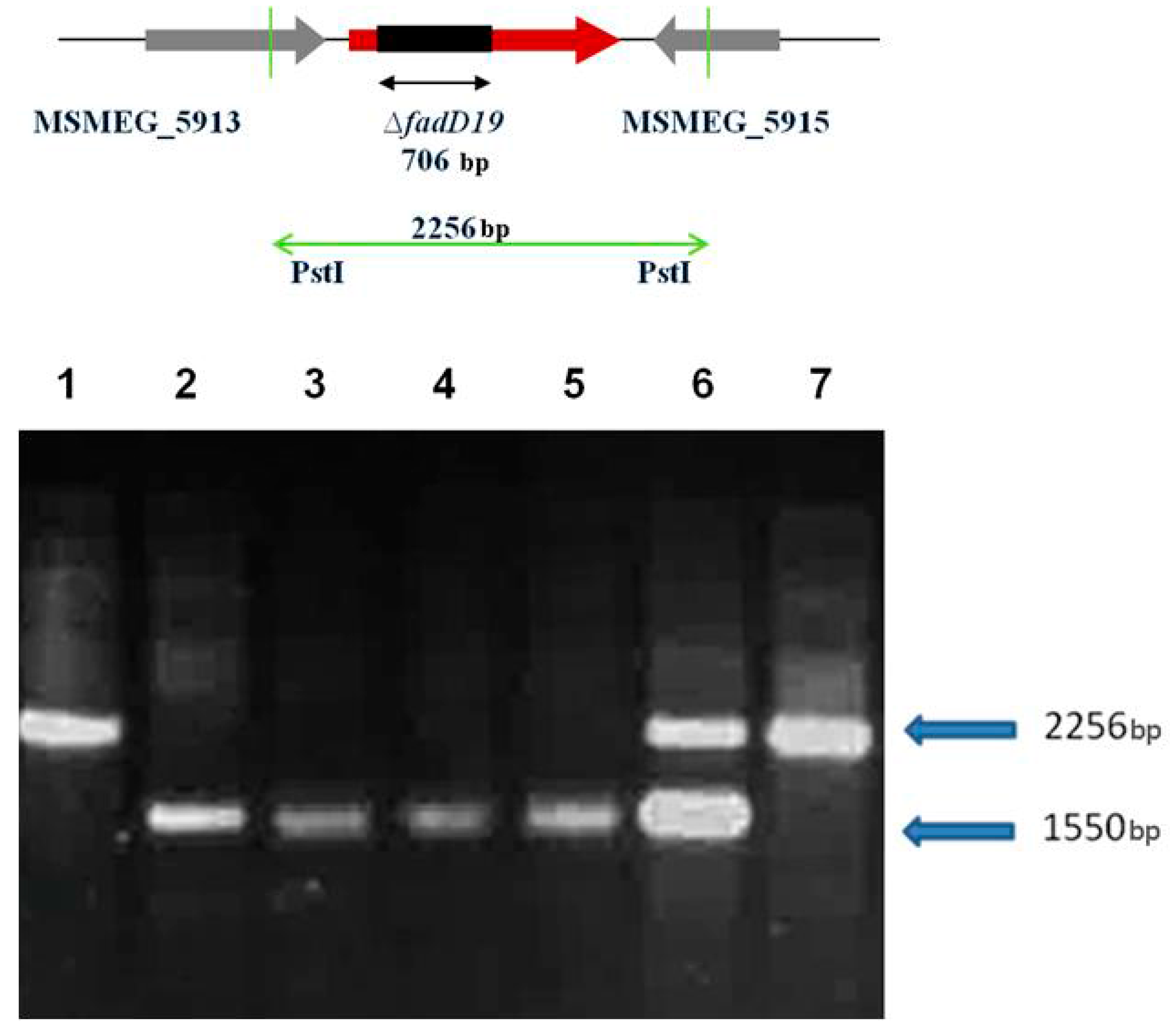
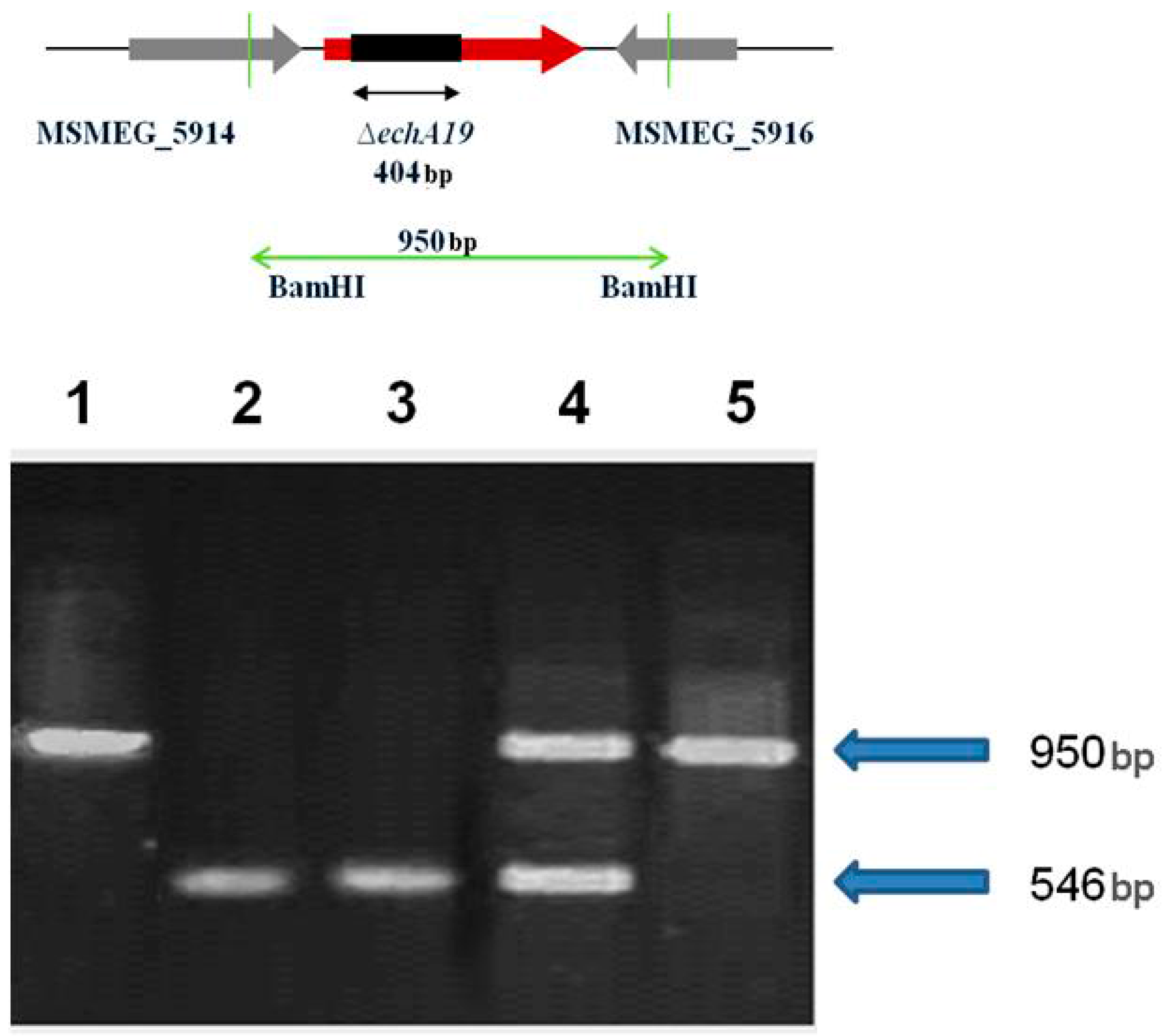
2.1. M. smegmatis fadD19 and echA19 Genes are not Essential for Viability

M. smegmatis has an ability to use cholesterol or β-sitosterol as a source of carbon and energy. Culturing of M. smegmatis in the presence of cholesterol results in fast complete degradation of this substrate which was observed as soon as at 24 h (Figure 1). In the case of β-sitosterol the complete degradation occurred at 96 h (Figure 2). The two-step recombination protocol of Parish and Stocker [37] was used to obtain the unmarked deletion of M. smegmatis fadD19 and echA19 genes. The successful engineering of mutants confirmed by PCR and Southern blot hybridization (Figure 3 and Figure 4) showed that neither fadD19 nor echa19 is essential for viability of M. smegmatis in rich media.

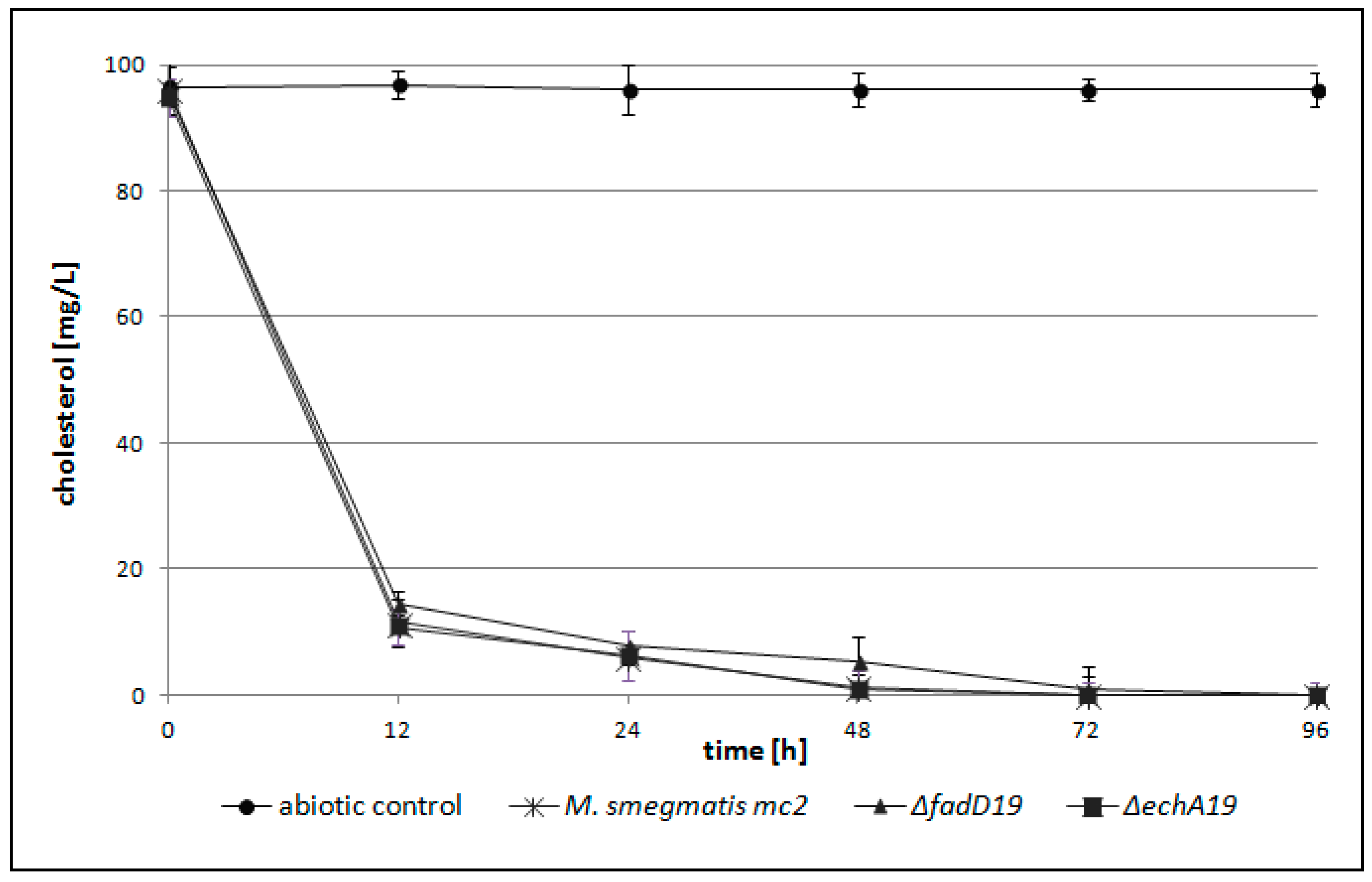
2.2. M. smegmatis fadD19 and echA19 Genes are not Essential for Cholesterol (Side Chain) Degradation

The ability of M. smegmatis ΔfadD19 and ΔechA19 mutants to utilize cholesterol was monitored using the LC-MS/MS technique. The very fast consumption of cholesterol (90% of the substrate was degraded completely at 24 h of culture) was observed for both mutants (Figure 1). M. smegmatis ΔfadD19 and ΔechA19 were capable of cholesterol degradation, as well as cholestenone, AD, ADD production (Table 1). The first metabolite identified in the degradation process was cholestenone, with the largest amount between 12 h and 24 h of the culture. Cholestenone was more abundant in ΔfadD19 in the 24 h culture, than in the culture of the parent strain.

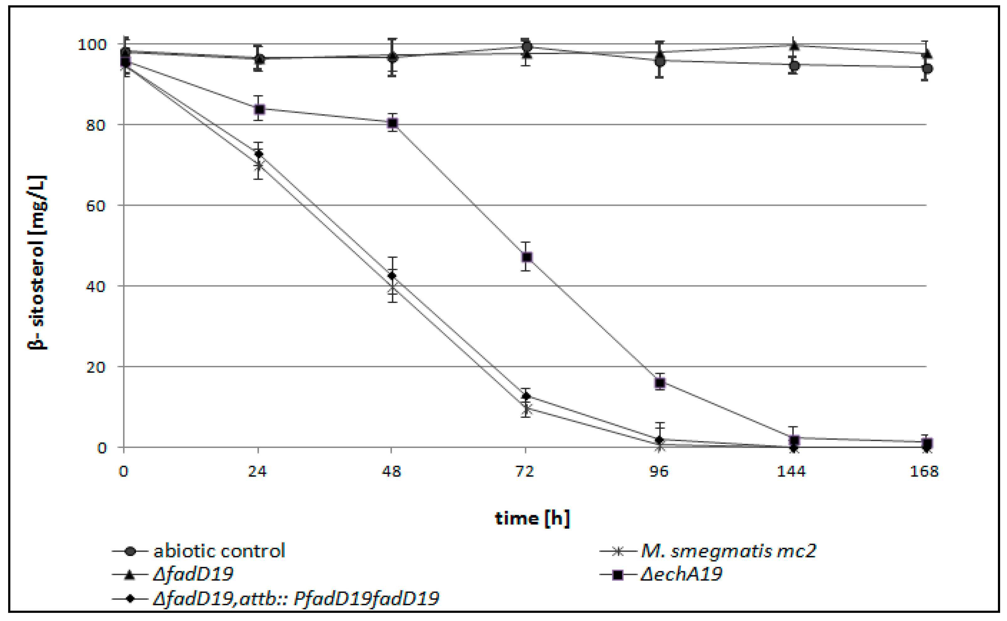
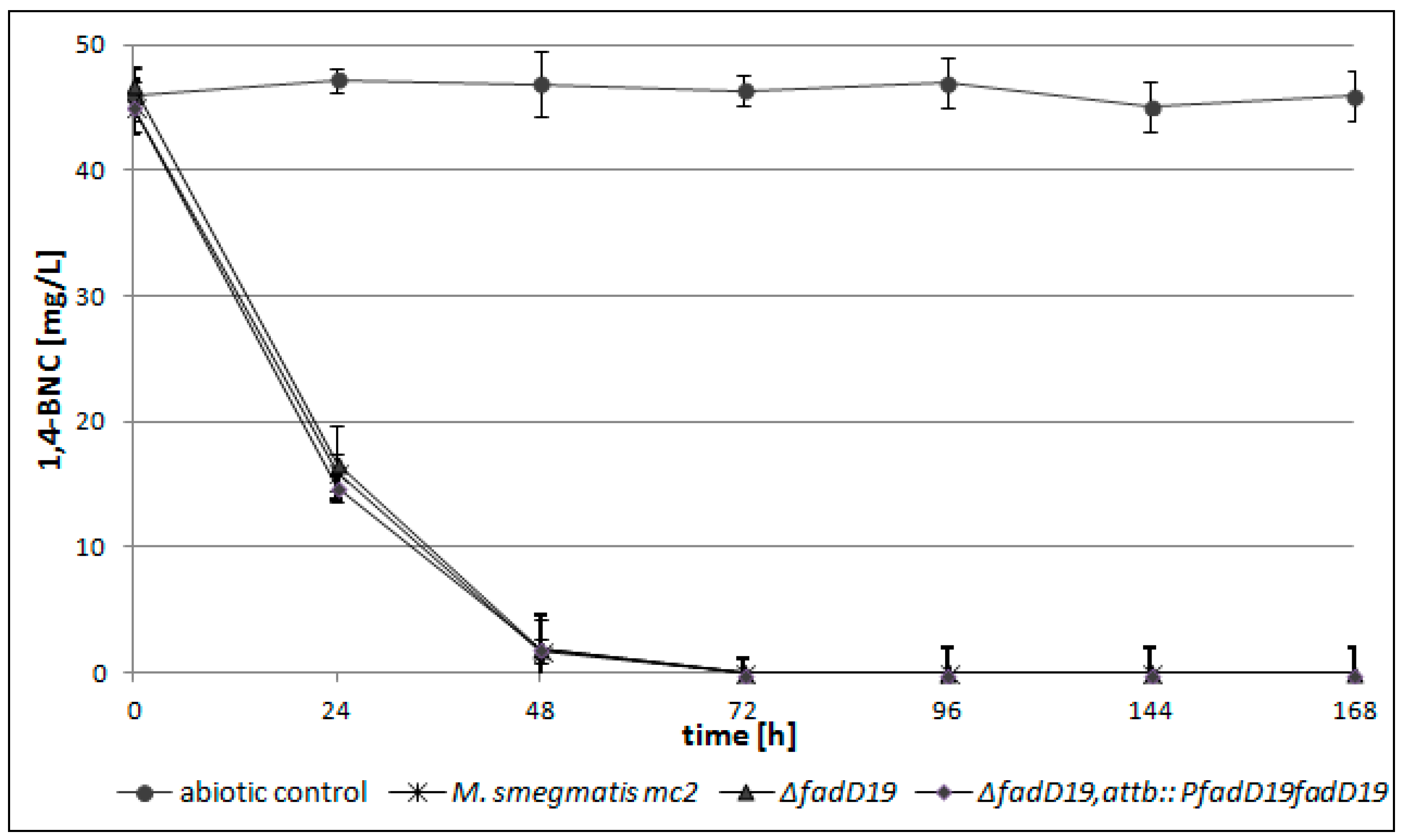
2.3. fadD19 but not echA19 is Required for β-Sitosterol (Side Chain) Degradation

We used β-sitosterol (C-24 branched side chain sterol) as a substrate to analyze the ability of wild type M. smegmatis and its ΔfadD19 and ΔechA19 mutants to degrade side-chain branched sterols. We found that mutant ∆fadD19 is unable to use β-sitosterol as the only source of carbon and energy, suggesting the fundamental role of FadD19 in β-sitosterol degradation by M. smegmatis (Figure 2, Table 2). Moreover, we observed that ΔechA19 inactivation delayed the β-sitosterol degradation by M. smegmatis ΔechA19 (Figure 2, Table 2), which was associated with a slower initial growth phase (24 h) (unpublished data).
We also tested the ability of the wild type strain and its mutants to utilize 1,4-BNC (3-oxo-23,24-bisnorchola-1,4-dien-22-oic acid), a steroid with a partially preserved aliphatic chain (Figure 5). It was observed that both wild type and ΔfadD19 strains were able to degrade 1,4-BNC.
To confirm the essential role of FadD19 in the early step of β-sitosterol degradation, a complemented mutant was constructed carrying an intact fadD19 under control of its natural putative promoter (M. smegmatisfadD19; attB::PfadD19fadD19). The complemented strain, but not the ∆fadD19 mutant, was able to degrade β-sitosterol with kinetics similar to that of wild type M. smegmatis (Figure 2). The complementation confirmed that the inhibition of β-sitosterol degradation by M. smegmatisfadD19 was directly due to the absence of an intact fad gene, not to a polar effect.
In the case of the ΔechA19 mutant we observed a delay in the degradation of β-sitosterol compared to a wild type strain (Figure 2, Table 2). Analysis of the growth kinetics (unpublished data) on minimal medium supplemented with β-sitosterol confirms that inactivation of echA19 results in slower consumption of this substrate.

3. Discussion

In this study we demonstrated for the first time an important role of fadD19 in the initial step of β-sitosterol degradation by M. smegmatis. Moreover, this is the first report on the molecular characterization of the gene playing an essential role in C 24-branched chain sterol degradation in M. smegmatis. In addition, we observed that echA19 inactivation delayed the process of β-sitosterol degradation by M. smegmatis ΔechA19. This effect is specific to β-sitosterol, because the addition of acetate or propionate abolishes this effect between the wild type strain M. smegmatis mc2155 and the ΔechA19 mutant (unpublished data). The literature data concerning EchA19 is very limited, so the role of EchA19 in sterol side-chain degradation will be further examined.
Fast growing mycobacteria, including M. smegmatis, are able to degrade sterols carrying straight or branched side chains. Here, we found that FadD19 in M. smegmatis is essential to utilize branched side chain sterols, but not those carrying a straight chain, like cholesterol. Using 1,4-BNC as a substrate, we found that FadD19 acts in the early step of β-sitosterol degradation. On the other hand, EchA19 appeared not to be essential for degrading either β-sitosterol or cholesterol. Similar observations were made by Wilbrink et al. [26] with Rhodococcus rhodochrous as a model strain. The mutant defective in the synthesis of FadD19 was not able to utilize sitosterol and campesterol but degraded cholesterol [26]. The essentiality for degradation of steroid substrates carrying branched but not straight side chains was also shown in the case of aldol-lyases Ltp3 and Ltp4 [24].
The knowledge of genes involved in sterol side-chain degradation is very limited in contrast to enzymes catalyzing the sterol ring degradation. However, a catabolic gene cluster was identified in M. smegmatis and R. jostii, as encoding the enzymes which are probably responsible for the β-oxidation process [22,25]. The β-oxidation process requires several enzymes, such as enoyl-CoA hydratase (EchA), acyl-CoA dehydrogenase (FadE), 3-hydroxy-acyl-CoA-dehydrogenase (FadB) and 3-keto-acyl-CoA thiolase (FadA) [38]. Steroid-CoA ligase (fadD19), which plays a key role in C-24 branched sterol degradation, has been identified by Wilbrink et al. [26]. A second gene which is analyzed in this study is echA19 (putative enoyl-CoA hydratase). EchA5 was described as non-essential for M. tuberculosis growth in vitro and in vivo [39]. Yang et al. showed that MaoC-like enoyl-CoA hydratase, ChsH1-ChsH2, catalyzes the hydratation of 3-oxo-4,17-pregnadiene-20-carboxyl-CoA to 17-hydroxy-3-oxo-4-pregnene-20-carboxyl-CoA in cholesterol side chain degradation [40]. Moreover, Casabon et al. characterized acyl-CoA synthetases involved in steroid side-chain degradation in M. tuberculosis and R. jostii RHA1 [41]. In the genome of pathogenic mycobacteria, Mtb, orthologs of analyzed genes fadD19 and echA19 can be identified with an 82.7% and 81.6% identity at the amino acids level, respectively. FadD19 was predicted to be specifically required in Mtb for growth on cholesterol [17]. However, the specific inactivation of both genes, Δ(echA19-fadD19), in Mtb did not affect the process of cholesterol degradation (Brzostek et al., unpublished data). There are no published data about the utilization of β-sitosterol by Mtb, however, a number of different sterols are available in the host.
The degradation of the sterol aliphatic side-chain is still not well understood. However, it is generally accepted that the side-chain of sterols is shortened by β-oxidation reactions. Some genes encoding putative β-oxidation enzymes in the cholesterol regulons of Mtb and Rhodococcus rhodochrous DSM43269 were identified. FadA5 catalyzes the thiolysis of acetoacetyl-CoA in vitro yielding androsterone metabolites [e.g., 4-androstenedione (AD) and 1,4-androstenedione (ADD)] [23]. Thiolase activity appeared to be required for growth on cholesterol and for Mtb virulence, especially during the late stage of mouse infection. Cytochrome P450 monooxygenase (Cyp125) was identified as a selective sterol side chain degradation enzyme in the Rhodococcus rhodochrous RG32 strain. The orthologue of cyp 125 in M. tuberculosis (rv3545c) was suggested to be associated with the process of pathogenesis [42].Understanding of the process of sterol degradation by mycobacteria is important not only from the point of view of Mtb pathogenesis but can also have practical implications in the production of steroid drugs (AD, ADD). These compounds might be synthesized using non-pathogenic mycobacterial strains [2,43].

4. Experimental Section

4.1. Bacterial Strains and Culture Conditions

The following bacterial strains were used: Escherichia coli Top 10 (Invitrogen, Carlsbad, CA, USA), Mycobacterium smegmatis mc2155 (Snapper et al., [44]). E. coli strains were grown in LB medium. For growth on solid medium, 1% (w/v) Difco agar (Becton, Franklin Lakes, NJ, USA) was added. The mycobacterial strains were cultured in Middlebrook 7H9 broth or 7H10 agar plates supplemented with albumin-dextrose-sodium chloride and/or kanamycin (25 μg·mL−1).
For steroid degradation experiments, mycobacterial strains were cultured in NB broth [8 gL−1 nutrient broth (Difco, Franklin Lakes, MJ, USA), 10 gL−1 glucose, supplemented with 2% Tween 80 (pH 6–6.2)] or in minimal medium [gL−1: MgSO4·7H2O, 5; Na2HPO4, 1; KH2PO4, 5; NH4NO3, 25; CaCl2·2H2O, 0.001; Fe2(SO4)3·nH2O, 0.01; MnSOnH2O, 0.0001; Co(NO3)·2.6H2O, 0.000005; (NH4)6Mo7O24·4H2O, 0.0001].
NB medium (100 mL) was inoculated with M. smegmatis and incubated overnight at 37 °C with shaking at 140 r.p.m. min−1. An overnight culture of M. smegmatis was harvested by centrifugation for 10 min at 5000 r.p.m. The cells were resuspended in 10 mL minimal medium and a sample (about 2–4 mL) was transferred to 96–98 mL fresh minimal medium in a 1 L flask. The optical density of bacteria was 0.1. Finally, cholesterol or β-sitosterol were added to the medium at a final concentration of 100 mg/L. The cultures were incubated on a shaker (140 r.p.m.) at 37 °C.

4.2. Plasmid Constructions

To perform unmarked deletion in fadD19 and echA19 genes of Mycobacterium smegmatis, suicidal recombination delivery vectors were constructed. In the first step the 5′ ends of the genes (fadD19 or echA19) and upstream regions were amplified using primers MsfadD19Gr1 (CCAGACGCCGGACGGATCCCAGGC) and MsfadD19Gr2 (CGCCTGTGTGGATACCAGCCATTG) for fadD19, and MsechA19Gr1 (GCTGCAGATCGGTGATGACGCGGTTGG) and MsechA19Gr2 (CAAGCTTGACGATGAGGGTG TGTCCGC) for echA19, and cloned into the KpnI/BamHI and PstI/HindIII sites of p2NIL to create pNW03 and pNW08, respectively. Subsequently, the 3′ ends of the genes (fadD19 or echA19) and downstream regions were amplified using primers MsfadD19Gr3 (CGGGATCCCCCGACGAGGTGTGGCAGG) and MsfadD19Gr4 (GCAAGCTTCCATCCAGACGCCGGACG) for fadD19, and MsechA19Gr3 (CAAGCTTTGGACAAGGCGCTGGAGATCG) and MsechA19Gr4 (CGGTACCTCACATTCGGGAACGGTGGGAC) for echA19, and cloned into the BamHI/HindIII sites of pNW03 and HindIII/KpnI sites of pNW08. The ligated 5′ and 3′ fragments of the fadD19 and echA19 genes in the resulting vectors were out of frame. The resultant vectors were named pNW04 and pNW09, respectively. Finally, PacI marker gene cassette from pGOAL17 carrying lacZ and sacB genes was cloned into the PacI site of pNW04 and pNW09 to create pNW05 and pNW010, respectively. The plasmids used in this work are listed in Table 3.

4.3. Disruption of fadD19 and echA19 Genes

The protocol of Parish and Stocker [29] was used to disrupt fadD19 or echA19 genes at their native loci on the chromosome. Plasmid DNA (pNW05, pNW10) was treated with NaOH (2 mM) and integrated into the M. smegmatis chromosome by homologous recombination. The resulting single crossover (SCO) recombinant mutant colonies were blue, KmR and sensitive to sucrose. The site of recombination was confirmed by PCR and Southern blot hybridization. The SCO strains were further processed to select DCO (double-crossover) mutants that were white, KmS and resistant to sucrose. PCR and Southern blot hybridization were used to distinguish between the wild-type and DCO mutants. The probes were generated by PCR with primers binding to the 5′ and 3′ of each gene and DNA of pNW04 and pNW09 as templates, and labeled with a nonradioactive primer extension system (DIG-labeling system, Amersham, Uppsala, Sweden).

4.4. Complementation Constructs

The complementation vector was engineered by amplifying the fad gene with its own promoter region (345 bp) using M. smegmatis chromosomal DNA as a template. The PCR product was initially cloned into pJet1.2, verified by sequencing and subsequently re-cloned into the pMV306Km integrative vector using XbaI and HindIII.

4.5. Sterols Degradation

4.5.1. Steroid Standards

Cholesterol, cholestenone, 4-androstene-3,17-dione (AD), 1,4-androstadiene-3,17-dione (ADD), β-sitosterol were purchased from Sigma (Darmstadt, Germany) and used as standards for LC MS/MS analyses; 3-oxo-3,24-bisnorchola-1,4-dien-22-oic acid (1,4-BNC) was synthesized by Steraloids (Newport, RI, USA).

4.5.2. Preparation of Cholesterol

The substrate was dissolved in a solution of 96% ethanol–Triton WR1339 (1:1) at 80 °C.

4.5.3. Preparation of β-Sitosterol

β-Sitosterol (15 g) and Tween 80 (2 g) were suspended in distilled water (50 mL). The suspension was homogenized at 94 °C for 20 min. Further the speed of homogenizer was reduced to 1000 rpm/min and gradually water was added to the mixture to a final volume of 200 mL. The suspension was then homogenized for 10 min. reaching up to 2000/min. In the final step the microscopic control of the substrate was performed by measuring the β-sitosterol crystal size. The resulting sample of β-sitosterol was in the shape of needles with a length of 2–4 μm and 1–2 μm in width. The steroid compound was sterilized by boiling three times in 1 h at 24 h intervals. β-Sitosterol was stored at 4 °C.

4.5.4. HPLC MS/MS Analysis of Sterol Degradation

Sterol degradation analysis was performed using an Agilent 1200 HPLC (Santa Clara CA, USA) system and a 3200 QTRAP mass spectrometer (AB Sciex, Framingham, MA, USA) with an atmospheric pressure chemical ionization APCI source. Chromatographic separation was performed using an XDB-C18 column (2.1 mm × 50 mm × 1.8 μm; Agilent Technologies). The mobile phase consisted of 2 mM ammonium formate and 0.2% formic acid in water (A) and 2 mM ammonium formate and 0.2% formic acid in methanol (B). The column temperature was maintained at 40 °C and the flow rate was 0.5 mL·min−1. The ion source settings were as follows: curtain gas: 25 (CUR), IS: 5500 V, nebulizer gas: 50 (GS1), drying gas: 50 (GS2) and temperature of 600 °C. Data analysis was performed with the Analyst™ v1.5.2 software (AB Sciex). Tandem mass spectrometry for the quantitation of sterols degradation courses was made using positive ionization in the multiple reaction monitoring (MRM) mode. The quantitative analysis of sterols and its degradation products was based on standard curves with a linearity range from 1 ng·mL−1 to 10 ng·mL−1 (correlation coefficient R2 = 0.951). All the experimental data represent the means of at least three independent experiments.

5. Conclusions

The obtained results allows us to conclude that the FadD19 plays an important role in the initial step of biodegradation of C-24 branched sterol side chains in Mycobacterium smegmatis mc2155. The extension of our knowledge in the area of sterol degradation could have significant implications for the production of steroid drugs. Additionally, a detailed explanation of the molecular mechanism of sterol degradation could help to explain M. tuberculosis pathogenesis.

Acknowledgments

This research was financed by a grant from the European Regional Development Fund (POIG.01.01.02-10-107/09) under the Operational Innovative Economy Programme.

Author Contributions

N.W., A.B. performed the experiments, drafted the manuscript; R.S., A.S. performed HPLC MS/MS analysis; K.L., J.D., performed the experiments, conceived and designed the experiments, analyzed the data, drafted the manuscript.

Conflicts of Interest

The authors declare no conflict of interest.

References

  1. Naghibi, F.; Tabatabai Yazdi, M.; Sahebgharani, M.; Noori Daloii, M.R. Microbial transformation of cholesterol. J. Sci. Islam. Repub. Iran 2002, 13, 103–106. [Google Scholar]
  2. Donova, M.V.; Egorova, O.V. Microbial steroid transformations: Current state and prospects. Appl. Microbiol. Biotechnol. 2012, 94, 1423–1447. [Google Scholar] [CrossRef] [PubMed]
  3. Lad, N. Optimalization of parameters for hydrocortisone succinate bioconversion. Malays. J. Microbiol. 2011, 7, 7–13. [Google Scholar]
  4. Field, J.A.; Bolesma, F.; Baten, H.; Rulkens, W.H. Oxidation of anthracene in water solvent mixtures by the white-rot fungus Bjerkandera sp. strain BOS55. Appl. Microbiol. Biotechnol. 1995, 44, 234–240. [Google Scholar] [CrossRef]
  5. Cerniglia, C.E. Biodegradation of polycyclic aromatic hydrocarbons. Biodegradation 1992, 3, 351–368. [Google Scholar] [CrossRef]
  6. Guerin, W.F.; Jones, G.E. Two-stage mineralization of phenanthrene by estuarine enrichment cultures. Appl. Environ. Microbiol. 1998, 54, 937–944. [Google Scholar]
  7. Mattos, K.A.; Oliveira, V.C.G.; Berrêdo-Pinho, M.; Amaral, J.J.; Antunes, L.C.M.; Melo, R.C.N.; Acosta, C.C.D.; Moura, D.F.; Olmo, R.; Han, J.; et al. Mycobacterium leprae intracellular survival relies on cholesterol accumulation in infected macrophages: A potential target for new drugs for leprosy treatment. Cell. Microbiol. 2014, 16, 797–815. [Google Scholar] [CrossRef] [PubMed]
  8. Fine-Coulson, K.; Reaves, B.J.; Karls, R.K.; Quinn, F.D. The role of lipid raft aggregation in the infection of type II pneumocytes by Mycobacterium tuberculosis. PLoS ONE 2012, 7, e45028. [Google Scholar] [CrossRef] [PubMed]
  9. Pandey, A.K.; Sassetti, C.M. Mycobacterial persistence requires the utilization of host cholesterol. Proc. Natl. Acad. Sci. USA 2008, 105, 4376–4380. [Google Scholar] [CrossRef] [PubMed]
  10. Brzostek, A.; Pawełczyk, J.; Rumijowska-Galewicz, A.; Dziadek, B.; Dziadek, J. Mycobacterium tuberculosis is able to accumulate and utilize cholesterol. J. Bacteriol. 2009, 191, 6584–6591. [Google Scholar] [CrossRef] [PubMed]
  11. Chang, J.C.; Miner, M.D.; Pandey, A.K.; Gill, W.P.; Harik, N.S.; Sassetti, C.M.; Sherman, D.R. Igr. gene and Mycobacterium tuberculosis cholesterol metabolism. J. Bacteriol. 2009, 191, 5232–5239. [Google Scholar] [CrossRef] [PubMed]
  12. Yam, K.C.; D’Angelo, I.; Kalscheuer, R.; Zhu, H.; Wang, J.X. Studies of a ring-cleaving dioxygenase illuminate the role of cholesterol metabolism in the pathogenesis of Mycobacterium tuberculosis. PLoS Pathog. 2009, 5, e1000344. [Google Scholar] [CrossRef] [PubMed]
  13. Hu, Y.; van der Geize, R.; Besra, G.S.; Gurcha, S.S.; Liu, A. 3-Ketosteroid 9alpha-hydroxylase is an essential factor in the pathogenesis of Mycobacterium tuberculosis. Mol. Microbiol. 2010, 75, 107–121. [Google Scholar] [CrossRef] [PubMed]
  14. Ouellet, H.; Johnston, J.B.; de Montellano, P.R. Cholesterol catabolism as a therapeutic target in Mycobacterium tuberculosis. Trends Microbiol. 2011, 19, 530–539. [Google Scholar] [CrossRef] [PubMed]
  15. Brzezinska, M.; Szulc, I.; Brzostek, A.; Klink, M.; Kiełbik, M. The role of 3-ketosteroid 1(2)-dehydrogenase in the pathogenicity of Mycobacterium tuberculosis. BMC Microbiol. 2013, 13, 43–55. [Google Scholar] [CrossRef] [PubMed]
  16. Van der Ven, B.C.; Fahey, R.J.; Lee, W.; Liu, Y.; Abramovitch, R.B.; Memmott, C.; Crowe, A.M.; Eltis, L.D.; Perola, E.; Deininger, D.D.; et al. Novel inhibitors of cholesterol degradation in Mycobacterium tuberculosis reveal how the bacterium’s metabolism is constrained by the intracellular environment. PLoS Pathog. 2015, 11, e1004679. [Google Scholar]
  17. Griffin, J.E.; Gawronski, J.D.; Dejesus, M.A.; Ioerger, T.R.; Akerley, B.J.; Sassetti, C.M. High-resolution phenotypic profiling defines genes essential for mycobacterial growth and cholesterol catabolism. PLoS Pathog. 2011, 7, e1002251. [Google Scholar] [CrossRef] [PubMed]
  18. Uhia, I.; Galan, B.; Morales, V.; Garcia, J.L. Initial step in the catabolism of cholesterol by Mycobacterium smegmatis mc2155. Environ. Microbiol. 2011, 13, 943–959. [Google Scholar] [CrossRef] [PubMed]
  19. Yang, X.; Dubnau, E.; Smith, I.; Sampson, N.S. Rv1106c from Mycobacterium tuberculosis is a 3β-hydroxysteroid dehydrogenase. Biochemistry 2007, 46, 9058–9067. [Google Scholar] [CrossRef] [PubMed]
  20. Brzostek, A.; Rumijowska-Galewicz, A.; Dziadek, B.; Wójcik, E.A.; Dziadek, J. ChoD and HsdD can be dispensable for cholesterol degradation in mycobacteria. J. Steroid Biochem. Mol. Biol. 2013, 134, 1–7. [Google Scholar] [CrossRef] [PubMed]
  21. Yang, X.; Gao, J.; Smith, I.; Dubnau, E.; Sampson, N.S. Cholesterol is not essential source of nutrition for Mycobacterium tuberculosis during infection. J. Bacteriol. 2011, 193, 1473–1476. [Google Scholar] [CrossRef] [PubMed]
  22. Van der Geize, R.; Yam, K.; Heuser, T.; Wilbrink, M.H.; Hara, H.; Anderton, M.C.; Sim, E.; Dijkhuizen, L.; Davies, J.E.; Mohn, W.W.; et al. A gene cluster encoding cholesterol catabolism in a soil actinomycete provides insight into Mycobacterium tuberculosis survival in macrophages. Proc. Natl. Acad. Sci. USA 2007, 104, 1947–1952. [Google Scholar] [CrossRef] [PubMed]
  23. Nesbitt, N.M.; Yang, X.; Fontan, P.; Kolesnikova, I.; Smith, I.; Sampson, N.S.; Dubnau, E. A thiolase of Mycobacterium tuberculosis is required for virulence and production of androstenedione and adrostadienedione from cholesterol. Infect. Immun. 2010, 78, 275–282. [Google Scholar] [CrossRef] [PubMed]
  24. Wilbrink, M.H.; van der Geize, R.; Dijkhuizen, L. Molecular characterization of ltp3 and ltp4, essential for C24-branched chain sterol-side-chain degradation in Rhodoccocus rhodochrous DSM 43269. Microbiology 2012, 158, 3054–3062. [Google Scholar] [CrossRef] [PubMed]
  25. Uhia, I.; Galan, B.; Kendall, S.L.; Stoker, N.G.; Garcia, J. Cholesterol metabolism in Mycobacterium smegmatis. Environ. Microbiol. Rep. 2012, 4, 168–182. [Google Scholar] [CrossRef] [PubMed]
  26. Wilbrink, M.H.; Petrusma, M.; Dijkhuizen, L.; van der Geize, R. FadD19 of Rhodococcus rhodochorus DSM43269, a s-coenzyme A ligase essential for degradation of C24 branched sterol side chains. Appl. Environ. Microbiol. 2011, 77, 4455–4464. [Google Scholar] [CrossRef] [PubMed]
  27. Frank, D.J.; Madrona, Y.; Ortiz de Montellano, P.R. Cholesterol ester oxidation by Mycobacterial Cytochrome P450. J. Biol. Chem. 2014, 289, 30417–30425. [Google Scholar] [CrossRef] [PubMed]
  28. Capyk, J.K.; Kalscheuer, R.; Stewart, G.R.; Liu, J.; Kwon, H.; Zhao, R.; Okamoto, S.; Jacobs, W.R.; Eltis, L.D.; Mohn, W.W. Enzyme Catalysis and Regulation: Mycobacterial Cytochrome P450 125 (Cyp125) Catalyzes the Terminal Hydroxylation of C27 Steroids. J. Biol. Chem. 2009, 284, 35534–35542. [Google Scholar] [CrossRef] [PubMed]
  29. McLean, K.J.; Lafite, P.; Levy, C.; Cheesman, R.E.; Mast, N.; Pikuleva, I.A.; Leys, D.; Munro, A.W. The structure of Mycobacterium tuberculosis CYP125: Molecular basis for cholesterol binding in a P450 needed for host infection. J. Biol. Chem. 2009, 284, 35524–35533. [Google Scholar] [CrossRef] [PubMed]
  30. Johnston, J.B.; Ouellet, H.; Ortiz de Montellano, P.R. Functional redundancy of steroid C26-monooxygenase activity in Mycobacterium tuberculosis revealed by biochemical and genetic analyses. J. Biol. Chem. 2010, 285, 36352–36360. [Google Scholar] [CrossRef] [PubMed]
  31. Driscoll, M.D.; McLean, K.J.; Levy, C.; Mast, N.; Pikuleva, I.A.; Lafite, P.; Rigby, S.E.J.; Leys, D.; Munro, A.W. Structural and biochemical characterization of Mycobacterium tuberculosis CYP142: Evidence for multiple cholesterol 27-hydroxylase activities in a human pathogen. J. Biol. Chem. 2010, 285, 38270–38282. [Google Scholar] [CrossRef] [PubMed]
  32. Thomas, S.T.; Sampson, N.S. Mycobacterium tuberculosis utilizes a unique heterotetrameric structure for dehydrogenation of the cholesterol side chain. Biochemistry 2013, 52, 2895–2904. [Google Scholar] [CrossRef] [PubMed]
  33. Ruprecht, A.; Maddox, J.; Stirling, A.J.; Visaggio, N.; Seah, S.Y.K. Characterization of novel acyl Coenzyme A dehydrogenases involved in bacterial steroid degradation. J. Bacteriol. 2015, 197, 1360–1367. [Google Scholar] [CrossRef] [PubMed]
  34. Yang, M.; Lu, R.; Guja, K.E.; Wipperman, M.F.; Clair, J.R.St.; Bonds, A.C.; Garcia-Diaz, M.; Sampson, N. Unraveling cholesterol catabolism in Mycobacterium tuberculosis: ChsE4-ChsE5 α2β2 acyl-CoA dehydrogenase initiates β-oxidation of 3-oxo-cholest-4-en-26-oyl CoA. ACS Infect. Dis. 2015, 1, 110–125. [Google Scholar] [CrossRef] [PubMed]
  35. Lu, R.; Schmitz, W.; Sampson, N.S. α-methyl acyl CoA racemase provides Mycobacterium tuberculosis catabolic access to cholesterol esters. Biochemistry 2015, 54, 5669–5672. [Google Scholar] [CrossRef] [PubMed]
  36. Thomas, S.T.; VanderVen, B.C.; Sherman, D.R.; Russell, D.G.; Sampson, N.S. Pathway profiling in Mycobacterium tuberculosis: Elucidation of cholesterol-derived catabolite and enzymes that catalyze its metabolism. J. Biol. Chem. 2011, 286, 43668–43678. [Google Scholar] [CrossRef] [PubMed]
  37. Parish, T.; Stocker, N.G. Use of a flexible cassette method to generate a double unmarked Mycobacterium tuberculosis tlyA plcABC mutant by gene replacement. Microbiology 2000, 146, 1969–1975. [Google Scholar] [CrossRef] [PubMed]
  38. Wipperman, M.F.; Sampson, N.S.; Thomas, S.T. Pathogen roid rage: Cholesterol utilization by Mycobacterium tuberculosis. Crit. Rev. Biochem. Mol. Biol. 2014, 49, 269–293. [Google Scholar] [CrossRef] [PubMed]
  39. Williams, K.J.; Boshoff, H.I.; Krishnan, N.; Gonzales, J.; Schnappinger, D.; Robertson, B.D. The Mycobacterium tuberculosis β-oxidation genes echA5 and fadB3 are dispensable for growth in vitro and in vivo. Tuberculosis 2011, 91, 549–555. [Google Scholar] [CrossRef] [PubMed]
  40. Yang, M.; Guja, K.E.; Thomas, S.T.; Garcia-Diaz, M.; Sampson, N. A distinct MaoC-like enoyl-CoA hydratase architecture mediates cholesterol catabolism in Mycobacterium tuberculosis. ACS Chem. Biol. 2014, 9, 2632–2645. [Google Scholar] [CrossRef] [PubMed]
  41. Casabon, I.; Swain, K.; Crowe, A.M.; Eltis, L.D.; Mohn, W.W. Actinobacterial acyl-CoA synthetases involved in steroid side chain catabolism. J. Bacteriol. 2014, 196, 579–587. [Google Scholar] [CrossRef] [PubMed]
  42. Rosłoniec, K.Z.; Wilbrink, M.H.; Capyk, J.K.; Mohn, W.W.; Ostendorf, M.; van der Geize, R.; Dijkhuizen, L.; Eltis, L.D. Cytochrome P450 125 (CYP125) catalyses C26-hydroxylation to initiate sterol side-chain degradation in Rhodococcus jostii RHA1. Mol. Microbiol. 2009, 74, 1031–1043. [Google Scholar] [CrossRef] [PubMed]
  43. Garcia, J.L.; Uhia, I.; Galan, B. Catabolism and biotechnological applications of cholesterol degrading bacteria. Microb. Biotechnol. 2012, 5, 679–699. [Google Scholar] [CrossRef] [PubMed]
  44. Snapper, S.B.; Melton, R.E.; Mustafa, S.; Kieser, T.; Jacobs, W.R., Jr. Isolation and characterization of efficient plasmid transformation mutants of Mycobacterium smegmatis. Mol. Microbiol. 1990, 4, 1911–1919. [Google Scholar] [CrossRef] [PubMed]
  • Sample Availability: Samples of the compounds are available from the authors.
Scheme 1. The basic pathways of cholesterol (a) and β-sitosterol (b) side chain degradation containing analyzed metabolites [14,24,25,35,37].
Molecules 21 00598 sch001aMolecules 21 00598 sch001b
Figure 1. Cholesterol degradation by M. smegmatis based on HPLC analysis. The curves represent the rate of cholesterol degradation in cultures of M. smegmatis mc2155 (×), ΔfadD19 (▲), ΔechA19 (■) in minimal medium. The substrate control (the stability of cholesterol in media) is marked by circles (●). Results are representative of three independent experiments.
Molecules 21 00598 g001
Figure 2. β-Sitosterol degradation by M. smegmatis based on HPLC analysis. The curves represent the rate of β-sitosterol degradation in cultures of M. smegmatis mc2155 (stars), ΔfadD19 (▲), ΔechA19 (■), ΔfadD19,attb::PfadD19fadD19 (♦) in minimal medium. The substrate control (the stability of β-sitosterol in media) is marked by circles (●). Results are representative of three independent experiments.
Molecules 21 00598 g002
Figure 3. Southern blot-based analysis of mycobacterial mutants generated by directed mutagenesis. (Top) Scheme showing the length of the restriction DNA fragment (2256 bp) and the internal deletion in the mutated gene (706 bp). The chromosomal localization of fadD19 is represented by red arrow, while the internal deletion is marked by black rectangle. (Bottom) Southern blot confirming the internal deletion in the fadD19 gene of M. smegmatis. The lanes represent genomic DNA from: 1, wild-type M. smegmatis; 2–5, double crossover mutants carrying the internally deleted ΔfadD19 gene; 6, single crossover mutant; 7, double crossover mutant carrying the wild-type fadD19 gene.
Molecules 21 00598 g003
Figure 4. Southern blot-based analysis of mycobacterial mutants generated by directed mutagenesis. (Top) Scheme showing the length of the restriction DNA fragment (950 bp) and the internal deletion in the mutated gene (404 bp). The chromosomal localization of echA19 is represented by red arrow, while the internal deletion is marked by black rectangle; (Bottom) Southern blot confirming the internal deletion in the echA19 gene of M. smegmatis. The lanes represent genomic DNA from: 1, wild-type M. smegmatis; 2–3, double crossover mutants carrying the internally deleted ΔechA19 gene; 4, single crossover mutant; 5, double crossover mutant carrying the wild-type echA19 gene.
Molecules 21 00598 g004
Figure 5. 1,4-BNC degradation by M. smegmatis based on HPLC analysis. The curves represent the rate of 1,4-BNC degradation in cultures of M. smegmatis mc2155 (×), ΔfadD19 (▲), ΔfadD19,attb::PfadD19fadD19 (♦) in minimal medium. The substrate control (the stability of β-sitosterol in media) is marked by circles (●). Results are representative of three independent experiments.
Molecules 21 00598 g005
Table 1. The content [%] of a selected metabolite intermediate of cholesterol degradation on minimal medium by ΔfadD19, ΔechA19, M. smegmatis mc2155.
Time (h)CholestenoneADADDCholestenoneADADDCholestenoneADADD
Mycobacterium smegmatis mc2155ΔfadD19ΔechA19
0000000000
1211.190011.020010.0100
2410.311.950.8314.132.251.0511.050.750.9
4804.516.20.743.946.981.053.026.14
7204.069.405.117.504.977.5
9604.048.5068.7107.349
14402.16.102.075.0901.796.01
Table 2. The content [%] of selected metabolite intermediate of β-sitosterol degradation on minimal medium by ΔfadD19, ΔechA19, M. smegmatis mc2155.
Time (h)1,4-BNCADD1,4-BNCADD1,4-BNCADD
Mycobacterium smegmatis mc2155ΔfadD19ΔechA19
0000000
2410.240.510013.050
7217.813.570029.12.1
1442.348.75004.757.05
16803.01000.218.95
Table 3. Plasmids used in this study.
PLASMIDDESCRIPTIONSOURCE
CLONING VECTORS
p2NILRecombination vector, non-replicating in mycobacteria, KmRParish and Stocker [ 29]
pGOAL17Source of PacI cassette, AmpRParish and Stocker [ 29]
pMV306Mycobacterial integrating vector, KmRMed-Imune Inc.
VECTORS USED FOR THE GENE REPLACEMENT
pNW03p2NIL carrying a fragment of the 5 ′fadD19 gene and the DNA located upstream of the gene (fadD19Gr1-2) restricted recognition sequences for Kpn I and BamH1This study
pNW04pNW03 carrying a fragment of the 3′fadD19 gene and the DNA located below of the gene (fadD19Gr3-4), bounded by recognition sequences BamH1 and HindIIIThis study
pNW05pNW04 with PacI cassette from pGOAL17This study
pNW08p2NIL carrying a fragment of the 5 ′echA19 gene and the DNA located upstream of the gene (echA19Gr1-2) restricted recognition sequences for Pst I and HindIIIThis study
pNW09pNW08 carrying a fragment of the 3′echA19 gene and the DNA located below of the gene (echA19Gr3-4), bounded by recognition sequences KpnI and HindIIIThis study
pNW10pNW09 with PacI cassette from pGOAL17This study

