Comprehensive Profiling and Quantification of Ginsenosides in the Root, Stem, Leaf, and Berry of Panax ginseng by UPLC-QTOF/MS

 , ,
, , 
Abstract
:1. Introduction
2. Materials and Methods
2.1. Standard Constituents and Reagents
2.2. Panax ginseng Samples
2.3. Extraction of Ginsenosides
2.4. UPLC-QTOF/MS Conditions for Ginsenoside Profiling
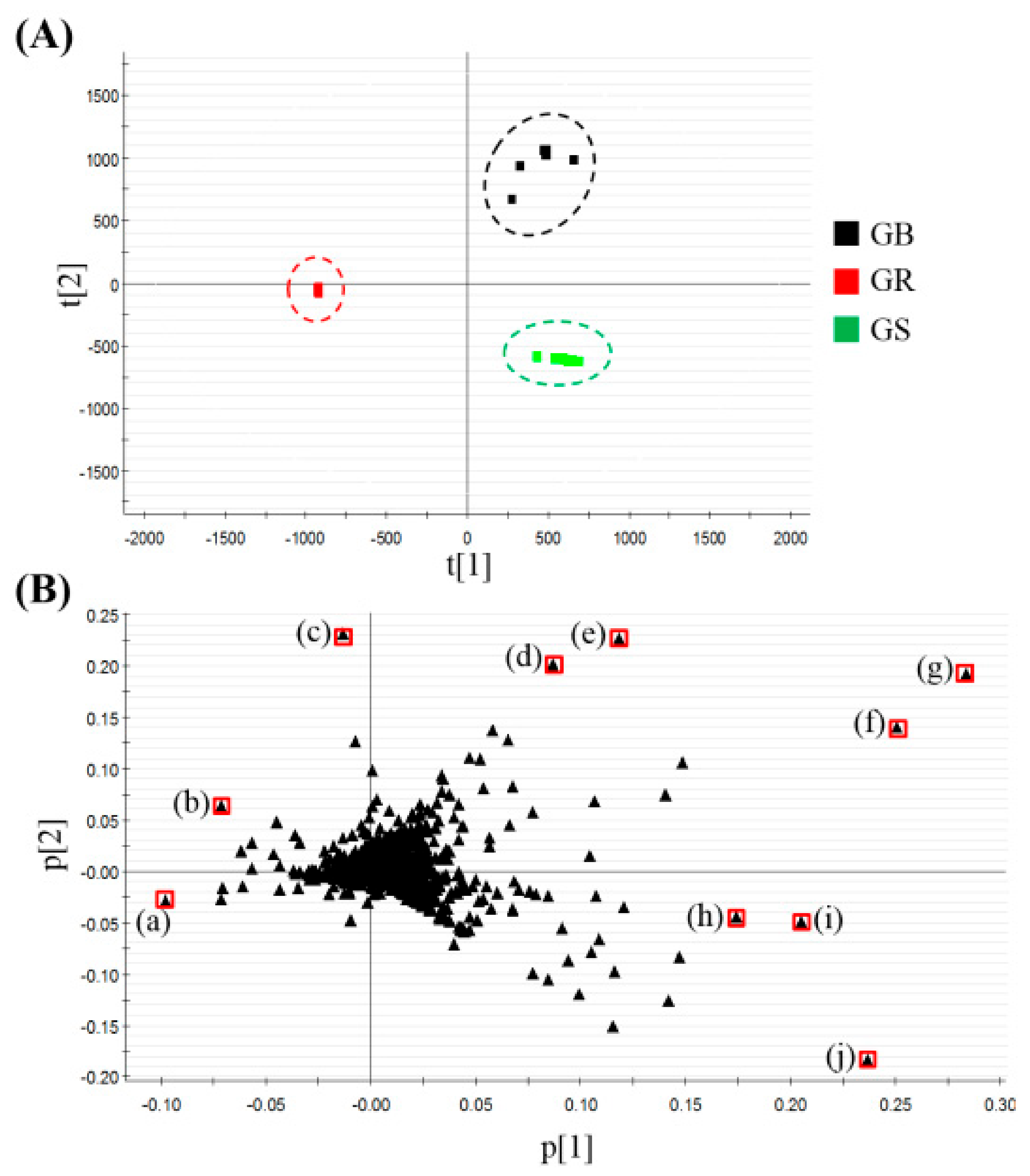
2.5. Data Processing and Multivariate Analysis
3. Results and Discussion
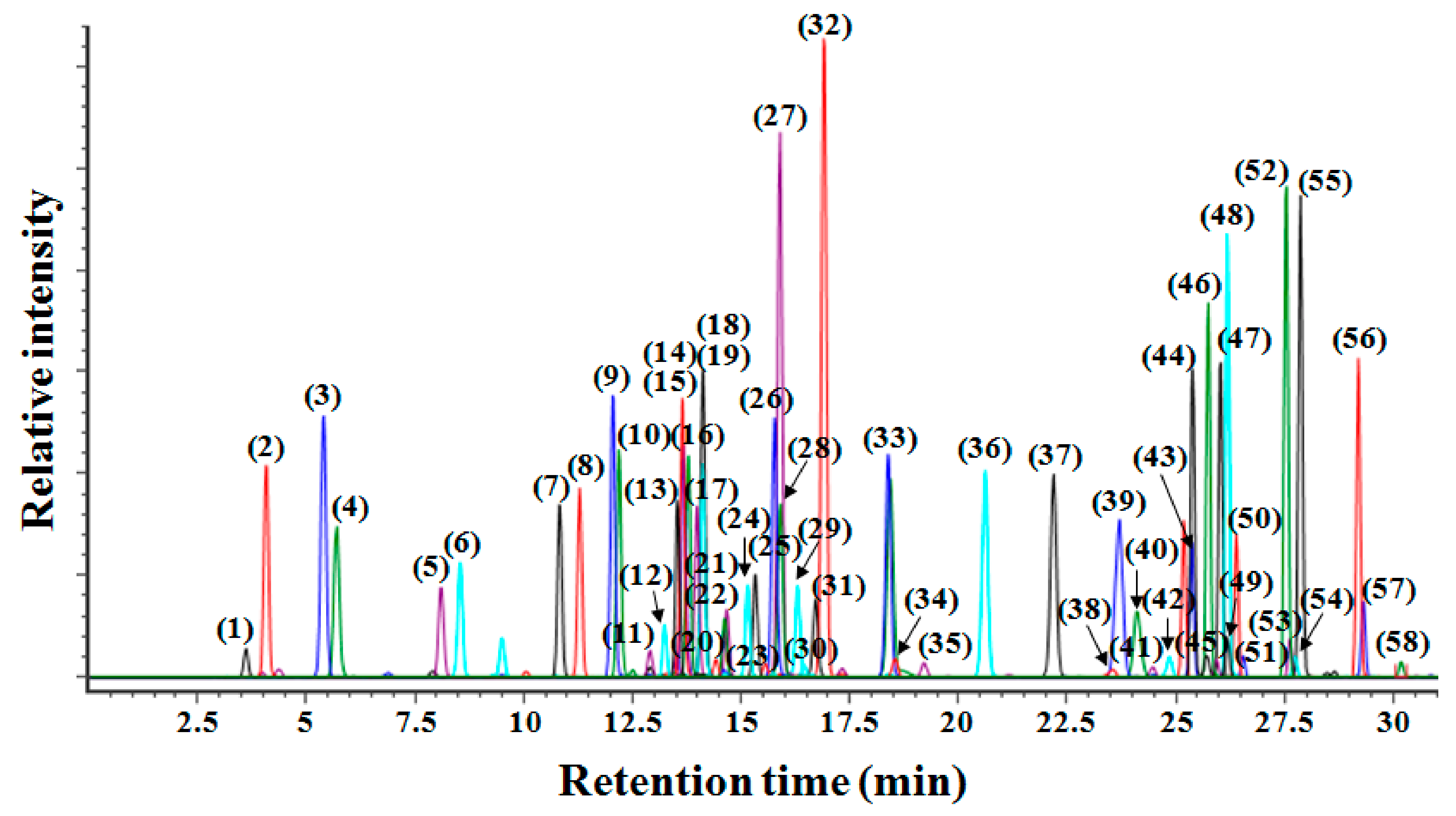
3.1. UPLC-QTOF/MS with an In-House Library to Profile Various Ginsenosides
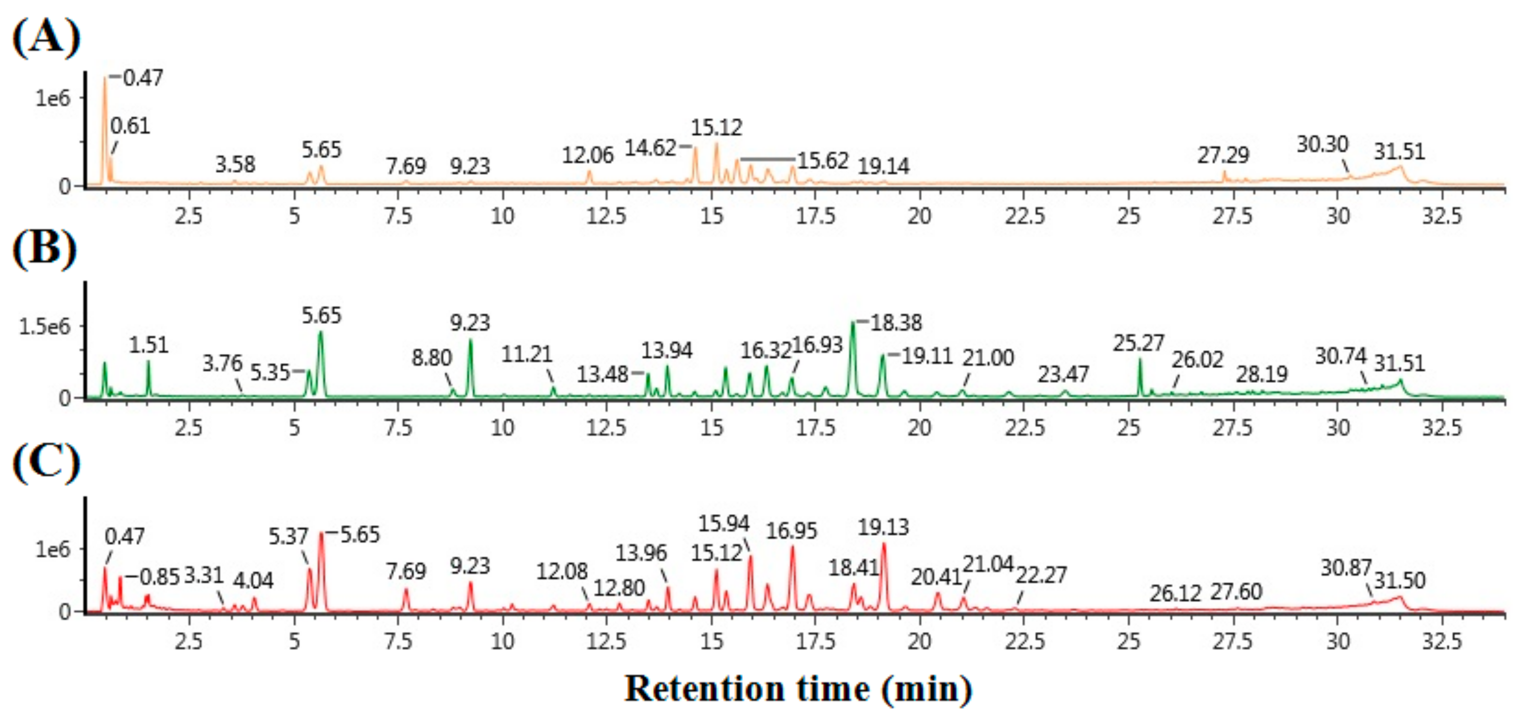
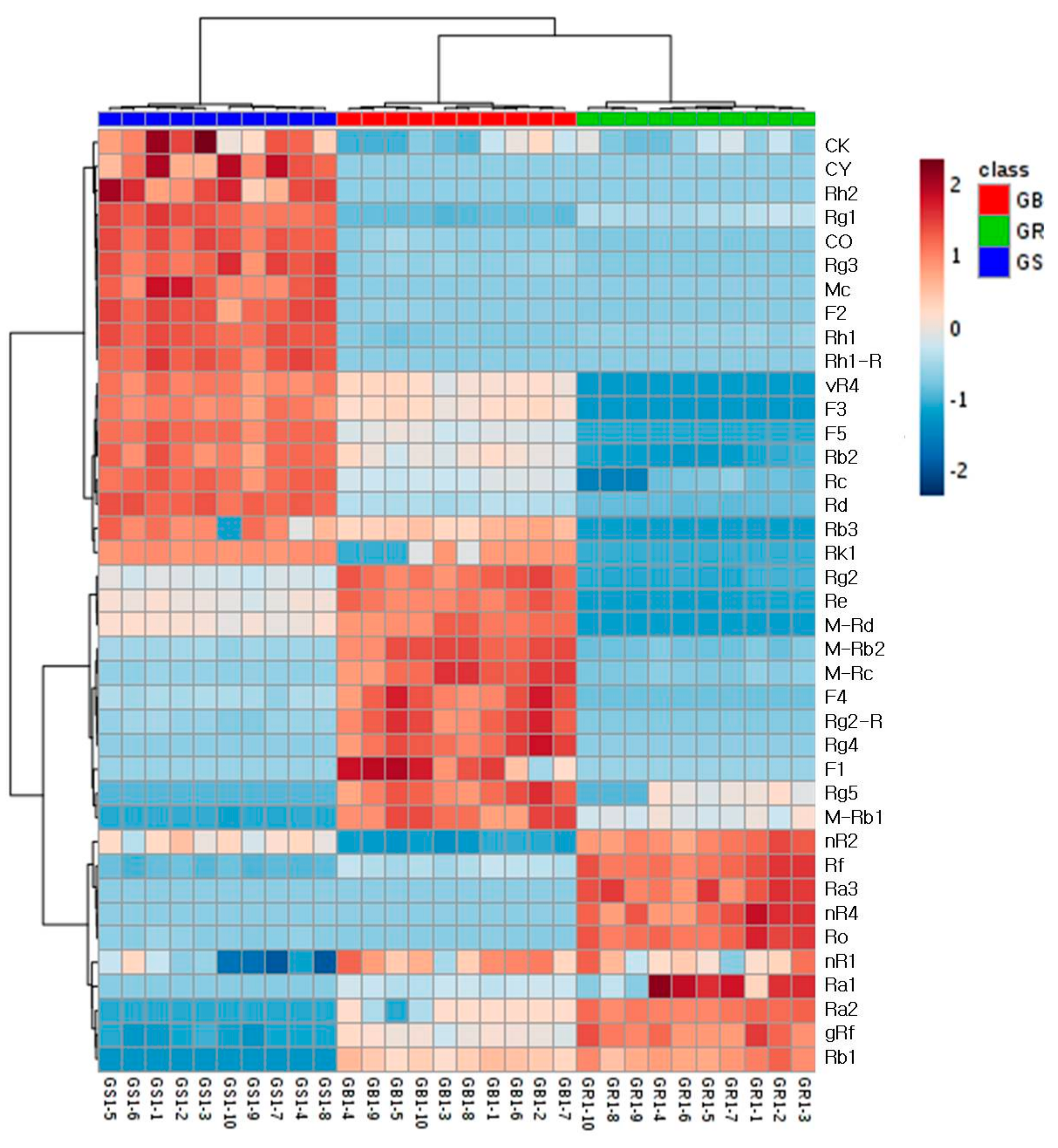
3.2. Profiling of Various Ginsenosides from the Roots, Stems, Leaves, and Berries of Panax ginseng
3.3. Quantification of Ginsenosides from the Roots, Stems and Leaves, and Berries of Panax ginseng
4. Conclusions
Supplementary Materials
Acknowledgments
Author Contributions
Conflicts of Interest
References
- Wang, Y.; Pan, J.Y.; Xiao, X.Y.; Lin, R.C.; Cheng, Y.Y. Simultaneous determination of ginsenosides in Panax ginseng with different growth ages using high-performance liquid chromatography-mass spectrometry. Phytochem. Anal. 2006, 17, 424–430. [Google Scholar] [CrossRef] [PubMed]
- Park, J.D.; Rhee, D.K.; Lee, Y.H. Biological activities and chemistry of saponins from Panax ginseng CA Meyer. Phytochem. Rev. 2005, 4, 159–175. [Google Scholar] [CrossRef]
- Wang, A.; Wang, C.Z.; Wu, J.A.; Osinski, J.; Yuan, C.S. Determination of major ginsenosides in Panax quinquefolius (American ginseng) using high-performance liquid chromatography. Phytochem. Anal. 2005, 16, 272–277. [Google Scholar] [CrossRef] [PubMed]
- Xie, J.-T.; Mehendale, S.R.; Li, X.; Quigg, R.; Wang, X.; Wang, C.-Z.; Wu, J.A.; Aung, H.H.; Rue, P.A.; Bell, G.I. Anti-diabetic effect of ginsenoside Re in ob/ob mice. Biochim. Biophys. Acta (BBA)-Mol. Basis Dis. 2005, 1740, 319–325. [Google Scholar] [CrossRef] [PubMed]
- Kenarova, B.; Neychev, H.; Hadjiivanova, C.; Petkov, V.D. Immunomodulating activity of ginsenoside Rg1 from Panax ginseng. Jpn. J. Pharmacol. 1990, 54, 447–454. [Google Scholar] [CrossRef] [PubMed]
- Yue, P.Y.K.; Mak, N.K.; Cheng, Y.K.; Leung, K.W.; Ng, T.B.; Fan, D.T.P.; Yeung, H.W.; Wong, R.N.S. Pharmacogenomics and the Yin/Yang actions of ginseng: Anti-tumor, angiomodulating and steroid-like activities of ginsenosides. Chin. Med. 2007, 2, 6. [Google Scholar] [CrossRef] [PubMed]
- Tang, W.; Zhang, Y.; Gao, J.; Ding, X.; Gao, S. The anti-fatigue effect of 20 (R)-ginsenoside Rg3 in mice by intranasally administration. Biol. Pharm. Bull. 2008, 31, 2024–2027. [Google Scholar] [CrossRef] [PubMed]
- Lee, S.Y.; Kim, Y.K.; Park, N.I.; Kim, C.S.; Lee, C.Y.; Park, S.U. Chemical constituents and biological activities of the berry of Panax ginseng. J. Med. Plants Res. 2010, 4, 349–353. [Google Scholar]
- Qiu, S.; Yang, W.-Z.; Yao, C.-L.; Qiu, Z.-D.; Shi, X.-J.; Zhang, J.-X.; Hou, J.-J.; Wang, Q.-R.; Wu, W.-Y.; Guo, D.-A. Nontargeted metabolomic analysis and “commercial-homophyletic” comparison-induced biomarkers verification for the systematic chemical differentiation of five different parts of Panax ginseng. J. Chromatogr. A 2016, 1453, 78–87. [Google Scholar] [CrossRef] [PubMed]
- Li, X.-G.; Yan, Y.Z.; Jin, X.-J.; Kim, Y.K.; Uddin, M.R.; Kim, Y.B.; Bae, H.; Kim, Y.C.; Lee, S.W.; Park, S.U. Ginsenoside content in the leaves and roots of Panax ginseng at different ages. Life Sci. J. 2012, 9, 670–683. [Google Scholar]
- Zhao, J.-M.; Li, N.; Zhang, H.; Wu, C.-F.; Piao, H.-R.; Zhao, Y.-Q. Novel dammarane-type sapogenins from Panax ginseng berry and their biological activities. Bioorg. Med. Chem. Lett. 2011, 21, 1027–1031. [Google Scholar] [CrossRef] [PubMed]
- Sritularak, B.; Morinaga, O.; Yuan, C.-S.; Shoyama, Y.; Tanaka, H. Quantitative analysis of ginsenosides Rb1, Rg1, and Re in American ginseng berry and flower samples by ELISA using monoclonal antibodies. J. Nat. Med. 2009, 63, 360–363. [Google Scholar] [CrossRef] [PubMed]
- Shi, W.; Wang, Y.; Li, J.; Zhang, H.; Ding, L. Investigation of ginsenosides in different parts and ages of Panax ginseng. Food Chem. 2007, 102, 664–668. [Google Scholar] [CrossRef]
- Wang, C.-Z.; Wu, J.A.; McEntee, E.; Yuan, C.-S. Saponins composition in American ginseng leaf and berry assayed by high-performance liquid chromatography. J. Agric. Food Chem. 2006, 54, 2261–2266. [Google Scholar] [CrossRef] [PubMed]
- Li, X.-L.; Wang, C.-Z.; Sun, S.; Mehendale, S.R.; Du, W.; He, T.-C.; Yuan, C.-S. American ginseng berry enhances chemopreventive effect of 5-FU on human colorectal cancer cells. Oncol. Rep. 2009, 22, 943–952. [Google Scholar] [PubMed]
- Lim, W.; Mudge, K.W.; Vermeylen, F. Effects of population, age, and cultivation methods on ginsenoside content of wild American ginseng (Panax quinquefolium). J. Agric. Food Chem. 2005, 53, 8498–8505. [Google Scholar] [CrossRef] [PubMed]
- Kim, S.N.; Ha, Y.W.; Shin, H.; Son, S.H.; Wu, S.J.; Kim, Y.S. Simultaneous quantification of 14 ginsenosides in Panax ginseng CA Meyer (Korean red ginseng) by HPLC-ELSD and its application to quality control. J. Pharm. Biomed. Anal. 2007, 45, 164–170. [Google Scholar] [CrossRef] [PubMed]
- Sun, B.-S.; Gu, L.-J.; Fang, Z.-M.; Wang, C.-Y.; Wang, Z.; Lee, M.-R.; Li, Z.; Li, J.-J.; Sung, C.-K. Simultaneous quantification of 19 ginsenosides in black ginseng developed from Panax ginseng by HPLC–ELSD. J. Pharm. Biomed. Anal. 2009, 50, 15–22. [Google Scholar] [CrossRef] [PubMed]
- Joo, K.-M.; Lee, J.-H.; Jeon, H.-Y.; Park, C.-W.; Hong, D.-K.; Jeong, H.-J.; Lee, S.J.; Lee, S.-Y.; Lim, K.-M. Pharmacokinetic study of ginsenoside Re with pure ginsenoside Re and ginseng berry extracts in mouse using ultra performance liquid chromatography/mass spectrometric method. J. Pharm. Biomed. Anal. 2010, 51, 278–283. [Google Scholar] [CrossRef] [PubMed]
- Xie, G.X.; Ni, Y.; Su, M.M.; Zhang, Y.Y.; Zhao, A.H.; Gao, X.F.; Liu, Z.; Xiao, P.G.; Jia, W. Application of ultra-performance LC-TOF MS metabolite profiling techniques to the analysis of medicinal Panax herbs. Metabolomics 2008, 4, 248–260. [Google Scholar] [CrossRef]
- Ji, Q.C.; Harkey, M.R.; Henderson, G.L.; Eric Gershwin, M.; Stern, J.S.; Hackman, R.M. Quantitative determination of ginsenosides by high-performance liquid chromatography-tandem mass spectrometry. Phytochem. Anal. 2001, 12, 320–326. [Google Scholar] [CrossRef] [PubMed]
- Wan, J.-Y.; Liu, P.; Wang, H.-Y.; Qi, L.-W.; Wang, C.-Z.; Li, P.; Yuan, C.-S. Biotransformation and metabolic profile of American ginseng saponins with human intestinal microflora by liquid chromatography quadrupole time-of-flight mass spectrometry. J. Chromatogr. A 2013, 1286, 83–92. [Google Scholar] [CrossRef] [PubMed]
- Mao, Q.; Yang, J.; Cui, X.-M.; Li, J.-J.; Qi, Y.-T.; Zhang, P.-H.; Wang, Q. Target separation of a new anti-tumor saponin and metabolic profiling of leaves of Panax notoginseng by liquid chromatography with eletrospray ionization quadrupole time-of-flight mass spectrometry. J. Pharm. Biomed. Anal. 2012, 59, 67–77. [Google Scholar] [CrossRef] [PubMed]
- Zhang, H.-M.; Li, S.-L.; Zhang, H.; Wang, Y.; Zhao, Z.-L.; Chen, S.-L.; Xu, H.-X. Holistic quality evaluation of commercial white and red ginseng using a UPLC-QTOF-MS/MS-based metabolomics approach. J. Pharm. Biomed. Anal. 2012, 62, 258–273. [Google Scholar] [CrossRef] [PubMed]
- Lee, J.W.; Ji, S.-H.; Lee, Y.-S.; Choi, D.J.; Choi, B.-R.; Kim, G.-S.; Baek, N.-I.; Lee, D.Y. Mass Spectrometry Based Profiling and Imaging of Various Ginsenosides from Panax ginseng Roots at Different Ages. Int. J. Mol. Sci. 2017, 18, 1114. [Google Scholar] [CrossRef] [PubMed]
- Bai, H.; Wang, S.; Liu, J.; Gao, D.; Jiang, Y.; Liu, H.; Cai, Z. Localization of ginsenosides in Panax ginseng with different age by matrix-assisted laser-desorption/ionization time-of-flight mass spectrometry imaging. J. Chromatogr. B 2016, 1026, 263–271. [Google Scholar] [CrossRef] [PubMed]
- Woo, S.-S.; Song, J.-S.; Lee, J.-Y.; In, D.S.; Chung, H.-J.; Liu, J.R.; Choi, D.-W. Selection of high ginsenoside producing ginseng hairy root lines using targeted metabolic analysis. Phytochemistry 2004, 65, 2751–2761. [Google Scholar] [CrossRef] [PubMed]
- Yang, W.; Zhang, J.; Yao, C.; Qiu, S.; Chen, M.; Pan, H.; Shi, X.; Wu, W.; Guo, D. Method development and application of offline two-dimensional liquid chromatography/quadrupole time-of-flight mass spectrometry-fast data directed analysis for comprehensive characterization of the saponins from Xueshuantong Injection. J. Pharm. Biomed. Anal. 2016, 128, 322–332. [Google Scholar] [CrossRef] [PubMed]
- Qi, L.-W.; Wang, H.-Y.; Zhang, H.; Wang, C.-Z.; Li, P.; Yuan, C.-S. Diagnostic ion filtering to characterize ginseng saponins by rapid liquid chromatography with time-of-flight mass spectrometry. J. Chromatogr. A 2012, 1230, 93–99. [Google Scholar] [CrossRef] [PubMed]
- Qiu, S.; Yang, W.-Z.; Shi, X.-J.; Yao, C.-L.; Yang, M.; Liu, X.; Jiang, B.-H.; Wu, W.-Y.; Guo, D.-A. A green protocol for efficient discovery of novel natural compounds: Characterization of new ginsenosides from the stems and leaves of Panax ginseng as a case study. Anal. Chim. Acta 2015, 893, 65–76. [Google Scholar] [CrossRef] [PubMed]
- Xu, X.-F.; Cheng, X.-L.; Lin, Q.-H.; Li, S.-S.; Jia, Z.; Han, T.; Lin, R.-C.; Wang, D.; Wei, F.; Li, X.-R. Identification of mountain-cultivated ginseng and cultivated ginseng using UPLC/oa-TOF MSE with a multivariate statistical sample-profiling strategy. J. Ginseng Res. 2016, 40, 344–350. [Google Scholar] [CrossRef] [PubMed]
- Sun, M.; Che, Y.; Liu, Z. A tractable method for the preparation of the ginsenoside compounds O and Mc1. Anal. Methods 2015, 7, 4757–4762. [Google Scholar] [CrossRef]
- Wu, Q.; Jang, M.; Piao, X.-L. Determination by UPLC-MS of four dammarane-type saponins from heat-processed Gynostemma pentaphyllum. Biosci. Biotechnol. Biochem. 2014, 78, 311–316. [Google Scholar] [CrossRef] [PubMed]
- Wang, R.-F.; Li, J.; Hu, H.-J.; Li, J.; Yang, Y.-B.; Yang, L.; Wang, Z.-T. Chemical transformation and target preparation of saponins in stems and leaves of Panax notoginseng. J. Ginseng Res. 2016. [Google Scholar] [CrossRef]
- Lee, G.-W.; Kim, K.-R.; Oh, D.-K. Production of rare ginsenosides (compound Mc, compound Y and aglycon protopanaxadiol) by β-glucosidase from Dictyoglomus turgidum that hydrolyzes β-linked, but not α-linked, sugars in ginsenosides. Biotechnol. Lett. 2012, 34, 1679–1686. [Google Scholar] [CrossRef] [PubMed]
- Qu, C.; Bai, Y.; Jin, X.; Wang, Y.; Zhang, K.; You, J.; Zhang, H. Study on ginsenosides in different parts and ages of Panax quinquefolius L. Food Chem. 2009, 115, 340–346. [Google Scholar] [CrossRef]
- Ko, S.K.; Bae, H.M.; Cho, O.S.; Im, B.O.; Chung, S.H.; Lee, B.Y. Analysis of ginsenoside composition of ginseng berry and seed. Food Sci. Biotechnol. 2008, 17, 1379–1382. [Google Scholar]
- Mai, T.T.; Moon, J.; Song, Y.; Viet, P.Q.; Van Phuc, P.; Lee, J.M.; Yi, T.-H.; Cho, M.; Cho, S.K. Ginsenoside F2 induces apoptosis accompanied by protective autophagy in breast cancer stem cells. Cancer Lett. 2012, 321, 144–153. [Google Scholar] [CrossRef] [PubMed]
- Xie, J.-T.; Wang, C.-Z.; Zhang, B.; Mehendale, S.R.; Li, X.-L.; Sun, S.; Han, A.H.; Du, W.; He, T.-C.; Yuan, C.-S. In vitro and in vivo anticancer effects of American ginseng berry: Exploring representative compounds. Biol. Pharm. Bull. 2009, 32, 1552–1558. [Google Scholar] [CrossRef]
- Lee, Y.; Jin, Y.; Lim, W.; Ji, S.; Choi, S.; Jang, S.; Lee, S. A ginsenoside-Rh1, a component of ginseng saponin, activates estrogen receptor in human breast carcinoma MCF-7 cells. J. Steroid Biochem. Mol. Biol. 2003, 84, 463–468. [Google Scholar] [CrossRef]
- Wang, Y.; Choi, K.-D.; Yu, H.; Jin, F.; Im, W.-T. Production of ginsenoside F1 using commercial enzyme Cellulase KN. J. Ginseng Res. 2016, 40, 121–126. [Google Scholar] [CrossRef] [PubMed]
- Attele, A.S.; Zhou, Y.-P.; Xie, J.-T.; Wu, J.A.; Zhang, L.; Dey, L.; Pugh, W.; Rue, P.A.; Polonsky, K.S.; Yuan, C.-S. Antidiabetic effects of Panax ginseng berry extract and the identification of an effective component. Diabetes 2002, 51, 1851–1858. [Google Scholar] [CrossRef] [PubMed]
- Kim, M.K.; Kang, H.; Baek, C.W.; Jung, Y.H.; Woo, Y.C.; Choi, G.J.; Shin, H.Y.; Kim, K.S. Antinociceptive and anti-inflammatory effects of ginsenoside Rf in a rat model of incisional pain. J. Ginseng Res. 2017. [Google Scholar] [CrossRef]
- Kim, C.-K.; Cho, D.H.; Lee, K.-S.; Lee, D.-K.; Park, C.-W.; Kim, W.G.; Lee, S.J.; Ha, K.-S.; Goo Taeg, O.; Kwon, Y.-G. Ginseng berry extract prevents atherogenesis via anti-inflammatory action by upregulating phase II gene expression. Evid.-Based Complement. Altern. Med. 2012. [Google Scholar] [CrossRef] [PubMed]
- Kim, D.-H. Chemical diversity of Panax ginseng, Panax quinquifolium, and Panax notoginseng. J. Ginseng Res. 2012, 36, 1–15. [Google Scholar] [CrossRef] [PubMed]
- Shan, S.-M.; Luo, J.-G.; Huang, F.; Kong, L.-Y. Chemical characteristics combined with bioactivity for comprehensive evaluation of Panax ginseng CA Meyer in different ages and seasons based on HPLC-DAD and chemometric methods. J. Pharm. Biomed. Anal. 2014, 89, 76–82. [Google Scholar] [CrossRef] [PubMed]
Sample Availability: Samples of the compounds are not available from the authors. |


| No. | RT (min) | Ginsenosides | Molecular Formula | Expected Neutral Mass (Da) | Observed Neutral Mass (Da) | QTOF/MS (ESI-) (m/z) | Mass Accuracy (ppm) | Adducts |
|---|---|---|---|---|---|---|---|---|
| 1 | 3.58 | 20-O-Glucoginsenoside Rf (gRf) | C48H82O19 | 962.545 | 962.5453 | 1007.5435 | 0.26 | +HCOO |
| 2 | 4.07 | Notoginsenoside R1 (nR1) | C47H80O18 | 932.5345 | 932.5343 | 977.5325 | 0.2 | +HCOO |
| 3 | 5.36 | Ginsenoisde Rg1 (Rg1) | C42H72O14 | 800.4922 | 800.4908 | 845.489 | 1.62 | +HCOO |
| 4 | 5.66 | Ginsenoside Re (Re) | C48H82O18 | 946.5501 | 946.5503 | 991.5485 | 0.16 | +HCOO |
| 5 | 8.05 | Floralginsenoside Ka (fKa) | C36H62O11 | 670.4292 | 670.4295 | 715.4277 | 0.34 | +HCOO |
| 6 | 8.46 | Ginsenoside Rh6 (Rh6) | C36H62O11 | 670.4292 | 670.4291 | 715.4273 | 0.19 | +HCOO |
| 7 | 10.72 | Ginsenoside Rh23 (Rh23) | C37H64O10 | 668.4499 | 668.4507 | 713.4489 | 1.06 | +HCOO |
| 8 | 11.2 | Vinaginsenoside R4 (vR4) | C48H82O19 | 962.545 | 962.5452 | 1007.5434 | 0.19 | +HCOO |
| 9 | 11.94 | Pseudo-ginsenoside F11 (pF11) | C42H72O14 | 800.4922 | 800.4932 | 845.4914 | 1.22 | +HCOO |
| 10 | 12.11 | Ginsenoside Rf (Rf) | C42H72O14 | 800.4922 | 800.4922 | 845.4904 | 0.01 | +HCOO |
| 11 | 12.81 | Notoginsenoside R2 (nR2) | C41H70O13 | 770.4816 | 770.4823 | 815.4805 | 0.83 | +HCOO |
| 12 | 13.15 | Notoginsenoside R4 (nR4) | C59H100O27 | 1240.6452 | 1240.6455 | 1285.6437 | 0.2 | +HCOO |
| 13 | 13.43 | Ginsenoside F5 (F5) | C41H70O13 | 770.4816 | 770.4819 | 815.4801 | 0.33 | +HCOO |
| 14 | 13.56 | Ginsenoside Rh1 (Rh1) | C36H62O9 | 638.4394 | 638.4392 | 683.4374 | 0.31 | +HCOO |
| 15 | 13.58 | 20(R)-Notoginsenoside R2 (nR2-R) | C41H70O13 | 770.4816 | 770.4815 | 815.4797 | 0.18 | +HCOO |
| 16 | 13.7 | Ginsenoside Rg2 (Rg2) | C42H72O13 | 784.4973 | 784.4984 | 829.4966 | 1.38 | +HCOO |
| 17 | 13.9 | Ginsenoside F3 (F3) | C41H70O13 | 770.4816 | 770.4811 | 815.4793 | 0.63 | +HCOO |
| 18 | 14.03 | 20(R)-Ginsenoside Rg2 (Rg2-R) | C42H72O13 | 784.4973 | 784.496 | 829.4942 | 1.58 | +HCOO |
| 19 | 14.03 | 20(R)-Ginsenoside Rh1 (Rh1-R) | C36H62O9 | 638.4394 | 638.4397 | 683.4379 | 0.47 | +HCOO |
| 20 | 14.35 | Ginsenoside Ra2 (Ra2) | C58H98O26 | 1210.6346 | 1210.6346 | 1255.6328 | 0.01 | +HCOO |
| 21 | 14.51 | Ginsenoside Ra3 (Ra3) | C59H100O27 | 1240.6452 | 1240.6447 | 1285.6429 | 0.36 | +HCOO |
| 22 | 14.53 | Ginsenoside Rb1 (Rb1) | C54H92O23 | 1108.6029 | 1108.6006 | 1153.5988 | 2.02 | +HCOO |
| 23 | 14.58 | Gypenoside XLIX (XLIX) | C52H86O21 | 1046.5662 | 1046.5658 | 1091.564 | 0.31 | +HCOO |
| 24 | 15.04 | Malonyl ginsenoside Rb1 (M-Rb1) | C57H94O26 | 1194.6033 | 1194.6057 | 1193.5984 | 2 | –H |
| 25 | 15.25 | Ginsenoside Rc (Rc) | C53H90O22 | 1078.5924 | 1078.5925 | 1123.5907 | 0.1 | +HCOO |
| 26 | 15.47 | Ginsenoside Ra1 (Ra1) | C58H98O26 | 1210.6346 | 1210.633 | 1255.6312 | 1.27 | +HCOO |
| 27 | 15.69 | Ginsenoside Ro (Ro) | C48H76O19 | 956.4981 | 956.4978 | 955.4905 | 0.28 | –H |
| 28 | 15.8 | Ginsenoside F1 (F1) | C36H62O9 | 638.4394 | 638.4395 | 683.4377 | 0.11 | +HCOO |
| 29 | 15.9 | Malonyl ginsenoside Rc (M-Rc) | C56H92O25 | 1164.5928 | 1164.5947 | 1163.5874 | 1.6 | –H |
| 30 | 16.25 | Ginsenoside Rb2 (Rb2) | C53H90O22 | 1078.5924 | 1078.5913 | 1123.5895 | 0.99 | +HCOO |
| 31 | 16.6 | Ginsenoside Rb3 (Rb3) | C53H90O22 | 1078.5924 | 1078.5915 | 1123.5897 | 0.81 | +HCOO |
| 32 | 16.9 | Malonyl ginsenoside Rb2 (M-Rb2) | C56H92O25 | 1164.5928 | 1164.5923 | 1163.585 | 0.4 | –H |
| 33 | 18.28 | Gypenoside A (GyA) | C46H74O17 | 898.4926 | 898.4915 | 943.4897 | 1.21 | +HCOO |
| 34 | 18.29 | Ginsenoside Rd (Rd) | C48H82O18 | 946.5501 | 946.5495 | 991.5477 | 0.59 | +HCOO |
| 35 | 18.97 | Malonyl ginsenoside Rd (M-Rd) | C51H84O21 | 1032.5505 | 1032.5498 | 1031.5425 | 0.7 | –H |
| 36 | 20.4 | Gypenoside XVII (GyXVII) | C48H82O18 | 946.5501 | 946.5497 | 991.5479 | 0.47 | +HCOO |
| 37 | 21.96 | Notoginsenoisde Fe (nFe) | C47H80O17 | 916.5396 | 916.5393 | 961.5375 | 0.22 | +HCOO |
| 38 | 23.29 | Compound O (CO) | C47H80O17 | 916.5396 | 916.5396 | 961.5378 | 0.1 | +HCOO |
| 39 | 23.46 | Ginsenoside Rg4 (Rg4) | C42H70O12 | 766.4867 | 766.4875 | 811.4857 | 0.91 | +HCOO |
| 40 | 23.84 | Ginsenoside Rk3 (Rk3) | C36H60O8 | 620.4288 | 620.4292 | 665.4274 | 0.51 | +HCOO |
| 41 | 24.28 | Ginsenoside F4 (F4) | C42H70O12 | 766.4867 | 766.4865 | 811.4847 | 0.27 | +HCOO |
| 42 | 24.68 | Ginsenoside Rh4 (Rh4) | C36H60O8 | 620.4288 | 620.4275 | 665.4257 | 2.01 | +HCOO |
| 43 | 25.15 | Gypenoside L (GyL) | C42H72O14 | 800.4922 | 800.4905 | 845.4887 | 2.1 | +HCOO |
| 44 | 25.19 | Ginsenoside F2 (F2) | C42H72O13 | 784.4973 | 784.4968 | 829.495 | 0.6 | +HCOO |
| 45 | 25.34 | Gypenoside LI (GyLI) | C42H72O14 | 800.4922 | 800.4909 | 845.4891 | 1.6 | +HCOO |
| 46 | 25.57 | Notoginsenoside Ft1 (nFt1) | C47H80O17 | 916.5396 | 916.5417 | 961.5399 | 2.26 | +HCOO |
| 47 | 25.78 | Protopanaxatiol (PPT) | C30H52O4 | 476.3866 | 476.3862 | 521.3844 | 0.71 | +HCOO |
| 48 | 26.01 | 20(S)-Ginsenoside Rg3 (Rg3) | C42H72O13 | 784.4973 | 784.498 | 829.4962 | 0.9 | +HCOO |
| 49 | 26.2 | 20(R)-Ginsenoside Rg3 (Rg3-R) | C42H72O13 | 784.4973 | 784.4989 | 829.4971 | 1.97 | +HCOO |
| 50 | 26.37 | Ginsenoisde Mc (Mc) | C41H70O12 | 754.4867 | 754.4867 | 799.4849 | 0 | +HCOO |
| 51 | 26.52 | Compound Y (CY) | C41H70O12 | 754.4867 | 754.4849 | 799.4831 | 2.3 | +HCOO |
| 52 | 27.35 | Compound K (CK) | C36H62O8 | 622.4445 | 622.4433 | 667.4415 | 1.8 | +HCOO |
| 53 | 27.43 | Ginsenoside Rk1 (Rk1) | C42H70O12 | 766.4867 | 766.4861 | 811.4843 | 0.81 | +HCOO |
| 54 | 27.55 | Ginsenoside Rg5 (Rg5) | C42H70O12 | 766.4867 | 766.4873 | 811.4855 | 0.73 | +HCOO |
| 55 | 27.7 | Ginsenoside Rh2 (Rh2) | C36H62O8 | 622.4445 | 622.4441 | 667.4423 | 0.52 | +HCOO |
| 56 | 29.04 | Ginsenoside Rk2 (Rk2) | C36H60O7 | 604.4339 | 604.4341 | 649.4323 | 0.35 | +HCOO |
| 57 | 29.15 | Ginsenoside Rh3 (Rh3) | C36H60O7 | 604.4339 | 604.434 | 649.4322 | 0.17 | +HCOO |
| 58 | 30.04 | Protopanaxadiol (PPD) | C30H52O3 | 460.3916 | 460.3917 | 505.3899 | 0.19 | +HCOO |
| No. | Ginsenosides | Roots | Stems and Leaves | Berries | No. | Ginsenosides | Root | Stems and Leaves | Berry |
|---|---|---|---|---|---|---|---|---|---|
| 1 | gRf | 1.8 ± 0.1 | 0.3 ± 0.1 | 3.6 ± 1.1 | 21 | Ro | 0.15 ± 0.01 | 0.014 ± 0.002 | 0.014 ± 0.002 |
| 2 | nR1 | 0.06 ± 0.01 | 0.03 ± 0.01 | 1.2 ± 0.2 | 22 | M-Rc | 0.9 ± 0.1 | 1.08 ± 0.04 | 4.7 ± 0.5 |
| 3 | Rg1 | 3.1 ± 0.2 | 9.6 ± 0.5 | 8.9 ± 4.3 | 23 | Rb2 | 0.6 ± 0.1 | 2.8 ± 0.1 | 2.3 ± 0.6 |
| 4 | Re | 3.0 ± 0.3 | 17 ± 1 | 26 ± 8 | 24 | Rb3 | not detected | 0.5 ± 0.1 | not detected |
| 5 | vR4 | 0.007 ± 0.001 | 1.2 ± 0.1 | 0.9 ± 0.1 | 25 | M-Rb2 | 0.9 ± 0.1 | 1.6 ± 0.1 | 6.8 ± 0.5 |
| 6 | Rf | 4.6 ± 0.2 | 0.30 ± 0.01 | 3.4 ± 1.1 | 26 | Rd | 0.15 ± 0.01 | 7.1 ± 0.2 | 2.3 ± 0.5 |
| 7 | nR2 | 1.1 ± 0.1 | 0.7 ± 0.1 | 4.5 ± 1.3 | 27 | M-Rd | 0.014 ± 0.001 | 3.1 ± 0.1 | 5.3 ± 0.4 |
| 8 | nR4 | 0.14 ± 0.02 | not detected | not detected | 28 | CO | 0.06 ± 0.01 | 12.6 ± 0.1 | 0.4 ± 0.1 |
| 9 | F5 | not detected | 5.8 ± 0.2 | 2.8 ± 0.3 | 29 | Rg4 | not detected | not detected | 0.06 ± 0.03 |
| 10 | Rh1 | 0.021 ± 0.002 | 0.24 ± 0.01 | 0.04 ± 0.02 | 30 | F4 | not detected | 0.4 ± 0.1 | 2.1 ± 0.8 |
| 11 | Rg2 | 0.17 ± 0.01 | 0.61 ± 0.03 | 0.6 ± 0.5 | 31 | F2 | 0.0019 ± 0.0006 | 2.7 ± 0.3 | 0.03 ± 0.01 |
| 12 | F3 | not detected | 5.3 ± 0.2 | 4.1 ± 0.4 | 32 | Rg3 | 0.0004 ± 0.0002 | 0.09 ± 0.01 | 0.007 ± 0.001 |
| 13 | Rg2-R | not detected | 0.008 ± 0.001 | 0.06 ± 0.03 | 33 | Mc | not detected | 0.24 ± 0.04 | not detected |
| 14 | F1 | not detected | not detected | 0.005 ± 0.001 | 34 | CY | not detected | 0.08 ± 0.02 | not detected |
| 15 | Ra2 | 3.2 ± 0.1 | not detected | not detected | 35 | CK | 0.0010±0.0004 | 0.003±0.001 | 0.002 ± 0.001 |
| 16 | Ra3 | 0.9 ± 0.1 | not detected | not detected | 36 | Rk1 | not detected | 1.18±0.02 | 1.153 ± 0.004 |
| 17 | Rb1 | 3.8 ± 0.3 | 0.08 ± 0.01 | 2.8 ± 0.6 | 37 | Rg5 | 0.031 ± 0.004 | not detected | 0.08 ± 0.01 |
| 18 | M-Rb1 | 4.7 ± 0.5 | 0.7 ± 0.1 | 9.9 ± 1.2 | 38 | Rh2 | not detected | 0.011 ± 0.002 | not detected |
| 19 | Rc | 0.9 ± 0.1 | 3.5 ± 0.1 | not detected | 39 | Rh1-R | not detected | 0.0028 ± 0.0004 | not detected |
| 20 | Ra1 | 0.6 ± 0.2 | not detected | not detected |
© 2017 by the authors. Licensee MDPI, Basel, Switzerland. This article is an open access article distributed under the terms and conditions of the Creative Commons Attribution (CC BY) license (http://creativecommons.org/licenses/by/4.0/).
Share and Cite
Lee, J.W.; Choi, B.-R.; Kim, Y.-C.; Choi, D.J.; Lee, Y.-S.; Kim, G.-S.; Baek, N.-I.; Kim, S.-Y.; Lee, D.Y. Comprehensive Profiling and Quantification of Ginsenosides in the Root, Stem, Leaf, and Berry of Panax ginseng by UPLC-QTOF/MS. Molecules 2017, 22, 2147. https://0-doi-org.brum.beds.ac.uk/10.3390/molecules22122147
Lee JW, Choi B-R, Kim Y-C, Choi DJ, Lee Y-S, Kim G-S, Baek N-I, Kim S-Y, Lee DY. Comprehensive Profiling and Quantification of Ginsenosides in the Root, Stem, Leaf, and Berry of Panax ginseng by UPLC-QTOF/MS. Molecules. 2017; 22(12):2147. https://0-doi-org.brum.beds.ac.uk/10.3390/molecules22122147
Chicago/Turabian StyleLee, Jae Won, Bo-Ram Choi, Young-Chang Kim, Doo Jin Choi, Young-Seob Lee, Geum-Soog Kim, Nam-In Baek, Seung-Yu Kim, and Dae Young Lee. 2017. "Comprehensive Profiling and Quantification of Ginsenosides in the Root, Stem, Leaf, and Berry of Panax ginseng by UPLC-QTOF/MS" Molecules 22, no. 12: 2147. https://0-doi-org.brum.beds.ac.uk/10.3390/molecules22122147