Article

Molecular Quantum Similarity, Chemical Reactivity and Database Screening of 3D Pharmacophores of the Protein Kinases A, B and G from Mycobacterium tuberculosis

by
Alejandro Morales-Bayuelo
Fondo Nacional de Desarrollo Científico y Tecnológico (FONDECYT), Proyecto Postdoctoral No. 3150035, Talca, 3660300, Chile
Submission received: 26 May 2017 / Revised: 13 June 2017 / Accepted: 16 June 2017 / Published: 21 June 2017
(This article belongs to the Section Molecular Diversity)

Abstract

:
Mycobacterium tuberculosis remains one of the world’s most devastating pathogens. For this reason, we developed a study involving 3D pharmacophore searching, selectivity analysis and database screening for a series of anti-tuberculosis compounds, associated with the protein kinases A, B, and G. This theoretical study is expected to shed some light onto some molecular aspects that could contribute to the knowledge of the molecular mechanics behind interactions of these compounds, with anti-tuberculosis activity. Using the Molecular Quantum Similarity field and reactivity descriptors supported in the Density Functional Theory, it was possible to measure the quantification of the steric and electrostatic effects through the Overlap and Coulomb quantitative convergence (alpha and beta) scales. In addition, an analysis of reactivity indices using global and local descriptors was developed, identifying the binding sites and selectivity on these anti-tuberculosis compounds in the active sites. Finally, the reported pharmacophores to PKn A, B and G, were used to carry out database screening, using a database with anti-tuberculosis drugs from the Kelly Chibale research group (http://www.kellychibaleresearch.uct.ac.za/), to find the compounds with affinity for the specific protein targets associated with PKn A, B and G. In this regard, this hybrid methodology (Molecular Mechanic/Quantum Chemistry) shows new insights into drug design that may be useful in the tuberculosis treatment today.

Graphical Abstract

1. Introduction

One of the United Nations’ main focuses is to eradicate communicable diseases such as tuberculosis, which affect millions of people worldwide and causing more critical problems in countries with low- and middle-income. Mycobacterium tuberculosis, the causative agent of tuberculosis, is one of the most lethal human pathogens, further characterized by being strongly resistant to current treatments. Despite more than 100 years of research performed to date, the disease still kills about two million people every year around the world. According to the World Health Organization (WHO), a third of the world’s population carries the infection in an inactive form known as latency [1]. Our current inability to control the spread can be explained by the lack of an effective vaccine, multidrug-resistance [2,3,4] and the great adaptability of Mycobacterium tuberculosis, which has great capacity for mutation, in different environments, it [5,6,7,8,9,10].
In order to find new drug targets for tuberculosis treatment, in this study we analyzed the Protein Kinases (PKs) involved in tuberculosis. PKs, which are enzymes that catalyze the protein phosphorylation process, play an important role in controlling the proliferation and differentiation of eukaryotic cells in living organisms. One reason to investigate protein phosphorylation is that this rationalization represents an attractive drug target for a variety of diseases such as cancer [11], Alzheimer’s [12], chronic inflammations [13], etc. PKs present in the human body have been widely studied due to their use in therapeutic targets. However, not much is known about the PKs involved in tuberculosis. Therefore the binding sites associated to PKs A, B, and G of Mycobacterium tuberculosis are studied with a particular set of inhibitors to each PK.
The inhibitors used are a series of compounds of Pkn A reported by Sipos et al. [14], of Pkn B reported by Székely et al. [15], Loughheed et al. [16], Chapman et al. [17] and Naqvi et al. [18], finally of PKn G reported by Sipos et al. [14]. These ligands were used with the aim of obtaining new information about their stabilization in the active site.
The process of drug discovery is very complex and requires an interdisciplinary effort to design effective and commercially feasible drugs. In addition, the objective of drug design is to find a drug that can interact with a specific drug target and modify its activity. For this reason, we used a hybrid methodology to search new insights for tuberculosis treatment involving the application of Molecular Mechanics (MM) to protein treatment and consequently identifying the more active poses of the ligands involved in the anti-tuberculosis activity using computational techniques such as 3D pharmacophore searching and docking molecular [19,20,21] to each PK.
With the goal of studying the selectivity of these inhibitors in the active site, we used considerations of Quantum Chemistry (QC), specifically the Molecular Quantum Similarity (MQS) field [22,23,24,25] and chemical reactivity descriptors within the Density Functional Theory (DFT) framework [26]. In previous works, the present author has reported his approaches to relate Molecular Mechanics with Quantum Chemistry (MM/QM) [27]. Hopefully, this hybrid approach (MM/QM) provides new considerations about the interactions and selectivity of these ligands in the active sites of the PKs. Taking into account that selectivity is a very important aspect that is today widely studied in drug development with selective targets in diseases which are difficult to control like tuberculosis.
The final aspect of our work is to carry out a database screening using the 3D pharmacophores of PKn A, B and G reported on a database with anti-tuberculosis drugs, to find the compounds with affinity for the specific protein target associated with PKn A, B or G. To accomplish this we created a database using 183 anti-tuberculosis compounds reported by the Chibale group [28,29,30,31,32]. The compounds reported by Chibale are racemic mixtures. Taking this into account, the chiral isomers were characterized from the computational viewpoint to find the specific isomers interacting with each characterized pharmacophore.

2. Results

The outcomes in this work are distributed as follows: (i) 3D pharmacophore searching for the protein kinases A, B and G, (ii) analysis of the 3D pharmacophores using molecular quantum similarity and chemical reactivity descriptors (selectivity analysis), and (iii) 3D pharmacophore-based database screening.

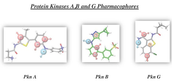
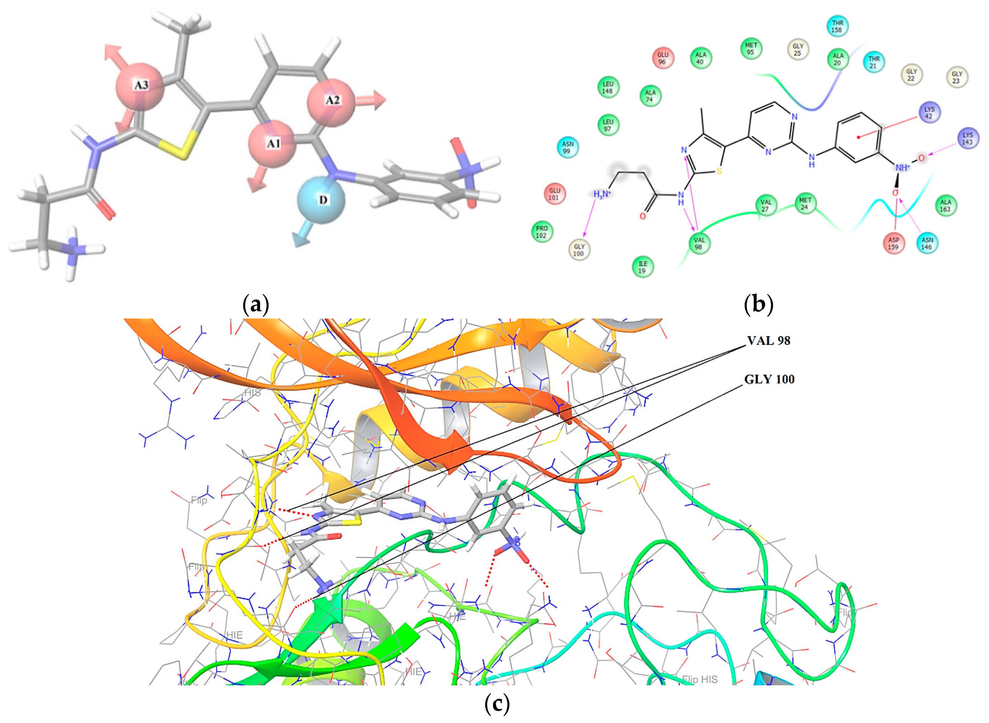
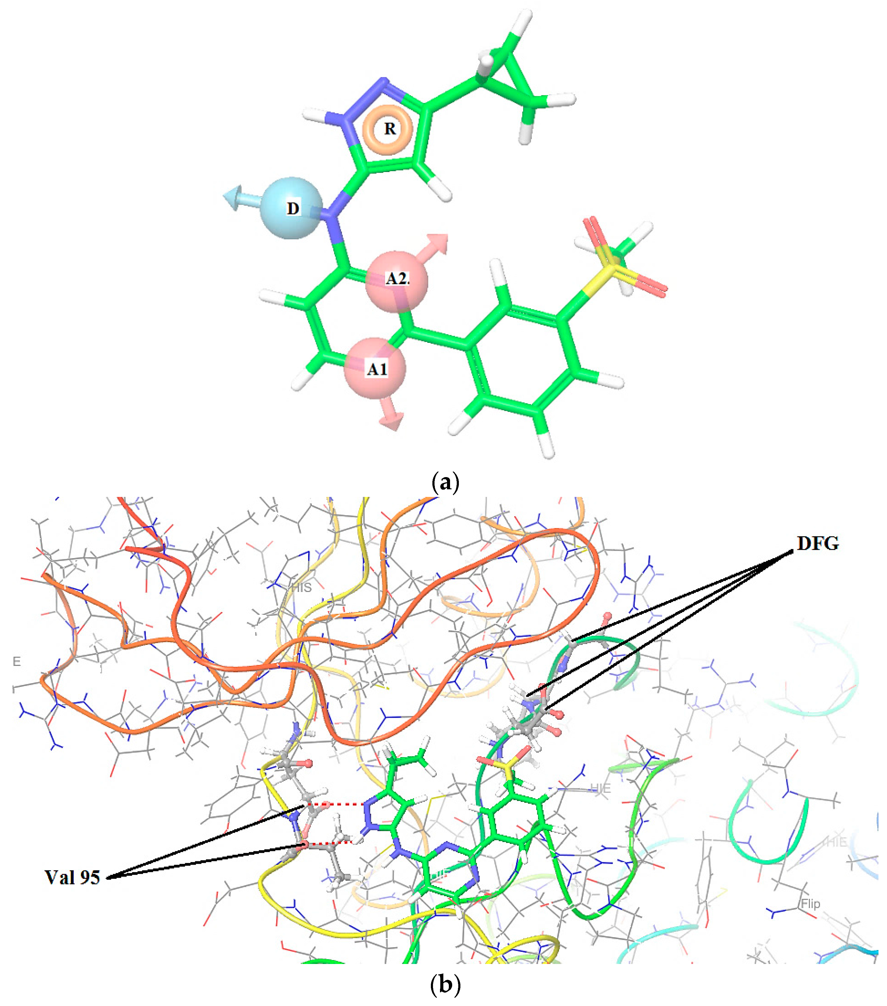
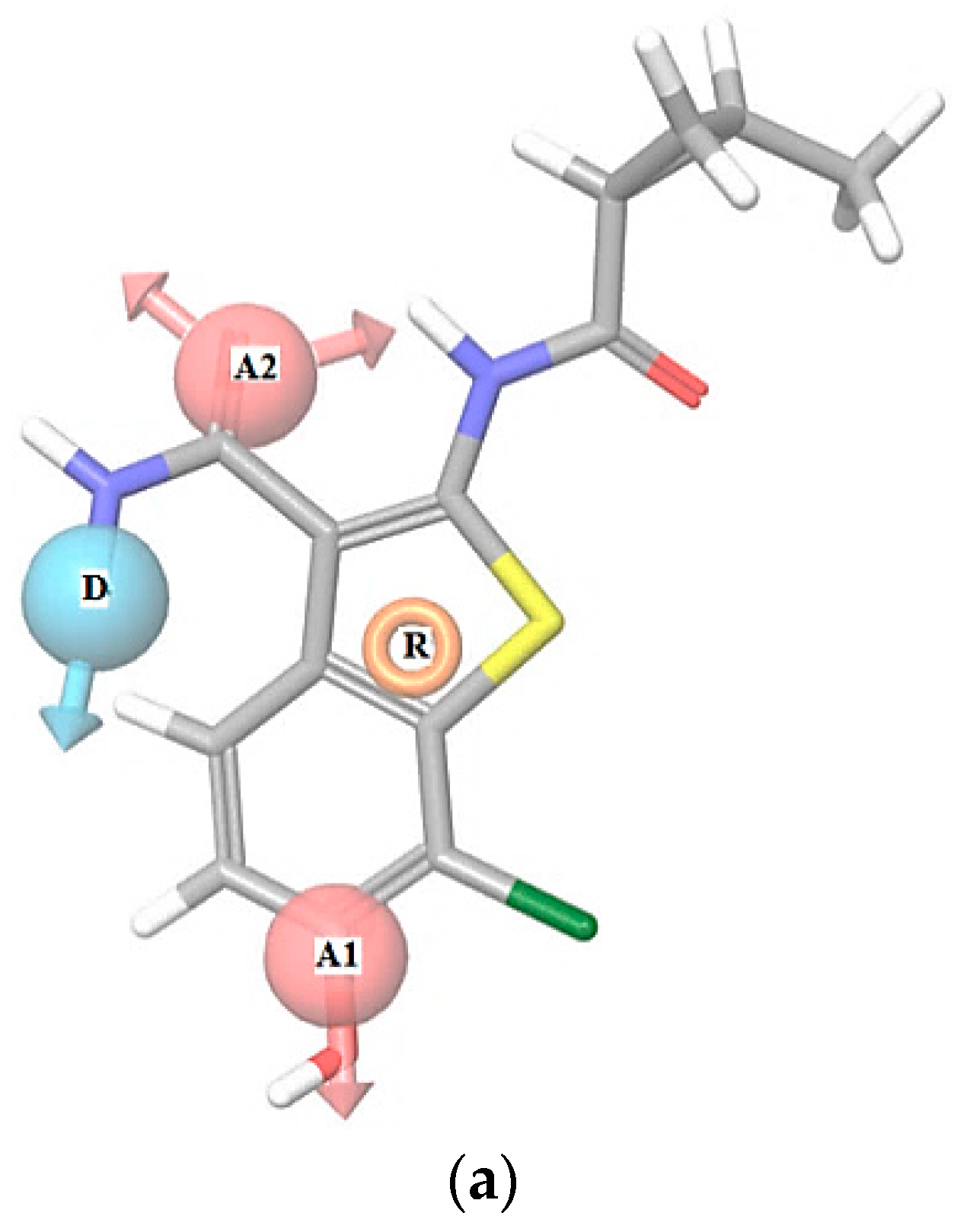
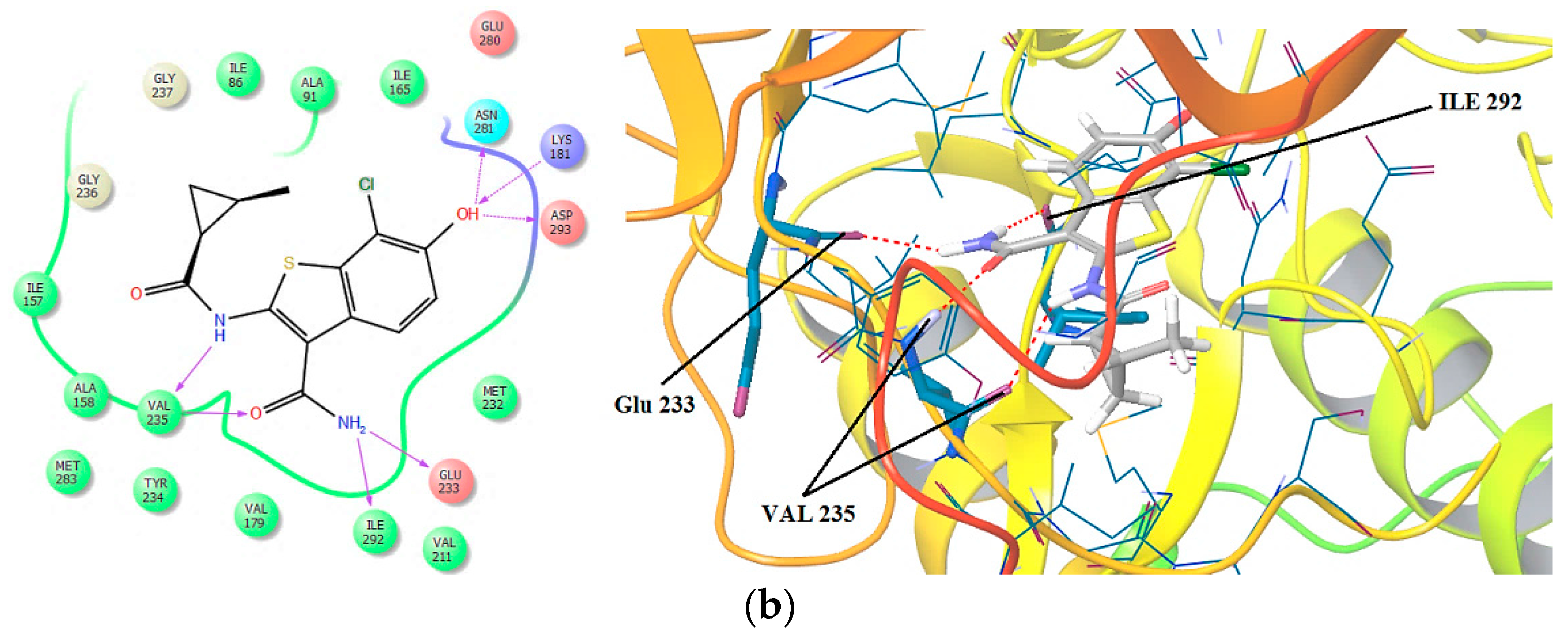
2.1. 3D Pharmacophore Searching: Mechanic Molecular Approach

For the 3D pharmacophores analysis, we considered the classification given by Zuccotto’s group [33]. Zuccotto’s work explains the active kinase conformation through the “gatekeeper door”. In this sense, the compounds were classified as type I1/2 inhibitors; recognize the target kinases in the DFG “out” form for PKn A and DFG “in” for Pkn B, the Pkn G have DLG instead of DFG and is DLG “in”. While developing the docking analysis, hydrogen bonds on the hinge zone and the non-covalent interactions near the “gatekeeper door”, helix-αC, C-terminal and N-terminal, were taken into account. The non-covalent interaction involved backbone, side chain hydrogen bonding and aromatic-aromatic interactions. Ligands with high scores have combinations of these non-covalent interactions, while the ligands with lower scores have few to no interaction forces. Many of the top scoring ligands that form hydrogen bonds and aromatic-aromatic interactions with the amino acid residues, are close to the hinge zone.
The PKs A and B are transmembrane proteins, while the Pkn G is a cytosolic protein, therefore their active sites have different characteristics. The Pkn A and B consist of a transmembrane receptor with a tyrosine kinase domain, protruding into the cytoplasm. As for the Pkn G, the unique multidomain topology of Pkn G reveals a central kinase domain that is flanked by N- and C-terminal rubredoxin and tetratrico-peptide repeat domains. Directed mutagenesis suggests that the rubredoxin domain functions as a regulator of Pkn G kinase activity [34], which was taken with its respective ligand to develop the docking analysis. To generate the pharmacophores the hypotheses with highest scoring were chosen, its features are shown in Table 1.
In Table 1 we can see a good reproducibility of each hypothesis shown above 70% to the molecular groups studied, these hypotheses are shown and characterized in Figure 1, Figure 2 and Figure 3.
In Figure 1, Figure 2 and Figure 3 are shown the interactions of the compounds 1 (Pkn A inhibitor), 7 (Pkn B inhibitor) and 23 (Pkn G inhibitor). Compound 1 presents three interactions on the hinge zone, while compound 7 presents two, and compound 23 also has three interactions. This criterion was crucial to define the actives poses and generate the hypothesis for each characterized pharmacophore. The results show the sites -H bond acceptor and -H bond donor on the hinge zone to all the ligands, establishing a structural model of the ligands on the active site with two or three interactions on this zone. With the focus to study the stabilization on the active site, we developed a study about chemical reactivity using DFT. The steric and electronic effects are characterized in terms of their chemical reactivity properties in the quantum chemistry context.

2.2. Selectivity Analysis: Quantum Chemistry Approach

2.2.1. Molecular Quantum Similarity Study

To analyze the steric and electronic effects, we calculate the Carbó indices to and the overlap and Coulomb similarity, the Carbó indices are restring on the interval (0, 1] where 1 means self-similarity and 0 means null quantum similarity.
One election characteristic of the molecular groups, is that they must have the highest structural difference in order to generate 3D pharmacophores that may have the largest possible chemical information. This can be seen in Table S6, see Supporting Information (SI), where the majority of values corresponding to the structural similarity have low values, considering the fact that a good index of Carbó begins above 0.500. The highest comparison is between the compounds 2 and 3 with a value of 0.684 (see Table S6) and an Euclidean distance of 4.191 (Table S7, see SI). The lowest value is found in the comparison between the compounds 1 and 4, with a value of 0.250 and an Euclidean distance of 6.317 (see Table S7, in the SI). Compound 1 has the highest activity (pIC50 = −1.568). We can see high structure differences along this molecular set. To study these features from the electronic point of view, the Coulomb indices are shown in Table S8, see SI.
The highest value using the Coulomb index is between the compounds 3 and 4 (0.902), and an Euclidean distance of 29.140 (Table S9, in SI). However, the lowest value is between compounds 1 and 4 (0.779), and Euclidean distance of 40.699. To analyze the steric effects and electronic effects reported on the most active compound 1 we propose the convergence quantitative alpha (α) scales for steric effect and beta (β) for electronic effect (Figure 4), with the goal of studying the variability of the steric and electronic effects along the Pkn A inhibitors from the biological activity point of view.
In Figure 4 we can see the variability in the steric and electronic effects with the Carbó indices of the most active compound 1. For this compound the highest similarity is with compound 2 (0.798) using the Coulomb operator (electronic effect) and with compound 3 (0.369) using the Dirac delta (steric effect).
For the Pkn B inhibitors, we can see the Carbó index in Tables S10–S13. For the overlap similarity, the highest comparison is between compounds 10 and 12 with a value of 0.832 (see Table S10) and Euclidean distance of 2.512 (see Table S11). The lowest value is obtained in the comparison between the compounds 10 and 16 with a value of 0.234 and Euclidean distance of 6.584 (see Table S11 in the SI). Among these Pkn B inhibitors, the compound with highest activity is 11 with pIC50 = 1.638, it has the highest structural difference with the compound 10 with a value of 0.741 (Table S10) and Euclidean distance of 3.241 (Table S11). The lowest structural difference for compound 7 is with compound 11 with a value of 0.388 and Euclidean distance of 4.825. To analyze the electronic effects, we calculated the Carbó indices (Table S12) using the Coulomb operators. The highest value in the Carbó index is obtain in the comparison between compounds 10 and 11 with the value 0.961 and Euclidean distance of 14.026 (see Table S13). The lowest value is obtained between the compounds 9 and 13 with a value of 0.589 and Euclidean distance of 43.709. To analyse the steric effects and electronic effects on the most active compound 11, we can see the alpha (α) and beta (β) scales in Figure 5, to study the variability of the steric and electronic effects along the Pkn B inhibitors from the biological activity viewpoint.
In Figure 5 we can see the variability of the steric and electronic effects with the Carbó indices of the most active compound 11. In both scales, compound 10 has the highest similarity and compound 16 has the lowest. These scales show how the chemical diversity selected can have influence on the biological activity, and also on the molecular space of the reported pharmacophore.
On the other hand, to the Pkn G inhibitors, the steric effects are shown in Table S14, see SI. In general, for this molecular group we obtained high values in the Carbó index of overlap; this fact is consistent with the common structural nucleus for this particular group. The highest value of overlap Similarity is obtained in the comparison between compounds 23 and 24 with the value of 0.993 and Euclidean distance of 0.492 (see Table S15), while the lowest value is obtained in the comparison between compounds 20 and 27 with a value of 0.532 and Euclidean distance of 4.556. These values show that the chemical diversity for this particular group is restricted with respect to the chemical diversity offered by the Pkn B inhibitors.
In general, the Coulomb indices show Carbó indices above of 0.900 (see Table S16). The highest value is obtained in the comparison between compounds 23 and 24 with a value of 0.999 and Euclidean distance of 1.246 (see Table S17, SI). The lowest value is obtained between compounds 24 and 27 with a value of 0.910 and Euclidean distance of 22.609. To analyze the variability of the steric and electronic effects on the biological activity, the alpha (α) and beta (β) scales are shown in Figure 6.
Figure 6 shows the variability of the steric and electronic effects on the biological activity of compound 26, the most active in this molecular group. In both scales, compound 19 has the highest structural and electronic similarity with compound 26.
On the other hand, in the analyses for the Pkn A, B and G inhibitors, the highest values were obtained using the Carbó Coulomb index. Taking into account these facts, we can say that the electronic effects have higher effect that the steric effects in the stabilization of these compounds in the active site associated to the Pkn A, B and G. With the aim to explore these outcomes a study on the chemical reactivity using global and local reactivity descriptors is developed in the DFT context.

2.2.2. Chemistry Reactivity Study

To study the electronic effects reported by the Carbó indices in the previous section, we calculated the global reactivity indices such as chemical potential, hardness, softness, electrophilicity (see Table 2, Table 3 and Table 4), and local as the Fukui functions in order to analyze the stabilization and interactions of these compounds in the active site.
In Table 2, compound 1 has the lowest chemical potential (−4.124 eV), hardness (3.839 eV), softness (0.261 eV−1) and the highest electrophilicity (2.215 eV). This electrophilicity value can be related with the highest biological activity for this series, compound pIC50 = −1.568. Compound 1 shows the highest electronic and structural similarity when it is compared with compound 2 (0.787) and (0.369) (see Figure 4). Therefore, an electronic parameter such as the electrophilicity can be important in the stabilization of these anti-tuberculosis compounds in the active site. Table 3 shows the reactivity values to Pkn B inhibitors.
In Table 3, compound 12 has the highest chemical potential and hardness with a value of −2.769 eV and 5.888 eV. Consequently, with these values this compound has the lowest softness (S = 0.170 eV−1) and electrophilicity (ω = 0.651 eV). These values are consistent with the lowest biological activity of this compound (pIC50 = −1.200). On the other hand, the highest values of softness and electrophilicity correspond to compound 16, S = 0.275 eV−1 and ω = 1.721 eV. This compound shows the highest steric and electronic effects when is compared with the most active compound 11, with a Carbó index of overlap 0.327 and Coulomb index of 0.733 (see Figure 5), therefore the steric and electronic effects large can have influence in the stability of such compounds, in the active site and consequently these aspects show low biological activity from experimental point of view. To analyze the chemical reactivity associated to the Pkn G inhibitors, we can consider their global indices listed in Table 4.
In Table 4 we can see the values of electrophility (1.500 eV) and softness (0.229 eV−1) of compound 26. These values are consistent with the highest biological activity to this compound (pIC50 = 2.000). However, the lowest values of electrophility and softness are found in compound 18 with 1.363 eV and 0.196 eV−1. Compound 18’s values are related to the steric and electronic effects, quantified by the Carbó index of overlap 0.651 and Coulomb index of 0.960, with respect to the most active compound of this series compound 26 (Figure 6). Therefore, for this molecular group, the high biological activity may be related to the ability of the inhibitors, to receive electrons from the external environment. The stabilization can be determined by the retro-donor process on the hinge zone.

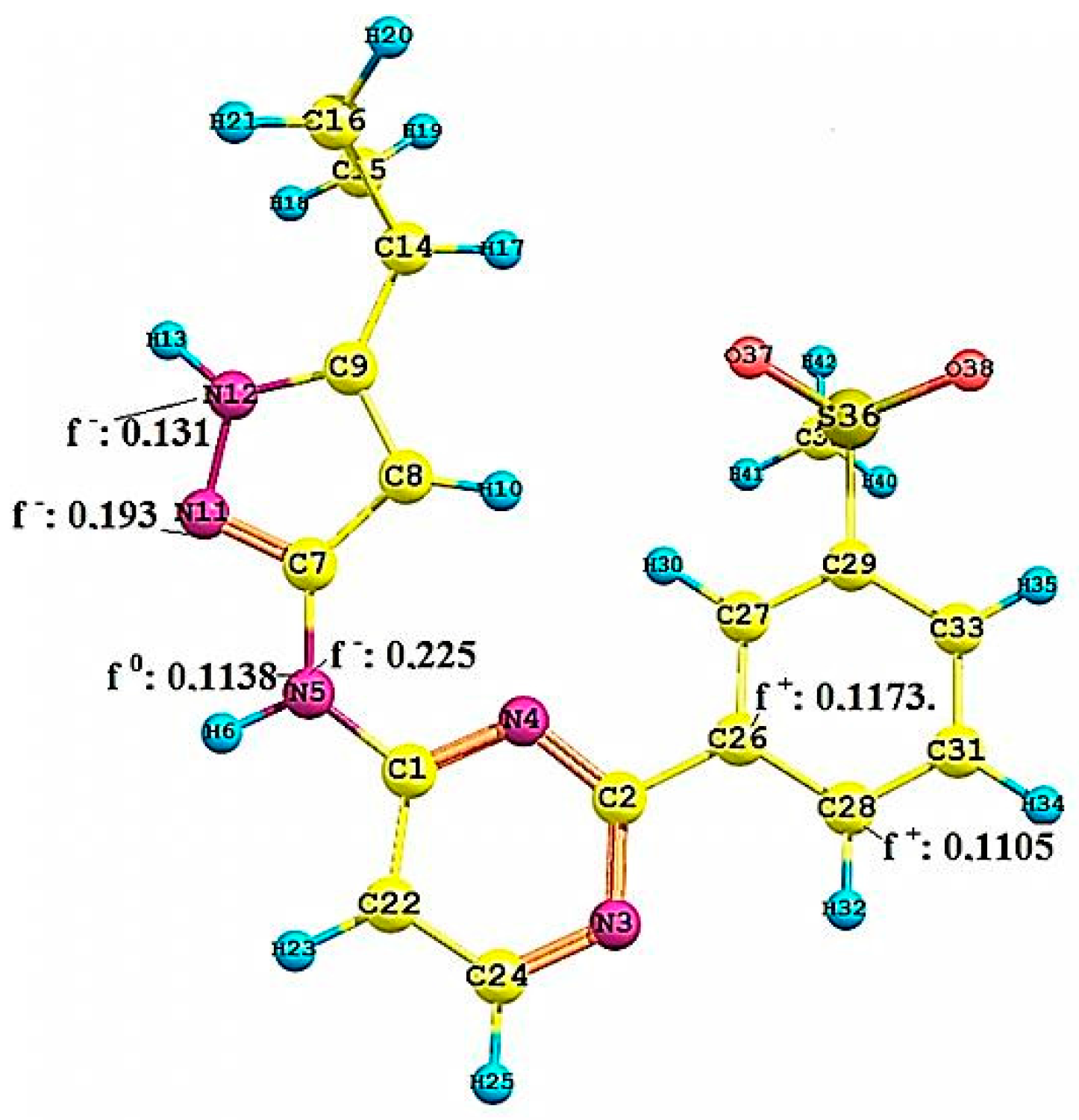
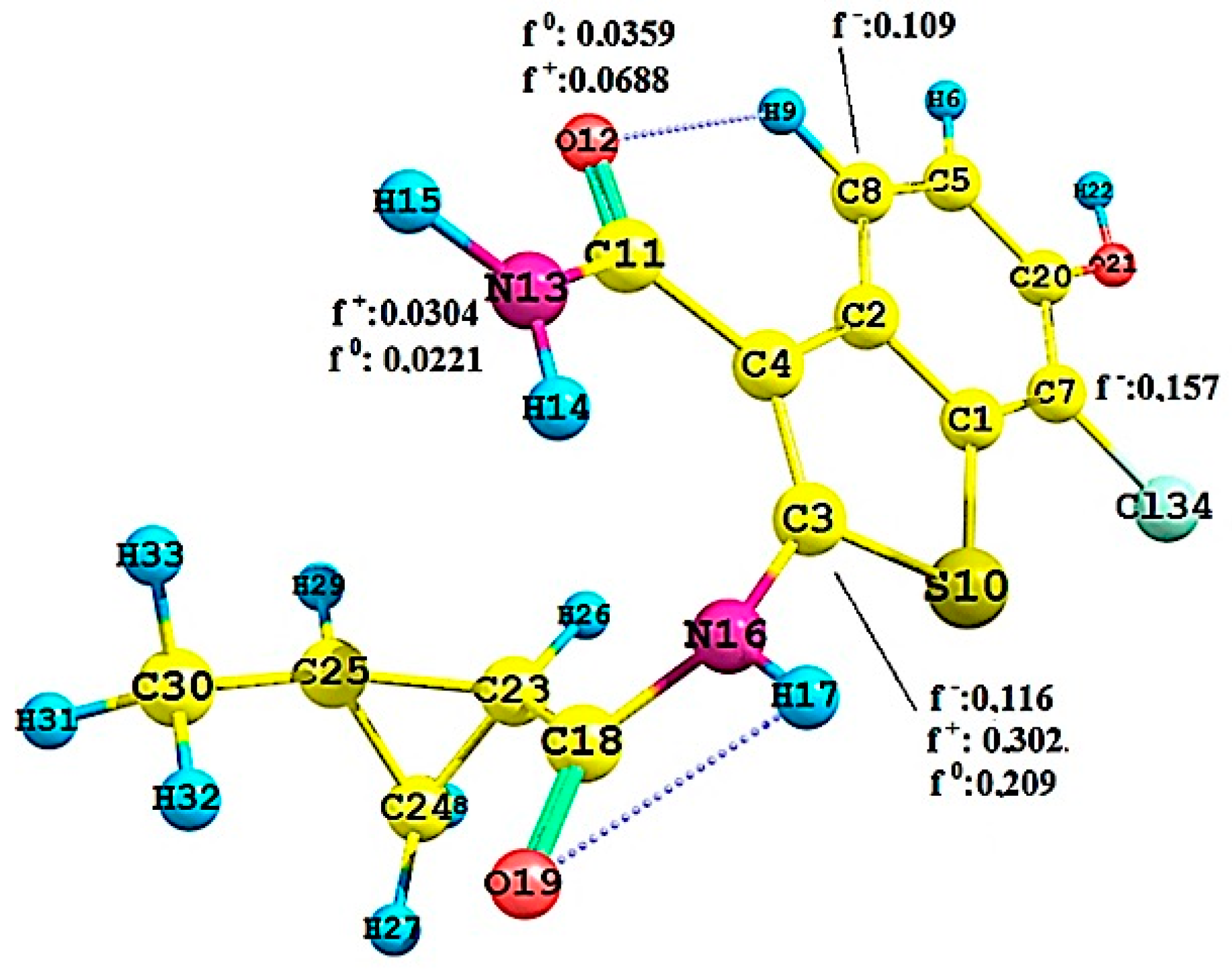
2.2.3. Local Reactivity Study

To quantify the molecular interactions and selectivity of the Pkn A, B and G inhibitors, we calculated the Fukui functions f(r) −,+,0 on the most active compounds 1 of Pkn A, 7 of Pkn B and 23 of Pkn G. The most significant values of these functions are shown in Figure 7, Figure 8 and Figure 9.
In Figure 7 we can see the interactions of N43 (f(r):0.022) with GLY 100, N33 (f(r):0.117) and N32 (f(r):0.047) with the VAL 98. The sites with affinity to nucleophilic attack are of the oxygen atoms (O12 (f(r)+:0.185) and O13 f(r)+:0.183) and on the nitrogen atom f(r)+:0.220. Finally, the site with neutral affinity is of the oxygen atom O12 (f(r)0:0.093) these interactions are responsible for the stabilization of the hinge zone and of the active site for these anti-tuberculosis compounds (see Figure 1).
Figure 8 shows the Fukui functions f(r) of the nitrogen atoms N12 (0.131), responsible for the hydrogen bond donor, and N11 (0.193) responsible for the acceptor site on the hinge zone. The other nitrogen atom, N5, besides showing a high f(r) = 0.225, shows a f(r)0 = 0.1138, representing that it can have either susceptibility to electrophilic attack, or toward neutral species. Therefore, this N5 can be crucial for the stabilization in the active site. The most sensitive values in the Fukui functions f(r)+ are shown on the carbon atoms C26 and C28, with values of 0.1173 and 0.1105. These values show the most susceptible zone to nucleophilic attacks. We can appreciate the nature of the molecular behaviour associated with the Pkn B inhibitors, and the type of stabilization that can happen on the active site. To know the nature of the chemical selectivity associated with the Pkn G inhibitors, is to know the Fukui functions of compound 23.
Figure 9 shows the Fukui functions f(r)+,0 of the nitrogen atom N13, with values of 0.0304 and 0.0221. These values justify the interactions between the nitrogen atom N13 and the residues Ile 292 and Glu 233 on the hinge zone (one interaction of nucleophilic character and another of neutral nature). Another important interaction is between the oxygen atom O12 and the residue Val 235, which can be related to the value of f(r)+ = 0.0688.
The values of f(r) relevant are shown in the carbon atoms C8 and C3 with 0.109 and 0.116, representing the areas susceptible to electrophilic attacks. In general, these values show the Fukui functions and the molecular behaviour of these Pkn G inhibitors. Compound 23 can also have intramolecular hydrogen bonds, that may be important in the stabilization of the active site. We can see the nature of the selectivity and the interactions of Pkn G inhibitors and how they can interact with the residues on the hinge zone (see Figure 3).

2.3. 3D Pharmacophore-Based Database Screening on the Chibale’s Database (CD)

The database screening [35] is carried out using the Chibale database (see SI), with the pharmacophores reported in Figure 1, Figure 2 and Figure 3, to determine the compounds that can have affinity to each pharmacophores. The CD was created with 183 compounds [28,29,30,31,32] (see SI) and resulted in the following; 60 compounds have affinity with the Pkn A pharmacophore, 50 have affinity with the Pkn B, and other group of 30 compounds presented affinity with the Pkn G pharmacophore. These respectively affinities are shown in Table 5 and Table 7 through matching properties such as QPpolz: Predicted polarizability in Å3, SASA: Total Solvent-accessible Surface are in Å2, FOSA: Hydrophobic component of the SASA (saturated carbons and attached hydrogen), FISA: Hydrophilic component of the SASA (SASA on N, O and H on heteroatom), PISA: Pi (carbon and attached hydrogen) component of the SASA and finally WPS: Weakly polar component of the SASA (halogens, P and S).
In Table 5 and Table 6 we can see the 60 compounds matching the Pkn A pharmacophore. 50 compounds matching Pkn B, and 30 compounds with affinity to the Pkn G. In these tables there are also compounds with affinities to Pkn A, B and G simultaneously, compounds only with affinity to Pkn B and G, and finally compounds with only affinity to the Pkn A and B pharmacophores. The Tables predicted that polarizability values are into the recommended ranges for 95% of known drugs (13.0 to 70.0). On the other hand, the volume Vmol (the total volume of molecule enclosed by solvent-accessible molecular surface, in Å3 (probe radius 1.4 Å)) and the globularity descriptor Glob = (4πr2 )/Smol, where r is the radius of the sphere whose volume is equal to the molecular volume, are also in the recommended ranges (Vmol: 500 to 2000 Å3) and (Glob: 0.75 to 0.95) [36,37,38,39,40,41,42,43,44].
In Table 7 we can see that the R and S isomers of compound 61 only present affinities to the pharmacophore Pkn G. Other important properties are the predicted skin permeability (Qlog Kp) and the number of likely metabolic reactions (#metab). These properties also are in the recommended range (Qlog Kp: −8.0 to −1.0) [45,46] and (#metab: 1 to 8) [36,37,38,39,40,41,42,43,44]. The results of this database screening may be helpful to characterize, from an experimental point of view, the R and S isomers, which is the goal of this work. We hope that this information can provide insights about the stereoisomers for a specific target Pkn A, B or G. To assess the ligands reported by the database screening, we used docking molecular with the structural model generated by the 3D pharmacophores, in order to verify the interaction of these ligands on the active site in each protein kinase.

3. Materials and Methods

The molecular dataset used in this study was taken from the literature as follow: four Pkn A inhibitors reported by Sipos et al. [14], 13 Pkn B inhibitor selected from the works of Székely et al. [15], Lougheed et al. [16], Chapman et al. [17] and Naqvi et al. [18] and 10 Pkn G inhibitors reported by Sipos et al. [14]). In this molecular dataset we considered structural diversity and uniform distribution of IC50. Logarithmic IC50 (μM) (pIC50 = −log IC50) was employed as a dependent variable instead of IC50. The pIC50 values of inhibition of the compounds are shown in Table 8 for Pkn A, Table 9 for Pkn B and Table 10 for Pkn G.

3.1. System Preparation to the Molecular Mechanic (MM) Approach

The crystal structure of PKn A (PBD code: 4OW8 [47]), B (PBD code: 1O6Y [48]) and PKn G (PBD code: 2PZI [34]) was prepared using Schrödinger Suite 2014-1’s Protein Preparation Wizard module [49,50], which refines the protein structure and optimizes the hydrogen bond (H-bond) network. Protonation states were determined using PropKa utility at a physiological pH. This was followed by a restrained molecular minimization using the Impact Refinement (Impref) module [51,52], with the heavy atoms restrained to remain within a root-mean-square deviation (RMSD) of 0.18 Å from the initial coordinates. The 3D molecular structures of the compounds were built using Maestro [53] and optimized using a B3LYP (hybrid-GGA exchange-correlation functional) at 6-311++G (2d,2p) level of theory [54,55] using Gaussian 09 [56], the ionization/tautomeric states were predicted at physiological pH conditions using Epik [57].

3.2. Docking Studies and Pharmacophore Research

Docking studies were carried out with Glide [58], using the Standard Precision (SP) mode with default parameters. Docking grid was generated with default settings centered at the co-crystallized ligand. A scaling factor down to 0.8 for the van der Waals radii of nonpolar protein atoms was used to accommodate the fact that the protein structure will not be optimized to fit larger ligands such as the studied in this work. The Induced Fit Docking (IFD) workflow [59,60] was employed to generate an alternative conformation of the receptor suitable to bind the studied ligands, by allowing the protein to undergo side-chain or backbone movements, or both, upon ligand docking. To develop the docking analysis we used protein-ligand complex molecular dynamics simulation of 25 ns in vacuo using GROMOS11 force field set, implemented through Gromacs 5.1.2 [61,62], to analyze the stability of each protein-ligand complex.
Finally, for the pharmacophore research we compared all the poses of the most and less active ligands for each congeneric family (PKn A, B and G), taking into account the extent of residue movement generated by the IFD calculation. The most energetically favourable conformation was selected by the best pose of each compound for further analysis, also ensuring that it exhibited interactions with the “hinge” residues of the PKs (i.e., Donor-Acceptor-Donor, DAD system). The pharmacophore research was carried out with Phase 3.7 [63] using four pharmacophore features: Hydrogen bond acceptor (A), Hydrogen bond donor (D), Hydrophobic group (H) and Aromatic ring (R); the pharmacophores of the best ligand poses in the active sites of the PKn A, B and G were examined and we chose the hypothesis with the highest score.

4. Theoretical and Computational Details to the Quantum Chemistry Approach

In this work, with the goal of studying the inhibitor from the quantum chemistry point of view, we used the MQS field and chemical descriptors within the DFT context, to analyze the group correlation and look for the facts that can determine the anti-tuberculosis activity of the Pkn A, B and G inhibitors considered.

4.1. Molecular Quantum Similarity: Steric and Electronic Effects Study

The similarity indices were introduced by Carbó and co-workers (see reviews on quantum similarity of [24,25,64,65,66]), and have been used to understand the steric and electronic effect on the molecular sets. The quantum similarity measure ZAB between compounds A and B, with electron density ρ A ( γ 1 ) and ρ B ( r 2 ) is defined, based on the idea of the minimizing of the expression for the Euclidean distance as:
D A B 2 = | ρ A ( r ) ρ B ( r ) | 2 d r = ( ρ A ( r 1 ) ) 2   d r 1 + ( ρ B ( r 2 ) ) 2   d r 2 2 ρ A ( r 1 ) ( r 2 ) d r 1 d r 2 = Z A A + Z B B 2 Z A B
Overlap integral involving the ZAB between the electronic density of the compound A and B, ZAA and ZBB are the self-similarity of compounds A and B [64,65,66]. The most common quantum similarity index is the one generalized by the cosine, introduced by Carbó et al. [64,65,66]. This index can be expressed mathematically as:
I A B = ρ A ( r 1 ) ρ B ( r 2 ) d r 1 d r 2 ( ρ A ( r 1 ) ) 2 d r 1 ( ρ B ( r 2 ) ) 2   d r 2
or using the elements of Z in an operator (Ω):
I A B = Z A B ( ) Z A A ( ) Z B B ( )
In Equation (3), the index is mathematically defined in the interval (0, 1) where 1 is self-similarity, and where only the measures of “shape similarity” are included. Another alternative is the Hodgkin-Richards index [67], which appears naturally when using the arithmetic mean and can be defined mathematically as:
I A B = 2 Z A B ( ) Z A A ( ) + Z B B ( )
This is (Ω) an operator for the measurement of quantum similarity. The Equation (4) shows another way to make Quantum Similarity Measures (QSM), but recent published work has shown that in fact, it is just an Euclidean distance, like shown in Equation (1) [68].
A simple way to make Quantum Similarity Measure (QSM) [22,23,24] involving two density functions, in the most usual form:
Z A B ( ) = ρ A ( r 1 ) ( r 1 , r 2 ) ρ B ( r 2 ) d r 1 d r 2
In this equation ρ A ( r 1 ) and ρ B ( r 2 ) are the density functions to quantum objects A and B, while ( r 1 , r 2 ) is a positive define weight operator. When the operator is chosen as the Dirac Delta function: δ ( r 1 r 2 ) we obtain overlap similarity measure, while Coulomb Similarity Measure appears when choosing: | r 1 r 2 | 1 These two operators are the most popular for similarity comparisons between molecules.

4.2. Molecular Alignment

To carry out the quantum similarity measures is an important optimal molecular alignment. As the integrals attached to the QSM produce, real positive defined results, the relative position problem can be addressed through a maximal QSM. For an overlap QSM, this situation can be expressed by means of the equation:
max T ; Z A B ( T ; ) = max T ; ρ A ( r ) ρ B ( r | T ; ) d r
In this equation, is implicitly supposed that ρ B ( r ) is translated and rotated by six possible ways, (T;φ) and are shown as explicit parameters in this integral [22,23,24,69]. This principle is used by the Topo Geometrical Superposition Algorithm (TGSA) [70] and is the program used in this study to calculate the Carbó indices using Equation (2).

4.3. Chemical Reactivity Descriptors: Selectivity Study

The global reactivity descriptors are defined within the DFT framework and were interpreted by Parr and coworkers [71,72,73,74,75,76,77,78]. The chemical potential can be written mathematically in terms of the energy of an electron in the frontier molecular orbitals (Higher Occupied Molecular Orbital) HOMO and (Lowest Unoccupied Molecular Orbital) LUMO as:
μ ε L + ε H 2
From (8), one can obtain quantitative expression for the chemical hardness (η) [79,80,81,82,83], meaning that the opposition of the system to distort the electron cloud and mathematically can be written as:
η ε L ε H
On the other hand, we have the global electrophilicity (ω) introduced by Parr et al. [84], which is a measure of the stabilization energy of the system when it is saturated by electrons from the external environment and represented mathematically as:
ω = μ 2 2 η
The Fukui function ( f ( r ) ) was one of the descriptors used in this work and defined as:
f ( r ) = ( ρ ( r ) N ) V = ( μ V ( r ) ) N
Due to that f ( r ) is discontinuous at integer values of 𝑁. There are three types according to Fukui et al., f+ which contain information on the reactivity local of nucleophilic attack, f (11) which does the same for an electrophilic attack. And finally f0, that measures the reactivity towards neutral or radical agents [84,85,86,87,88,89,90,91], using the condensation scheme on specific sites of the molecule to obtain the following Fukui indices [92,93,94,95]:
f x + ( r ) ρ N + 1 x ( r ) ρ N x ( r ) q x ( N + 1 ) q x ( N ) f x ( r ) ρ N x ( r ) ρ N 1 x ( r ) q x ( N ) q x ( N 1 ) f x 0 ( r ) [ f x + + f x 2 ] ρ N + 1 x ( r ) ρ N 1 x ( r ) 2 q x ( N + 1 ) q x ( N 1 ) 2
Hopefully using this equation scheme will help understanding the biological activity (selectivity) of the PKn A, B and G inhibitors studied from global and local point of view.

4.4. Creating the 3D Chibale’s Database: Database Screening

Computational Aided Drug Design (CADD) is presently a key component in the process of drug discovery and development associated with the tuberculosis disease. It offers great promise to drastically reduce cost and time requirements. In a pharmaceutical context, database screening is normally regarded as the top CADD tool to screen large libraries of chemical structures, and reduce them to a key set of likely drug candidates regarding a specific protein target [96,97].
In this work, the 3D Chibale database was created using 103 anti-tuberculosis compounds reported by the Chibale group [28,29,30,31,32]. Each anti-tuberculosis compound was characterized taking account their chiral centers (chiral isomers), which leaves the final molecular group with 183 compounds (see Tables S1–S5 in Supporting Information (SI)). This 3D database was created using the command line tools of Accompany Phase version 3.5, which is part of the Schrödinger Suite 2013 release [98,99,100]. It was used to evaluate the pharmacophores to Pkn A, B and G reported, to identify the compound that may have affinity with these specific protein targets.

5. Conclusions

In conclusion, the 3D pharmacophore reported were selected according to the hypotheses with highest score (Acceptor: A1/A2/A3/Donor:D) of Pkn A and (A/A/D/aromatic ring:R) of Pkn B and G. The three 3D pharmacophores generated were characterized using the molecular quantum similarity field and reactivity descriptors supported in the DFT framework.
To develop the study of quantum similarity, we used the Carbó indices. Through these indices are proposed the convergence quantitative alpha (α) scale to steric effects and beta (β) to electronic effects with respect to the most active compound of each inhibitor set. In this analysis, the Carbó indices with the highest values are the Coulomb values. Considering that the electronic effects are more relevant than the steric, we develop a study using global reactivity descriptors such as Chemical Potential, Harness, Softness and the Fukui functions as local descriptor, to understand the interaction of these compounds in the active sites.
The conclusion regarding the chemistry reactivity of Pkn A inhibitors is that the electronic factors as the electrophilicity can be important in the stabilization of these anti-tuberculosis compounds. In the active site of Pkn B inhibitors, there are steric and electronic effects that can have a big influence in the stability of such compounds in the active site. Consequently these aspects show low biological activity from experimental point of view. Regarding Pkn G inhibitors, the high biological activity may be related to the ability of the inhibitors to receive electrons from the external environment. The stabilization can be determined by the retro-donor process on the hinge zone. In this order of ideas, the reactivity descriptors reported can be related to the experimental data.
Finally, the database screening was developed using the Chibale’s Database created with 183 anti-tuberculosis compounds and using the Pkn A, B and G pharmacophores reported. We found 60 compounds with affinity to the Pkn A pharmacophore, 50 compounds for the PKn B and 30 compounds to the Pkn G. These predictions were tested with properties related such as QPpolz: Predicted Polarizability in Å3, SASA: total Solvent-Accessible Surface Are in Å2, FOSA: Hydrophobic component of the SASA (saturated carbons and attached hydrogen) and WPS: Weakly Polar Component of the SASA (halogens, P and S), among others. These properties related are within the recommended ranges for 95% of known drugs. In addition, the docking results for these ligands show the sites –H bond acceptor and –H bond donor, which are the characteristic interactions on the hinge zone in the active site, of each protein kinase studied.

Supplementary Materials

The Supplementary Materials following are available online.

Acknowledgments

A.M.-B. Thanks to CONICYT/FONDECYT through postdoctoral project No. 3150035 and to the drug discovery group of Professor Kelly Chibale, University of Cape Town, South Africa.

Author Contributions

This work was modeled and evaluated computationally by the present author Alejandro Morales-Bayuelo.

Conflicts of Interest

This work has not conflict of interest.

References

  1. World Health Organization (WHO). Health Topics (Tuberculosis). Available online: http://www.who.int/campaigns/tb-day/2017/event/en/ (accessed on 24 March 2017).
  2. Cully, M. Redesigned antibiotic combats drug-resistant tuberculosis. Nat. Rev. Drug Discov. 2014, 13, 256. [Google Scholar] [CrossRef] [PubMed]
  3. Lee, R.E.; Hurdle, J.G.; Liu, J.; Bruhn, D.F.; Matt, T.; Scherman, M.S.; Vaddady, P.K.; Zheng, Z.; Qi, J.; Akbergenov, R.; et al. Spectinamides: A new class of semisynthetic antituberculosis agents that overcome native drug efflux. Nat. Med. 2014, 20, 152–158. [Google Scholar] [CrossRef] [PubMed]
  4. Zumla, A.; Nahid, P.; Cole, S.T. Advances in the development of new tuberculosis drugs and treatment regimens. Nat. Rev. Drug Discov. 2013, 12, 388–404. [Google Scholar] [CrossRef] [PubMed]
  5. Smaill, F.; Jeyanathan, M.; Smieja, M.; Medina, M.F.; Zganiacz, N.T.A.; Yin, C.; Heriazon, A.; Damjanovic, D.; Puri, L.; Hamid, J.; et al. A Human Type 5 Adenovirus–Based Tuberculosis Vaccine Induces Robust T Cell Responses in Humans Despite Preexisting Anti-Adenovirus Immunity. Sci. Transl. Med. 2013, 5, 205ra134. [Google Scholar] [CrossRef] [PubMed]
  6. Raviglione, M.C.; Smith, I.M. XDR Tuberculosis—Implications for Global Public Health. N. Engl. J. Med. 2007, 356, 656–659. [Google Scholar] [CrossRef] [PubMed]
  7. Rao, S.P.S.; Lakshminarayana, S.B.; Kondreddi, R.R.; Herve, M.; Camacho, L.R.; Bifani, P.; Kalapala, S.K.; Jiricek, J.; Ma, N.L.; Tan, B.H.; et al. Indolcarboxamide is a preclinical candidate for treating multidrug-resistant tuberculosis. Sci. Transl. Med. 2013, 5, 214ra168. [Google Scholar] [CrossRef] [PubMed]
  8. Mills, H.L.; Cohen, T.; Colijn, C. Community-wide isoniazid preventive therapy drives drug-resistant tuberculosis: A model-based analysis. Sci. Transl. Med. 2013, 5, 180ra49. [Google Scholar]
  9. Migliori, G.B.; Lange, C.; Centis, R.; Sotgiu, G.; Hoffmann, R.M.H.; Kliiman, K.; de laco, G.; Lauria, F.N.; Richardson, M.D.; Spanevello, A.; et al. Resistance to second-line injectables and treatment outcomes in multidrug-resistant and extensively drug-resistant tuberculosis cases. Eur. Resp. J. 2008, 31, 1155–1159. [Google Scholar] [CrossRef] [PubMed]
  10. Dye, C.; Williams, B.G.; Espinal, M.A.; Raviglione, M.C. Erasing the world’s slow stain: Strategies to beat multidrug-resistant tuberculosis. Science 2002, 295, 2042–2046. [Google Scholar] [CrossRef] [PubMed]
  11. Hallows, K.R.; Alzamora, R.; Li, H. AMP-activated protein kinase inhibits alkaline pH- and PKA-induced apical vacuolar H+-ATPase accumulation in epididymal clear cells. Am. J. Physiol. Cell Physiol. 2009, 296, C672–C681. [Google Scholar] [CrossRef] [PubMed]
  12. Salminen, A.; Kaarniranta, K.; Haapasalo, A.; Soininen, H.; Hiltunen, M. AMP-activated protein kinase: A potential player in Alzheimer’s disease. J. Neurochem. 2011, 118, 460–474. [Google Scholar] [CrossRef] [PubMed]
  13. Kotlyarov, A.; Neininger, A.; Schubert, C.; Eckert, R.; Birchmeier, C.; Volk, H.D.; Gaestel, M. MAPKAP kinase 2 is essential for LPS-induced TNF-αbiosynthesis. Nat. Cell. Biol. 1999, 1, 94–97. [Google Scholar] [PubMed]
  14. Sipos, A.; Pató, J.; Székely, R.; Hartkoorn, R.C.; Kékesi, L.; Őrfi, L.; Szántai-Kis, C.; Mikušová, K.; Svetlíková, Z.; Korduláková, J.; et al. Lead selection and characterization of antitubercular compounds using the Nested Chemical Library. Tuberculosis 2015, 95, S200–S206. [Google Scholar] [CrossRef] [PubMed]
  15. Székely, R.; Wáczek, F.; Szabadkai, I.; Németh, G.; Hegymegi-Barakonyi, B.; Erős, D.; Szokol, B.; Pató, J.; Hafenbradl, D.; Satchell, J.; et al. A novel drug discovery concept for tuberculosis: Inhibition of bacterial and host cell signalling. Immunol. Lett. 2008, 116, 225–231. [Google Scholar] [CrossRef] [PubMed]
  16. Lougheed, K.E.A.; Osborne, S.A.; Saxty, B.; Whalley, D.; Chapman, T.; Bouloc, N.; Chugh, J.; Nott, T.J.; Patel, D.; Spivey, V.L.; et al. Effective inhibitors of the essential kinase PknB and their potential as anti-mycobacterial agents. Tuberculosis 2011, 91, 277–286. [Google Scholar] [CrossRef] [PubMed]
  17. Chapman, T.M.; Bouloc, N.; Buxton, R.S.; Chugh, J.; Lougheed, K.E.A.; Osborne, S.A.; Saxty, B.; Smerdon, S.J.; Taylor, D.L.; Whalley, D. Substituted aminopyrimidine protein kinase B (PknB) inhibitors show activity against Mycobacterium tuberculosis. Bioorg. Med. Chem. Lett. 2012, 22, 3349–3353. [Google Scholar] [CrossRef] [PubMed]
  18. Naqvi, A.; Malasoni, R.; Srivastava, A.; Pandey, R.R.; Dwivedi, A.K. Design, synthesis and molecular docking of substituted 3-hydrazinyl-3-oxo-propanamides as anti-tubercular agents. Bioorg. Med. Chem. Lett. 2014, 24, 5181–5184. [Google Scholar] [CrossRef] [PubMed]
  19. Kitchen, D.B.; Decornez, H.; Furr, J.R.; Bajorath, J. Docking and scoring in virtual screening for drug discovery: Methods and applications. Nat. Rev. Drug Discov. 2004, 3, 935–949. [Google Scholar] [CrossRef] [PubMed]
  20. Goldman, B.B.; Proteins, W.T.W. QSD quadratic shape descriptors. 2. Molecular docking using quadratic shape descriptors (QSDock). Proteins Struct. Funct. Bioinform. 2000, 38, 79–94. [Google Scholar] [CrossRef]
  21. Meng, E.C.; Shoichet, B.K.; Kuntz, I.D. Automated docking with grid-based energy evalution. J. Comp. Chem. 2004, 13, 505–524. [Google Scholar] [CrossRef]
  22. Carbó-Dorca, R. Collective Euclidian distances and quantum similarity. J. Math. Chem. 2013, 51, 338–353. [Google Scholar] [CrossRef]
  23. Lipkowitz, K.B.; Larter, R.; Cundari, T.R. Biomolecular applications of Poisson-Boltzmann methods. Revis. Comput. Chem. 2005, 21, 1–125. [Google Scholar]
  24. Gironés, X.; Carbó-Dorca, R. Modelling Toxicity using Molecular Quantum Similarity Measures. QSAR Comb. Sci. 2006, 25, 579–589. [Google Scholar] [CrossRef]
  25. Carbó-Dorca, R.; Gironés, X. Foundation of Quantum Similarity Measures and Their Relationship to QSPR: Density Function Structure, Approximations, and Application Examples. Int. J. Quant. Chem. 2005, 101, 8–20. [Google Scholar] [CrossRef]
  26. Parr, R.G.; Yang, W. Density Functional Theory of Atoms and Compounds; Oxford University Press: New York, NY, USA, 1989. [Google Scholar]
  27. Morales-Bayuelo, A. Analyzing the substitution effect on the CoMFA results within the framework of density functional theory (DFT). J. Mol. Mod. 2016, 22, 164. [Google Scholar] [CrossRef] [PubMed]
  28. Hans, R.H.; Wiid, I.J.F.; van Helden, P.D.; Wanc, B.; Franzblau, S.G.; Gut, J.; Rosenthal, P.J.; Chibale, K. Novel thiolactone–isatin hybrids as potential antimalarial and antitubercular agents. Bioorg. Med. Chem. Lett. 2011, 21, 2055–2058. [Google Scholar] [CrossRef] [PubMed]
  29. Hans, R.H.; Guantai, E.M.; Lategan, C.; Smith, P.J.; Wanc, B.; Franzblau, S.G.; Gut, J.; Rosenthal, P.J.; Chibale, K. Synthesis, antimalarial and antitubercular activity of acetylenic chalcones. Bioorg. Med. Chem. Lett. 2010, 20, 942–944. [Google Scholar] [CrossRef] [PubMed]
  30. Singh, K.; Kumar, M.; Pavadai, E.; Naran, K.; Warner, D.F.; Ruminski, P.G.; Chibale, K. Synthesis of new verapamil analogues and their evaluation in combination with rifampicin against Mycobacterium tuberculosis and molecular docking studies in the binding site of efflux protein Rv1258c. Bioorg. Med. Chem. Lett. 2014, 24, 2985–2990. [Google Scholar] [CrossRef] [PubMed]
  31. Singh, K.; Singh, K.; Wan, B.; Franzblau, S.; Chibale, K.; Balzarini, J. Facile transformation of Biginelli pyrimidin-2(1H)-ones to pyrimidines. In vitro evaluation as inhibitors of Mycobacterium tuberculosis and modulators of cytostatic activity. Eur. J. Med. Chem. 2011, 46, 2290–2294. [Google Scholar] [CrossRef] [PubMed]
  32. Khanye, S.D.; Wan, B.; Franzblau, S.G.; Gut, J.; Rosenthal, P.J.; Smith, G.S.; Chibale, K. Synthesis and in vitro antimalarial and antitubercular activity of gold(III) complexes containing thiosemicarbazone ligands. J. Organomet. Chem. 2011, 696, 3392–3396. [Google Scholar] [CrossRef]
  33. Zuccotto, F.; Ardini, E.; Casale, E.; Angiolini, M. Through the “Gatekeeper Door”: Exploiting the Active Kinase Conformation. J. Med. Chem. 2010, 53, 2681–2694. [Google Scholar] [CrossRef] [PubMed]
  34. Scherr, N.; Honnappa, S.; Kunz, G.; Mueller, P.; Jayachandran, R.; Winkler, F.; Pieters, J.; Steinmetz, M.O. Structural basis for the specific inhibition of protein kinase G, a virulence factor of Mycobacterium tuberculosis. Proc. Natl. Acad. Sci. USA 2014, 104, 12151–12156. [Google Scholar] [CrossRef] [PubMed]
  35. Lionta, E.; Spyrou, G.; Vassilatis, D. K.; Cournia, Z. Structure-based virtual screening for drug discovery: Principles, applications and recent advances. Curr. Top. Med. Chem. 2014, 14, 1923–1938. [Google Scholar] [CrossRef] [PubMed]
  36. Jorgensen, W.L.; Duffy, E.M. Prediction of Drug Solubility from Structure. Adv. Drug Deliv. Rev. 2002, 54, 355–366. [Google Scholar] [CrossRef]
  37. Duffy, E.M.; Jorgensen, W.L. Prediction of Properties from Simulations:  Free Energies of Solvation in Hexadecane, Octanol, and Water. J. Am. Chem. Soc. 2000, 122, 2878–2888. [Google Scholar] [CrossRef]
  38. Colmenarejo, G.; Alvarez-Pedraglio, A.; Lavandera, J.L. Cheminformatic Models to Predict Binding Affinities to Human Serum Albumin. J. Med. Chem. 2001, 44, 4370–4378. [Google Scholar] [CrossRef] [PubMed]
  39. Luco, J.M. Prediction of the Brain–Blood Distribution of a Large Set of Drugs from Structurally Derived Descriptors Using Partial Least-Squares (PLS) Modeling. J. Chem. Inf. Comput. Sci. 1999, 39, 396–404. [Google Scholar] [CrossRef] [PubMed]
  40. Kelder, J.; Grootenhuis, P.D.; Bayada, D.M.; Delbresine, L.P.; Ploemen, J.P. Polar molecular surface as a dominating determinant for oral absorption and brain penetration of drugs. Pharm. Res. 1999, 16, 1514–1519. [Google Scholar] [CrossRef] [PubMed]
  41. Ajay; Bermis, G.W.; Murkco, M.A. Designing libraries with CNS activity. J. Med. Chem. 1999, 42, 4942–4951. [Google Scholar] [CrossRef] [PubMed]
  42. Yazdanian, M.; Glynn, S.L.; Wright, J.L.; Hawi, A. Correlating partitioning and caco-2 cell permeability of structurally diverse small molecular weight compounds. Pharm. Res. 1998, 15, 1490–1494. [Google Scholar] [CrossRef] [PubMed]
  43. Irvine, J.D.; Takahashi, L.; Lockhart, K.; Cheong, J.; Tolan, J.W.; Selick, H.E.; Grove, J.R. MDCK (Madin-Darby canine kidney) cells: A tool for membrane permeability screening. Pharm. Sci. 1999, 88, 28–33. [Google Scholar] [CrossRef] [PubMed]
  44. Stenberg, P.; Norinder, U.; Luthman, K.; Artursson, P. Experimental and computational screening models for the prediction of intestinal drug absorption. J. Med. Chem. 2001, 44, 1927–1937. [Google Scholar] [CrossRef] [PubMed]
  45. Potts, R.O.; Guy, R.H. Predicting Skin Permeability. Pharm. Res. 1992, 9, 663–669. [Google Scholar] [CrossRef] [PubMed]
  46. Potts, R.O.; Guy, R.H. A Predictive Algorithm for Skin Permeability: The Effects of Molecular Size and Hydrogen Bond Activity. Pharm. Res. 1995, 12, 1628–1633. [Google Scholar] [CrossRef] [PubMed]
  47. Ravala, S.K.; Singh, S.; Yadav, G.S.; Kumar, S.; Karthikeyan, S.; Chakraborti, P.K. Evidence that phosphorylation of threonine in the GT motif triggers activation of Pkn A, a eukaryotic-type serine/threonine kinase from Mycobacterium tuberculosis. FEBS J. 2015, 282, 1419–1431. [Google Scholar] [CrossRef] [PubMed]
  48. Ortiz-Lombardía, M.; Pompeo, F.; Boitel, B.; Alzari, P.M. Crystal Structure of the Catalytic Domain of the PknB Serine/Threonine Kinase from Mycobacterium tuberculosis. J. Biol. Chem. 2003, 278, 13094–13100. [Google Scholar] [CrossRef] [PubMed]
  49. Schrödinger Suite 2014-1 Protein Preparation Wizard, Epik, version 2.7; Schrödinger, LLC: New York, NY, USA, 2013.
  50. Olsson, M.H.M.; Søndergaard, C.R.; Rostkowski, M.; Jensen, J.H. PROPKA3: Consistent Treatment of Internal and Surface Residues in Empirical pKa Predictions. J. Chem. Theor. Comp. 2011, 7, 525–537. [Google Scholar] [CrossRef] [PubMed]
  51. Madhavi Sastry, G.; Adzhigirey, M.; Day, T.; Annabhimoju, R.; Sherman, W.J. Protein and ligand preparation: Parameters, protocols, and influence on virtual screening enrichments. Comput. Aided Mol. Des. 2013, 27, 221–234. [Google Scholar] [CrossRef] [PubMed]
  52. Banks, J.L.; Beard, H.S.; Cao, Y.; Cho, A.E.; Damm, W.; Farid, R. Integrated Modeling Program, Applied Chemical Theory (IMPACT). J. Comput. Chem. 2005, 26, 1752–1780. [Google Scholar] [CrossRef] [PubMed]
  53. Maestro, version 9.7; Schrödinger, LLC: New York, NY, USA, 2014.
  54. Becke, A.D. Density-functional exchange-energy approximation with correct asymptotic behavior. Phys. Rev. A 1988, 38, 3098–3100. [Google Scholar] [CrossRef]
  55. Lee, C.; Yang, W.; Parr, R.G. Development of the Colle-Salvetti correlation-energy formula into a functional of the electron density. Phys. Rev. B 1998, 37, 785–789. [Google Scholar] [CrossRef]
  56. Frisch, M.J.; Trucks, G.W.; Schlegel, H.B.; Scuseria, G.E.; Robb, M.A.; Cheeseman, J.R.; Scalmani, G.; Barone, V.; Petersson, G.A.; Nakatsuji, H.; et al. Gaussian 09, Revision A.02; Gaussian, Inc.: Wallingford, CT, USA, 2016.
  57. Epik, version 2.7; Schrödinger, LLC: New York, NY, USA, 2014.
  58. Glide, version 6.2; Schrödinger, LLC: New York, NY, USA, 2014.
  59. Schrödinger Suite 2014-2 Induced Fit Docking Protocol, Glide, Version 6.3; Schrödinger, LLC: New York, NY, USA, 2014.
  60. Prime, version 3.6; Schrödinger, LLC: New York, NY, USA, 2014.
  61. GROMACS Tutorials. Available online: https://www.pdc.kth.se/software/software/GROMACS/centos7/5.1.2/index.html (accessed on 1 March 2017).
  62. Karplus, M.; McCammon, J.A. Molecular dynamics simulations of biomolecules. Nat. Struct. Biol. 2001, 9, 646–652. [Google Scholar] [CrossRef] [PubMed]
  63. Phase 3.7 User Manual. Available online: http://helixweb.nih.gov/schrodinger-2013.3-docs/phase/phase_user_manual.pdf (accessed on 1 April 2014).
  64. Carbó-Dorca, R.; Besalú, E. A general survey of molecular quantum similarity. J. Mol. Struct. THEOCHEM 1998, 451, 11–23. [Google Scholar] [CrossRef]
  65. Gallegos, A.; Carbó-Dorca, R.; Ponec, R.; Waisser, K. Similarity approach to QSAR: Application to antimycobacterial benzoxazines. Int. J. Pharm. 2004, 269, 51–60. [Google Scholar] [CrossRef] [PubMed]
  66. Carbó-Dorca, R.; Damme, S.V. A new insight on the quantum quantitative structure-properties relationships. Int. J. Quant. Chem. 2008, 108, 1721–1734. [Google Scholar] [CrossRef]
  67. Ferro, N.; Gallegos, A.; Bultinck, P.; Jacobsen, H.; Carbó-Dorca, R.; Reinard, T. Coulomb and Overlap Self-Similarities:  A Comparative Selectivity Analysis of Structure–Function Relationships for Auxin–like Molecules. J. Chem. Inf. Mod. 2006, 46, 1751–1762. [Google Scholar] [CrossRef] [PubMed]
  68. Reynolds, C.; Burt, C.; Graham, R. A Linear Molecular Similarity Index. Quant. Struct. Act. Relat. 1992, 11, 34–35. [Google Scholar] [CrossRef]
  69. Carbó-Dorca, R. Scaled Euclidian distances: A general dissimilarity index with a suitably defined geometrical foundation. J. Math. Chem. 2012, 50, 734–740. [Google Scholar] [CrossRef]
  70. Gironés, X.; Robert, D.; Carbó-Dorca, R. TGSA: A molecular superposition program based on topo-geometrical considerations. J. Comput. Chem. 2001, 22, 255–263. [Google Scholar] [CrossRef]
  71. Ayers, P.W.; Anderson, J.S.M.; Bartolotti, L.J. Perturbative perspectives on the chemical reaction prediction problem. Int J. Quantum Chem. 2005, 101, 520–534. [Google Scholar] [CrossRef]
  72. Geerlings, P.; de Proft, F.; Langenaeker, W. Conceptual density functional theory. Chem. Rev. 2003, 103, 1793–1874. [Google Scholar] [CrossRef] [PubMed]
  73. Parr, R.G.; Pearson, R.G. Absolute Hardness: Companion Parameter to Absolute Electronegativity. J. Am. Chem. Soc. 1983, 105, 7512–7516. [Google Scholar] [CrossRef]
  74. Melin, J.; Aparicio, F.; Subramanian, V.; Galván, M.; Chattaraj, P.K. Is the Fukui Function a Right Descriptor of Hard–Hard Interactions? J. Phys. Chem. A 2004, 108, 2487–2491. [Google Scholar] [CrossRef]
  75. Chermette, H. Chemical reactivity indexes in density functional theory. J. Comp. Chem. 1999, 20, 129–154. [Google Scholar] [CrossRef]
  76. Chattaraj, P.K.; Maiti, B.; Sarkar, U. Philicity:  A Unified Treatment of Chemical Reactivity and Selectivity. J. Phys. Chem. A 2003, 107, 4973–4975. [Google Scholar] [CrossRef]
  77. Liu, S.B. Conceptual density functional theory and some recent developments. Acta Physico-Chim. Sin. 2009, 25, 590–600. [Google Scholar]
  78. Harbola, M.K.; Chattaraj, P.K.; Parr, R.G. Aspects of the Softness and Hardness Concepts of Density-Functional Theory. Isr. J. Chem. 1991, 31, 395–402. [Google Scholar] [CrossRef]
  79. Pearson, R.G. Chemical Hardness, Applications from Molecules to Solids; John Wiley and Sons: Weinheim, Germany, 1997. [Google Scholar]
  80. Ayers, P.W. The physical basis of the hard/soft acid/base principle. Faraday Discuss. 2007, 135, 161–190. [Google Scholar] [CrossRef] [PubMed]
  81. Mineva, T.; Sicilia, E.; Russo, N. Density-Functional Approach to Hardness Evaluation and Its Use in the Study of the Maximum Hardness Principle. J. Am. Chem. Soc. 1998, 120, 9053–9058. [Google Scholar] [CrossRef]
  82. DeLuca, G.; Sicilia, E.; Russo, N.; Mineva, T. On the hardness evaluation in solvent for neutral and charged systems. J. Am. Chem. Soc. 2002, 124, 1494–1499. [Google Scholar] [CrossRef]
  83. Mineva, T.; Parvanov, V.; Petro, I.; Neshev, N.; Russo, N. Fukui Indices from Perturbed Kohn–Sham Orbitals and Regional Softness from Mayer Atomic Valences. J. Phys Chem. A 2001, 105, 1959–1967. [Google Scholar] [CrossRef]
  84. Parr, R.G.; von Szentpaly, L.; Liu, S. Electrophilicity Index. J. Am. Chem. Soc. 1999, 121, 1922–1924. [Google Scholar] [CrossRef]
  85. Parr, R.G.; Yang, W. Density Functional Approach to the Frontier-Electron Theory of Chemical Reactivity. J. Am. Chem. Soc. 1984, 106, 4049–4050. [Google Scholar] [CrossRef]
  86. Perdew, J.P.; Parr, R.G.; Levy, M.; Balduz, J.L., Jr. Density-Functional Theory for Fractional Particle Number: Derivative Discontinuities of the Energy. Phys. Rev. Lett. 1982, 49, 1691–1694. [Google Scholar] [CrossRef]
  87. Yang, W.T.; Zhang, Y.K.; Ayers, P.W. Degenerate Ground States and a Fractional Number of Electrons in Density and Reduced Density Matrix Functional Theory. Phys. Rev. Lett. 2000, 84, 5172–5175. [Google Scholar] [CrossRef] [PubMed]
  88. Yan, W.T.; Parr, R.G.; Pucci, R. Electron density, Kohn–Sham frontier orbitals, and Fukui functions. J. Chem. Phys. 1984, 81, 2862–2863. [Google Scholar]
  89. Ayers, P.W.; Yang, W.T.; Bartolotti, L.J. Fukui function. In Chemical Reactivity Theory: A Density Functional View; Chattaraj, P.K., Ed.; CRC Press: Boca Raton, FL, USA, 2009; pp. 255–267. [Google Scholar]
  90. Ayers, P.W.; Levy, M. Perspective on “Density functional approach to the frontier-electron theory of chemical reactivity”. Theor. Chem. Acc. 2000, 103, 353–360. [Google Scholar] [CrossRef]
  91. Fuentealba, P.; Pérez, P.; Contreras, R. On the condensed Fukui function. J. Chem. Phys. 2000, 113, 2544–2551. [Google Scholar] [CrossRef]
  92. Bultnick, P.; Carbó-Dorca, R.; Langenaeker, W. Negative Fukui functions: New insights based on electronegativity equalization. J. Chem. Phys. 2003, 118, 4349–4356. [Google Scholar] [CrossRef]
  93. Roy, R.K.; Tajima, N.; Hirao, K.J. A Simple Model to Predict Preferable Aldol Products from Unsymmetrical Ketones Using Local Hard–Soft Acid–Base Concept. J. Phys. Chem. A 2001, 105, 2117–2124. [Google Scholar] [CrossRef]
  94. Ayers, P.W.; Morrison, R.C.; Roy, R.K. Variational principles for describing chemical reactions: Condensed reactivity indices. J. Chem. Phys. 2002, 116, 8731–8734. [Google Scholar] [CrossRef]
  95. Petersen, G.O.; Saxena, S.; Renuka, J.; Soni, V.; Yogeeswari, P.; Santos, D.S.; Bizarro, C.V.; Sriram, D. Structure-based virtual screening as a tool for the identification of novel inhibitors against Mycobacterium tuberculosis 3-dehydroquinate dehydratase. J. Mol. Graph. Model. 2015, 60, 124–131. [Google Scholar] [CrossRef] [PubMed]
  96. Ren, J.-X.; Qian, H.-L.; Huang, Y.-X.; Zhu, N.-Y.; Si, S.-Y.; Xie, Y. Virtual screening for the identification of novel inhibitors of Mycobacterium tuberculosis cell wall synthesis: Inhibitors targeting RmlB and RmlC. Comp. Biol. Med. 2015, 58, 110–117. [Google Scholar] [CrossRef] [PubMed]
  97. Wang, Y.; Hess, T.N.; Jones, V.; Zhou, J.Z.; McNeil, M.R.; McCammon, J.A. Novel inhibitors of Mycobacterium tuberculosis dTDP-6-deoxy-l-lyxo-4-hexulose reductase (RmlD) identified by virtual screening. Bioorg. Med. Chem. Lett. 2011, 21, 7064–7067. [Google Scholar] [CrossRef] [PubMed]
  98. Agrawal, H.; Kumar, A.; Chandra, N.; Imran, B.M.; Arora, S.A. Effect of lipophilicity modulation on inhibition of human rhinovirus capsid binders. Bioorg. Med. Chem. Lett. 2011, 21, 6021–6035. [Google Scholar]
  99. Poyraz, Ö.; Saxena, S.; Schnell, R.; Yogeeswari, P.; Schneider, G.; Sriram, D. Discovery of novel inhibitors targeting the Mycobacterium tuberculosis O-acetylserine sulfhydrylase (CysK1) using virtual high-throughput screening. Bioorg. Med. Chem. Lett. 2013, 23, 1182–1186. [Google Scholar]
  100. Schrödinger Suite 2014-1 Command Line Tutorial: Creating and Searching 3D Databases, Epik Version 3.5; Schrödinger, LLC: New York, NY, USA, 2013.
Figure 1. (a) Hypotheses AAAD to Pkn A on the Compound 1, pIC50 = −1.569; (b) Ligands interactions between Gly 100, Val 98, Lys 42, 143, Asn 146 and Asp 159 residues and finally (c) Ligands interactions between Val 98 and Gly 100 residues on the Hinge zone to the compound 1. PKn A (PBD code: 4OW8).
Molecules 22 01027 g001
Figure 2. (a) Hypotheses AADR to Pk B on the Compound 7, pIC50 = 1.066; (b) Zuccotto classification: type I1/2-Hinge Region/Back Pocket pharmacophore, DFG “in” Kinase (see Ref. [33]) and ligand interactions between Val 95 residues in the Hinge zone with the compound 7. Pkn B (PBD code: 1O6Y).
Molecules 22 01027 g002
Figure 3. (a) Hypotheses AADR to Pk G on compound 23 (R isomer), pIC50 = 1.699; (b) Ligand interactions between Glu 233, Val 235 and Ile 292 residues in the hinge zone with compound 23. PKn G (PBD code: 2PZI).
Molecules 22 01027 g003aMolecules 22 01027 g003b
Figure 4. Convergence quantitative scales α to steric effects (overlap similarity) and β to electronic effects (coulomb similarity) for Pkn A inhibitors with respect to the most active compound 1.
Molecules 22 01027 g004
Figure 5. Convergence quantitative scales α for steric effects (overlap similarity) and β for electronic effects (Coulomb similarity) for Pkn B inhibitors with respect to the most active compound 11.
Molecules 22 01027 g005
Figure 6. Convergence quantitative scales α for steric effects (overlap similarity) and β for electronic effects (coulomb similarity) for Pkn G inhibitors with respect to the most active compound 26.
Molecules 22 01027 g006
Figure 7. Local reactivity indices (Fukui functions) on the compound 1, see Figure 1.
Molecules 22 01027 g007
Figure 8. Local reactivity indices (Fukui functions) on the compound 7, see Figure 2.
Molecules 22 01027 g008
Figure 9. Local reactivity indices (Fukui functions) on the compound 23, see Figure 3.
Molecules 22 01027 g009
Table 1. Statistical data for the hypothesis of Pkn A, B and G, respectively.
PKn ASurvivalSiteVectorVolumeMatches
AAAD *3.1450.850.9560.4844 of 4 compounds
ADRR3.6800.4580.8540.4213 compounds
AADR3.8450.5690.5480.4013 compounds
PKn BSurvivalSiteVectorVolumeMatches
AADR *4.3160.930.9920.6279 of 13 compounds
AADR4.3080.910.9880.6407 compounds
DHRR4.2530.800.9620.5478 compounds
PKn GSurvivalSiteVectorVolumeMatches
AADR *3.0560.470.8880.69610 of 10 compounds
AADR2.4660.460.7530.25710 compounds
AADR2.1800.160.7100.31110 compounds
* Hyphotheses selected.
Table 2. Global Chemical Reactivity Indices for Pkn A inhibitors. Chemical Potential (μ), Chemical Hardness (ƞ) and Electrophilicity (ω) in eV and Softness (S) in (eV)−1.
CompoundC. Potential (μ)C. Hardness (η)Softness (S)Electrophilicity (ω)
1−4.1243.8390.2612.215
2−3.4853.5430.2821.714
3−3.4183.9590.2531.476
4−3.4913.1540.3171.932
Table 3. Global Chemical Reactivity Indices for Pkn B inhibitors. Chemical Potential (μ), Chemical Hardness (η) and Electrophilicity (ω) in eV and Softness (S) in (eV)−1.
CompoundC. Potential (μ)C. Hardness (η)Softness (S)Electrophilicity (ω)
5−3.5755.6550.1771.129
6−2.9654.1180.2431.067
7−3.5754.0890.2451.563
8−2.6215.0300.1990.683
9−2.9814.3510.2301.021
10−3.5314.9370.2021.268
11−3.3133.7640.2661.458
12−2.7695.8880.1700.651
13−2.8874.6300.2160.900
14−3.4944.3990.2271.386
15−3.1114.7050.2131.028
16−3.5493.6420.2751.721
17−3.2523.4280.2921.543
Table 4. Global Chemical Reactivity Indices for Pkn G inhibitors. Chemical Potential (μ), Chemical Hardness (η) and Electrophilicity (ω) in eV and Softness (S) in (eV)−1.
CompoundC. Potential (μ)C. Hardness (η)Softness (S)Electrophilicity (ω)
18−3.7305.1030.1961.363
19−3.3863.9400.2541.455
20−3.9055.1270.1951.487
21−3.6224.4710.2241.467
22−3.7314.4730.2241.556
23−3.7454.4780.2231.566
24−4.1314.6510.2151.835
25−3.2414.4070.2271.198
26−3.6234.3750.2291.500
27−3.6114.4900.2231.459
Table 5. Properties related and selected from the virtual screening on the Chibale’s Database (CB).
Compound with Pkn A AffinityRnQPpolz dSASA eFOSA fFISA gPISA hWPSA iClog P
CB a-11 b RR cH333.435519.287246.394108.866134.09529.9322.12
CB-11RS ′H333.444519.272263.24277.672149.32229.0352.12
CB-11SR *H332.996509.874250.606107.808124.50326.9572.12
CB-11SS *H333.785524.334265.15485.176149.12424.8812.12
CB-12RR ′Br335.103548.141246.203108.88686.116106.9373.12
CB-12RS ′Br335.102548.265263.25277.672101.105106.2373.12
CB-12SR ′Br334.852533.186249.293105.63876.888101.3673.12
CB-12SS *Br335.515554.864265.19385.184102.419102.0683.12
CB-13RR ′I335.500553.805246.169108.88585.600113.1513.38
CB-13RS *I335.055542.675250.252106.17378.136108.1143.38
CB-13SR ′I335.498553.944263.25377.671100.562112.4583.38
CB-13SS *I335.911560.530265.18085.178101.877108.2943.38
CB-14RR ′F333.723528.294246.429108.87596.13576.8552.40
CB-14RS *F333.510515.890249.540107.36285.70373.2842.40
CB-14SR ′F333.727528.286263.24877.672111.46275.9052.40
CB-14SS *F334.140534.883265.18985.18785.18771.7392.40
CB-15RR ′Cl334.752543.124246.230108.88386.801101.2102.97
CB-15RS *Cl334.511528.669249.320105.93877.56095.8512.97
CB-15SR ′Cl334.752543.237263.25377.672101.828100.4832.97
CB-15SS *Cl335.165549.828265.18685.186103.13796.3192.97
CB-16RR ′CH3335.277549.512331.983108.86978.72829.9332.62
CB-16RS*CH3334.849538.256333.659105.14772.44427.0062.62
CB-16SR ′CH3335.306551.436351.23977.67093.49829.0292.62
CB-16SS*CH3335.715557.934353.12785.178112.76824.8852.62
CB-17RR ′NO2335.116556.139245.355204.84076.01829.9272.16
CB-17RS *NO2334.684544.945250.325198.13769.52526.9582.16
CB-17SR ′NO2335.157557.839263.310174.70990.79229.0292.16
CB-17SS ′NO2334.555548.924242.772205.49470.92129.7372.16
CB-22RR ′H536.703547.504292.222101.786124.21129.2853.24
CB-22RS*H537.311561.538269.138131.258151.7639.3793.24
CB-22SR ′H536.593545.737297.89597.651122.48627.7053.24
CB-22SS*H537.288558.397290.577105.866137.39324.5613.24
CB-23RR ′Cl538.013571.319292.026101.81876.893100.5824.09
CB-23RS ′Cl538.624585.586269.148131.276104.292104.2924.09
CB-23SR *Cl538.638587.477314.80777.67395.41399.5844.09
CB-23SS *Cl538.182578.661287.071124.58095.66571.3454.09
CB-24RR ′F536.985556.516292.243101.80786.27476.1923.52
CB-24RS ′F537.595570.571269.155131.263113.86956.2833.52
CB-24SR ′F537.691574.185314.78877.664106.72375.0093.52
CB-24SS *F537.061548.024313.18778.43497.48658.9183.52
CB-25RR ′Br538.384576.756291.976101.823101.823106.2984.24
CB-25RS *Br538.799586.992267.539128.972104.13086.3524.24
CB-25SR ′Br538.987592.508314.80677.67494.692105.3364.24
CB-25SS *Br538.522583.254287.106124.24594.98376.9204.24
CB-26RR ′I538.781582.399291.923101.82076.160112.4954.50
CB-26RS *I539.003590.994269.419128.332100.81692.4274.50
CB-26SR ′I539.384598.197314.81077.67994.151111.5574.50
CB-26SS *I539.003582.006283.206121.66694.06883.0664.50
a CB: Chibale’s Database; b Numeration in the Chibale’s Database (see SI); c Chiral isomers: RR, RS, SR and SS; d QPpolz: Predicted polarizability in Å3; e SASA: Total Solvent-accessible Surface are in Å2; f FOSA: Hydrophobic component of the SASA (saturated carbons and attached hydrogen); g FISA: Hydrophilic component of the SASA (SASA on N, O and H on heteroatom); h PISA: Pi (carbon and attached hydrogen) component of the SASA; i WPS: Weakly polar component of the SASA (halogens, P and S); * Compound with Pkn A, B and G affinity; Compound with only Pkn A and B affinity.
Table 6. The virtual screening on Chibale’s Database (CD).
Molecules 22 01027 i001 Molecules 22 01027 i002 Molecules 22 01027 i003
CB-61CB-83-94CB-90-98
Compound with Pkn A AffinityR1R2XR3pIC50
CB a-61 b R cCH(CH3)21,2-Dimethoxy-phenylethylamine--−2.699
CB-61S ′CH(CH3)21,2-Dimethoxy-phenylethylamine--−2.699
CB-83HC2H5NHH-
CB-84C6H5C2H5NHH-
CB-85HC2H5NHC6H5CH2-
CB-86C6H5C2H5NHC6H5CH2−1.805
CB-88 ′C6H5C2H5NH(CH2)3OH>−2.107
CB-90C6H5C2H5NHn-C4H9−2.071
CB-92 ″C6H5C2H5NH2-OHC6H4−2.086
CB-93 ′C6H5C2H5NH4-OHC6H4-
CB-94 ″C6H5C2H5NH3-NH2C6H4−1.494
CB-98 ′3-Methoxy-4-hydroxyphenylH-->−2.107
a CB: Chibale’s Database; b Numbering in the Chibale Database (see SI); c Chiral isomerism; ′ Compound with Pkn A and G affinity; ″ Compound with Pkn A and B affinity.
Table 7. Properties related and selected from the virtual screening to the compounds of Table 6.
CompoundQPpolz dSASA eFOSA fFISA gPISA hWPSA i
CB a-61 b R c46.733809.665554.71561.543193.4080.000
CB-61S ′46.917824.012572.50256.988194.5220.000
CB-8318.594409.242196.278155.09357.8710.000
CB-8427.711506.534191.029126.786188.7190.000
CB-8531.732572.272279.92791.991200.3540.000
CB-8641.980693.239280.02162.464350.7530.000
CB-88 ′34.639641.288314.312123.633203.3430.000
CB-9036.787661.635391.15867.097203.3790.000
CB-92 ″39.959668.341190.82795.529381.9850.000
CB-93 ′39.974673.321191.826117.150364.3450.000
CB-94 ″40.209675.980191.376123.448361.1560.000
CB-98 ′21.225452.473106.409133.125139.41673.522
a CB: Chibale’s Database; b Numbering in Chibale’s Database (see SI); c Chiral isomerism d QPpolz: Predicted polarizability in Å3; e SASA: Total Solvent-accessible Surface are in Å2; f FOSA: Hydrophobic component of the SASA (saturated carbons and attached hydrogen); g FISA: Hydrophilic component of the SASA (SASA on N, O and H on heteroatom); h PISA: Pi (carbon and attached hydrogen) component of the SASA; i WPS: Weakly polar component of the SASA (halogens, P and S); ′ Compound with Pkn A and G affinity; Compound with Pkn A and B affinity.
Table 8. Structures, pIC50 values of the Pkn A inhibitors.
CompoundStructurepIC50
1 Molecules 22 01027 i004−1.569
2 Molecules 22 01027 i005−1.839
3 Molecules 22 01027 i006−1.875
4 Molecules 22 01027 i007−1.934
Table 9. Structures, pIC50 values of the Pkn B inhibitors.
CompoundStructurepIC50
5 Molecules 22 01027 i0080.800
6 Molecules 22 01027 i0090.971
7 Molecules 22 01027 i0101.066
8 Molecules 22 01027 i0111.076
9 Molecules 22 01027 i0121.137
10 Molecules 22 01027 i013−0.360
11 Molecules 22 01027 i0141.638
12 Molecules 22 01027 i015−1.200
13 Molecules 22 01027 i0161.276
14 Molecules 22 01027 i0170.285
15 Molecules 22 01027 i0181.187
16 Molecules 22 01027 i019−0.375
17 Molecules 22 01027 i0200.086
Table 10. Structures, pIC50 values of the Pkn G inhibitors.
Molecules 22 01027 i021
C aR1R2R3R4XBond C3-XBond C1-C2pIC50
18CyclopropylHHHCC3-XC1-C20.523
19CyclohexylH-HOC3-XC1-C20.167
20CyclopropylH-HOC3-XC1-C20.276
21IsopropylH–OHHCC3=XC1=C21.523
22CyclopropylBr–OHHCC3=XC1=C21.699
23MethylcyclopropylCl–OHHCC3=XC1=C21.699
24CyclopropylCl–OCH3ClCC3=XC1=C21.398
25EthanolH–OHHCC3=XC1=C21.301
261,3-BenzodioxoleCl–OHHCC3=XC1=C22.000
27MethylcyclopropylH–OHHCC3=XC3=X1.301
a C: compound.
Sample Availability: Samples of the compounds are not available from the authors.

