
Anticancer Phenolics from Dryopteris fragrans (L.) Schott
by
 , ,
, ,
Zhen-Dong Liu
1, 
Dan-Dan Zhao
2,
Shuai Jiang
2,
Bei Xue
3,
Yan-Long Zhang
2,* and
Xiu-Feng Yan
1,* 1
Key Laboratory of Saline-alkali Vegetation Ecology Restoration, Ministry of Education/Alkali Soil Natural Environmental Science Center, Northeast Forestry University, Harbin 150040, China
2
Sino-Russian Joint Laboratory of Bioactive Substance, College of Life Science, Heilongjiang University, Harbin 150080, China
3
Department of Food Science, Tibet Agriculture and Animal Husbandry University, Tibet 860000, China
*
Authors to whom correspondence should be addressed.
Molecules 2018, 23(3), 680; https://0-doi-org.brum.beds.ac.uk/10.3390/molecules23030680
Submission received: 1 March 2018
/
Revised: 16 March 2018
/
Accepted: 16 March 2018
/
Published: 17 March 2018
(This article belongs to the Collection Natural Products: Anticancer Potential and Beyond)
Abstract
:Cancer is one of the most major diseases that threatens human health and life. The aim of this work was to obtain novel anticancer molecules from D. fragrans, a kind of medicinal plant. The structure of the new compound was identified using spectroscopic data (1H-NMR, 13C-NMR and two dimensions NMR). Its anticancer properties were evaluated using the 3-(4,5-dimethyl-2-thiazolyl)-2,5-diphenyl-2-H-tetrazolium bromide (MTT) assay against four human cells including lung cancer cells (A549), breast cancer cells (MCF-7), gastric cancer cells (SGC7901) and noncancerous human umbilical vein endothelial cells (HUVEC). A new phenylpropanoid—(E)-caffeic acid-9-O-β-d-xylpyranosyl-(1→2)-β-d-glucopyranosyl ester (1), with seven known compounds (2–8)—was isolated. The IC50 value of compound 1 against MCF-7 cells was 2.65 ± 0.14 µM, and the IC50 values of compound 8 against three cancer cells were below 20 µM.
1. Introduction
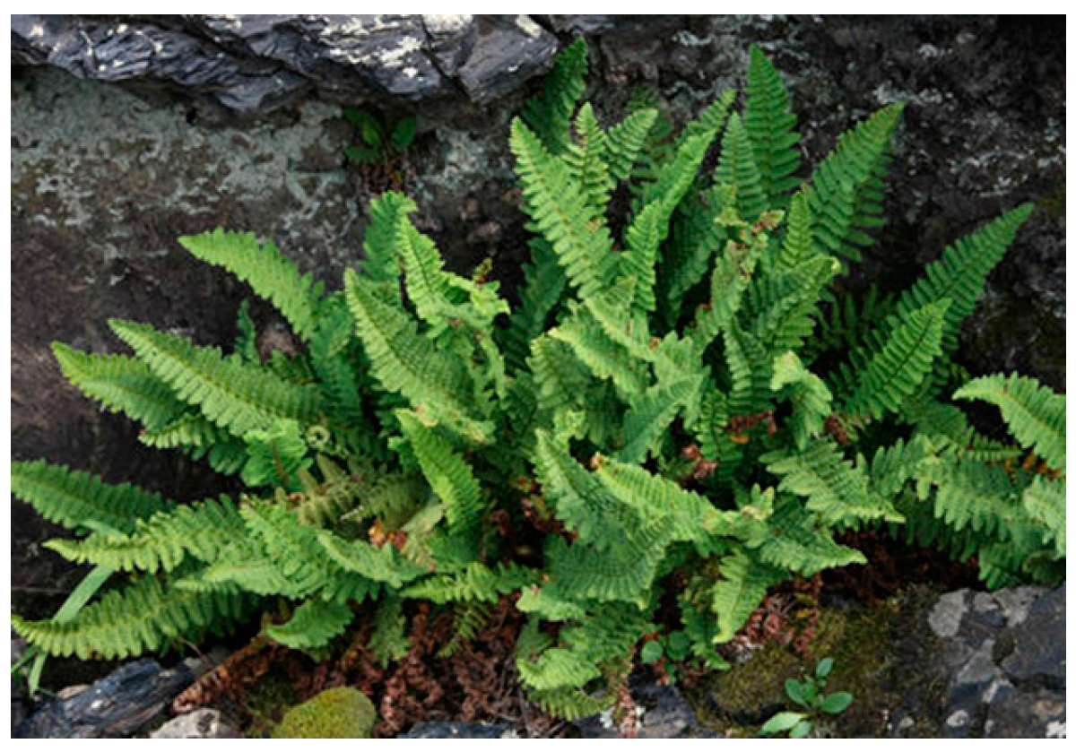
D. fragrans (Figure 1), from the Dryopteris genus of the Dryopteris family, is a deciduous perennial herb that is mostly distributed in Northeast China, Korea Japan, Russia and North America. [1,2]. The chemical components isolated from D. fragrans have exerted many biological effects [3,4,5,6]. In recent years, there have been many studies regarding the anticancer components of D. fragrans. Zhao [7] isolated a new coumarin—Dryofracoumarin A—from D. fragrans, which has cytotoxic activity. Su [8] reported that Dryofragin, a phloroglucinol derivative from D. fragrans, could stop human osteosarcoma U2OS cells from migrating and invading by lowering MMP-2 and MMP-9 expression and up-regulating the expression of TIMP-2 and TIMP-1 through the p38 MAPK and PI3K/AKT signal pathways. Zhong [9] obtained a novel sesquiterpene—Dryofraterpene A—from D. fragrans, which could significantly inhibit the proliferation of five kinds of cancer cell lines.
Thus, to discover new anticancer molecules, the chemical components of D. fragrans and its anticancer bioactivity were studied. This work can provide new resources for research and the development of new drugs against cancer.
2. Results and Discussion
2.1. Identification of Isolated Compounds
Compound 1 (Figure 2) was puce powder. Its acid hydrolysis yielded d-glucose and d-xylose [10]. The molecular formula—C20H26O13—was deduced from High Resolution Electrospray Ionization Mass Spectrometry (HR-ESI-MS) with an [M + Na]+ peak at 497.1274 (calcd. for 497.1271). In the infrared spectrum, it displayed a hydroxyl group (3412 cm−1), a carbonyl group (1706 cm−1), a double-bond group (1680 cm−1) and a benzene ring group (1602 cm−1).
The 1H-NMR spectrum (Table 1) exhibited three proton signals at δ 7.05 (d, J = 2.0 Hz), δ 6.78 (d, J = 8.0 Hz) and δ 6.96 (dd, J = 2.0, 8.0 Hz), corresponding to a 1,3,4-trisubstituted phenyl group. Moreover, it showed two trans-double-bond protons at δ 7.64 (d, J = 15.8 Hz) and δ 6.26 (d, J = 15.8 Hz). There were two anomeric proton signals at δ 5.68 (d, J = 7.6 Hz) and δ 4.48 (d, J = 7.4 Hz), suggesting two sugars of β-type [11].
In the 13C-NMR spectrum and DEPT spectrum, it showed an ester carbonyl carbon (δ 167.3) and a pair of trans-double-bond carbons at δ 148.2 and δ 114.5.
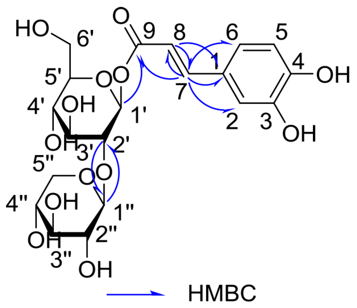
In the 1H Detected Heteronuclear Multiple Bond Correlation (HMBC) spectrum (Figure 3), the correlation of the anomeric proton signal of β-d-glucose at δ 5.68 with the ester carbonyl carbon at δ 167.3 indicated that the ester carbonyl group was located at C-1′. The linkage between C-1′′ from β-D-xylose and C-2′ from β-d-glucose by oxygen was revealed through the HMBC correlations of H-1′′/C-2′. On the basis of nuclear magnetic resonance (NMR) data and the relevant literature [12], compound 1 was identified as (E)-caffeic acid-9-O-β-d-xylpyranosyl-(1→2)-β-d-glucopyranosyl ester. The new compound was trivially named Fragranoside B.
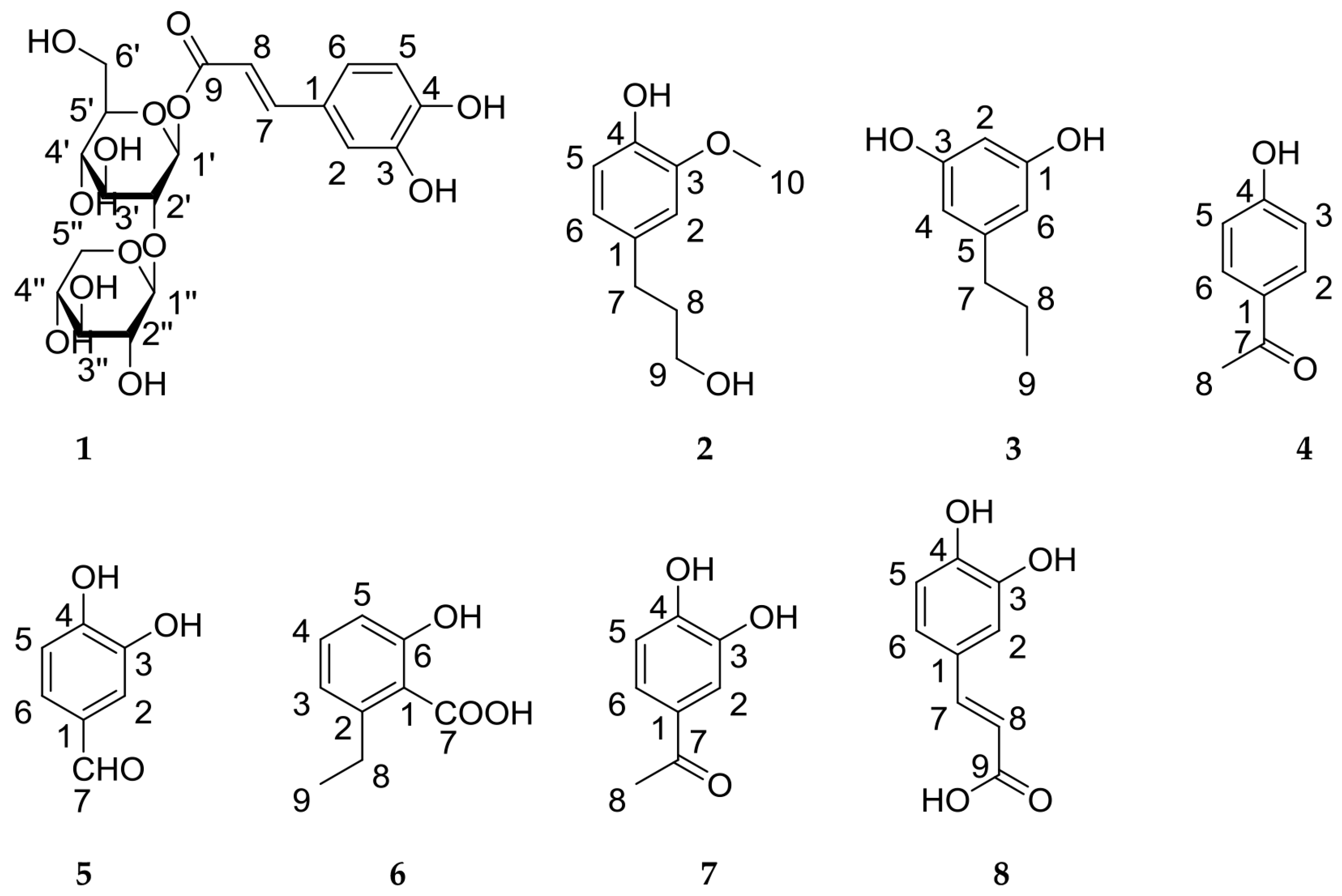
The known compounds (Figure 2) were identified as dihydroconiferyl alcohol (2) [13], 1,3-dihydroxyl-5-propylbenzene (3) [14], 4-hydroxyacetophenone (4) [15], 3,4-dihydroxybenzaldehyde (5) [16], 2-ethyl-6-hydroxybenzoic acid (6) [17], 3,4-dihydroxyacetophenone (7) [18] and caffeic acid (8) [19] through a comparison with the NMR data in the literature.
2.2. In Vitro Cytotoxicity Assay
A549, MCF-7, SGC7901 and human umbilical vein endothelial (HUVEC) cells were measured for cytotoxicity using the MTT assay with taxol as the positive control. As shown in Table 2, compound 1 showed significant inhibitory activity against MCF-7 cells with an IC50 value of 2.65 ± 0.14 μM. Compound 8 showed good cytotoxic activity against A549, MCF-7 and SGC7901 cells with IC50 values of 8.96–19.44 μM. However, the others were not active (IC50 > 50 μM). Notably, compound 1 exhibited no cytotoxic activities against A549, SGC7901 and noncancerous HUVEC cells. It showed good selectivity toward MCF-7. Thus, compound 1 can be considered as a promising lead compound and its anticancer mechanism should be further studied.
3. Materials and Methods
3.1. General Procedures
1D and 2D NMR spectra were obtained using a Bruker AM-400 (Bruker, Fällanden, Switzerland) instrument with tetramethyl silane as the internal standard. HR-ESI-MS was performed on VG Autospec-3000 mass spectrometers (VG, Manchester, UK). Semi-preparative High Performance Liquid Chromatography (HPLC) was performed using an Agilent 1100 liquid chromatography (Agilent Technologies, Waldbronn, Germany). Silica gel (200–300 mesh, Haiyang Chemical Co. Ltd., Qingdao, China) and Sephadex LH-20 (RuiDaHengHui Science and Technology Development Co., Ltd., Beijing, China) were used for column chromatography.
3.2. Plant Material
D. fragrans was collected from Wudalianchi City, Heilongjiang Province, China, in July 2015 and identified by Prof. Zhen-Yue Wang (Heilongjiang University of Chinese Medicine). The voucher specimen (No. XLMJ-20150828) of this plant was deposited in the Herbarium of Heilongjiang University of Chinese Medicine, Harbin, China.
3.3. Extraction and Isolation
The dried and powdered whole D. fragrans (8 kg) was extracted three times with water vapor. The concentrated extract (640 g) was fractionated by AB-8 macroporous resin eluted with EtOH–H2O (0:100, 30:70, 60:40, 95:5, v/v). The EtOH–H2O (30:70) extract (45 g) was chromatographed by silica gel eluted with CHCl3–MeOH (95:5, 90:10, 85:15, 80:20 and 50:50, v/v) to yield five fractions (A–E), respectively. Fraction A was subjected to column chromatography on silica gel eluted with CHCl3–MeOH (100:0, 98:2, 90:10 and 0:100, v/v) to afford four subfractions (A1–A4). Compound 2 (22 mg) was obtained from subfraction A1 by Sephadex LH-20 column chromatography with CHCl3. Compound 3 (3 mg) was purified by semi-preparative HPLC using MeOH–H2O (37:63, v/v) from subfraction A3. Fraction B was chromatographed by silica gel column (CHCl3–MeOH, 90:10, v/v), Sephadex LH-20 column (CHCl3) and semi-preparative HPLC (MeOH–H2O, 45:55, v/v) to give compound 4 (8 mg). Fraction C was separated by silica gel (CHCl3–MeOH, 80:20, v/v), Sephadex LH-20 column chromatography (CHCl3) and recrystallization to afford compound 5 (12 mg). Fraction D was repeatedly chromatographed using silica gel columns (CHCl3–MeOH) to yield three subfractions (D1–D3). Subfraction D1 was subject to Sephadex LH-20 columns chromatography (CHCl3) and recrystallization to yield compound 6 (20 mg). Compounds 7 (4 mg) and 8 (10 mg) were obtained from subfraction D3 by Sephadex LH-20 columns chromatography (CHCl3) and recrystallization. Compound 1 (9 mg) was isolated by column chromatography using Sephadex LH-20 (CHCl3–MeOH, 50:50, v/v), silica gel and semi-preparative HPLC (MeOH–H2O, 15:85, v/v) from fraction E.
3.4. Acid Hydrolysis
Compound 1 (5 mg) was hydrolyzed with 10 mL of 0.01 M H2SO4 for 4 h at 100 °C. After cooling, the hydrolysate was neutralized by 0.02 M KOH then extracted by CH2Cl2. The sugars in the aquatic layer were monitored by Thin-Layer Chromatography (TLC) with BuOH–H2O–AcOH (40:10:50, v/v/v, upper BuOH layer) as a developing system when compared with authentic sugars. The TLC plate was sprayed with a vanillin–H2SO4 solvent [20].
3.5. Cell Culture
Human A549, MCF-7, SGC7901 and HUVEC cells were obtained from the Cell Library of Committee on Type Culture Collection of Chinese Academy of Sciences (Shanghai, China). Cells were cultured at 37 °C, 5% CO2 in the Roswell Park Memorial Institute (RPMI) medium containing 10% FBS, 100 U/mL penicillin and 100 U/mL streptomycin.
3.6. MTT Assay
Anticancer activity was evaluated by the MTT assay. Compounds 1–8 were dissolved in dimethyl sulfoxide (DMSO) and diluted with RPMI medium for appropriate concentrations (0 μM, 0.08 μM, 0.4 μM, 2 μM, 10 μM and 50 μM). A549, MCF-7, SGC7901 and HUVEC cells were seeded in 96-well microtiter plates (100 μL, 5000 cells/well). After 24 h, the medium was removed and 100 μL of tested compounds with various concentrations were added into 96-well microtiter plates for 48 h. Next, 10 μL of MTT was added and the 96-well microtiter plates were incubated for another 4 h. The medium was removed and 150 μL DMSO was added to each well to dissolve the formazan crystals. The absorbance was measured by microplate spectrophotometer (Molecular Devices, Palo Alto, CA, USA) at 570 nm. Taxol was used as the positive control (0 μM, 0.01 μM, 0.04 μM, 0.16 μM, 0.64 μM, and 2.56 µM for 48 h). Half Maximal Inhibitory Concentration (IC50) values were calculated by GraphPad Prism. Data were obtained from three independent assays.
4. Conclusions
A new phenylpropanoid, (E)-caffeic acid-9-O-β-d-xylpyranosyl-(1→2)-β-d-glucopyranosyl ester (1), and seven known phenolics (2–8) were isolated from the medicinal plant D. fragrans. Compounds 1 and 8 showed good anticancer activity.
Acknowledgments
This work was supported by the Natural Science Foundation Project, Tibet, China (No. 2016-ZR-NZ-03) and the Science and Technology Project, Nyingchi Municipality, Tibet, China (No. 2016-03).
Author Contributions
Y.-L.Z., X.-F.Y. and Z.-D.L. conceived and designed the experiments; Z.-D.L. and D.-D.Z. performed the experiments; S.J. and B.X. analyzed the data and wrote the paper.
Conflicts of Interest
The authors declare no conflict of interest.
References
- Huang, Q.Y.; Li, W.H.; Fan, R.F.; Chang, Y. New mads-box gene in fern: Cloning and expression analysis of dfmads1 from dryopteris fragrans. PLoS ONE 2014, 9, e86349. [Google Scholar] [CrossRef] [PubMed]
- Kuang, H.X.; Sun, C.; Zhang, Y.L.; Zhang, Y.L.; Chen, D.; Yang, B.Y.; Xia, Y.G. Three drimane sesquiterpene glucoside from the aerial parts of dryopteris fragrans (L.) schot. Fitoterapia 2009, 80, 134–137. [Google Scholar] [CrossRef] [PubMed]
- Li, X.J.; Wang, W.; Luo, M.; Li, C.Y.; Zu, Y.G.; Mu, P.S.; Fu, Y.J. Solvent-free microwave extraction of essential oil from dryopteris fragrans and evaluation of antioxidant activity. Food Chem. 2012, 133, 437–444. [Google Scholar] [CrossRef] [PubMed]
- Li, N.; Gao, C.; Peng, X.; Wang, W.; Luo, M.; Fu, Y.J.; Zu, Y.G. Aspidin bb, a phloroglucinol derivative, exerts its antibacterial activity against staphylococcus aureus by inducing the generation of reactive oxygen species. Res. Microbiol. 2014, 165, 263–272. [Google Scholar] [CrossRef] [PubMed]
- Huang, Y.H.; Zeng, W.M.; Li, G.Y.; Liu, G.Q.; Zhao, D.D.; Wang, J.; Zhang, Y.L. Characterization of a new sesquiterpene and antifungal activities of chemical constituents from dryopteris fragrans (L.) schott. Molecules 2014, 19, 507–513. [Google Scholar] [CrossRef] [PubMed]
- Peng, B.; Bai, R.F.; Li, P.; Han, X.Y.; Wang, H.; Zhu, C.C.; Zeng, Z.P.; Chai, X.Y. Two new glycosides from dryopteris fragrans with anti-inflammatory activities. J. Asian Nat. Prod. Res. 2016, 18, 59–64. [Google Scholar] [CrossRef] [PubMed]
- Zhao, D.D.; Zhao, Q.S.; Liu, L.; Chen, Z.Q.; Zeng, W.M.; Lei, H.; Zhang, Y.L. Compounds from dryopteris fragrans (L.) schott with cytotoxic activity. Molecules 2014, 19, 3345–3355. [Google Scholar] [CrossRef] [PubMed]
- Su, Y.; Wan, D.Q.; Song, W.Q. Dryofragin inhibits the migration and invasion of human osteosarcoma U2OS cells by suppressing MMP-2/9 and elevating TIMP-1/2 through PI3K/AKT and p38 MAPK signaling pathways. Anti-Cancer Drug 2016, 27, 660–668. [Google Scholar] [CrossRef] [PubMed]
- Zhong, Z.C.; Zhao, D.D.; Liu, Z.D.; Jiang, S.; Zhang, Y.L. A new human cancer cell proliferation inhibition sesquiterpene, dryofraterpene a, from medicinal plant dryopteris fragrans (L.) schott. Molecules 2017, 22, 180. [Google Scholar] [CrossRef] [PubMed]
- Eskander, J.; Lavaud, C.; Pouny, I.; Soliman, H.S.; Abdel-Khalik, S.M.; Mahmoud, I.I. Saponins from the seeds of mimusops laurifolia. Phytochemistry 2006, 67, 1793–1799. [Google Scholar] [CrossRef] [PubMed]
- Grant, D.M.; Harris, R.K. Encyclopedia of Nuclear Magnetic Resonance; Wiley: Hoboken, NJ, USA, 2002. [Google Scholar]
- Zhang, Y.L.; Li, J.P.; Li, G.Y. Two new compounds from dryopteris fragrans. Zhong Yao Cai 2014, 37, 599–602. [Google Scholar] [PubMed]
- Li, L.Y.; Seeram, N.P. Further investigation into maple syrup yields 3 new lignans, a new phenylpropanoid, and 26 other phytochemicals. J. Agric. Food Chem. 2011, 59, 7708–7716. [Google Scholar] [CrossRef] [PubMed]
- Alonso, E.; Ramon, D.J.; Yus, M. Simple synthesis of 5-substituted resorcinols: A revisited family of interesting bioactive molecules. J. Org. Chem. 1997, 62, 417–421. [Google Scholar] [CrossRef] [PubMed]
- Dhanuskodi, S.; Manikandan, S. Epr investigations on γ-irradiated 4-hydroxyacetophenone single crystals: An nlo material. Radiat. Effects Defects Solids 2005, 160, 197–205. [Google Scholar] [CrossRef]
- Jeschke, T.; Wensbo, D.; Annby, U.; Gronowitz, S.; Cohen, L.A. A novel approach to bz-substituted tryptophans via pd-catalysed coupling/annulation. Tetrahedron Lett. 1993, 34, 6471–6474. [Google Scholar] [CrossRef]
- Dain, J.G.; Ernst, L.A.; Campbell, I.M.; Bentley, R. The formation of 6-ethylsalicylic acid by mycobacterium phlei. Biomed. Mass Spectrom. 1974, 1, 57–61. [Google Scholar] [CrossRef] [PubMed]
- Zhao, Y.B.; Shen, Y.M.; He, H.P.; Mu, Q.Z.; Hao, X.J. Antifungal agent and other constituents from cynanchum otophyllum. Nat. Prod. Res. 2007, 21, 203–210. [Google Scholar] [CrossRef] [PubMed]
- Damtoft, S.; Jensen, S.R. Three phenylethanoid glucosides of unusual structure from chirita sinensis (gesneriaceae). Phytochemistry 1994, 37, 441–443. [Google Scholar] [CrossRef]
- Al-Sayed, E.; Eldahshan, O.A.; Bahgat, D.M.; Singab, A.N. Cytotoxic oleanane-type saponins from the leaves of albizia anthelmintica brongn. Chem. Biodivers. 2016, 13, 1666–1673. [Google Scholar] [CrossRef] [PubMed]
Sample Availability: Samples of the compounds are unavailable from the authors. |
Figure 1.
D. fragrans plant.

Figure 2.
Structures of 1–8 isolated from D. fragrans.

Figure 3.
Heteronuclear Multiple Bond Correlation (HMBC) correlations of 1.


Table 1.
13C-NMR (100 MHz) and 1H-NMR (400 MHz) spectral data of compound 1 in MeOD.
| No. | δC | δH (J in Hz) | No. | δC | δH (J in Hz) |
|---|---|---|---|---|---|
| 1 | 127.6 (C) | 1′ | 94.3 (CH) | 5.68 (d, 7.6) | |
| 2 | 115.1 (CH) | 7.05 (d, 2.0) | 2′ | 83.5 (CH) | 3.60 (m) |
| 3 | 146.9 (C) | 3′ | 77.6 (CH) | 3.65 (m) | |
| 4 | 149.9 (C) | 4′ | 70.7 (CH) | 3.42 (m) | |
| 5 | 116.5 (CH) | 6.78 (d, 8.0) | 5′ | 78.7 (CH) | 3.40 (m) |
| 6 | 123.2 (CH) | 6.96 (dd, 8.0, 2.0) | 6′ | 62.2 (CH2) | 3.84 (dd, 1.6, 12.5), 3.68 (d, 6.3) |
| 7 | 148.2 (CH) | 7.64 (d, 15.8) | 1′′ | 106.6 (CH) | 4.48 (d, 7.4) |
| 8 | 114.5 (CH) | 6.26 (d, 15.8) | 2′′ | 75.7 (CH) | 3.18 (m) |
| 9 | 167.3 (C) | 3′′ | 77.5 (CH) | 3.30 (m) | |
| 4′′ | 71.0 (CH) | 3.32 (m) | |||
| 5′′ | 67.4 (CH2) | 3.70 (d, 5.3), 3.14 (m) |
Table 2.
Cytotoxicity of compounds 1–8 against A549, MCF-7, SGC7901 and human umbilical vein endothelial (HUVEC) cells.
Table 2.
Cytotoxicity of compounds 1–8 against A549, MCF-7, SGC7901 and human umbilical vein endothelial (HUVEC) cells.
| Compounds | IC50 (μM) 1 | |||
|---|---|---|---|---|
| A549 | MCF-7 | SGC7901 | HUVEC | |
| 1 | >50 | 2.65 ± 0.14 | >50 | >50 |
| 2 | >50 | >50 | >50 | ND 3 |
| 3 | >50 | >50 | >50 | ND |
| 4 | >50 | >50 | >50 | ND |
| 5 | >50 | >50 | >50 | ND |
| 6 | >50 | >50 | >50 | ND |
| 7 | >50 | >50 | >50 | ND |
| 8 | 10.41 ± 1.02 | 19.44 ± 1.74 | 8.96 ± 0.99 | ND |
| Taxol 2 | 0.047 ± 0.08 | 0.073 ± 0.11 | 0.069 ± 0.03 | ND |
1 IC50 values represent mean ± standard deviation of three individual observations; 2 Taxol was used as the positive control; 3 ND, not determined.
© 2018 by the authors. Licensee MDPI, Basel, Switzerland. This article is an open access article distributed under the terms and conditions of the Creative Commons Attribution (CC BY) license (http://creativecommons.org/licenses/by/4.0/).
Share and Cite
MDPI and ACS Style
Liu, Z.-D.; Zhao, D.-D.; Jiang, S.; Xue, B.; Zhang, Y.-L.; Yan, X.-F. Anticancer Phenolics from Dryopteris fragrans (L.) Schott. Molecules 2018, 23, 680. https://0-doi-org.brum.beds.ac.uk/10.3390/molecules23030680
AMA Style
Liu Z-D, Zhao D-D, Jiang S, Xue B, Zhang Y-L, Yan X-F. Anticancer Phenolics from Dryopteris fragrans (L.) Schott. Molecules. 2018; 23(3):680. https://0-doi-org.brum.beds.ac.uk/10.3390/molecules23030680
Chicago/Turabian StyleLiu, Zhen-Dong, Dan-Dan Zhao, Shuai Jiang, Bei Xue, Yan-Long Zhang, and Xiu-Feng Yan. 2018. "Anticancer Phenolics from Dryopteris fragrans (L.) Schott" Molecules 23, no. 3: 680. https://0-doi-org.brum.beds.ac.uk/10.3390/molecules23030680