
Antitumor, Antiviral, and Anti-Inflammatory Efficacy of Essential Oils from Atractylodes macrocephala Koidz. Produced with Different Processing Methods


Abstract
:1. Introduction
2. Results
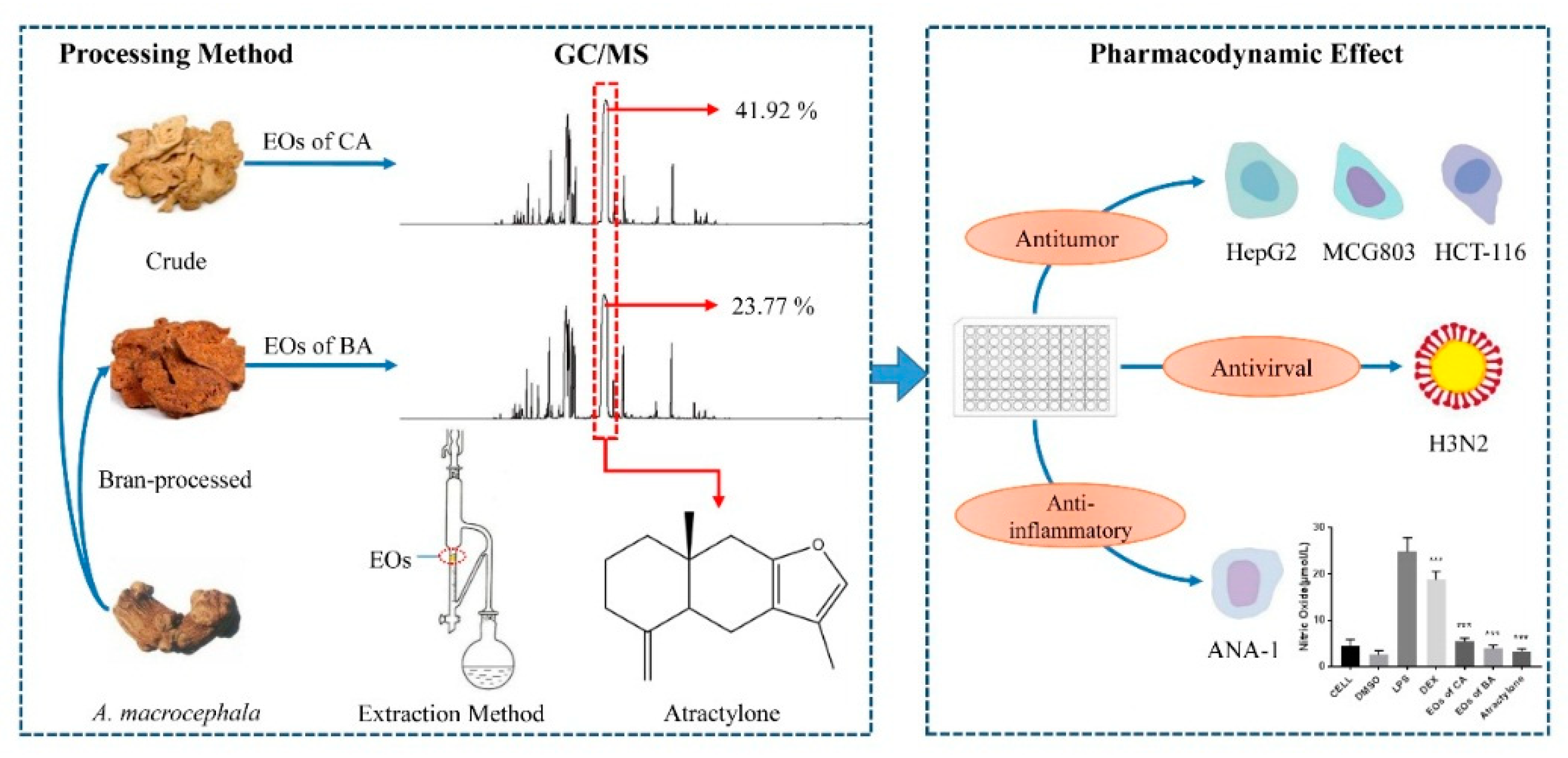
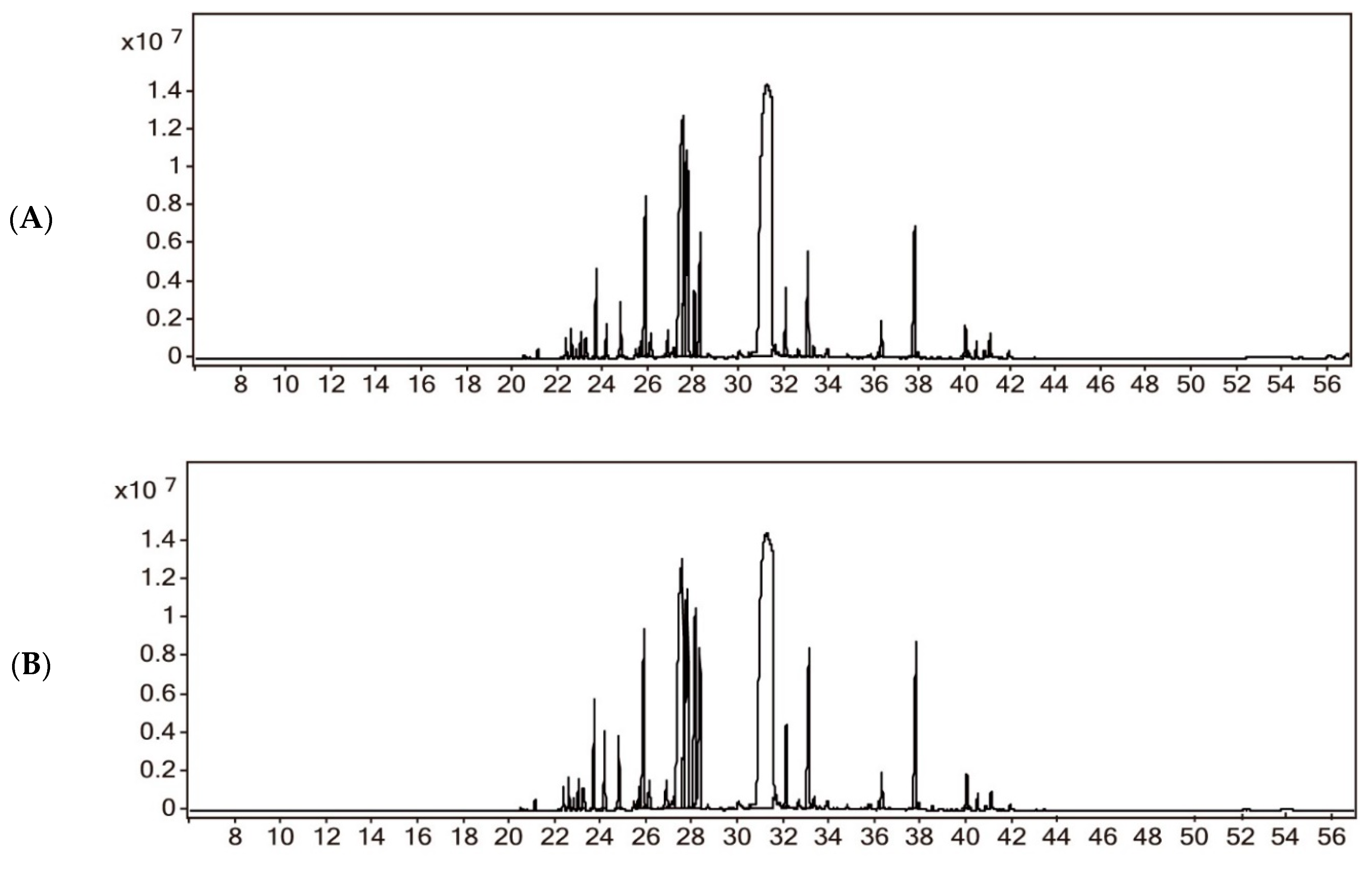
2.1. Chemical Composition of Essential Oils
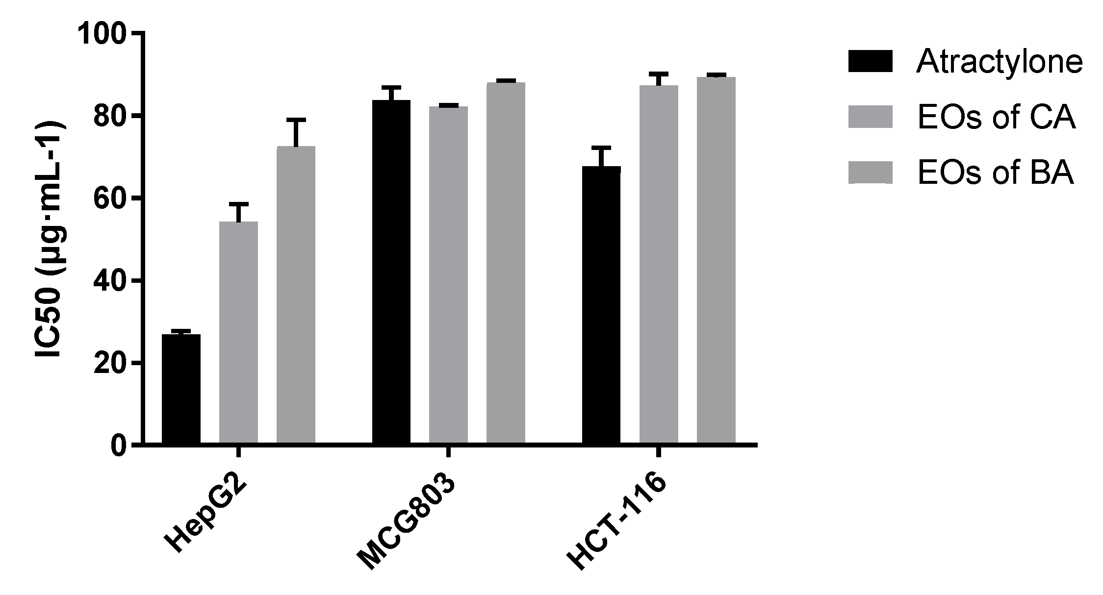
2.2. Antitumor Activity of Essential Oil from A. macrocephala
2.3. Antiviral Activity of Essential Oil from A. macrocephala
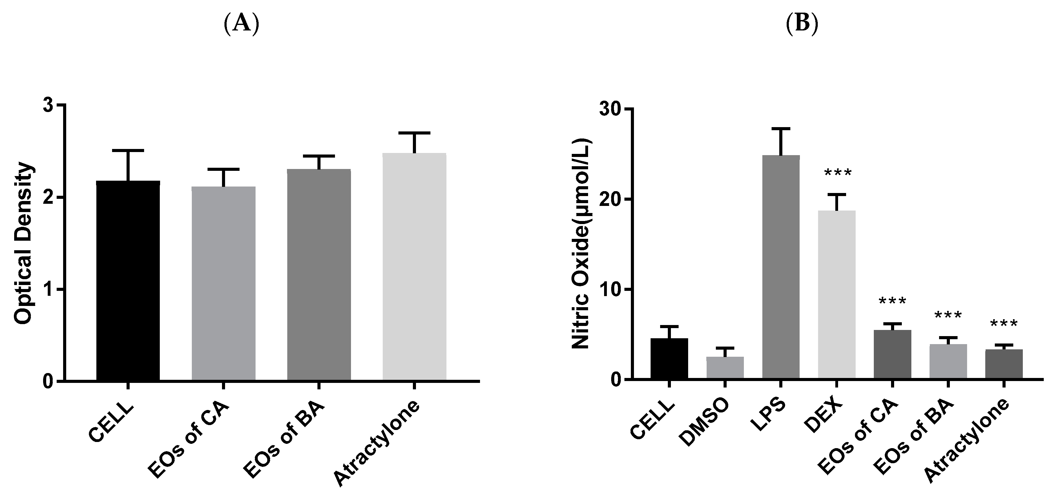
2.4. Anti-Inflammatory Activity of EOs from A. macrocephala
3. Discussion
4. Materials and Methods
4.1. Plant Materialsand Chemicals
4.2. Extraction Methods
4.3. Chemical Components Analysis by GC/MS
4.4. Effects on Liver Cancer, Gastric Cancer, and Intestinal Cancer Cells
4.5. Anti-H3N2 Virucidal Activity
4.5.1. Sample Cytotoxicity Test
4.5.2. In vitro Antiviral Virus Test
4.6. Anti-Inflammatory Activity
4.6.1. MTT Method to Investigate the Effect of Test Drugs on Cell Viability
4.6.2. Inhibition of ANA-1 Cell Inflammatory Model Induced by LPS Stimulation
5. Conclusions
Author Contributions
Funding
Acknowledgments
Conflicts of Interest
References
- Hoang, L.S.; Tran, M.H.; Lee, J.S.; Ngo, Q.M.; Woo, M.H.; Min, B.S. Inflammatory inhibitory activity of sesquiterpenoids from Atractylodes macrocephala rhizomes. Chem. Pharm. Bull. (Tokyo) 2016, 64, 507–511. [Google Scholar] [CrossRef] [PubMed]
- Cai, H.; Xu, Z.; Luo, S.; Zhang, W.; Cao, G.; Liu, X.; Lou, Y.; Ma, X.; Qin, K.; Cai, B. Study on chemical fingerprinting of crude and processed Atractylodes macrocephala from different locations in Zhejiang province by reversed-phase high-performance liquid chromatography coupled with hierarchical cluster analysis. Pharm. Mag. 2012, 8, 300–307. [Google Scholar] [CrossRef] [PubMed]
- Yu, L.; Jia, T.; Qian, C. Rearch on the chemical references of Atractylodes macrocephala Koidz. Asia-Pac. Tradit. Med. 2010, 6, 36–39. [Google Scholar]
- Liu, P.; Teng, J.; Zhang, Y.W.; Takaishi, Y.; Duan, H.Q. Chemical constituents from rhizome of Phlomis umbrosa. Yao Xue Xue Bao Acta Pharm. Sin. 2007, 42, 401–404. [Google Scholar]
- Pharmacopoeia Commission of the Ministry of Health of the People’s Republic of China. Pharmacopoeia of the People’s Republic of China; China Medical Science Press: Beijing, China, 2015. [Google Scholar]
- Wang, X.T.; Li, L.H.; Ran, X.K.; Dou, D.Q.; Li, B.; Yang, B.Y.; Li, W.; Koike, K.; Kuang, H.X. What caused the changes in the usage of Atractylodis macrocephalae Rhizoma from ancient to current times? J. Nat. Med. Tokyo 2016, 70, 36–44. [Google Scholar] [CrossRef] [PubMed]
- Zhang, J.D.; Cao, G.; Xia, Y.H.; Wen, C.P.; Fan, Y.S. Fast analysis of principal volatile compounds in crude and processed Atractylodes macrocephala by an automated static headspace gas chromatography-mass spectrometry. Pharm. Mag. 2014, 10, 249–253. [Google Scholar] [CrossRef] [PubMed]
- Ji, G.Q.; Chen, R.Q.; Wang, L. Anti-inflammatory activity of atractylenolide III through inhibition of nuclear factor-kappa B and mitogen-activated protein kinase pathways in mouse macrophages. Immunopharm. Immunot. 2016, 38, 98–102. [Google Scholar] [CrossRef] [PubMed]
- Chen, J.; Liu, X.; Dou, D.Q. Bidirectional effective components of Atractylodis macrocephalae Rhizoma on gastrointestinal peristalsis. Int. J. Pharm. 2016, 12, 108–115. [Google Scholar] [CrossRef]
- Song, H.P.; Hou, X.Q.; Li, R.Y.; Yu, R.; Li, X.; Zhou, S.N.; Huang, H.Y.; Cai, X.; Zhou, C. Atractylenolide I stimulates intestinal epithelial repair through polyamine-mediated Ca2+ signaling pathway. Phytomedicine 2017, 28, 27–35. [Google Scholar] [CrossRef]
- Huang, H.L.; Lin, T.W.; Huang, Y.L.; Huang, R.L. Induction of apoptosis and differentiation by atractylenolide-1 isolated from Atractylodes macrocephala in human leukemia cells. Bioorg. Med. Chem. Lett. 2016, 26, 1905–1909. [Google Scholar] [CrossRef]
- Liu, H.Y.; Zhu, Y.J.; Zhang, T.; Zhao, Z.G.; Zhao, Y.; Cheng, P.; Li, H.; Gao, H.; Su, X.M. Anti-tumor effects of Atractylenolide I isolated from Atractylodes macrocephala in human lung carcinoma cell lines. Molecules 2013, 18, 13357–13368. [Google Scholar] [CrossRef] [PubMed]
- Long, F.Y.; Wang, T.; Jia, P.; Wang, H.F.; Qing, Y.; Xiong, T.T.; He, M.J.; Wang, X.L. Anti-tumor effects of Atractylenolide-I on human ovarian cancer cells. Med. Sci. Monit. 2017, 23, 571–579. [Google Scholar] [CrossRef] [PubMed]
- Zhou, J.R.; Yuan, X.R.; Li, L.; Zhang, T.; Wang, B. Comparison of different methods for extraction of Cinnamomi ramulus: Yield, chemical composition and in vitro antiviral activities. Nat. Prod. Res. 2017, 31, 2909–2913. [Google Scholar] [CrossRef] [PubMed]
- Comelli, N.C.; Romero, O.E.; Diez, P.A.; Marinho, C.F.; Schliserman, P.; Carrizo, A.; Ortiz, E.V.; Duchowicz, P.R. QSAR study of biologically active essential oils against beetles infesting the walnut in Catamarca, Argentina. J. Agr. Food Chem. 2018, 66, 12855–12865. [Google Scholar] [CrossRef] [PubMed]
- Perez-Outeiral, J.; Elcoroaristizabal, S.; Amigo, J.M.; Vidal, M. Development and validation of a method for the determination of regulated fragrance allergens by High-Performance Liquid Chromatography and parallel factor analysis 2. J. Chromatography A 2017, 1526, 82–92. [Google Scholar] [CrossRef] [PubMed]
- Szoke, E.; Maday, E.; Kiss, S.A.; Sonnewend, L.; Lemberkovics, E. Effect of magnesium on essential oil formation of genetically transformed and non-transformed chamomile cultures. J. Am. Coll. Nutr. 2004, 23, 763s–767s. [Google Scholar] [CrossRef] [PubMed]
- Jiang, T.; Li, K.F.; Liu, H.H.; Yang, L. Extraction of biomedical compounds from the wood of Pterocarpus macarocarpus Kurz heartwood. Pak. J. Pharm. Sci. 2018, 31, 913–918. [Google Scholar]
- Li, X.F.; Mou, Z.; Wang, X.P.; Wu, H.M.; Xu, F.; Zhu, C.X.; Zhang, M. Geographic analysis of the cultivation region of Ai pian derived from Blumea balsamifera through the determination of volatiles in the medicinal product and blood of treated mice by gas chromatography-mass spectrometry (GC-MS). Instrum. Sci. Technol. 2019, 47, 597–610. [Google Scholar] [CrossRef]
- Sedaghat Doost, A.; Stevens, C.V.; Claeys, M.; Van Der Meeren, P. Fundamental study on the salt tolerance of oregano essential oil-in-water nanoemulsions containing tween 80. Langmuir: ACS J. Surf. Colloids 2019. [Google Scholar] [CrossRef]
- Zhang, X.; Li, M.; Cheng, Z.; Ma, L.; Zhao, L.; Li, J. A comparison of electronic nose and gas chromatography-mass spectrometry on discrimination and prediction of ochratoxin A content in Aspergillus carbonarius cultured grape-based medium. Food Chem. 2019, 297, 124850. [Google Scholar] [CrossRef]
- El Mokni, R.; Majdoub, S.; Chaieb, I.; Jlassi, I.; Joshi, R.K.; Hammami, S. Chromatographic analysis, antimicrobial and insecticidal activities of the essential oil of Phlomis floccosa D. Don. Biomed. Chromatogr. BMC 2019. [Google Scholar] [CrossRef] [PubMed]
- Chen, F.; Jia, J.; Zhang, Q.; Gu, H.; Yang, L. A modified approach for isolation of essential oil from fruit of Amorpha fruticosa Linn using microwave-assisted hydrodistillation concatenated liquid-liquid extraction. J. Chromatography A 2017, 1524, 254–265. [Google Scholar] [CrossRef] [PubMed]
- Gong, F.; Zhang, Q.; Wang, B.T. Chemical characterization of herbal formula yupingfeng powder and its single herbs (i) volatile components. Anal. Lett. 2009, 42, 2610–2624. [Google Scholar] [CrossRef]
- Andriana, Y.; Xuan, T.D.; Quy, T.N.; Tran, H.D.; Le, Q.T. Biological activities and chemical constituents of essential oils from Piper cubeba Bojer and Piper nigrum L. Molecules 2019, 24, 1876. [Google Scholar] [CrossRef] [PubMed]
- Cao, G.; Cai, H.; Cong, X.; Liu, X.; Ma, X.; Lou, Y.; Qin, K.; Cai, B. Global detection and analysis of volatile components from sun-dried and sulfur-fumigated herbal medicine by comprehensive two-dimensional gas chromatography/time-of-flight mass spectrometry. Analyst 2012, 137, 3828–3835. [Google Scholar] [CrossRef] [PubMed]
- Karakaya, S.; Koca, M.; Yilmaz, S.V.; Yildirim, K.; Pinar, N.M.; Demirci, B.; Brestic, M.; Sytar, O. Molecular docking studies of coumarins isolated from extracts and essential oils of zosima absinthifolia link as potential inhibitors for Alzheimer’s disease. Molecules 2019, 24, 722. [Google Scholar] [CrossRef] [PubMed]
- Le, T.B.; Beaufay, C.; Nghiem, D.T.; Pham, T.A.; Mingeot-Leclercq, M.P.; Quetin-Leclercq, J. Evaluation of the anti-trypanosomal activity of vietnamese essential oils, with emphasis on curcuma longa L. and its components. Molecules 2019, 24, 1158. [Google Scholar] [CrossRef]
- Stoev, G. Application and chiral recognition of heptakis (2,6-di-O-methyl-3-O-trifluoroacetyl)-beta-cyclodextrin as a stationary phase for the gas-chromatographic separation of enantiomers. J Chromatogr. 1992, 589, 257–263. [Google Scholar] [CrossRef]
- Guo, F.Q.; Huang, L.F.; Zhou, S.Y.; Zhang, T.M.; Liang, Y.Z. Comparison of the volatile compounds of Atractylodes medicinal plants by headspace solid-phase microextraction-gas chromatography-mass spectrometry. Anal. Chim. Acta 2006, 570, 73–78. [Google Scholar] [CrossRef]
- Kapetanos, C.; Karioti, A.; Bojovic, S.; Marin, P.; Veljic, M.; Skaltsa, H. Chemical and principal-component analyses of the essential oils of apioideae taxa (Apiaceae) from central Balkan. Chem. Biodivers. 2008, 5, 101–119. [Google Scholar] [CrossRef]
- Borisov, R.S.; Esparza, C.; Goriainov, S.V.; Zaikin, V.G. Suitable in-situ derivatization of alcohols by reaction with basic amines in direct analysis in real time mass spectrometry. Talanta 2019, 200, 31–40. [Google Scholar] [CrossRef] [PubMed]
- Liao, G.; Chen, H.M.; Shi, B.F. Synthesis of phthalic acid derivatives via Pd-catalyzed alkoxycarbonylation of aromatic C–H bonds with alkyl chloroformates. Chem. Commun. 2018, 54, 10859–10862. [Google Scholar] [CrossRef] [PubMed]
- Hamad, Y.K.; Abobakr, Y.; Salem, M.Z.M.; Ali, H.M.; Al-Sarar, A.S.; Al-Zabib, A.A. Activity of plant extracts/essential oils against three plant pathogenic fungi and mosquito larvae: GC/MS analysis of bioactive compounds. Bioresources 2019, 14, 4489–4511. [Google Scholar] [CrossRef]
- El-Gawad, A.A.; Elshamy, A.; El Gendy, A.E.; Gaara, A.; Assaeed, A. Volatiles profiling, allelopathic activity, and antioxidant potentiality of xanthium strumarium leaves essential oil from egypt: Evidence from chemometrics analysis. Molecules 2019, 24, 584. [Google Scholar] [CrossRef] [PubMed]
- Lin, Y.; Wang, G.H.; Yuan, K. Extraction and GC-MS analysis of the volatile constituents of a tractylodes macrocephala. Asian J. Chem. 2011, 23, 551–555. [Google Scholar]
- Zhou, Q.; Liu, S.; Liu, Y.; Song, H. Comparative analysis of volatiles of 15 brands of extra-virgin olive oils using solid-phase micro-extraction and solvent-assisted flavor evaporation. Molecules 2019, 24, 1512. [Google Scholar] [CrossRef] [PubMed]
- He, X.W.; Zhang, L.T.; Chen, J.P.; Sui, J.L.; Yi, G.H.; Wu, J.Y.; Ma, Y.Z. Correlation between chemical composition and antifungal activity of clausena lansium essential oil against candida spp. Molecules 2019, 24, 1394. [Google Scholar] [CrossRef] [PubMed]
- Andrade, M.A.; das Gracas Cardoso, M.; de Andrade, J.; Silva, L.F.; Teixeira, M.L.; Valerio Resende, J.M.; da Silva Figueiredo, A.C.; Barroso, J.G. Chemical composition and antioxidant activity of essential oils from cinnamodendron dinisii schwacke and siparuna guianensis aublet. Antioxidants 2013, 2, 384–397. [Google Scholar] [CrossRef] [PubMed]
- Wang, B.; Ge, L.; Mo, J.G.; Su, L.; Li, Y.J.; Yang, K.D. Essential oils and ethanol extract from Camellia nitidissima and evaluation of their biological activity. J. Food Sci. Tech. Mys. 2018, 55, 5075–5081. [Google Scholar] [CrossRef]
- Mahanta, B.P.; Sut, D.; Kemprai, P.; Paw, M.; Lal, M.; Haldar, S. A (1) H-NMR spectroscopic method for the analysis of thermolabile chemical markers from the essential oil of black turmeric (Curcuma caesia) rhizome: Application in post-harvest analysis. Phytochem. Anal. PCA 2019. [Google Scholar] [CrossRef]
- Benedetto, C.; D’Auria, M.; Mecca, M.; Prasad, P.; Singh, P.; Singh, S.; Sinisgalli, C.; Milella, L. Chemical and biological evaluation of essential oil from Saussurea costus (Falc.) Lipsch. from Garhwal Himalaya collected at different harvesting periods. Nat. Prod. Res. 2019, 33, 2355–2358. [Google Scholar] [CrossRef] [PubMed]
- Dai, C.X.; Huang, X.Y.; Lv, R.Q.; Zhang, Z.C.; Sun, J.; Aheto, J.H. Analysis of volatile compounds of Tremella aurantialba fermentation via electronic nose and HS-SPME-GC-MS. J. Food Saf. 2018, 38. [Google Scholar] [CrossRef]
- Mirza, B.; Samiei, S.S.; Taherkhani, M.; Fathizadeh, M. Composition of the essential oils of Anthemis hyalina DC., Achillea nobilis L. and Cichorium intybus L. Three asteraceae herbs growing wild in Iran. Asian J. Chem. 2012, 24, 1151–1154. [Google Scholar]
- Ling, Y.; Wang, X.M.; Wang, C.N.; Xu, C.J.; Zhang, W.; Zhang, Y.H.; Zhang, Y.N. Hybrids from farnesylthiosalicylic acid and hydroxamic acid as dual ras-related signaling and histone deacetylase (hdac) inhibitors: Design, synthesis and biological evaluation. Chemmedchem 2015, 10, 971–976. [Google Scholar] [CrossRef] [PubMed]
- Elmassry, M.M.; Kormod, L.; Labib, R.M.; Farag, M.A. Metabolome based volatiles mapping of roasted umbelliferous fruits aroma via HS-SPME GC/MS and peroxide levels analyses. J. Chromatogr. B 2018, 1099, 117–126. [Google Scholar] [CrossRef] [PubMed]
- Zhang, L.; Yang, Z.; Huang, Z.; Zhao, M.; Li, P.; Zhou, W.; Zhang, K.; Zheng, X.; Lin, L.; Tang, J.; et al. Variation in essential oil and bioactive compounds of curcuma kwangsiensis collected from natural habitats. Chem. Biodivers. 2017, 14. [Google Scholar] [CrossRef]
- Zhu, J.; Jia, R.F.; Lai, P.X. Chemical composition of the essential oil of chloranthus serratus from China. Chem. Nat. Compd. 2017, 53, 159–161. [Google Scholar] [CrossRef]
- Xu, S.Z.; Qi, X.J.; Liu, Y.Q.; Liu, Y.H.; Lv, X.; Sun, J.Z.; Cai, Q. UPLC-MS/MS of Atractylenolide I, Atractylenolide II, Atractylenolide III, and Atractyloside A in rat plasma after oral administration of raw and wheat bran-processed atractylodis rhizoma. Molecules 2018, 23, 3234. [Google Scholar] [CrossRef]
- Su, H.H.; Wang, W.D.; Bao, L.Z.; Wang, S.S.; Cao, X.F. Synthesis and evaluation of essential oil-derived beta-methoxyacrylate derivatives as high potential fungicides. Molecules 2017, 22, 763. [Google Scholar] [CrossRef]
- Zeng, M.N.; Li, M.; Li, M.; Zhang, B.B.; Li, B.K.; Zhang, L.; Feng, W.S.; Zheng, X.K. 2-Phenylacetamide isolated from the seeds of lepidium apetalum and its estrogen-like effects in vitro and in vivo. Molecules 2018, 23, 2293. [Google Scholar] [CrossRef]
- Guo, Z.B.; Liu, Z.Z.; Yue, H.F.; Wang, J.Y. Beta-elemene increases chemosensitivity to 5-fluorouracil through down-regulating microRNA-191 expression in colorectal carcinoma cells. J. Cell Biochem. 2018, 119, 7032–7039. [Google Scholar] [CrossRef] [PubMed]
- Chen, J.C.; Wang, T.Y.; Xu, S.T.; Lin, A.J.; Yao, H.Q.; Xie, W.J.; Zhu, Z.Y.; Xu, J.Y. Novel hybrids of natural beta-elemene bearing isopropanolamine moieties: Synthesis, enhanced anticancer profile, and improved aqueous solubility. Fitoterapia 2017, 120, 117–125. [Google Scholar] [CrossRef] [PubMed]
- Ding, W.; Ning, L.P.; Xiong, Y.; Shi, H.; Wang, T.S.; An, R.M. Essential oils extracted from Phoebe hui Cheng ex Yang: Chemical constituents, antitumor and antibacterial activities, and potential use as a species identifier. J. Wood Chem. Technol. 2017, 37, 201–210. [Google Scholar] [CrossRef]
- Oliveira, A.P.; Franca, H.S.; Kuster, R.M.; Teixeira, L.A.; Rocha, L.M. Chemical composition and antibacterial activity of Brazilian propolis essential oil. J. Venom. Anim. Toxins 2010, 16, 121–130. [Google Scholar] [CrossRef] [Green Version]
- Jaradat, N.; Al-Lahham, S.; Abualhasan, M.N.; Bakri, A.; Zaide, H.; Hammad, J.; Hussein, F.; Issa, L.; Mousa, A.; Speih, R. Chemical constituents, antioxidant, cyclooxygenase inhibitor, and cytotoxic activities of teucrium pruinosum boiss. essential oil. Biomed. Res. Int. 2018, 2018, 4034689. [Google Scholar] [CrossRef] [PubMed]
- Kim, H.Y.; Nam, S.Y.; Hwang, S.Y.; Kim, H.M.; Jeong, H.J. Atractylone, an active constituent of KMP6, attenuates allergic inflammation on allergic rhinitis in vitro and in vivo models. Mol. Immunol. 2016, 78, 121–132. [Google Scholar] [CrossRef] [PubMed]
- Landy, J.; Ronde, E.; English, N.; Clark, S.K.; Hart, A.L.; Knight, S.C.; Ciclitira, P.J.; Al-Hassi, H.O. Tight junctions in inflammatory bowel diseases and inflammatory bowel disease associated colorectal cancer. World J. Gastroenterol. 2016, 22, 3117–3126. [Google Scholar] [CrossRef]
- Vivinus-Nebot, M.; Frin-Mathy, G.; Bzioueche, H.; Dainese, R.; Bernard, G.; Anty, R.; Filippi, J.; Saint-Paul, M.C.; Tulic, M.K.; Verhasselt, V.; et al. Functional bowel symptoms in quiescent inflammatory bowel diseases: Role of epithelial barrier disruption and low-grade inflammation. Gut 2014, 63, 744–752. [Google Scholar] [CrossRef]
- Schulzke, J.D.; Ploeger, S.; Amasheh, M.; Fromm, A.; Zeissig, S.; Troeger, H.; Richter, J.; Bojarski, C.; Schumann, M.; Fromm, M. Epithelial tight junctions in intestinal inflammation. Ann. New York Acad. Sci. 2009, 1165, 294–300. [Google Scholar] [CrossRef]
- Ying, S.; Qing, S.; Li, C.Y. The effect of gentian violet on virulent properties of candida albicans. Mycopathologia 2010, 169, 279–285. [Google Scholar] [CrossRef]
- Amparo, T.R.; Seibert, J.B.; Mathias, F.A.S.; Vieira, J.F.P.; Soares, R.; Freitas, K.M.; Cabral, V.A.R.; Brandao, G.C.; Santos, O.; de Souza, G.H.B.; et al. Anti-inflammatory activity of Protium spruceanum (Benth.) Engler is associated to immunomodulation and enzymes inhibition. J. Ethnopharmacol. 2019, 241, 112024. [Google Scholar] [CrossRef] [PubMed]
- Green, L.C.; Wagner, D.A.; Glogowski, J.; Skipper, P.L.; Wishnok, J.S.; Tannenbaum, S.R. Analysis of nitrate, nitrite, and [15N]nitrate in biological fluids. Anal. Biochem. 1982, 126, 131–138. [Google Scholar] [CrossRef]
- Zhao, Y.; Wei, C.; Chen, X.; Liu, J.; Yu, Q.; Liu, Y.; Liu, J. Drug delivery system based on near-infrared light-responsive molybdenum disulfide nanosheets controls the high-efficiency release of dexamethasone to inhibit inflammation and treat osteoarthritis. ACS Appl. Mater. Interf. 2019, 11, 11587–11601. [Google Scholar] [CrossRef] [PubMed]
Sample Availability: Samples of atractylone is available from the authors. |


| No. | Retention (min) | Compounds | Molecular Formula | EOs of CA | EOs of BA | CAS No. | Reference | ||
|---|---|---|---|---|---|---|---|---|---|
| Area (%) | Similarity | Area (%) | Similarity | ||||||
| 1 | 21.085 | α-Guaiene | C15H24 | ND | 0.13 | 87.5 | 3691-12-1 | [15] | |
| 2 | 22.327 | α-Amylcinnamyl alcohol | C14H20O | 0.35 | 87.8 | 0.29 | 87.9 | 101-85-9 | [16] |
| 3 | 22.561 | Berkheyaradulene | C15H24 | 0.54 | 87.8 | 0.42 | 87.7 | 65372-78-3 | [17] |
| 4 | 22.769 | Eremophylene | C15H24 | ND | 0.15 | 83.7 | 10219-75-7 | [18] | |
| 5 | 22.987 | α-Gurjunene | C15H24 | 0.48 | 93.1 | 0.97 | 92.8 | 489-40-7 | [19] |
| 6 | 23.189 | β-Isocomene | C15H24 | ND | 0.26 | 71.1 | 74311-15-2 | [17] | |
| 7 | 23.236 | (−)-β-Caryophyllene | C15H24 | ND | 0.28 | 70 | 87-44-5 | [20] | |
| 8 | 23.674 | Isoledene | C15H24 | 1.85 | 96.3 | 1.75 | 96.3 | 95910-36-4 | [7] |
| 9 | 24.135 | γ-Elemene | C15H24 | 0.64 | 93.5 | 1.14 | 93.7 | 29873-99-2 | [21] |
| 10 | 24.753 | α-Caryophyllene | C15H24 | 1.21 | 92.5 | 1.07 | 92.5 | 6753-98-6 | [22] |
| 11 | 25.622 | 1,2,3,4,4a,7-Hexahydro-1,6-dimethyl-4-(1-methylethyl)naphthalene | C15H24 | 0.33 | 81.1 | 0.34 | 83.2 | 16728-99-7 | [23] |
| 12 | 25.850 | Cedrol | C15H26O | ND | 0.14 | 51.6 | 77-53-2 | [24] | |
| 13 | 25.864 | β-Selinene | C15H24 | 4.88 | 95.4 | 4.36 | 95.3 | 17066-67-0 | [25] |
| 14 | 26.085 | trans-Nuciferol | C15H22O | 0.69 | 76 | 0.67 | 78.6 | 39599-18-3 | [26] |
| 15 | 26.829 | Calarene | C15H24 | 0.84 | 91.5 | 0.74 | 91.7 | 17334-55-3 | [27] |
| 16 | 27.136 | Zingiberene | C15H24 | 0.33 | 76.1 | 0.25 | 65.8 | 495-60-3 | [28] |
| 17 | 27.441 | (−)-Norbornenone | C7H8O | 1.06 | 57.5 | ND | 16620-79-4 | [29] | |
| 18 | 27.459 | 1,2,3,6-Tetramethylbicyclo[2.2.2] octa-2,5-diene | C12H18 | 1.68 | 58.5 | ND | 62338-43-6 | [30] | |
| 19 | 27.486 | 2-Methoxy-4-methyl-1-(1-methylethyl)benzene | C11H16O | ND | 0.55 | 60.9 | 1076-56-8 | [30] | |
| 20 | 27.486 | 3,7-Guaiadiene | C15H24 | 9.57 | 75.7 | ND | 6754-04-7 | [31] | |
| 21 | 27.497 | Eudesma-4(14),11-diene | C15H24 | 5.34 | 72.6 | 5.38 | 70.2 | 17066-67-0 | [30] |
| 22 | 27.500 | 1-Heptanal | C7H14O | ND | 1.74 | 57.9 | 111-71-7 | [24] | |
| 23 | 27.507 | 1-Adamantylethanol | C12H20O | ND | 3.66 | 61.4 | 6240-11-5 | [32] | |
| 24 | 27.519 | 2-Phenylacetamide | C8H9NO | ND | 6.32 | 64.3 | 103-81-1 | [33] | |
| 25 | 27.674 | Eudesma-3,7(11)-diene | C15H24 | 5.57 | 80.6 | 4.36 | 79.6 | 6813-21-4 | [34] |
| 26 | 27.708 | Caryophyllene | C15H24 | 0.57 | 61.9 | ND | 87-44-5 | [30] | |
| 27 | 27.721 | β-Himachalene | C15H24 | ND | 0.19 | 61.4 | 1461-03-6 | [30] | |
| 28 | 27.723 | Isolongifolene | C15H24 | 4.33 | 81.3 | 3.04 | 75.9 | 1135-66-6 | [35] |
| 29 | 27.732 | β-Eudesmol | C15H26O | ND | 0.77 | 54.3 | 473-15-4 | [36] | |
| 30 | 27.738 | trans-2-Heptenal | C7H12O | ND | 0.21 | 61.8 | 18829-55-5 | [37] | |
| 31 | 27.756 | (9E,12E)-9,12-Octadecadienoic acid methylester | C19H34O2 | ND | 0.41 | 76.8 | 2566-97-4 | [24] | |
| 32 | 28.089 | γ-Gurjunene | C15H24 | 1.31 | 95 | 4.37 | 94.6 | 22567-17-5 | [38] |
| 33 | 28.297 | Aromadendrene | C15H24 | 3.23 | 91.1 | 3.29 | 91.9 | 489-39-4 | [7] |
| 34 | 29.978 | β-Vatirenene | C15H22 | 0.37 | 78.5 | 0.35 | 77.4 | 27840-40-0 | [7] |
| 35 | 31.173 | Atractylone | C15H20O | 41.92 | 95 | 23.77 | 95 | 6989-21-5 | [39] |
| 36 | 31.239 | (Z)-3-decen-1-ol | C10H20O | ND | 8.99 | 78.2 | 10340-22-4 | [30] | |
| 37 | 31.514 | Agarospirol | C15H26O | ND | 6.25 | 77.9 | 1460-73-7 | [40] | |
| 38 | 31.559 | β-Elemene | C15H24 | 0.25 | 79 | ND | 515-13-9 | [41] | |
| 39 | 31.616 | 4,11,11-Trimethyl-8-methylenebicyclo[7.2.0] undec-4-ene | C15H24 | ND | 0.16 | 80.3 | 13877-93-5 | [42] | |
| 40 | 32.066 | 10S,11S-Himachala-3(12),4-diene | C15H24 | 1.54 | 94.3 | 1.38 | 94.4 | 60909-28-6 | [43] |
| 41 | 32.611 | Dehydroaromadendrene | C15H22 | 0.29 | 81.5 | 0.24 | 83.1 | 698388-95-3 | [30] |
| 42 | 33.039 | 4,5-Dehydroisolongifolene | C15H22 | ND | 0.45 | 79.5 | 1246777-02-5 | [36] | |
| 43 | 33.072 | Aristolone | C15H22O | 2.69 | 87.1 | 3.58 | 82.7 | 6831-17-0 | [44] |
| 44 | 33.283 | 3,7,11-Trimethyl-dodeca-2,4,6,10-tetraenal | C15H22O | 0.34 | 78.2 | 0.28 | 81.2 | 13832-89-8 | [45] |
| 45 | 33.865 | Spathulenol | C15H24O | 0.34 | 76.7 | ND | 6750-60-3 | [7] | |
| 46 | 33.874 | 3,4,7,8-Tetrahydro-8,8,9,9-tetramethyl-2H-2,4a-methanonaphthalene | C15H22 | ND | 0.25 | 77.9 | 67517-14-0 | [46] | |
| 47 | 36.250 | Velleral | C15H20O2 | 1.02 | 85.3 | 0.70 | 84.2 | 50656-61-6 | [47] |
| 48 | 37.741 | α-Curcumene | C15H22 | 3.65 | 89.2 | 3.95 | 88.8 | 644-30-4 | [7] |
| 49 | 39.958 | Procerin | C15H18O2 | 0.83 | 72.3 | 0.67 | 74.2 | 552-96-5 | [48] |
| 50 | 40.439 | 8,9-dehydro-9-formyl-Cycloisolongifolene | C16H22O | 0.31 | 69 | 0.25 | 68.8 | 1206188-76-2 | [7] |
| 51 | 41.022 | 3a,5,6,7,8,8a,9,9a-Octahydro-5,8a-dimethyl-3-methylenenaphtho[2-b]furan-2(3H)-one | C15H20O2 | 0.72 | 89.8 | 0.36 | 89.4 | 80367-94-8 | [48] |
| 52 | 41.861 | 1,2,3,3a,4,5-Hexahydro-1,1,4,4-tetramethyl-2,3b-methano-3bH-cyclopenta[1,3]cyclopropa[1,2]benzene-6-carboxaldehyde | C16H22O | 0.23 | 68.7 | 0.13 | 67.9 | 59820-24-5 | [48] |
| Total | 98.44 | 98.02 | |||||||
© 2019 by the authors. Licensee MDPI, Basel, Switzerland. This article is an open access article distributed under the terms and conditions of the Creative Commons Attribution (CC BY) license (http://creativecommons.org/licenses/by/4.0/).
Share and Cite
Gu, S.; Li, L.; Huang, H.; Wang, B.; Zhang, T. Antitumor, Antiviral, and Anti-Inflammatory Efficacy of Essential Oils from Atractylodes macrocephala Koidz. Produced with Different Processing Methods. Molecules 2019, 24, 2956. https://0-doi-org.brum.beds.ac.uk/10.3390/molecules24162956
Gu S, Li L, Huang H, Wang B, Zhang T. Antitumor, Antiviral, and Anti-Inflammatory Efficacy of Essential Oils from Atractylodes macrocephala Koidz. Produced with Different Processing Methods. Molecules. 2019; 24(16):2956. https://0-doi-org.brum.beds.ac.uk/10.3390/molecules24162956
Chicago/Turabian StyleGu, Sihao, Ling Li, Hai Huang, Bing Wang, and Tong Zhang. 2019. "Antitumor, Antiviral, and Anti-Inflammatory Efficacy of Essential Oils from Atractylodes macrocephala Koidz. Produced with Different Processing Methods" Molecules 24, no. 16: 2956. https://0-doi-org.brum.beds.ac.uk/10.3390/molecules24162956