
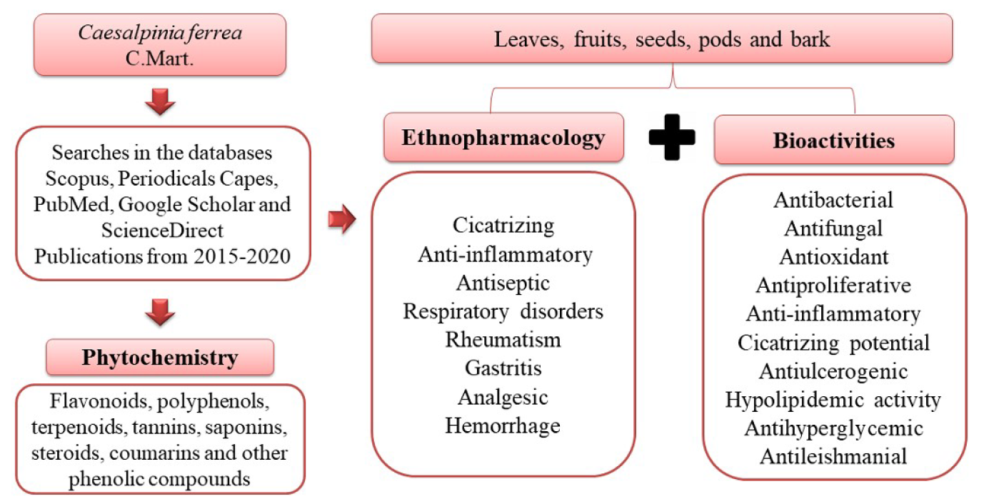
Caesalpinia ferrea C. Mart. (Fabaceae) Phytochemistry, Ethnobotany, and Bioactivities: A Review

 , , , , ,
  ,
,
Abstract
:
1. Introduction
2. Results and Discussion
2.1. Botanical Characterization
2.2. Ethnobotany
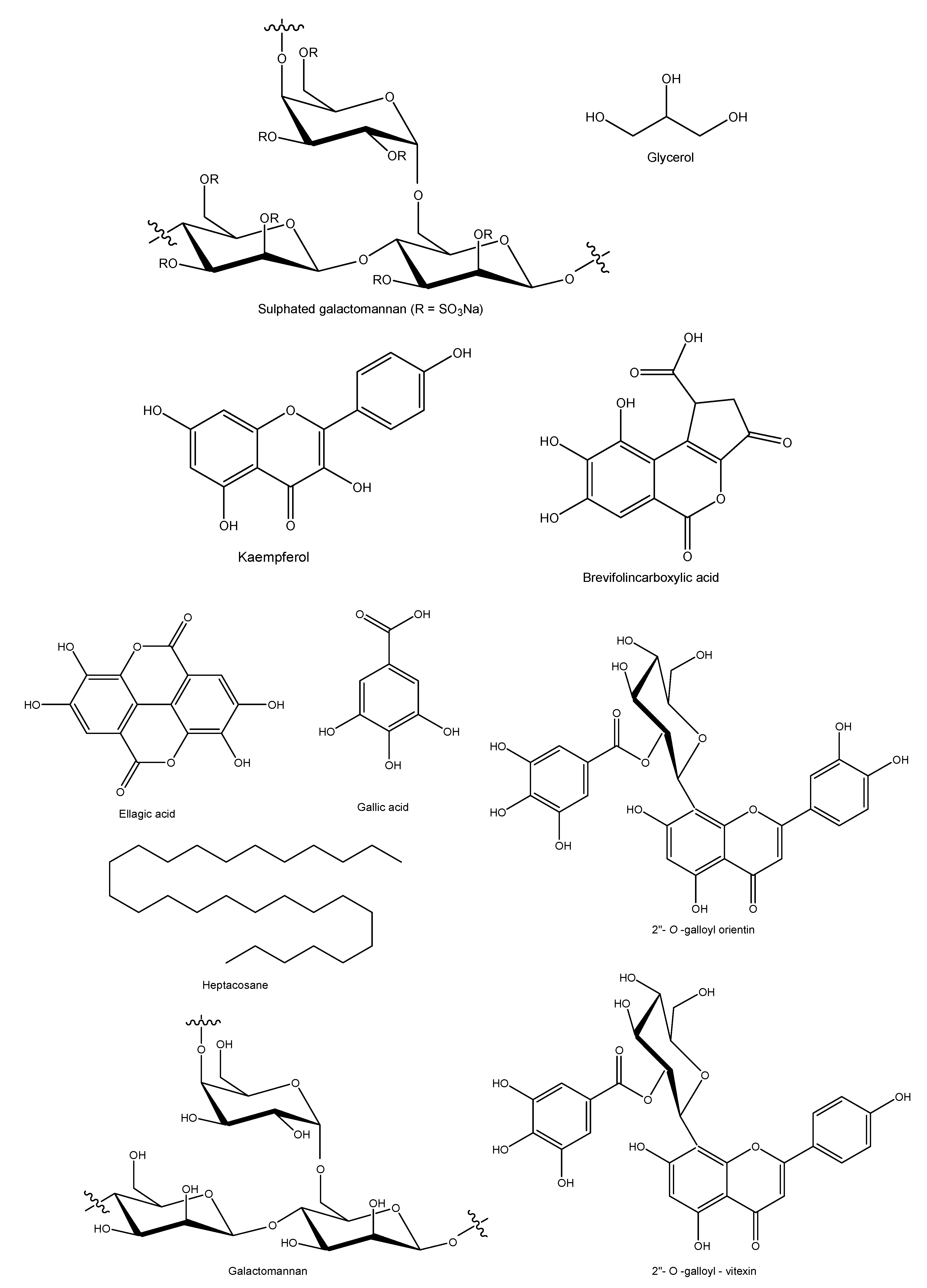
2.3. Phytochemical Aspects
2.3.1. Leaves
2.3.2. Bark
2.3.3. Fruits or Pods
2.3.4. Seeds and Roots
2.3.5. Compostos De Valor Quimiotaxônomico
2.4. Bioactivities
2.4.1. Antimicrobial Activity
2.4.2. Anti-Halitosis Activity
2.4.3. Antifungal Activity
2.4.4. Anti-Inflammatory Activity
2.4.5. Antioxidant Activity
2.4.6. Antileishmanial Activity
2.4.7. Antiproliferative and Apoptotic Effects
2.4.8. Anti-Wrinkle and Anti-Melanogenic Activity
2.4.9. Anti-Hyperglycemic Activity
2.4.10. Antiviral Activity
2.4.11. Antinociceptive Activity
2.4.12. Antiulcerogenic Activity
2.4.13. Hypolipidemic Effects
2.4.14. Toxicity
2.4.15. Genotoxicity
2.4.16. Cytotoxicity
2.4.17. Cicatrizing Activity
2.4.18. Repellent Action
2.4.19. Insecticidal Activity
2.4.20. Fertilizer
2.4.21. Allelopathic Potential
2.4.22. Biosorbent
2.4.23. Other Bioactivities
3. Materials and Methods
4. Critical Analysis
5. Conclusions
Author Contributions
Funding
Acknowledgments
Conflicts of Interest
References
- Ferreira, D.Q.; Ferraz, T.O.; Araújo, R.S.; Cruz, R.A.S.; Fernandes, C.P.; Souza, G.C.; Ortiz, B.L.S.; Sarquis, R.S.F.R.; Miranda, J.C.M.M.; Garrett, R.; et al. Libidibia ferrea (Jucá), a traditional anti-inflammatory: A study of acute toxicity in adult and embryos Zebrafish (Danio rerio). Pharmaceuticals 2019, 12, 175. [Google Scholar] [CrossRef] [PubMed] [Green Version]
- Bueno, N.R.; Campos, É.P.; Silva, M.S.; Rezende, K.S.; Lima, B.B.M. Levantamento Etnofarmacológico e Farmacológico de Plantas Medicinais Comercializadas em Rondonópolis (MT). Biodiversidade 2019, 2, 2–20. [Google Scholar]
- Leal, J.B.; Silva, M.M.; Costa, J.M.; Albuquerque, L.C.S.; Pereira, M.D.G.S.; Sousa, R.L. Etnobotânica de plantas medicinais com potencial anti-inflamatório utilizadas pelos moradores de duas comunidades no município de Abaetetuba, Pará. Biodiversidade 2019, 3, 110–125. [Google Scholar]
- Sacramento, A.A.; Martins Filho, I.E.; Dos Reis, L.A. Estudo etnobotânico das plantas medicinais comercializadas numa feira livre num município do interior da Bahia. Rev. Enferm. Atual Derme 2019, 89, 27. [Google Scholar] [CrossRef]
- Santos, E.Q.; Costa, J.F.D.S.; Pereira, M.D.G.D.S.; Costa, J.M.; Sousa, R.L. Etnobotânica da flora medicinal de quintais na comunidade Mamangal, Rio Meruú, Igarapé-Miri, Pará. Sci. Plena 2019, 15, 1–11. [Google Scholar] [CrossRef] [Green Version]
- Andrade, B.A.; Corrêa, A.J.C.; Gomes, A.K.S.; Neri, P.M.S.; Sobrinho, T.J.S.P.; Araújo, T.A.S.; Castro, V.T.N.A.; Amorim, E.L.C. Photoprotective Activity of Medicinal Plants from the Caatinga Used as Anti-inflammatories. Pharmacogn. Mag. 2019, 15, 356–361. [Google Scholar] [CrossRef]
- Falcão, T.R.; Rodrigues, C.A.O.; Araújo, A.A.; Medeiros, C.A.C.X.; Soares, L.A.L.; Ferreira, M.R.A.; Vasconcelos, R.C.; Araújo-Júnior, R.F.; Sousa Lopes, M.L.D.; Guerra, G.C.B. Crude extract from Libidibia ferrea (Mart. ex. Tul.) L.P. Queiroz leaves decreased intra articular inflammation induced by zymosan in rats. BMC Complement. Altern. Med. 2019, 19, 1–10. [Google Scholar] [CrossRef]
- Luna, M.S.M.; Paula, R.A.; Brandão Costa, R.M.P.; Anjos, J.V.; Silva, M.V.; Correia, M.T.S. Bioprospection of Libidibia ferrea var. ferrea: Phytochemical properties and antibacterial activity. South. African J. Bot. 2020, 130, 103–108. [Google Scholar] [CrossRef]
- Hassan, S.K.; El-Sammad, N.M.; Mousa, A.M.; Mohammed, M.H.; Farrag, A.E.R.H.; Hashim, A.N.E.; Werner, V.; Lindequist, U.; Nawwar, M.A.E.-M. Hypoglycemic and antioxidant activities of Caesalpinia ferrea Martius leaf extract in streptozotocin-induced diabetic rats. Asian Pac. J. Trop. Biomed. 2015, 5, 462–471. [Google Scholar] [CrossRef] [Green Version]
- Nascimento, J.; Reatgui, W.; Araújo, L.; Ribeiro, M.E.; Maia, D.; Giacomin, L.; Kitagawa, R.; Baratto, L. Avaliação do potencial antioxidante e anti-Helicobacter pylori in vitro de extratos de plantas medicinais utilizadas popularmente na região amazônica. Rev. Fitos 2017, 11, 140–152. [Google Scholar] [CrossRef]
- Soares, M.R.P.S.; Caneschi, C.A.; Chaves, M.D.G.A.M.; Mota, M.; Stroppa, P.H.F.; Barbosa, W.; Raposo, N.R.B. In Vitro Antifungal Activity and Cytotoxicity Screening of Dry Crude Extracts from Brazilian Amazonia Plants. Afr. J. Tradit. Complement. Altern. Med. 2018, 15, 13. [Google Scholar] [CrossRef]
- Prazeres, L.D.K.T.; Aragão, T.P.; Brito, S.A.; Almeida, C.L.F.; Silva, A.D.; Paula, M.M.F.; Farias, J.S.; Vieira, L.D.; Damasceno, B.P.G.L.; Rolim, L.A.; et al. Antioxidant and Antiulcerogenic Activity of the Dry Extract of Pods of Libidibia ferrea Mart. ex Tul. (Fabaceae). Oxid. Med. Cell. Longev. 2019, 2019. [Google Scholar] [CrossRef] [PubMed] [Green Version]
- Falcão, T.R.; Araújo, A.A.; Soares, L.A.L.; Farias, I.B.; Silva, W.A.V.; Ferreira, M.R.A.; Araújo, R.F.; Medeiros, J.S.; Lopes, M.L.D.D.S.; Guerra, G.C.B. Libidibia ferrea Fruit Crude Extract and Fractions Show Anti-Inflammatory, Antioxidant, and Antinociceptive Effect in Vivo and Increase Cell Viability in Vitro. Evid.-Based Complement. Altern. Med. 2019. [Google Scholar] [CrossRef] [Green Version]
- Veloso, D.J.; Abrão, F.; Martins, C.H.G.; Bronzato, J.D.; Gomes, B.P.F.A.; Higino, J.S.; Sampaio, F.C. Potential antibacterial and anti-halitosis activity of medicinal plants against oral bacteria. Arch. Oral Biol. 2020, 110. [Google Scholar] [CrossRef] [PubMed]
- Ferreira, M.R.A.; Soares, L.A.L. Libidibia ferrea (Mart. Ex Tul.) LP Queiroz: A review of activities biological and phytochemical composition. J. Med. Plants Res. 2015, 140–145. [Google Scholar] [CrossRef] [Green Version]
- Andrade, G.C.; Silva, L.C. Responses of tropical legumes from the Brazilian Atlantic Rainforest to simulated acid rain. Protoplasma 2017, 254, 1639–1649. [Google Scholar] [CrossRef]
- Matos, S.S.; Melo, A.L.; Santos-Silva, J. Caesalpinioideae e Cercidoideae (Leguminosae) no Parque Estadual Mata da Pimenteira, Semiárido de Pernambuco, Brasil. Rodriguesia 2019, 70. [Google Scholar] [CrossRef]
- Medeiros, J.G.F.; Araújo-Neto, A.C.; Silva, E.C.; Huang, M.-F.N.; Nascimento, L.C. Qualidade Sanitária de Semente de Caesalpinia ferrea: Incidência de Fungos, Controle e Efeitos na Qualidade Fisiológica com o Uso de Extratos Vegetais. Rev. Floresta 2015, 45, 163–174. [Google Scholar] [CrossRef] [Green Version]
- Bragante, R.B.; Hell, A.F.; Paulo, J.P.; Silva, N.D.C.; Centeno, R.D.C.L.; Figueiredo-Ribeiro, C.J.B. Physiological and metabolic responses of immature and mature seeds of Libidibia ferrea ((Mart. ex Tul.) L.P. Queiroz) under contrasting storage temperatures. Braz. J. Bot. 2018, 41, 43–55. [Google Scholar] [CrossRef]
- Santos, S.F.; Santos, A.S.; Corpes, R.S.; Leão, N.V.M. Aspectos do cultivo in vitro de Libidibia ferrea (Mart. Ex Tul.) L. P. QUEIROZ (Leguminosae-Caesalpinioideae) como fonte alternativa para produção de metabólitos secundários. Rev. Espac. 2018, 39, 17–24. [Google Scholar]
- Matos, A.C.B.; Ataíde, G.M.; Borges, E.E.L. Physiological, physical, and morpho-anatomical changes in Libidibia ferrea ((Mart. ex Tul.) L.P. Queiroz) seeds after overcoming dormancy. J. Seed Sci. 2015, 37, 26–32. [Google Scholar] [CrossRef]
- Carvalho, S.M.C.; Torres, S.B.; Benedito, C.P.; Nogueira, N.W.; Souza, A.A.T.; Souza Neta, M.L. Viability of Libidibia ferrea (Mart. ex Tul.) L.P. Queiroz var. ferrea) seeds by tetrazolium test. J. Seed Sci. 2017, 39, 7–12. [Google Scholar] [CrossRef] [Green Version]
- Ferreira, W.N.; Lacerda, C.F.; Costa, R.C.; Filho, S.M. Effect of water stress on seedling growth in two species with different abundances: The importance of Stress Resistance Syndrome in seasonally dry tropical forest. Acta Bot. Bras. 2015, 29, 375–382. [Google Scholar] [CrossRef] [Green Version]
- De David, M.; Pasa, M.C. As plantas medicinais e a etnobotânica em Várzea Grande, MT, Brasil. Interações 2015, 16, 97–108. [Google Scholar] [CrossRef] [Green Version]
- Almeida-Neto, J.R.; Barros, R.F.M.; Silva, P.R.R. Uso de plantas medicinais em comunidades rurais da Serra do Passa-Tempo, estado do Piauí, Nordeste do Brasil. Braz. J. Biosci. 2015, 13, 165–175. [Google Scholar]
- Ferreira, A.B.; Ming, L.C.; Haverroth, M.; Daly, D.C.; Caballero, J.; Ballesté, A.M. Plants Used to Treat Malaria in the Regions of Rio Branco-Acre State and Southern Amazonas State-Brazil. Int. J. Phytocosmetics Nat. Ingredients 2015, 2, 1–5. [Google Scholar] [CrossRef]
- Gonçalves, K.G.; Pasa, M.C. O saber local e as plantas medicinais na comunidade sucuri, cuiabá, mt, brasil. Biodiversidade 2015, 14, 50–73. [Google Scholar]
- Silva, C.G.; Marinho, M.G.V.; Lucena, M.F.A.; Costa, J.G.M. Levantamento etnobotânico de plantas medicinais em área de Caatinga na comunidade do Sítio Nazaré, município de Milagres, Ceará, Brasil. Rev. Bras. Plantas Med. 2015, 17, 133–142. [Google Scholar] [CrossRef] [Green Version]
- Silva, M.P.; Barros, R.F.M.; Moita Neto, J.M. Farmacopeia natural de comunidades rurais no estado do Piauí, Nordeste do Brasil. Desenvolv. e Meio Ambient. 2015, 33, 193–207. [Google Scholar] [CrossRef]
- Cajaiba, R.L.; Silva, W.B.; Sousa, R.D.N.; Sousa, A.S. Levantamento etnobotânico de plantas medicinais comercializadas no município de Uruará, Pará, Brasil. Biotemas 2016, 29, 115. [Google Scholar] [CrossRef] [Green Version]
- Castro, K.N.C.; Wolschick, D.; Leite, R.R.S.; Andrade, I.M.; Magalhães, J.A.; Mayo, S.J. Ethnobotanical and ethnoveterinary study of medicinal plants used in the municipality of Bom Princpio do Piau, Piau, Brazil. J. Med. Plants Res. 2016, 10, 318–330. [Google Scholar] [CrossRef] [Green Version]
- Kffuri, C.W.; Lopes, M.A.; Ming, L.C.; Odonne, G.; Kinupp, V.F. Antimalarial plants used by indigenous people of the Upper Rio Negro in Amazonas, Brazil. J. Ethnopharmacol. 2016, 178, 188–198. [Google Scholar] [CrossRef] [PubMed] [Green Version]
- Lima, I.E.O.; Nascimento, L.A.M.; Silva, M.S. Comercialização de plantas medicinais no município de Arapiraca-AL. Rev. Bras. Plantas Med. 2016, 18, 462–472. [Google Scholar] [CrossRef] [Green Version]
- Moraes Rego, C.A.R.; Rocha, A.E.; De Oliveira, C.A.; Pacheco, F.P.F. Levantamento etnobotânico em comunidade tradicional do assentamento pedra suada, do município de cachoeira grande, Maranhão, Brasil. Acta Agron. 2016, 65, 284–291. [Google Scholar] [CrossRef]
- Oliveira, M.S.; Silva, E.O.; Ferreira, A.W.C.; Guarçoni, E.A.E. Conhecimento e uso tradicional das espécies madeireiras e medicinais no município de Aldeias Altas, Maranhão, Brasil. Enciclopédia Biosf. 2016, 13, 1160. [Google Scholar] [CrossRef]
- Silva, F.J.; Silveira, A.P.; Gomes, V.S. Plantas medicinais e suas indicações ginecológicas: Estudo de caso com moradoras de Quixadá, CE, Brasil. Rev. Bras. Biociências 2016, 14, 193–201. [Google Scholar]
- Souza, L.F.; Dias, R.F.; Guilherme, F.A.G.; Coelho, C.P. Plantas medicinais referenciadas por raizeiros no município de Jataí, estado de Goiás. Rev. Bras. Plantas Med. 2016, 18, 451–461. [Google Scholar] [CrossRef]
- Saraiva, M.E.; Ulisses, A.V.R.D.A.; Ribeiro, D.A.; Oliveira, L.G.S.; Macêdo, D.G.; Sousa, F.D.F.S.; Menezes, I.R.A.; Sampaio, E.V.D.S.B.; Souza, M.M.D.A. Plant species as a therapeutic resource in areas of the savanna in the state of Pernambuco, Northeast Brazil. J. Ethnopharmacol. 2015, 171, 141–153. [Google Scholar] [CrossRef] [Green Version]
- Ribeiro, S.C.; Melo, N.D.P.; Barros, A.B. Etnoconhecimento de Pequenos Agricultores Tradicionais sobre Plantas Medicinais no Tratamento de Dores Provocadas pelo Trabalho. Cad. Ter. Ocup. UFSCar. 2016, 24, 563–574. [Google Scholar] [CrossRef]
- Cordeiro, M.C.; Botrel, R.T.; Holanda, A.C. Levantamento etnobotânico de espécies arbóreas no assentamento Tabuleiro Grande, Apodi, Rio Grande do Norte. Rev. Verde Agroecol. Desenvolv. Sustentável. 2017, 12, 122. [Google Scholar] [CrossRef] [Green Version]
- Palheta, I.C.; Tavares-Martins, A.C.C.; Lucas, F.C.A.; Jardim, M.A.G. Ethnobotanical study of medicinal plants in urban home gardens in the city of abaetetuba, Paá state, Brazil. Bol. Latinoam. Caribe Plantas. Med. Aromat. 2017, 16, 206–262. [Google Scholar]
- Leandro, Y.A.S.; Jardim, I.N.; Gavilanes, M.L. The use of medicinal plants in health care practices by the residents of a communitty settlement in Anapu, Pará, Brazil. Biodiversidade 2017, 16, 30–44. [Google Scholar]
- Pereira, M.G.S.; Coelho-Ferreira, M. Uso e diversidade de plantas medicinais em uma comunidade quilombola na Amazônia Oriental, Abaetetuba, Pará. Biota Amaz. 2017, 7, 57–68. [Google Scholar]
- Silva, R.C.; Roriz, B.C.; Scareli-Santos, C. Etnoconhecimento sobre as espécies medicinais utilizadas pela população de Araguaína, TO. Rev. São Luís Orione. 2018, 1, 1–21. [Google Scholar]
- Albergaria, E.T.; Silva, M.V.; Silva, A.G. Levantamento etnobotânico de plantas medicinais em comunidades rurais do município de Lagoa Grande, Pernambuco, Brasil. Rev. Fitos 2019, 13, 137–154. [Google Scholar] [CrossRef]
- Ribeiro, R.V.; Bieski, I.G.C.; Balogun, S.O.; Martins, D.T.O. Ethnobotanical study of medicinal plants used by Ribeirinhos in the North Araguaia microregion, Mato Grosso, Brazil. J. Ethnopharmacol. 2017, 205, 69–102. [Google Scholar] [CrossRef]
- Lima, B.B.; Fernandes, F.P. Uso e diversidade de plantas medicinais no município de Aracati—CE, Brasil. J. Appl. Pharm. Sci. 2020, 24–42. Available online: https://www.researchgate.net/publication/340082226 (accessed on 23 August 2020).
- Leandro, C.S.; Bezerra, J.W.A.; Rodrigues, M.D.P.; Silva, A.K.F.; Silva, D.L.; Santos, M.A.F.; Linhares, K.V.; Boligon, A.A.; Silva, V.B.; Rodrigues, A.S.; et al. Phenolic Composition and Allelopathy of Libidibia ferrea Mart. ex Tul. in Weeds. J. Agric. Sci. 2019, 11, 109. [Google Scholar] [CrossRef]
- Hussein, S.A.M.; El-Mesallamy, A.M.D.; Souleman, A.M.A.; Mousa, M.A. Cytotoxic activity of bioactive compound from Caesalpinia ferrea Martius, Fabaceae. Int. J.Pharm. Phytochem. Res. 2016, 8, 2080–2084. [Google Scholar]
- Nawwar, M.A.; Hussein, S.A.; El-Mousallami, A.M.; Hashim, A.N.; Mousa, M.A.; Hetta, M.H.; Hamed, M.A.; Werner, V.; Becker, A.; Haertel, B.; et al. Phenolics from Caesalpinia ferrea Mart.: Antioxidant, cytotoxic and hypolipidemic activity. Pharmazie 2015, 70, 553–558. [Google Scholar] [CrossRef]
- Indriani, D.; Elya, B.; Noviani, A. Arginase inhibitory activity and total flavonoid content on Caesalpinia ferrea C. Mart stem bark extracts. Pharm. J. 2018, 10, 1180–1183. [Google Scholar] [CrossRef] [Green Version]
- Alves, F.S.; Viturino, W.A.S.; Ferreira, M.R.A.; Soares, L.A.L.; Barbosa, F.S.S. Bark of the Stem of Libidibia ferrea Associated with Mycorrhizal Fungi: An Alternative to Produce High Levels of Phenolic Acids. Open Microbiol. J. 2018, 12, 412–418. [Google Scholar] [CrossRef] [Green Version]
- Pedrosa, T.N.; Barros, A.O.; Nogueira, J.R.; Fruet, A.C.; Rodrigues, I.C.; Calcagno, D.Q.; Smith, M.A.C.; Souza, T.P.; Barros, S.B.M.; Vasconcellos, M.C.; et al. Anti-wrinkle and anti-whitening effects of jucá (Libidibia ferrea Mart.) extracts. Arch. Dermatol. Res. 2016, 308, 643–654. [Google Scholar] [CrossRef] [PubMed]
- Guerra, A.C.V.A.; Soares, L.A.L.; Ferreira, M.R.A.; Araújo, A.A.; Rocha, H.A.O.; Medeiros, J.S.; Cavalcante, R.S.; Júnior, R.F.A. Libidibia ferrea presents antiproliferative, apoptotic and antioxidant effects in a colorectal cancer cell line. Biomed. Pharmacother. 2017, 92, 696–706. [Google Scholar] [CrossRef] [PubMed]
- Ferreira, M.R.A.; Fernandes, M.T.M.; Silva, W.A.V.; Bezerra, I.C.F.; Souza, T.P.; Pimentel, M.F.; Soares, L.A.L. Chromatographic and Spectrophotometric Analysis of Phenolic Compounds from Fruits of Libidibia ferrea Martius. Pharmacogn Mag. 2016, 12, S285–S291. [Google Scholar] [CrossRef] [Green Version]
- Azevedo, L.F.C.; Ferreira, T.A.A.; Melo, K.M.; Dias, C.L.P.; Bastos, C.E.M.C.; Santos, S.F.; Santos, A.D.S.; Nagamachi, C.Y.; Pieczarka, J.C. Aqueous ethanol extract of Libidibia ferrea (Mart. Ex Tul) L.P. Queiroz (juca) exhibits antioxidant and migration-inhibiting activity in human gastric adenocarcinoma (ACP02) cells. PLoS ONE 2020, 15, e0226979. [Google Scholar] [CrossRef] [Green Version]
- Magalhães, L.S.; Pussente, C.G.; Azevedo, L.R.; Crespo, J.M.R.S. Avaliação da atividade antibacteriana do extrato de Caesalpinia ferrea Martius e desenvolvimento de uma formulação fitocosmética. Rev. Científica Faminas 2015, 11, 27–43. [Google Scholar]
- Pereira, L.D.P.; Mota, M.R.L.; Brizeno, L.A.C.; Nogueira, F.C.; Ferreira, E.G.M.; Pereira, M.G.; Assreuy, A.M.S. Modulator effect of a polysaccharide-rich extract from Caesalpinia ferrea stem barks in rat cutaneous wound healing: Role of TNF-α, IL-1β, NO, TGF-β. J. Ethnopharmacol. 2016, 187, 213–223. [Google Scholar] [CrossRef]
- Pickler, T.B.; Lopes, K.P.; Magalhães, S.A.; Krueger, C.M.A.; Martins, M.M.; Filho, V.C.; Jozala, A.F.; Grotto, D.; Gerenutti, M. Effect of Libidibia ferrea bark and seed in maternal reproductive and biochemical outcomes and fetal anomaly in rats. Birth Defects Res. 2019, 111, 863–871. [Google Scholar] [CrossRef]
- Lins, T.R.S.; Braz, R.L.; Silva, T.C.; Araujo, E.C.G.; Medeiros, J.X.; Reis, C.A. Tannin Content of the Bark and Branch of Caatinga Species. J. Exp. Agric. Int. 2019, 31, 1–8. [Google Scholar] [CrossRef] [Green Version]
- Kobayashi, Y.T.S.; Almeida, V.T.; Bandeira, T.; Alcántara, B.N.; Silva, A.S.B.; Barbosa, W.L.R.; Silva, P.B.; Monteiro, M.V.B.; Almeida, M.B. Avaliação fotoquímica e potencial cicatrizante do extrato etanólico dos frutos de Jucá (Libidibia ferrea) em ratos Wistar. Braz. J. Vet. Res. Anim. Sci. 2015, 52, 34–40. [Google Scholar] [CrossRef]
- Moura, V.M.; Freitasa, L.A.S.; Santos, M.C.; Raposo, J.D.A.; Lima, A.E.; Oliveira, R.B.; Silva, M.N.; Mourão, R.H.V. Plants used to treat snakebites in Santarém, western Pará, Brazil: An assessment of their effectiveness in inhibiting hemorrhagic activity induced by Bothrops jararaca venom. J. Ethnopharmacol. 2015, 161, 224–232. [Google Scholar] [CrossRef] [PubMed]
- Buckeridge, M.S.; Dietrich, S.M.C.; Lima, D.U. Galactomannan as the reserve carbohydrate in legume seeds. Dev. Crop. Sci. 2000, 26, 283–316. Available online: http://0-www-sciencedirect-com.brum.beds.ac.uk/science/article/pii/S0378519×0080015X (accessed on 23 August 2020).
- Gallão, M.I.; Normando, L.D.O.; Vieira, Í.G.; Mendes, F.N.; Ricardo, N.M.; Brito, E.S. Morphological, chemical and rheological properties of the main seed polysaccharide from Caesalpinia ferrea Mart. Ind. Crop Prod. 2013, 47, 58–62. Available online: http://0-dx-doi-org.brum.beds.ac.uk/10.1016/j.indcrop.2013.02.035 (accessed on 23 August 2020).
- Cunha, A.P.; Ribeiro, A.C.B.; Ricardo, N.M.P.S.; Oliveira, A.C.; Dávila, L.S.P.; Cardoso, J.H.L.; Rodrigues, D.C.; Azeredo, H.M.C.; Silva, L.M.A.; Brito, E.S.; et al. Polysaccharides from Caesalpinia ferrea seeds – Chemical characterization and anti-diabetic effects in Wistar rats. Food Hydrocoll. 2017, 65, 68–76. [Google Scholar] [CrossRef]
- Hegnauer, R.; Gpayer-Barkmeijer, R.J. Relevance of seed polysaccharides and flavonoids for the classification of the Leguminosae: A chemotaxonomic approach. Phytochemistry 1993, 34, 3–16. [Google Scholar] [CrossRef]
- Jozala, A.F.; Santos, J.R.; Santos, G.R.; Viroel, F.J.M.; Pickler, T.B.; Santos, C.A.; Rebelo, M.A.; Chaud, M.V.; Hataka, A.; Groto, D.; et al. Libidibia ferrea loaded in bacterial nanocellulose: Evaluation of antimicrobial activity and wound care. J. Biomed. Biotechnol. 2020, 6, 6212–6226. [Google Scholar] [CrossRef]
- Sousa, A.C.J.; Oliveira, J.S.; Porcy, C.; Souza, M.J.C.; Menezes, R.A.O. Potencial antimicrobiano de extratos vegetais frente a cepas bacterianas de interesse médico em Macapá, Amapá, Amazônia Brasileira. Diagn. Trat. 2019, 24, 85–90. [Google Scholar]
- Malafaia, C.B.; Jardelino, A.C.S.; Silva, A.G.; Souza, E.B.; Macedo, A.J.; Correia, M.T.S.; Silva, M.V. Effects of Caatinga Plant Extracts in Planktonic Growth and Biofilm Formation in Ralstonia solanacearum. Microb. Ecol. 2018, 75, 555–561. [Google Scholar] [CrossRef]
- Ferreira, J.V.A.; Ferreira, L.L.; Gomes, F.F.; Matias, E.F.F.; Sobral, E.S.; Cosmo, J.A.; Tintino, S.R.; Leite, N.F.; Albuquerque, R.S.; Morais Braga, M.F.B.; et al. Evaluation of antimicrobial and modulatory activity of the ethanol extract of Libidibia ferrea (Mart. ex tul.) l.p. queiroz. Rev. Cuba. Plantas Med. 2016, 21, 71–82. [Google Scholar]
- Paiva, W.S.; Souza Neto, F.E.; Bandeira, M.G.L.; Abrantes, M.R.; Batista, A.C.L.; Silva, J.B.A. Atividade antibacteriana da casca do jucá (Libidibia ferrea (mart. ex tul.) l. p. queiroz), frente a Staphylococcus spp. isolados do leite de cabras com mastite. Arch. Vet. Sci. 2015, 20, 141–146. [Google Scholar] [CrossRef]
- Conde, N.C.O.; Pereira, M.S.V.; Bandeira, M.F.C.L.; Venâncio, G.N.; Oliveira, G.P.; Sampaio, F.C. In vitro antimicrobial activity of plants of the Amazon on oral biofilm micro-organisms. J. Dent. Sci. Orig. 2015, 30, 179–183. [Google Scholar]
- Nascimento, P.; Silva, T.; Gomes, J.; Silva, M.; Souza, S.; Falcão, R.; Silva, T.; Moreira, K. Antioxidant and antimicrobial properties of ethanolic extract of Libidibia ferrea pods. Rev. Fitos 2015, 9, 207–216. [Google Scholar] [CrossRef]
- Agostini, V.O.; Macedo, A.J.; Muxagata, E.; Silva, M.V.; Pinho, G.L.L. Non-toxic antifouling potential of Caatinga plant extracts: Effective inhibition of marine initial biofouling. Hydrobiologia 2020, 847, 45–60. [Google Scholar] [CrossRef]
- Soares, R.P.S.M.; Corrêa, R.O.; Stroppa, P.H.F.; Marques, F.C.; Andrade, G.F.S.; Corrêa, C.C.; Brandão, M.A.F.; Raposo, N.R.B. Biosynthesis of silver nanoparticles using Caesalpinia ferrea (Tul.) Martius extract: Physicochemical characterization, antifungal activity and cytotoxicity. PeerJ 2018, 1–16. [Google Scholar] [CrossRef] [PubMed] [Green Version]
- Melo, P.A.F.R.; Medeiros, R.L.S.; Ferreira-Júnior, D.C.; Alves, E.U.A.; Valeri, S.V.; Mondego, J.M.; Araújo, J.R.G.L.R.N.S.L. Protection of Sideroxylon obtusifolium seeds against Colletotrichum sp. with Caesalpinia ferrea extract. Afr. J. Agric. Res. 2017, 12, 3433–3440. [Google Scholar] [CrossRef] [Green Version]
- Melo, P.A.F.R.; Alves, E.U.; Martins, C.C.; Anjos Neto, A.P.; Pinto, K.M.S.; Araújo, L.R.; Vieira, C.P.; Nascimento, L.C. Extracts of Caesalpinia ferrea and Trichoderma sp. on the control of Colletotrichum sp. transmission in Sideroxylon obtusifolium seeds. Rev. Bras. Plantas Med. 2016, 18, 494–501. [Google Scholar] [CrossRef] [Green Version]
- Biasi-Garbin, R.P.; Demitto, F.O.; Amaral, R.C.R.; Ferreira, M.R.A.; Soares, L.A.L.; Svidzinski, T.I.E.; Baeza, L.C.; Yamada-Ogatta, S.F. Antifungal potential of plant species from brazilian caatinga against dermatophytes. Rev. Inst. Med. Trop. 2016, 58, 18–22. [Google Scholar] [CrossRef]
- Peixinho, G.S.; Santos, C.M.G.; Ribeiro, V.G.; Amorim, E.P.R.; Bispo, J.S.; Carvalho, V.N. Avaliação da eficiência de extratos de plantas nativas da caatinga sobre o controle da podridão seca (Lasiodiplodia theobromae) em cachos da videira cv. Itália. Summa Phytopathol. 2017, 43, 155–157. [Google Scholar] [CrossRef]
- Comandolli-Wyrepkowski, C.D.; Jensen, B.B.; Grafova, I.; Santos, P.A.; Barros, A.M.C.; Soares, F.V.; Barcellos, J.F.M.; Silva, A.F.; Grafov, A.; Franco, A.M.R. Antileishmanial activity of extracts from Libidibia ferrea: Development of in vitro and in vivo tests. Acta Amaz. 2017, 47, 331–340. [Google Scholar] [CrossRef]
- Lima, M.F.F.; Araujo Silva, J.W.S.; Silva, J.K.; Moura, A.H.N.; Lopes, R.L.F.; Cordeiro, B.A.; Cordeiro, R.P.; Melo, A.F.M. Avaliação toxicológica através do bioensaio com Artemia salina Leach de espécimes vegetais pertencentes à caatinga. Braz. J. Heal. Rev. 2019, 2, 5950–5963. [Google Scholar] [CrossRef]
- Sousa, M.J.B.; Campos, C.B.M.; Duarte, S.S.M.; Sousa, D.F.; Manso, J.A.X.; Cruz, A.D.; Minasi, L.B.; Silva, C.C. Genotoxicity of Brosimum gaudichaudii (Moraceae) and Caesalpinia ferrea (Fabaceae) in Astyanax sp. (Characidae) based on a comet assay. Genet. Mol. Res. 2018, 17, 1–8. [Google Scholar] [CrossRef]
- Silva, F.; Sales, M.; Sá, O.; Santana, G.; Deus, M.; Sousa, J.; Ferreira, P.; Peron, A. Potencial citotóxico, genotóxico e citoprotetor de extratos aquosos de Caesalpinia pyramidalis Tul., Caesalpinia ferrea Mart. eCaesalpinia pulcherrima Sw. Rev. Bras. Biociências 2015, 101–109. [Google Scholar]
- Batista, E.K.F.; Trindade, H.I.; Farias, I.S.; Martins, F.M.M.; Silva-Filho, O.F.; Batista, M.C.S. Avaliação da Atividade Cicatizante de Preparados à base de Jucá (Caesalpinia ferrea Mart.). Arch. Vet. Sci. 2017, 22, 30–39. [Google Scholar] [CrossRef] [Green Version]
- Carvalho, F.G.; Sampaio, J.P.S.; Araújo, M.M.S.; Pinto, L.S.S.; Rocha, A.J. Assessment of the healing activity of jucá pods [Libidibia ferrea (Mart. ex Tul.) L. P. Queiroz] in cutaneous lesions of rats. Acta Sci.-Technol. 2016, 38, 137–143. [Google Scholar] [CrossRef]
- Pereira, L.D.P.; Queiroz, C.V.G.; Pereira, G.; Assreuy, A.M.S. Uso de extratos de polissacarídicos da planta medicinal Caesalpinea ferrea na estimulação do edema na pata de ratos. Ciência Anim. 2018, 28, 56–69. [Google Scholar]
- Martins, I.C.F.; Fagundes, M.R.; Mockdeci, H.R.; Amarante, C.B.; Chaves, M.D.G.A.M.; Raposo, N.R.B. Ex vivo evaluation of the erosive effect of acid tea widely consumed in Brazil. J. Clin. Diagn. Res. 2018, 12, ZC50–ZC53. [Google Scholar] [CrossRef]
- Fernandes, C.P.M.; Machado, C.; Lopes, T.V.; Cunha Filho, N.; Bretanha, P.R.; Schons, S.; Félix, S.R.; Nobre, M.O. Repellent Action of Carapa guianensis and Caesalpinia ferrea for flies species of Calliphoridae family. Ciência Rural 2016, 46, 867–870. [Google Scholar] [CrossRef]
- Lopes, R.S.; Martins, M.C.B.; Oliveira, L.G.; Costa, A.F.; Santos, V.F.; Correia, M.T.S.; Silva, N.H.; Albuquerque, A.C.; Lima, E.Á.L.-A.; Lima, V.L.M. Termiticidal Activity of Libidibia ferrea var. ferrea and of the Association with Isaria spp. against Nasutitermes corniger. J. Agric. Sci. 2020, 12, 159. [Google Scholar] [CrossRef] [Green Version]
- Gomes, V.E.V.; Dutra, J.A.C.; Almeida, M.M.M. Controlling the cowpea black aphid (Aphis craccivora Koch) with botanical extracts. Biotemas 2019, 32, 117–121. [Google Scholar] [CrossRef]
- Lopes, R.S.; Oliveira, L.G.; Lima, G.; Costa, A.F.; Lima, E.Á.L.A.; Lima, V.L.M. Controle Biológico e Alternativo de Dactylopius opuntiae por Fungo Entomopatogênico e Extratos Vegetais em Plantação de Opuntia fícus-indica (Pernambuco/Brasil). Pesqui. Agropecuária Pernambucana 2018, 23, 23–26. [Google Scholar] [CrossRef]
- Lopes, R.S.; Oliveira, L.G.; Costa, A.F.; Correia, M.T.S.; Lima, E.A.L.-A.; Lima, V.L.M. Efficacy of Libidibia ferrea var. ferrea and Agave sisalana Extracts against Dactylopius opuntiae (Hemiptera: Coccoidea). J. Agric. Sci. 2018, 10, 255. [Google Scholar] [CrossRef]
- Demartelaere, A.C.F.; Nascimento, L.C.; Abraão, P.C.; Gomes, R.S.S.; Marinho, C.O.; Nunes, M.C. Alternativas no controle da mancha marrom de alternaria em tangerineira ‘Dancy’. Summa Phytopathol. 2018, 44, 164–169. [Google Scholar] [CrossRef] [Green Version]
- Pinto, K.M.S.; Melo, P.A.F.R.; Mondego, J.M.; Nascimento, L.C.; Cortez, M.I.M.M.; Aires, A.A.C.; Anjos-Neto, A.P.; Medeiros, R.L.S.; Araujo, J.R.G.; Silva, H.F. Plant extracts enhancers of defense response in ponkan mandarin Seedlings against Alternaria alternate f. spp. citri infection. Afr. J. Agric. Res. 2018, 13, 650–656. [Google Scholar] [CrossRef] [Green Version]
- Primo, A.A.; Melo, M.D.; Pereira, G.A.C.; Silva, L.A.; Fernandes, F.É.P.; Souza, H.A. Potencial fertilizante da serapilheira de espécies lenhosas da Caatinga na recuperação de um solo degradado. Rev. Ceres 2018, 65, 74–84. [Google Scholar] [CrossRef] [Green Version]
- Oliveira, A.K.; Coelho, M.F.B.; Torres, S.B.; Diógenes, F.É.P. Allelopathy by extracts of Caatinga species on melon seeds. Semin. Agrar. 2016, 37, 557–566. [Google Scholar] [CrossRef] [Green Version]
- Alves, R.M.; Silva, M.A.D.; Silva, J.N.; Costa, R.S.; Santos, B.K.L.; Lima, E.D.S. Efeito alelopático de Libidibia ferrea Mart. sobre o vigor das sementes de feijão-caupi. Rev. Verde Agroecol. Desenvolv. Sustentável. 2019, 14, 476–479. [Google Scholar] [CrossRef] [Green Version]
- Carvalho, L.B.; Chagas, P.M.B.; Pinto, L.M.A. Caesalpinia ferrea Fruits as a Biosorbent for the Removal of Methylene Blue Dye from an Aqueous Medium. Water Air. Soil Pollut. 2018, 229, 37200. [Google Scholar] [CrossRef]
- Kasperiski, F.M.; Lima, E.C.; Umpierres, C.S.; Reis, G.S.; Thue, P.S.; Lima, D.R.; Dias, S.L.P.; Saucier, C.; Costa, J.B. Production of porous activated carbons from Caesalpinia ferrea seed pod wastes: Highly efficient removal of captopril from aqueous solutions. J. Clean. Prod. 2018, 197, 919–929. [Google Scholar] [CrossRef]
- Marques, M.M.M.; Morais, S.M.; Silva, A.R.A.; Barroso, N.D.; Pontes Filho, T.R.; Araujo, F.M.D.C.; Vieira, I.G.P.; Lima, D.M.; Guedes, M.I.F. Antiviral and Antioxidant Activities of Sulfated Galactomannans from Plants of Caatinga Biome. Evid.-Based Complement. Altern. Med. 2015. [Google Scholar] [CrossRef]


| Characteristics | Attributes | Citations |
|---|---|---|
| Habit | Arboreal | [17] |
| Height | 10–15 m | [18] |
| Leaves | Alternating and composed | [16] |
| Flowers | Inflorescences with yellow petals | [17,19] |
| Fruits | Flattened pods | [19] |
| Seeds | Brown when ripe | [19] |
| Part Used | Method of Preparation or Use | Therapeutic Indication | Citation |
|---|---|---|---|
| Leaf | Tea | Vermifuge | [24] |
| Leaf, bark and fruit | Decoction, “lambedor”, maceration, medicinal wine | Asthma, bones pain, flu, kidney pain, cough, shaking | [25] |
| Bark | Decoction | Liver/bleeding | [26] |
| Pod, fruit, seed, and bark | Tanned in wine, tea, bath, macerated, cooked beaten with water | Anti-inflammatory and healing | [27] |
| Bark and fruit | Tea, “lambedor” and syrup | Flu, kidney inflammation and soothing | [28] |
| Fruit | “Lambedor” | Flu | [29] |
| Bark and seeds | Hurt the seed and soak it in the water | Pneumonia, anemia, diarrhea, colic and gastritis | [30] |
| Stem bark, fruit, and seed | Maceration | Anti-inflammatory, kidneys, bruises, back pain, healing, analgesic | [31] |
| Bark | Decoction | Malaria | [32] |
| Bark and root | Tea and bottles | Rheumatism and diabetes | [33] |
| Bark and fruit | Bottles | Anti-inflammatory | [34] |
| Fruit | Tea | Diarrhea, liver and healing | [35] |
| Stalk | Tea | Anti-inflammatory | [36] |
| Roots | Decoction | Hemorrhoids, inflammation of the eyes and injuries | [37] |
| Whole shell | Immersed in water | Hemorrhage, anti-inflammatory, infection and pain | [38] |
| Dry bark | Decoction | Back pain | [39] |
| Stem bark and fruit | Maceration and cooking | Back pain, vision problems, anti-inflammatory and healing | [40] |
| Fruit | Tea (decoction), tea (maceration), maceration in a bath | Sore throat, hoarseness, leg pain, toothache, uterine inflammation, wounds, anemia, gastritis | [41] |
| Fruit, bark, roots and seed | Tea and tincture | Asthma, bronchitis, flu, fever, sore throat, sinusitis, diarrhea, rheumatism, blood clearance, kidneys and soothing | [42] |
| Fruits | Tea | Urinary infection | [43] |
| Fruits | Bottles | Infection | [44] |
| Stem bark, bast, fruits and seeds | Tea, “lambedor”, in natura and in powder | Infectious diseases, parasitic, circulatory, immune, cardiovascular, digestive, respiratory, genitourinary, musculoskeletal, conjunctive, injuries and poisoning | [45] |
| Seeds | Tea and immersed in water | Skin cuts, cough, flu and depression | [3] |
| Bark, fruit, and seeds | Decoction, infusion, maceration and syrup | Syphilis, cancer, depurative, diabetes, asthma, gastritis, bronchitis, sinusitis, stomach ache, rheumatism, sexual impotence, healing, bone fracture, headache, fever and throat infection | [46] |
| Whole plant and fruits | Infusion and maceration | Spine, blow, inflammation and kidneys | [47] |
| Leafs | Tea | All kinds of infection and inflammation | [5] |
| Part Used | Solvent | Analytical Technique | Constituents | Citations |
|---|---|---|---|---|
| Leafs | Cyclohexane | CG/MS | Octacosane, docosane, and heptacosan | [8] |
| Leafs | Water | HPLC | Ellagic acid and gallic | [7] |
| Leafs | Water at 25 and 100 °C | HPLC-DAD | Gallic acid, caffeic and ellagic epicatechin, quercetin, and luteolin and catechin | [48] |
| Leafs | Ethanol at 70% | HPLC | Ellagic acid and gallic, orientin and isovitexin | [49] |
| Leafs | Ethanol at 70% | NMR 1D e 2D | Gallic acid, brevifolin carboxylic acid, and brevifolin | [50] |
| Barks and seeds | Ethanol at 70% | HPLC | Ellagic acid | [51] |
| Barks | Water at 25 and 100 °C | HPLC-DAD | Gallic, caffeic and ellagic acids, catechin, epicatechin and quercetin | [48] |
| Barks | Water | RP-HPLC | Ellagic acid and gallic | [52] |
| Barks | Ethanol and water | LC-MS/MS | Kaempferol, quinolinic acid and gallic | [53] |
| Fruit | Ethanol at 96% | LC-HRMS/MS | Corilagin and ellagic acid and gallic | [1] |
| Fruit | Water, ethanol at 20–80% | HPLC-DAD | Ellagic acid and gallic | [7] |
| Fruit | Ethanol | HPLC | Ellagic acid and gallic | [54] |
| Fruit | Water | HPTLC e HPLC | Ellagic acid and gallic | [55] |
| Pods | N-hexane | GC-MS | N-dodecanol, myristic acid, methyl palmitate, palmitic acid | [56] |
| Pods | Chloroform | GC-MS | n-valeric acid, caproic acid, heptanoic acid, and octanoic acid | [56] |
| Pods | Ethyl acetate | GC-MS | Oxalic acid, butanedioic acid, pyrotartaric acid, and pentanoic acid | [56] |
| Pods | Alcohol at 70% | GC-MS | Glycerol, D-fructose, myo-inositol, and glucopyranose | [56] |
| Pods | Alcohol at 40% | HPLC-MS | Valonium dilactone acid, gallic acid derivatives, and ellagic acid | [12] |
| Pods | Ethanol and Water | LC-MS/MS | Ellagic acid, chlorogenic acid, and rutin | [54] |
| Pods | Water at 25 and 100 °C | HPLC-DAD | Ellagic acid and gallic, catechin, epicatechin, quercetin, and luteolin | [48] |
| Parts Used/Solvents | Target or Model | Bioactivities Evaluated | Formulations/Dosage | Control (s) | Results | Citations |
|---|---|---|---|---|---|---|
| Full pod/Methanol | Parvimonas micra and Porphyromonas gingivalis | Antibacterial and anti-halitosis | In vitro 50–400 μg/mL for 72 h | Positive: chlorhexidine Negative: liquid medium | MIC: 50 and 120 µg/mL, respectively MBC: >50 and 130 µg/mL, respectively | [14] |
| Leafs/ Cyclohexane | Bacillus subtilis, Escherichia coli, Proteus vulgaris, Pseudomonas aeruginosa, and Staphylococcus aureus | Antibacterial | In vitro 0.04–25 mg/mL for 24 h | Positive: ampicillin Negative: DMSO 10% | MIC: 0.039, 0.039, 0.039, 0.39, 0.078 mg/mL, respectively | [8] |
| Leafs/Chloroform | B. subtilis, E. coli, P. vulgaris, P. aeruginosa, and S. aureus | Antibacterial | In vitro 0.04–25 mg/mL for 24 h | Positive: ampicillin Negative: DMSO 10% | MIC: 1.56, 6.25, 12.5, 3.12, 0.78 mg/mL, respectively | [8] |
| Leafs/Ethyl acetate | B. subtilis, E. coli, P. vulgaris, P. aeruginosa, and S. aureus | Antibacterial | In vitro 0.04–25 mg/mL for 24 h | Positive: ampicillin Negative: DMSO 10% | MIC: 0.78, 6.25, 12.5, 3.12, 1.56 mg/mL, respectively | [8] |
| Leafs/Methanol | B. subtilis, E. coli, P. vulgaris, P. aeruginosa, and S. aureus | Antibacterial | In vitro 0.04–25 mg/mL for 24 h | Positive: ampicillin Negative: DMSO | MIC: 6.25, 12.5, 25, 3.12, 3.12 mg/mL, respectively | [8] |
| Barks/Alcohol | S. aureus ATCC 10390, P. aeruginosa ATCC 9721, and E. coli ATCC 25,922 Wistar rats | Antibacterial and healing activity | In vitro 10 mg/mL for 24 h In vivo10 mg/mL for 28 days | Negative: bacterial nanocellulose membranes with extract | MIC: 0.39, 0.79 and 0.19 mg·mL−1, respectively | [67] |
| Fruits/Alcohol | S. aureus, E. coli, Klebsiella pneumoniae, and P. aeruginosa | Antibacterial | In vitro 20 μL of the crude extracts and in dilutions 1:2, 1:4, 1:8; 1:16 for 24 h | Negative: sterile water | Inhibition halos: 18, 12, 10 and 11 mm, respectively | [68] |
| Leafs and Fruits/Water | Ralstonia solanacearum | Antibacterial | In vitro 0.4–4.0 mg/mL for 24 h | Negative: water | 70% inhibition at a concentration of 0.4 mg/mL | [69] |
| Pods and bark/Ethanol | S. aureus, E. coli, and P. aeruginosa | Antibacterial | In vitro 512–8 μg/mL for 24 h | Positive: amikacin, gentamicin, and clindamycin | MIC: 1024 µg/mL for all strains | [70] |
| Barks/Alcohol | Staphylococcus spp. | Antibacterial | In vitro Crude extract, 70% and 50% for 24 h | Positive: ampicillin, cephalexin, gentamicin, oxacillin, and penicillin Negative: saline | Inhibition halos: 61.1; 27.78 and 5.56% for the crude extract and concentrations of 70 and 50%, respectively | [71] |
| Leafs/ Propylene Glycol | S. aureus ATCC 6538 | Antibacterial | In vitro Glycolic extract in concentrations of 3%, 5%, and 10% for 24 and 48 h | Negative: liquid soap | Average inhibition halo: 0.97 cm | [51] |
| Pods/Ethanol | Streptococcus mutans, Streptococcus mitis, Streptococcus sanguis, Streptococcus sobrinus, and Lactobacillus casei | Antibacterial | In vitro 0.97–500 mg/mL for 24 h | Positive: chlorhexidine gluconate | MIC: 15, 14, 14, 15, 15 mg/mL, respectively, and MICA: 31.2 mg/mL for all strains | [72] |
| Pods/Ethanol | Staphylococcus aureus, Enterococcus faecalis, B. subtilis, E. coli, K. pneumoniae, and P. aeruginosa | Antibacterial and antioxidant | In vitro 500–25 μg/mL for 24 h; 100–500 μg/mL for 30 min; 20 to 120 μg/mL for 10 min respectively | Positive: ascorbic acid and Trolox. Negative: specific medium | MIC: 125, 50, 50, 50, 125, 50 μg/mL, respectively; DPPH: EC50 4.4 μg/mL and ABTS: EC50 2.5 μg/mL | [73] |
| Pods/Alcohol | Helicobacter pylori Wistar rats | Antibacterial, antioxidant, antiulcerogenic and toxicity | In vitro 32–1024 μg/mL for 24 h In vivo 200 mg/kg for 14 days | Positive: amoxicillin, trolox, ranitidine, respectively. Negative: NaCl | MIC: 512 µg/mL; DPPH and ABTS: IC50 of 28.96 and 145.10 μg/mL, respectively; ED: 113 and 185.7 mg/kg; LD greater than 2000 mg/kg | [12] |
| Pods | Proteobacteria and Bacteroidetes | Antibiofilm | In vitro 0.5, 1, 2, 4, and 8 mg/mL for 48 h | Negative: sterile water | Inhibited growth by 82% at a concentration of 4 mg·mL−1 | [74] |
| Seeds/ Ethanol | Candida albicans ATCC 10231, Candida glabrata CCT 0728, Candida krusei CCT 1517, and Candida guilliermondii CCT 1890 | Antifungal | In vitro 4.8–5000 μg/mL for 48 h | Positive: ethanol 70%; amphotericin B and nystatin. Negative: specific medium | MIC: 9.7, 19.53, 78 and 39.06 µg/mL, respectively | [11] |
| Seeds/Ethanol | C. albicans ATCC 10231, C. glabrata CCT 0728, C. krusei CCT 1517, and C. guilliermondii CCT 1890; L929 fibroblast cells | Antifungal and Cytotoxicity | In vitro 7.81–1.000 μg/mL for 48 h | Positive: ethanol 70% | MIC: 9.7; 19; 78 and 4.8 µg/mL, respectively; toxicity at concentrations of 1000; 500 and e 250 µg/mL | [75] |
| Leafs/Water | Colletotrichum sp. | Antifungal | In vitro 0.156 mg/200 mL for 24 h | Positive: captan | Up to 96% inhibition at a concentration of 0.075 mg.mL−1 | [76] |
| Leafs/Alcohol | Colletotrichum sp. | Antifungal | In vitro 0.156 mg/200 mL for 24 h | Positive: captan | 100% inhibition of symptoms in treated seeds | [77] |
| Stem bark/Water, Ethanol and acetone | Trichophyton rubrum ATCC 28,189 and Trichophyton mentagrophytes ATCC 11481 | Antifungal | In vitro 1.96–1000 mg/mL for 7 days | Positive: terbinafine Negative: DMSO | MIC: 62.5 and 31.3 μg/mL, respectively | [78] |
| --- | Lasiodiplodia theobromae | Antifungal | In vitro 10, 20, and 30% for 5 days | Negative: sterile distilled water | Inhibited mycelial growth by 85.6% at a concentration of 30% | [79] |
| Leafs/Water | Wistar rats | Anti-inflammatory and antioxidant | In vitro 100, 200 and 300 mg for 24 h | Negative: saline 0.9% Positive: diclofenac 100 mg/kg | Effective doses: 100, 200 and 300 mg/kg; | [7] |
| Seeds/Water or Ethanol (20 - 80%) | Swiss mice and mouse embryonic fibroblast 3T3 cell line | Anti-inflammatory, antioxidant, antinociceptive, and cytotoxicity | In vitro 10, 15, 20, 25, and 30 µg/mL for 24, 48, and 72 h In vivo 50, 100, and 200 mg/kg for 20 and 30 min | Positive: diclofenac, cisplatin and ascorbic acid, and morphine Negative: saline | Effective doses: 50, 100 and 200 mg/kg; | [13] |
| Pods/Alcohol and ethyl acetate | ACP02 gastric adenocarcinoma cell line | Antioxidant and antimetastatic | In vitro 6.25 or 400 µg/mL for 20 min 6.25, 12.5, 25, 50, 100, 200, and 400 μg/mL for 24 and 48 h | Positive: doxorubicin Negative: medium RPMI | DPPH: IC50 74.36 and 116.10 μg/mL ABTS: IC50 9.76 and 29.13 μg/mL Decreased cell migration at concentrations of 50 µg/mL | [56] |
| Leafs/Ethanol | HaCaT and Wistar rats | Antioxidant, cytotoxicity, and hypolipidemic activity | In vitro 12.45 mg/L for 90 min; extract 50% for 45 min In vivo 300 mg/kg for 4 weeks | Positive: trolox, etoposide, lipanthyl, respectively | ED50: 12.5 µg/mL, IC50, 114.4 µg/mL | [50] |
| Leafs/Ethanol | Male Sprague–Dawley rats | Antioxidant, antihyperglycemic, and toxicity | In vitro 1 µg/mL for 30 min In vivo 250–500 mg/kg for 72 h and 1600, 2900, and 5000 mg/kg for 24 h | Positive: ascorbic acid Negative: normal rats | ED50: 12.45 µg/mL; reduced levels of liver function, serum glucose and a-amylase; non-toxic profile; | [9] |
| Leafs/Ethanol | --- | Antioxidant | In vitro 0.39–100 μg/mL for 30 min | Positive: trolox | DPPH: IC50 10.57 µg/mL e ABTS: IC50 2.77 µg/mL | [10] |
| Leafs, branches and fruits/Ethanol and hexane | Leishmania (Leishmania) amazonensis and Leishmania (Viannia) guyanensis | Antileishmanial | In vitro 32–500 μg·mL−1 for 24, 48 and 72 h | Positive: pentamidine Negative: DMSO | Methanol extract from fruits and hexane from leaves: IC50 of 15.04 and 53.09 μg·mL−1L. (L.) amazonensis | [80] |
| Fruits/Ethanol | HT-29 e HEK-293 | Antiproliferative, apoptotic and antioxidant | In vitro 12.5; 25; 50; 100 µg/mL for 24 and 48 h | Negative: untreated cells | Effective doses: 25–100 μg/mL | [54] |
| Barks and pods/Ethanol | B16F10 e NHF | Anti-wrinkle, anti-whitening and cytotoxicity | In vitro 0–250 μg/mL for 48 h | Negative: IBMX 25 μM | Effective doses: 25 and 250 μg/mL | [53] |
| Bark and seed/ethanol | Wistar rats | Acute toxicity maternal and fetal | In vivo 1.0; 2.5 and 5.0 g/kg for 14 days | Positive: 0.9% saline solution | Increase in creatinine levels in maternal serum and morphological changes in the fetus | [59] |
| Fruit/Ethanol | Danio rerio (Zebrafish) | Toxicity | In vivo 25, 50, 75, 125, 250, and 500 mg/L | Positive: water Negative: 1% propylene glycol | Concentrations of 25, 50, and 125 mg/L caused lethality in the embryos | [1] |
| Bark/Alcohol | Larvae of Artemia salina L. | Toxicity | In vitro 50, 100, 250, 500, 750, and 1000 µg/mL; 750 µg/mL for 24 h | Positive: sea water | CL50 of 822.6334 µg/mL | [81] |
| Fruit/Alcohol | Wistar rats | Toxicity and healing activity | In vivo C. ferrea 12.5 and 50% for 9 days | Positive: chlorhexidine digluconate Negative: NaCl 0.9% | Concentration of 12.5% exhibited epidermis constituted in all animals | [61] |
| Seed/Ethanol | Astyanax sp. | Genotoxicity | In vivo and In vitro 5, 10 and 20 mg/L for 96 h | Negative: not exposed | Increase of 2.5× in the level of DNA strands breaks in erythrocytes exposed to doses of 5, 10, and 20 mg/L | [82] |
| Leafs/Ethanol | HepG-2, Hep2, MCF-7, and HCT-116 | Cytotoxicity | In vitro 5, 12.5, 25, and 50 μg/mL for 48 h | Positive: not exposed | IC50 of 19.3, 20, 21.8, and 24.47 μg/mL, respectively | [49] |
| Pods/Water | Meristematic roots cells of Allium cepa | Cytotoxic, genotoxic, and cytoprotective potential | In vitro 1 g/500 mL and 1 g/1000 mL for 24 and 48 h | Positive: water | Cytotoxic at concentrations 1 g/500 mL and 1 g/1000 mL after times 24 and 48 h of exposure | [83] |
| Pods | Oryctolagus cuniculus | Healing activity | In vivo Ointment in 16 and 24% for 21 days | Negative: glycerin and water | Ointment in 24% inhibited the lesion area | [84] |
| Stem barks/NaOH | Wistar rats | Healing activity | In vivo 0.025–0.1% for 21 days | Positive: collagenase 0.1 mL; negative: NaCl 0.9% | Effective concentrations: contractions 0.025, 0.05, 0.75, and 0.1% | [58] |
| Pods | Wistar rats | Healing activity | In vivo Ointment in 50% for 21 days | Positive: ointment collagenase | Significant reduction in the lesion area | [85] |
| Barks/Ethanol | --- | Photoprotective activity and antioxidant | In vitro 0.005; 0.025; 0.050 e 0.100 mg/mL | Positive: Ascorbic acid | SPF of 3.29 in concentration 0.100 mg/mL and IC50 27.53 µg/mL | [6] |
| Stem barks/Methanol | --- | Arginase inhibitory activity | In vitro 10 μL for 30 min | Positive: Nor-NOHA | Inhibited 12.81% in the concentration 100 μg/mL | [51] |
| Seeds/water | Swiss mice | Inhibition of the hemorrhagic activity | In vitro Two venom to plant extract ratios 1:12 and 1:48 for 1 h | Positive: crude venom + saline Negative: crude venom + plant extract + saline | Showed no activity | [62] |
| Pods, bark and leafs/Methanol | Wistar rats | Edematogenic effect | In vivo 0.01, 0.1, 1 mg/Kg for 8 h | Negative: NaCl 0.9% | Effects at doses of 0.01–1 mg/kg | [86] |
| Stem barks/Water | Human third molars | Erosive potential | In vitro 50 mL tea + 0.1 mL 0.1 mol/L NaOH for 5 days | Positive: 1% citric acid | Loss of 37.03% dental enamel | [87] |
| Fruit barks | Flies of the Calliphoridae family | Repellent action | In vivo 20 and 50 % for 24 h | Positive: deteriorated bovine liver | Repellency of 97.5 and 100% in the concentrations 20 and 50% | [88] |
| Leafs and pods/Water and methanol | Nasutitermes corniger (Termite) | Insecticidal activity | In vitro 10, 25, 50, and 100 mg·mL−1 for 11 days | Negative: 0.1% Tween 80 | Workers: CL50 0.255–1.279 mg·mL−1 Soldiers: CL50 0.146–8.003 mg·mL−1 | [89] |
| Leafs/Alcohol | Aphis craccivora (Black aphid) | Insecticidal activity | In vivo 2.5 and 5 % | Positive: insecticide Negative: water | Efficiency of 51.71% | [90] |
| Leafs and pods/Water and methanol | Dactylopius opuntiae (Carmine cochineal) | Insecticidal activity | In vivo 200 mg/mL | Negative: 0.1% Tween 80 | 72.46–99.33% of mortality | [91] |
| Leafs and pods/Water and methanol | Dactylopius opuntiae (Carmine cochineal) | Insecticidal activity | In vivo 100, 50, 25, and 10 mg/mL for 10 days | Positive: chlorpyrifos, acetamiprid and thiamethoxam | Nymphs: CL50 20–150 mg/mL Adults: CL50 43–50 mg/mL | [92] |
| Leafs/Ethanol | Alternaria alternata | Control of brown spot of Alternaria | In vitro 100, 50, 25, and 10 mg/mL for 10 days for 12 days | Positive: cibenzolar-S-methyl Negative: water | Concentration of 500 μg/mL reduced in 52.0% the severity of disease | [93] |
| Leafs/Water and ethanol | Alternaria alternata | Control of brown spot of Alternaria | In vitro 1.0 mg/mL for 96 h | Positive: acibenzolar-S-methyl Negative: water | Concentration of 1 mg/mL reduced in 96.49 and 99.12% the severity of disease | [94] |
| Leafs | Sorghum bicolor L. (Sorghum) | Fertilizer | --- | --- | Increased the levels of potassium, calcium, and magnesium in the soil | [95] |
| Leafs and seeds/Ethanol | Seeds of Cucumis melo L. | Allelopathic potential | In vitro 1; 0.5; 0.25 and 0.125% for 8 days | Positive: water | 30% abnormal seedlings at the concentration of 1% | [96] |
| Leafs, barks and roots/Water | Calotropis procera and Cenchrus echinatus | Allelopathic potential | In vitro Crude extract for 5 and 7 days | Negative: water | Inhibition of germination of both species | [48] |
| Dry leaves | Vigna unguiculata | Allelopathic potential | In vivo Proportion of sand: leaves 1:1/2; 1:1 e 1:2 for 70 days | Positive: water | Abnormalities in seedlings | [97] |
| Fruits | Meio aquoso contendo MB | Biosorbent | --- | --- | Fast kinetics and good adsorption in the removal of MB | [98] |
| Residues of pods | Captopril aqueous solutions | Biosorbent | Proportion of pod waste: ZnCl2 0.5: 1; 1: 1 and 1.5:1 | --- | 97.67% removal | [99] |
| Compound | Target or Model | Bioactivities Evaluated | Formulations/Dosage | Control(s) | Results | Citations |
|---|---|---|---|---|---|---|
| Galactomannan | Wistar rats | Antihyperglycemic and toxicity | In vivo10 mg/kg for 5 weeks | Positive: non-diabetic animals | Efficient dose of 10 mg/kg; No toxicity | [65] |
| Sulfated galactomannan | DENV-2 virus in Vero cells | Antiviral, antioxidant and cytotoxicity | In vitro 25, 50 and 100 μg/mL for 7 days | Positive: Vero cells infected DENV-2 Negative: Normal Vero Cells | 96% inhibition against DENV-2 in the concentration of 25 g/mL; IC50 of 0.94 μg/mL | [100] |
| Brevifolin carboxylic acid | HaCaT | Antioxidant and cytotoxicity | In vitro 1–500 μg/mL for 72 h | Positive: Not exposed | ED50 5 µg/mL and IC50 124.9 μg/mL | [50] |
| 2″-O-Galloylorientin | HaCaT | Antioxidant and cytotoxicity | In vitro 1–500 μg/mL for 72 h | Positive: Not exposed | ED50 1.9 µg/mL and IC50 67.5 μg/mL | [50] |
| 2″-O-Galloylvitexin | HaCaT | Antioxidant and cytotoxicity | In vitro 1–500 μg/mL for 72 h | Positive: Not exposed | ED50 3.8 µg/mL and IC50 59.7 μg/mL | [50] |
| 2″-O-Galloylvitexin | HepG-2, HCT-116, Hep2 and MCF-7 | Cytotoxicity | In vitro 5, 12.5, 25, and 50 μg/mL for 48 h | Positive: Not exposed | IC50: 18.5; 22.6; 24.2 and 28.4 μg/mL, respectively | [49] |
© 2020 by the authors. Licensee MDPI, Basel, Switzerland. This article is an open access article distributed under the terms and conditions of the Creative Commons Attribution (CC BY) license (http://creativecommons.org/licenses/by/4.0/).
Share and Cite
Macêdo, N.S.; Silveira, Z.d.S.; Bezerra, A.H.; Costa, J.G.M.d.; Coutinho, H.D.M.; Romano, B.; Capasso, R.; Cunha, F.A.B.d.; da Silva, M.V. Caesalpinia ferrea C. Mart. (Fabaceae) Phytochemistry, Ethnobotany, and Bioactivities: A Review. Molecules 2020, 25, 3831. https://0-doi-org.brum.beds.ac.uk/10.3390/molecules25173831
Macêdo NS, Silveira ZdS, Bezerra AH, Costa JGMd, Coutinho HDM, Romano B, Capasso R, Cunha FABd, da Silva MV. Caesalpinia ferrea C. Mart. (Fabaceae) Phytochemistry, Ethnobotany, and Bioactivities: A Review. Molecules. 2020; 25(17):3831. https://0-doi-org.brum.beds.ac.uk/10.3390/molecules25173831
Chicago/Turabian StyleMacêdo, Nair Silva, Zildene de Sousa Silveira, Antonio Henrique Bezerra, José Galberto Martins da Costa, Henrique Douglas Melo Coutinho, Barbara Romano, Raffaele Capasso, Francisco Assis Bezerra da Cunha, and Márcia Vanusa da Silva. 2020. "Caesalpinia ferrea C. Mart. (Fabaceae) Phytochemistry, Ethnobotany, and Bioactivities: A Review" Molecules 25, no. 17: 3831. https://0-doi-org.brum.beds.ac.uk/10.3390/molecules25173831