
Effectiveness of Cymbopogon citratus Oil Encapsulated in Chitosan on Colletotrichum gloeosporioides Isolated from Capsicum annuum


Abstract
: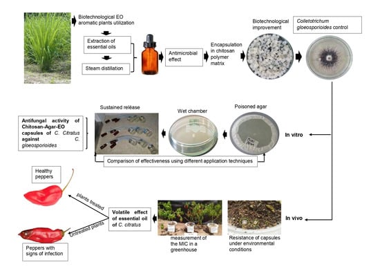
1. Introduction
2. Results
2.1. Isolation, Identification, Reactivation, and Virulence of C. gloeosporioides
2.2. Essential Oil Extraction and Characterization
2.3. Preparation of Chitosan-EO Capsules of C. citratus
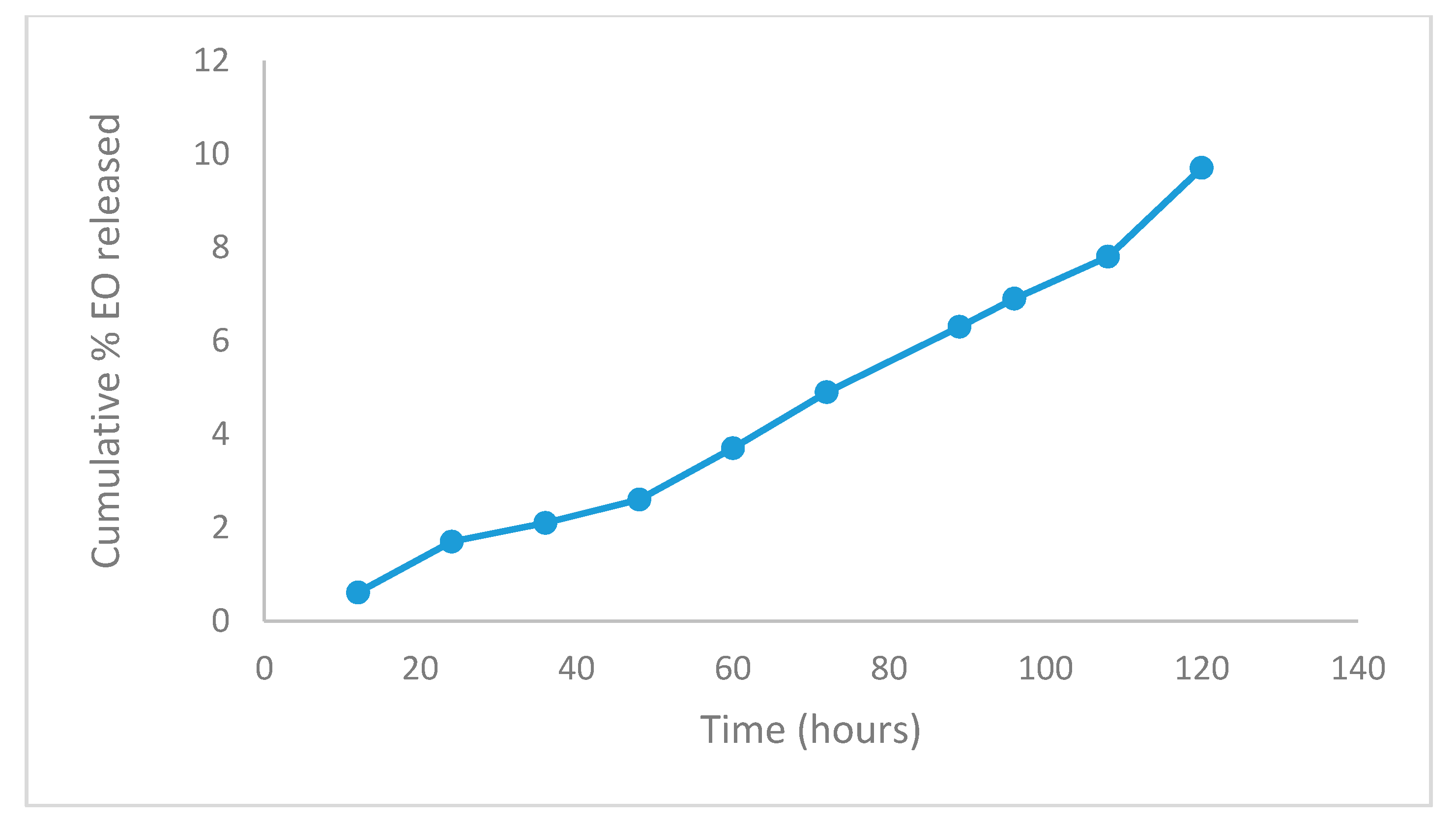
2.4. In Vitro Release
2.5. Percentage of Encapsulated Active Ingredient
2.6. Mass Encapsulation Efficiency
2.7. Chitosan Antifungal Activity Against C. gloeosporioides
2.8. Antifungal Activity of Chitosan-Agar-EO Capsules of C. citratus Against C. gloeosporioides
2.9. Comparison of Antifungal Activity Effectiveness Using Different Application Techniques of EO against C. gloeosporioides
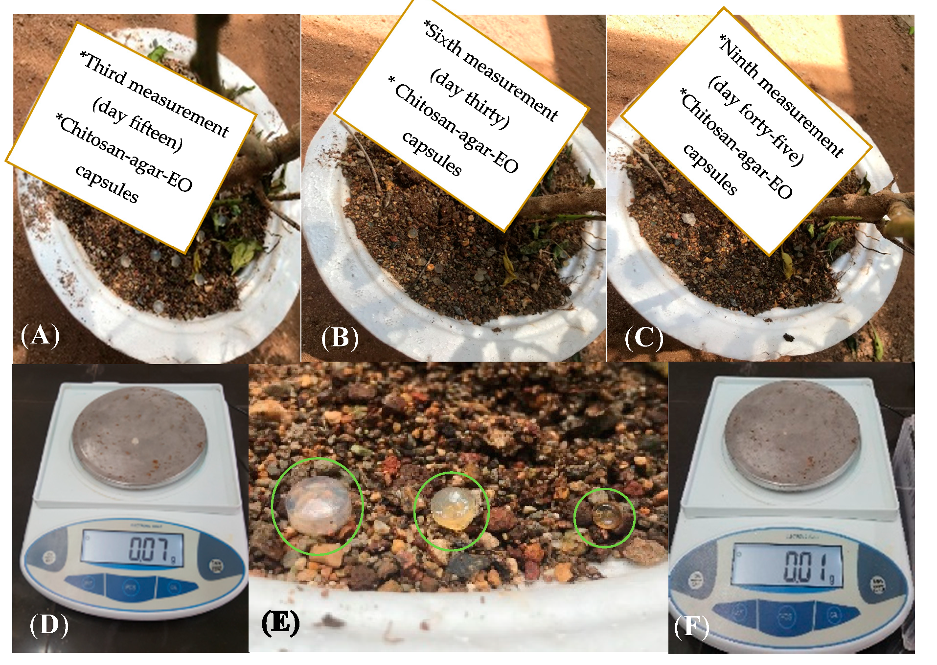
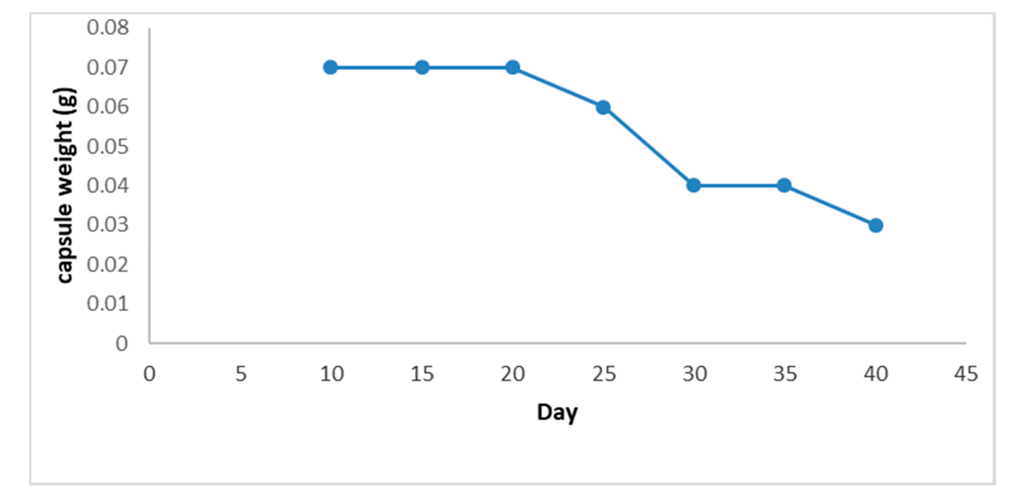
2.10. Evaluation of the Resistance of (Chitosan-Agar-EO) Capsules under Environmental Conditions
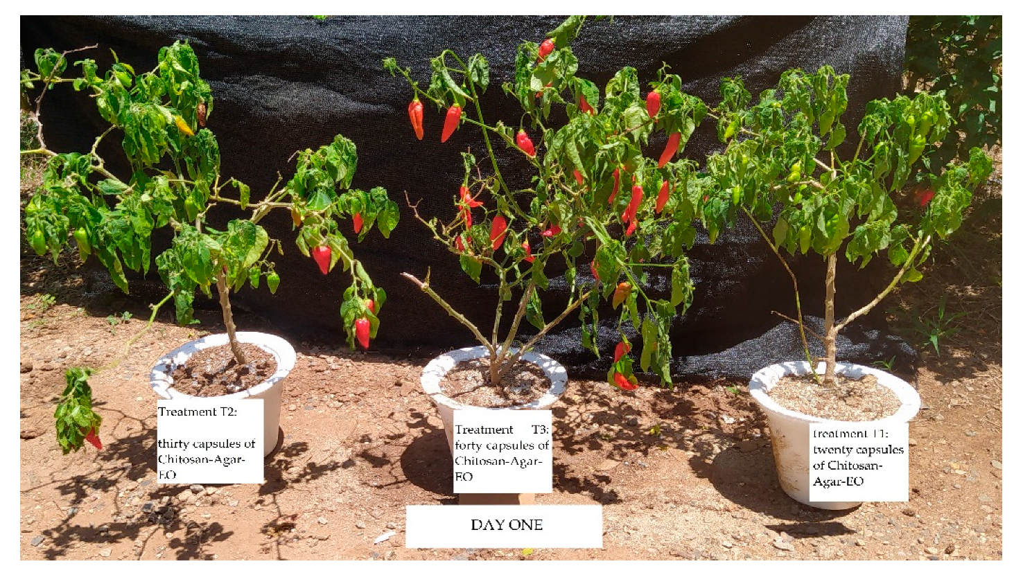
2.11. Antifungal Activity of (Chitosan-Agar-EO) Capsules of C. citratus against C. gloeosporioides in Topito Pepper Plants: Measurement of the MIC Test in a Greenhouse.
3. Discussion
4. Materials and Methods
4.1. Pathogen Selection
4.2. Collection of Plant Material of Cymbopogon citratus (Poaceae) and Extraction of Essential Oils
4.3. Obtaining and Characterizing Chitosan Capsules
4.4. In Vitro Release
4.5. Percentage of Encapsulated Active Ingredient
4.6. Total Oil Content
4.7. Content of Surface Oil
- EE = Encapsulation efficiency as a percentage of mass.
- TO = Total oil retained in g.
- SO = Content of superficial oil in g.
4.8. Evaluation of the Antifungal Activity of Chitosan Against C. gloeosporioides
4.9. Determination of the Minimum Inhibitory Concentration of Chitosan Capsules with EO for the Control C. gloeosporioides
4.10. Efficiency of Chitosan-EO Capsules in Relation to Other EO Application Techniques
4.11. Evaluation of the Resistance of Chitosan-Agar-EO Capsules under Environmental Conditions
4.12. Antifungal Activity of Chitosan-Agar-EO Capsules of Cymbopogon citratus against Colletotrichum gloeosporioides in Topito Pepper Plants: Measurement of the MIC Test in a Greenhouse
4.13. Incidence (%) of Anthracnose in Evaluated Plants
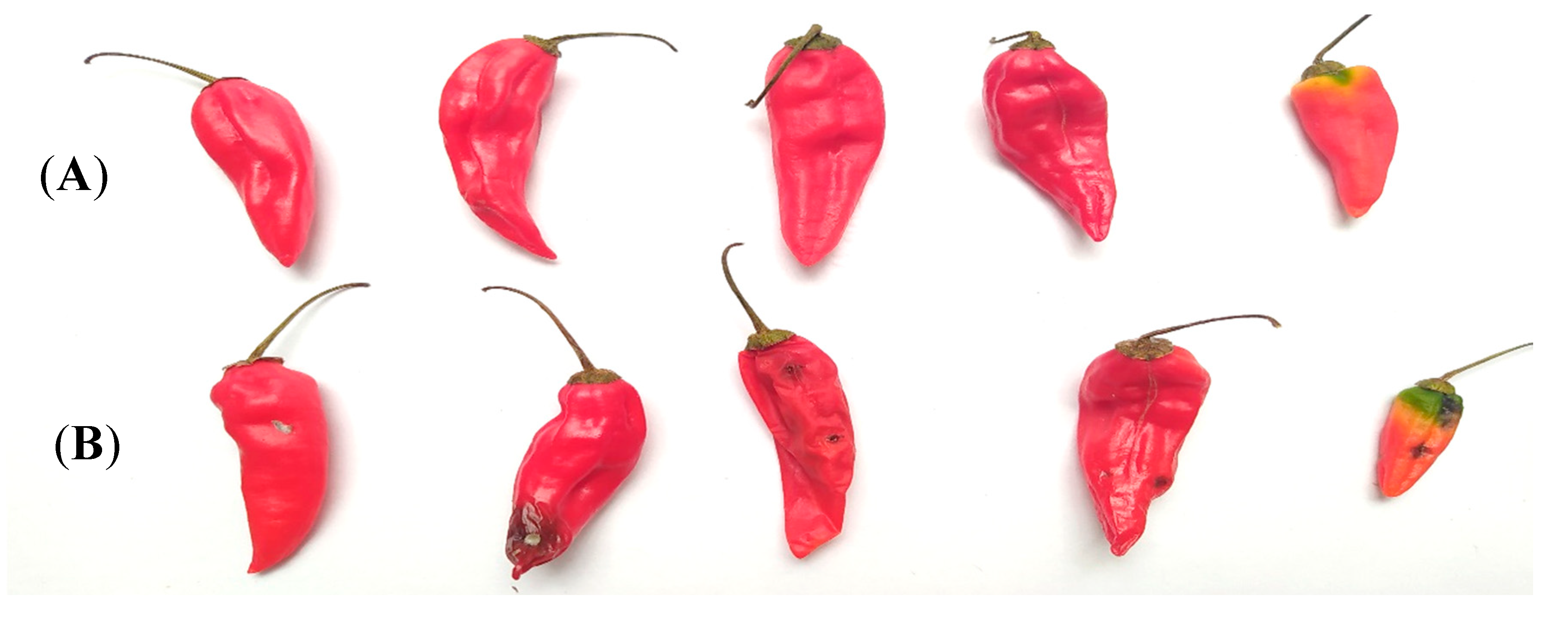
4.14. Severity (%) of Anthracnose in Fruit
5. Conclusions
Supplementary Materials
Author Contributions
Funding
Conflicts of Interest
References
- Ávila, F.; León, L.; Pinzón, M.; Londoño, A.; Gutiérrez, J. Residualidad de fitosanitarios en tomate y uchuva cultivados en Quindío (Colombia). Rev. Corpoica Cienc. Tecnol. Agropecu. 2017, 18, 571–582. [Google Scholar] [CrossRef]
- Beghin, J.; Maertens, M.; Swinnen, J. Nontariff measures and standards in trade and global value chains. Annu. Rev. Resour. Econ. 2015, 7, 425–450. [Google Scholar] [CrossRef] [Green Version]
- Melo, A.; Ariza, P.; Lissbrant, S.; Tofiño, A. Evaluation of agrochemicals and bioinputs for sustainable bean management on the Caribbean coast of Colombia. Agron. Colomb. 2015, 33, 203–211. [Google Scholar] [CrossRef]
- Ali, A.; Bordoh, P.K.; Singh, A.; Siddiqui., Y.; Droby, S. Post-harvest development of anthracnose in pepper (Capsicum spp.): Etiology and management strategies. Crop Prot. 2016, 90, 132–141. [Google Scholar] [CrossRef]
- Villa-Martínez, A.; Pérez-Leal, R.; Morales-Morales, H.A.; Basurto-Sotelo, M.; Soto-Parra, J.M.; Martínez-Escudero, E. Situación actual en el control de Fusarium spp. y evaluación de la actividad antifúngica de extractos vegetales. Acta Agronómica 2014, 6, 433–458. [Google Scholar] [CrossRef]
- Pérez-Cordero, A.; Chamorro-Anaya, L.; Vitola-Romero, D.; Hernández-Gómez, J. Actividad antifúngica de Cymbopogon citratus contra Colletotrichum gloeosporioides. Agron. Mesoam. 2017, 28, 465–475. [Google Scholar] [CrossRef] [Green Version]
- Moretti, M.D.; Sanna-Passino, G.; Demontis, S.; Bazzoni, E. Essential oil formulation useful as a new tool for insect pest control. AAPs PharmSciTech 2002, 3, 64–74. [Google Scholar] [CrossRef] [Green Version]
- Lai, F.; Wissing, S.; Müller, R.; Fadda, A. Artemisia arborescens L essential oil–loaded solid lipid nanoparticles for potential agricultural application: Preparation and characterization. AAPs PharmSciTech 2002, 7, 1–9. [Google Scholar] [CrossRef] [Green Version]
- Dadalioglu, I.; Evrendilek, G.A. Chemical compositions and antibacterial effects of essential oils of Turkish oregano (Origanum minutiflorum), bay laurel (Laurus nobilis), Spanish lavender (Lavandula stoechas L.) and fennel (Foeniculum vulgare) on common foodborne pathogens. J. Agric. Food Chem. 2004, 52, 8255–8260. [Google Scholar] [CrossRef]
- Younes, I.; Rinaudo, M. Chitin and Chitosan Preparation from Marine Sources. Structure, Properties and Applications. Mar. Drugs 2015, 13, 1133–1174. [Google Scholar] [CrossRef] [Green Version]
- Sánchez-González, L.; González-Martínez, C.; Chiralt, A.; Cháfer, M. Physical and antimicrobial properties of chitosan–tea tree essential oil composite films. J. Food Eng. 2010, 98, 443–452. [Google Scholar] [CrossRef]
- Reyes-Chaparro, P.; Gutiérrez-Mendez, N.; Salas-Muñoz, E.; Ayala-Soto, J.G.; Chavez-Flores, D.; Hernández-Ochoa, L. Effect of the addition of essential oils and functional extracts of clove on physicochemical properties of chitosan-based films. Int. J. Polym. Sci. 2015, 714254. [Google Scholar] [CrossRef] [Green Version]
- Sayas-Barberá, E.; Quesada, J.; Sánchez-Zapata, E.; Viuda-Martos, M.; Fernández-López, F.; Pérez-Alvarez, J.A.; Sendra, E. Effect of the molecular weight and concentration of chitosan in pork model burgers. Meat Sci. 2011, 88, 740–749. [Google Scholar] [CrossRef]
- Ogawa, M.; Reis, V.; Godoy, P.; de Menezes, F.G.; Enokihara, M.; Tomimori, J. Feohifomicosis causada por Colletotrichum gloeosporioides y Alternaria infectoria en un paciente trasplantado renal. Rev. Chil. Infectol. 2014, 31, 468–472. [Google Scholar] [CrossRef] [PubMed] [Green Version]
- Alves, M.; Santos, D.; Silva, L.C.; Pontes, E.; Souza, M.A. Essential oils composition and toxicity tested by fumigation against Callosobruchus maculatus (Coleoptera: Bruchidae) pest of stored cowpea. Rev. Virtual Quím. 2015, 7, 2387–2399. [Google Scholar] [CrossRef]
- Tofiño-Rivera, A.; Ortega-Cuadros, M.; Galvis-Pareja, D.; Jiménez-Rios, H.; Merini, J.; Martínez-Pabón, M.C. Effect of Lippia alba and Cymbopogon citratus essential oils on biofilms of Streptococcus mutans and cytotoxicity in CHO cells. J. Ethnopharmacol. 2016, 194, 749–754. [Google Scholar] [CrossRef] [Green Version]
- Kaur, G.; Deepak, D.; Bist, V.; Verma, P.C. Antifungal and larvicidal activities of two acyclic monoterpenes; citral and geraniol against phytopathogenic fungi and insects. Arch. Phytopathol. Plant Prot. 2019, 52, 458–469. [Google Scholar] [CrossRef]
- Avelelas, F.; Horta, A.; Pinto, L.; Marques, S.; Marques Nunes, P.; Pedrosa, R.; Leandro, S.M. Antifungal and antioxidant properties of chitosan polymers obtained from nontraditional Polybius henslowii sources. Mar. Drugs 2019, 17, 239. [Google Scholar] [CrossRef] [Green Version]
- Hoyos, N.; Correa, A.; Jepsen, S.; Wemple, B.; Valencia, S.; Marsik, M.; Doria, R.; Escobar, J.; Restrepo, J.; Velez, M. Modeling streamflow response to persistent drought in a coastal tropical mountainous watershed, Sierra Nevada de Santa Marta, Colombia. Water 2019, 11, 94. [Google Scholar] [CrossRef] [Green Version]
- Rojo-Báez, I.; Álvarez-Rodríguez, B.; García-Estrada, R.S.; León-Félix, J.; Sañudo-Barajas, A.; Allende-Molar, R. Current status of Colletotrichum spp. in Mexico: Taxonomy, characterization, pathogenesis and control. Rev. Mex. Fitopatol. 2017, 35, 549–570. [Google Scholar] [CrossRef] [Green Version]
- Mishra, A.; Dubey, N. Evaluation of some essential oils for their toxicity against fungi causing deterioration of stored food commodities. Appl. Environ. Microbiol. 1994, 60, 1101–1105. [Google Scholar] [CrossRef] [Green Version]
- Janmohammad, M.; Mostafavi, H.; Kazemi, H.; Mahdavinia, G.; Sabaghnia, N. Effect of chitosan application on the performance of lentil genotypes under rainfed conditions. Acta Technol. Agric. 2014, 17, 86–90. [Google Scholar] [CrossRef]
- Pięta, D.; Pastucha, A.; Struszczyk, H.; Wójcik, W. The formation of microorganism communities in the soil under the effect of chitosan and runner bean (Phaseolus coccineus L.) cultivation. Acta Agrobot. 2013, 54, 105–115. [Google Scholar] [CrossRef]
- Kong, M.; Chen, X.G.; Xing, K.; Park, H.J. Antimicrobial properties of chitosan and mode of action: A state of the art review. Int. J. Food Microbiol. 2010, 144, 51–63. [Google Scholar] [CrossRef] [PubMed]
- Gutiérrez-Martínez, P.; Bautista-Baños, S.; Berúmen-Varela, G.; Ramos-Guerrero, A.; Hernández-Ibañez, A.M. In vitro response of Colletotrichum to chitosan. Effect on incidence and quality on tropical fruit. Enzymatic expression in mango. Acta Agron. 2017, 66, 282–289. [Google Scholar] [CrossRef]
- Aquino, C.F.; Sales, N.L.P.; Soares, E.P.S.; Martins, E.R.; Costa, C.A. Composição química e atividade in vitro de três óleos essenciais sobre Colletotrichum gloeosporioides do maracujazeiro. Rev. Bras. Plantas Med. 2014, 16, 329–336. [Google Scholar] [CrossRef] [Green Version]
- Ramos, K.; Andreani, J.; Kozusny, D. Essential and vegetal oils in the in vitro control of Colletotrichum gloeosporioides. Rev. Bras. Plantas Med. 2016, 18, 605–612. [Google Scholar] [CrossRef]
- Marcondes, M.M.; Martins Marcondes, M.; Baldin, I.; Maia, A.J.; Leite, C.D.; Faria, C.M.D.R. Influence of different aqueous extracts of medicinal plants in the development of Colletotrichum gloeosporioides and Fusarium moniliforme. Rev. Bras. Plantas Med. 2014, 16, 896–904. [Google Scholar] [CrossRef] [Green Version]
- Hernández-Ochoa, L.; Macías-Castañeda, C.; Nevárez, G.; Salas, E.; Sandoval, F. Antimicrobial activity of chitosan-based films including spices essential oils and functional extracts. Cyta J. Food 2012, 10, 85–91. [Google Scholar] [CrossRef] [Green Version]
- Ali, E.O.M.; Shakil, N.A.; Rana, V.S.; Sarkar, D.J.; Majumder, S.; Kaushik, P.; Singh, B.B.; Kumar, J. Antifungal activity of nano emulsions of neem and citronella oils against phytopathogenic fungi, Rhizoctonia solani and Sclerotium rolfsii. Ind. Crop. Prod. 2017, 108, 379–387. [Google Scholar] [CrossRef]
- Li, C.M.; Yu, J.P. Chemical composition, antimicrobial activity and mechanism of action of essential oil from the leaves of Macleaya Cordata (Willd.) R. Br. J. Food Saf. 2015, 35, 227–236. [Google Scholar] [CrossRef]
- Shukla, S.K.; Mishra, A.K.; Arotiba, O.A.; Mamba, B.B. Chitosan-based nanomaterials: A state-of-the-art review. Int. J. Biol. Macromol. 2013, 59, 46–58. [Google Scholar] [CrossRef] [PubMed]
- Tofiño-Rivera, A.; Ortega-Cuadros, M.; Melo-Ríos, A.; Mier-Giraldo, H. Vigilancia tecnológica de plantas aromáticas: Desde la investigación a la consolidación de la agrocadena colombiana. Cienc. Technol. Agropecu. 2017, 18, 353–377. [Google Scholar] [CrossRef]
- Admasu, W.; Sahile, S.; Kibret, M. Assessment of potential antagonists for anthracnose (Colletotrichum gloeosporioides) disease of mango (Mangifera indica L.) in North Western Ethiopia (Pawe). Arch. Phytopathol. Plant Prot. 2014, 47, 2176–2186. [Google Scholar] [CrossRef]
- Tofiño-Rivera, A.; Ortega-Cuadros, M.; Pedraza, B.; Perdomo, S.; Moya, D. Efectividad de Beauveria bassiana (Baubassil®) sobre la garrapata común del ganado bovino Rhipicephalus microplus en el Departamento de la Guajira, Colombia. Rev. Argent. Microbiol. 2018, 50, 426–430. [Google Scholar] [CrossRef]
- Gañán, L.; Álvarez, E.; Castaño, J. Identificación genética de aislamientos de Colletotrichum spp. causantes de antracnosis en frutos de aguacate, banano, mango y tomate de árbol. Rev. Acad. Colomb. Cienc. Exactasfísicas Nat. 2015, 39, 339–347. [Google Scholar] [CrossRef] [Green Version]
- Gomes, C.; Costa, L.; Loretti, F.; Borges, E.; de Aguiar, A.; de Moura, M.; Gomes, I. Aspergillus in endodontic infection near the maxillary sinus. Braz. J. Otorhinolaryngol. 2015, 81, 527–532. [Google Scholar] [CrossRef] [Green Version]
- Jovanović, G.D.; Klaus, A.S.; Nikšić, M. Antimicrobial activity of chitosan coatings and films against Listeria monocytogenes on black radish. Rev. Argent. Microbiol. 2016, 48, 128–136. [Google Scholar] [CrossRef] [Green Version]
- Mishra, N.; Kumar, V.R.; Singh, K.Y.; Sinha, P.; Kanaujia, A.; Chanda, D.; Jakhmola, A.; Saikia, D.; Yadav, N. Encapsulation of Mentha Oil in Chitosan Polymer Matrix Alleviates Skin Irritation. AAPs PharmSciTech 2015, 17, 482–492. [Google Scholar] [CrossRef]
- Vila, J.L. Aspectos fundamentales de los sistemas farmacéuticos y operaciones básicas. Madr. Sínt. 2001, 1, 623. [Google Scholar]
- Official Method 922.06. Acid Hydrolysis Method. Retrieved from: AOAC. 2005. Available online: http://www.eoma.aoac.org/methods/info.asp?ID=26940 (accessed on 15 September 2020).
- Calvo, P.; Hernández, T.; Lozano, M.; González-Gómez, D. Microencapsulation of extra-virgin olive oil by spray-drying: Influence of wall material and olive quality. Eur. J. Lipid Sci. Technol. 2010, 112, 852–858. [Google Scholar] [CrossRef]
- Velásquez, M.A.; Álvarez, R.; Tamayo, P.; Carvalho, C. Evaluación in vitro de la actividad fungistática del aceite esencial de mandarina sobre el crecimiento de Penicillium sp. Cienc. Tecnol. Agropecu. 2014, 15, 7–14. [Google Scholar]
- Chen, J.L.; Liu, K.; Miao, C.P.; Sun, S.Z.; Chen, Y.W.; Xu, L.H.; Guan, H.L.; Zhao, L.X. Salt tolerance of endophytic Trichoderma koningiopsis YIM PH30002 and its volatile organic compounds (VOCs) allelopathic activity against phytopathogens associated with Panax notoginseng. Ann. Microbiol. 2016, 66, 981–990. [Google Scholar] [CrossRef]
- Males, I. Estudio del Efecto de Aditivos Oxobiodegradables en los Cambios Estructurales de Materiales Nanoestructurados a Base de Mezclas de LLDPE/COC con Nanoarcillas. Centro de Investigación en Química Aplicada. 2013. Available online: https://ciqa.repositorioinstitucional.mx/jspui/bitstream/1025/40/1/Tesis%20MTP%20Iris%20Candy%20Males%20Casta%C3%B1eda%20Nov%2021%202013.pdf (accessed on 17 June 2020).
- Instituto de Hidrologia, Meteorologia y Estudios Ambientales [IDEAM]. Boletin de Predicción Climática y Recomendación Sectorial 2018–2019: Para Planear y Decidir. 2019. Available online: http://www.ideam.gov.co/web/tiempo-y-clima/prediccion-climatica?p_p_id=110_INSTANCE_ljPLJWRaQzCm&p_p_lifecycle=0&p_p_state=normal&p_p_mode=view&p_p_col_id=column-1&p_p_col_count=1 (accessed on 26 June 2020).
- Andrades, I.; Yender, F.; Labarca, J.; Ulacio, D.; Paredes, C.; Marín, Y. Evaluación de la antracnosis (Colletotrichum sp.) en guanábana (Annona muricata L.) tipo Gigante en el sector Moralito del estado Zulia, Venezuela. Rev. Cient. UDO Agríc. 2009, 9, 148–157. [Google Scholar]
Sample Availability: Samples of the compounds are not available from the authors. |


| Identification | Relative Quantity (%) |
|---|---|
| 6-methyl-5-hepten-2-one | 1.2 |
| Myrcene | 14.0 |
| α-felandrene | 0.1 |
| (Z)-β-ocimene | 0.6 |
| N.I. | 1.4 |
| 3-4-methyl-3-pentyl)-furan | 0.3 |
| Linalool | 2.8 |
| N.I | 1.6 |
| N.I | 1.3 |
| cis-verbenol | 3.4 |
| trans-verbenol | 4.1 |
| N.I. | 0.7 |
| Citronellol | 1.3 |
| Neral | 22.1 |
| 3,7-dimethyl-2,6-octadien-1-ol | 2.6 |
| Geranial | 32.9 |
| 2-undecanone | 0.7 |
| Eugenol | 0.9 |
| geraniol acetate | 1.5 |
| (E)-Caryophylene | 1.7 |
| α-zingibirene | 0.4 |
| β-sesquifelandrene | 1.6 |
| Tumeron | 1.5 |
| Curlona | 0.9 |
| mono-2-ethylhexylphthalate | 1.2 |
| Treatment | Inhibition of Mycelial Growth | |||
|---|---|---|---|---|
| Day 5 | Day 10 | Day 15 | Day 30 | |
| Capsules with corn oil (control) | 0 a | 0 a | 0 a | 0 a |
| Poisoned food | 88.5 c | 68.6 c | 30.6 b | 0 a |
| Wet chamber | 77.5 b | 61.2 b | 49 c | 0 a |
| Chitosan-Agar-EO capsules | 100 d | 100 e | 100 e | 100 b |
| Trichoderma | 88.9 c | 90.9 d | 96.3 e | 98.8 b |
| Percentage and Degree of Severity of Anthracnose in Topito Peppers with Treatments of EO Capsules of C. citratus and Corn Oil Controls | ||||
|---|---|---|---|---|
| Capsules of Chitosan-Agar-EO C. citratus | Treatments | T1 | T2 | T3 |
| Average number of peppers with signs of infection | 12 | 0 | 0 | |
| Number of healthy peppers | 25 | 35 | 36 | |
| Average number of peppers per plant | 37 | 35 | 36 | |
| Total percentage of damage in each treatment | 32% ab | 0% a | 0% a | |
| Degree of severity | 5 | 0 | 0 | |
| Capsules of Chitosan-Agar-Corn Oil | Controls | C1 | C2 | C3 |
| Average number of peppers with signs of infection | 33 | 37 | 35 | |
| Number of healthy peppers | 6 | 4 | 2 | |
| Average number of peppers per plant | 39 | 41 | 37 | |
| Total percentage of damage in each control | 84.6% b | 90.2% b | 94.6% b | |
| Degree of severity | 7 | 8 | 9 | |
| Type of Capsules | Number of Capsules (µL Oil) | Treatments and Controls Utilized | ||
|---|---|---|---|---|
| System 1 (186 cc) | System 2 (161 cc) | System 3 (147 cc) | ||
| (Chitosan-Agar-EO) C. citratus (treatments) | 20 (170 µL) | (913 ppm) | (1056 ppm) | (1156 ppm) |
| 30 (255 µL) | (1370 ppm) | (1584 ppm) | (1734 ppm) | |
| 40 (340 µL) | (1827 ppm) | (2111 ppm) | (2312 ppm) | |
| Chitosan-Agar-Corn oil (control) | 20 (170 µL) | (913 ppm) | (1056 ppm) | (1156 ppm) |
| 30 (255 µL) | (1370 ppm) | (1584 ppm) | (1734 ppm) | |
| 40 (340 µL) | (1827 ppm) | (2111 ppm) | (2312 ppm) | |
| Air Temperature at a Height of 12 m | Precipitation (mm) | Period of Sunlight (hours) | Relative Humidity (%) | |||||
|---|---|---|---|---|---|---|---|---|
| Month | Average (°C) | Month | Average | Month | Average | Month | Average | |
| Highest | March | 35.8 | October | 199 | June | 12.7 | May | 69 |
| July | 36.1 | July | 12.7 | October | 75 | |||
| Lowest | May | 32 | February | 10 | December | 11.5 | January | 59 |
| October | 23 | February | 55 | |||||
| March | 56 | |||||||
© 2020 by the authors. Licensee MDPI, Basel, Switzerland. This article is an open access article distributed under the terms and conditions of the Creative Commons Attribution (CC BY) license (http://creativecommons.org/licenses/by/4.0/).
Share and Cite
Tofiño-Rivera, A.P.; Castro-Amaris, G.; Casierra-Posada, F. Effectiveness of Cymbopogon citratus Oil Encapsulated in Chitosan on Colletotrichum gloeosporioides Isolated from Capsicum annuum. Molecules 2020, 25, 4447. https://0-doi-org.brum.beds.ac.uk/10.3390/molecules25194447
Tofiño-Rivera AP, Castro-Amaris G, Casierra-Posada F. Effectiveness of Cymbopogon citratus Oil Encapsulated in Chitosan on Colletotrichum gloeosporioides Isolated from Capsicum annuum. Molecules. 2020; 25(19):4447. https://0-doi-org.brum.beds.ac.uk/10.3390/molecules25194447
Chicago/Turabian StyleTofiño-Rivera, Adriana Patricia, Glorismar Castro-Amaris, and Fánor Casierra-Posada. 2020. "Effectiveness of Cymbopogon citratus Oil Encapsulated in Chitosan on Colletotrichum gloeosporioides Isolated from Capsicum annuum" Molecules 25, no. 19: 4447. https://0-doi-org.brum.beds.ac.uk/10.3390/molecules25194447