Article

Syntheses and Structural Investigations of Penta-Coordinated Co(II) Complexes with Bis-Pyrazolo-S-Triazine Pincer Ligands, and Evaluation of Their Antimicrobial and Antioxidant Activities

by
Saied M. Soliman
1,*,
Raghdaa A. Massoud
1,
Hessa H. Al-Rasheed
2 and
Ayman El-Faham
1,2,*
1
Department of Chemistry, Faculty of Science, Alexandria University, P.O. Box 426, Ibrahimia, Alexandria 21321, Egypt
2
Department of Chemistry, College of Science, King Saud University, P.O. Box 2455, Riyadh 11451, Saudi Arabia
*
Authors to whom correspondence should be addressed.
Submission received: 29 April 2021 / Revised: 10 June 2021 / Accepted: 11 June 2021 / Published: 14 June 2021

Abstract

:
Two penta-coordinated [Co(MorphBPT)Cl2]; 1 and [Co(PipBPT)Cl2]; 2 complexes with the bis-pyrazolyl-s-triazine pincer ligands MorphBPT and PipBPT were synthesized and characterized. Both MorphBPT and PipBPT act as NNN-tridentate pincer chelates coordinating the Co(II) center with one short Co-N(s-triazine) and two longer Co-N(pyrazole) bonds. The coordination number of Co(II) is five in both complexes, and the geometry around Co(II) ion is a distorted square pyramidal in 1, while 2 shows more distortion. In both complexes, the packing is dominated by Cl…H, C-H…π, and Cl…C (anion-π stacking) interactions in addition to O…H interactions, which are found only in 1. The UV-Vis spectral band at 564 nm was assigned to metal–ligand charge transfer transitions based on TD-DFT calculations. Complexes 1 and 2 showed higher antimicrobial activity compared to the respective free ligand MorphBPT and PipBPT, which were not active. MIC values indicated that 2 had better activity against S. aureus, B. subtilis, and P. vulgaris than 1. DPPH free radical scavenging assay revealed that all the studied compounds showed weak to moderate antioxidant activity where the nature of the substituent at the s-triazine core has a significant impact on the antioxidant activity.

1. Introduction

Triazine is a prototypal molecule that has, together with its derivatives, a wide commercial uses, for example, in resins, dyes, herbicides, or as sulfide removal agents [1,2]. These compounds are a well-suited model system in molecular imprinting [3]. s-Triazines are widely used within the pharmaceutical, textile, plastic, and rubber industries, and as pesticides, dyestuffs, optical bleaches, explosives, and surface-active agents [4,5,6]. s-Triazine and its compounds are also used as subunits in the formation of supramolecular structures because they possess good optical and electronic properties and are able to form three strong hydrogen bonds with the host molecule [7].
On the other hand, bacterial and fungal infectious diseases are very common all over the world. Due to the rapid development in drug resistance, tolerance, and side effects, there is a critical need for new antibacterial and antifungal agents that exhibit improved pharmacological properties and drug-resistance profiles [8,9]. In this aspect, the triazine class [10,11,12,13,14,15,16,17,18,19] and their metal complexes [20,21,22,23,24] have been received a great deal of attention as they demonstrate wide range of therapeutic activities and a great array of biological applications including antimicrobial, antituberculosis, anticancer, antiviral, antibacterial, antifungal, anti-HIV, and antimalarial activities [9,25,26]. In addition, a number of metal complexes based on triazine derivatives have been studied for their interesting magnetic [27,28,29] and catalytic [30,31,32,33,34,35,36,37,38,39,40,41] applications.
Among the s-triazine derivatives, 2,4-bis(3,5-dimethyl-1H-pyrazol-1-yl)-6-methoxy-1,3,5-triazine (MBPT) pincer ligand has been extensively used in the synthesis of a wide range of homoleptic and heteroleptic metal(II) complexes with coordination numbers ranging from five to eight [42,43,44,45,46,47,48,49,50]. Recently, we reported that the reaction of MBPT with CoCl2 afforded the [Co(MBPT)Cl(H2O)2]Cl pincer complex, which was found to be the best candidate as an antimicrobial agent compared to the [Co(MBPT)(NO3)2] and [Co(MBPT)(H2O)3](ClO4)2 analogues [48]. Furthermore, a number of [ML]Cl2 and [ML2]Cl2 complexes (where M = Cu(II), Ni(II), and Co(II), and L is 2,4-bis(3,5-dimethyl-1H-pyrazol-1-yl)-6-phenylamino-1,3,5-triazine) were synthesized and were also investigated for their antimicrobial activities [51]. Following the research on the same class of these pincer ligands, we present here the self-assembly of the s-triazine functional ligands shown in Figure 1 with CoCl2 in order to synthesize new biologically active complexes bearing both bioactive species; the ligand and Co(II) ion, which could lead to the synthesis of powerful antimicrobial agents. The structural aspects of the synthesized complexes were analyzed using single-crystal X-ray diffraction, Hirshfeld, and DFT calculations, as well as spectroscopic analysis. In addition, the antimicrobial and antioxidant activities of both complexes were examined and compared with the free ligands.

2. Results and Discussion
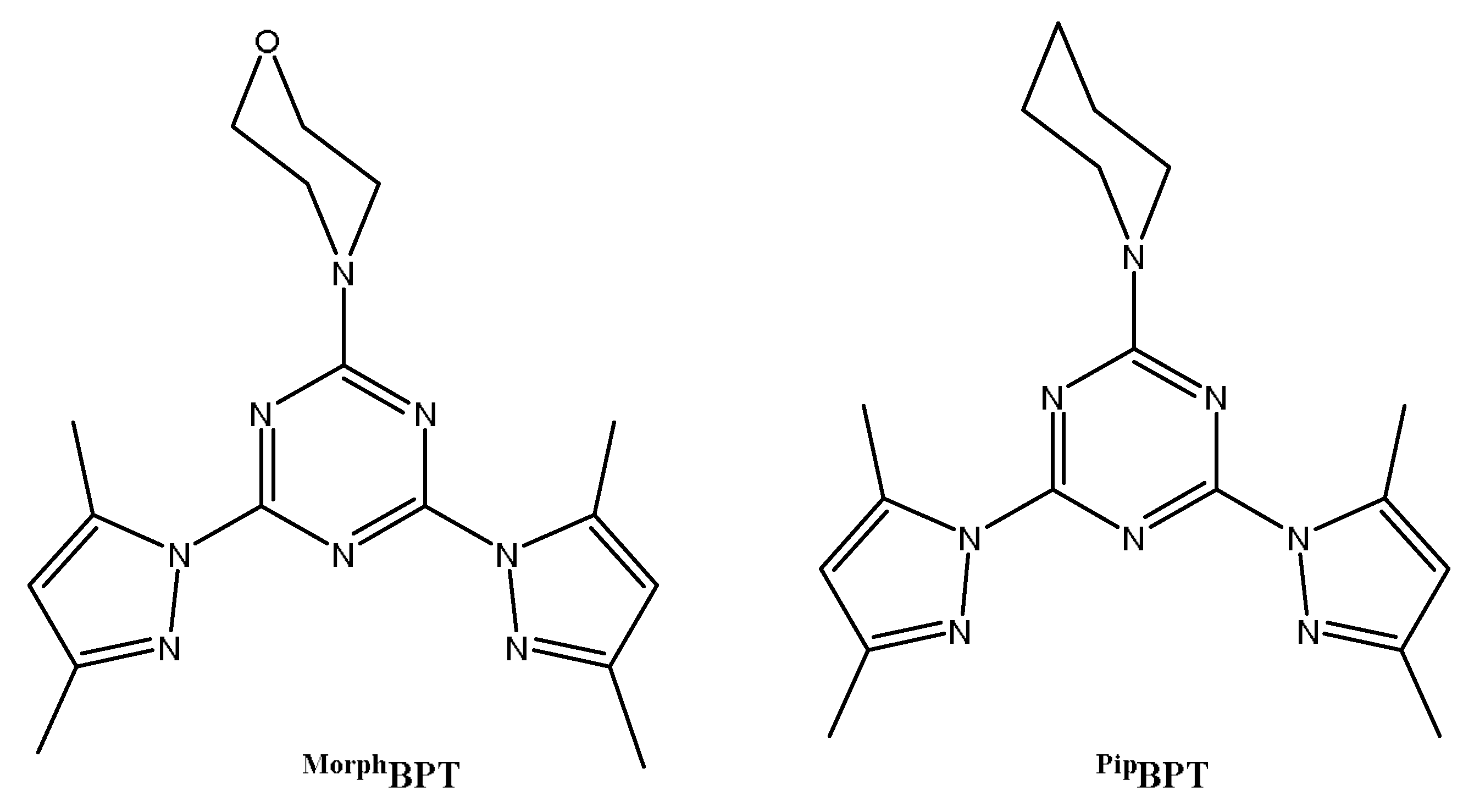
2.1. Chemistry

The self-assembly of the functional ligands 4-(4,6-bis(3,5-dimethyl-1H-pyrazol-1-yl)-1,3,5-triazin-2-yl)morpholine (MorphBPT) and 2,4-bis(3,5-dimethyl-1H-pyrazol-1-yl)-6-(piperidin-1-yl)-1,3,5-triazine (PipBPT) with CoCl2 in methanol afforded the corresponding heteroleptic neutral complexes [Co(MorphBPT)Cl2] (1) and [Co(pipBPT)Cl2] (2) in good yield. The Structure aspects of complexes 1 and 2 were analyzed using different spectroscopic tools such as FTIR, UV-Vis, and low-temperature X-ray single-crystal diffraction combined with Hirshfeld calculations. Crystallographic details are listed in Table 1. In addition, biological evaluations of these new complexes as antimicrobial and antioxidant agents were performed and compared with the corresponding free ligands.

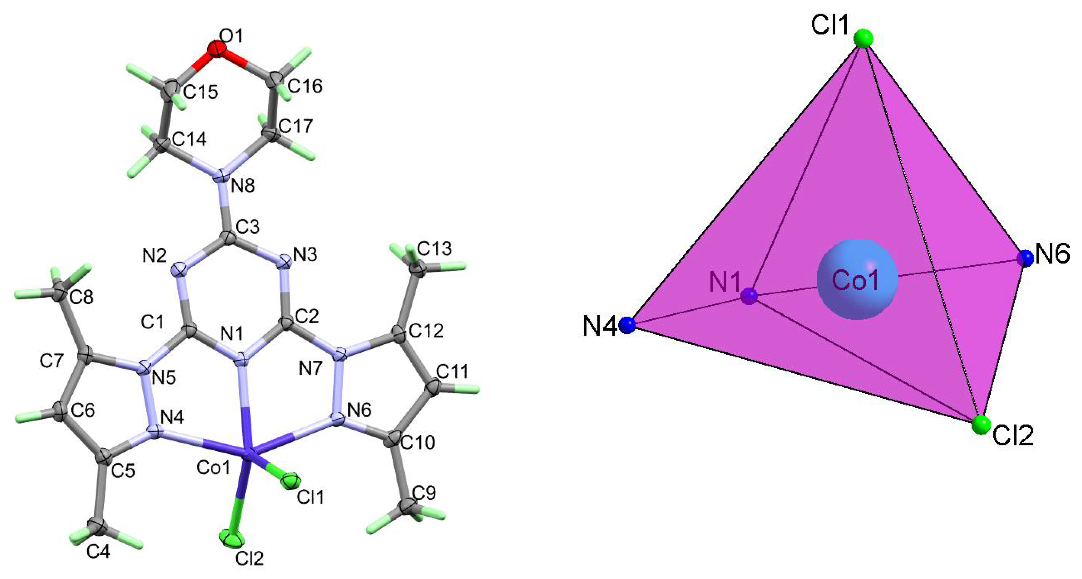
2.1.1. Structure Description of [Co(MorphBPT)Cl2] (1)

The neutral complex [Co(MorphBPT)Cl2] (1) crystallized in the monoclinic crystal system and the centrosymmetric C2/c space group. The asymmetric unit comprised one [Co(MorphBPT)Cl2] with Z = 8. The structure of 1 revealed a penta-coordinated Co(II) complex with one tridentate MorphBPT chelate and two chloride anionic ligands in the inner sphere (Figure 2; right part). The MorphBPT ligand coordinated the Co(II) via one short Co-N(s-triazine) and relatively longer Co-N(pyrazole) bonds. The corresponding Co1-N1, Co1-N4, and Co1-N6 distances are 2.0387(15), 2.2008(15), and 2.2304(16) Å, respectively. The two Co-Cl distances are very similar, where the Co1-Cl1 and Co1-Cl2 distances are 2.2718(7) and 2.2968(7) Å, respectively. The bite angles in the coordinated MorphBPT are 74.01(6) and 73.07(5)° for N1-Co1-N4 and N1-Co1-N6, respectively, while the N4-Co1-N6 and Cl2-Co1-Cl1 bond angles are 146.87(5) and 111.40(3)°, respectively (Table 2). The ring systems in MorphBPT are not perfectly coplanar where the angle between the s-triazine mean plane and each of the two pyrazolyl rings are 4.88 and 8.49° for the pyrazole moieties with lower and higher atom numbering, respectively. The distortion in the CoN3Cl2 coordination sphere of complex 1 was described using the criterion reported by Addison [52]. The geometry around Co(II) is a distorted square pyramidal with N4-Co1-N6 (β= of 146.87(5)°) and N1-Co1-Cl2 (α = 138.89(4)°), giving a τ value of 0.133. The distorted square pyramidal configuration comprised Cl2N1N4N6 donor atoms in the basal plane and Cl1 as apical (Figure 2; left part).
The packing of complex molecules in 1 is controlled by O1…H11 and Cl1…H6 contacts shown in Figure 3 (upper part). The donor (D)-acceptor (A) distances are 3.503(3) and 3.550(2) Å for O1…H11 and Cl1…H6 hydrogen bond contacts, respectively (Table 3). Packing of complex units via C-H…Cl and C-H…O interactions is shown in Figure 3 (lower part).

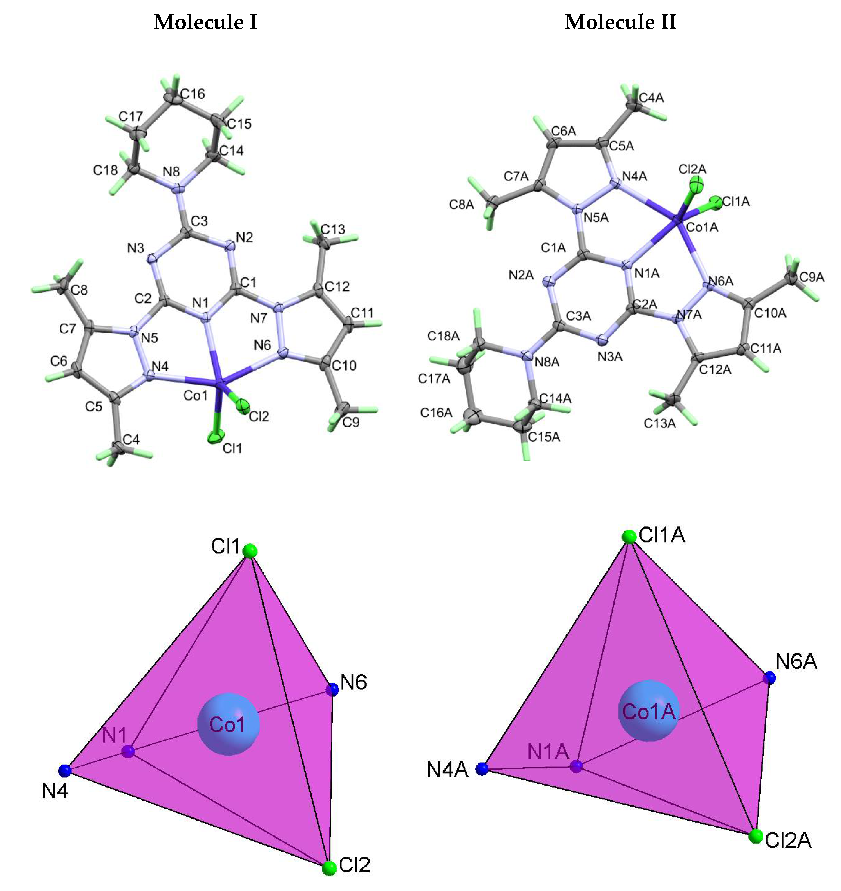
2.1.2. Structure Description of [Co(PipBPT)Cl2] (2)

The [Co(PipBPT)Cl2] complex (2) crystallized in the less symmetric triclinic crystal system and P-1 space group with Z = 4 and two molecular units as an asymmetric formula. In both units, the Co(II) is penta-coordinated with CoN3Cl2 coordination sphere comprising one PipBPT ligand chelating the Co(II) ion in a pincer fashion augmented with two Co-Cl bonds at almost equal distances (Figure 4). Generally, the bond distances and angles of the two asymmetric formulas are very similar (Table 2). The τ values are 0.42 and 0.37 for the two molecular units I and II of [Co(PipBPT)Cl2] complex indicating more distorted square pyramidal compared to complex 1. It could be considered as an intermediate structure between square pyramidal and trigonal bipyramidal configurations. For molecule I, the angle between the s-triazine mean plane and each of the two pyrazolyl rings are 4.17 and 0.69° for the pyrazole moieties with lower and higher atom numbering, respectively. On the other hand, for molecule II the corresponding values are 4.71 and 7.76°, respectively.
The packing of complex molecules in 2 is controlled by Cl2…H6A, Cl2…H8A, and Cl2…H13A contacts shown in Figure 5 (upper part). The donor (D)-acceptor (A) distances are 3.524(2), 3.768(2) and 3.611(2) Å, respectively (Table 3). The hydrogen-bonding network in which the complex units are interconnected via C-H…Cl interactions as shown in Figure 5 (lower part).

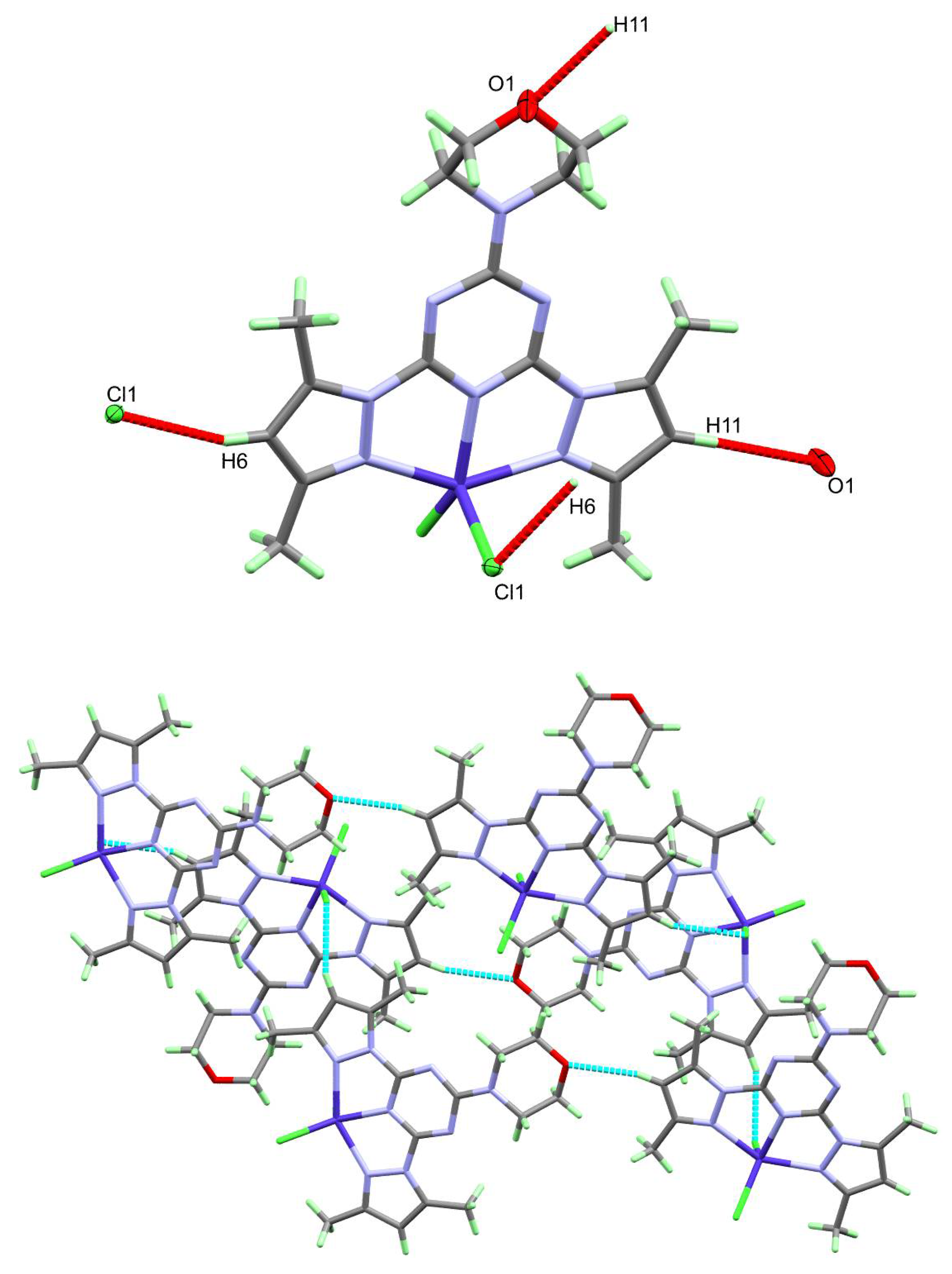
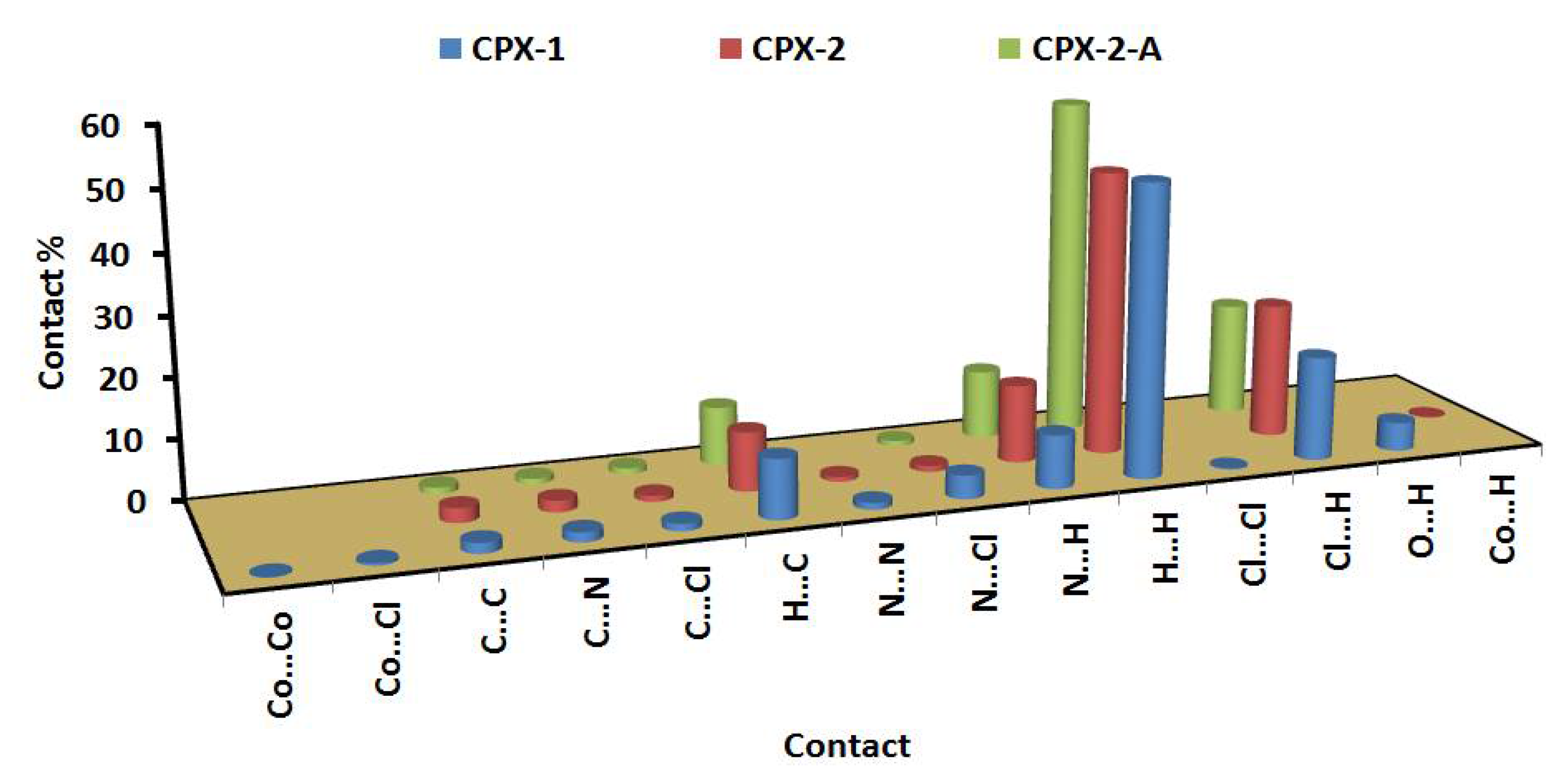
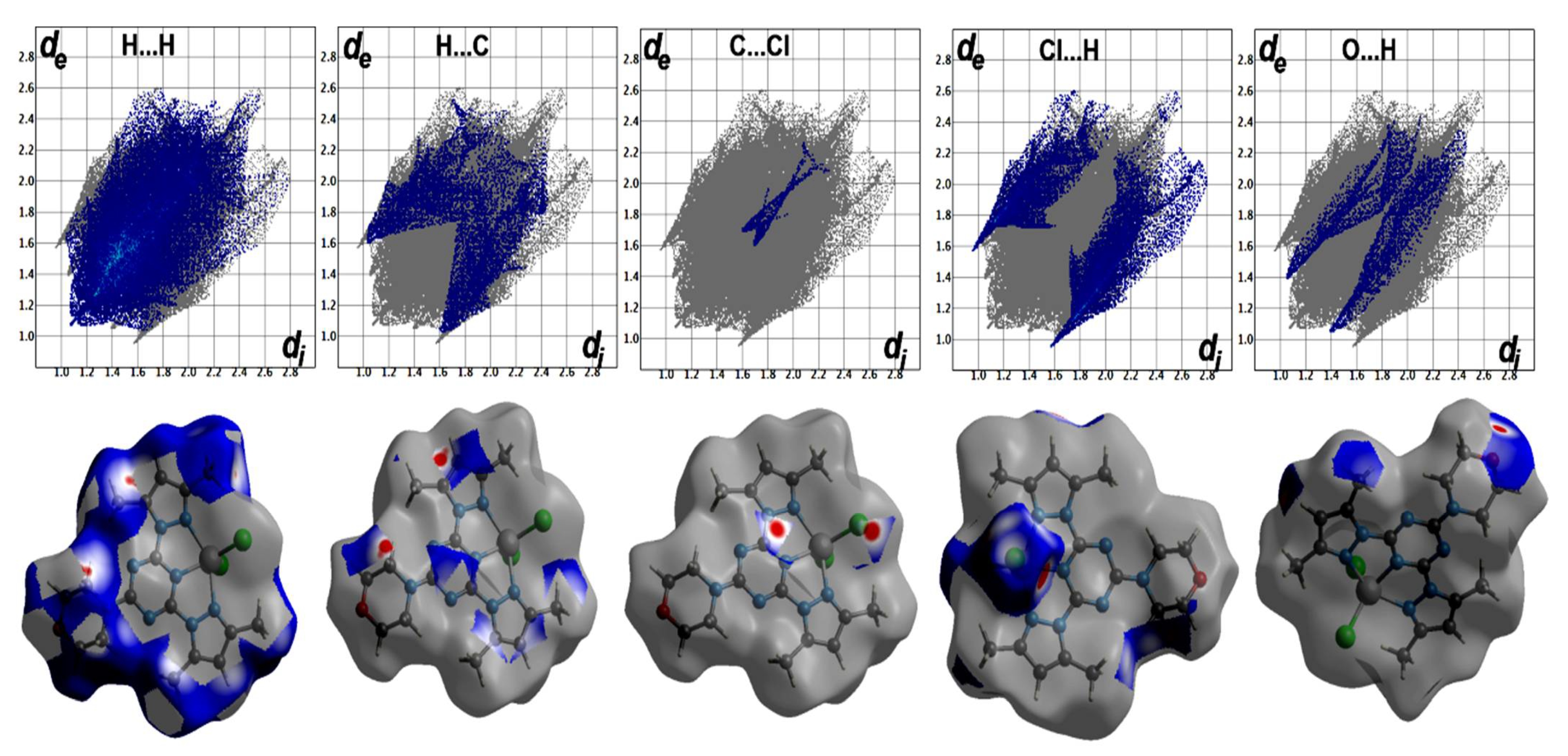
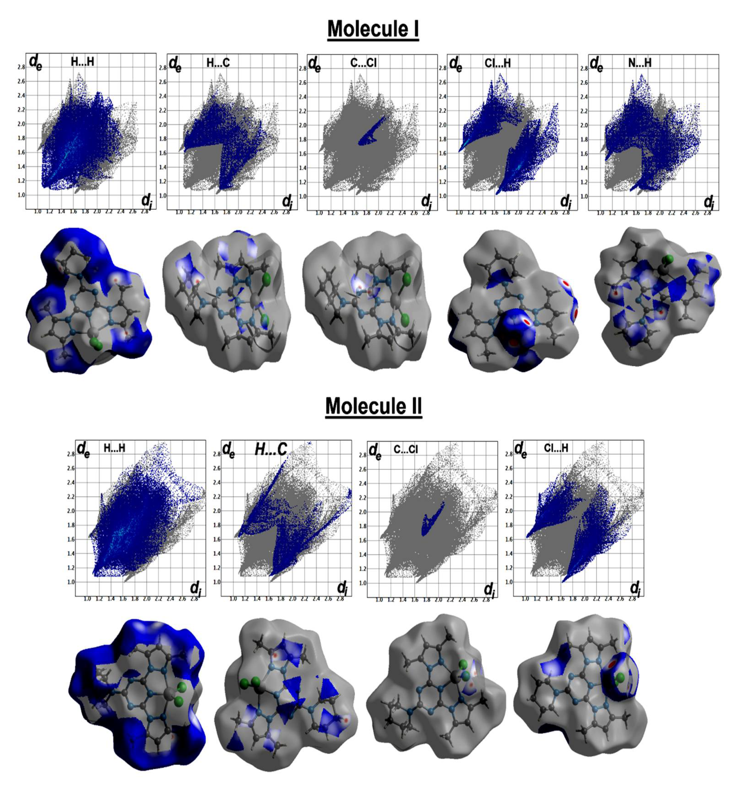
2.2. Hirshfeld Topology Analyses
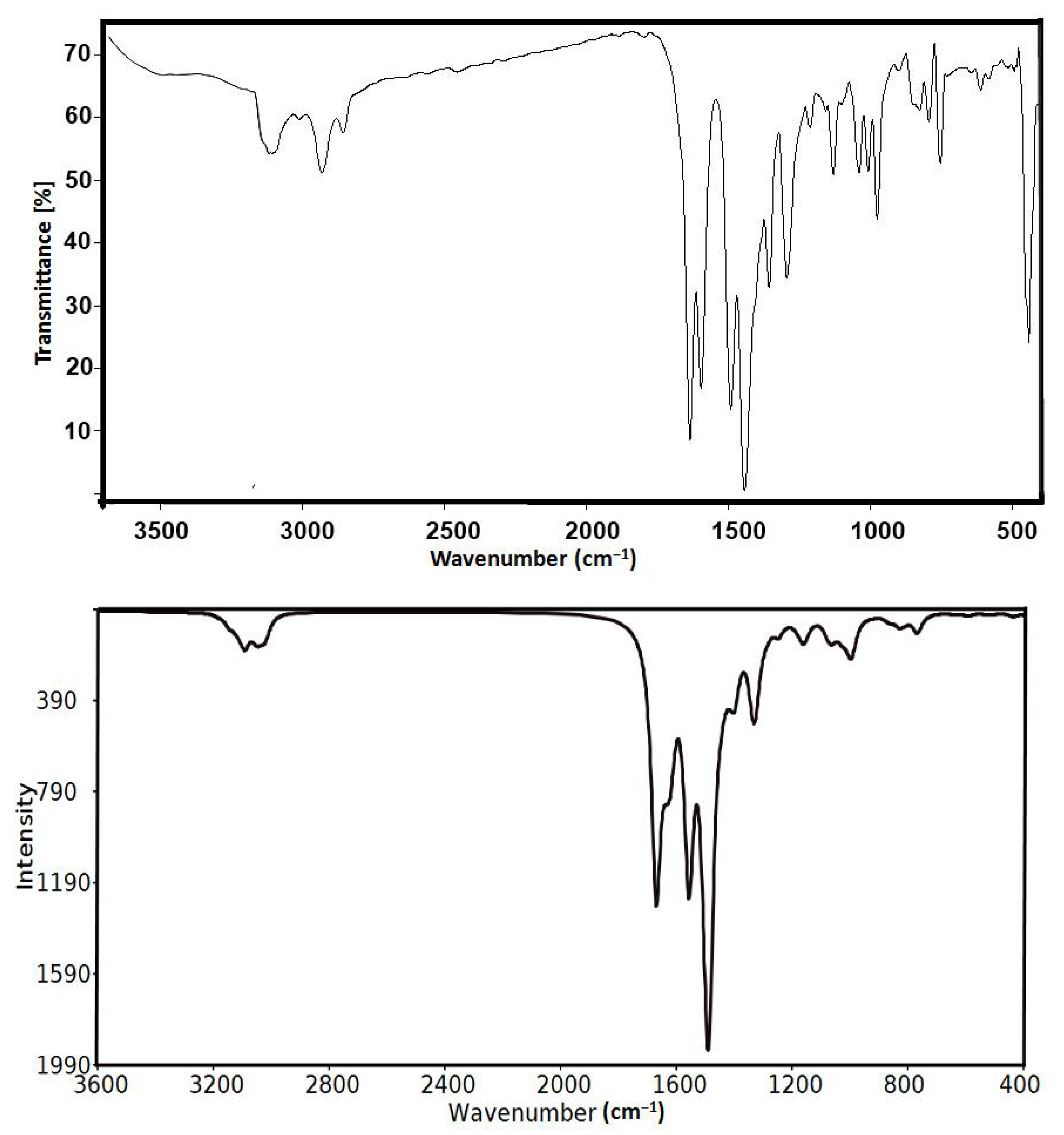
In order to further explore the different intermolecular contacts in the solid-state structure of the studied complexes, we employed Hirshfeld calculations (Figures S1–S3; Supplementary Data). Quantitative analysis results of all possible interactions are presented in Figure 6.
The percentages of the H…H contacts are 48.8% and 47.7–56.1% from the whole contacts detected in [Co(MorphBPT)Cl2] and [Co(PipBPT)Cl2], respectively, while the Cl…H contact percentages are 17.4 and 18.9–22.5%, respectively. It is clear from the decomposed fingerprint and dnorm maps that these interactions have the characteristics of short contacts (Figure 7 and Figure 8). The shortest intermolecular contacts are listed in Table 4. In complex 1, the packing is controlled by O…H and Cl…H hydrogen bonds as well as C-H…π and Cl…C (anion-π stacking) interactions. The latter belongs to the interaction between the coordinated chloride anion Cl2 and the C1 atom from the electron-deficient s-triazine moiety. In complex 2, the packing is also dominated by short Cl…H, C-H…π and Cl…C (anion-π stacking) interactions. It is noted that the Cl2…C1 (3.297 Å) in complex 1 is significantly shorter than the Cl2A…C3 (3.407 Å) and Cl2A…C2 (3.412 Å) interactions in complex 2. The H…C(π-system) are in the range of 2.642–2.751 and 2.722–2.727 Å in complexes 1 and 2, respectively. Also, the C…C/C…N contacts having longer distances than the vdWs radii sum of the interacting elements indicated weak π-π interactions.

2.3. FTIR Spectra

The FTIR spectra of [Co(MorphBPT)Cl2] (1) and [Co(PipBPT)Cl2] (2) showed some variations compared to the free ligands. The free MorphBPT and PipBPT showed the C=N stretching modes at 1609 and 1603 cm−1, respectively. The corresponding values in complexes 1 and 2 showed significant shifts toward higher wavenumbers of 1633 and 1636 cm−1, respectively, due to the coordination of the Co(II) with the pincer ligand. Additionally, the νC = C modes in the free ligands were observed at 1529 and 1512 cm−1 for MorphBPT and PipBPT, respectively. The νC = C modes are also significantly shifted to higher wave numbers of 1589 and 1597 cm−1 in [Co(MorphBPT)Cl2] (1) and [Co(PipBPT)Cl2] (2), respectively. The presentation of the calculated vibrational spectra of complex 2 compared with the experimental FTIR spectra is shown in Figure 9. The results indicated two sharp bands at 1670.7 and 1558.1 cm−1 with relatively high intensity corresponding to the mixed C=N and C=C stretching vibrations. A comprehensive comparison of the experimental and calculated vibrational characteristics for complex 2 is provided in Table S1 (Supplementary Data). Generally, the calculated results are in fair agreement with the experimental results. For example, the calculated aromatic νC-H modes of the pyrazolyl moiety are calculated at 3272.4 cm−1 (exp. 3114.5 cm−1) while the asymmetric and symmetric aliphatic νC-H modes are calculated at 3146.7 cm−1 (exp. 2931.9 cm−1) and 3089.3–3021.0 cm−1 (exp. 2856.9 cm−1), respectively. The overestimations of the calculated vibrational frequencies compared to the experimental results are expected since the calculation was performed for a single molecule in the gas phase and hence neglects the anharmonicity present in the real system.

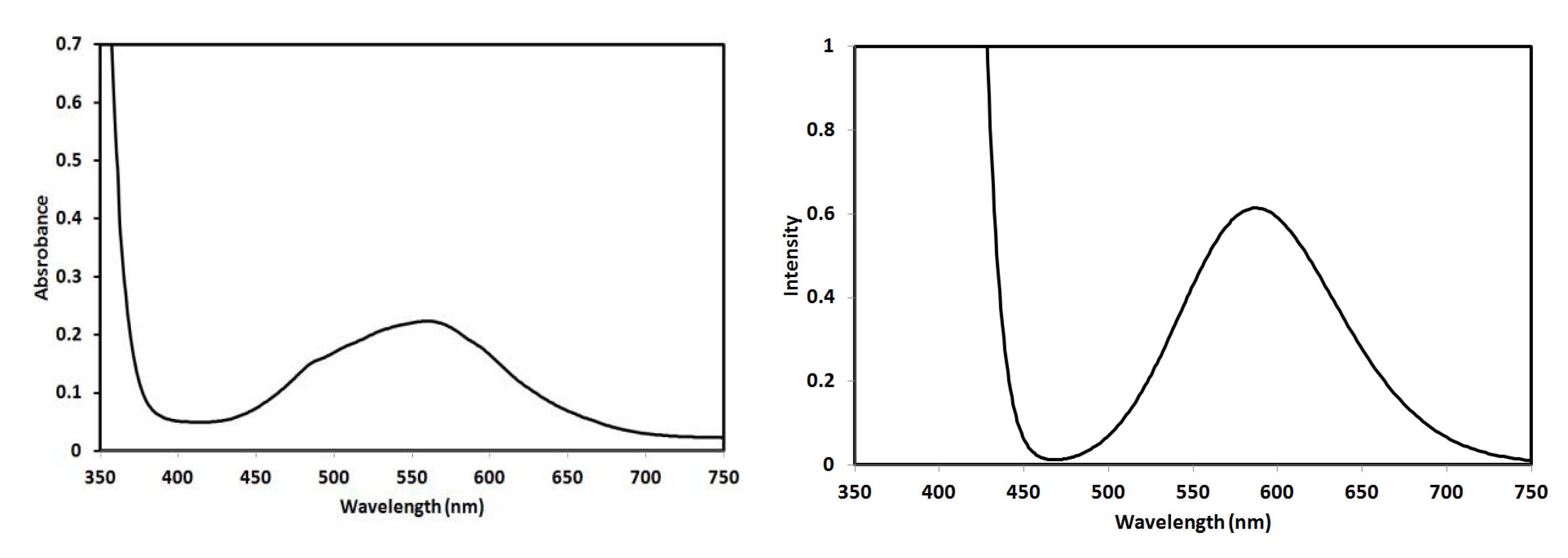
2.4. Electronic Spectra

The electronic spectra of 4 × 10⁻³ M solution of complex 2 were recorded in ethanol as solvent. The experimentally observed UV-Vis spectra along with the simulated electronic spectra calculated using the TD-DFT method for complex 2 are shown in Figure 10. The recorded electronic spectra showed a broad spectral band at 564 nm, which was calculated at 589.9 nm.
In order to assign the origin of this electronic spectral band, the calculated excited and ground states included in this spectral band are shown in Figure 11. The band observed in the visible region could be assigned to electronic transitions from HOMO, HOMO-1, HOMO-9, and HOMO-10 as ground states to LUMO+4 as an excited state where all are β-type orbitals. This electronic transition could be described as mainly metal-ligand (PipBPT) charge transfer-based transition.

2.5. Antimicrobial Activity

The biological activity of the free ligands (MorphBPT and PipBPT), as well as the Co(II) complexes [Co(MorphBPT)Cl2] (1) and [Co(PipBPT)Cl2] (2), were evaluated against S. aureus and B. subtilis as Gram-positive bacteria, E. coli and P. vulgaris as Gram-negative bacteria and two fungi (A. fumigatus and C. albicans). Minimum inhibition zone diameters were determined for the studied compounds (10 mg/mL) and the results are listed in Table 5.
The results shown in Table 5 indicated that the free ligands have no antimicrobial activity against all the studied microbes at the applied concentration (10 mg/mL) except PipBPT, which is active only against the Gram-positive bacteria B. subtilis (13 mm). In contrast, the Co(II) complexes showed interesting antibacterial activities. Complex 1 is active against the two tested Gram-positive bacteria (S. aureus (20 mm) and B. subtilis (24 mm)) and one Gram-negative bacteria (E. coli (16 mm)). On the other hand, complex 2 showed significant antibacterial activities against all the studied bacteria strains with inhibition zone diameters ranging from 15 mm (E. coli) to 30 mm (P. vulgaris). An additional observation that could be concluded from these results; complex 2 has better antibacterial activity against P. vulgaris (30 mm) and very close antibacterial activities against B. subtilis (26 mm) compared to control (gentamycin: 27 mm). Both complexes showed no antifungal activity against the two tested fungi at the experimental conditions. The results indicated that the synthesized Co(II) complexes are promising antibacterial agents rather than antifungal agents.
Moreover, the minimum inhibitory concentrations (MIC) in μg/mL were determined and the results are depicted in Table 6. The results are in accord with our observations. The MIC values are the lowest for complex 2 against B. subtilis, P. vulgaris, and S. aureus indicated potent activities against these microbes. It is also more potent (complex 2; 39 μg/mL) than PipBPT against B. subtilis (87 μg/mL). Complex 1 has lower potency against the studied bacteria with higher MIC values ranging from 156–625 μg/mL.

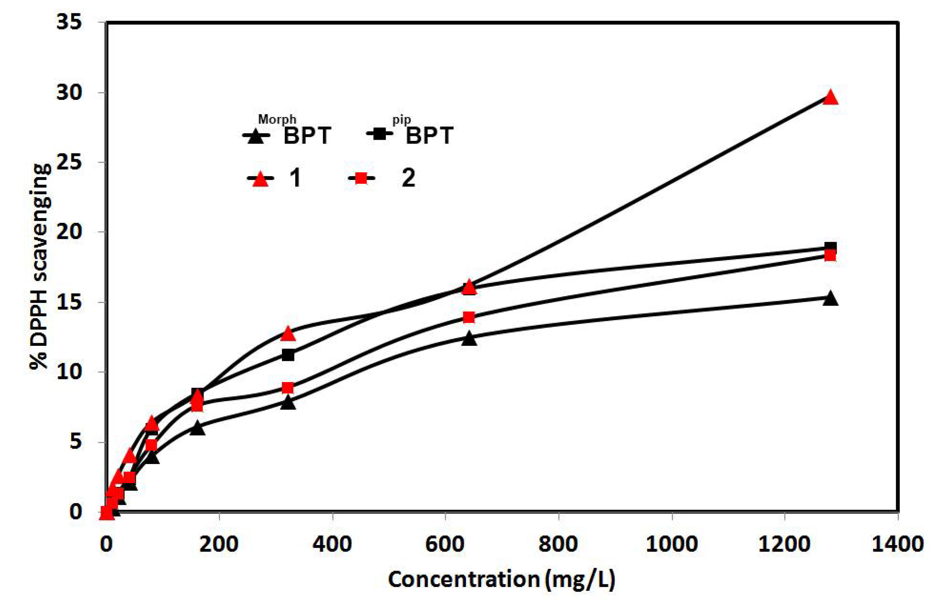
2.6. Antioxidant Activity

The DPPH free radical scavenging assay enabled us to determine the antioxidant activity of the studied complexes compared to the free ligands. The detailed results are tabulated in Tables S2–S5 (Supplementary Data) and summarized graphically in Figure 12. Although the results showed that the studied systems have weak to moderate antioxidant activity, especially for the free ligands and complex 2, but the most significant conclusion is that complex 1 has improved antioxidant activity compared to the free ligand MorphBPT while the antioxidant activity of PipBPT and its [Co(PipBPT)Cl2]; 2 are comparable indicating that varying the substituent at the s-triazine core of the functional ligand have a significant impact on the antioxidant activity of this class of Co(II) complexes.

3. Materials and Methods

Chemicals were purchased from Sigma-Aldrich Company (Chemie GmbH, 82024 Taufkirchen, Germany). The CHN analyses were determined using a Perkin-Elmer 2400 instrument (PerkinElmer, Inc., 940 Winter Street, Waltham, MA, USA). Cobalt content was determined using Shimadzu atomic absorption spectrophotometer (AA-7000 series, Shimadzu, Ltd., Kyoto, Japan). An Alpha Bruker spectrophotometer (Billerica, MA, USA) was used to measure the FTIR spectra in KBr pellets (Figures S4 and S5, Supplementary Data). The FTIR spectra were recorded in the range of 4000–400 cm−1 at a spectral resolution of 2 cm−1 and with 40 scans. The UV-Vis electronic spectra were recorded in ethanol using Pg instruments T80+ spectrophotometer (Alma Park, Wibtoft, UK). The melting points were ascertained in open capillary tubes using a Gallenkamp melting point apparatus (Sigma-Aldrich Chemie GmbH, Taufkirchen, Germany) and were uncorrected.

3.1. Syntheses of [Co(MorphBPT)(Cl)2]; (1) and [Co(PipBPT)(Cl)2]; (2)

The ligands were prepared following the same method reported by us [53]. Details regarding the ligand preparations were given in (Supplementary Material Method S1, Figures S6 and S7).
A 10 mL methanolic solution of 0.5 mmol of the functional s-triazine chelate was added to 10 mL aqueous solution of the CoCl2 (64.9 mg, 0.5 mmol). Purple color crystals of the titled complexes were obtained after five days.
Yield; C17H22Cl2CoN8O (1) 86%; mp > 360 °C (dec). Anal. Calc. C, 42.16; H, 4.58; N, 23.14; Co, 12.17%. Found: C, 41.93; H, 4.49; N, 23.01; Co, 12.05%. IR (KBr, cm−1): 3090, 2983, 2924, 2859, 1633, 1589, 1499, 1448, 1067, 1020.
Yield; C18H24Cl2CoN8 (2) 81%; mp > 360 °C (dec). Anal. Calc. C, 44.83; H, 5.02; N, 23.23; Co, 12.22%. Found: C, 44.98; H, 4.94; N, 23.09; Co, 12.10%. IR (KBr, cm−1): 3115, 2932, 2857, 1636, 1597, 1493, 1444, 1041, 1008.

3.2. Crystal Structure Determination

A Bruker D8 Quest diffractometer was used to determine the crystal structures of complexes 12 with the aid of SHELXTL and SADABS programs [54,55,56]. Refinement and crystal details were given in Table 1. Hirshfeld calculations were performed using the Crystal Explorer 17.5 program [57,58,59,60,61,62].

3.3. Antimicrobial Studies

The antimicrobial activity of the free MorphBPT and PipBPT ligands, as well as the corresponding Co(II) complexes, against two Gram-positive bacteria (S. aureus (ATCC 25923) and B. subtilis (RCMB015(1)NRR LB-543)), two Gram-negative bacteria (E. coli (ATCC 25922) and P. vulgaris (RCMB 004(1)ATCC 13315)), and two fungi (A. fumigatus (RCMB 002008) and C. albicans (RCMB 005003(1) ATCC 10231)). Minimum inhibition zone diameters at 10 mg/mL of the studied compounds, as well as the minimum inhibitory concentrations (MIC), were determined against these microbes [53]. Gentamycin and ketoconazole were used as standard antibacterial and antifungal agents, respectively. More details are found in (Method S2 Supplementary Data).

3.4. Antioxidant Activity

The antioxidant activity of complexes 1 and 2 was determined at the Regional Center for Mycology and Biotechnology (RCMB) at the Al-Azhar University by the DPPH free radical scavenging assay in triplicate and average values were considered [63,64]. More details regarding the bio-experiments are found in (Method S3 Supplementary Data).

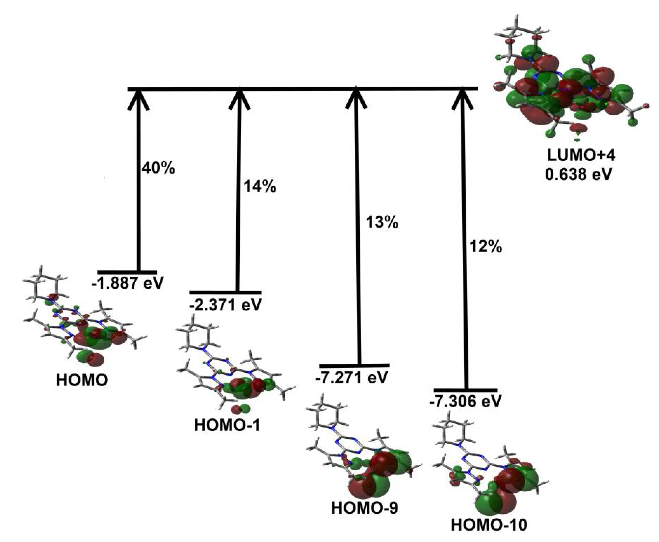
3.5. DFT Calculations

The structure of complex 2 was optimized in the gas phase using the B3LYP method employing 6–31G(d,p) for nonmetal atoms and LANL2DZ for Co [65] with the aid of Gaussian 09 software [66]. All frequency results are positive and no imaginary frequency indicating real minima. The gas-phase optimized structure was used as the input for simulating the structure in ethanol as solvent followed by TD-DFT calculations in the same solvent in order to simulate and assign the experimentally observed UV-Vis spectra [67,68].

4. Conclusions

Two penta-coordinated Co(II) complexes with bis-pyrazolo-s-triazine pincer ligands bearing morpholino (MorphBPT) and piperidino (PipBPT) substituents were synthesized and their structure aspects were analyzed using single-crystal X-ray diffraction and Hirshfeld analysis. The mononuclear [Co(MorphBPT)Cl2]; 1 and [Co(PipBPT)Cl2]; 2 pincer complexes have a similar coordination environment comprising a tridentate functional ligand and two coordinated chloride ions. Complex 2 has higher potency against all the studied bacteria (except E. coli) than complex 1. In addition, the antioxidant activity of complex 1 is higher than MorphBPT, while both 2 and PipBPT have comparable results. These outcomes shed light on the importance of the nature of the substituent on the s-triazine ring of the coordinated functional ligand on the antioxidant activity. The design of s-triazine ligands carrying different substituents could improve the antioxidant activity, which is one of our future perspectives.

Supplementary Materials

The following are available online at; Method S1: General method for the synthesis of bis-pyrazolo-s-triazine derivatives. Method S2: Antimicrobial studies. Method S3: DPPH radical scavenging activity. Figure S1 Hirshfeld surfaces of complex 1. Figure S2 Hirshfeld surfaces of complex 2 (molecule I). Figure S3 Hirshfeld surfaces of complex 2 (molecule II). Figure S4 FTIR spectra of the free ligand MorphBPT (upper) and its Co(II) complex [Co(MorphBPT)Cl2]; 1 (lower). Figure S5 FTIR spectra of the free ligand PipBPT (upper) and its Co(II) complex [Co(PipBPT)Cl2]; 2 (lower). Figure S6: 1H NMR and 13C NMR of compound MorphBPT. Figure S7: 1H NMR and 13C NMR of compound PipBPT. Table S1 Calculated and experimental vibrational characteristics for complex 2. Table S2 Evaluation of antioxidant activity using DPPH scavenging assay for MorphBPT. Table S3 Evaluation of antioxidant activity using DPPH scavenging assay for [Co(MorphBPT)Cl2]; 1. Table S4 Evaluation of antioxidant activity using DPPH scavenging assay for PipBPT. Table S5 Evaluation of antioxidant activity using DPPH scavenging assay for [Co(PipBPT)Cl2]; 2.

Author Contributions

The work was designed and supervised by S.M.S. X-ray structure analyses were performed by S.M.S. Computational calculations as well as the synthesis of complexes 1–2 were carried out by S.M.S. and R.A.M., A.E.-F. and H.H.A.-R. carried out the preparations of the organic ligands and their analyses. All authors contributed to the first draft and the final version. All authors have read and agreed to the published version of the manuscript.

Funding

Deanship of Scientific Research at King Saud University, group no. RG-1441-365, Saudi Arabia.

Institutional Review Board Statement

Not applicable.

Informed Consent Statement

Not applicable.

Data Availability Statement

The data presented in this study are available on request from the corresponding author.

Acknowledgments

The authors extend their thanks to the Deanship of Scientific Research at King Saud University for funding this work through research group no. (RG-1441-365, Saudi Arabia).

Conflicts of Interest

The authors declare no conflict of interest.

Sample Availability

Samples of the compounds are not available from the authors.

References

  1. Updegraff, I.H.; Moore, S.T.; Herbes, W.F.; Roth, P.B. Amino Resins and Plastics. In Kirk-Othmer’s Encyclopedia of Chemical Technology, 3rd ed.; Grayson, M., Eckroth, D., Eds.; Wiley: New York, NY, USA, 1978; Volume 2, pp. 440–469. [Google Scholar]
  2. Brown, A.W.A. Ecology of Pesticides; Wiley: New York, NY, USA, 1978; pp. 10–11; 329–339; 369–373. [Google Scholar]
  3. Shah, D.R.; Modh, R.P.; Chikhalia, K.H. Privileged s-triazines: Structure and pharmacological applications. Fut. Med. Chem. 2014, 6, 463–477. [Google Scholar] [CrossRef]
  4. Hatfield, S.E. Applications of Triazine Chemistry: Education, Remediation, and Drug Delivery. Ph.D. Thesis, Texas A & M University, College Station, TX, USA, 2007. [Google Scholar]
  5. Tobe, A.; Kobayashi, T. Pharmacological studies on triazine derivatives V. Sedative and neuroleptic actions of 2-amino-4-(4-(2hydroxyethyl)-piperazin-1-yl)-6-trifluoromethyl-s-triazine (TR10). Jpn. J. Aerosp. Med. Psychol. 1976, 26, 559–570. [Google Scholar]
  6. Patel, R.V.; Kumari, P.; Rajani, D.P.; Pannecouque, C.; De Clercq, E.; Chikhalia, K.H. Antimicrobial, anti-TB, anticancer and anti-HIV evaluation of new s-triazine-based heterocycles. Fut. Med. Chem. 2012, 4, 1053–1065. [Google Scholar] [CrossRef] [PubMed]
  7. Aksenov, A.V.; Aksenov, I.V. Use of the ring opening reactions of 1,3,5-triazines in organic synthesis. Chem. Heterocyc. Comp. 2009, 45, 130–150. [Google Scholar] [CrossRef]
  8. Pandey, V.K.; Tusi, S.; Tusi, Z.; Joshi, M.; Bajpai, S. Synthesis and biological activity of substituted 2,4,6-s-triazines. Acta Pharm. 2014, 54, 1–12. [Google Scholar]
  9. Agrawal, A.; Shrivastav, K.; Puri, S.K.; Chauhan, M.S. Syntheses of 2,4,6-trisubstituted triazines as antimalarial agents. Bioorg. Med. Chem. Lett. 2005, 15, 531–533. [Google Scholar] [CrossRef]
  10. Bartholomew, D. 1,3,5-Triazines, In Comprehensive Heterocyclic Chemistry II; Boulton, A.J., Ed.; Pergamon: Oxford, UK, 1996. [Google Scholar]
  11. Giacomelli, G.; Porcheddu, A.; Luca, L.D. [1,3,5]-Triazine: A Versatile heterocycle in current applications of organic chemistry. Curr. Org. Chem. 2004, 15, 1497–1519. [Google Scholar] [CrossRef]
  12. Blotny, G. Recent applications of 2,4,6-trichloro-1,3,5-triazine and its derivatives in organic synthesis. Tetrahedron 2006, 62, 9507–9522. [Google Scholar] [CrossRef]
  13. Kumar, A.; Menon, S.K. Fullerene derivatized s-triazine analogues as antimicrobial agents. Eur. J. Med. Chem. 2009, 44, 2178–2183. [Google Scholar] [CrossRef] [PubMed]
  14. Menicagli, R.; Samaritani, S.; Signore, G.; Vaglini, F.; Via, L.D. In Vitro Cytotoxic Activities of 2-Alkyl-4,6-diheteroalkyl-1,3,5-triazines:  New molecules in anticancer research. J. Med. Chem. 2004, 47, 4649–4652. [Google Scholar] [CrossRef]
  15. Henke, B.R.; Consler, T.G.; Go, N.; Hale, R.L.; Hohman, D.R.; Jones, S.A.; Lu, A.T.; Moore, L.B.; Moore, J.T.; Orband-Miller, L.A.; et al. A New series of estrogen receptor modulators that display selectivity for estrogen receptor β. J. Med. Chem. 2002, 45, 5492–5505. [Google Scholar] [CrossRef]
  16. Jensen, N.P.; Ager, A.L.; Bliss, R.A.; Canfield, C.J.; Kotecka, B.M.; Rieckmann, K.H.; Terpinski, J.; Jacobus, D.P. Phenoxypropoxybiguanides, prodrugs of DHFR−inhibiting diaminotriazine antimalarials. J. Med. Chem. 2001, 44, 3925–3931. [Google Scholar] [CrossRef] [PubMed]
  17. kala, R.S.; Tharmaraj, P.; Sheela, C.D.; Anitha, C. Synthesis and studies on s-Triazine-based ligand and its metal complexes. Int. J. Inorg. Chem. 2012, 2012, 1–7. [Google Scholar] [CrossRef]
  18. Ghaib, A.; Ménager, S.; Vérité, P.; Lafont, O. Synthesis of variously 9,9-dialkylated octahydropyrimido [3,4-a]-s-triazines with potential antifungal activity. Farmaco 2002, 57, 109–116. [Google Scholar] [CrossRef]
  19. Lübbers, T.; Angehrn, P.; Gmünder, H.; Herzig, S.; Kulhanek, J. Design, synthesis, and structure-activity relationship studies of ATP analogues as DNA gyrase inhibitors. Bioorg. Med. Chem. Lett. 2000, 10, 821–826. [Google Scholar] [CrossRef]
  20. Vathanaruba, M.; Tharmaraj, P.; Sheela, C.D. Studies on A Novel s-triazine based ONO donor heterocyclic ligand and its transition Metal(II) complexes. Int. J. Adv. Res. Chem. Sci. 2014, 1, 21–27. [Google Scholar]
  21. Marandi, F.; Moeini, K.; Arkak, A.; Mardani, Z.; Krautscheid, H. Docking studies to evaluate the biological activities of the Co(II) and Ni(II) complexes containing the triazine unit: Supported by structural, spectral, and theoretical studies. J. Coord. Chem. 2018, 71, 3893–3911. [Google Scholar] [CrossRef]
  22. Katugampala, S.; Perera, I.C.; Nanayakkara, C.; Perera, T. Synthesis, characterization, and antimicrobial activity of novel sulfonated copper-triazine complexes. Bioinorg. Chem. Appl. 2018, 2018, 2530851. [Google Scholar] [CrossRef] [Green Version]
  23. kala, R.S.; Tharmaraja, P.; Sheela, C.D. Synthesis, spectral studies, NLO, and biological studies on metal(II) complexes of s-triazine-based ligand. Synth. React. Inorg. Met. Org. Nano Met. Chem. 2014, 44, 1487–1496. [Google Scholar] [CrossRef]
  24. Singh, K.; Thakur, R.; Kumar, V. Co(II), Ni(II), Cu(II), and Zn(II) complexes derived from 4-[{3-(4-bromophenyl)-1-phenyl-1H-pyrazol-4-ylmethylene}-amino]-3-mercapto-6-methyl-5-oxo-1,2,4-triazine. Beni-Suef Univ. J. Basic App. Sci. 2016, 5, 21–30. [Google Scholar] [CrossRef] [Green Version]
  25. Baldaniya, B.B.; Patel, P.K. Synthesis, antibacterial and antifungal activities of s-triazine derivatives. Egypt. J. Chem. 2009, 6, 673–680. [Google Scholar]
  26. Jain, S.; Sharma, A.M.; Agrawal, M.; Sharma, S.; Dwivedi, J.; Kishore, D. Synthesis and antimicrobial evaluation of some novel trisubstituted s-triazine derivatives based on isatinimino, sulphonamido, and azacarbazole. J. Chem. 2012, 2013, 1–9. [Google Scholar] [CrossRef]
  27. Osman, A.H. Synthesis and characterization of cobalt(II) and nickel(II) complexes of some Schiff bases derived from 3-hydrazino-6-methyl[1,2,4] triazin-5(4H)one. Transit. Met. Chem. 2006, 31, 35–41. [Google Scholar] [CrossRef]
  28. Shao, D.; Shi, L.; Wei, H.Y.; Wang, X.Y. Field-induced single-ion magnet behavior in two new cobalt(II) coordination polymers with 2,4,6-tris(4-pyridyl)-1,3,5-triazine. Inorganics 2017, 5, 90. [Google Scholar] [CrossRef] [Green Version]
  29. Asgharpour, Z.; Farzaneh, F.; Abbasi, A. Synthesis, characterization and immobilization of a new cobalt(ii) complex on modified magnetic nanoparticles as catalyst for epoxidation of alkenes and oxidation of activated alkanes. RSC Adv. 2016, 6, 95729–95739. [Google Scholar] [CrossRef]
  30. Tilly, D.; Dayaker, G.; Bachu, P. Cobalt mediated C–H bond functionalization: Emerging tools for organic synthesis. Catal. Sci. Technol. 2014, 4, 2756–2777. [Google Scholar] [CrossRef]
  31. Yoshino, T.; Matsunaga, S. Cobalt-catalyzed C(sp3)−H functionalization reactions. Asian J. Org. Chem. 2018, 7, 1193–1205. [Google Scholar] [CrossRef]
  32. Pototschnig, G.; Maulide, N.; Schnürch, M. Direct functionalization of C−H bonds by iron, nickel, and cobalt catalysis. Chem. Eur. J. 2017, 23, 9206–9232. [Google Scholar] [CrossRef]
  33. Tordin, E.; List, M.; Monkowius, U.; Schindler, S.; Knör, G. Synthesis and characterization of cobalt, nickel and copper complexes with tripodal 4N ligands as novel catalysts for the homogeneous partial oxidation of alkanes. Inorg. Chim. Acta 2013, 402, 90–96. [Google Scholar] [CrossRef] [Green Version]
  34. Aktaş, A.; Saka, E.T.; Bıyıklıoğlu, Z.; Acar, İ.; Kantekin, H. Investigation of catalytic activity of new Co(II) phthalocyanine complexes in cyclohexene oxidation using different type of oxidants. J. Organomet. Chem. 2013, 745, 18–24. [Google Scholar] [CrossRef]
  35. Junge, K.; Papa, V.; Beller, M. Cobalt–pincer complexes in catalysis. Chem. Eur. J. 2019, 25, 122–143. [Google Scholar] [CrossRef]
  36. Judy-Azar, A.R.; Mohebbi, S.J. A novel magnetic hybrid nanomaterial as a highly efficient and selective catalyst for alcohol oxidation based on new Schiff base complexes of transition metal ions. Mol. Catal. A Chem. 2015, 397, 158–165. [Google Scholar] [CrossRef]
  37. Menati, S.; Rudbari, H.A.; Askari, B.; Farsani, M.R.; Jalilian, F.; Dini, G. Synthesis and characterization of insoluble cobalt(II), nickel(II), zinc(II) and palladium(II) Schiff base complexes: Heterogeneous catalysts for oxidation of sulfides with hydrogen peroxide. Compets Rendus Chim. 2016, 19, 347–356. [Google Scholar] [CrossRef]
  38. Cahiez, G.R.; Moyeux, A. Cobalt-catalyzed cross-coupling reactions. Chem. Rev. 2010, 110, 1435–1462. [Google Scholar] [CrossRef]
  39. Hebrard, F.; Kalck, P. Cobalt-catalyzed hydroformylation of alkenes: Generation and recycling of the carbonyl species, and catalytic cycle. Chem. Rev. 2009, 109, 4272–4282. [Google Scholar] [CrossRef]
  40. Omae, I. Three characteristic reactions of organocobalt compounds in organic synthesis. Appl. Organomet. Chem. 2007, 21, 318–344. [Google Scholar] [CrossRef]
  41. Sarkheil, M.; Lashanizadegan, M. Schiff base ligand derived from (±)trans-1,2-cyclohexane-diamine and its Cu(II), Co(II), Zn(II) and Mn(II) Complexes: Synthesis, characterization, styrene oxidation and a hydrolysis study of the imine bond in the Cu(II) Schiff base complex. J. Serb. Chem. Soc. 2016, 81, 369–382. [Google Scholar] [CrossRef]
  42. Soliman, S.M.; El-Faham, A. One pot synthesis of two Mn(II) perchlorate complexes with s-triazine NNN-pincer ligand; molecular structure, Hirshfeld analysis and DFT studies. J. Mol. Struct. 2018, 1164, 344–353. [Google Scholar] [CrossRef]
  43. Soliman, S.M.; El-Faham, A. Synthesis, characterization, and structural studies of two heteroleptic Mn(II) complexes with tridentate N,N,N-pincer type ligand. J. Coord. Chem. 2018, 71, 2373–2388. [Google Scholar] [CrossRef]
  44. Soliman, S.M.; Almarhoon, Z.; Sholkamy, E.N.; El-Faham, A. Bis-pyrazolyl-s-triazine Ni(II) pincer complexes as selective Gram-positive antibacterial agents; synthesis, structural and antimicrobial studies. J. Mol. Struct. 2019, 1195, 315–322. [Google Scholar] [CrossRef]
  45. Soliman, S.M.; Almarhoon, Z.; El-Faham, A. Synthesis, molecular and supramolecular structures of new Cd(II) pincer-type complexes with s-triazine core ligand. Crystals 2019, 9, 226. [Google Scholar] [CrossRef] [Green Version]
  46. Soliman, S.M.; El-Faham, A. Synthesis, X-ray structure, and DFT studies of five- and eight-coordinated Cd(II) complexes with s-triazine N-pincer chelate. J. Coord. Chem. 2019, 72, 1621–1636. [Google Scholar] [CrossRef]
  47. Soliman, S.M.; Elsilk, S.E.; El-Faham, A. Synthesis, structure and biological activity of zinc(II) pincer complexes with 2,4-bis(3,5-dimethyl-1H-pyrazol-1-yl)-6-methoxy-1,3,5-triazine. Inorg. Chim. Acta 2020, 508, 119627. [Google Scholar] [CrossRef]
  48. Soliman, S.M.; Elsilk, S.E.; El-Faham, A. Syntheses, structure, Hirshfeld analysis and antimicrobial activity of four new Co(II) complexes with s-triazine-based pincer ligand. Inorg. Chim. Acta 2020, 510, 119753. [Google Scholar]
  49. Soliman, S.M.; El-Faham, A.; El Silk, S.E. Novel one-dimensional polymeric Cu(II) complexes via Cu(II)-assisted hydrolysis of the 2,4-bis(3,5-dimethyl-1H-pyrazol-1-yl)-6-methoxy-1,3,5-triazine pincer ligand: Synthesis, structure, and antimicrobial activities. Appl. Organomet. Chem. 2020, 510, e5941. [Google Scholar]
  50. Lasri, J.; Haukka, M.; Al-Rasheed, H.H.; Abutaha, N.; El-Faham, A.; Soliman, S.M. Synthesis, structure and in vitro anticancer activity of Pd(II) complex of pyrazolyl-s-triazine ligand; A new example of metal-mediated hydrolysis of s-triazine pincer ligand. Crystals 2021, 11, 119. [Google Scholar] [CrossRef]
  51. Kala, R.S.; Tharmaraj, P.; Sheela, C.D. Synthesis and spectral studies on metal complexes of s-triazine based ligand and non linear optical properties. J. Mol. Struct. 2014, 1076, 606–613. [Google Scholar]
  52. Addison, A.W.; Rao, T.N.; Reedijk, J.; Rijn, J.V.; Verschoor, G.C. Synthesis, structure, and spectroscopic properties of copper(II) compounds containing nitrogen–sulphur donor ligands; the crystal and molecular structure of aqua[1,7-bis(N-methylbenzimidazol-2′-yl)-2,6-dithiaheptane]copper(II) perchlorate. J. Chem. Soc. Dalton Trans. 1984, 1349–1356. [Google Scholar] [CrossRef]
  53. Sharma, A.; Ghabbour, H.; Khan, S.T.; de la Torre, B.G.; Albericio, F.; El-Faham, A. Novel pyrazolyl-s-triazine derivatives, molecular structure and antimicrobial activity. J. Mol. Struct. 2017, 1145, 244–253. [Google Scholar] [CrossRef]
  54. Sheldrick, G.M. Program for Empirical Absorption Correction of Area Detector Data; University of Göttingen: Göttingen, Germany, 1996. [Google Scholar]
  55. Sheldrick, G.M. SHELXT—Integrated space-group and crystal-structure determination. Acta Cryst. A 2015, 71, 3–8. [Google Scholar] [CrossRef] [Green Version]
  56. Spek, A.L. Structure validation in chemical crystallography. Acta Cryst. D 2009, 65, 148–155. [Google Scholar] [CrossRef]
  57. Turner, M.J.; McKinnon, J.J.; Wolff, S.K.; Grimwood, D.J.; Spackman, P.R.; Jayatilaka, D.; Spackman, M.A. Crystal Explorer 17; University of Western Australia: Perth, Australia, 2017. Available online: http://hirshfeldsurface.net (accessed on 12 June 2017).
  58. Hirshfeld, F.L. Bonded-atom fragments for describing molecular charge densities. Theor. Chim.Acta 1977, 44, 129–138. [Google Scholar] [CrossRef]
  59. Spackman, M.A.; Jayatilaka, D. Jayatilaka, Hirshfeld surface analysis. Cryst. Eng. Commun. 2009, 11, 19–32. [Google Scholar] [CrossRef]
  60. Spackman, M.A.; McKinnon, J.J. Fingerprinting intermolecular interactions in molecular crystals. Cryst. Eng. Commun. 2002, 4, 378–392. [Google Scholar] [CrossRef]
  61. Bernstein, J.; Davis, R.E.; Shimoni, L.; Chang, N.-L. Patterns in hydrogen bonding: Functionality and graph set analysis in crystals. Angew. Chem. Int. Ed. 1995, 34, 1555–1573. [Google Scholar] [CrossRef]
  62. McKinnon, J.J.; Jayatilaka, D.; Spackman, M.A. Towards quantitative analysis of intermolecular interactions with Hirshfeld surfaces. Chem. Commun. 2007, 3814–3816. [Google Scholar] [CrossRef]
  63. Yen, G.C.; Duh, P.D. Scavenging Effect of Methanolic Extracts of Peanut Hulls on Free-Radical and Active-Oxygen Species . J. Agric. Food. Chem. 1994, 42, 629–632. [Google Scholar] [CrossRef]
  64. Al Zahrani, N.A.; El-Shishtawy, R.M.; Elaasser, M.M.; Asiri, A.M. Synthesis of Novel Chalcone-Based Phenothiazine Derivatives as Antioxidant and Anticancer Agents. Molecules 2020, 25, 4566. [Google Scholar] [CrossRef] [PubMed]
  65. Nogheu, L.N.; Ghogomu, J.N.; Mama, D.B.; Nkungli, N.K.; Younang, E.; Gadre, S.R. Structural, spectral (IR and UV/Visible) and thermodynamic properties of some 3d transition metal(II) chloride complexes of glyoxime and Its derivatives: A DFT and TD-DFT study. Comput. Chem. 2016, 4, 119–136. [Google Scholar] [CrossRef] [Green Version]
  66. Frisch, M.J.; Trucks, G.W.; Schlegel, H.B.; Scuseria, G.E.; Robb, M.A.; Cheeseman, J.R.; Scalmani, G.; Barone, V.; Mennucci, B.; Petersson, G.A.; et al. GAUSSIAN 09. Revision A02; Gaussian Inc.: Wallingford, CT, USA, 2009. [Google Scholar]
  67. Casida, M.E. Recent Advances in Density Functional Methods, Part I; World Scientific: Singapore, 1995. [Google Scholar]
  68. Gross, E.K.U.; Dobson, J.F.; Petersilka, M. Introduction in Density Functional Theory II; Springer: Heidelberg, Germany, 1996. [Google Scholar]
Figure 1. Structure of the pincer ligands.
Molecules 26 03633 g001
Figure 2. Structure with atom numbering (left) and the distorted square pyramidal (right) of 1.
Molecules 26 03633 g002
Figure 3. The hydrogen bond contacts (upper) and hydrogen bonding network (lower) in 1.
Molecules 26 03633 g003
Figure 4. Structure with atom numbering (upper) and the distorted square pyramidal (lower) of 2.
Molecules 26 03633 g004
Figure 5. The hydrogen bond contacts (upper) and hydrogen bonding network (lower) in 2.
Molecules 26 03633 g005
Figure 6. The important contacts and their percentages.
Molecules 26 03633 g006
Figure 7. Hirshfeld surfaces of complex 1.
Molecules 26 03633 g007
Figure 8. Hirshfeld surfaces of complex 2.
Molecules 26 03633 g008
Figure 9. The experimental (upper) and calculated (lower) vibrational spectra of complex 2.
Molecules 26 03633 g009
Figure 10. The experimental (left) and calculated (right) electronic spectra of complex 2.
Molecules 26 03633 g010
Figure 11. Origin of the electronic spectral band observed in ethanol for complex 2.
Molecules 26 03633 g011
Figure 12. The antioxidant activity of the studied compounds.
Molecules 26 03633 g012
Table 1. Crystal data and structure refinement for the studied complexes.
Compound[Co(MorphBPT)Cl2] (1)[Co(pipBPT)Cl2] (2)
Empirical formulaC17 H22 Cl2 Co N8 OC18 H24 Cl2 Co N8
Formula weight484.25 g/mol482.28 g/mol
Temperature121(2) K121(2) K
Wavelength0.71073 Å0.71073 Å
Crystal systemMonoclinicTriclinic
Space groupC2/cP-1
Unit cell dimensionsa = 17.241(5) Åa = 11.094(3) Å
b = 12.832(3) Åb = 12.785(3) Å
c = 20.165(7) Åc = 16.527(4) Å
α = 90°α = 97.125(6)°
β = 109.687(9)°β = 103.496(6)°
γ = 90°γ = 102.933(6)°
Volume4200.(2) Å32182.8(10) Å3
Z84
Density (calculated)1.531 g/cm31.468 g/cm3
Absorption coefficient1.098 mm−11.053 mm−1
F(000)1992996
Crystal size0.14 × 0.21 × 0.25 mm30.20 × 0.27 × 0.32 mm3
Theta range for data collection2.51 to 25.34°2.21 to 25.32°
Index ranges−20 ≤ h ≤ 20−13 ≤ h ≤ 13
−15 ≤ k ≤ 13−15 ≤ k ≤ 15
−24 ≤ l ≤ 24−19 ≤ l ≤ 19
Reflections collected14,32335,520
Independent reflections3834 [R(int) = 0.0293]7949 [R(int) = 0.0373]
Completeness to theta99.60%99.70%
Refinement methodFull-matrix least-squares on F2
Data/restraints/parameters3834/0/2677949/0/532
Goodness-of-fit on F21.0331.041
Final R indices (I > 2sigma(I))R1 = 0.0250, wR2 = 0.0581R1 = 0.0246, wR2 = 0.0548
R indices (all data)R1 = 0.0310, wR2 = 0.0616R1 = 0.0324, wR2 = 0.0584
Extinction coefficient0.00125(12)0.0036(3)
Largest difference peak and hole0.342 and −0.3530.315 and −0.272
CCDC2080583320805834
Table 2. Bond lengths (Å) and angles (°) for complexes 1 and 2.
[Co(MorphBPT)Cl2] (1)[Co(PipBPT)Cl2] (2)
Co1-N12.0387(15)Co1-N12.0291(15)Co1A-N1A2.0325(15)
Co1-N42.2008(15)Co1-N42.1829(15)Co1A-N4A2.2023(15)
Co1-N62.2304(16)Co1-N62.1935(15)Co1A-N6A2.2184(15)
Co1-Cl22.2718(7)Co1-Cl22.2766(7)Co1A-Cl2A2.2667(6)
Co1-Cl12.2968(7)Cl1-Co12.2822(7)Co1A-Cl1A2.2686(8)
N1-Co1-N474.01(6)N1-Co1-N473.73(6)N1A-Co1A-N4A73.83(6)
N1-Co1-N673.07(5)N1-Co1-N674.23(6)N1A-Co1A-N6A73.72(6)
N4-Co1-N6146.87(5)N4-Co1-N6147.94(5)N4A-Co1A-N6A147.43(5)
N1-Co1-Cl2138.89(4)N1-Co1-Cl2119.81(4)N1A-Co1A-Cl2A115.61(5)
N4-Co1-Cl2102.34(4)N4-Co1-Cl297.95(4)N4A-Co1A-Cl2A100.41(4)
N6-Co1-Cl299.89(4)N6-Co1-Cl298.88(4)N6A-Co1A-Cl2A95.97(4)
N1-Co1-Cl1109.63(5)N1-Co1-Cl1122.66(4)N1A-Co1A-Cl1A124.96(4)
N4-Co1-Cl197.96(4)N4-Co1-Cl1100.29(4)N4A-Co1A-Cl1A95.76(4)
N6-Co1-Cl196.42(4)N6-Co1-Cl195.81(4)N6A-Co1A-Cl1A100.40(4)
Cl2-Co1-Cl1111.40(3)Cl2-Co1-Cl1117.51(2)Cl2A-Co1A-Cl1A119.43(2)
Table 3. Geometric parameters of the hydrogen bonds [Å and °] in complexes 1 and 2.
1
C6-H6…Cl1 i0.952.653.550(2)158
C11-H11…O1 ii0.952.583.503(3)165
i 1/2-x,-1/2+y,3/2-z; ii 1/2+x,3/2-y,1/2+z
2
C6A-H6A…Cl2 i0.952.743.524(2)140
C8-H8A…Cl2 ii0.982.823.768(2)162
C13-H13A…Cl2 iii0.982.763.611(2)146
i 1+x,y,z; ii -x,-y,1-z; iii 1-x,1-y,1-z.
Table 4. The most important contacts in complexes 1 and 2.
1 2
ContactDistanceContactDistance
C6…H14B2.745C5A…H16D2.727
C7…H14B2.642C10A…H17A2.722
C8…H14B2.718Cl2A…C33.407
C3…H4A2.751Cl2A…C23.412
Cl2…C13.297Cl2…H6A2.642
Cl1…H62.527Cl1A…H4C2.780
O1…H112.447Cl2A…H4A2.797
H4C…H15A2.223Cl2…H13A2.670
C6…C23.495Cl2…H8A2.724
C6…N73.390Cl1...H18B2.816
H9A…H13B2.294
C11…N4A3.347
C11…N5A3.341
N2…H8B2.578
Table 5. Inhibition zone diameters of the studied compounds and control against different microbes a.
MicorbeMorphBPT1PipBPT2Control
A. fumigatus----17 a
C. albicans----20 a
S. aureus-20-1825 b
B. subtilis-24132627 b
E. coli-16-1530 b
P. vulgaris---3027 b
a Ketoconazole and b Gentamycin.
Table 6. MIC values (μg/mL) for the studied compounds.
MicorbeMorphBPT1PipBPT2
A. fumigatus----
C. albicans----
S. aureus-156-20
B. subtilis-3127839
E. coli-625-1250
P. vulgaris---20
Publisher’s Note: MDPI stays neutral with regard to jurisdictional claims in published maps and institutional affiliations.

