
Gas Plasma-Augmented Wound Healing in Animal Models and Veterinary Medicine


1
ZIK Plasmatis, Leibniz Institute for Plasma Science and Technology (INP), Felix-Hausdorff-Str. 2, 17489 Greifswald, Germany
2
Institute for Hygiene and Environmental Medicine, Greifswald University Medical Center, Sauerbruchstr., 17475 Greifswald, Germany
*
Author to whom correspondence should be addressed.
Molecules 2021, 26(18), 5682; https://0-doi-org.brum.beds.ac.uk/10.3390/molecules26185682
Submission received: 12 August 2021
/
Revised: 14 September 2021
/
Accepted: 17 September 2021
/
Published: 19 September 2021
(This article belongs to the Special Issue Wound Infection: Emerging Challenges in Normal and Diabetic Skin Wounds)
Abstract
:The loss of skin integrity is inevitable in life. Wound healing is a necessary sequence of events to reconstitute the body’s integrity against potentially harmful environmental agents and restore homeostasis. Attempts to improve cutaneous wound healing are therefore as old as humanity itself. Furthermore, nowadays, targeting defective wound healing is of utmost importance in an aging society with underlying diseases such as diabetes and vascular insufficiencies being on the rise. Because chronic wounds’ etiology and specific traits differ, there is widespread polypragmasia in targeting non-healing conditions. Reactive oxygen and nitrogen species (ROS/RNS) are an overarching theme accompanying wound healing and its biological stages. ROS are signaling agents generated by phagocytes to inactivate pathogens. Although ROS/RNS’s central role in the biology of wound healing has long been appreciated, it was only until the recent decade that these agents were explicitly used to target defective wound healing using gas plasma technology. Gas plasma is a physical state of matter and is a partially ionized gas operated at body temperature which generates a plethora of ROS/RNS simultaneously in a spatiotemporally controlled manner. Animal models of wound healing have been vital in driving the development of these wound healing-promoting technologies, and this review summarizes the current knowledge and identifies open ends derived from in vivo wound models under gas plasma therapy. While gas plasma-assisted wound healing in humans has become well established in Europe, veterinary medicine is an emerging field with great potential to improve the lives of suffering animals.
1. Introduction
The skin protects the body from environmental stressors, infectious agents, and loss of homeostasis. Being the body’s largest organ in vertebrates, the skin has multifunctional roles due to its complex structure and multiple compartments such as a layer of dead keratinocytes (corneocytes) in the stratum corneum and the stratum lucidum, stratum granulosum, stratum spinosum, and stratum basale in the epidermis. Below the epidermis, the dermis is host to a rich meshwork of vasculature, immune cells, hair follicles, and glands [1].
Skin trauma is inevitable in life. The skin has extensive renewing capacities in both trauma and constitutive conditions. Constitutively, the epidermis renews every 25 to 70 days in an age-dependent fashion. In non-extensive trauma, wound healing usually occurs within a week, with further cellular proliferation and maturation along with intracutaneous matrix remodeling going on for several days to weeks after that. The wound healing phases include hemostasis, inflammation, proliferation and maturation, and matrix remodeling (Figure 1). The phases have defined yet partially overlapping sequences that have been recently reviewed [2]. Extensive trauma, infection, and underlying disease can extend the time to heal significantly. If healing is not achieved within four weeks despite standard care, the wounds are defined as being chronic, and can persist for several months to years. Such insufficient cutaneous wound healing is a burden to patients and health care systems worldwide. Despite a wealth of non-invasive and invasive options to support healing, a considerable number of wounds are refractory to therapy. An aging society and increased prevalence of widespread diseases like diabetes and vascular insufficiencies add to this issue. Antimicrobial resistance (AMR) is also rising globally [3], making efficient wound therapy a critical issue to address.
Wound healing has a redox dimension [4]. Reactive oxygen species (ROS), which include reactive nitrogen species (RNS), as these mostly contain oxygen, contribute to all stages of healing as signaling molecules and are therefore part of signal transduction processes. For details on primary and secondary ROS/RNS relevant in plasma medicine, the reader is referred to a recent review [5]. ROS/RNS are moreover integral components of the antimicrobial defense elicited by phagocytes [6]. Hence, it appears logical to use therapeutic ROS/RNS supplied exogenously to the wound microenvironment to aid in healing by promoting signaling and anti-infective action. About a decade ago, gas plasma technology began to be systemically investigated globally as a novel tool to target defective wound healing based on clinically relevant findings [7,8,9]. Gas plasma is a partially ionized gas generating a plethora of ROS/RNS simultaneously and in a spatiotemporally-controlled manner [10]. The two main types of devices are gas plasma jets [11] and dielectric barrier discharges (DBDs) [12], with the kINPen MED [13,14] and the SteriPlas [15] being well-described and clinically certified examples of each category. Today, based on clinical evidence, several gas plasma devices have been marketed in Europe as accredited medical products class IIa for dermatological applications with a particular focus on wound healing. Animal models and animal patients have been vital in understanding this technology’s efficacy, safety, and suitability in wound healing promotion and identifying ways to improve clinical response. This review aims at summarizing the current knowledge on animal models in investigating gas plasma-assisted wound healing.
2. Wound Healing in Animals and Experimental Models
An array of experimental models is available in veterinary science for studying wound healing. These can relate to mainly regenerative models of healing in species such as starfish, zebrafish, frogs, and salamander, as well as models more closely resembling the human wound healing stages, including rodents, ruminants, and pigs [16]. Details on different wound healing models and species in veterinary science and experimental medicine have been thoroughly reviewed recently [17,18,19,20].
2.1. Rodent Models
Rodent models, especially mice and rats, are by far the most used models in wound healing research [16]. Both species are easy to house and breed, and genetic engineering tools are well-established for generating constitutive or conditional knock-out and knock-in variants that allow the molecular mechanisms of wound healing to be deciphered. Moreover, small rodent models benefit from fast birth-to-adulthood times and modest housing costs, facilitating the use of larger groups within experiments to obtain robust statistical analysis. For instance, SCID (severe combined immunodeficient) mice are void of T-cells, and these mice heal wounds as efficiently as non-SCID mice do, suggesting a minor role of T-cells in wound healing [21]. Due to rodent models’ small size, a cost-effective manipulation of metabolic or immunological traits by pharmacological or immunotherapeutic interventions is also possible. As an example, the minor relevance of T-cells in wound healing has also been shown by depleting T-cells experimentally using monoclonal antibodies [22].
Although chronic wound healing models closely resembling human pathology are currently not available, there are several options to induce wounding in general to generate models of acute healing, impaired healing, and semi-chronic wounds, as has been summarized before [23]. Briefly, acute models can be generated using punch biopsies, wound chambers, burns, cold, laser, and pressure ulcers. Impaired healing can be achieved by pharmacological, genetic, physical, or microbial means. Examples are the usage of ionizing radiation, malnutrition, glucocorticoid-mediated immunosuppression, mitomycin C and 5-fluorouracil-mediated dermonecrosis, zinc and vitamin deficiencies, ischemia, wound contamination with Pseudomonas and Staphylococcus strains, and models of diabetes and obesity. Additionally, aging is a parameter suitable to investigate impaired healing [24]. With the emergence of two-photon microscopy, real-time in vivo imaging has much accelerated the knowledge on cellular dynamics in wounds. For instance, Lämmermann and colleagues used a reporter mouse model with fluorescently-tagged neutrophils and macrophages to observe their infiltration and swarming phenome following small-scale laser-induced wounding [25]. For larger-scale wounds, the rabbit ear model has been frequently employed [26,27].
2.2. Non-Rodent Models
Tissue repair and regeneration in non-rodent models is a field of intense investigation. The perhaps best-investigated vertebrate model is zebrafish in both larval and adult stages. Zebrafish genetics is well-understood, and many genetic variants and reporter models have been generated [28]. Zebrafish larvae with genetically encoded redox sensors and fluorescent reporters for immune cells have helped researchers to understand the first events during wounding [29]. Immediately after wounding, NADPH (nicotinamide adenine dinucleotide phosphate)-oxidase (NOX)-generated superoxide dismutates to hydrogen peroxide (H2O2), which serves as a gradient for infiltrating leukocytes [30]. If the ROS/RNS production is inhibited by pharmacological means, leukocyte infiltration into the wound does not occur. These data lead to the hypothesis that gas plasma-released ROS/RNS affects the wound microenvironment’s cellular composition, independent of infection. Adult zebrafish are also an excellent model of wound healing, as demonstrated previously [31]. Apart from fish, large animal models are also frequently employed in wound healing research [32]. In particular, pigs and mini pigs are favorable for dermatological research, as their skin architecture closely resembles that found in humans [33]. Porcine ears are a particularly well-investigated ex vivo model system and are also employed in gas plasma medical research to study the consequences of exogenous agents such as ROS/RNS on the skin [34]. In general, non-rodent in vivo models often require extensive and non-standard infrastructure at most research institutions, such that the majority of gas plasma studies have been performed in rodents.
3. Gas Plasma Treatment of Skin and Wounds in Animal and Veterinary Models
Much has been learned on the efficacy, mechanisms, and safety of gas plasma treatment of wounds in rodent models, as outlined in the following sections. In this review, only studies on mammals that have received gas plasma treatment of the skin or skin wounds are included, since other model organisms, such as zebrafish, have only started to be investigated in the gas plasma medicine field [35].
3.1. Murine Models of Gas Plasma in Wound Healing
Gas plasma-assisted wound healing has been investigated using various devices and rodent models, mainly mice, rats, and rabbits. Most studies were conducted using mice (Table 1) which received a punch or incision biopsies to generate wounds; gas plasma jets were then used for gas plasma-assisted wound healing. In a study investigating a full-thickness model of ear wounds in hairless but immunocompetent SKH-1 mice, treatment with the atmospheric pressure argon (Ar) plasma jet kINPen accelerated healing and angiogenesis [36], as was also found for other argon plasma jets [37,38]. The healing observed with the kINPen was corroborated by improved tissue oxygenation and microcirculation in superficial and deep layers of the wound [39]. Mechanistically, these findings correlated with increased expression and activation of Nrf2 (nuclear factor erythroid 2–related factor 2) and modulation of inflammation and granulation [40]. The healing responses were guided by changes in focal adhesion complexes and junction protein expression, along with epithelial to mesenchymal and fibroblast to myofibroblast transitions (EMT, FMT) [41]. Notably, a one-year follow-up study in those mice, which translated to a 60 year equivalent time span for humans, did not find any side effects or risks associated with carcinogenesis or defective healing [42]. The device is also commercially available as a class II medical product in the EU, the kINPen MED [13]. Another study using a similar mouse model but a different gas plasma jet suggested that feed gas conditions other than pure argon might be more beneficial in mediating wound healing [43]. The wound healing-promoting effect of an argon plasma jet was enhanced if the number of gas plasma treatment sessions was adapted [44], as it was shown that long gas plasma exposure times, in general, can lead to wound necrosis [45]. Leveled gas plasma exposure, by contrast, supports re-epithelization [46] and changes in collagen and vimentin [47] and αSMA (alpha-smooth muscle actin) deposition [48]. Especially for nitrogen (N2) plasma jets, positive effects on wound contraction were observed [49], which was accompanied by elevated RNS deposition [50] and laminin production, along with decreased MMP (matrix metalloproteinase)-3 expression [51]. Additionally, helium (He) plasma jets accelerate wound healing [52,53]. Independent of the feed gas, a decreased microbial burden of the natural wound flora [54] and experimentally infected wounds were observed [55]. One study found argon and helium plasma jet-mediated healing to be superior to DBD treatment [56]. In general, jets seem to be preferred to DBD devices in experimental wound healing mouse models, presumably because jets can penetrate small cavities and have pen-like operated guidance while DBDs ‘hide’ the treated surface, making direct proof of uniform plasma discharge treatment difficult. An alternative is gas-driven DBDs with a ROS/RNS-rich afterglow. For instance, an experimental argon plasma torch, the prototype of the commercially available SteriPlas device [15], successfully improved wound healing and modulated inflammation [57] while simultaneously spurring angiogenesis and growth [58].
Apart from the direct gas plasma treatment of incisional wounds of healthy mice, other mouse models have been reported in gas plasma wound healing medicine. For instance, gas plasma-supported wound healing was observed for a helium plasma jet in diabetic mice [65], which was found to be enhanced when adding low amounts of oxygen (O2) [69]. For an argon plasma jet, beneficial healing was also observed for burn wounds and infected burn wounds [68]. It is critical to design the gas plasma source with care to keep it at body temperature, as gas plasma jets exceeding those temperatures readily induce burns themselves at longer treatment times and inappropriate gas fluxes [64]. Clinically, burns are often therapeutically addressed using skin grafts. One study investigated the effect of gas plasma treatment on supporting skin engraftment and found elevated angiogenesis and growth factors at the grafted sites [61]. Such a procedure often leads to intense scar formation, and another study focused on gas plasma treatment of scars, finding that scar thickness and vascularization were reduced due to the treatment [66]. Traditional wound treatments involve lavaging with liquids and ointments with creams, oils, or hydrogels. Accordingly, several studies have investigated the effect of gas plasma-treated liquids, oils, and hydrogels compared to non-treated counterparts on wound closure. For hydrogels formed using gas plasma-treated liquids, improved wound healing was observed in random-pattern skin-flap full-thickness wounds [67]. In other models, gas plasma-treated liquids were used as lavage. While one study found beneficial effects of direct gas plasma jet treatment and jet-treated liquids on wound healing [59], another report did not observe such results [60]. Both studies were done in Balb/c mice, with the former using helium and the latter an argon plasma jet. From the findings above, it can be concluded that the lack of findings must be owed to device- or wound model-specific traits, as gas plasma-supported wound healing has been observed for most feed gas conditions so far. In approaches of DBD-treated water used for wound healing, positive results on wound healing and antimicrobial efficacy were achieved [70] that were related to changes in the inflammatory profiles of the wounds [71]. One further study used gas plasma jet-treated oil emulsion to identify improved wound closure upon application [72]. A complimentary report applied oil to the wound first, followed by Ar/air or Ar/O2 plasma jet treatment, also reporting positive results [73]. In contrast, the combination of gas plasma jet wound treatment and medical honey did not lead to beneficial healing effects [62,63].
From the findings mentioned above, it can be concluded that gas plasma treatment supports wound healing independent of its antimicrobial efficacy. Even more, gas plasma treatment times or energy deposition need to be adjusted carefully to remain within the therapeutic window of wound healing promotion. Mechanistically, it appears that gas plasma exposure accelerates the sequence and intensity of regular wound healing phases, unambiguously demonstrating that wound healing is subject to redox control that can be targeted by gas plasma-derived ROS/RNS. Improved wound healing was found in all mouse strains investigated and was largely independent of the specific type of device used. In general, it appeared that direct gas plasma treatment of wounds is more promising than the application of gas plasma-treated vehicles such as liquids and oils, as these will quickly deteriorate the short-lived ROS/RNS that make gas plasmas so unique.
3.2. Rat and Rabbit Models of Gas Plasma in Wound Healing
Rat and rabbit wound healing models are favorable because their skin is thicker, mimicking human wound healing to a greater extent compared to mice. Furthermore, their larger body surface allows the generation of larger wound areas, which is less conceivable and ethically acceptable in mice. We identified six studies using helium plasma jets and four using argon plasma jets which reported that gas plasma-mediated wound healing was advantageous (Table 2). All argon plasma jet studies were done in Sprague Dawley rats and found increased re-epithelization and less fibrosis in incisional wounds [89], faster wound closure in healthy rats as well as Type I and Type II diabetic animals accompanied by accelerated re-epithelization and fewer neutrophils [90], and decelerated injury progression regarding necrosis and neutrophil influx in burn wounds [91]. Additionally, elevated levels of superoxide dismutates (SOD), catalase (CAT), and glutathione peroxidases (GPx) were found, linking the responses identified to redox control. Interestingly, the fourth study found a helium/argon mixture to be superior to helium alone in terms of improved granulation tissue formation and wound healing, but only for short treatment times [92]. Overall similar results were obtained for helium plasma jets. The treatment accelerated wound healing in both non-diabetic [93] and diabetic animals [94], improved angiogenesis and neovascularization [95], amended wound contraction and epithelization [96], and decreased collagen deposition and scar formation [97].
Five studies using DBSs were done to investigate wound healing in Wistar rats. Improved wound healing and attenuated long-term inflammation were observed [100], which was confirmed in diabetic animals, accompanied by increased epidermal thickness, re-epithelization, collagen deposition, angiogenesis, proliferation, TGF-β (tumor growth factor-beta) release, and an increased number of fibroblasts [101]. Apart from non-infected wounds, augmented healing was also observed in gas plasma-treated infected wounds [100]. Mechanistically, one study identified an increase of free thiols and neutrophil activity, as observed by increased cellular influx and myeloperoxidase (MPO) levels [98]. Gas plasma was also used as an enabling technology, functionalizing polypropylene particles to increasingly absorb betaine hydrochloride, which improved wound healing in diabetic rats [102]. In New Zealand white rabbits, DBD-treated wounds reduced the microbial burden of methicillin-resistant Staphylococcus aureus (MRSA) and accelerated re-epithelization and healing [103]. In another study using the same rodent species, DBD-treated wounds led to markedly elevated leukocyte infiltrates within short times following plasma exposure, suggesting plasma-derived ROS/RNS serve as chemoattractants for immune cells that may help to facilitate faster healing responses [104]. One study in diabetic rabbits reported gas plasma jet treatment of the skin to alter the tender structure in joints of the animals [105]. The results are questionable, as it is unclear how ROS/RNS or any other gas plasma agents should be able to directly penetrate such long distances through skin and muscles to reach such targets. More conclusive studies on the effects of gas plasma on the skin have been performed in mice, as described in the following.
3.3. Animal Models of Gas Plasma Treatment of Intact Skin
Besides wound healing, an array of studies have investigated the effects of gas plasma on the skin in live animals, predominantly using mice (Table 1). Treating healthy murine skin, Arndt and colleagues used 129Sv/Ev mice and an argon plasma torch as a prototype of the commercially available SteriPlas device [15]. They did not observe any changes in keratinocytes’ proliferation, migration, or apoptosis, concluding the device to be safe [74]. In addition, they found β-defensins, a class of host defense peptides characterized by their antimicrobial activity and capacity for immune stimulation, to be increased. Experiments using a different type of argon-driven DBD found, in addition, an enhanced expression of cytokines and chemokines, such as TGF-β, VEGF (vascular endothelial growth factor), GM-CSF (granulocyte/macrophage colony-stimulating factor), and EGF (epidermal growth factor), along with enhanced epidermal thickness [82]. A lack of side effects, inflammation, or collagen production was also found for another DBD that moreover led to increased NO (nitric oxide) deposition into the tissues [76]. With an air DBD, increased uptake of the anesthetic lidocaine in the skin was observed following gas plasma exposure [84]. For gas plasma jets, kINPen treatment increased the uptake of the model drug curcumin concomitant with altered gene and protein expression of junctional proteins and led to elevated tissue oxygenation and microcirculation [86]. In addition, this first report on a lipidomics analysis of murine gas plasma-treated skin revealed changes in lipid composition in the upper layers of the stratum corneum, which was corroborated in a different study finding decreased E-cadherin expression and increased EGF absorption [81]. These studies and the following study were performed in nude, immunocompetent mice. Here, kINPen treatment did not induce dermal degeneration and increased the levels of Nrf2 and catalase in the skin [83]. Questionable results were reported in Balb/c mice with induced diabetes, with study results stating that local skin treatment changed the levels of malondialdehyde (MDA) and the overall advanced oxidation protein products (AOPP) in the blood circulation, even at longer times following treatment [77]. We did not find these results for an argon plasma jet [83]. From these studies, it can be concluded that gas plasma treatment of murine healthy skin was well-tolerated and changed the composition of proteins and lipids, ultimately leading to enhanced drug uptake. Such findings were reported more than a decade ago in human and pig skin [106,107], constituting a promising novel application for gas plasma in dermatology.
Another field of interest is the gas plasma treatment of pathological skin conditions, such as dermatitis and psoriasis. Psoriatic lesions and epidermal thickness were found to be decreased using a He/O2 plasma jet [75], which was also confirmed for argon and N2 plasma jet conditions [88]. In addition, another study found decreased leukocyte infiltrates and abrogated expression psoriasis-associated inflammatory mediators, such as IL (interleukin)-17 and IL-22 [79]. These are promising results, since psoriatic patients often experience therapy-resistant lesions, making gas plasma a valuable option in those patients. Although the first clinical case reports were only partially encouraging in this regard [108,109], several parameters may be modulated, e.g., gas plasma source, feed gas, treatment time, treatment frequency, and combination therapy, to support the treatment of this disease. A skin condition also investigated for gas plasma therapy effects is atopic dermatitis. In a DBD model, humidified argon was found to best decrease disease severity compared to N2 or O2 plasma in Balb/c mice [78]. A more detailed study investigated disease immuno-infiltrates and identified decreased numbers of mast cells and eosinophils along with lower epidermal thickness, all of which are associated with disease severity [85]. A report on gas plasma-treated water also suggested this agent could actively reduce the harshness of the disease [104]. Since mainly long-lived ROS/RNS such as H2O2 and NO3− are present in such liquids [5], it is likely that the results might be recapitulated using chemically enriched liquids without the gas plasma process. In terms of hypersensitive skin, allergy is another condition causing patient discomfort. To investigate whether gas plasma treatment causes skin allergies, the kINPen was used to treat the skin of CBA mice. Comparison against allergy-inducing agents as positive controls showed no allergic response to the gas plasma exposure [80]. These results showed gas plasma treatment to be well-tolerated and safe and suggested possible indications of this technology apart from wound healing in dermatology.
3.4. Larger Animal Models and Veterinary Patients of Wound Treatment
Several studies have been conducted in larger animal models to investigate the effects of gas plasma exposure on wound healing (Table 3). In two studies using Bergamasca sheep, a novel helium plasma torch designed explicitly for veterinary purposes was studied [110]. The advantage of the source is its large treatment surface area, reducing the long exposure times required for larger wounds. In the first study, improved healing of experimental wounds was observed [110] along with a lower microbial burden. Subsequent wound tissue analysis revealed higher cell proliferation and VEGF presence in gas plasma-treated wounds than controls during early wound healing phases. The results were re-iterated in a follow-up study that included an additional group receiving mesenchymal stem cells (MSCs) [111]. Interestingly, the combination of both approaches led to the best wound healing results. The results may be interesting for small sheep farms that rely on wool production throughout animals’ lifetimes.
Two studies investigated the effects of gas plasma treatment on porcine skin and skin wounds in vivo. In the first study, a floating-electrode DBD was used at different energy intensities and treatment times. Extended exposure times and energies led to tissue necrosis and thermal damage, similar to burns. By contrast, modest treatment modalities did not harm the tissue or wounds being treated. There was no conclusion on overall healing responses. Interestingly, the gas plasma treatment led to rapid hemostasis in pig wounds, a finding that was recently confirmed quantitatively in mice [119] and mechanistically in human donor blood [120,121] using the kINPen argon plasma jet. In another study using this jet, superficial and deep skin wounds in live pigs were exposed to gas plasma [9]. It was found that both untreated and gas plasma-treated wounds healed equally well and that there were fewer inflammatory cell infiltrates in fully healed tissues of the gas plasma group.
Several reports have been published on animal patients in veterinary medicine, mainly cats and dogs. In a case report using the kINPen VET, the only medical product-accredited and commercially available gas plasma device dedicated to veterinary medicine, a dog suffering from cutaneous Alternaria spp. infection experienced complete clinical remission after several gas plasma therapy sessions. This led to the cessation of immunosuppressive drugs that the dog had to take during the long time of persistent infection and improved its life quality [113]. Two more extensive studies in 40 and 85 dogs, respectively, did not confirm a dedicated antimicrobial effect of gas plasma in canine bite wounds, however [114,115]. Comparators were saline and polyhexanide, with the latter showing the best antimicrobial results. There was no conclusion on the overall wound healing performance in this first prospective blinded randomized clinical trial in veterinary medicine reported so far. However, the wounds were not characterized as being chronic by definition. In a case report series of eight cats or dogs suffering from therapy-resistant wounds for months to years, repeated treatment with the kINPen led to complete wound remission in seven pet patients and partial remission in all eight patients [116,117]. Compared to initial wound sizes and conditions, the results were remarkable and essential as a proof-of-concept that gas plasma can heal chronic wounds. A second case-cohort of pets suffering from insufficient wound healing re-iterated these findings. All 12 pet patients experienced complete wound healing in the course of gas plasma therapy using the kINPen [116]. These results are promising for using gas plasma in veterinary medicine, especially when targeting problematic wounds resistant to standard-of-care.
4. Opportunities of Gas Plasma in Wound Healing Science and Veterinary Medicine
The in vivo studies on gas plasma-aided wound healing in rodents and non-rodents are highly promising for clinical applications. Two main routes can be identified for future research in this field. The first is the further improvement of wound-healing responses using gas plasma devices in human patients. The second is the widespread use of gas plasma technology in veterinary medicine, which so far is underacknowledged.
Despite the clinical success of medical gas plasma devices in wound healing [122,123,124,125,126,127,128,129,130,131,132,133,134,135,136,137,138,139], which goes beyond the reported clinical trials as evidenced by hundreds of devices that were successfully operated in daily practice in many dermatological centers across Europe, several questions remain not fully addressed. Some examples are the identification of ideal exposure times, exposure frequencies onset and offset of therapy, combination with existing wound healing methods, and type of devices to be used that need to be stratified against patient and wound characteristics such as age, underlying disease, medication, wound location, wound size, wound history, infection status, and immunological fitness. Certainly, utilizing small animal models will help to understand such causal relations better. However, strategic designs are necessary to help implement these clinical questions to a greater extent, especially using gas plasma setups already available in the clinics. Additionally, model complexity should be increased to account for the clinical situation, e.g., using a diabetic model of full-thickness wounds contaminated with multi-species biofilms.
It goes without saying that veterinary medicine offers tremendous potential for gas plasma-supported wound healing. Defective wound healing is a prominent issue across several disciplines of this field [140,141], such as farming, pets, and animals used in sports, e.g., racing horses and camels. The economic impact of non-healing wounds is not to be underestimated, as these often require isolation of the animals, repeated visits by veterinarians, and in the case of sports, a lack of participation in competitive events. Proper wound healing is an intense field of research in veterinary sciences [142,143], but—apart from the few cited reports from Germany—published reports on systemic studies or case reports are scarce. The question remains whether this is due to the lack of awareness of the promising traits of gas plasma technology or the lack of appropriate and commercially available large-area devices to target extensive wounds. Another aspect possibly complicating the translation of preclinical and human clinical results to veterinary medicine is potential differences in the wound healing physiology across several species, as described, for instance, between horses and ponies [144], and cats and dogs [145]. Notwithstanding, the published case reports suggest that gas plasma technology work regardless of the mammal species investigated so far.
5. Conclusions
The ROS/RNS-generating technology of gas plasmas is highly auspicious in modulating the inflammatory state of wounds to promote healing. Experimental gas plasma-assisted wound healing models have helped to close significant knowledge gaps on molecular mechanisms and favorable physical plasma conditions. Open questions relate to gas plasma effects on the detrimental contribution of different microbial strains, including drug-resistant bacteria, in rendering wound healing ineffective. Apart from supporting wound healing in humans, a highly promising gas plasma application relates to defective healing in veterinary medicine, especially for pets and animals engaged in sports events.
Funding
At the Leibniz Institute for Plasma Science and Technology and the Greifswald University Medical Center, funding on this topic has been received from the German Federal Ministry of Education and Research (BMBF; grant numbers 13N11182, 13N11183, 03Z2DN11, 03Z2DN12, 03Z22DN11, 03Z22DN11, and 03Z22Di1).
Institutional Review Board Statement
Not applicable.
Informed Consent Statement
Not applicable.
Data Availability Statement
Not applicable.
Conflicts of Interest
There is no conflict of interest regarding the publication of this paper.
References
- Shaw, T.J.; Martin, P. Wound repair at a glance. J. Cell Sci. 2009, 122, 3209–3213. [Google Scholar] [CrossRef] [PubMed] [Green Version]
- Almadani, Y.H.; Vorstenbosch, J.; Davison, P.G.; Murphy, A.M. Wound healing: A comprehensive review. Semin. Plast. Surg. 2021, 35, 141–144. [Google Scholar] [CrossRef] [PubMed]
- O’Meara, S.; Al-Kurdi, D.; Ologun, Y.; Ovington, L.G. Antibiotics and antiseptics for venous leg ulcers. Cochrane Database Syst. Rev. 2010, CD003557. [Google Scholar] [CrossRef]
- Sen, C.K. The general case for redox control of wound repair. Wound Repair Regen. 2003, 11, 431–438. [Google Scholar] [CrossRef]
- Wende, K.; von Woedtke, T.; Weltmann, K.D.; Bekeschus, S. Chemistry and biochemistry of cold physical plasma derived reactive species in liquids. Biol. Chem. 2018, 400, 19–38. [Google Scholar] [CrossRef] [PubMed]
- Winterbourn, C.C.; Kettle, A.J. Reactions of superoxide with myeloperoxidase and its products. Jpn. J. Infect. Dis. 2004, 57, S31–S33. [Google Scholar]
- Kramer, A.; Hubner, N.O.; Weltmann, K.D.; Lademann, J.; Ekkernkamp, A.; Hinz, P.; Assadian, O. Polypragmasia in the therapy of infected wounds—Conclusions drawn from the perspectives of low temperature plasma technology for plasma wound therapy. GMS Krankenh. Interdiszip. 2008, 3, Doc13. [Google Scholar]
- Heinlin, J.; Morfill, G.; Landthaler, M.; Stolz, W.; Isbary, G.; Zimmermann, J.L.; Shimizu, T.; Karrer, S. Plasma medicine: Possible applications in dermatology. J. Dtsch. Dermatol. Ges. 2010, 8, 968–976. [Google Scholar] [CrossRef]
- Kramer, A.; Lademann, J.; Bender, C.; Sckell, A.; Hartmann, B.; Münch, S.; Hinz, P.; Ekkernkamp, A.; Matthes, R.; Koban, I.; et al. Suitability of tissue tolerable plasmas (ttp) for the management of chronic wounds. Clin. Plasma Med. 2013, 1, 11–18. [Google Scholar] [CrossRef]
- Privat-Maldonado, A.; Schmidt, A.; Lin, A.; Weltmann, K.D.; Wende, K.; Bogaerts, A.; Bekeschus, S. Ros from physical plasmas: Redox chemistry for biomedical therapy. Oxidative Med. Cell. Longev. 2019, 2019, 9062098. [Google Scholar] [CrossRef] [Green Version]
- Winter, J.; Brandenburg, R.; Weltmann, K.D. Atmospheric pressure plasma jets: An overview of devices and new directions. Plasma Sources Sci. Technol. 2015, 24, 064001. [Google Scholar] [CrossRef]
- Brandenburg, R. Dielectric barrier discharges: Progress on plasma sources and on the understanding of regimes and single filaments. Plasma Sources Sci. Technol. 2017, 26, 053001. [Google Scholar] [CrossRef]
- Bekeschus, S.; Schmidt, A.; Weltmann, K.-D.; von Woedtke, T. The plasma jet kinpen—A powerful tool for wound healing. Clin. Plasma Med. 2016, 4, 19–28. [Google Scholar] [CrossRef]
- Reuter, S.; von Woedtke, T.; Weltmann, K.D. The kinpen—A review on physics and chemistry of the atmospheric pressure plasma jet and its applications. J. Phys. D Appl. Phys. 2018, 51. [Google Scholar] [CrossRef] [Green Version]
- Herbst, F.; van Schalkwyk, J.; McGovern, M. Microplaster and steriplas. In Comprehensive Clinical Plasma Medicine; Metelmann, H.-R., von Woedtke, T., Weltmann, K.-D., Eds.; Springer International Publishing: Cham, Switzerland, 2018; pp. 503–509. [Google Scholar]
- Parnell, L.K.S.; Volk, S.W. The evolution of animal models in wound healing research: 1993–2017. Adv. Wound Care 2019, 8, 692–702. [Google Scholar] [CrossRef]
- Grada, A.; Mervis, J.; Falanga, V. Research techniques made simple: Animal models of wound healing. J. Investig. Dermatol. 2018, 138, 2095–2105. [Google Scholar] [CrossRef] [Green Version]
- Sami, D.G.; Heiba, H.H.; Abdellatif, A. Wound healing models: A systematic review of animal and non-animal models. Wound Med. 2019, 24, 8–17. [Google Scholar] [CrossRef]
- Masson-Meyers, D.S.; Andrade, T.A.M.; Caetano, G.F.; Guimaraes, F.R.; Leite, M.N.; Leite, S.N.; Frade, M.A.C. Experimental models and methods for cutaneous wound healing assessment. Int. J. Exp. Pathol. 2020, 101, 21–37. [Google Scholar] [CrossRef]
- Trøstrup, H.; Thomsen, K.; Calum, H.; Hoiby, N.; Moser, C. Animal models of chronic wound care: The application of biofilms in clinical research. Chronic Wound Care Manag. Res. 2016, 3, 123–132. [Google Scholar] [CrossRef] [Green Version]
- Barbul, A.; Shawe, T.; Rotter, S.M.; Efron, J.E.; Wasserkrug, H.L.; Badawy, S.B. Wound healing in nude mice: A study on the regulatory role of lymphocytes in fibroplasia. Surgery 1989, 105, 764–769. [Google Scholar] [PubMed]
- Barbul, A.; Breslin, R.J.; Woodyard, J.P.; Wasserkrug, H.L.; Efron, G. The effect of in vivo t helper and t suppressor lymphocyte depletion on wound healing. Ann. Surg. 1989, 209, 479–483. [Google Scholar] [CrossRef] [PubMed]
- Davidson, J.M. Animal models for wound repair. Arch. Dermatol. Res. 1998, 290, S1–S11. [Google Scholar] [CrossRef] [PubMed]
- Jackson, S.J.; Andrews, N.; Ball, D.; Bellantuono, I.; Gray, J.; Hachoumi, L.; Holmes, A.; Latcham, J.; Petrie, A.; Potter, P.; et al. Does age matter? The impact of rodent age on study outcomes. Lab. Anim. 2017, 51, 160–169. [Google Scholar] [CrossRef] [PubMed] [Green Version]
- Lammermann, T.; Afonso, P.V.; Angermann, B.R.; Wang, J.M.; Kastenmuller, W.; Parent, C.A.; Germain, R.N. Neutrophil swarms require ltb4 and integrins at sites of cell death in vivo. Nature 2013, 498, 371–375. [Google Scholar] [CrossRef] [PubMed]
- Ahn, S.T.; Mustoe, T.A. Effects of ischemia on ulcer wound healing: A new model in the rabbit ear. Ann. Plast. Surg. 1990, 24, 17–23. [Google Scholar] [CrossRef]
- Brennan, S.S.; Leaper, D.J. The effect of antiseptics on the healing wound: A study using the rabbit ear chamber. Br. J. Surg. 1985, 72, 780–782. [Google Scholar] [CrossRef]
- Koster, R.; Sassen, W.A. A molecular toolbox for genetic manipulation of zebrafish. Adv. Genom. Genet. 2015, 5, 151–163. [Google Scholar] [CrossRef] [Green Version]
- Martin, P.; Feng, Y. Inflammation: Wound healing in zebrafish. Nature 2009, 459, 921–923. [Google Scholar] [CrossRef]
- Niethammer, P.; Grabher, C.; Look, A.T.; Mitchison, T.J. A tissue-scale gradient of hydrogen peroxide mediates rapid wound detection in zebrafish. Nature 2009, 459, 996–999. [Google Scholar] [CrossRef]
- Richardson, R.; Slanchev, K.; Kraus, C.; Knyphausen, P.; Eming, S.; Hammerschmidt, M. Adult zebrafish as a model system for cutaneous wound-healing research. J. Investig. Dermatol. 2013, 133, 1655–1665. [Google Scholar] [CrossRef] [Green Version]
- Ribitsch, I.; Baptista, P.M.; Lange-Consiglio, A.; Melotti, L.; Patruno, M.; Jenner, F.; Schnabl-Feichter, E.; Dutton, L.C.; Connolly, D.J.; van Steenbeek, F.G.; et al. Large animal models in regenerative medicine and tissue engineering: To do or not to do. Front. Bioeng. Biotechnol. 2020, 8, 972. [Google Scholar] [CrossRef]
- Mitra, A.; Leyes, A.; Manser, K.; Roadcap, B.; Mestre, C.; Tatosian, D.; Jin, L.; Uemura, N. Use of minipig skin biopsy model as an innovative tool to design topical formulation to achieve desired pharmacokinetics in humans. J. Pharm. Sci. 2015, 104, 1701–1708. [Google Scholar] [CrossRef] [PubMed]
- Vandersee, S.; Lademann, J.; Richter, H.; Patzelt, A.; Lange-Asschenfeldt, B. Comparison of tissue damage caused by various laser systems with tissue tolerable plasma by light and laser scan microscopy. Laser Phys. Lett. 2013, 10, 105602. [Google Scholar] [CrossRef]
- Gandhirajan, R.K.; Endlich, N.; Bekeschus, S. Zebrafish larvae as a toxicity model in plasma medicine. Plasma Process. Polym. 2020, 18, e2000188. [Google Scholar] [CrossRef]
- Schmidt, A.; Bekeschus, S.; Wende, K.; Vollmar, B.; von Woedtke, T. A cold plasma jet accelerates wound healing in a murine model of full-thickness skin wounds. Exp. Dermatol. 2017, 26, 156–162. [Google Scholar] [CrossRef] [PubMed]
- Nasruddin; Nakajima, Y.; Mukai, K.; Rahayu, H.S.E.; Nur, M.; Ishijima, T.; Enomoto, H.; Uesugi, Y.; Sugama, J.; Nakatani, T. Cold plasma on full-thickness cutaneous wound accelerates healing through promoting inflammation, re-epithelialization and wound contraction. Clin. Plasma Med. 2014, 2, 28–35. [Google Scholar] [CrossRef] [Green Version]
- Garcia-Alcantara, E.; Lopez-Callejas, R.; Morales-Ramirez, P.R.; Pena-Eguiluz, R.; Fajardo-Munoz, R.; Mercado-Cabrera, A.; Barocio, S.R.; Valencia-Alvarado, R.; Rodriguez-Mendez, B.G.; Munoz-Castro, A.E.; et al. Accelerated mice skin acute wound healing in vivo by combined treatment of argon and helium plasma needle. Arch. Med. Res. 2013, 44, 169–177. [Google Scholar] [CrossRef]
- Schmidt, A.; Niesner, F.; von Woedtke, T.; Bekeschus, S. Hyperspectral imaging of wounds reveals augmented tissue oxygenation following cold physical plasma treatment in vivo. IEEE Trans. Radiat. Plasma Med. Sci. 2021, 5, 412–419. [Google Scholar] [CrossRef]
- Schmidt, A.; von Woedtke, T.; Vollmar, B.; Hasse, S.; Bekeschus, S. Nrf2 signaling and inflammation are key events in physical plasma-spurred wound healing. Theranostics 2019, 9, 1066–1084. [Google Scholar] [CrossRef]
- Schmidt, A.; Liebelt, G.; Niessner, F.; von Woedtke, T.; Bekeschus, S. Gas plasma-spurred wound healing is accompanied by regulation of focal adhesion, matrix remodeling, and tissue oxygenation. Redox Biol 2021, 38, 101809. [Google Scholar] [CrossRef]
- Schmidt, A.; Woedtke, T.V.; Stenzel, J.; Lindner, T.; Polei, S.; Vollmar, B.; Bekeschus, S. One year follow-up risk assessment in skh-1 mice and wounds treated with an argon plasma jet. Int. J. Mol. Sci. 2017, 18, 868. [Google Scholar] [CrossRef]
- Kim, H.Y.; Kang, S.K.; Park, S.M.; Jung, H.Y.; Choi, B.H.; Sim, J.Y.; Lee, J.K. Characterization and effects of ar/air microwave plasma on wound healing. Plasma Process. Polym. 2015, 12, 1423–1434. [Google Scholar] [CrossRef]
- Lee, J.; Kim, W.S.; Bae, K.B.; Yun, G.; Lee, J.K. Variation of physical parameters and anomalous healing observation in plasma wound healing. IEEE Trans. Plasma Sci. 2019, 47, 4833–4839. [Google Scholar] [CrossRef]
- Xu, G.M.; Shi, X.M.; Cai, J.F.; Chen, S.L.; Li, P.; Yao, C.W.; Chang, Z.S.; Zhang, G.J. Dual effects of atmospheric pressure plasma jet on skin wound healing of mice. Wound Repair Regen. 2015, 23, 878–884. [Google Scholar] [CrossRef] [PubMed]
- Darmawati, S.; Rohmani, A.; Nurani, L.H.; Prastiyanto, M.E.; Dewi, S.S.; Salsabila, N.; Wahyuningtyas, E.S.; Murdiya, F.; Sikumbang, I.M.; Rohmah, R.N.; et al. When plasma jet is effective for chronic wound bacteria inactivation, is it also effective for wound healing? Clin. Plasma Med. 2019, 14, 100085. [Google Scholar] [CrossRef]
- Shi, X.M.; Xu, G.M.; Zhang, G.J.; Liu, J.R.; Wu, Y.M.; Gao, L.G.; Yang, Y.; Chang, Z.S.; Yao, C.W. Low-temperature plasma promotes fibroblast proliferation in wound healing by ros-activated nf-kappab signaling pathway. Curr. Med. Sci. 2018, 38, 107–114. [Google Scholar] [CrossRef] [PubMed]
- Jung, J.M.; Yoon, H.K.; Jung, C.J.; Jo, S.Y.; Hwang, S.G.; Lee, H.J.; Lee, W.J.; Chang, S.E.; Won, C.H. Cold plasma treatment promotes full-thickness healing of skin wounds in murine models. Int. J. Low. Extrem. Wounds 2021. [Google Scholar] [CrossRef] [PubMed]
- Nguyen, T.N.S.; Jiunn Der, L.; Huynh, L.Q.; Ngo, H.T.M. Stimulation of wound healing process through ros/rns signals indirectly generated by n2/ar micro-plasma—In vitro and in vivo studies. Sci. Technol. Dev. J. 2015, 18, 29–37. [Google Scholar] [CrossRef]
- Ngo Thi, M.-H.; Shao, P.-L.; Liao, J.-D.; Lin, C.-C.K.; Yip, H.-K. Enhancement of angiogenesis and epithelialization processes in mice with burn wounds through ros/rns signals generated by non-thermal n2/ar micro-plasma. Plasma Process. Polym. 2014, 11, 1076–1088. [Google Scholar] [CrossRef]
- Shao, P.L.; Liao, J.D.; Wong, T.W.; Wang, Y.C.; Leu, S.; Yip, H.K. Enhancement of wound healing by non-thermal n2/ar micro-plasma exposure in mice with fractional-co2-laser-induced wounds. PLoS ONE 2016, 11, e0156699. [Google Scholar] [CrossRef]
- Shahbazi Rad, Z.; Abbasi Davani, F.; Etaati, G. Determination of proper treatment time for in vivo blood coagulation and wound healing application by non-thermal helium plasma jet. Australas. Phys. Eng. Sci. Med. 2018, 41, 905–917. [Google Scholar] [CrossRef] [PubMed]
- Kim, D.W.; Park, T.J.; Jang, S.J.; You, S.J.; Oh, W.Y. Plasma treatment effect on angiogenesis in wound healing process evaluated in vivo using angiographic optical coherence tomography. Appl. Phys. Lett. 2016, 109. [Google Scholar] [CrossRef]
- Yu, Y.; Tan, M.; Chen, H.; Wu, Z.; Xu, L.; Li, J.; Cao, J.; Yang, Y.; Xiao, X.; Lian, X.; et al. Non-thermal plasma suppresses bacterial colonization on skin wound and promotes wound healing in mice. J. Huazhong Univ. Sci. Technol. Med. Sci. 2011, 31, 390–394. [Google Scholar] [CrossRef] [PubMed]
- Darmawati, S.; Nasruddin, N.; Putri, G.S.A.; Iswara, A.; Kurniasiwi, P.; Wahyuningtyas, E.S.; Nurani, L.H.; Hayati, D.N.; Ishijima, T.; Nakatani, T.; et al. Accelerated healing of chronic wound under the combinatorial therapeutic regimen based on contact and non-contact styles of the cold atmospheric plasma jet. Plasma Med. 2021, 11. [Google Scholar] [CrossRef]
- Shahbazi Rad, Z.; Abbasi Davani, F. Measurements of the electrical parameters and wound area for investigation on the effect of different non-thermal atmospheric pressure plasma sources on wound healing time. Measurement 2020, 155. [Google Scholar] [CrossRef]
- Arndt, S.; Unger, P.; Wacker, E.; Shimizu, T.; Heinlin, J.; Li, Y.F.; Thomas, H.M.; Morfill, G.E.; Zimmermann, J.L.; Bosserhoff, A.K.; et al. Cold atmospheric plasma (cap) changes gene expression of key molecules of the wound healing machinery and improves wound healing in vitro and in vivo. PLoS ONE 2013, 8, e79325. [Google Scholar] [CrossRef] [Green Version]
- Arndt, S.; Unger, P.; Berneburg, M.; Bosserhoff, A.K.; Karrer, S. Cold atmospheric plasma (cap) activates angiogenesis-related molecules in skin keratinocytes, fibroblasts and endothelial cells and improves wound angiogenesis in an autocrine and paracrine mode. J. Dermatol. Sci. 2018, 89, 181–190. [Google Scholar] [CrossRef]
- Duchesne, C.; Frescaline, N.; Lataillade, J.-J.; Rousseau, A. Comparative study between direct and indirect treatment with cold atmospheric plasma on in vitro and in vivo models of wound healing. Plasma Med. 2018, 8, 379–401. [Google Scholar] [CrossRef]
- Nasruddin; Nakajima, Y.; Mukai, K.; Komatsu, E.; Rahayu, H.S.E.; Nur, M.; Ishijima, T.; Enomoto, H.; Uesugi, Y.; Sugama, J.; et al. A simple technique to improve contractile effect of cold plasma jet on acute mouse wound by dropping water. Plasma Process. Polym. 2015, 12, 1128–1138. [Google Scholar] [CrossRef] [Green Version]
- Duchesne, C.; Banzet, S.; Lataillade, J.J.; Rousseau, A.; Frescaline, N. Cold atmospheric plasma modulates endothelial nitric oxide synthase signalling and enhances burn wound neovascularisation. J. Pathol. 2019, 249, 368–380. [Google Scholar] [CrossRef]
- Wahyuningtyas, E.S.; Iswara, A.; Sari, Y.; Kamal, S.; Santosa, B.; Ishijima, T.; Nakatani, T.; Putri, I.K.; Nasruddin, N. Comparative study on manuka and indonesian honeys to support the application of plasma jet during proliferative phase on wound healing. Clin. Plasma Med. 2018, 12, 1–9. [Google Scholar] [CrossRef]
- Nasruddin; Putri, I.K.; Kamal, S.; Esti Rahayu, H.S.; Lutfiyati, H.; Pribadi, P.; Kusuma, T.M.; Muhlisin, Z.; Nur, M.; Nurani, L.H.; et al. Evaluation the effectiveness of combinative treatment of cold plasma jet, indonesian honey, and micro-well dressing to accelerate wound healing. Clin. Plasma Med. 2017, 5–6, 14–25. [Google Scholar] [CrossRef] [Green Version]
- Kos, S.; Blagus, T.; Cemazar, M.; Filipic, G.; Sersa, G.; Cvelbar, U. Safety aspects of atmospheric pressure helium plasma jet operation on skin: In vivo study on mouse skin. PLoS ONE 2017, 12, e0174966. [Google Scholar] [CrossRef]
- Jacofsky, M.C.; Lubahn, C.; McDonnell, C.; Seepersad, Y.; Fridman, G.; Fridman, A.A.; Dobrynin, D. Spatially resolved optical emission spectroscopy of a helium plasma jet and its effects on wound healing rate in a diabetic murine model. Plasma Med. 2014, 4, 177–191. [Google Scholar] [CrossRef] [Green Version]
- Hoon Lee, D.; Lee, J.-O.; Jeon, W.; Choi, I.-G.; Kim, J.-S.; Hoon Jeong, J.; Kang, T.-C.; Hoon Seo, C. Suppression of scar formation in a murine burn wound model by the application of non-thermal plasma. Appl. Phys. Lett. 2011, 99. [Google Scholar] [CrossRef]
- Lee, H.R.; Lee, H.-Y.; Heo, J.; Jang, J.Y.; Shin, Y.S.; Kim, C.-H. Liquid-type nonthermal atmospheric plasma enhanced regenerative potential of silk-fibrin composite gel in radiation-induced wound failure. Mater. Sci. Eng. C 2021, 128. [Google Scholar] [CrossRef]
- Dang, C.P.; Weawseetong, S.; Charoensappakit, A.; Sae-Khow, K.; Thong-Aram, D.; Leelahavanichkul, A. Non-thermal atmospheric pressure argon-sourced plasma flux promotes wound healing of burn wounds and burn wounds with infection in mice through the anti-inflammatory macrophages. Appl. Sci. 2021, 11, 5343. [Google Scholar] [CrossRef]
- Pan, S.; Zhang, S.; Chen, H. Low temperature plasma promotes the healing of chronic wounds in diabetic mice. J. Phys. D Appl. Phys. 2020, 53. [Google Scholar] [CrossRef]
- Xu, D.; Wang, S.; Li, B.; Qi, M.; Feng, R.; Li, Q.; Zhang, H.; Chen, H.; Kong, M.G. Effects of plasma-activated water on skin wound healing in mice. Microorganisms 2020, 8, 1091. [Google Scholar] [CrossRef] [PubMed]
- Wang, S.; Xu, D.; Qi, M.; Li, B.; Peng, S.; Li, Q.; Zhang, H.; Liu, D. Plasma-activated water promotes wound healing by regulating inflammatory responses. Biophysica 2021, 1, 297–310. [Google Scholar] [CrossRef]
- Pan, S.; Xu, M.; Gan, L.; Zhang, S.; Chen, H.; Liu, D.; Li, Y.; Lu, X. Plasma activated radix arnebiae oil as innovative antimicrobial and burn wound healing agent. J. Phys. D Appl. Phys. 2019, 52. [Google Scholar] [CrossRef]
- Xu, M.; Li, Y. Infected wound healing using plasma activated oil. IEEE Trans. Plasma Sci. 2019, 47, 4827–4832. [Google Scholar] [CrossRef]
- Arndt, S.; Landthaler, M.; Zimmermann, J.L.; Unger, P.; Wacker, E.; Shimizu, T.; Li, Y.F.; Morfill, G.E.; Bosserhoff, A.K.; Karrer, S. Effects of cold atmospheric plasma (cap) on ss-defensins, inflammatory cytokines, and apoptosis-related molecules in keratinocytes in vitro and in vivo. PLoS ONE 2015, 10, e0120041. [Google Scholar] [CrossRef] [PubMed] [Green Version]
- Gan, L.; Duan, J.; Zhang, S.; Liu, X.; Poorun, D.; Liu, X.; Lu, X.; Duan, X.; Liu, D.; Chen, H. Cold atmospheric plasma ameliorates imiquimod-induced psoriasiform dermatitis in mice by mediating antiproliferative effects. Free Radic. Res. 2019, 53, 269–280. [Google Scholar] [CrossRef]
- Rajasekaran, P.; Oplander, C.; Hoffmeister, D.; Bibinov, N.; Suschek, C.V.; Wandke, D.; Awakowicz, P. Characterization of dielectric barrier discharge (dbd) on mouse and histological evaluation of the plasma-treated tissue. Plasma Process. Polym. 2011, 8, 246–255. [Google Scholar] [CrossRef]
- Rezaeinezhad, A.; Eslami, P.; Mirmiranpour, H.; Ghomi, H. The effect of cold atmospheric plasma on diabetes-induced enzyme glycation, oxidative stress, and inflammation; in vitro and in vivo. Sci. Rep. 2019, 9, 19958. [Google Scholar] [CrossRef]
- Xiong, Q.; Wang, X.; Yin, R.; Xiong, L.; Chen, Q.; Zheng, M.-X.; Xu, L.; Huang, Q.-H.; Hamblin, M.R. Surface treatment with non-thermal humid argon plasma as a treatment for allergic contact dermatitis in a mouse model. Clin. Plasma Med. 2018, 12, 10–16. [Google Scholar] [CrossRef] [PubMed]
- Lee, Y.S.; Lee, M.H.; Kim, H.J.; Won, H.R.; Kim, C.H. Non-thermal atmospheric plasma ameliorates imiquimod-induced psoriasis-like skin inflammation in mice through inhibition of immune responses and up-regulation of pd-l1 expression. Sci. Rep. 2017, 7, 15564. [Google Scholar] [CrossRef]
- van der Linde, J.; Liedtke, K.R.; Matthes, R.; Kramer, A.; Heidecke, C.-D.; Partecke, L.I. Repeated cold atmospheric plasma application to intact skin does not cause sensitization in a standardized murine model. Plasma Med. 2017, 7, 383–393. [Google Scholar] [CrossRef]
- Choi, J.H.; Nam, S.H.; Song, Y.S.; Lee, H.W.; Lee, H.J.; Song, K.; Hong, J.W.; Kim, G.C. Treatment with low-temperature atmospheric pressure plasma enhances cutaneous delivery of epidermal growth factor by regulating e-cadherin-mediated cell junctions. Arch. Dermatol. Res. 2014, 306, 635–643. [Google Scholar] [CrossRef]
- Choi, B.B.R.; Choi, J.H.; Ji, J.; Song, K.W.; Lee, H.J.; Kim, G.C. Increment of growth factors in mouse skin treated with non-thermal plasma. Int. J. Med. Sci. 2018, 15, 1203–1209. [Google Scholar] [CrossRef] [PubMed]
- Pasqual-Melo, G.; Nascimento, T.; Sanches, L.J.; Blegniski, F.P.; Bianchi, J.K.; Sagwal, S.K.; Berner, J.; Schmidt, A.; Emmert, S.; Weltmann, K.D.; et al. Plasma treatment limits cutaneous squamous cell carcinoma development in vitro and in vivo. Cancers 2020, 12, 1993. [Google Scholar] [CrossRef] [PubMed]
- Xin, Y.; Wen, X.; Hamblin, M.R.; Jiang, X. Transdermal delivery of topical lidocaine in a mouse model is enhanced by treatment with cold atmospheric plasma. J. Cosmet. Dermatol. 2021, 20, 626–635. [Google Scholar] [CrossRef] [PubMed]
- Lee, M.H.; Lee, Y.S.; Kim, H.J.; Han, C.H.; Kang, S.U.; Kim, C.H. Non-thermal plasma inhibits mast cell activation and ameliorates allergic skin inflammatory diseases in nc/nga mice. Sci. Rep. 2019, 9, 13510. [Google Scholar] [CrossRef]
- Schmidt, A.; Liebelt, G.; Striesow, J.; Freund, E.; von Woedtke, T.; Wende, K.; Bekeschus, S. The molecular and physiological consequences of cold plasma treatment in murine skin and its barrier function. Free Radic. Biol. Med. 2020, 161, 32–49. [Google Scholar] [CrossRef]
- Ara, J.; Bajgai, J.; Sajo, M.E.J.; Fadriquela, A.; Kim, C.S.; Kim, S.K.; Lee, K.J. The immunological and oxidative stress regulation of non-thermal plasma-aided water on atopic dermatitis-like lesion in dinitrochlorobenzene-induced skh-1 hairless mice. Mol. Cell. Toxicol. 2019, 15, 199–208. [Google Scholar] [CrossRef]
- Ma, S.; Lee, M.-H.; Kang, S.U.; Lee, Y.S.; Kim, C.-H.; Kim, K. Development of an atmospheric nonthermal multineedle dielectric barrier discharge jet for large area treatment of skin diseases. Curr. Appl. Phys. 2021, 24, 24–31. [Google Scholar] [CrossRef]
- Breathnach, R.; McDonnell, K.A.; Chebbi, A.; Callanan, J.J.; Dowling, D.P. Evaluation of the effectiveness of kinpen med plasma jet and bioactive agent therapy in a rat model of wound healing. Biointerphases 2018, 13, 051002. [Google Scholar] [CrossRef] [Green Version]
- Cheng, K.Y.; Lin, Z.H.; Cheng, Y.P.; Chiu, H.Y.; Yeh, N.L.; Wu, T.K.; Wu, J.S. Wound healing in streptozotocin-induced diabetic rats using atmospheric-pressure argon plasma jet. Sci. Rep. 2018, 8, 12214. [Google Scholar] [CrossRef]
- Lee, Y.; Ricky, S.; Lim, T.H.; Jang, K.-S.; Kim, H.; Song, Y.; Kim, S.-Y.; Chung, K.-S. Wound healing effect of nonthermal atmospheric pressure plasma jet on a rat burn wound model: A preliminary study. J. Burn. Care Res. 2019, 40, 923–929. [Google Scholar] [CrossRef]
- Lou, B.S.; Hsieh, J.H.; Chen, C.M.; Hou, C.W.; Wu, H.Y.; Chou, P.Y.; Lai, C.H.; Lee, J.W. Helium/argon-generated cold atmospheric plasma facilitates cutaneous wound healing. Front. Bioeng. Biotechnol. 2020, 8, 683. [Google Scholar] [CrossRef]
- Kubinova, S.; Zaviskova, K.; Uherkova, L.; Zablotskii, V.; Churpita, O.; Lunov, O.; Dejneka, A. Non-thermal air plasma promotes the healing of acute skin wounds in rats. Sci. Rep. 2017, 7, 45183. [Google Scholar] [CrossRef] [Green Version]
- Fathollah, S.; Mirpour, S.; Mansouri, P.; Dehpour, A.R.; Ghoranneviss, M.; Rahimi, N.; Safaie Naraghi, Z.; Chalangari, R.; Chalangari, K.M. Investigation on the effects of the atmospheric pressure plasma on wound healing in diabetic rats. Sci. Rep. 2016, 6, 19144. [Google Scholar] [CrossRef] [Green Version]
- Chatraie, M.; Torkaman, G.; Khani, M.; Salehi, H.; Shokri, B. In vivo study of non-invasive effects of non-thermal plasma in pressure ulcer treatment. Sci. Rep. 2018, 8, 5621. [Google Scholar] [CrossRef] [PubMed]
- Zhang, J.-P.; Guo, L.; Chen, Q.-L.; Zhang, K.-Y.; Wang, T.; An, G.-Z.; Zhang, X.-F.; Li, H.-P.; Ding, G.-R. Effects and mechanisms of cold atmospheric plasma on skin wound healing of rats. Contrib. Plasma Phys. 2019, 59, 92–101. [Google Scholar] [CrossRef] [Green Version]
- Wang, X.F.; Fang, Q.Q.; Jia, B.; Hu, Y.Y.; Wang, Z.C.; Yan, K.P.; Yin, S.Y.; Liu, Z.; Tan, W.Q. Potential effect of non-thermal plasma for the inhibition of scar formation: A preliminary report. Sci. Rep. 2020, 10, 1064. [Google Scholar] [CrossRef] [PubMed] [Green Version]
- Souza, L.B.; Silva, J.I.S.; Bagne, L.; Pereira, A.T.; Oliveira, M.A.; Lopes, B.B.; Amaral, M.; de Aro, A.A.; Esquisatto, M.A.M.; Santos, G.; et al. Argon atmospheric plasma treatment promotes burn healing by stimulating inflammation and controlling the redox state. Inflammation 2020, 43, 2357–2371. [Google Scholar] [CrossRef]
- Shekhter, A.B.; Serezhenkov, V.A.; Rudenko, T.G.; Pekshev, A.V.; Vanin, A.F. Beneficial effect of gaseous nitric oxide on the healing of skin wounds. Nitric Oxide 2005, 12, 210–219. [Google Scholar] [CrossRef] [PubMed]
- Shekhter, A.B.; Pekshev, A.V.; Vagapov, A.B.; Telpukhov, V.I.; Panyushkin, P.V.; Rudenko, T.G.; Fayzullin, A.L.; Sharapov, N.A.; Vanin, A.F. Physicochemical parameters of no-containing gas flow affect wound healing therapy. An experimental study. Eur. J. Pharm. Sci. 2018, 128, 193–201. [Google Scholar] [CrossRef]
- Guo, P.; Liu, Y.; Li, J.; Zhang, N.; Zhou, M.; Li, Y.; Zhao, G.; Wang, N.; Wang, A.; Wang, Y.; et al. A novel atmospheric-pressure air plasma jet for wound healing. Int. Wound J. 2021. [Google Scholar] [CrossRef]
- Zahedi, L.; Ghourchi Beigi, P.; Shafiee, M.; Zare, F.; Mahdikia, H.; Abdouss, M.; Abdollahifar, M.A.; Shokri, B. Development of plasma functionalized polypropylene wound dressing for betaine hydrochloride controlled drug delivery on diabetic wounds. Sci. Rep. 2021, 11, 9641. [Google Scholar] [CrossRef]
- Li, G.; Li, D.; Li, J.; Jia, Y.-N.; Zhu, C.; Zhang, Y.; Li, H.-P. Promotion of the wound healing of in vivo rabbit wound infected with methicillin-resistant staphylococcus aureus treated by a cold atmospheric plasma jet. IEEE Trans. Plasma Sci. 2021, 49, 2329–2339. [Google Scholar] [CrossRef]
- Ding, C.; Huang, P.; Feng, L.; Jin, T.; Zhou, Y.; He, Y.; Wu, Z.; Liu, Y. Immediate intervention effect of dielectric barrier discharge on acute inflammation in rabbit’s ear wound. AIP Adv. 2020, 10. [Google Scholar] [CrossRef]
- Amini, M.; Momeni, M.; Jahandideh, A.; Ghoranneviss, M.; Soudmand, S.; Yousefi, P.; Khandan, S.; Amini, M. Tendon repair by plasma jet treatment. J. Diabetes Metab. Disord. 2021, 20, 621–626. [Google Scholar] [CrossRef]
- Lademann, O.; Richter, H.; Kramer, A.; Patzelt, A.; Meinke, M.C.; Graf, C.; Gao, Q.; Korotianskiy, E.; Ruhl, E.; Weltmann, K.D.; et al. Stimulation of the penetration of particles into the skin by plasma tissue interaction. Laser Phys. Lett. 2011, 8, 758–764. [Google Scholar] [CrossRef]
- Lademann, O.; Richter, H.; Meinke, M.C.; Patzelt, A.; Kramer, A.; Hinz, P.; Weltmann, K.D.; Hartmann, B.; Koch, S. Drug delivery through the skin barrier enhanced by treatment with tissue-tolerable plasma. Exp. Dermatol. 2011, 20, 488–490. [Google Scholar] [CrossRef]
- Klebes, M.; Lademann, J.; Philipp, S.; Ulrich, C.; Patzelt, A.; Ulmer, M.; Kluschke, F.; Kramer, A.; Weltmann, K.D.; Sterry, W.; et al. Effects of tissue-tolerable plasma on psoriasis vulgaris treatment compared to conventional local treatment: A pilot study. Clin. Plasma Med. 2014, 2, 22–27. [Google Scholar] [CrossRef]
- Zheng, L.; Gao, J.; Cao, Y.; Yang, X.; Wang, N.; Cheng, C.; Yang, C. Two case reports of inverse psoriasis treated with cold atmospheric plasma. Dermatol. Ther. 2020, 33, e14257. [Google Scholar] [CrossRef]
- Martines, E.; Brun, P.; Cavazzana, R.; Cordaro, L.; Zuin, M.; Martinello, T.; Gomiero, C.; Perazzi, A.; Melotti, L.; Maccatrozzo, L.; et al. Wound healing improvement in large animals using an indirect helium plasma treatment. Clin. Plasma Med. 2020, 17–18. [Google Scholar] [CrossRef]
- Melotti, L.; Martinello, T.; Perazzi, A.; Martines, E.; Zuin, M.; Modenese, D.; Cordaro, L.; Ferro, S.; Maccatrozzo, L.; Iacopetti, I.; et al. Could cold plasma act synergistically with allogeneic mesenchymal stem cells to improve wound skin regeneration in a large size animal model? Res. Vet. Sci. 2021, 136, 97–110. [Google Scholar] [CrossRef]
- Wu, A.S.; Kalghatgi, S.; Dobrynin, D.; Sensenig, R.; Cerchar, E.; Podolsky, E.; Dulaimi, E.; Paff, M.; Wasko, K.; Arjunan, K.P.; et al. Porcine intact and wounded skin responses to atmospheric nonthermal plasma. J. Surg. Res. 2013, 179, e1–e12. [Google Scholar] [CrossRef]
- Classen, J.; Dengler, B.; Klinger, C.J.; Bettenay, S.V.; Rickerts, V.; Mueller, R.S. Cutaneous alternariosis in an immunocompromised dog successfully treated with cold plasma and cessation of immunosuppressive medication. Tierärztliche Prax. Ausg. K Kleintiere/Heimtiere 2017, 45, 337–343. [Google Scholar] [CrossRef]
- Winter, S.; Nolff, M.C.; Reese, S.; Meyer-Lindenberg, A. Comparison of the antibacterial efficacy of polyhexanide, cold atmospheric argon plasma and saline in the treatment of canine bite wounds. Tierärztliche Prax. Ausg. K Kleintiere/Heimtiere 2018, 46, 73–82. [Google Scholar] [CrossRef] [PubMed]
- Nolff, M.C.; Winter, S.; Reese, S.; Meyer-Lindenberg, A. Comparison of polyhexanide, cold atmospheric plasma and saline in the treatment of canine bite wounds. J. Small Anim. Pract. 2019, 60, 348–355. [Google Scholar] [CrossRef]
- Bender, C.; Kramer, A. Therapy of wound healing disorders in pets with atmospheric pressure plasma. Tieraerztliche Umsch. 2016, 71, 262–268. [Google Scholar]
- Bender, C.P.; Hübner, N.-O.; Weltmann, K.-D.; Scharf, C.; Kramer, A. Tissue Tolerable Plasma and Polihexanide: Are Synergistic Effects Possible to Promote Healing of Chronic Wounds? In Vivo and In Vitro Results; Springer: Dordrecht, The Netherlands, 2012; pp. 321–334. [Google Scholar]
- Metelmann, H.-R.; von Woedtke, T.; Weltmann, K.-D. Comprehensive Clinical Plasma Medicine; Springer: New York, NY, USA, 2018. [Google Scholar]
- Bekeschus, S.; Brüggemeier, J.; Hackbarth, C.; von Woedtke, T.; Partecke, L.-I.; van der Linde, J. Platelets are key in cold physical plasma-facilitated blood coagulation in mice. Clin. Plasma Med. 2017, 7–8, 58–65. [Google Scholar] [CrossRef]
- Bekeschus, S.; Kading, A.; Schroder, T.; Wende, K.; Hackbarth, C.; Liedtke, K.R.; van der Linde, J.; von Woedtke, T.; Heidecke, C.D.; Partecke, L.I. Cold physical plasma-treated buffered saline solution as effective agent against pancreatic cancer cells. Anticancer Agents Med. Chem. 2018, 18, 824–831. [Google Scholar] [CrossRef]
- Bekeschus, S.; Poschkamp, B.; van der Linde, J. Medical gas plasma promotes blood coagulation via platelet activation. Biomaterials 2020. [Google Scholar] [CrossRef]
- Hartwig, S.; Doll, C.; Voss, J.O.; Hertel, M.; Preissner, S.; Raguse, J.D. Treatment of wound healing disorders of radial forearm free flap donor sites using cold atmospheric plasma: A proof of concept. J. Oral Maxillofac. Surg. 2017, 75, 429–435. [Google Scholar] [CrossRef] [PubMed]
- Chuangsuwanich, A.; Assadamongkol, T.; Boonyawan, D. The healing effect of low-temperature atmospheric-pressure plasma in pressure ulcer: A randomized controlled trial. Int. J. Low. Extrem. Wounds 2016, 15, 313–319. [Google Scholar] [CrossRef]
- Naderi, N.; Zaefizadeh, M. Expression of growth factors in re-epithelialization of diabetic foot ulcers after treatment with non-thermal plasma radiation. Biomed. Res. 2017, 28, 3402–3407. [Google Scholar]
- Mirpour, S.; Fathollah, S.; Mansouri, P.; Larijani, B.; Ghoranneviss, M.; Mohajeri Tehrani, M.; Amini, M.R. Cold atmospheric plasma as an effective method to treat diabetic foot ulcers: A randomized clinical trial. Sci. Rep. 2020, 10, 10440. [Google Scholar] [CrossRef] [PubMed]
- Moelleken, M.; Jockenhofer, F.; Wiegand, C.; Buer, J.; Benson, S.; Dissemond, J. Pilot study on the influence of cold atmospheric plasma on bacterial contamination and healing tendency of chronic wounds. J. Dtsch. Dermatol. Ges. 2020, 18, 1094–1101. [Google Scholar] [CrossRef] [PubMed]
- Pekbagriyanik, T.; Dadas, F.K.; Enhos, S. Effects of non-thermal atmospheric pressure plasma on palatal wound healing of free gingival grafts: A randomized controlled clinical trial. Clin. Oral Investig. 2021. [Google Scholar] [CrossRef]
- Miyamoto, K.; Ikehara, S.; Sakakita, H.; Ikehara, Y. Low temperature plasma equipment applied on surgical hemostasis and wound healings. J. Clin. Biochem. Nutr. 2017, 60, 25–28. [Google Scholar] [CrossRef] [PubMed] [Green Version]
- Rutkowski, R.; Schuster, M.; Unger, J.; Seebauer, C.; Metelmann, H.R.; Woedtke, T.V.; Weltmann, K.D.; Daeschlein, G. Hyperspectral imaging for in vivo monitoring of cold atmospheric plasma effects on microcirculation in treatment of head and neck cancer and wound healing. Clin. Plasma Med. 2017, 7–8, 52–57. [Google Scholar] [CrossRef]
- Hartwig, S.; Preissner, S.; Voss, J.O.; Hertel, M.; Doll, C.; Waluga, R.; Raguse, J.D. The feasibility of cold atmospheric plasma in the treatment of complicated wounds in cranio-maxillo-facial surgery. J. Cranio-Maxillofac. Surg. 2017, 45, 1724–1730. [Google Scholar] [CrossRef] [PubMed]
- Assadian, O.; Ousey, K.J.; Daeschlein, G.; Kramer, A.; Parker, C.; Tanner, J.; Leaper, D.J. Effects and safety of atmospheric low-temperature plasma on bacterial reduction in chronic wounds and wound size reduction: A systematic review and meta-analysis. Int. Wound J. 2019, 16, 103–111. [Google Scholar] [CrossRef] [Green Version]
- Gao, J.; Wang, L.; Xia, C.; Yang, X.; Cao, Z.; Zheng, L.; Ko, R.; Shen, C.; Yang, C.; Cheng, C. Cold atmospheric plasma promotes different types of superficial skin erosion wounds healing. Int. Wound J. 2019, 16, 1103–1111. [Google Scholar] [CrossRef]
- van Welzen, A.; Hoch, M.; Wahl, P.; Weber, F.; Rode, S.; Tietze, J.K.; Boeckmann, L.; Emmert, S.; Thiem, A. The response and tolerability of a novel cold atmospheric plasma wound dressing for the healing of split skin graft donor sites: A controlled pilot study. Skin Pharmacol. Physiol. 2021, 1–9. [Google Scholar] [CrossRef]
- Isbary, G.; Heinlin, J.; Shimizu, T.; Zimmermann, J.L.; Morfill, G.; Schmidt, H.U.; Monetti, R.; Steffes, B.; Bunk, W.; Li, Y.; et al. Successful and safe use of 2 min cold atmospheric argon plasma in chronic wounds: Results of a randomized controlled trial. Br. J. Dermatol. 2012, 167, 404–410. [Google Scholar] [CrossRef] [PubMed]
- Isbary, G.; Morfill, G.; Schmidt, H.U.; Georgi, M.; Ramrath, K.; Heinlin, J.; Karrer, S.; Landthaler, M.; Shimizu, T.; Steffes, B.; et al. A first prospective randomized controlled trial to decrease bacterial load using cold atmospheric argon plasma on chronic wounds in patients. Br. J. Dermatol. 2010, 163, 78–82. [Google Scholar] [CrossRef]
- Isbary, G.; Shimizu, T.; Zimmermann, J.L.; Heinlin, J.; Al-Zaabi, S.; Rechfeld, M.; Morfill, G.E.; Karrer, S.; Stolz, W. Randomized placebo-controlled clinical trial showed cold atmospheric argon plasma relieved acute pain and accelerated healing in herpes zoster. Clin. Plasma Med. 2014, 2, 50–55. [Google Scholar] [CrossRef]
- Klebes, M.; Ulrich, C.; Kluschke, F.; Patzelt, A.; Vandersee, S.; Richter, H.; Bob, A.; von Hutten, J.; Krediet, J.T.; Kramer, A.; et al. Combined antibacterial effects of tissue-tolerable plasma and a modern conventional liquid antiseptic on chronic wound treatment. J. Biophotonics 2015, 8, 382–391. [Google Scholar] [CrossRef]
- Stratmann, B.; Costea, T.C.; Nolte, C.; Hiller, J.; Schmidt, J.; Reindel, J.; Masur, K.; Motz, W.; Timm, J.; Kerner, W.; et al. Effect of cold atmospheric plasma therapy vs standard therapy placebo on wound healing in patients with diabetic foot ulcers: A randomized clinical trial. JAMA Netw. Open 2020, 3, e2010411. [Google Scholar] [CrossRef]
- Ulrich, C.; Kluschke, F.; Patzelt, A.; Vandersee, S.; Czaika, V.A.; Richter, H.; Bob, A.; von Hutten, J.; Painsi, C.; Hugel, R.; et al. Clinical use of cold atmospheric pressure argon plasma in chronic leg ulcers: A pilot study. J. Wound Care 2015, 24, 196–203. [Google Scholar] [CrossRef]
- Soares, C.S.; Babo, P.S.; Reis, R.L.; Carvalho, P.P.; Gomes, M.E. Platelet-derived products in veterinary medicine: A new trend or an effective therapy? Trends Biotechnol. 2021, 39, 225–243. [Google Scholar] [CrossRef]
- Balsa, I.M.; Culp, W.T. Wound care. Vet. Clin. N. Am. Small Anim. Pract. 2015, 45, 1049–1065. [Google Scholar] [CrossRef]
- Davidson, J.R. Current concepts in wound management and wound healing products. Vet. Clin. N. Am. Small Anim. Pract. 2015, 45, 537–564. [Google Scholar] [CrossRef]
- Demetriou, J.; Stein, S. Causes and management of complications in wound healing. Practice 2011, 33, 392–400. [Google Scholar] [CrossRef] [Green Version]
- Wilmink, J.M.; van Weeren, P.R.; Stolk, P.W.; Van Mil, F.N.; Barneveld, A. Differences in second-intention wound healing between horses and ponies: Histological aspects. Equine Vet. J. 1999, 31, 61–67. [Google Scholar] [CrossRef] [PubMed]
- Bohling, M.W.; Henderson, R.A.; Swaim, S.F.; Kincaid, S.A.; Wright, J.C. Cutaneous wound healing in the cat: A macroscopic description and comparison with cutaneous wound healing in the dog. Vet. Surg. 2004, 33, 579–587. [Google Scholar] [CrossRef] [PubMed]
Figure 1.
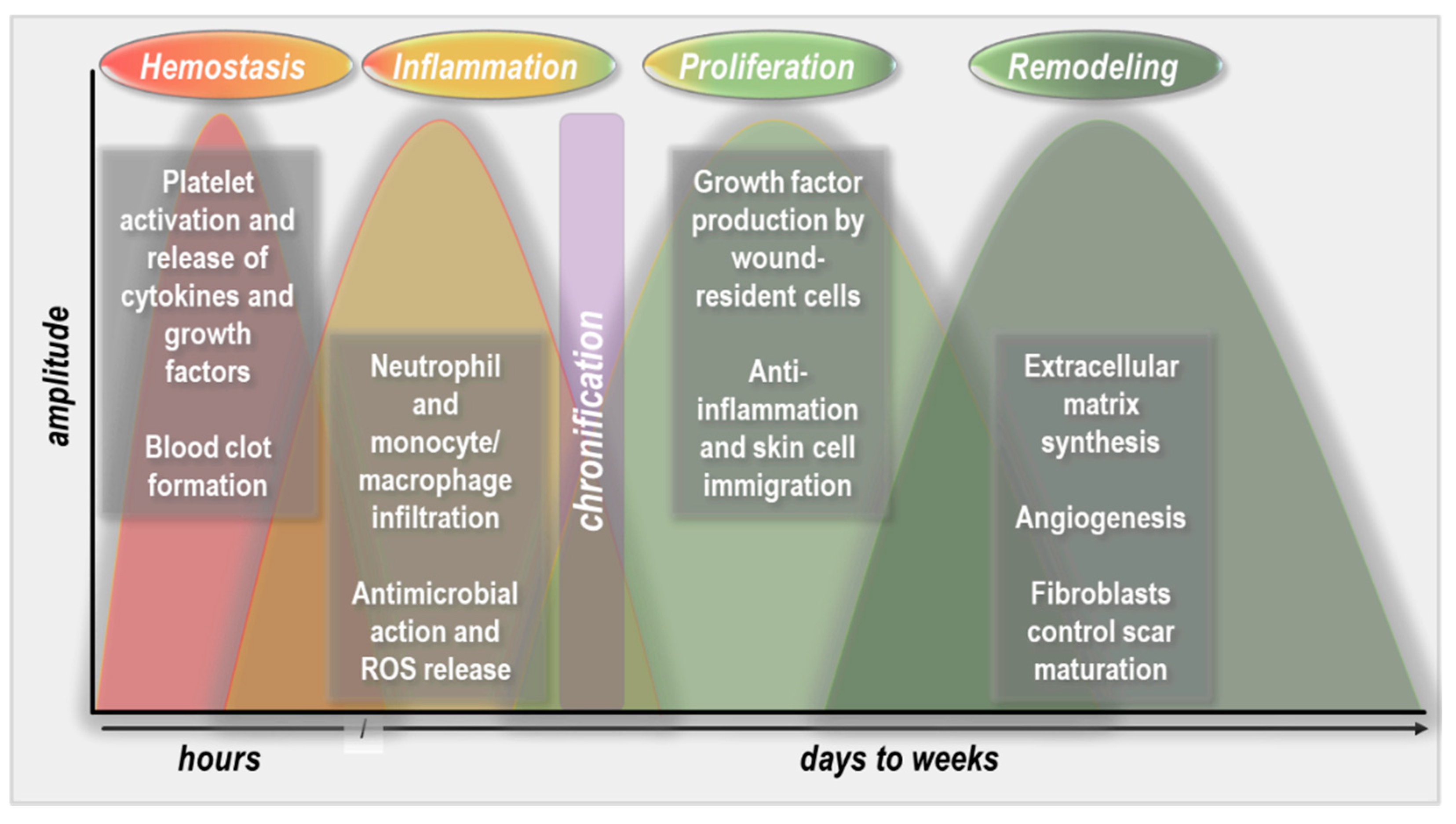
Wound healing stages. During hemostasis, platelets become activated to contribute to blood clot formation and release cytokines and growth factors. This is followed by the inflammation phase, which is characterized by extensive immigration of phagocytes, predominantly neutrophils and macrophages, to promote inflammation and pathogen clearance. During the subsequent proliferation phase, profound anti-inflammatory and growth-promoting processes are induced that allow the immigration of fibroblasts and keratinocytes into the wound bed, guided by growth factor production and gradients. Once the wound is closed, the final step is the remodeling of the skin architecture, which is mainly driven by fibroblasts and extracellular matrix synthesis and accompanied by angiogenesis to restore near-original tissue strength.
Figure 1.
Wound healing stages. During hemostasis, platelets become activated to contribute to blood clot formation and release cytokines and growth factors. This is followed by the inflammation phase, which is characterized by extensive immigration of phagocytes, predominantly neutrophils and macrophages, to promote inflammation and pathogen clearance. During the subsequent proliferation phase, profound anti-inflammatory and growth-promoting processes are induced that allow the immigration of fibroblasts and keratinocytes into the wound bed, guided by growth factor production and gradients. Once the wound is closed, the final step is the remodeling of the skin architecture, which is mainly driven by fibroblasts and extracellular matrix synthesis and accompanied by angiogenesis to restore near-original tissue strength.


Table 1.
Study overview on gas plasma-treated skin and wound healing in mice.
| Animal Model | Discharge Type | Key Outcomes | Ref. |
|---|---|---|---|
| Wounded | |||
| 129Sv/Ev | Ar plasma torch system | Elevated FGF-2 production and angiogenesis | [58] |
| 129Sv/Ev | Ar plasma torch system | Faster wound closure; elevated neutrophil and macrophage levels; higher production of IL-6 and MCP-1; collagen type I production increased | [57] |
| Balb/c | He plasma jet or treated liquid | Direct gas plasma exposure did not improve wound healing; gas plasma-treated liquid did not improve healing | [59] |
| Balb/c | Ar or He plasma jet | Accelerated wound healing; faster hemostasis | [38] |
| Balb/c | He plasma jet | Improved vascularization and angiogenesis | [53] |
| Balb/c | Ar plasma jet or treated water | Reduced feed gas flux increased wound temperature; direct gas plasma treatment or exposure to gas plasma-treated water improved wound healing; elevated numbers of myofibroblast | [60] |
| Balb/c | Ar plasma jet | Promotion of inflammation, re-epithelization, and wound contraction | [37] |
| Balb/c | DBD or Ar or He plasma jet | Ar and He plasma jets showed best wound healing promotion; DBD plasma showed good wound healing promotion | [56] |
| Balb/c | He plasma jet | Improved wound healing | [52] |
| Balb/c | Ar plasma jet | Wound healing not compared; exposure time-dependent changes of collagen and vimentin | [47] |
| Balb/c | Ar plasma jet | Improved wound healing for short but not long exposure times; long treatment showed necrosis and inflammatory cell influx | [45] |
| Balb/c | He/O2/N2 plasma jet | Accelerated neovascularization and epithelization; decreased microbial burden of natural wound flora | [54] |
| Balb/c | He plasma jet | Skin grafts on wounds exposed to gas plasma elevated angiogenesis and CD31 and hemoglobin expression; increased VEGFR2, PDGFRβ, and eNOS; less TSP-1 expression | [61] |
| Balb/c | Ar plasma jet | Short remote but not long direct exposure promoted wound healing and re-epithelization | [46] |
| Balb/c | Ar plasma jet + medical honey | Lack of beneficial effect of the combination of gas plasma with hydrocolloid dressings and medical honey | [62] |
| Balb/c | Ar plasma jet + medical honey | No improved healing | [63] |
| Balb/c | Plasma jet | Infection model; gas plasma jet-to-wound contact reduced infection while remote gas plasma jet treatment was better to stimulate wound healing | [55] |
| Balb/c | Hot He plasma jet | This gas plasma jet reached up to 90 °C and hence damaged the skin under various feed gas fluxes and treatment times | [64] |
| BKS.CG | He plasma jet | Improved wound closure in diabetic mice with moderate gas plasma treatment; short or long exposure did not improve healing as much | [65] |
| C57/BL6 | N2 plasma jet | Wound healing rates dependent on times points investigated; elevated secondary RNS in wound tissue; improved angiogenesis; earlier epithelization and wound contraction | [50] |
| C57/BL6 | N2/air plasma jet | Improved wound contraction and healing | [49] |
| C57/BL6 | N2/Ar plasma jet | Repeated but not single exposure increased wound healing; elevated blood flow and RNS deposition into tissue; augmented wound strength and laminin production; decreased MMP3 | [51] |
| C57/BL6 | Air/He DBD | Treatment of scars two weeks after wounding led to reduction of scar tissue, thickness, and vascularization | [66] |
| C57/BL6 | Plasma jet-treated liquid for hydrogels | Gas plasma-treated liquid enriched in hydrogel improved wound healing in random-pattern skin-flap full-thickness wounds | [67] |
| C57/BL6 | Ar plasma jet | Improved healing in sterile and infected burn wounds; lower TNFα levels; bacterial burden unchanged | [68] |
| db/db | He or He/O2 plasma jet | Faster wound healing in a diabetic model, predominantly in He/0.1% O2 compared to He and He/1% O2 plasma; elevated bFGF and VEGF in all treatment groups investigated | [69] |
| ICR | Air DBD-treated water | Improved wound healing; enhanced antimicrobial efficacy; altered wound microbiome | [70] |
| ICR | DBD-treated water | Improved wound healing; enhanced pro-inflammatory and anti-inflammatory cytokines and growth factors | [71] |
| SKH-1 | Ar plasma jet | Elevated wound tissue oxygenation in deep and superficial layers; enhanced tissue hemoglobin and water index | [39] |
| SKH-1 | Ar or Ar/air plasma jet | Best wound closure in and IL-6 mRNA in Ar/Air over Ar plasma | [43] |
| SKH-1 | Ar/air plasma jet | Dual-frequency but not single-frequency and 2 but not 1, 3, or 4 gas plasma treatment cycles improved healing; scab hampered gas plasma effects | [44] |
| SKH-1 | Ar plasma jet | Improved angiogenesis and wound closure | [36] |
| SKH-1 | Ar plasma jet | Lack of adverse events or cancerogenesis one year after repeated gas plasma exposure; improved wound healing | [42] |
| SKH-1 | Ar plasma jet | Improved angiogenesis and wound healing; faster re-epithelization; elevated keratin production and collagen fibers; augmented p53 activation, Nrf2 response, macrophage infiltration, inflammation, and granulation | [40] |
| SKH-1 | Ar plasma jet | Focal adhesion signaling complexes and junctional protein expression along with EMT and FMT accompanied improved wound healing | [41] |
| (not stated) | Ar/O2 plasma-treated oil emulsion | Improved wound closure | [72] |
| (not stated) | Ar/air or Ar/O2 plasma jet-treated oil | Accelerated wound healing with oil application previously treated with either air or O2 plasma regimens | [73] |
| (not stated) | Plasma jet | Improved wound healing and formation of keratin and granular layers along with collagen and αSMA deposition | [48] |
| Non-Wounded | |||
| 129Sv/Ev | Ar plasma torch | Keratinocyte proliferation, migration, and apoptosis did not change; β-defensins were found upregulated | [74] |
| Balb/c | He/O2 plasma jet | Ameliorated morphological manifestation and reduced epidermal proliferation in imiquimod-induced psoriatic lesions | [75] |
| Balb/c | Air DBD | Increased NO deposition; lack of inflammation or changes in skin collagen | [76] |
| Balb/c | Ar plasma jet | Local treatment through healthy skin in diabetic mice; presumable systemic effects on MDA, AOPP, oxLDL, and cytokines observed | [77] |
| Balb/c | Ar/N2/O2/humidity DBD | Allergic contact dermatitis models; humidified argon decreased disease severity greater than N2 or O2 admixture | [78] |
| C57/BL6 | N2 plasma jet | Imiquimod-induced psoriasis-like inflammation; reduced epidermal thickness, leukocyte infiltrate, chemokine/cytokine expression (IL-6, IL-17, IL-22, CXCL1, CCL20) | [79] |
| CBA | Ar plasma jet | Lack of allergic response in intact skin | [80] |
| HRM-2 | Ar plasma jet | E-cadherin decreased; EGF absorption through skin increased | [81] |
| HRM-2 | Ar DBD | Enhanced TGF-β, VEGF, GM-CSF, and EGF levels, and epidermal thickness | [82] |
| HRS | Ar plasma jet | No dermal degeneration in intact skin; lack of follicular atrophy; elevated catalase and Nrf2 expression | [83] |
| KM | Air DBD | Increased lidocaine drug absorption in gas plasma-treated skin; increased permeability was reversible 30 min post-treatment | [84] |
| NC/Nga | N2 DBD | Atopic dermatitis-like allergic skin inflammation model; less recruited mast cell and eosinophils; decreased epidermal thickness; less TH2 differentiation | [85] |
| SKH-1 | Ar plasma jet | Increased uptake of model drug and tissue oxygenation and microcirculation; changes in lipid composition; changes in junctional protein expression | [86] |
| SKH-1 | Plasma jet-treated water | Decrease of symptoms in a model of atopic dermatitis; decreased catalase and increased SOD activity | [87] |
| (not stated) | He/Ar/N2/air plasma jet | Psoriasis model; effectiveness in treating psoriasis shown | [88] |
Table 2.
Study overview on gas plasma-treated skin and wound healing in medium-sized rodents (rats, rabbits).
Table 2.
Study overview on gas plasma-treated skin and wound healing in medium-sized rodents (rats, rabbits).
| Animal Model | Discharge Type | Key Outcomes | Ref. |
|---|---|---|---|
| Rats | He plasma jet | In non-diabetic and diabetic rats, improved wound healing, acute inflammation, and neovascularization | [94] |
| Sprague Dawley rats | Ar plasma jet | Increased re-epithelization and wound closure; more acute inflammation and less fibrosis | [89] |
| Sprague Dawley rats | Ar plasma jet | Faster wound closure in rats as well as Type I and Type II diabetic rats; accelerated re-epithelization and fewer neutrophils; elevated SOD, catalase, and GPx in treated tissues | [90] |
| Sprague Dawley rats | Ar plasma jet | Decelerated burn wound injury progression regarding necrosis and neutrophil influx | [91] |
| Sprague Dawley rats | He/Ar plasma jet | Improved granulation tissue formation and wound healing; He/Ar better than He plasma; short treatment time better than intermediate and long treatment times | [92] |
| Sprague Dawley rats | He plasma jet | Decreased collagen deposition, scar formation, and TGF-β/pSmad2/pSmad3/αSMA positive cells; improved healing | [97] |
| Sprague Dawley rats | He plasma jet | Improved wound contraction, healing, and epithelization; unchanged collagen; increased TNFα and IL-1β but not IL-10 | [96] |
| Wistar rats | He plasma jet | Improved wound closure, angiogenesis, re-epithelization, inflammation; stronger force resistance; elevated elastic stiffness | [95] |
| Wistar rats | He plasma jet | Elevated expression of interleukin-6, nitric oxide synthase 2, prostaglandin-endoperoxide synthase 2 mRNA; decreased expression of nuclear factor ‘kappa-light-chain-enhancer’ of activated B-cells and superoxide dismutase 1 mRNA; improved wound closure speed | [93] |
| Wistar rats | Ar DBD | Elevated angiogenesis, free thiols, leukocyte influx, and MPO release | [98] |
| Wistar rats | Air DBD | Augmented healing of non-infected and infected wounds; elevated angiogenesis and granulation; shortened inflammation | [99] |
| Wistar rats | Air DBD | Attenuated long-term inflammation and improved wound healing and collagen I/collagen III ratio | [100] |
| Wistar rats | Air DBD | Improved healing in diabetic rats; increased epidermal thickness, re-epithelization, collagen deposition, angiogenesis, proliferation, TGF-β, and fibroblasts counts; decreased T cells | [101] |
| Wistar rats | Air DBD-functionalized PEG | Gas plasma-increased adhesion of polypropylene to absorb betaine hydrochloride, which improved wound healing in diabetic rats | [102] |
| New Zealand white rabbits | He DBD | Removal of MRSA in infected wounds; modulation of cytokine secretion, inflammation; accelerated re-epithelization and healing | [103] |
| New Zealand white rabbits | Air DBD | Lower leukocyte infiltration and decreased viability when investigated early after gas plasma treatment | [104] |
| New Zealand white rabbits | Plasma jet | Gas plasma treatment of the skin in diabetic animals was proposed to alter the tender structure in joints | [105] |
Table 3.
Study overview on gas plasma-treated skin and wound healing in other mammals and pet patients.
Table 3.
Study overview on gas plasma-treated skin and wound healing in other mammals and pet patients.
| Animal Model | Discharge Type | Key Outcomes | Ref. |
|---|---|---|---|
| Large Animal Models | |||
| Bergamasca sheep | He plasma torch | Improved wound healing; lower microbial wound burden; elevated proliferation and VEGF during early wound healing phases | [110] |
| Bergamasca sheep | He plasma torch | Improved wound healing efficacy when combined with the application of mesenchymal stem cells | [111] |
| Pigs | Ar plasma jet | Both superficial dermal wounds and deep wounds healed well following gas plasma treatment with no side effects; compared to untreated wounds, there were fewer inflammatory (immune) cells in fully healed gas plasma-treated wounds | [9] |
| Pigs | Air DBD | Intact skin was damaged with extended exposure times; moderate energy deposition/exposure time did not harm tissue; blood coagulation observed | [112] |
| Animal Patients | |||
| Dog (case report) | Ar plasma jet | Cutaneous infection with Alternaria spp. was alleviated; cessation of immunosuppressive drugs; complete clinical remission | [113] |
| Dogs | Ar plasma jet | Study in 40 dogs with canine bite wounds revealed no superior antimicrobial efficacy of gas plasma; gas plasma exposure was void of side effects | [114] |
| Dogs | Ar plasma jet | Study in 85 dogs with canine bite wounds revealed no superior antimicrobial efficacy of gas plasma; gas plasma exposure was void of side effects | [115] |
| Dogs and cats | Ar plasma jet | 8 case studies using pets suffering from chronic wounds since 1, 2, 6, 24, 36, 48, 60, and 80 months; complete healing achieved in 7 out of 8 patients | [116,117] |
| Dogs, cats, Guinea pigs | Ar plasma jet | 12 case studies using pets suffering from insufficient wound healing; all 12 patients experienced complete remission with repeated gas plasma treatment | [116,118] |
Publisher’s Note: MDPI stays neutral with regard to jurisdictional claims in published maps and institutional affiliations. |
© 2021 by the authors. Licensee MDPI, Basel, Switzerland. This article is an open access article distributed under the terms and conditions of the Creative Commons Attribution (CC BY) license (https://creativecommons.org/licenses/by/4.0/).
Share and Cite
MDPI and ACS Style
Bekeschus, S.; Kramer, A.; Schmidt, A. Gas Plasma-Augmented Wound Healing in Animal Models and Veterinary Medicine. Molecules 2021, 26, 5682. https://0-doi-org.brum.beds.ac.uk/10.3390/molecules26185682
AMA Style
Bekeschus S, Kramer A, Schmidt A. Gas Plasma-Augmented Wound Healing in Animal Models and Veterinary Medicine. Molecules. 2021; 26(18):5682. https://0-doi-org.brum.beds.ac.uk/10.3390/molecules26185682
Chicago/Turabian StyleBekeschus, Sander, Axel Kramer, and Anke Schmidt. 2021. "Gas Plasma-Augmented Wound Healing in Animal Models and Veterinary Medicine" Molecules 26, no. 18: 5682. https://0-doi-org.brum.beds.ac.uk/10.3390/molecules26185682