
Psychoactive Substances of Natural Origin: Toxicological Aspects, Therapeutic Properties and Analysis in Biological Samples


Abstract
:1. Introduction
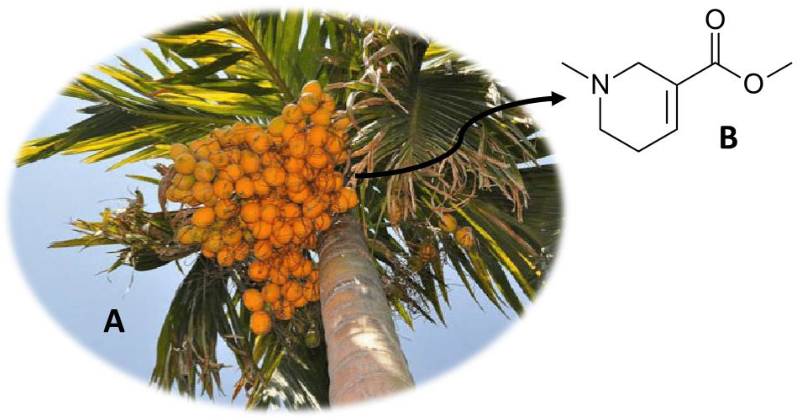
2. Areca catechu (Betel Quid)
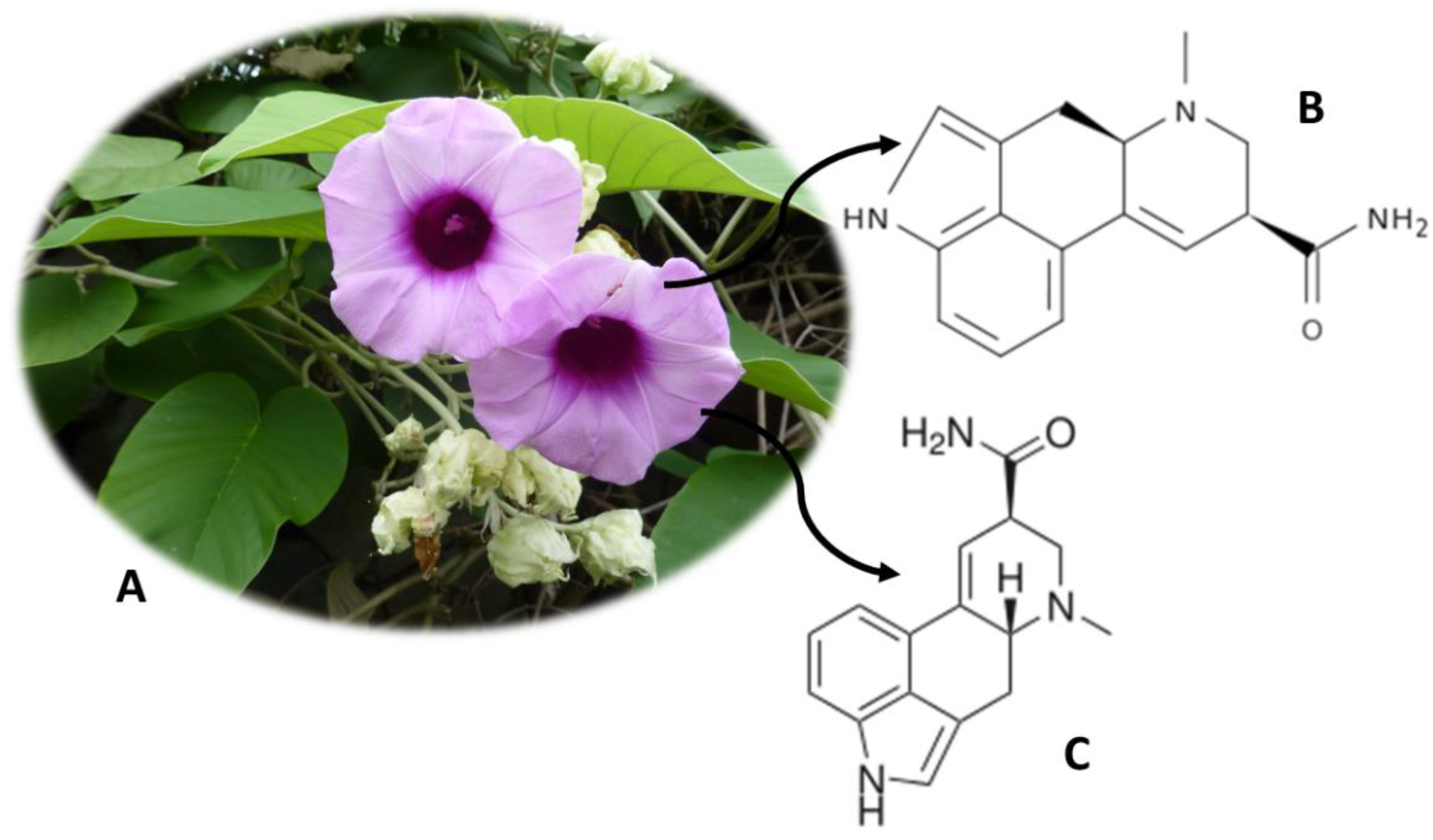
3. Argyreia nervosa (Adhoguda)
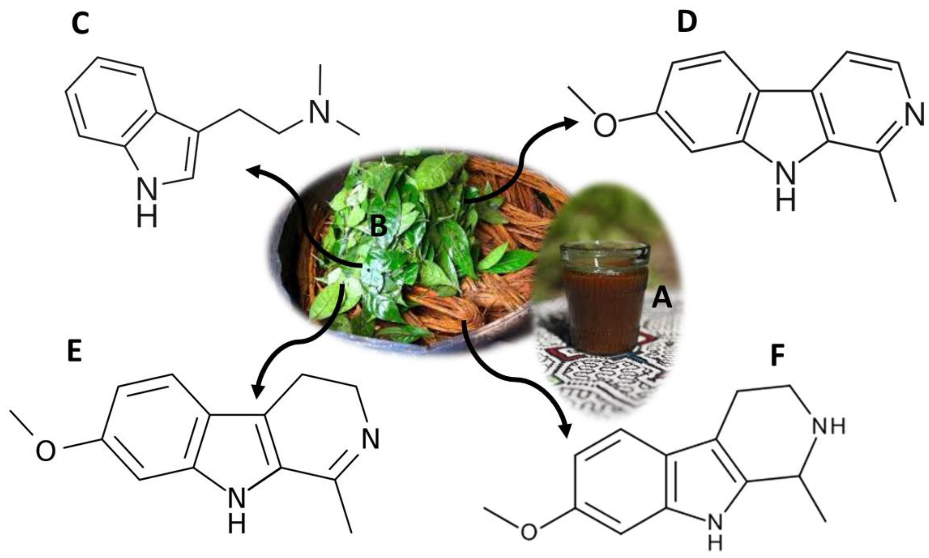
4. Ayahuasca (“Hoasca”)
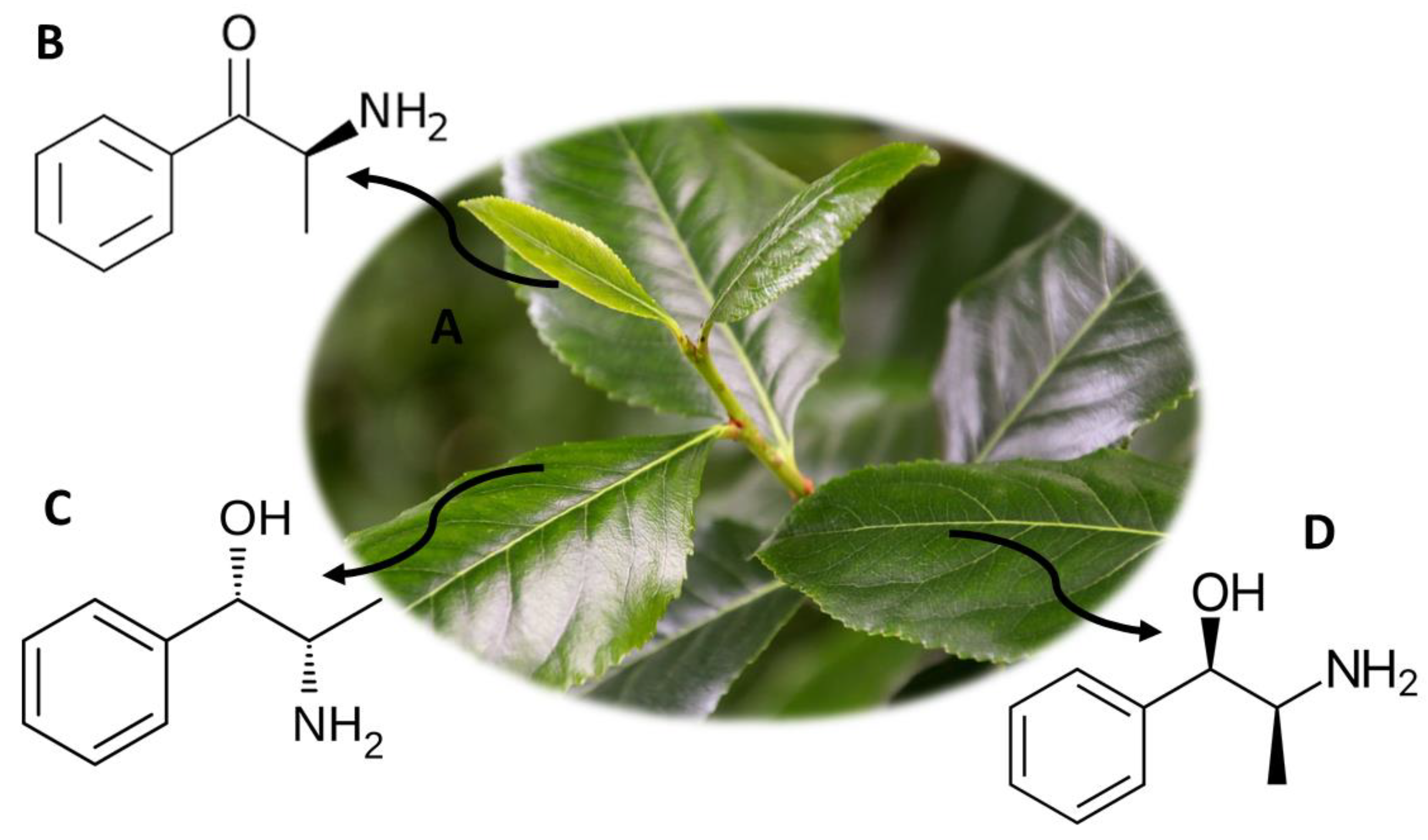
5. Catha edulis (Khat)
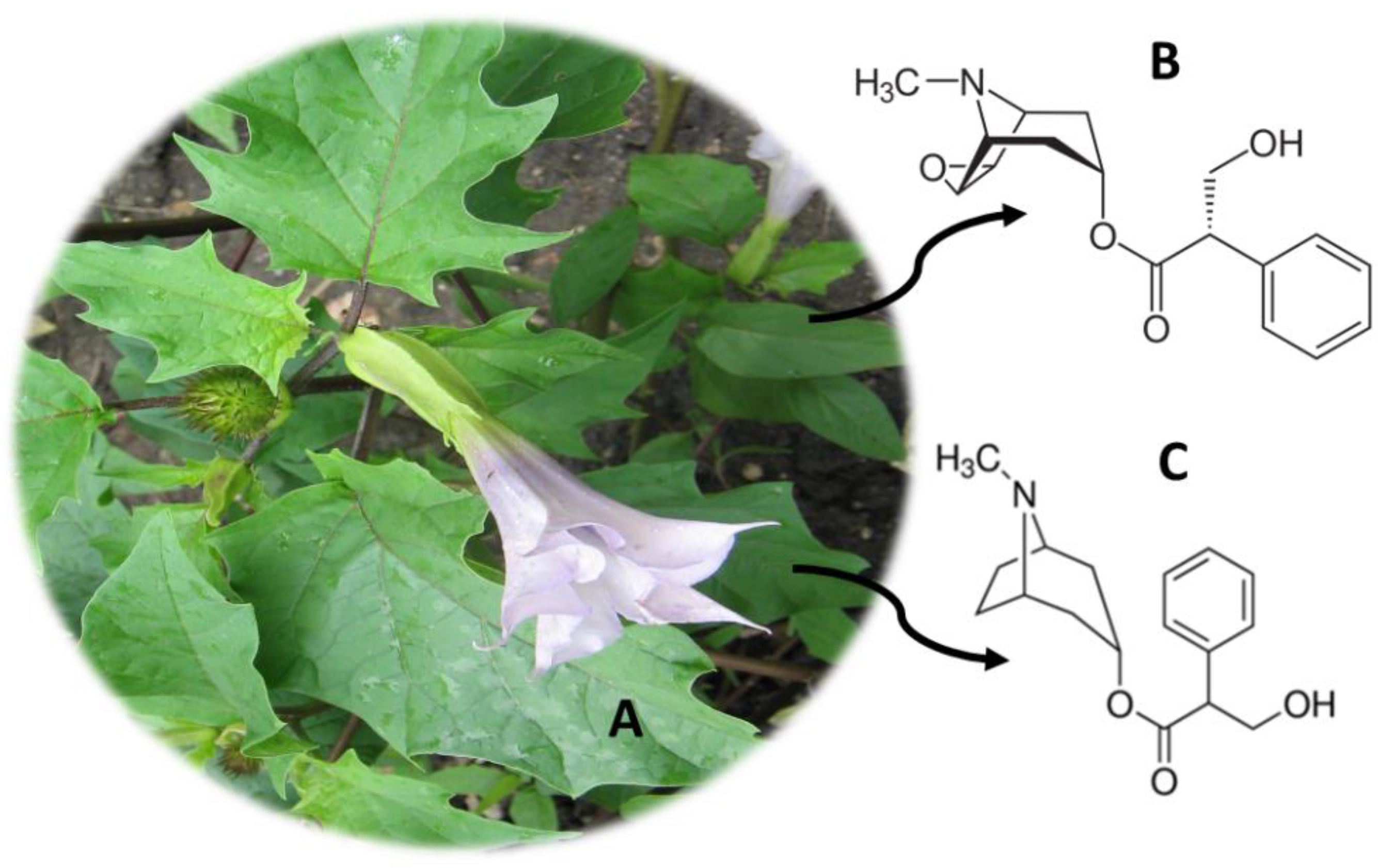
6. Datura stramonium (Jimson Weed)
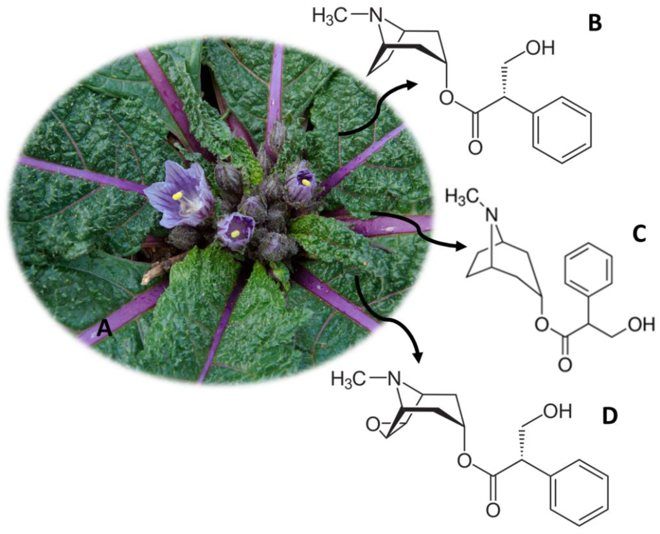
7. Mandragora officinarum (Mandrake)
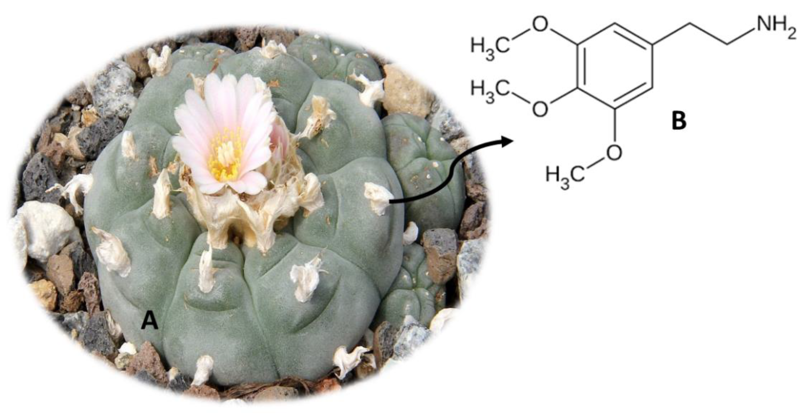
8. Lophophora williamsii (Peyote)
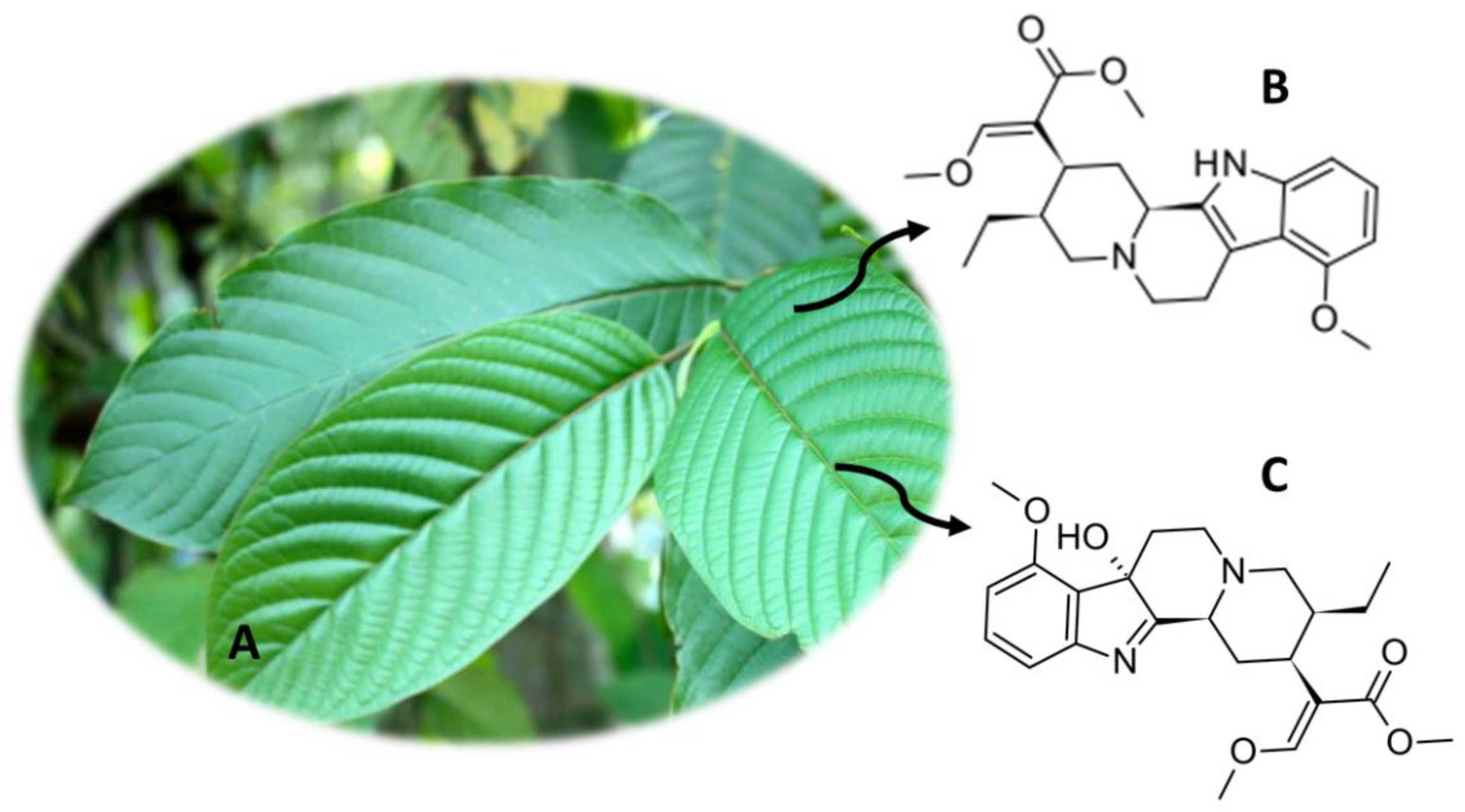
9. Mitragyna speciosa (Kratom)
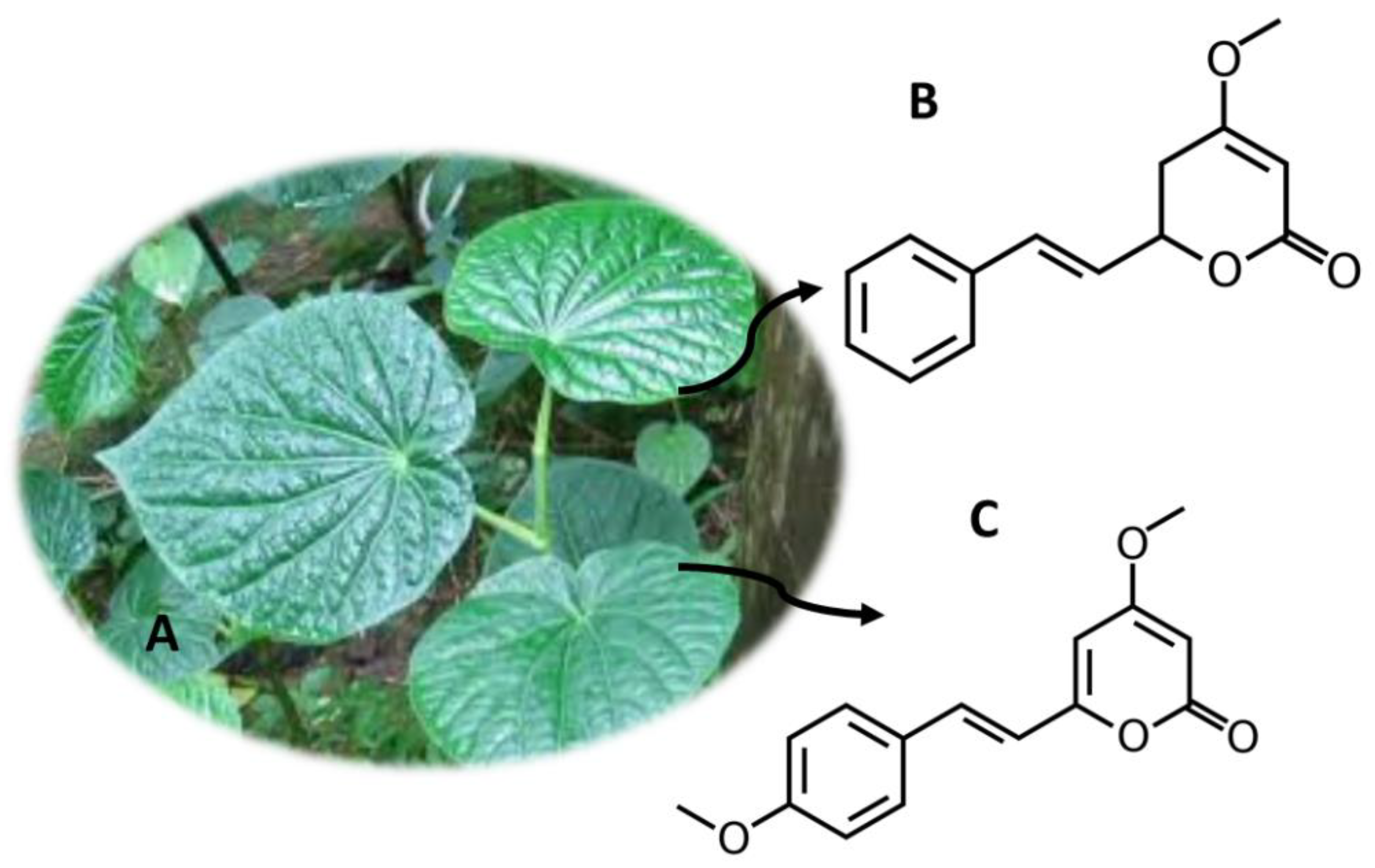
10. Piper methysticum Forst (Kava)
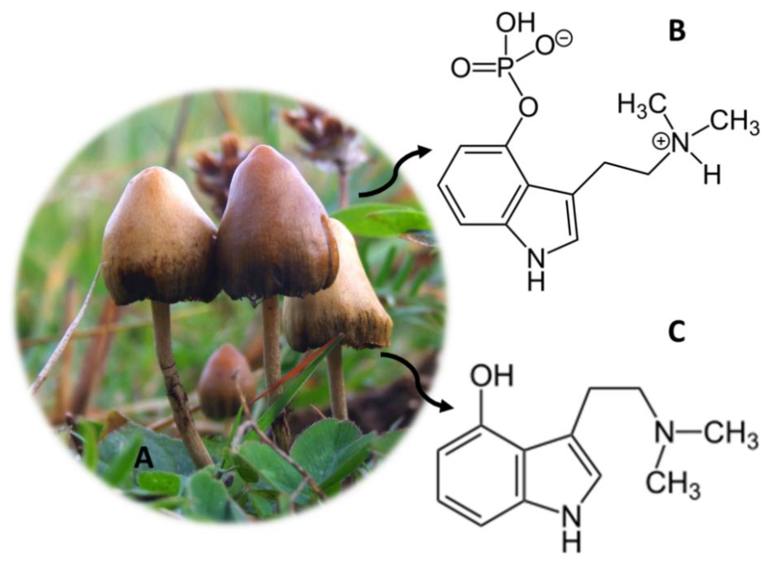
11. Psilocybe Genus (Magic Mushrooms)
12. Salvia divinorum (“Hierba de Maria”)
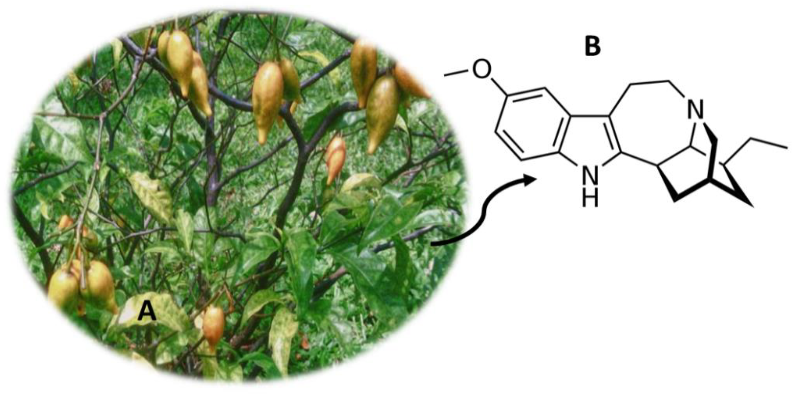
13. Tabernanthe iboga (Iboga)
14. Conclusions
Author Contributions
Funding
Institutional Review Board Statement
Informed Consent Statement
Conflicts of Interest
References
- European Monitoring Centre for Drugs and Drug Addiction. EU Drug Markets Report 2019; Publications Office of the European Union: Luxembourg, 2019; pp. 1–260. [CrossRef]
- Simão, A.Y.; Antunes, M.; Marques, H.; Rosado, T.; Soares, S.; Gonçalves, J.; Barroso, M.; Andraus, M.; Gallardo, E. Recent bionalytical methods for the determination of new psychoactive substances in biological specimens. Bioanalysis 2020, 12, 1557–1595. [Google Scholar] [CrossRef]
- Lo Faro, A.F.; Di Trana, A.; La Maida, N.; Tagliabracci, A.; Giorgetti, R.; Busardò, F.P. Biomedical analysis of New Psychoactive Substances (NPS) of natural origin. J. Pharm. Biomed. Anal. 2020, 179, 112945. [Google Scholar] [CrossRef]
- Guirguis, A. New psychoactive substances: A public health issue. Int. J. Pharm. Practice 2017, 25, 323–325. [Google Scholar] [CrossRef] [PubMed] [Green Version]
- Salomone, A.; Palamar, J.J.; Gerace, E.; Di Corcia, D.; Vincenti, M. Hair testing for drugs of abuse and new psychoactive substances in a high-risk population. J. Anal. Toxicol. 2017, 41, 376–381. [Google Scholar] [CrossRef] [PubMed] [Green Version]
- Archer, J.R.H.; Mendes, F.; Hudson, S.; Layne, K.; Dargan, P.I.; Wood, D.M. Evaluation of long-term detection trends of new psychoactive substances in pooled urine from city street portable urinals (London, UK). Br. J. Clin. Pharmacol. 2020, 86, 517–527. [Google Scholar] [CrossRef] [PubMed]
- Peacock, A.; Bruno, R.; Gisev, N.; Degenhardt, L.; Hall, W.; Sedefov, R.; White, J.; Thomas, K.V.; Farrell, M.; Griffiths, P. New psychoactive substances: Challenges for drug surveillance, control, and public health responses. Lancet 2019, 394, 1668–1684. [Google Scholar] [CrossRef]
- Meyer, M.R. New psychoactive substances: An overview on recent publications on their toxicodynamics and toxicokinetics. Arch. Toxicol. 2016, 90, 2421–2444. [Google Scholar] [CrossRef]
- Evans-Brown, M.; Sedefov, R. New psychoactive substances: Driving greater complexity into the drug problem. Addiction 2017, 112, 36–38. [Google Scholar] [CrossRef] [PubMed]
- United Nations Office on Drugs and Crime. UNODC Early Warning Advisory (EWA) on New Psychoactive Substances (NPS). Available online: https://www.unodc.org/LSS/Home/NPS (accessed on 28 January 2021).
- European Monitoring Centre for Drugs and Drug The EU Early Warning System on new psychoactive substances (NPS). Available online: https://www.emcdda.europa.eu/publications/topic-overviews/eu-early-warning-system_en (accessed on 28 January 2021).
- Smith, J.P.; Sutcliffe, O.B.; Banks, C.E. An overview of recent developments in the analytical detection of new psychoactive substances (NPSs). Analyst 2015, 140, 4932–4948. [Google Scholar] [CrossRef] [PubMed] [Green Version]
- Graziano, S.; Orsolini, L.; Rotolo, M.C.; Tittarelli, R.; Schifano, F.; Pichini, S. Herbal Highs: Review on Psychoactive Effects and Neuropharmacology. Curr. Neuropharmacol. 2017, 15, 750–761. [Google Scholar] [CrossRef] [Green Version]
- Souza, R.C.Z.; Zandonadi, F.S.; Freitas, D.P.; Tófoli, L.F.F.; Sussulini, A. Validation of an analytical method for the determination of the main ayahuasca active compounds and application to real ayahuasca samples from Brazil. J. Chromatogr. B Anal. Technol. Biomed. Life Sci. 2019, 1124, 197–203. [Google Scholar] [CrossRef] [PubMed]
- Peng, W.; Liu, Y.J.; Wu, N.; Sun, T.; He, X.Y.; Gao, Y.X.; Wu, C.J. Areca catechu L. (Arecaceae): A review of its traditional uses, botany, phytochemistry, pharmacology and toxicology. J. Ethnopharmacol. 2015, 164, 340–356. [Google Scholar] [CrossRef]
- Gupta, P.C.; Warnakulasuriya, S. Global epidemiology of areca nut usage. Addict. Biol. 2002, 7, 77–83. [Google Scholar] [CrossRef] [PubMed]
- Papke, R.L.; Horenstein, N.A.; Stokes, C. Nicotinic Activity of Arecoline, the Psychoactive Element of “Betel Nuts”, Suggests a Basis for Habitual Use and Anti-Inflammatory Activity. PLoS ONE 2015, 10, e0140907. [Google Scholar] [CrossRef]
- Coppola, M.; Mondola, R. Potential action of betel alkaloids on positive and negative symptoms of schizophrenia: A review. Nord. J. Psychiatry 2012, 66, 73–78. [Google Scholar] [CrossRef]
- Taylor, R.F.H.; Al-Jarad, N.; John, L.M.E.; Barnes, N.C.; Conroy, D.M. Betel-nut chewing and asthma. Lancet 1992, 339, 1134–1136. [Google Scholar] [CrossRef]
- Chen, Y.-C.; Lee, H.-C.; Lee, H.-H.; Su, H.-M.; Lin, T.-H.; Hsu, P.-C. Areca Nut Chewing Complicated with Non-Obstructive and Obstructive ST Elevation Myocardial Infarction. Acta Cardiol. Sin. 2016, 32, 103–107. [Google Scholar] [PubMed]
- Salehi, B.; Konovalov, D.A.; Fru, P.; Kapewangolo, P.; Peron, G.; Ksenija, M.S.; Cardoso, S.M.; Pereira, O.R.; Nigam, M.; Nicola, S.; et al. Areca catechu—From farm to food and biomedical applications. Phyther. Res. 2020, 34, 2140–2158. [Google Scholar] [CrossRef] [PubMed]
- Keshava Bhat, S.; Sarpangala, M.; Ashwin, D. Antilipidemic activity of arecanut, Areca catechu L.: A valuable herbal medicine. Int. J. Herb. Med. 2017, 5, 35–38. [Google Scholar]
- Sreeramulu, D.; Reddy, C.V.K.; Chauhan, A.; Balakrishna, N.; Raghunath, M. Natural antioxidant activity of commonly consumed plant foods in India: Effect of domestic processing. Oxid. Med. Cell. Longev. 2013, 369479, 1–12. [Google Scholar] [CrossRef] [PubMed]
- Wu, M.; Fang, M.; Hu, Y.; Wang, X. Four types of traditional Chinese medicine inducing epileptic seizures. Seizure 2012, 21, 311–315. [Google Scholar] [CrossRef] [PubMed] [Green Version]
- Hamsar, M.N.; Ismail, S.; Mordi, M.N.; Ramanathan, S.; Mansor, S.M. Antioxidant activity and the effect of different parts of areca catechu extracts on Glutathione-S-Transferase activity in vitro. Free Radic. Antioxid. 2011, 1, 28–33. [Google Scholar] [CrossRef]
- Wu, I.C.; Chen, P.H.; Wang, C.J.; Wu, D.C.; Tsai, S.M.; Chao, M.R.; Chen, B.H.; Lee, H.H.; Lee, C.H.; Ko, Y.C. Quantification of blood betel quid alkaloids and urinary 8-hydroxydeoxyguanosine in humans and their association with betel chewing habits. J. Anal. Toxicol. 2010, 34, 325–331. [Google Scholar] [CrossRef]
- Pichini, S.; Pellegrini, M.; Pacifici, R.; Marcheil, E.; Murillo, J.; Puig, C.; Vall, O.; García-Algar, O. Quantification of arecoline (Areca nut alkaloid) in neonatal biological matrices by high-performance liquid chromatography/electrospray quadrupole mass spectrometry. Rapid Commun. Mass Spectrom. 2003, 17, 1958–1964. [Google Scholar] [CrossRef] [PubMed]
- Krais, S.; Klima, M.; Huppertz, L.M.; Auwärter, V.; Altenburger, M.J.; Neukamm, M.A. Betel Nut Chewing in Iron Age Vietnam? Detection of Areca catechu Alkaloids in Dental Enamel. J. Psychoact. Drugs 2017, 49, 11–17. [Google Scholar] [CrossRef]
- Franke, A.A.; Li, X.; Custer, L.J.; Lai, J.F. Chemical Markers for Short- and Long-Term Areca Nut Exposure. Subst. Use Misuse 2020, 55, 1395–1402. [Google Scholar] [CrossRef] [PubMed]
- Lee, H.H.; Chen, L.Y.; Wang, H.L.; Chen, B.H. Quantification of salivary arecoline, arecaidine and N-methylnipecotic acid levels in volunteers by liquid chromatography-tandem mass spectrometry. J. Anal. Toxicol. 2015, 39, 714–719. [Google Scholar] [CrossRef] [PubMed] [Green Version]
- Pellegrini, M.; Marchei, E.; Rossi, S.; Vagnarelli, F.; Durgbanshi, A.; García-Algar, Ó.; Vall, O.; Pichini, S. Liquid chromatography/electrospray ionization tandem mass spectrometry assay for determination of nicotine and metabolites, caffeine and arecoline in breast milk. Rapid Commun. Mass Spectrom. 2007, 21, 2693–2703. [Google Scholar] [CrossRef]
- Marchei, E.; Durgbanshi, A.; Rossi, S.; Garcia-Algar, Ó.; Zuccaro, P.; Pichini, S. Determination of arecoline (areca nut alkaloid) and nicotine in hair by high-performance liquid chromatography/electrospray quadrupole mass spectrometry. Rapid Commun. Mass Spectrom. 2005, 19, 3416–3418. [Google Scholar] [CrossRef]
- Austin, D.; Huáman, Z. A synopsis of Ipomoea (Convolvulaceae) in the Americas. Taxon 1996, 45, 3–38. [Google Scholar] [CrossRef]
- Paulke, A.; Kremer, C.; Wunder, C.; Wurglics, M.; Schubert-Zsilavecz, M.; Toennes, S.W. Studies on the alkaloid composition of the Hawaiian Baby Woodrose Argyreia nervosa, a common legal high. Forensic Sci. Int. 2015, 249, 281–293. [Google Scholar] [CrossRef]
- Chao, J.-M.; Der Marderosian, A.H. Ergoline alkaloidal constituents of hawaiian baby wood rose, argyreia nervosa (Burm. f.) bojer. J. Pharm. Sci. 1973, 62, 588–591. [Google Scholar] [CrossRef] [PubMed]
- Larson, B.T.; Harmon, D.L.; Piper, E.L.; Griffis, L.M.; Bush, L.P. Alkaloid binding and activation of D2 dopamine receptors in cell culture. J. Anim. Sci. 1999, 77, 942. [Google Scholar] [CrossRef] [PubMed]
- Modi, A.J.; Khadabadi, S.S.; Deokate, U.A.; Farooqui, I.A.; Deore, S.L.; Gangwani, M.R. Argyreia speciosa Linn. f.: Phytochemistry, pharmacognosy and pharmacological studies. J. Pharmacogn. Phyther. 2010, 2, 34–42. [Google Scholar]
- Weber, J.M.; Ma, T.S. Microchemical investigations of medicinal plants. XIV—Identification of the alkaloids in the leaves of Ipomoea violacea using preparative thin layer chromatography and solid probe mass spectrometry. Mikrochim. Acta 1976, 65, 227–242. [Google Scholar] [CrossRef]
- Paulke, A.; Kremer, C.; Wunder, C.; Toennes, S.W. Analysis of lysergic acid amide in human serum and urine after ingestion of Argyreia nervosa seeds. In Proceedings of the Analytical and Bioanalytical Chemistry; Springer: Berlin, Germany, 2012; Volume 404, pp. 531–538. [Google Scholar]
- Simão, A.Y.; Gonçalves, J.; Duarte, A.P.; Barroso, M.; Cristóvão, A.C.; Gallardo, E. Toxicological Aspects and Determination of the Main Components of Ayahuasca: A Critical Review. Medicines 2019, 6, 106. [Google Scholar] [CrossRef] [Green Version]
- Andrade, T.S.; de Oliveira, R.; da Silva, M.L.; Von Zuben, M.V.; Grisolia, C.K.; Domingues, I.; Caldas, E.D.; Pic-Taylor, A. Exposure to ayahuasca induces developmental and behavioral alterations on early life stages of zebrafish. Chem. Biol. Interact. 2018, 293, 133–140. [Google Scholar] [CrossRef] [PubMed]
- Gable, R.S. Risk assessment of ritual use of oral dimethyltryptamine (DMT) and harmala alkaloids. Addiction 2007, 102, 24–34. [Google Scholar] [CrossRef] [PubMed]
- Ramachandran, P.; Zhang, N.; McLaughlin, W.B.; Luo, Y.; Handy, S.; Duke, J.A.; Vasquez, R.; Ottesen, A. Sequencing the vine of the soul: Full chloroplast genome sequence of Banisteriopsis caapi. Genome Announc. 2018, 6. [Google Scholar] [CrossRef] [PubMed] [Green Version]
- Dos Santos, R.G.; Balthazar, F.M.; Bouso, J.C.; Hallak, J.E.C. The current state of research on ayahuasca: A systematic review of human studies assessing psychiatric symptoms, neuropsychological functioning, and neuroimaging. J. Psychopharmacol. 2016, 30, 1230–1247. [Google Scholar] [CrossRef]
- dos Santos, R.G.; Osório, F.L.; Crippa, J.A.S.; Hallak, J.E.C. Antidepressive and anxiolytic effects of ayahuasca: A systematic literature review of animal and human studies. Rev. Bras. Psiquiatr. 2016, 38, 65–72. [Google Scholar] [CrossRef] [PubMed] [Green Version]
- Riba, J.; Valle, M.; Urbano, G.; Yritia, M.; Morte, A.; Barbanoj, M.J. Human pharmacology of ayahuasca: Subjective and cardiovascular effects, monoamine metabolite excretion, and pharmacokinetics. J. Pharmacol. Exp. Ther. 2003, 306, 73–83. [Google Scholar] [CrossRef] [Green Version]
- De Araujo, D.B.; Ribeiro, S.; Cecchi, G.A.; Carvalho, F.M.; Sanchez, T.A.; Pinto, J.P.; de Martinis, B.S.; Crippa, J.A.; Hallak, J.E.C.; Santos, A.C. Seeing with the eyes shut: Neural basis of enhanced imagery following ayahuasca ingestion. Hum. Brain Mapp. 2012, 33, 2550–2560. [Google Scholar] [CrossRef] [PubMed]
- Riba, J.; Romero, S.; Grasa, E.; Mena, E.; Carrió, I.; Barbanoj, M.J. Increased frontal and paralimbic activation following ayahuasca, the pan-amazonian inebriant. Psychopharmacology 2006, 186, 93–98. [Google Scholar] [CrossRef] [Green Version]
- Morales-García, J.A.; De La Fuente Revenga, M.; Alonso-Gil, S.; Rodríguez-Franco, M.I.; Feilding, A.; Perez-Castillo, A.; Riba, J. The alkaloids of Banisteriopsis caapi, the plant source of the Amazonian hallucinogen Ayahuasca, stimulate adult neurogenesis in vitro. Sci. Rep. 2017, 7, 1–13. [Google Scholar] [CrossRef] [Green Version]
- Hassan, Z.; Bosch, O.G.; Singh, D.; Narayanan, S.; Kasinather, B.V.; Seifritz, E.; Kornhuber, J.; Quednow, B.B.; Müller, C.P. Novel psychoactive substances-recent progress on neuropharmacological mechanisms of action for selected drugs. Front. Psychiatry 2017, 8, 152. [Google Scholar] [CrossRef] [PubMed] [Green Version]
- Callaway, J.C.; Raymon, L.P.; Hearn, W.L.; McKenna, D.J.; Grob, C.S.; Brito, G.S.; Mash, D.C. Quantitation of N,N-dimethyltryptamine and harmala alkaloids in human plasma after oral dosing with ayahuasca. J. Anal. Toxicol. 1996, 20, 492–497. [Google Scholar] [CrossRef] [Green Version]
- Gonçalves, J.; Luís, Â.; Gradillas, A.; García, A.; Restolho, J.; Fernández, N.; Domingues, F.; Gallardo, E.; Duarte, A.P. Ayahuasca Beverages: Phytochemical Analysis and Biological Properties. Antibiotics 2020, 9, 731. [Google Scholar] [CrossRef] [PubMed]
- Simão, A.Y.; Gonçalves, J.; Gradillas, A.; García, A.; Restolho, J.; Fernández, N.; Rodilla, J.M.; Barroso, M.; Duarte, A.P.; Cristóvão, A.C.; et al. Evaluation of the Cytotoxicity of Ayahuasca Beverages. Molecules 2020, 25, 5594. [Google Scholar] [CrossRef]
- De Osório, F.L.; Sanches, R.F.; Macedo, L.R.; dos Santos, R.G.; Maia-De-Oliveira, J.P.; Wichert-Ana, L.; de Araujo, D.B.; Riba, J.; Crippa, J.A.; Hallak, J.E. Antidepressant effects of a single dose of ayahuasca in patients with recurrent depression: A preliminary report. Rev. Bras. Psiquiatr. 2015, 37, 13–20. [Google Scholar] [CrossRef] [Green Version]
- Sanches, R.F.; De Lima Osório, F.; Santos, R.G.D.; Macedo, L.R.H.; Maia-De-Oliveira, J.P.; Wichert-Ana, L.; De Araujo, D.B.; Riba, J.; Crippa, J.A.S.; Hallak, J.E.C. Antidepressant effects of a single dose of ayahuasca in patients with recurrent depression a SPECT study. J. Clin. Psychopharmacol. 2016, 36, 77–81. [Google Scholar] [CrossRef] [Green Version]
- Santos, R.G.; Landeira-Fernandez, J.; Strassman, R.J.; Motta, V.; Cruz, A.P.M. Effects of ayahuasca on psychometric measures of anxiety, panic-like and hopelessness in Santo Daime members. J. Ethnopharmacol. 2007, 112, 507–513. [Google Scholar] [CrossRef] [PubMed]
- Grob, C.S.; McKenna, D.J.; Callaway, J.C.; Brito, G.S.; Neves, E.S.; Oberlaender, G.; Saide, O.L.; Labigalini, E.; Tacla, C.; Miranda, C.T.; et al. Human psychopharmacology of hoasca, a plant hallucinogen used in ritual context in Brazil. J. Nerv. Ment. Dis. 1996, 184, 86–94. [Google Scholar] [CrossRef] [PubMed] [Green Version]
- Thomas, G.; Lucas, P.; Capler, N.R.; Tupper, K.W.; Martin, G. Ayahuasca-assisted therapy for addiction: Results from a preliminary observational study in Canada. Curr. Drug Abus. Rev. 2013, 6, 30–42. [Google Scholar] [CrossRef] [PubMed]
- Oliveira-Lima, A.J.; Santos, R.; Hollais, A.W.; Gerardi-Junior, C.A.; Baldaia, M.A.; Wuo-Silva, R.; Yokoyama, T.S.; Costa, J.L.; Malpezzi-Marinho, E.L.A.; Ribeiro-Barbosa, P.C.; et al. Effects of ayahuasca on the development of ethanol-induced behavioral sensitization and on a post-sensitization treatment in mice. Physiol. Behav. 2015, 142, 28–36. [Google Scholar] [CrossRef] [Green Version]
- Da Silveira, D.X.; Grob, C.S.; de Rios, M.D.; Lopez, E.; Alonso, L.K.; Tacla, C.; Doering-Silveira, E. Ayahuasca in adolescence: A preliminary psychiatric assessment. J. Psychoact. Drugs 2005, 37, 129–133. [Google Scholar] [CrossRef]
- Barbosa, P.C.R.; Cazorla, I.M.; Giglio, J.S.; Strassman, R.S. A six-month prospective evaluation of personality traits, psychiatric symptoms and quality of life in Ayahuasca-Naïve Subjects. J. Psychoact. Drugs 2009, 41, 205–212. [Google Scholar] [CrossRef] [PubMed]
- McIlhenny, E.H.; Riba, J.; Barbanoj, M.J.; Strassman, R.; Barker, S.A. Methodology for determining major constituents of ayahuasca and their metabolites in blood. Biomed. Chromatogr. 2012, 26, 301–313. [Google Scholar] [CrossRef]
- Yritia, M.; Riba, J.; Ortuño, J.; Ramirez, A.; Castillo, A.; Alfaro, Y.; de la Torre, R.; Barbanoj, M.J. Determination of N, N-dimethyltryptamine and β-carboline alkaloids in human plasma following oral administration of Ayahuasca. J. Chromatogr. B Anal. Technol. Biomed. Life Sci. 2002, 779, 271–281. [Google Scholar] [CrossRef]
- Oliveira, C.D.R.; Okai, G.G.; Da Costa, J.L.; De Almeida, R.M.; Oliveira-Silva, D.; Yonamine, M. Determination of dimethyltryptamine and β-carbolines (ayahuasca alkaloids) in plasma samples by LC-MS/MS. Bioanalysis 2012, 4, 1731–1738. [Google Scholar] [CrossRef]
- Mcilhenny, E.H.; Riba, J.; Barbanoj, M.J.; Strassman, R.; Barker, S.A. Methodology for and the determination of the major constituents and metabolites of the amazonian botanical medicine ayahuasca in human urine. Biomed. Chromatogr. 2011, 25, 970–984. [Google Scholar] [CrossRef] [PubMed]
- Riba, J.; Mcilhenny, E.H.; Bouso, J.C.; Barker, S.A. Metabolism and urinary disposition of N,N-dimethyltryptamine after oral and smoked administration: A comparative study. Drug Test. Anal. 2015, 7, 401–406. [Google Scholar] [CrossRef] [PubMed]
- Pichini, S.; Marchei, E.; García-Algar, O.; Gomez, A.; Di Giovannandrea, R.; Pacifici, R. Ultra-high-pressure liquid chromatography tandem mass spectrometry determination of hallucinogenic drugs in hair of psychedelic plants and mushrooms consumers. J. Pharm. Biomed. Anal. 2014, 100, 284–289. [Google Scholar] [CrossRef] [PubMed]
- Feyissa, A.M.; Kelly, J.P. A review of the neuropharmacological properties of khat. Prog. Neuro Psychopharmacol. Biol. Psychiatry 2008, 32, 1147–1166. [Google Scholar] [CrossRef]
- Brenneisen, R.; Geisshüsler, S.; Schorno, X. Metabolism of cathinone to (−)-norephedrine and (−)-norpseudoephedrine. J. Pharm. Pharmacol. 1986, 38, 298–300. [Google Scholar] [CrossRef] [PubMed]
- Sørensen, L.K. Determination of cathinones and related ephedrines in forensic whole-blood samples by liquid-chromatography-electrospray tandem mass spectrometry. J. Chromatogr. B Anal. Technol. Biomed. Life Sci. 2011, 879, 727–736. [Google Scholar] [CrossRef]
- Beyer, J.; Peters, F.T.; Kraemer, T.; Maurer, H.H. Detection and validated quantification of nine herbal phenalkylamines and methcathinone in human blood plasma by LC-MS/MS with electrospray ionization. J. Mass Spectrom. 2007, 42, 150–160. [Google Scholar] [CrossRef] [PubMed]
- Toennes, S.W.; Kauert, G.F. Excretion and Detection of Cathinone, Cathine, and Phenylpropanolamine in Urine after Kath Chewing. Clin. Chem. 2002, 48, 1715–1719. [Google Scholar] [CrossRef] [PubMed] [Green Version]
- Mohamed, K.M.; Al-Hazmi, A.H.; Alasiri, A.M.; Ali, M.E.S. A GC-MS Method for Detection and Quantification of Cathine, Cathinone, Methcathinone and Ephedrine in Oral Fluid. J. Chromatogr. Sci. 2016, 54, 1271–1276. [Google Scholar] [CrossRef]
- Margalho, C.; Castanheira, A.; Real, F.C.; Gallardo, E.; López-Rivadulla, M. Determination of “new psychoactive substances” in postmortem matrices using microwave derivatization and gas chromatography-mass spectrometry. J. Chromatogr. B Anal. Technol. Biomed. Life Sci. 2016, 1020, 14–23. [Google Scholar] [CrossRef]
- Margalho, C.; Almeida, P.; Rosado, T.; Corte-Real, F.; Gallardo, E. Determination of New Psychoactive Substances in Whole Blood Using Microwave Fast Derivatization and Gas Chromatography/Mass Spectrometry. J. Anal. Toxicol. 2020, 44, 92–102. [Google Scholar] [CrossRef]
- Mahler, D.A. Anticholinergic poisoning from Jimson weed. J. Am. Coll. Emerg. Physicians 1976, 5, 440–442. [Google Scholar] [CrossRef]
- Oerther, S.; Behrman, A.D.; Ketcham, S. Herbal hallucinations: Common abuse situations seen in the emergency department. J. Emerg. Nurs. 2010, 36, 594–596. [Google Scholar] [CrossRef]
- Forrester, M.B. Jimsonweed (Datura stramonium) exposures in Texas, 1998–2004. J. Toxicol. Environ. Health Part A Curr. Issues 2006, 69, 1757–1762. [Google Scholar] [CrossRef] [PubMed]
- Sutter, M.E.; Chenoweth, J.; Albertson, T.E. Alternative drugs of abuse. Clin. Rev. Allergy Immunol. 2014, 46, 3–18. [Google Scholar] [CrossRef]
- Krenzelok, E.P. Aspects of datura poisoning and treatment. Clin. Toxicol. 2010, 48, 104–110. [Google Scholar] [CrossRef]
- Gowdy, J.M. Stramonium intoxication: Review of symptomatology in 212 cases. JAMA J. Am. Med. Assoc. 1972, 221, 585–587. [Google Scholar] [CrossRef]
- Levy, R. Jimson seed poisoning—A new hallucinogen on the horizon. J. Am. Coll. Emerg. Physicians 1977, 6, 58–61. [Google Scholar] [CrossRef]
- Hanuš, L.O.; Řezanka, T.; Spížek, J.; Dembitsky, V.M. Substances isolated from Mandragora species. Phytochemistry 2005, 66, 2408–2417. [Google Scholar] [CrossRef] [PubMed]
- Riley, S.C.E.; Blackman, G. Between prohibitions: Patterns and meanings of magic mushroom use in the UK. Subst. Use Misuse 2008, 43, 55–71. [Google Scholar] [CrossRef] [PubMed]
- Suleiman, R.K.; Zarga, M.A.; Sabri, S.S. New withanolides from mandragora officinarum: First report of withanolides from the Genus Mandragora. Fitoterapia 2010, 81, 864–868. [Google Scholar] [CrossRef] [PubMed]
- Frasca, T. Mandrake Toxicity. Arch. Intern. Med. 1997, 157, 2007. [Google Scholar] [CrossRef]
- Vidal, F.; Templado, C.; Navarro, J.; Marina, S.; Egozcue, J. Meiotic and synaptonemal complex studies in a 14/21 translocation carrier. Int. J. Androl. 1982, 5, 21–26. [Google Scholar] [CrossRef]
- Pietsch, J.; Günther, J.; Henle, T.; Dreßler, J. Simultaneous determination of thirteen plant alkaloids in a human specimen by SPE and HPLC. J. Sep. Sci. 2008, 31, 2410–2416. [Google Scholar] [CrossRef]
- Carlier, J.; Guitton, J.; Romeuf, L.; Bévalot, F.; Boyer, B.; Fanton, L.; Gaillard, Y. Screening approach by ultra-high performance liquid chromatography-tandem mass spectrometry for the blood quantification of thirty-four toxic principles of plant origin. Application to forensic toxicology. J. Chromatogr. B Anal. Technol. Biomed. Life Sci. 2015, 975, 65–76. [Google Scholar] [CrossRef]
- Namera, A.; Yashiki, M.; Hirose, Y.; Yamaji, S.; Tani, T.; Kojima, T. Quantitative analysis of tropane alkaloids in biological materials by gas chromatography-mass spectrometry. Forensic Sci. Int. 2002, 130, 34–43. [Google Scholar] [CrossRef]
- Björnstad, K.; Beck, O.; Helander, A. A multi-component LC-MS/MS method for detection of ten plant-derived psychoactive substances in urine. J. Chromatogr. B Anal. Technol. Biomed. Life Sci. 2009, 877, 1162–1168. [Google Scholar] [CrossRef]
- Halpern, J.H.; Sewell, R.A. Hallucinogenic botanicals of America: A growing need for focused drug education and research. Life Sci. 2005, 78, 519–526. [Google Scholar] [CrossRef]
- Carstairs, S.D.; Cantrell, F.L. Peyote and mescaline exposures: A 12-year review of a statewide poison center database. Clin. Toxicol. 2010, 48, 350–353. [Google Scholar] [CrossRef]
- Forsyth, B.; Machado, L.; Jowett, T.; Jakobi, H.; Garbe, K.; Winter, H.; Glue, P. Effects of low dose ibogaine on subjective mood state and psychological performance. J. Ethnopharmacol. 2016, 189, 10–13. [Google Scholar] [CrossRef]
- Halberstadt, A.L.; Geyer, M.A. Multiple receptors contribute to the behavioral effects of indoleamine hallucinogens. Neuropharmacology 2011, 61, 364–381. [Google Scholar] [CrossRef] [Green Version]
- Hollister, L.E.; Hartman, A.M. Mescaline, lysergic acid diethylamide and psilocybin: Comparison of clinical syndromes, effects on color perception and biochemical measures. Compr. Psychiatry 1962, 3, 235–241. [Google Scholar] [CrossRef]
- Johnson, M.W.; Andrew Sewell, R.; Griffiths, R.R. Psilocybin dose-dependently causes delayed, transient headaches in healthy volunteers. Drug Alcohol Depend. 2012, 123, 132–140. [Google Scholar] [CrossRef] [PubMed] [Green Version]
- Hermle, L.; Fünfgeld, M.; Oepen, G.; Botsch, H.; Borchardt, D.; Gouzoulis, E.; Fehrenbach, R.A.; Spitzer, M. Mescaline-induced psychopathological, neuropsychological, and neurometabolic effects in normal subjects: Experimental psychosis as a tool for psychiatric research. Biol. Psychiatry 1992, 32, 976–991. [Google Scholar] [CrossRef]
- Hollister, L.E.; Sjoberg, B.M. Clinical syndromes and biochemical alterations following mescaline, lysergic acid diethylamide, psilocybin and a combination of the three psychotomimetic drugs. Compr. Psychiatry 1964, 5, 170–178. [Google Scholar] [CrossRef]
- Dinis-Oliveira, R.J.; Pereira, C.L.; da Silva, D.D. Pharmacokinetic and Pharmacodynamic Aspects of Peyote and Mescaline: Clinical and Forensic Repercussions. Curr. Mol. Pharmacol. 2018, 12, 184–194. [Google Scholar] [CrossRef]
- Singh, D.; Müller, C.P.; Vicknasingam, B.K.; Mansor, S.M. Social Functioning of Kratom (Mitragyna speciosa) Users in Malaysia. J. Psychoact. Drugs 2015, 47, 125–131. [Google Scholar] [CrossRef]
- Singh, D.; Narayanan, S.; Vicknasingam, B. Traditional and non-traditional uses of Mitragynine (Kratom): A survey of the literature. Brain Res. Bull. 2016, 126, 41–46. [Google Scholar] [CrossRef] [PubMed]
- Brown, P.N.; Lund, J.A.; Murch, S.J. A botanical, phytochemical and ethnomedicinal review of the genus Mitragyna korth: Implications for products sold as kratom. J. Ethnopharmacol. 2017, 202, 302–325. [Google Scholar] [CrossRef]
- Vicknasingam, B.; Narayanan, S.; Beng, G.T.; Mansor, S.M. The informal use of ketum (Mitragyna speciosa) for opioid withdrawal in the northern states of peninsular Malaysia and implications for drug substitution therapy. Int. J. Drug Policy 2010, 21, 283–288. [Google Scholar] [CrossRef]
- Boyer, E.W.; Babu, K.M.; Adkins, J.E.; McCurdy, C.R.; Halpern, J.H. Self-treatment of opioid withdrawal using kratom (Mitragynia speciosa korth). Addiction 2008, 103, 1048–1050. [Google Scholar] [CrossRef] [Green Version]
- Cinosi, E.; Martinotti, G.; Simonato, P.; Singh, D.; Demetrovics, Z.; Roman-Urrestarazu, A.; Bersani, F.S.; Vicknasingam, B.; Piazzon, G.; Li, J.H.; et al. Following “the Roots” of Kratom (Mitragyna speciosa): The Evolution of an Enhancer from a Traditional Use to Increase Work and Productivity in Southeast Asia to a Recreational Psychoactive Drug in Western Countries. Biomed Res. Int. 2015, 2015. [Google Scholar] [CrossRef] [Green Version]
- Warner, M.L.; Kaufman, N.C.; Grundmann, O. The pharmacology and toxicology of kratom: From traditional herb to drug of abuse. Int. J. Leg. Med. 2016, 130, 127–138. [Google Scholar] [CrossRef]
- Suhaimi, F.W.; Yusoff, N.H.M.; Hassan, R.; Mansor, S.M.; Navaratnam, V.; Müller, C.P.; Hassan, Z. Neurobiology of Kratom and its main alkaloid mitragynine. Brain Res. Bull. 2016, 126, 29–40. [Google Scholar] [CrossRef]
- Ahmad, K.; Aziz, Z. Mitragyna speciosa use in the northern states of Malaysia: A cross-sectional study. J. Ethnopharmacol. 2012, 141, 446–450. [Google Scholar] [CrossRef] [PubMed]
- Meireles, V.; Rosado, T.; Barroso, M.; Soares, S.; Gonçalves, J.; Luís, Â.; Caramelo, D.; Simão, A.; Fernández, N.; Duarte, A.; et al. Mitragyna speciosa: Clinical, Toxicological Aspects and Analysis in Biological and Non-Biological Samples. Medicines 2019, 6, 35. [Google Scholar] [CrossRef] [PubMed] [Green Version]
- Shellard, E.J.; Phillipson, J.D.; Gupta, D. The Mitragyna species of Asia. XV. The alkaloids from the bark of Mitragyna parvifolia (Roxb.) Korth and a possible biogenetic route for the oxindole alkaloids. Planta Med. 1969, 17, 146–163. [Google Scholar] [CrossRef]
- Ujvary, I. Kratom and Other Mitragynines: The Chemistry and Pharmacology of Opioids from a Non-Opium Source. J. Addict. Res. Ther. 2015, 6, 1–2. [Google Scholar] [CrossRef]
- Hassan, Z.; Muzaimi, M.; Navaratnam, V.; Yusoff, N.H.M.; Suhaimi, F.W.; Vadivelu, R.; Vicknasingam, B.K.; Amato, D.; von Hörsten, S.; Ismail, N.I.W.; et al. From Kratom to mitragynine and its derivatives: Physiological and behavioural effects related to use, abuse, and addiction. Neurosci. Biobehav. Rev. 2013, 37, 138–151. [Google Scholar] [CrossRef] [PubMed]
- Yusoff, N.H.M.; Mansor, S.M.; Müller, C.P.; Hassan, Z. Opioid receptors mediate the acquisition, but not the expression of mitragynine-induced conditioned place preference in rats. Behav. Brain Res. 2017, 332, 1–6. [Google Scholar] [CrossRef] [PubMed]
- Lu, J.; Wei, H.; Wu, J.; Jamil, M.F.A.; Tan, M.L.; Adenan, M.I.; Wong, P.; Shim, W. Evaluation of the Cardiotoxicity of Mitragynine and Its Analogues Using Human Induced Pluripotent Stem Cell-Derived Cardiomyocytes. PLoS ONE 2014, 9, e115648. [Google Scholar] [CrossRef] [Green Version]
- Matsumoto, K.; Yamamoto, L.T.; Watanabe, K.; Yano, S.; Shan, J.; Pang, P.K.T.; Ponglux, D.; Takayama, H.; Horie, S. Inhibitory effect of mitragynine, an analgesic alkaloid from Thai herbal medicine, on neurogenic contraction of the vas deferens. Life Sci. 2005, 78, 187–194. [Google Scholar] [CrossRef] [PubMed]
- Utar, Z.; Majid, M.I.A.; Adenan, M.I.; Jamil, M.F.A.; Lan, T.M. Mitragynine inhibits the COX-2 mRNA expression and prostaglandin E 2 production induced by lipopolysaccharide in RAW264.7 macrophage cells. J. Ethnopharmacol. 2011, 136, 75–82. [Google Scholar] [CrossRef] [PubMed]
- Farah Idayu, N.; Taufik Hidayat, M.; Moklas, M.A.M.; Sharida, F.; Nurul Raudzah, A.R.; Shamima, A.R.; Apryani, E. Antidepressant-like effect of mitragynine isolated from Mitragyna speciosa Korth in mice model of depression. Phytomedicine 2011, 18, 402–407. [Google Scholar] [CrossRef]
- Nelsen, J.L.; Lapoint, J.; Hodgman, M.J.; Aldous, K.M. Seizure and Coma Following Kratom (Mitragynina speciosa Korth) Exposure. J. Med. Toxicol. 2010, 6, 424–426. [Google Scholar] [CrossRef] [Green Version]
- Davidson, L.; Rawat, M.; Stojanovski, S.; Chandrasekharan, P. Natural drugs, not so natural effects: Neonatal abstinence syndrome secondary to “kratom”. J. Neonatal. Perinat. Med. 2019, 12, 109–112. [Google Scholar] [CrossRef] [PubMed] [Green Version]
- Lee, M.J.; Ramanathan, S.; Mansor, S.M.; Yeong, K.Y.; Tan, S.C. Method validation in quantitative analysis of phase I and phase II metabolites of mitragynine in human urine using liquid chromatography-tandem mass spectrometry. Anal. Biochem. 2018, 543, 146–161. [Google Scholar] [CrossRef]
- Basiliere, S.; Bryand, K.; Kerrigan, S. Identification of five Mitragyna alkaloids in urine using liquid chromatography-quadrupole/time of flight mass spectrometry. J. Chromatogr. B Anal. Technol. Biomed. Life Sci. 2018, 1080, 11–19. [Google Scholar] [CrossRef]
- Le, D.; Goggin, M.M.; Janis, G.C. Analysis of Mitragynine and Metabolites in Human Urine for Detecting the Use of the Psychoactive Plant Kratom. J. Anal. Toxicol. 2012, 36, 616–625. [Google Scholar] [CrossRef] [Green Version]
- Fu, H. A Mass Spectrometric Study of Kratom Compounds by Direct Infusion Electrospray Ionization Triple Quadrupole Mass Spectrometry. Detection 2016, 4, 66–72. [Google Scholar] [CrossRef]
- Pantano, F.; Tittarelli, R.; Mannocchi, G.; Zaami, S.; Ricci, S.; Giorgetti, R.; Terranova, D.; Busardò, F.; Marinelli, E. Hepatotoxicity Induced by “the 3Ks”: Kava, Kratom and Khat. Int. J. Mol. Sci. 2016, 17, 580. [Google Scholar] [CrossRef] [PubMed]
- Teschke, R.; Qiu, S.X.; Lebot, V. Herbal hepatotoxicity by kava: Update on pipermethystine, flavokavain B, and mould hepatotoxins as primarily assumed culprits. Dig. Liver Dis. 2011, 43, 676–681. [Google Scholar] [CrossRef]
- Villain, M.; Cirimele, V.; Tracqui, A.; Ricaut, F.X.; Ludes, B.; Kintz, P. Testing for kavain in human hair using gas chromatography-tandem mass spectrometry. J. Chromatogr. B Anal. Technol. Biomed. Life Sci. 2003, 798, 351–354. [Google Scholar] [CrossRef]
- Tarbah, F.; Barguil, Y.; Müller, C.; Rickert, A.; Weinmann, W.; Nour, M.; Kintz, P.; Daldrup, T. Chromatographic hair analysis for natural kavalactones and their metabolites. A preliminary study. Ann. Toxicol. Anal. 2013, 25, 109–119. [Google Scholar] [CrossRef] [Green Version]
- Wang, Y.; Eans, S.O.; Stacy, H.M.; Narayanapillai, S.C.; Sharma, A.; Fujioka, N.; Haddad, L.; McLaughlin, J.; Avery, B.A.; Xing, C. A stable isotope dilution tandem mass spectrometry method of major kavalactones and its applications. PLoS ONE 2018, 13. [Google Scholar] [CrossRef]
- Tarbah, F.; Mahler, H.; Kardel, B.; Weinmann, W.; Hafner, D.; Daldrup, T. Kinetics of kavain and its metabolites after oral application. J. Chromatogr. B Anal. Technol. Biomed. Life Sci. 2003, 789, 115–130. [Google Scholar] [CrossRef]
- Halpern, J.H. Hallucinogens and dissociative agents naturally growing in the United States. Pharmacol. Ther. 2004, 102, 131–138. [Google Scholar] [CrossRef] [PubMed]
- Vollenweider, F.X.; Kometer, M. The neurobiology of psychedelic drugs: Implications for the treatment of mood disorders. Nat. Rev. Neurosci. 2010, 11, 642–651. [Google Scholar] [CrossRef]
- Harries, A.D.; Evans, V. Sequelae of a “magic mushroom banquet.”. Postgrad. Med. J. 1981, 57, 571–572. [Google Scholar] [CrossRef] [PubMed] [Green Version]
- Wittmann, M.; Carter, O.; Hasler, F.; Cahn, B.R.; Grimberg, U.; Spring, P.; Hell, D.; Flohr, H.; Vollenweider, F.X. Effects of psilocybin on time perception and temporal control of behaviour in humans. J. Psychopharmacol. 2007, 21, 50–64. [Google Scholar] [CrossRef]
- Griffiths, R.R.; Johnson, M.W.; Richards, W.A.; Richards, B.D.; McCann, U.; Jesse, R. Psilocybin occasioned mystical-type experiences: Immediate and persisting dose-related effects. Psychopharmacol. 2011, 218, 649–665. [Google Scholar] [CrossRef] [PubMed] [Green Version]
- Vargas, A.S.; Luís, Â.; Barroso, M.; Gallardo, E.; Pereira, L. Psilocybin as a New Approach to Treat Depression and Anxiety in the Context of Life-Threatening Diseases—A Systematic Review and Meta-Analysis of Clinical Trials. Biomedicines 2020, 8, 331. [Google Scholar] [CrossRef]
- Pellegrini, M.; Rotolo, M.C.; Marchei, E.; Pacifici, R.; Saggio, F.; Pichini, S. Magic truffles or Philosopher’s stones: A legal way to sell psilocybin? Drug Test. Anal. 2013, 5, 182–185. [Google Scholar] [CrossRef]
- Kamata, T.; Nishikawa, M.; Katagi, M.; Tsuchihashi, H. Optimized glucuronide hydrolysis for the detection of psilocin in human urine samples. J. Chromatogr. B Anal. Technol. Biomed. Life Sci. 2003, 796, 421–427. [Google Scholar] [CrossRef]
- Kamata, T.; Nishikawa, M.; Katagi, M.; Tsuchihashi, H. Direct detection of serum psilocin glucuronide by LC/MS and LC/MS/MS: Time-courses of total and free (unconjugated) psilocin concentrations in serum specimens of a “magic mushroom” user. Forensic Toxicol. 2006, 24, 36–40. [Google Scholar] [CrossRef]
- Valdés, L.J.; Díaz, J.; Paul, A.G. Ethnopharmacology of ska María Pastora (Salvia divinorum, Epling AND Játiva-M.). J. Ethnopharmacol. 1983, 7, 287–312. [Google Scholar] [CrossRef] [Green Version]
- Medana, C.; Massolino, C.; Pazzi, M.; Baiocchi, C. Determination of salvinorins and divinatorins inSalvia divinorum leaves by liquid chromatography/multistage mass spectrometry. Rapid Commun. Mass Spectrom. 2006, 20, 131–136. [Google Scholar] [CrossRef]
- Roth, B.L.; Baner, K.; Westkaemper, R.; Siebert, D.; Rice, K.C.; Steinberg, S.A.; Ernsberger, P.; Rothman, R.B. Salvinorin A: A potent naturally occurring nonnitrogenous κ opioid selective agonist. Proc. Natl. Acad. Sci. USA. 2002, 99, 11934–11939. [Google Scholar] [CrossRef] [Green Version]
- Paudel, M.K.; Shirota, O.; Sakamoto, S.; Morimoto, S.; Tanaka, H. An immunochromatographic assay for rapid etection of salvinorin A. J. Immunoass. Immunochem. 2017, 38, 438–448. [Google Scholar] [CrossRef]
- Paudel, M.K.; Shirota, O.; Sasaki-Tabata, K.; Tanaka, H.; Sekita, S.; Morimoto, S. Development of an Enzyme Immunoassay Using a Monoclonal Antibody against the Psychoactive Diterpenoid Salvinorin, A. J. Nat. Prod. 2013, 76, 1654–1660. [Google Scholar] [CrossRef]
- Braida, D.; Limonta, V.; Capurro, V.; Fadda, P.; Rubino, T.; Mascia, P.; Zani, A.; Gori, E.; Fratta, W.; Parolaro, D.; et al. Involvement of κ-Opioid and Endocannabinoid System on Salvinorin A-Induced Reward. Biol. Psychiatry 2008, 63, 286–292. [Google Scholar] [CrossRef]
- Braida, D.; Capurro, V.; Zani, A.; Rubino, T.; Viganò, D.; Parolaro, D.; Sala, M. Potential anxiolytic—And antidepressant-like effects of salvinorin A, the main active ingredient of Salvia divinorum, in rodents. Br. J. Pharmacol. 2009, 157, 844–853. [Google Scholar] [CrossRef] [PubMed] [Green Version]
- Ebner, S.R.; Roitman, M.F.; Potter, D.N.; Rachlin, A.B.; Chartoff, E.H. Depressive-like effects of the kappa opioid receptor agonist salvinorin A are associated with decreased phasic dopamine release in the nucleus accumbens. Psychopharmacology 2010, 210, 241–252. [Google Scholar] [CrossRef] [Green Version]
- Fichna, J.; Dicay, M.; Lewellyn, K.; Janecka, A.; Zjawiony, J.K.; MacNaughton, W.K.; Storr, M.A. Salvinorin A has antiinflammatory and antinociceptive effects in experimental models of colitis in mice mediated by KOR and CB1 receptors. Inflamm. Bowel Dis. 2012, 18, 1137–1145. [Google Scholar] [CrossRef]
- Hanes, K.R. Antidepressant effects of the herb salvia divinorum: A case report [12]. J. Clin. Psychopharmacol. 2001, 21, 634–635. [Google Scholar] [CrossRef] [PubMed]
- Kivell, B.M.; Ewald, A.W.M.; Prisinzano, T.E. Salvinorin A analogs and other kappa-opioid receptor compounds as treatments for cocaine abuse. Adv. Pharmacol. 2014, 69, 481–511. [Google Scholar] [PubMed] [Green Version]
- McCurdy, C.R.; Sufka, K.J.; Smith, G.H.; Warnick, J.E.; Nieto, M.J. Antinociceptive profile of salvinorin A, a structurally unique kappa opioid receptor agonist. Pharmacol. Biochem. Behav. 2006, 83, 109–113. [Google Scholar] [CrossRef] [PubMed]
- Su, D.; Riley, J.; Armstead, W.M.; Liu, R. Salvinorin a pretreatment preserves cerebrovascular autoregulation after brain hypoxic/ischemic injury via extracellular signal-regulated kinase/mitogen-activated protein kinase in piglets. Anesth. Analg. 2012, 114, 200–204. [Google Scholar] [CrossRef] [Green Version]
- Xin, J.; Zhang, Y.; He, Z.; Wang, Z. Highly selective non-opioid kappa opioid receptor (KOR) agonist salvinorin A protects against forebrain ischemia-induced brain injury in rats. Brain Res. 2016, 1637, 168–176. [Google Scholar] [CrossRef]
- Zhang, Y.; Butelman, E.R.; Schlussman, S.D.; Ho, A.; Kreek, M.J. Effects of the plant-derived hallucinogen salvinorin A on basal dopamine levels in the caudate putamen and in a conditioned place aversion assay in mice: Agonist actions at kappa opioid receptors. Psychopharmacology 2005, 179, 551–558. [Google Scholar] [CrossRef] [PubMed]
- Cruz, A.; Domingos, S.; Gallardo, E.; Martinho, A. A unique natural selective kappa-opioid receptor agonist, salvinorin A, and its roles in human therapeutics. Phytochemistry 2017, 137, 9–14. [Google Scholar] [CrossRef] [PubMed]
- Silva, A.; Simão, A.; Gonçalves, J.; Caramelo, D.; Soares, S.; Luís, Â.; Rosado, T.; Barroso, M.; Duarte, A.P.; Gallardo, E. Salvia Divinorum: Beyond Its Therapeutic Uses. In Salvia: Chemistry and Effects; Villalobos, J.R.R., Ed.; Nova Publisher: Hauppauge, NY, USA, 2020; Volume 1, pp. 175–211. ISBN 978-1-53617-819-7. [Google Scholar]
- Barnes, B.B.; Snow, N.H. Analysis of Salvinorin A in plants, water, and urine using solid-phase microextraction-comprehensive two-dimensional gas chromatography-time of flight mass spectrometry. J. Chromatogr. A 2012, 1226, 110–115. [Google Scholar] [CrossRef]
- McDonough, P.C.; Holler, J.M.; Vorce, S.P.; Bosy, T.Z.; Magluilo, J.; Past, M.R. The Detection and Quantitative Analysis of the Psychoactive Component of Salvia divinorum, Salvinorin A, in Human Biological Fluids Using Liquid Chromatography-Mass Spectrometry*. J. Anal. Toxicol. 2008, 32, 417–421. [Google Scholar] [CrossRef] [PubMed] [Green Version]
- Pichini, S.; Abanades, S.; Farré, M.; Pellegrini, M.; Marchei, E.; Pacifici, R.; De La Torre, R.; Zuccaro, P. Quantification of the plant-derived hallucinogen Salvinorin A in conventional and non-conventional biological fluids by gas chromatography/mass spectrometry after Salvia divinorum smoking. Rapid Commun. Mass Spectrom. 2005, 19, 1649–1656. [Google Scholar] [CrossRef]
- Moreno, I.; Da Fonseca, B.; Oppolzer, D.; Martinho, A.; Barroso, M.; Cruz, A.; Queiroz, J.A.; Gallardo, E. Analysis of Salvinorin A in urine using microextraction in packed syringe and GC-MS/MS. Bioanalysis 2013, 5, 661–668. [Google Scholar] [CrossRef]
- Margalho, C.; Gallardo, E.; Castanheira, A.; Vieira, D.N.; López-Rivadulla, M.; Real, F.C. A validated procedure for detection and quantitation of salvinorin a in pericardial fluid, vitreous humor, whole blood and plasma using solid phase extraction and gas chromatography-mass spectrometry. J. Chromatogr. A 2013, 1304, 203–210. [Google Scholar] [CrossRef]
- Alper, K.R.; Stajić, M.; Gill, J.R. Fatalities Temporally Associated with the Ingestion of Ibogaine. J. Forensic Sci. 2012, 57, 398–412. [Google Scholar] [CrossRef]
- Paškulin, R.; Jamnik, P.; Danevčič, T.; Koželj, G.; Krašovec, R.; Krstić-Milošević, D.; Blagojević, D.; Štrukelj, B. Metabolic plasticity and the energy economizing effect of ibogaine, the principal alkaloid of Tabernanthe iboga. J. Ethnopharmacol. 2012, 143, 319–324. [Google Scholar] [CrossRef]
- Alper, K.R. Ibogaine: A review. Alkaloids Chem. Biol. 2001, 56, 1–38. [Google Scholar] [PubMed]
- Glick, S.D.; Maisonneuve, I.M.; Szumlinski, K.K. Mechanisms of action of ibogaine: Relevance to putative therapeutic effects and development of a safer iboga alkaloid congener. Alkaloids Chem. Biol. 2001, 56, 39–53. [Google Scholar]
- Goutarel, R.; Gollnhofer, O.; Sillans, R. Pharmacodynamics and therapeutic applications of iboga and ibogaine. Psychedelic Monogr. 1993, 6, 71–111. [Google Scholar]
- Kontrimavičiut, V.; Larroque, M.; Briedis, V.; Margout, D.; Bressolle, F. Quantitation of ibogaine and 12-hydroxyibogamine in human plasma by liquid chromatography with fluorimetric detection. J. Chromatogr. B Anal. Technol. Biomed. Life Sci. 2005, 822, 285–293. [Google Scholar] [CrossRef] [PubMed]


| Compounds | Sample (Amount) | Sample Preparation | Analytical Technique | Limits of Detection | Limits of Quantitation | Recovery (%) | Reference |
|---|---|---|---|---|---|---|---|
| Arecoline | Teeth (50 mg) | Pulverization and sonication (methanol) | LC-MS/MS (ESI); LC-HR-ToF-MS (ESI) | - | - | - | [28] |
| Arecoline | Saliva (950 mL) | LLE (ethylacetate) | HPLC-UV-VIS | - | - | - | [29] |
| Arecoline | Hair (50 mg) | Pulverization; alkaline digestion (NaOH 12 M) and LLE (cloroform/isopropanol (95:5, v/v) | LC-MS (ESI) | 0.09 μg/g | 0.3 μg/g | 81.2 ± 2.6 | [32] |
| Arecoline | Meconium (1000 mg), urine (1 mL) and cord serum (1 mL) | LLE (chloroform/isopropanol (95:5, v/v)) | LC-MS (ESI) | 0.0004–0.001 μg/g | 0.001–0.005 μg/g | 86.5–90.7 | [27] |
| Arecoline | Breast milk (1 mL) | LLE (chloroform/isopropanol (95:5, v/v)) | LC-MS/MS (ESI) | 16 μg/L | 50 μg/L | 76.8–84.7 | [31] |
| Arecoline, Arecaidine and N-methylnipecotic acid | Saliva (0.05 mL) | PP (acetonitrile) | LC-MS/MS (ESI) | 0.156 μg/L | 1.25 μg/L | 72.5–100.1 | [30] |
| Compounds | Sample (Amount) | Sample Preparation | Analytical Technique | Limits of Detection | Limits of Quantitation | Recovery (%) | Reference |
|---|---|---|---|---|---|---|---|
| 5-OH-DMT, DMK, Harmol, Harmalol, NMT, 5-MeO-DMT, 2-MTHBC, THH, DMT-NO, Harmine and Harmaline | Urine (0.1 mL) | Dilution (90% water– 0.1% formic acid:10% acetonitrile–0.1% formic acid) and enzyme hydrolysis (glucuronidase–sulfatase–acetate buffer) | LC-MS/MS (ESI) | 0.04–0.57 μg/L | 5 μg/L | - | [65] |
| DMT, Harmine, Harmaline, THH, Harmol and Harmalol | Plasma (1 mL) | LLE (n-pentane) and SPE (C18) | GC–NPD and HPLC–FLD | - | 0.3-1.6 μg/L | 74–87 | [63] |
| DMT, Harmine, Harmaline, THH, Harmol, Harmalol, 5-OH-DMT, THH-OH, DMK, NMT, 5-MeODMT, 2-MTHBC, DMT-NO | Blood (0.2 mL) | PP (96-well plates), Dilution (formic acid (0.1% in water); formic acid (0.1% inacetonitrile)) | LC–MS/MS (HESI) | 0.09–0.45 μg/L | 1.0 μg/L | 60.28–76.31 | [62] |
| DMT, Harmine, Harmaline, THH | Plasma (1 mL) | SPE (C18) | LC–MS/MS (ESI) | 0.1 μg/L | 0.2–0.4 μg/L | 88.4–107.7 | [64] |
| DMT | Hair (25 mg) | Hydrolysis (M3 reagent) | UHPLC-MS/MS (ESI) | 0.01–0.02 μg/g | 0.03–0.05 μg/g | 79.6–97.4 | [67] |
| DMT and DMT-NO | Urine (0.1 mL) | Dilution (97:3 water with 0.1% formic acid:acetonitrile with 0.1% formic acid) | LC-MS/MS (ESI) | - | 5.0 μg/L | - | [66] |
| Compounds | Sample (Amount) | Sample Preparation | Analytical Technique | Limits of Detection | Limits of Quantitation | Recovery (%) | Reference |
|---|---|---|---|---|---|---|---|
| Cathinone, Cathine and Phenylpropanolamine | Urine (0.2 mL) | SPE (C8) | GC-MS (EI) | <10µg/L | - | 73–82 | [72] |
| d-cathine (d-norpseudoehedrine), ephedrine, methcathinone, 1-(4-methoxyphenyl)-propan-2-amine, mephedrone, methedrone, 2,5-dimethoxy-4-methylamphetamine, 4-bromo-2,5-dimethoxyamphetamine, 2,5-dimethoxyphenethylamine, 4-bromo-2,5-dimethoxyphenethylamine, 4-iodo-2,5-dimethoxyphenethylamine, 2-[2,5-dimethoxy-4-(ethylthio)phenyl]ethanamine, 2,5-dimethoxy-4-isopropylthiophenethylamine and 2-[2,5-dimethoxy-4-(propylthio)phenyl]ethanamine | vitreous humor (0.1 mL), pericardial fluid (0.25 mL) and whole blood (0.25 mL) | SPE (Oasis® MCX) | GC-MS (EI) | 5 µg/L | 5 µg/L | 100 | [74] |
| Cathinone, Methcathinone, Ethcathinone, Amfepramone, Mephedrone, Flephedrone, Methedrone, Methylone, Butylone, Cathine, Norephedrine, Ephedrine, Pseudoephedrine, Methylephedrine and Methylpseudoephedrine | Blood (0.3 mL) | PP (methanol) and Ultrafiltration | LC-MS/MS (ESI) | 0.5–3 μg/L | - | 87–106 | [70] |
| cathinone, flephedrone, buphedrone, 4-MTA, α-PVP, methylone, 2C-P, ethylone, pentylone, MDPV and bromo-dragonFLY | whole blood (0.25 mL) | SPE (Oasis® MCX) | GC-MS | 40–5 µg/L | 40–5 µg/L | 70.3–116.6 | [75] |
| Cathine, Cathinone, Methcathinone and Ephedrine | Oral fluid (0.5 mL) | LLE (ethyl acetate) | GC-MS (EI) | 10.0 μg/L | 20.0 μg/L | - | [73] |
| Compounds | Sample (Amount) | Sample Preparation | Analytical Technique | Limits of Detection | Limits of Quantitation | Recovery (%) | Reference |
|---|---|---|---|---|---|---|---|
| Hyoscyamine and Scopolamine | Serum (0.5 mL) and Urine (0.5 mL) | SPE (Extrelut1) | GC-MS (EI) | 5.0 μg/L | - | >80 | [90] |
| Atropine, DMT, Ephedrine, Harmaline, Harmine, Ibogaine, LSA, Psilocin, Scopolamine and Yohimbine | Urine (0.05 mL) | Dilution (distilled water) | LC–MS/MS (ESI) | 2.0–10.0 μg/L | - | - | [91] |
| α-lobeline, α-solanine, Aconitine, Ajmaline, Atropine, Brucine, Cephalomannine, Colchicine, Convallatoxin, Cymarine, Cytisine, Digitoxin, Digoxin, Emetine, Gelsemine, Ibogaine, Jervine, Kavain, Lanatoside C, Lupanine, Mitragynine, Neriifolin, Oleandrin, Ouabain, Paclitaxel, Physostigmine, Pilocarpine, Podophyllotoxin, Proscillaridin A, Reserpine, Retrorsine, Ricinine, Scopolamine, Senecionine, Sparteine, Strophanthidin, Strychnine, Veratridine and Yohimbine | Blood (1 mL) | SPE (HLB Oasis ®) | UHPLC-MS/MS (ESI) | 0.1–1.6 μg/L | 10 μg/L | 33–106 | [89] |
| Compounds | Sample (Amount) | Sample Preparation | Analytical Technique | Limits of Detection | Limits of Quantitation | Recovery (%) | Reference |
|---|---|---|---|---|---|---|---|
| Mitragynine, 7-hydroxymitragynine, Speciociliatine, Speciogynine and Paynantheine | Urine (1 mL) | SPE (PolyChrom ClinII 3 cm3 (35 mg)) | LC-Q/TOF-MS | 0.25–1 μg/L | 0.5–1 μg/L | - | [122] |
| Mitragynine, 7-hydroxy-mitragynine, 5-desmethyl-mitragynine, 17-desmethyldihydromitragynine and mitraphylline | Urine (0.2 mL) | Hydrolysis (β-Glucuronidase) and LLE (methyl tert-butyl ether) | LC-MS/MS (ESI) | - | 1 μg/L | - | [123] |
| Mitragynine, 16-carboxy mitragynine and 9-O-demethyl mitragynine | Urine (1 mL) | Hydrolysis (β-glucuronidase/arylsulfatase) and SPE (Bond Elut Certify (200 mg, 3 mL) and Abs Elut-Nexus SPE (60 mg, 3 mL)) | LC-MS/MS (ESI) | - | 1–50 μg/L | - | [121] |
| Mitragynine and 7-hydroxymitragynine | Urine (1 mL) | Dilution (water with 0.1% formic acid) | LC-MS/MS (ESI) | 0.012–0.069 μg/L | 0.0356–0.215 μg/L | - | [124] |
| Compounds | Sample (Amount) | Sample Preparation | Analytical Technique | Limits of Detection | Limits of Quantitation | Recovery (%) | Reference |
|---|---|---|---|---|---|---|---|
| Kavain, Dihydrokavain, Methysticin, Dihydromethysticin and Desmethoxyyangonin | Urine (0.1 mL) and Plasma (0.1 mL) | PP (Methanol), LLE (ethyl acetate) and SPE (SOLA HRP cartridge) | UHPLC-MS/MS (HESI) | 0.015–0.137 μg/L | 0.0457–0.4165 μg/L | - | [129] |
| Kavain | Hair (29–50 mg) | Decontamination (methylene chloride) and digestion (methanol) | GC-MS/MS (EI) | 0.030 μg/g | 0.1 μg/g | - | [127] |
| Kavain, p-hydroxykavain, p-hydroxy-5,6-dehydrokavain and p-hydroxy-7,8-dihydrokavain | Blood (1 mL), Urine (1 mL) and Serum (1 mL) | LLE (dichlormethane:diethylether (7:3, v/v)) | HPLC-DAD and LC-MS | 1 μg/L | 5 μg/L | 91–97 | [130] |
| Kavain, 7,8-dihydrokavain, Yangonin, 5,6-dehydrokavain, 12-hydroxy-5,6-dehydrokavain, Methysticin, 7,8-dihydromethysticin, 11-hydroxy-5,6-dehydrokavain, 12-hydroxykavain and 12-hydroxy-7,8-dihydrokavain | Hair (21–253 mg) | Decontamination (HPLC water, acetone and petroleumbenzene) and digestion (methanol) | HPLC-DAD, LC-MS/MS (ESI) e GC/TOF-MS | - | - | - | [128] |
| Compounds | Sample (Amount) | Sample Preparation | Analytical Technique | Limits of Detection | Limits of Quantitation | Recovery (%) | Reference |
|---|---|---|---|---|---|---|---|
| Psilocin glucuronide and Psilocin | Urine (0.1mL) | Enzymatic hydrolyses (β-glucuronidase), alkaline hydrolyses (potassium hydroxide) acid hydrolysis (concentratedhydrochloric acid) and deproteinization (methanol) | LC-MS (ESI) and LC-MS/MS (ESI) | 0.5 μg/L | - | - | [138] |
| Psilocin glucuronide | Serum (0.1mL) | Enzymatic hydrolysis (β-glucuronidase) and deproteinization (methanol) | LC-MS (ESI) and LC-MS/MS (ESI) | 0.5 μg/L | - | - | [139] |
| Mescaline, DMT, Psilocin, Psilocybin and Salvinorin A | Hair (25 mg) | Hydrolysis (M3reagent) | UHPLC-MS/MS (ESI) | 0.01–0.02 μg/g | 0.03–0.05 μg/g | 79.6–97.4 | [67] |
| Compounds | Sample (Amount) | Sample Preparation | Analytical Technique | Limits of Detection | Limits of Quantitation | Recovery (%) | Reference |
|---|---|---|---|---|---|---|---|
| Salvinorin A | Urine (20 mL) | LLE (chloroform) and SPME (85 µm polyacrylate fiber) | GC × GC − ToF-MS | 4–200 μg/L | - | - | [157] |
| Salvinorin A | Urine (1 mL) | SPE (Waters Oasis ®HLB) | LC-MS (ESI) | 5 μg/L | 2.5 μg/L | - | [158] |
| Salvinorin A | Plasma (1 mL), Urine (1 mL), Saliva (1 mL) and Sweat (1 patch cut into littlepieces) | LLE (chloroform/isopropanol (9:1, v/v)) | GC-MS (EI) | 3–5 μg/L | 10–15 μg/L | 77.1–92.7 | [159] |
| Salvinorin A | Pericardial fluid (0.25 mL), Vitreous humor (0.1 mL), Blood (0.25 mL) and Plasma (0.25 mL) | SPE (Waters Oasis®HLB) | GC-MS (EI) | 5.0 μg/L | 5.0 μg/L | - | [161] |
| Salvinorin A | Urine (0.2 mL) | MEPS (C18) | GC-MS/MS (EI) | 5.0 μg/L | 20 μg/L | 71–80 | [160] |
Publisher’s Note: MDPI stays neutral with regard to jurisdictional claims in published maps and institutional affiliations. |
© 2021 by the authors. Licensee MDPI, Basel, Switzerland. This article is an open access article distributed under the terms and conditions of the Creative Commons Attribution (CC BY) license (http://creativecommons.org/licenses/by/4.0/).
Share and Cite
Gonçalves, J.; Luís, Â.; Gallardo, E.; Duarte, A.P. Psychoactive Substances of Natural Origin: Toxicological Aspects, Therapeutic Properties and Analysis in Biological Samples. Molecules 2021, 26, 1397. https://0-doi-org.brum.beds.ac.uk/10.3390/molecules26051397
Gonçalves J, Luís Â, Gallardo E, Duarte AP. Psychoactive Substances of Natural Origin: Toxicological Aspects, Therapeutic Properties and Analysis in Biological Samples. Molecules. 2021; 26(5):1397. https://0-doi-org.brum.beds.ac.uk/10.3390/molecules26051397
Chicago/Turabian StyleGonçalves, Joana, Ângelo Luís, Eugenia Gallardo, and Ana Paula Duarte. 2021. "Psychoactive Substances of Natural Origin: Toxicological Aspects, Therapeutic Properties and Analysis in Biological Samples" Molecules 26, no. 5: 1397. https://0-doi-org.brum.beds.ac.uk/10.3390/molecules26051397