Review

Progress in Targeted Alpha-Particle-Emitting Radiopharmaceuticals as Treatments for Prostate Cancer Patients with Bone Metastases

by
Chirayu M. Patel
1,
Thaddeus J. Wadas
2 and
Yusuke Shiozawa
1,*
1
Department of Cancer Biology and Comprehensive Cancer Center, Wake Forest University Health Sciences, Winston-Salem, NC 27157, USA
2
Department of Radiology, University of Iowa, Iowa City, IA 52242, USA
*
Author to whom correspondence should be addressed.
Submission received: 18 March 2021 / Revised: 5 April 2021 / Accepted: 6 April 2021 / Published: 9 April 2021
(This article belongs to the Special Issue Radiopharmaceutical Chemistry and Radiotherapy)

Abstract

:
Bone metastasis remains a major cause of death in cancer patients, and current therapies for bone metastatic disease are mainly palliative. Bone metastases arise after cancer cells have colonized the bone and co-opted the normal bone remodeling process. In addition to bone-targeted therapies (e.g., bisphosphonate and denosumab), hormone therapy, chemotherapy, external beam radiation therapy, and surgical intervention, attempts have been made to use systemic radiotherapy as a means of delivering cytocidal radiation to every bone metastatic lesion. Initially, several bone-seeking beta-minus-particle-emitting radiopharmaceuticals were incorporated into the treatment for bone metastases, but they failed to extend the overall survival in patients. However, recent clinical trials indicate that radium-223 dichloride (223RaCl2), an alpha-particle-emitting radiopharmaceutical, improves the overall survival of prostate cancer patients with bone metastases. This success has renewed interest in targeted alpha-particle therapy development for visceral and bone metastasis. This review will discuss (i) the biology of bone metastasis, especially focusing on the vicious cycle of bone metastasis, (ii) how bone remodeling has been exploited to administer systemic radiotherapies, and (iii) targeted radiotherapy development and progress in the development of targeted alpha-particle therapy for the treatment of prostate cancer bone metastasis.

1. Introduction

As a result of improvements in cancer research, prevention, early diagnosis, and treatment, the survival time of cancer patients with localized disease has increased. However, the prognosis for cancer patients with disseminated disease has decreased dramatically; distant metastases are responsible for 90% of all cancer-related deaths [1]. Although cancer cells may spread to any part of the body, different cancers have been observed to colonize different organs of the body at different rates and bone is a major metastatic site for several cancers. The relative incidence of bone metastasis is 65–75% in breast cancer, 65–75% in prostate cancer, 60% in thyroid cancer, 30–40% in lung cancer, 40% in bladder cancer, 20–25% in renal cell carcinoma, and 14–45% in melanoma [2]. Furthermore, the median survival time of patients with bone metastases is 19–25 months for breast cancer, 12–53 months for prostate cancer, 28 months for thyroid cancer, 6 months for lung cancer, 6 months for bladder cancer, 12 months for renal carcinoma, and 6 months for melanoma [3]. Importantly, the presence of metastatic bone disease alters the course of clinical care of patients, increases the immense physical and emotional burdens faced by patients, and augments the economic burden faced by patients and society [4,5,6,7,8,9,10,11,12,13]. For example, prostate cancer patients experiencing bone metastases incurred health care costs that were approximately $8000 more than those incurred by men without bone metastasis [14]. Interestingly, it has been demonstrated that the total medical care costs for breast cancer patients with bone metastases who experienced skeletal-related events (SREs) are nearly $50,000 greater than for those without SREs [4,7].
Since bone metastasis is one of the major causes of death of cancer patients, eradicating cancer-induced bone diseases represents one of the greatest challenges of modern health care. Thus, there is a critical need to integrate our current understanding of cancer metastasis with emerging concepts in bone biology to advance our understanding of cancer-induced bone diseases, with the goal of improving treatment strategies and clinical outcomes, while reducing the financial difficulties experienced by patients. The treatment strategies for bone metastases are somewhat unique when compared to those for other metastases. Normally, the treatment strategies for both primary and metastatic tumors are similar—targeting the tumors themselves or inducing the immune system surrounding the tumors. However, for bone metastases, the treatments target the function of the metastatic organ, which is bone (an organ that continuously remodels throughout life by coupling osteoclast and osteoblast activity, which is called bone remodeling [15]). It has been suggested that the cells involved in bone remodeling (e.g., osteoclasts, osteoblasts, and osteocytes) and bone metastatic cancer cells interact with each other, and this crosstalk between bone-related cells and bone metastatic cancer cells stimulates further bone metastatic progression, known as “the vicious cycle of bone metastases” [16]. It is therefore natural to target bone remodeling to interfere with this cycle. Indeed, bisphosphonate and denosumab, a human monoclonal anti-receptor activator of nuclear factor κB ligand (RANKL) antibody, which decreases osteoclastic activity, have been used as treatments for bone metastases [17,18]. These treatments have been effective in reducing the painful complications of bone metastases but ultimately fail to improve the overall survival of cancer patients with bone metastases [17,18]. However, recent clinical trials indicate that an alpha-particle-emitting radiopharmaceutical radium-223 dichloride (223RaCl2), which targets hydroxyapatite or osteoblastic bone metastatic lesions, improves the overall survival of prostate cancer patients with bone metastases. Importantly, to date, this is the only bone-targeted treatment modality that can prolong the survival time of cancer patients with bone metastases, although several combinations of systemic treatments (e.g., hormone therapies and chemotherapies) are known to enhance the overall survival of metastatic castration-resistant prostate cancer patients, including patients with bone metastases [19]. Although 223RaCl2’s success holds promise for alpha-particle-emitting radiopharmaceuticals for bone metastatic disease and has renewed interest in the development of these therapies [20,21,22,23,24,25,26,27,28,29,30,31,32], little is known as to the targeted treatment strategies for bone metastatic disease using alpha-particle-emitting radiopharmaceuticals.
While many excellent reviews relating to targeted radiotherapy have been published [22,33,34,35], this review will highlight (i) the biology of bone metastases by emphasizing the vicious cycle of bone metastasis, (ii) the treatment strategies for bone metastasis by mainly focusing on radiopharmaceuticals, and (iii) the future directions for targeted alpha-particle-emitting radiopharmaceutical treatment strategies in bone metastasis.

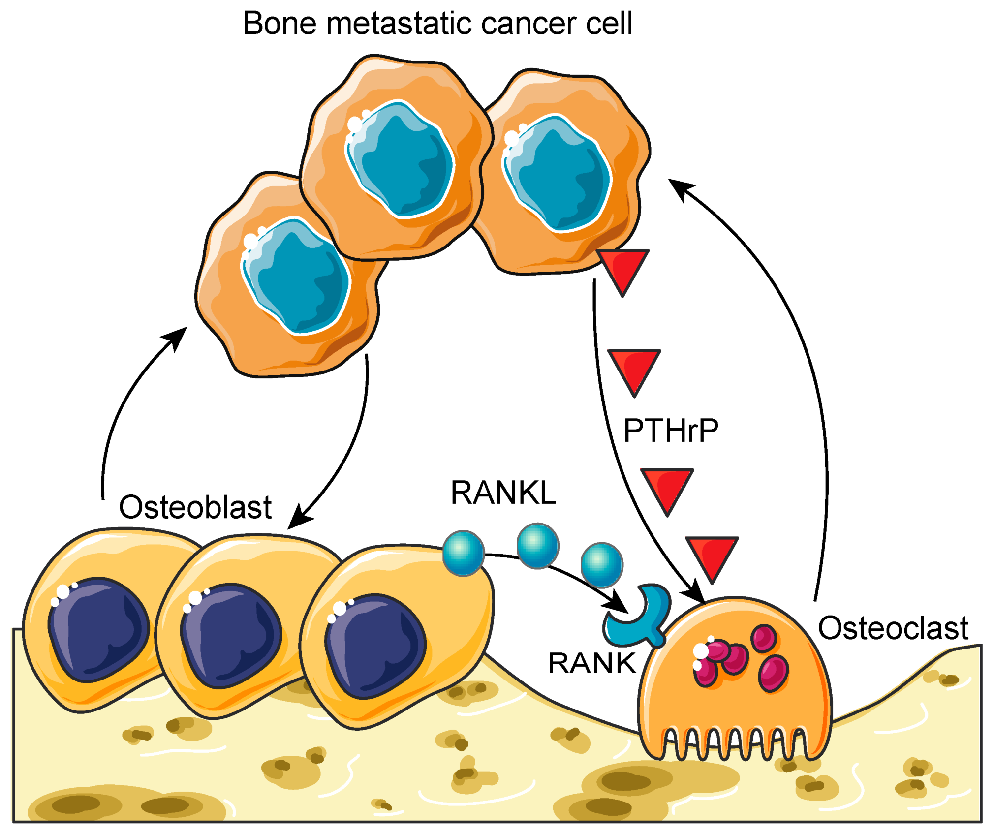
2. The Biology of the Vicious Cycle of Bone Metastases

Although personalized medicine strategies continue to be adopted in the clinic, the tumor phenotype at the primary site typically dictates the treatment regimen. However, these conventional treatment strategies usually fail to eradicate metastases. This leads to a more aggressive combination treatment, including chemotherapies, radiotherapies, immunotherapies, and/or targeted therapies, for cancer patients with metastases. Importantly, when considering the treatment of bone metastasis, the dynamics of bone turnover also needs to be considered during treatment planning.
Unlike other organs, bone is continuously renewed throughout life to maintain its structural integrity. Bone is composed of three parts: compact bone, trabecular bone, and bone marrow. Compact bone is a hard, solid bone tissue and forms the outside layer of bone. Trabecular bone (or spongy bone) and bone marrow are found in the inside of bones. A part of trabecular bone eventually converts into compact bone. The bone marrow is composed of two distinct stem cell lineages, cells of hematopoietic origin and those of mesenchymal origin. Hematopoietic stem cells (HSCs) give rise to all blood cell types, including macrophages that differentiate into osteoclasts, while mesenchymal stem cells (MSCs) are responsible for the generation of stromal cells, osteoblasts, and osteocytes [36]. Additionally, these cells of mesenchymal origin are crucial for trabecular bone development. Interestingly, these two cell lineages interact with each other to maintain each other’s functions. For example, osteoblasts serve as the microenvironment for HSCs, or the HSC niche [37,38,39,40,41,42], while HSCs support osteoblastic differentiation to establish the HSC niche [43]. Another example is that osteoblasts are responsible for the activation of osteoclasts [44], while these activated osteoclasts resorb the bone matrix to create the space for osteoblasts to form new bone [45]. As a result, compact bone, trabecular bone, and bone marrow crosstalk to maintain healthy bone development. This process, called bone remodeling, is a delicate balance that is often exploited by cancer cells that successfully colonize the bone [46].
Since Paget’s seed and soil theory was proposed over a century ago [47], efforts have been made to understand why particular cancers prefer specific organs over others. Although blood flow and anatomical structure are considered as among the major contributing factors to the development of bone metastasis [48,49], proper physiological mechanisms of why particular types of cancers disseminate to the bone have not been fully uncovered. Additionally, recent evidence suggests that the establishment of organ-specific metastases is the result of not only the passive reception of circulating tumor cells through blood flow and anatomical structure but also the fact that the bone microenvironment selectively and actively recruits these circulating tumor cells [50]. This indicates that the interactions between the microenvironment of the bone marrow and bone metastatic cancer cells are crucial for the bone metastatic progression process.
Once these tumor cells have effectively seeded on the bone marrow, they begin to proliferate and interact with the cells involved in bone remodeling (e.g., osteoclasts, osteoblasts, and osteocytes) through paracrine and juxtacrine signaling events. This interaction creates an imbalance in the normal bone remodeling process in what has been termed the vicious cycle of bone metastases (Figure 1) [51]. Recent studies have revealed that bone metastatic prostate cancer cells hijack the interaction between the cells of hematopoietic and mesenchymal lineages, which is important for maintaining healthy bone remodeling, to establish metastatic growth within the marrow. For example, bone metastatic prostate cancer cells target the osteoblastic HSC niche during their dissemination to the bone and compete for occupancy of the HSC niche [52,53]. These observations are consistent with previous research that has demonstrated that metastatic colonization is frequently observed in bones that contain red marrow, where blood cell formation and bone formation are active (e.g., the axial skeleton, vertebrae, ribs, and the pelvis) [54].
Additionally, once the vicious cycle has been initiated, bone metastases may present as osteolytic (bone destructive), osteoblastic (bone forming), or mixed metastases, depending on how the infiltrating cancer cells have exploited the normal bone remodeling mechanisms. Although there are osteolytic- and osteoblastic-only bone metastases, most metastases arising from solid tumors have a heterogenous phenotype [55]. Osteolytic bone metastatic lesions are characterized by the destruction of normal bone formation, primarily mediated by the hyper-activation of osteoclasts (multinucleated cells that resorb the bone matrix and develop from a monocyte–macrophage lineage) [56]. In this destructive mechanism, bone metastatic cells induce the release of parathyroid-hormone-related peptide (PTHrP), which is known to activate osteoclasts and induce bone resorption [57,58]. Conversely, osteoblastic bone metastatic lesions are characterized by the deposition of new bone. Although the mechanisms of osteoblastic bone metastases are still poorly understood, they are believed to occur through the hyper-activation of osteoblasts [59]. However, the newly formed bone matrix is poorly organized, weak, and fragile [60,61]. This leads to a lack of mechanical strength and frequent fracture [60,61]. Moreover, as osteoblasts continue to proliferate, they can inadvertently cause increased bone resorption, since they release cytokines, such as RANKL, which stimulates osteoclast differentiation and activation [59,60,62].
Taken together, the bone marrow microenvironment, especially where active hematopoiesis and bone remodeling take place, may play a crucial role in the establishment and development of bone metastases, and therefore bone-marrow-microenvironment-targeting strategies have been used to treat bone metastatic disease.

3. Treatment of Bone Metastases

Current treatment options for bone metastasis include agents targeting bone remodeling (e.g., bisphosphonate and denosumab), hormone therapy, chemotherapy, external beam radiation therapy (EBRT), systemic radiotherapy, and surgical intervention [3,63,64]. Often, more than one intervention is used to suppress bone metastatic growth and maintain a patient’s quality of life [65]. Although EBRT, bisphosphonate, and denosumab can reduce the onset of the painful complications of bone metastasis [17,18,66,67,68,69,70,71], delivering EBRT to every bone metastatic lesion is not practical and bisphosphonate and denosumab fail to improve the overall survival of bone metastatic patients (since these therapies mainly target bone remodeling but not cancer cells within the bone). Because of these limitations, bone metastasis is currently considered a hard-to-treat disease. Therefore, significant effort has been expended to develop ways to use the systemic delivery of therapeutic radionuclides to every bone metastatic lesion so that bone metastatic cancer cells can be exposed to the cytocidal effects of radiation.

3.1. Radiopharmaceuticals for Bone Metastases

Radionuclides derive their cytotoxicity from the particles they release during radioactive decay, which has been considered a systemic radiotherapy for bone metastasis. These particles include auger electrons (e), beta minus (β) particles, and alpha (α++) particles (Table 1) [72].
Auger electrons are released from an atom that undergoes electron capture or internal conversion. These particles have low energy, travel a maximum distance of a micron in tissue and have moderately high linear energy transfer (LET), which allows for many destructive ionization events along the path traveled by the particles [73]. These properties require that the emitting atom be localized in close proximity to the cancer cell’s DNA to have a therapeutic effect [74,75].
Beta-minus-particle-emitting radionuclides decay by β emission. These negatively charged electrons have widely varying energies and path lengths, which may range from 0.05 keV to 2.3 MeV and from 0.05 mm to 12 mm, respectively. As these particles travel, they exhibit a lower LET than auger electrons. This results in few ionization events along the particles’ path. Their cytocidal effect is believed to occur through the particles’ ability to generate reactive oxygen species (ROS). In turn, these ROS generate single-stranded DNA breaks within the cancer cell. High concentrations of these β-particles are required to create enough single-stranded DNA breaks to overwhelm DNA damage repair mechanisms and yield a therapeutic benefit.
Alpha-particle-emitting radionuclides are, by far, the most cytocidal radionuclides that have been used as systemic radiotherapy [76,77]. These particles have mass and charge equivalent to those of a helium nucleus. Alpha-particles are emitted with an energy range of 5–9 MeV, travel short distances (equivalent to no more than 10 cell diameters), and have an LET that surpasses the LET of auger electrons to ensure a significant number of ionization events along the alpha-particle’s path through tissue. These properties make the therapeutic efficacy of these radionuclides less dependent on chemo- and radio-resistance mechanisms or hypoxia. Although their cytotoxic properties have been recognized for decades, they are only now achieving clinical translation due to successful research initiatives that have explored how to efficiently attach these α++-emitting radionuclides to targeting ligands such as antibodies and peptides for effective delivery to cancer cells.
Historically, the Food and Drug Administration (FDA) has approved beta-minus-particle-emitting radiopharmaceuticals as adjunct treatments for bone metastases because of their chemical properties that give them an affinity for the bone matrix [78,79,80,81]. These radiotherapies are either administered as an ionic salt solution or chelated to bone-seeking ligands, such as hydroxyethylidene diphosphonic acid (HEDP) or tetramethylene phosphonic acid (EDTMP) (Figure 2A); a brief review of each radionuclide is outlined below.
Phosphorus-32 and strontium-89: Phosphorus-32 (32P: t1/2 = 14.3 d; Eβmax = 1.71 MeV; maximum penetration in tissue (average) = 8 mm (3 mm)) is produced by the irradiation of sulfur-32 (32S) through neutron capture and is distributed as 32P-orthophosphate. It has been investigated as a bone-pain-palliating radiopharmaceutical for more than 50 years [82]. After injection, approximately 90% of the injected dose is known to be incorporated at sites of bone remodeling due to the radiopharmaceutical’s affinity for the hydroxyapatite within the bone matrix. In several studies, palliative response rates approached 80% [83,84,85] and were observed to last more than a year in patients who received multiple doses of the radiopharmaceutical. Unfortunately, due to the long particle path length and relatively high energy of the β-particle, pancytopenia was observed in most patients, resulting in disfavor of its clinical use [86,87]. Strontium-89 (89Sr: t1/2 = 50.5 d; Eβmax = 1.46 MeV; maximum penetration in tissue (average) = 6 mm (2.4 mm)) is produced by nuclear fission or through neutron capture and is distributed as 89SrCl2. Since 89Sr2+ ions exhibit similar chemical properties to Ca2+ ions, they are readily incorporated into the hydroxyapatite where active bone remodeling is occurring due to metastatic disease [78]. Typically, overall response rates, as defined by a reduction in bone pain, for patients receiving 89Sr therapy approached 80%, with the palliative effects lasting approximately 15 months [87,88]. Although significant changes in red blood cell counts are not observed with this therapy, a decrease in white blood cell counts and platelets has been observed in 80% of patients [89,90,91,92], but these toxicities are resolved within 4 months of treatment cessation.
Rhenium-186 and Rhenium-188: Rhenium-186 (186Re: t1/2 = 3.8 d; Eβmax = 1.07 MeV; maximum penetration in tissue (average) = 4.5 mm (1.1 mm)) is routinely produced in nuclear reactors by direct neutron activation of metallic-enriched 185Re. Rhenium-186 decays with a maximum beta energy of 1.07 MeV and a low abundance 137 keV gamma emission. It has a physical half-life of 89.3 h. Because of its maximum beta energy and particle path length, efforts have been made to determine its ability to treat bone metastasis, but since its chemical properties are not similar to those of calcium, it must be chelated to a bone-seeking ligand before injection. In the comparative study between the skeletal and soft-tissue uptake of 186Re-HEDP (0.13 GBq) and 153Sm-EDTMP (37 MBq/kg-body weight) in patients with confirmed bone metastasis, significantly less bone uptake was observed in patients receiving 186Re-HEDP therapy, while soft-tissue retention was comparable for both radiopharmaceuticals [78]. Despite this fact, several studies have demonstrated that more than 80% of patients receiving 186Re therapy reported an improvement in their bone pain [93,94,95,96,97]. Similar to other agents, thrombocytopenia was observed to be a major but reversible side effect of treatment [98,99]. Rhenium-188 (188Re: t1/2 = 0.7 d; Eβmax = 2.12 MeV; maximum penetration in tissue (average) = 10.4 mm (3.1 mm)), although not approved for clinical use, has also been investigated as an agent for bone pain management, since it can be produced and shipped to clinical sites as a 188W/188Re generator [100]. When chelated to HEDP, it demonstrated therapeutic efficacy, and repeated administrations improved progression-free and overall survival [101]. Additionally, it was observed that only 63% of the patients receiving 188Re-HEDP therapy reported grade I thrombocytopenia, 3% reported Grade II thrombocytopenia, and 3% of the patients reported grade I leukopenia, which were comparable for patients treated with 186Re-HEDP or 153Sm-EDTMP [101]. Furthermore, platelet and leukocyte counts returned to pretreatment levels within 3 months, suggesting that these toxicities are clinically manageable.
Samarium-153: Samarium-153 (153Sm: t1/2 = 1.9 d; Eβmax = 0.81 MeV; maximum penetration in tissue (average) = 2.5 mm (0.6 mm)), which is produced by the neutron irradiation of a samarium-152 target, has a half-life of 46.3 h and a range in bone of 1.7 mm [102]. To be effective, however, 153Sm needs to be chelated to a ligand such as EDTMP, which like other phosphonic acid complexes has a high affinity for skeletal tissue while being rapidly cleared from the blood and local soft tissue. This radiopharmaceutical has shown clinical benefit in several clinical trials, with the majority of patients experiencing pain relief [103]. In one study involving 100 patients with confirmed metastatic disease, subjects received 18–37 MBq/kg of 153Sm-EDTMP and were followed for several weeks after treatment [102]. Nearly 70% of the patients experienced symptomatic relief of their bone pain. Myelotoxicity was the main side effect of treatment, with nearly 90% of the subjects experiencing grade II thrombocytopenia or leukopenia, which resolved approximately 2 months after therapy. While several patients experienced grade III/IV myelotoxicity, these patients were observed to have a depleted hematopoietic reserve because of previous therapeutic interventions.
Lutetium-177: Lutetium-177 (177Lu: t1/2 = 6.7 d; Eβmax = 0.50 MeV; maximum penetration in tissue (average) = 2.2 mm (0.67 mm)) is produced by neutron irradiation of 176Lu or 176Yb targets. Although several cancer-targeting agents, such as Lutathera® and 117Lu-PSMA-617, have been used to deliver targeted radiation therapy to cancer cells expressing specific cancer cell antigens [104], additional studies have sought to use the radioactive decay properties of 177Lu for treating bone metastasis. Several groups have investigated the utility of 177Lu-EDTMP as a bone-seeking radiopharmaceutical since the energy of the emitted β is low enough to reduce bone marrow suppression, which has consistently been a major disadvantage of other β-emitting bone-seeking agents [105,106]. Recently, the use of low- and high-dose 177Lu-EDTMP was examined in a phase II study in patients with metastatic breast or prostate cancer [107]. In this study, subjects received either a low (0.13 GBq) or a high (0.26 GBq) dose of 177Lu-EDTMP and were followed for 16 weeks post-treatment. Based upon pain assessment scores, the overall response rate was 86%. Additionally, treatment-related toxicity was evaluated in all patients. Grade I/II hematological toxicities were observed in 34% of the subjects, while grade III/IV toxicities were observed in 23% of the study participants. However, the observed toxicities were not significantly different in either the low- or the high-dose cohort, and this finding prompted the authors to conclude that 177Lu-EDTMP is safe and effective as a treatment for bone pain palliation. More recently, several groups investigated whether using a bisphosphonate to deliver the cytocidal radiation released by 177Lu to the metastatic deposit in bone would provide greater relief. While many bisphosphonates were studied, zoledronic acid was chosen for further study since it had significantly higher incorporation into hydroxyapatite [108,109]. Consequently, several groups studied 177Lu-DOTA-zoledronic acid (177Lu-DOTA-ZOL) and demonstrated that it has pharmacokinetics comparable to those of 177Lu-EDTMP [110,111]. These studies also revealed higher absorption of the dose by the trabecular bone in patients who received 177Lu-DOTA-ZOL vs. 177Lu-EDTMP, but the dose absorbed by critical organs was much lower for the former radiopharmaceutical. Recently, the safety and efficacy of 177Lu-DOTA-ZOL in 40 patients with metastatic bone disease was evaluated [112]. Eligible subjects received 0.13 GBq of 177Lu-DOAT-ZOL at monthly intervals and then were monitored for a 12-week period after therapy. Based upon criteria such as pain palliation, an overall response rate of 90% was observed. However, unlike patients receiving 177Lu-EDTMP, grade III/IV hematological toxicities were not observed, although several patients did experience grade II anemia. Moreover, renal toxicity and hyper-calcemia, which can be complications of bisphosphonate administration, were not observed in study participants. Interestingly, pain palliation was observed within 7 days of treatment, with relief lasting 10 months, which is more than twice that experienced by patients receiving 177Lu-EDTMP.
Not only beta-minus-particle-emitting radiopharmaceuticals but also alpha-particle-emitting radiopharmaceuticals have been used as a treatment modality for bone metastatic disease. Radium-223 dichloride (223RaCl2: T1/2 = 11.4 d; Eαmax = 6–7 MeV) is a water-soluble salt that was approved nearly a decade ago by the FDA for the treatment of bone metastasis associated with metastatic prostate cancer. Similar to calcium ions, this alkaline earth ion accumulates in bone, where it decays through seven daughter radionuclides, while releasing four α++-particles and two β-particles, generating approximately 30 MeV of total kinetic energy, which is deposited in the surrounding bone cancer microenvironment [67,68,69,78,113]. This large energy deposition is believed to generate irreparable double-stranded DNA breaks within the DNA of tumor cells that have localized in the bone, causing cancer cell death [114,115,116,117]. This strategy has yielded clinical success. For example, in the ALSYMPCA trial (NCT00699751), which was a randomized, double-blind, placebo-controlled phase III trial that investigated the role of 223RaCl2 in metastatic castration-resistant prostate cancer patients (n = 921), the median overall survival of patients treated with 223RaCl2 (n = 614, 14.9 months) significantly improved compared with those treated with a placebo (n = 307, 11.3 months) (hazard ratio, 0.70; 95% confidence interval, 0.58 to 0.83; p < 0.001) [66]. Moreover, patients treated with 223RaCl2 experienced decreased pain levels that correlated with increased overall survival [118]. Diarrhea, nausea, and vomiting were the most common side effects reported by patients. Additionally, pancytopenia was observed in many patients with grade III/IV thrombocytopenia, and neutropenia was observed in patients with compromised bone marrow function [119]. Since those initial trials, the use of 223RaCl2 therapy in combination with other agents using a strategy that has proven successful with non-radioactive therapeutics has been sought [120]. For example, a post hoc exploratory analysis of an international, early access, open-label, single-arm phase IIIb trial of the testing efficacy of 223RaCl2 in metastatic castration-resistant prostate cancer patients following the ALSYMPCA trial revealed that a combination of 223RaCl2 and androgen deprivation therapies (ADTs; abiraterone or enzalutamide) extended the patients’ overall survival compared to therapy with 223RaCl2 alone [118]. However, a recent multinational, multicenter, randomized, double-blind, placebo-controlled phase III trial of the combination of abiraterone and 223RaCl2 in patients with metastatic castration-resistant prostate cancer (NCT02043678) not only failed to improve skeletal event-free survival but also increased the frequency of bone fractures compared to the placebo [121]. These contradictory results suggest that additional prospective studies are needed and caution is warranted when choosing an effective combination strategy that involves the use of ADTs and 223RaCl2 therapy for treating prostate cancer patients with bone metastasis.

3.2. Targeted Radiopharmaceuticals for Bone Metastases

To date, the strategy for integrating systemic radiotherapies into the treatment plans of cancer patients with bone metastases has relied on bone metabolism to target bone metastatic disease. While successful, the results of this strategy have been primarily palliative, since the bone metastatic cancer cells are not specifically targeted by the radiopharmaceutical. As a result, efforts are being made to expand this strategy by developing agents that can selectively target biomarkers that are over-expressed on the cancer cells within the bone metastatic microenvironment, with the goal of improving therapeutic efficacy and patient outcomes. For example, one such strategy that has been successful in the treatment of metastatic prostate cancer has been the development of radiotherapies that specifically target prostate-specific membrane antigen (PSMA) [122]. PSMA is known to be highly expressed on prostate cancer cells at the primary tumor and within visceral and bone metastases [123]. PSMA-617, a small molecule that binds to PSMA with high affinity, was designed to target PSMA-expressing prostate cancer cells (Figure 2B) [124]. In a recent single-arm, single-center, phase II trial, 177Lu-PSMA-617 was administered to men with metastatic castration-resistant prostate cancer (n =30) [125]. After four cycles of radiotherapy, 57% of these patients experienced minimal toxicity and achieved a greater-than-50% reduction in prostate-specific antigen (PSA) levels, which is one of the clinical surrogate markers for prostate cancer treatment response [125]. Based on these promising results, an international prospective open-label randomized phase III trial comparing the treatment efficacy between 177Lu-PSMA-617 and the best standard of care in men with metastatic castration-resistant prostate cancer (VISION trial, NCT03511664) is currently underway [126], and as of 23 March 2021, the initial result that 177Lu-PSMA-617 significantly improves the overall survival and radiographic progression-free survival of PSMA-positive metastatic castration-resistant prostate cancer patients was announced (https://www.novartis.com/news/media-releases/novartis-announces-positive-result-phase-iii-study-radioligand-therapy-177lu-psma-617-patients-advanced-prostate-cancer, (accessed on 1 April 2021)).
Despite the promise of 177Lu-PSMA-617 therapy, approximately 30% of patients do not respond to this treatment [127]. These observations have led to the investigation of the use of 225Ac-PSMA-617 as an alternative alpha-particle-emitting radiotherapy for refractory patients [127,128,129]. Actinium-225 (225Ac: t1/2 = 10 d; Eαmax = 6–8 MeV) is a radioactive metal of the actinide series. Similar to 223RaCl2, 225Ac has a relatively long half-life and emits four α++- and two β-particles per nuclear decay. However, unlike 223RaCl2, 225Ac can be linked to a variety of targeting ligands [130,131,132,133,134,135,136,137]. In the early stage of the development of 225Ac-PSMA-617, two patients received this therapy [127]. The first patient exhausted conventional chemotherapies, bone remodeling therapy, ADTs, and six cycles of 223RaCl2 therapy. The patient received three cycles of approximately 10 MBq (100 kBq/kg body weight) of 225Ac-PSMA-617. After 8 weeks, all visible visceral and bone metastases had decreased to a size that was below the limit of detection of clinical imaging scanners, while PSA levels had decreased from 3000 to 0.26 ng/mL. Similar to the first patient, the second patient was refractory to conventional chemotherapies and ADTs and failed to respond to several cycles of 177Lu-PSMA-617 therapy. This patient received three cycles of 225Ac-PSMA-617 therapy (100 kBq/kg body weight) at bimonthly intervals. After completing the three cycles, the patient experienced complete remission, as indicated by the results of the restaging PSMA-PET/CT scans (Figure 3). Similar to the first patient, no relevant hematological toxicities were observed, although both subjects did experience moderate xerostomia.
In additional studies by this same group, chemotherapy-naive prostate cancer patients with extensive bone and lymph node metastases were selected to receive 225Ac-PSMA-617 therapy [138]. All patients (n = 17) had failed prior treatments, which included radical prostatectomy, EBRT, ADT, brachytherapy, and 177Lu-PSMA-617 therapy. Patients received between two and six cycles of 225Ac-PSMA-617, with an average administered activity range between 4 and 13 MBq. Following treatment, 82% of the patients experienced a ≥90% reduction in PSA levels. Moreover, PSMA PET/CT revealed that 65% of the treated patients demonstrated a complete absence of metastatic lesions at the sites of metastases, previously identified on baseline imaging scans [138]. Similar to the initial two patients, no acute toxicities were experienced by any of the surviving patients, though all patients did experience xerostomia, which prompted additional investigations into the most efficient ways to salvage salivary gland function [139,140].
While a recent report of a patient achieving remission lasting longer than 5 years was published in the literature [141], these results should be tempered by recent observations that 17% of patients, many with bone metastasis, do not respond to 225Ac-PSMA-617 therapy despite sufficient tumor uptake on PSMA-PET/CT. Investigations using CT-guided biopsy and targeted next-generation sequencing have revealed a total of 15 mutations in DNA-damage-repair-associated genes, which suggests that patients harboring these mutations may need a treatment strategy that combines 225Ac-PSMA-617 with chemotherapies that target DNA damage repair pathways to achieve maximum clinical benefit [142].
Although 225Ac-PSMA-617 is a promising treatment agent for metastatic castration-resistant prostate cancer, PSMA-targeted agents (e.g., antibody, nanobody, small molecule) labeled with other alpha-particle-emitting radiopharmaceuticals have also been explored in both preclinical and clinical settings (Table 2). Since PSMA is expressed by not only bone metastatic prostate cancer cells but also those at the primary site and soft-tissue metastatic sites, this strategy is not specific for bone metastases. However, a majority of the patients with metastatic castration-resistant prostate cancer experience bone metastases during the course of disease progression. Therefore, the development of PSMA-targeted alpha-particle-emitting radiopharmaceuticals is crucial if eradicating bone metastatic prostate cancer is to be an achievable clinical goal. Further studies, including the assessments of their clinical efficacy and safety, are clearly warranted.

4. Conclusions

Bone metastasis creates enormous physical, emotional, and financial burdens for cancer patients and society; it is a significant contributor to cancer mortality rates. Systemically delivered radionuclides have been explored as potential therapies for bone metastasis. Initially, β-emitting radionuclides were used because they could be delivered easily to sites of active bone remodeling. However, their efficacy has been limited to pain mitigation. Recently, systemically delivered 223RaCl2, which decays through α++-particle emission, has been observed to extend the overall survival of men with metastatic prostate cancer. These results have renewed interest in alpha-particle-emitting radiotherapies, and new molecularly targeted strategies to deliver this highly cytotoxic radiation directly to bone metastatic cancer cells within the bone marrow microenvironment are being investigated. Using this strategy, 225Ac-PSMA-617, which has been an effective treatment for men with metastatic prostate cancer, which was previously believed to be refractory to conventional chemotherapies and standard of care radiotherapy, has been developed. While further clinical evaluation is warranted, current data suggest that targeted alpha-particle-emitting radiopharmaceuticals used alone or in concert with current standard treatments may lead to much-needed improvements in the clinical management of cancer bone metastasis.

Author Contributions

Conceptualization, T.J.W. and Y.S.; writing—original draft preparation, C.M.P., T.J.W., and Y.S.; writing—review and editing, T.J.W. and Y.S.; supervision, T.J.W. and Y.S.; and funding acquisition, T.J.W. and Y.S. All authors have read and agreed to the published version of the manuscript.

Funding

This research was funded by the National Cancer Institute, R01-CA238888; R44-CA203184; R21-CA227709; and P30-CA012197; the U.S. Department of Defense, W81XWH-17-1-0541; W81XWH-19-1-0045; and W81XWH-19-1-0046; and METAvivor, METAvivor Research Award. The APC was funded by W81XWH-19-1-0045.

Institutional Review Board Statement

Not applicable.

Informed Consent Statement

Not applicable.

Data Availability Statement

Not applicable.

Acknowledgments

This work is directly supported by the National Cancer Institute (R01-CA238888, Y.S.; R44-CA203184, Y.S.; and R21-CA227709, T.J.W.), the U.S. Department of Defense (W81XWH-17-1-0541, Y.S.; W81XWH-19-1-0045, Y.S.; and W81XWH-19-1-0046, T.J.W.), METAvivor (METAvivor Research Award, Y.S.), and the Wake Forest Baptist Comprehensive Cancer Center Internal Pilot Funding (Y.S.). This work is also supported by the National Cancer Institute’s Cancer Center Support Grant award number P30-CA012197, issued to the Wake Forest Baptist Comprehensive Cancer Center. The content is solely the responsibility of the authors and does not necessarily represent the official views of the National Cancer Institute and Department of Defense.

Conflicts of Interest

Y.S. has received research funding from TEVA Pharmaceuticals, but not relevant to this study. No conflict of interest exists for the remaining authors.

References

  1. Seyfried, T.N.; Huysentruyt, L.C. On the origin of cancer metastasis. Crit. Rev. Oncog. 2013, 18, 43–73. [Google Scholar] [CrossRef] [PubMed] [Green Version]
  2. Macedo, F.; Ladeira, K.; Pinho, F.; Saraiva, N.; Bonito, N.; Pinto, L.; Goncalves, F. Bone Metastases: An Overview. Oncol. Rev. 2017, 11, 321. [Google Scholar] [PubMed]
  3. Selvaggi, G.; Scagliotti, G.V. Management of bone metastases in cancer: A review. Crit. Rev. Oncol. Hematol. 2005, 56, 365–378. [Google Scholar] [CrossRef] [PubMed]
  4. Hagiwara, M.; Delea, T.E.; Chung, K. Healthcare costs associated with skeletal-related events in breast cancer patients with bone metastases. J. Med. Econ. 2014, 17, 223–230. [Google Scholar] [CrossRef]
  5. Barlev, A.; Song, X.; Ivanov, B.; Setty, V.; Chung, K. Payer costs for inpatient treatment of pathologic fracture, surgery to bone, and spinal cord compression among patients with multiple myeloma or bone metastasis secondary to prostate or breast cancer. J. Manag. Care Pharm. 2010, 16, 693–702. [Google Scholar] [CrossRef] [PubMed]
  6. Delea, T.; Langer, C.; McKiernan, J.; Liss, M.; Edelsberg, J.; Brandman, J.; Sung, J.; Raut, M.; Oster, G. The cost of treatment of skeletal-related events in patients with bone metastases from lung cancer. Oncology 2004, 67, 390–396. [Google Scholar] [CrossRef]
  7. Delea, T.; McKiernan, J.; Brandman, J.; Edelsberg, J.; Sung, J.; Raut, M.; Oster, G. Retrospective study of the effect of skeletal complications on total medical care costs in patients with bone metastases of breast cancer seen in typical clinical practice. J. Support. Oncol. 2006, 4, 341–347. [Google Scholar]
  8. Delea, T.E.; McKiernan, J.; Brandman, J.; Edelsberg, J.; Sung, J.; Raut, M.; Oster, G. Impact of skeletal complications on total medical care costs among patients with bone metastases of lung cancer. J. Thorac. Oncol. 2006, 1, 571–576. [Google Scholar] [CrossRef]
  9. Lage, M.J.; Barber, B.L.; Harrison, D.J.; Jun, S. The cost of treating skeletal-related events in patients with prostate cancer. Am. J. Manag. Care 2008, 14, 317–322. [Google Scholar]
  10. Krupski, T.L.; Foley, K.A.; Baser, O.; Long, S.; Macarios, D.; Litwin, M.S. Health care cost associated with prostate cancer, androgen deprivation therapy and bone complications. J. Urol. 2007, 178, 1423–1428. [Google Scholar] [CrossRef]
  11. Groot, M.T.; Boeken Kruger, C.G.; Pelger, R.C.; Uyl-de Groot, C.A. Costs of prostate cancer, metastatic to the bone, in the Netherlands. Eur. Urol. 2003, 43, 226–232. [Google Scholar] [CrossRef]
  12. Duran, I.; Garzon, C.; Sanchez, A.; Garcia-Carbonero, I.; Perez-Gracia, J.L.; Segui-Palmer, M.A.; Wei, R.; Restovic, G.; Gasquet, J.A.; Gutierrez, L. Cost analysis of skeletal-related events in Spanish patients with bone metastases from solid tumours. Clin. Transl. Oncol. 2014, 16, 322–329. [Google Scholar] [CrossRef] [Green Version]
  13. Hechmati, G.; Cure, S.; Gouepo, A.; Hoefeler, H.; Lorusso, V.; Luftner, D.; Duran, I.; Garzon-Rodriguez, C.; Ashcroft, J.; Wei, R.; et al. Cost of skeletal-related events in European patients with solid tumours and bone metastases: Data from a prospective multinational observational study. J. Med. Econ. 2013, 16, 691–700. [Google Scholar] [CrossRef] [PubMed]
  14. Tangirala, K.; Appukkuttan, S.; Simmons, S. Costs and Healthcare Resource Utilization Associated with Hospital Admissions of Patients with Metastatic or Nonmetastatic Prostate Cancer. Am. Health Drug Benef. 2019, 12, 306–312. [Google Scholar]
  15. Hadjidakis, D.J.; Androulakis, I.I. Bone remodeling. Ann. N. Y. Acad. Sci. 2006, 1092, 385–396. [Google Scholar] [CrossRef] [PubMed]
  16. Guise, T.A. The vicious cycle of bone metastases. J. Musculoskelet. Neuronal. Interact. 2002, 2, 570–572. [Google Scholar] [PubMed]
  17. Stopeck, A.T.; Lipton, A.; Body, J.J.; Steger, G.G.; Tonkin, K.; de Boer, R.H.; Lichinitser, M.; Fujiwara, Y.; Yardley, D.A.; Viniegra, M.; et al. Denosumab compared with zoledronic acid for the treatment of bone metastases in patients with advanced breast cancer: A randomized, double-blind study. J. Clin. Oncol. 2010, 28, 5132–5139. [Google Scholar] [CrossRef] [Green Version]
  18. Fizazi, K.; Carducci, M.; Smith, M.; Damiao, R.; Brown, J.; Karsh, L.; Milecki, P.; Shore, N.; Rader, M.; Wang, H.; et al. Denosumab versus zoledronic acid for treatment of bone metastases in men with castration-resistant prostate cancer: A randomised, double-blind study. Lancet 2011, 377, 813–822. [Google Scholar] [CrossRef] [Green Version]
  19. McCain, J. Drugs that offer a survival advantage for men with bone metastases resulting from castration-resistant prostate cancer: New and emerging treatment options. Pharm. Ther. 2014, 39, 130–143. [Google Scholar]
  20. Nguyen, N.C.; Shah, M.; Appleman, L.J.; Parikh, R.; Mountz, J.M. Radium-223 Therapy for Patients with Metastatic Castrate-Resistant Prostate Cancer: An Update on Literature with Case Presentation. Int. J. Mol. Imaging 2016, 2016, 2568031. [Google Scholar] [CrossRef] [PubMed]
  21. Vogelzang, N.J.; Coleman, R.E.; Michalski, J.M.; Nilsson, S.; O’Sullivan, J.M.; Parker, C.; Widmark, A.; Thuresson, M.; Xu, L.; Germino, J.; et al. Hematologic Safety of Radium-223 Dichloride: Baseline Prognostic Factors Associated with Myelosuppression in the ALSYMPCA Trial. Clin. Genitourin Cancer 2017, 15, 42–52. [Google Scholar] [CrossRef] [Green Version]
  22. Wadas, T.J.; Pandya, D.N.; Solingapuram Sai, K.K.; Mintz, A. Molecular targeted alpha-particle therapy for oncologic applications. AJR Am. J. Roentgenol. 2014, 203, 253–260. [Google Scholar] [CrossRef] [PubMed] [Green Version]
  23. Scheinberg, D.A.; McDevitt, M.R. Actinium-225 in targeted alpha-particle therapeutic applications. Curr. RadioPharm. 2011, 4, 306–320. [Google Scholar] [CrossRef] [PubMed] [Green Version]
  24. Yamazaki, H.; Nakamura, S.; Suzuki, G.; Yoshida, K.; Yoshioka, Y.; Koizumi, M.; Ogawa, K. Hypofractionated Radiotherapy for Localized Prostate Cancer: A Challenging Accelerated Hypofractionated Radiotherapy. Anticancer Res. 2015, 35, 5167–5177. [Google Scholar] [PubMed]
  25. Lewis, B.; Chalhoub, E.; Chalouhy, C.; Sartor, O. Radium-223 in Bone-Metastatic Prostate Cancer: Current Data and Future Prospects. Oncology (Williston Park) 2015, 29, 483–488. [Google Scholar] [PubMed]
  26. de Kruijff, R.M.; Wolterbeek, H.T.; Denkova, A.G. A Critical Review of Alpha Radionuclide Therapy-How to Deal with Recoiling Daughters? Pharmaceuticals (Basel) 2015, 8, 321–336. [Google Scholar] [CrossRef]
  27. Guerard, F.; Barbet, J.; Chatal, J.F.; Kraeber-Bodere, F.; Cherel, M.; Haddad, F. Which radionuclide, carrier molecule and clinical indication for alpha-immunotherapy? Q. J. Nucl. Med. Mol. Imaging 2015, 59, 161–167. [Google Scholar]
  28. McGann, S.; Horton, E.R. Radium-223 dichloride: A novel treatment option for castration-resistant prostate cancer patients with symptomatic bone metastases. Ann. Pharmacother. 2015, 49, 469–476. [Google Scholar] [CrossRef]
  29. Qi, W.X.; Fu, S.; Zhang, Q.; Guo, X.M. Charged particle therapy versus photon therapy for patients with hepatocellular carcinoma: A systematic review and meta-analysis. Radiother. Oncol. 2015, 114, 289–295. [Google Scholar] [CrossRef] [PubMed]
  30. Buroni, F.E.; Persico, M.G.; Pasi, F.; Lodola, L.; Nano, R.; Aprile, C. Radium-223: Insight and Perspectives in Bone-metastatic Castration-resistant Prostate Cancer. Anticancer Res. 2016, 36, 5719–5730. [Google Scholar] [CrossRef] [Green Version]
  31. Cordier, D.; Krolicki, L.; Morgenstern, A.; Merlo, A. Targeted Radiolabeled Compounds in Glioma Therapy. Semin. Nucl. Med. 2016, 46, 243–249. [Google Scholar] [CrossRef]
  32. Ettari, R.; Previti, S.; Bitto, A.; Grasso, S.; Zappala, M. Immunoproteasome-Selective Inhibitors: A Promising Strategy to Treat Hematologic Malignancies, Autoimmune and Inflammatory Diseases. Curr. Med. Chem. 2016, 23, 1217–1238. [Google Scholar] [CrossRef]
  33. Sgouros, G.; Bodei, L.; McDevitt, M.R.; Nedrow, J.R. Radiopharmaceutical therapy in cancer: Clinical advances and challenges. Nat. Rev. Drug Discov. 2020, 19, 589–608. [Google Scholar] [CrossRef]
  34. Tafreshi, N.K.; Doligalski, M.L.; Tichacek, C.J.; Pandya, D.N.; Budzevich, M.M.; El-Haddad, G.; Khushalani, N.I.; Moros, E.G.; McLaughlin, M.L.; Wadas, T.J.; et al. Development of Targeted Alpha Particle Therapy for Solid Tumors. Molecules 2019, 24, 4314. [Google Scholar] [CrossRef] [Green Version]
  35. Schaue, D.; McBride, W.H. Opportunities and challenges of radiotherapy for treating cancer. Nat. Rev. Clin. Oncol. 2015, 12, 527–540. [Google Scholar] [CrossRef]
  36. Laurenti, E.; Gottgens, B. From haematopoietic stem cells to complex differentiation landscapes. Nature 2018, 553, 418–426. [Google Scholar] [CrossRef]
  37. Shiozawa, Y.; Taichman, R.S. Getting blood from bone: An emerging understanding of the role that osteoblasts play in regulating hematopoietic stem cells within their niche. Exp. Hematol. 2012, 40, 685–694. [Google Scholar] [CrossRef] [Green Version]
  38. Galan-Diez, M.; Kousteni, S. The osteoblastic niche in hematopoiesis and hematological myeloid malignancies. Curr. Mol. Biol. Rep. 2017, 3, 53–62. [Google Scholar] [CrossRef] [PubMed]
  39. Yin, T.; Li, L. The stem cell niches in bone. J. Clin. Investig. 2006, 116, 1195–1201. [Google Scholar] [CrossRef]
  40. Askmyr, M.; Sims, N.A.; Martin, T.J.; Purton, L.E. What is the true nature of the osteoblastic hematopoietic stem cell niche? Trends Endocrinol. Metab. 2009, 20, 303–309. [Google Scholar] [CrossRef]
  41. Szade, K.; Gulati, G.S.; Chan, C.K.F.; Kao, K.S.; Miyanishi, M.; Marjon, K.D.; Sinha, R.; George, B.M.; Chen, J.Y.; Weissman, I.L. Where Hematopoietic Stem Cells Live: The Bone Marrow Niche. Antioxid. Redox Signal. 2018, 29, 191–204. [Google Scholar] [CrossRef]
  42. Pinho, S.; Frenette, P.S. Haematopoietic stem cell activity and interactions with the niche. Nat. Rev. Mol. Cell Biol. 2019, 20, 303–320. [Google Scholar] [CrossRef]
  43. Jung, Y.; Song, J.; Shiozawa, Y.; Wang, J.; Wang, Z.; Williams, B.; Havens, A.; Schneider, A.; Ge, C.; Franceschi, R.T.; et al. Hematopoietic stem cells regulate mesenchymal stromal cell induction into osteoblasts thereby participating in the formation of the stem cell niche. Stem Cells 2008, 26, 2042–2051. [Google Scholar] [CrossRef] [PubMed] [Green Version]
  44. Chen, X.; Wang, Z.; Duan, N.; Zhu, G.; Schwarz, E.M.; Xie, C. Osteoblast-osteoclast interactions. Connect. Tissue Res. 2018, 59, 99–107. [Google Scholar] [CrossRef]
  45. Sims, N.A.; Martin, T.J. Coupling the activities of bone formation and resorption: A multitude of signals within the basic multicellular unit. Bonekey Rep. 2014, 3, 481. [Google Scholar] [CrossRef] [PubMed] [Green Version]
  46. Vessella, R.L.; Corey, E. Targeting factors involved in bone remodeling as treatment strategies in prostate cancer bone metastasis. Clin. Cancer Res. 2006, 12, 6285s–6290s. [Google Scholar] [CrossRef] [Green Version]
  47. Paget, S. The distribution of secondary growths in cancer of the breast. 1889. Cancer Metastasis Rev. 1989, 8, 98–101. [Google Scholar]
  48. Raubenheimer, E.J.; Noffke, C.E. Pathogenesis of bone metastasis: A review. J. Oral. Pathol. Med. 2006, 35, 129–135. [Google Scholar] [CrossRef]
  49. Guise, T.A.; Mundy, G.R. Cancer and bone. Endocr. Rev. 1998, 19, 18–54. [Google Scholar]
  50. Chantrain, C.F.; Feron, O.; Marbaix, E.; DeClerck, Y.A. Bone marrow microenvironment and tumor progression. Cancer Microenviron. 2008, 1, 23–35. [Google Scholar] [CrossRef] [Green Version]
  51. Cook, L.M.; Shay, G.; Araujo, A.; Lynch, C.C. Integrating new discoveries into the “vicious cycle” paradigm of prostate to bone metastases. Cancer Metastasis Rev. 2014, 33, 511–525. [Google Scholar] [CrossRef] [PubMed] [Green Version]
  52. Shiozawa, Y.; Pedersen, E.A.; Havens, A.M.; Jung, Y.; Mishra, A.; Joseph, J.; Kim, J.K.; Patel, L.R.; Ying, C.; Ziegler, A.M.; et al. Human prostate cancer metastases target the hematopoietic stem cell niche to establish footholds in mouse bone marrow. J. Clin. Investig. 2011, 121, 1298–1312. [Google Scholar] [CrossRef] [Green Version]
  53. Shiozawa, Y.; Berry, J.E.; Eber, M.R.; Jung, Y.; Yumoto, K.; Cackowski, F.C.; Yoon, H.J.; Parsana, P.; Mehra, R.; Wang, J.; et al. The marrow niche controls the cancer stem cell phenotype of disseminated prostate cancer. Oncotarget 2016, 7, 41217–41232. [Google Scholar] [CrossRef] [Green Version]
  54. Hage, W.D.; Aboulafia, A.J.; Aboulafia, D.M. Incidence, location, and diagnostic evaluation of metastatic bone disease. Orthop. Clin. N. Am. 2000, 31, 515–528. [Google Scholar] [CrossRef]
  55. Mundy, G.R. Metastasis to bone: Causes, consequences and therapeutic opportunities. Nat. Rev. Cancer 2002, 2, 584–593. [Google Scholar] [CrossRef]
  56. Taube, T.; Elomaa, I.; Blomqvist, C.; Beneton, M.N.; Kanis, J.A. Histomorphometric evidence for osteoclast-mediated bone resorption in metastatic breast cancer. Bone 1994, 15, 161–166. [Google Scholar] [CrossRef]
  57. Martin, T.J. Osteoblast-derived PTHrP is a physiological regulator of bone formation. J. Clin. Investig. 2005, 115, 2322–2324. [Google Scholar] [CrossRef] [Green Version]
  58. Iwamura, M.; di Sant’Agnese, P.A.; Wu, G.; Benning, C.M.; Cockett, A.T.; Deftos, L.J.; Abrahamsson, P.A. Immunohistochemical localization of parathyroid hormone-related protein in human prostate cancer. Cancer Res. 1993, 53, 1724–1726. [Google Scholar] [PubMed]
  59. Weilbaecher, K.N.; Guise, T.A.; McCauley, L.K. Cancer to bone: A fatal attraction. Nat. Rev. Cancer 2011, 11, 411–425. [Google Scholar] [CrossRef]
  60. Keller, E.T.; Brown, J. Prostate cancer bone metastases promote both osteolytic and osteoblastic activity. J. Cell Biochem. 2004, 91, 718–729. [Google Scholar] [CrossRef] [Green Version]
  61. Ganguly, S.S.; Li, X.; Miranti, C.K. The host microenvironment influences prostate cancer invasion, systemic spread, bone colonization, and osteoblastic metastasis. Front. Oncol. 2014, 4, 364. [Google Scholar] [CrossRef] [PubMed] [Green Version]
  62. Kohli, S.S.; Kohli, V.S. Role of RANKL-RANK/osteoprotegerin molecular complex in bone remodeling and its immunopathologic implications. Indian J. Endocrinol. Metab. 2011, 15, 175–181. [Google Scholar] [CrossRef]
  63. Pinski, J.; Dorff, T.B. Prostate cancer metastases to bone: Pathophysiology, pain management, and the promise of targeted therapy. Eur. J. Cancer 2005, 41, 932–940. [Google Scholar] [CrossRef]
  64. Lipton, A. Management of bone metastases in breast cancer. Curr. Treat. Options Oncol. 2005, 6, 161–171. [Google Scholar] [CrossRef]
  65. Woolf, D.K.; Padhani, A.R.; Makris, A. Assessing response to treatment of bone metastases from breast cancer: What should be the standard of care? Ann. Oncol. 2015, 26, 1048–1057. [Google Scholar] [CrossRef]
  66. Parker, C.; Nilsson, S.; Heinrich, D.; Helle, S.I.; O’Sullivan, J.M.; Fossa, S.D.; Chodacki, A.; Wiechno, P.; Logue, J.; Seke, M.; et al. Alpha emitter radium-223 and survival in metastatic prostate cancer. N. Engl. J. Med. 2013, 369, 213–223. [Google Scholar] [CrossRef] [Green Version]
  67. Badrising, S.K.; van der Noort, V.; Hamberg, P.; Coenen, J.L.; Aarts, M.J.; van Oort, I.M.; van den Eertwegh, A.J.; Los, M.; van den Berg, H.P.; Gelderblom, H.; et al. Enzalutamide as a Fourth- or Fifth-Line Treatment Option for Metastatic Castration-Resistant Prostate Cancer. Oncology 2016, 91, 267–273. [Google Scholar] [CrossRef]
  68. Vignani, F.; Bertaglia, V.; Buttigliero, C.; Tucci, M.; Scagliotti, G.V.; Di Maio, M. Skeletal metastases and impact of anticancer and bone-targeted agents in patients with castration-resistant prostate cancer. Cancer Treat. Rev. 2016, 44, 61–73. [Google Scholar] [CrossRef]
  69. Abou, D.S.; Ulmert, D.; Doucet, M.; Hobbs, R.F.; Riddle, R.C.; Thorek, D.L. Whole-Body and Microenvironmental Localization of Radium-223 in Naive and Mouse Models of Prostate Cancer Metastasis. J. Natl Cancer Inst. 2016, 108, djv380. [Google Scholar] [CrossRef] [Green Version]
  70. Laird, B.J.; Walley, J.; Murray, G.D.; Clausen, E.; Colvin, L.A.; Fallon, M.T. Characterization of cancer-induced bone pain: An exploratory study. Support. Care Cancer 2011, 19, 1393–1401. [Google Scholar] [CrossRef]
  71. De Felice, F.; Piccioli, A.; Musio, D.; Tombolini, V. The role of radiation therapy in bone metastases management. Oncotarget 2017, 8, 25691–25699. [Google Scholar] [CrossRef] [Green Version]
  72. Kassis, A.I.; Adelstein, S.J. Radiobiologic principles in radionuclide therapy. J. Nucl. Med. 2005, 46 (Suppl. 1), 4S–12S. [Google Scholar] [PubMed]
  73. Cole, A. Absorption of 20-eV to 50,000-eV electron beams in air and plastic. Radiat. Res. 1969, 38, 7–33. [Google Scholar] [CrossRef] [PubMed]
  74. Kassis, A.I.; Adelstein, S.J.; Haydock, C.; Sastry, K.S.; McElvany, K.D.; Welch, M.J. Lethality of Auger electrons from the decay of bromine-77 in the DNA of mammalian cells. Radiat. Res. 1982, 90, 362–373. [Google Scholar] [CrossRef]
  75. Kassis, A.I.; Sastry, K.S.; Adelstein, S.J. Kinetics of uptake, retention, and radiotoxicity of 125IUdR in mammalian cells: Implications of localized energy deposition by Auger processes. Radiat. Res. 1987, 109, 78–89. [Google Scholar] [CrossRef]
  76. Walicka, M.A.; Vaidyanathan, G.; Zalutsky, M.R.; Adelstein, S.J.; Kassis, A.I. Survival and DNA damage in Chinese hamster V79 cells exposed to alpha particles emitted by DNA-incorporated astatine-211. Radiat. Res. 1998, 150, 263–268. [Google Scholar] [CrossRef]
  77. Goddu, S.M.; Howell, R.W.; Rao, D.V. Cellular dosimetry: Absorbed fractions for monoenergetic electron and alpha particle sources and S-values for radionuclides uniformly distributed in different cell compartments. J. Nucl. Med. 1994, 35, 303–316. [Google Scholar] [PubMed]
  78. Choi, J.Y. Treatment of Bone Metastasis with Bone-Targeting Radiopharmaceuticals. Nucl. Med. Mol. Imaging 2018, 52, 200–207. [Google Scholar] [CrossRef]
  79. Paes, F.M.; Serafini, A.N. Systemic metabolic radiopharmaceutical therapy in the treatment of metastatic bone pain. Semin Nucl. Med. 2010, 40, 89–104. [Google Scholar] [CrossRef]
  80. Lepareur, N.; Lacoeuille, F.; Bouvry, C.; Hindre, F.; Garcion, E.; Cherel, M.; Noiret, N.; Garin, E.; Knapp, F.F.R., Jr. Rhenium-188 Labeled Radiopharmaceuticals: Current Clinical Applications in Oncology and Promising Perspectives. Front. Med. (Lausanne) 2019, 6, 132. [Google Scholar] [CrossRef] [Green Version]
  81. Alavi, M.; Omidvari, S.; Mehdizadeh, A.; Jalilian, A.R.; Bahrami-Samani, A. Metastatic Bone Pain Palliation using (177)Lu-Ethylenediaminetetramethylene Phosphonic Acid. World J. Nucl. Med. 2015, 14, 109–115. [Google Scholar] [CrossRef] [PubMed]
  82. Friedell, H.L.; Storaasli, J.P. The use of radioactive phosphorus in the treatment of carcinoma of the breast with widespread metastases to bone. Am. J. Roentgenol. Radium Ther. 1950, 64, 559–575. [Google Scholar] [PubMed]
  83. Maxfield, J.R., Jr.; Maxfield, J.G.; Maxfield, W.S. The use of radioactive phosphorus and testosterone in metastatic bone lesions from breast and prostate. South. Med. J. 1958, 51, 320–327. [Google Scholar] [CrossRef]
  84. Miller, A.D. Radiophosphorous (P32) treatment in carcinoma of the breast and prostate: Report of 39 cases. J. Am. Osteopath Assoc. 1974, 74, 217–222. [Google Scholar] [PubMed]
  85. Cheung, A.; Driedger, A.A. Evaluation of radioactive phosphorus in the palliation of metastatic bone lesions from carcinoma of the breast and prostate. Radiology 1980, 134, 209–212. [Google Scholar] [CrossRef]
  86. Silberstein, E.B. The treatment of painful osseous metastases with phosphorus-32-labeled phosphates. Semin. Oncol. 1993, 20 (Suppl. 2), 10–21. [Google Scholar]
  87. Nair, N. Relative efficacy of 32P and 89Sr in palliation in skeletal metastases. J. Nucl. Med. 1999, 40, 256–261. [Google Scholar]
  88. Blake, G.M.; Zivanovic, M.A.; McEwan, A.J.; Ackery, D.M. Sr-89 therapy: Strontium kinetics in disseminated carcinoma of the prostate. Eur. J. Nucl. Med. 1986, 12, 447–454. [Google Scholar] [CrossRef]
  89. Finlay, I.G.; Mason, M.D.; Shelley, M. Radioisotopes for the palliation of metastatic bone cancer: A systematic review. Lancet Oncol. 2005, 6, 392–400. [Google Scholar] [CrossRef]
  90. Silberstein, E.B.; Williams, C. Strontium-89 therapy for the pain of osseous metastases. J. Nucl. Med. 1985, 26, 345–348. [Google Scholar] [PubMed]
  91. Robinson, R.G.; Preston, D.F.; Spicer, J.A.; Baxter, K.G. Radionuclide therapy of intractable bone pain: Emphasis on strontium-89. Semin. Nucl. Med. 1992, 22, 28–32. [Google Scholar] [CrossRef]
  92. Mertens, W.C.; Stitt, L.; Porter, A.T. Strontium 89 therapy and relief of pain in patients with prostatic carcinoma metastatic to bone: A dose response relationship? Am. J. Clin. Oncol. 1993, 16, 238–242. [Google Scholar] [CrossRef]
  93. Maxon, H.R., 3rd; Schroder, L.E.; Thomas, S.R.; Hertzberg, V.S.; Deutsch, E.A.; Scher, H.I.; Samaratunga, R.C.; Libson, K.F.; Williams, C.C.; Moulton, J.S.; et al. Re-186(Sn) HEDP for treatment of painful osseous metastases: Initial clinical experience in 20 patients with hormone-resistant prostate cancer. Radiology 1990, 176, 155–159. [Google Scholar] [CrossRef]
  94. Giannakenas, C.; Kalofonos, H.P.; Apostolopoulos, D.J.; Zarakovitis, J.; Kosmas, C.; Vassilakos, P.J. Preliminary results of the use of Re-186-HEDP for palliation of pain in patients with metastatic bone disease. Am. J. Clin. Oncol. 2000, 23, 83–88. [Google Scholar] [CrossRef]
  95. Kolesnikov-Gauthier, H.; Carpentier, P.; Depreux, P.; Vennin, P.; Caty, A.; Sulman, C. Evaluation of toxicity and efficacy of 186Re-hydroxyethylidene diphosphonate in patients with painful bone metastases of prostate or breast cancer. J. Nucl. Med. 2000, 41, 1689–1694. [Google Scholar]
  96. Sciuto, R.; Tofani, A.; Festa, A.; Giannarelli, D.; Pasqualoni, R.; Maini, C.L. Short- and long-term effects of 186Re-1,1-hydroxyethylidene diphosphonate in the treatment of painful bone metastases. J. Nucl. Med. 2000, 41, 647–654. [Google Scholar]
  97. Quirijnen, J.M.; Han, S.H.; Zonnenberg, B.A.; de Klerk, J.M.; van het Schip, A.D.; van Dijk, A.; ten Kroode, H.F.; Blijham, G.H.; van Rijk, P.P. Efficacy of rhenium-186-etidronate in prostate cancer patients with metastatic bone pain. J. Nucl. Med. 1996, 37, 1511–1515. [Google Scholar]
  98. Maxon, H.R., 3rd; Schroder, L.E.; Hertzberg, V.S.; Thomas, S.R.; Englaro, E.E.; Samaratunga, R.; Smith, H.; Moulton, J.S.; Williams, C.C.; Ehrhardt, G.J.; et al. Rhenium-186(Sn)HEDP for treatment of painful osseous metastases: Results of a double-blind crossover comparison with placebo. J. Nucl. Med. 1991, 32, 1877–1881. [Google Scholar]
  99. Han, S.H.; de Klerk, J.M.; Tan, S.; van het Schip, A.D.; Derksen, B.H.; van Dijk, A.; Kruitwagen, C.L.; Blijham, G.H.; van Rijk, P.P.; Zonnenberg, B.A. The PLACORHEN study: A double-blind, placebo-controlled, randomized radionuclide study with (186)Re-etidronate in hormone-resistant prostate cancer patients with painful bone metastases. Placebo Controlled Rhenium Study. J. Nucl. Med. 2002, 43, 1150–1156. [Google Scholar]
  100. Liepe, K. (188)Re-HEDP therapy in the therapy of painful bone metastases. World J. Nucl. Med. 2018, 17, 133–138. [Google Scholar] [CrossRef]
  101. Liepe, K.; Kotzerke, J. A comparative study of 188Re-HEDP, 186Re-HEDP, 153Sm-EDTMP and 89Sr in the treatment of painful skeletal metastases. Nucl. Med. Commun. 2007, 28, 623–630. [Google Scholar] [CrossRef]
  102. Resche, I.; Chatal, J.F.; Pecking, A.; Ell, P.; Duchesne, G.; Rubens, R.; Fogelman, I.; Houston, S.; Fauser, A.; Fischer, M.; et al. A dose-controlled study of 153Sm-ethylenediaminetetramethylenephosphonate (EDTMP) in the treatment of patients with painful bone metastases. Eur. J. Cancer 1997, 33, 1583–1591. [Google Scholar] [CrossRef]
  103. Tian, J.H.; Zhang, J.M.; Hou, Q.T.; Oyang, Q.H.; Wang, J.M.; Luan, Z.S.; Chuan, L.; He, Y.J. Multicentre trial on the efficacy and toxicity of single-dose samarium-153-ethylene diamine tetramethylene phosphonate as a palliative treatment for painful skeletal metastases in China. Eur. J. Nucl. Med. 1999, 26, 2–7. [Google Scholar] [CrossRef] [PubMed]
  104. Yadav, M.P.; Ballal, S.; Sahoo, R.K.; Dwivedi, S.N.; Bal, C. Radioligand Therapy With (177)Lu-PSMA for Metastatic Castration-Resistant Prostate Cancer: A Systematic Review and Meta-Analysis. AJR Am. J. Roentgenol. 2019, 213, 275–285. [Google Scholar] [CrossRef] [PubMed]
  105. Chakraborty, S.; Das, T.; Banerjee, S.; Balogh, L.; Chaudhari, P.R.; Sarma, H.D.; Polyak, A.; Mathe, D.; Venkatesh, M.; Janoki, G.; et al. 177Lu-EDTMP: A viable bone pain palliative in skeletal metastasis. Cancer Biother. Radio Pharm. 2008, 23, 202–213. [Google Scholar] [CrossRef] [PubMed]
  106. Mathe, D.; Balogh, L.; Polyak, A.; Kiraly, R.; Marian, T.; Pawlak, D.; Zaknun, J.J.; Pillai, M.R.; Janoki, G.A. Multispecies animal investigation on biodistribution, pharmacokinetics and toxicity of 177Lu-EDTMP, a potential bone pain palliation agent. Nucl. Med. Biol. 2010, 37, 215–226. [Google Scholar] [CrossRef]
  107. Agarwal, K.K.; Singla, S.; Arora, G.; Bal, C. (177)Lu-EDTMP for palliation of pain from bone metastases in patients with prostate and breast cancer: A phase II study. Eur. J. Nucl. Med. Mol. Imaging 2015, 42, 79–88. [Google Scholar] [CrossRef] [PubMed]
  108. Chakraborty, S.; Das, T.; Sarma, H.D.; Venkatesh, M.; Banerjee, S. Comparative studies of 177Lu-EDTMP and 177Lu-DOTMP as potential agents for palliative radiotherapy of bone metastasis. Appl. Radiat. Isot. 2008, 66, 1196–1205. [Google Scholar] [CrossRef]
  109. Das, T.; Shinto, A.; Karuppuswamy Kamaleshwaran, K.; Banerjee, S. Theranostic Treatment of Metastatic Bone Pain with 177Lu-DOTMP. Clin. Nucl. Med. 2016, 41, 966–967. [Google Scholar] [CrossRef]
  110. Bahrami-Samani, A.; Anvari, A.; Jalilian, A.R.; Shirvani-Arani, S.; Yousefnia, H.; Aghamiri, M.R.; Ghannadi-Maragheh, M. Production, Quality Control and Pharmacokinetic Studies of (177)Lu-EDTMP for Human Bone Pain Palliation Therapy Trials. Iran. J. Pharm. Res. 2012, 11, 137–144. [Google Scholar]
  111. Yousefnia, H.; Zolghadri, S.; Jalilian, A.R. Absorbed dose assessment of (177)Lu-zoledronate and (177)Lu-EDTMP for human based on biodistribution data in rats. J. Med. Phys. 2015, 40, 102–108. [Google Scholar] [PubMed]
  112. Yadav, M.P.; Ballal, S.; Meckel, M.; Roesch, F.; Bal, C. [(177)Lu]Lu-DOTA-ZOL bone pain palliation in patients with skeletal metastases from various cancers: Efficacy and safety results. EJNMMI Res. 2020, 10, 130. [Google Scholar] [CrossRef] [PubMed]
  113. Wissing, M.D.; van Leeuwen, F.W.; van der Pluijm, G.; Gelderblom, H. Radium-223 chloride: Extending life in prostate cancer patients by treating bone metastases. Clin. Cancer Res. 2013, 19, 5822–5827. [Google Scholar] [CrossRef] [PubMed] [Green Version]
  114. Kassis, A.I. Therapeutic radionuclides: Biophysical and radiobiologic principles. Semin Nucl. Med. 2008, 38, 358–366. [Google Scholar] [CrossRef] [PubMed] [Green Version]
  115. Baidoo, K.E.; Yong, K.; Brechbiel, M.W. Molecular pathways: Targeted alpha-particle radiation therapy. Clin. Cancer Res. 2013, 19, 530–537. [Google Scholar] [CrossRef] [PubMed] [Green Version]
  116. Sgouros, G.; Hobbs, R.F.; Song, H. Modelling and dosimetry for alpha-particle therapy. Curr. Radio Pharm. 2011, 4, 261–265. [Google Scholar] [CrossRef] [PubMed] [Green Version]
  117. Song, H.; Hobbs, R.F.; Vajravelu, R.; Huso, D.L.; Esaias, C.; Apostolidis, C.; Morgenstern, A.; Sgouros, G. Radioimmunotherapy of breast cancer metastases with alpha-particle emitter 225Ac: Comparing efficacy with 213Bi and 90Y. Cancer Res. 2009, 69, 8941–8948. [Google Scholar] [CrossRef] [Green Version]
  118. Saad, F.; Carles, J.; Gillessen, S.; Heidenreich, A.; Heinrich, D.; Gratt, J.; Levy, J.; Miller, K.; Nilsson, S.; Petrenciuc, O.; et al. Radium-223 and concomitant therapies in patients with metastatic castration-resistant prostate cancer: An international, early access, open-label, single-arm phase 3b trial. Lancet Oncol. 2016, 17, 1306–1316. [Google Scholar] [CrossRef]
  119. Delgado Bolton, R.C.; Giammarile, F. Bone radionuclide therapy and increased survival with radium-223 is the way to go for nuclear medicine: The offer that oncologists cannot refuse. Eur. J. Nucl. Med. Mol. Imaging 2018, 45, 822–823. [Google Scholar] [CrossRef] [PubMed] [Green Version]
  120. Gravis, G. Systemic treatment for metastatic prostate cancer. Asian J. Urol. 2019, 6, 162–168. [Google Scholar] [CrossRef]
  121. Smith, M.; Parker, C.; Saad, F.; Miller, K.; Tombal, B.; Ng, Q.S.; Boegemann, M.; Matveev, V.; Piulats, J.M.; Zucca, L.E.; et al. Addition of radium-223 to abiraterone acetate and prednisone or prednisolone in patients with castration-resistant prostate cancer and bone metastases (ERA 223): A randomised, double-blind, placebo-controlled, phase 3 trial. Lancet Oncol. 2019, 20, 408–419. [Google Scholar] [CrossRef]
  122. Chang, S.S. Overview of prostate-specific membrane antigen. Rev. Urol. 2004, 6 (Suppl. 10), S13–S18. [Google Scholar] [PubMed]
  123. Hupe, M.C.; Philippi, C.; Roth, D.; Kumpers, C.; Ribbat-Idel, J.; Becker, F.; Joerg, V.; Duensing, S.; Lubczyk, V.H.; Kirfel, J.; et al. Expression of Prostate-Specific Membrane Antigen (PSMA) on Biopsies Is an Independent Risk Stratifier of Prostate Cancer Patients at Time of Initial Diagnosis. Front. Oncol. 2018, 8, 623. [Google Scholar] [CrossRef]
  124. Sun, M.; Niaz, M.O.; Nelson, A.; Skafida, M.; Niaz, M.J. Review of 177Lu-PSMA-617 in Patients with Metastatic Castration-Resistant Prostate Cancer. Cureus 2020, 12, e8921. [Google Scholar] [CrossRef]
  125. Hofman, M.S.; Violet, J.; Hicks, R.J.; Ferdinandus, J.; Thang, S.P.; Akhurst, T.; Iravani, A.; Kong, G.; Ravi Kumar, A.; Murphy, D.G.; et al. [(177)Lu]-PSMA-617 radionuclide treatment in patients with metastatic castration-resistant prostate cancer (LuPSMA trial): A single-centre, single-arm, phase 2 study. Lancet Oncol. 2018, 19, 825–833. [Google Scholar] [CrossRef]
  126. Rahbar, K.; Bodei, L.; Morris, M.J. Is the Vision of Radioligand Therapy for Prostate Cancer Becoming a Reality? An Overview of the Phase III VISION Trial and Its Importance for the Future of Theranostics. J. Nucl. Med. 2019, 60, 1504–1506. [Google Scholar] [CrossRef] [Green Version]
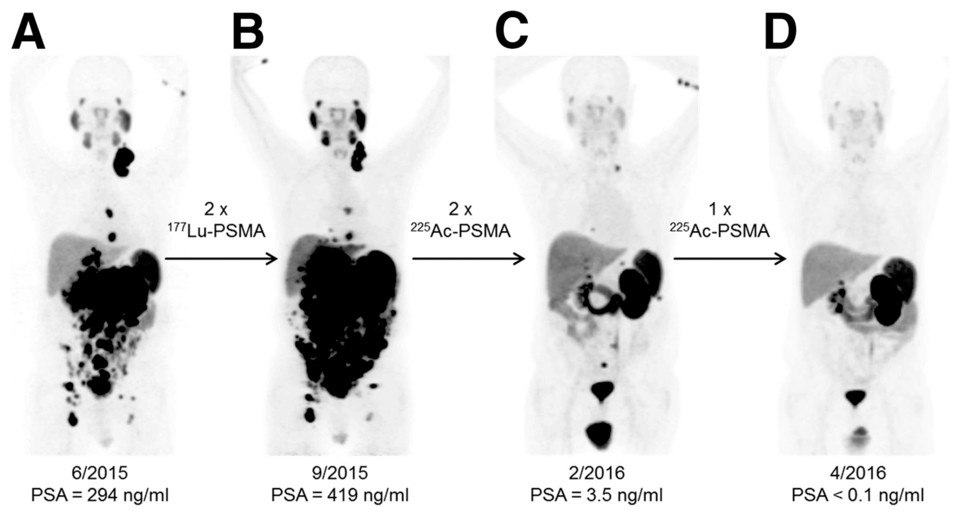
  127. Kratochwil, C.; Bruchertseifer, F.; Giesel, F.L.; Weis, M.; Verburg, F.A.; Mottaghy, F.; Kopka, K.; Apostolidis, C.; Haberkorn, U.; Morgenstern, A. 225Ac-PSMA-617 for PSMA-Targeted alpha-Radiation Therapy of Metastatic Castration-Resistant Prostate Cancer. J. Nucl. Med. 2016, 57, 1941–1944. [Google Scholar] [CrossRef] [PubMed] [Green Version]
  128. Yadav, M.P.; Ballal, S.; Sahoo, R.K.; Tripathi, M.; Seth, A.; Bal, C. Efficacy and safety of (225)Ac-PSMA-617 targeted alpha therapy in metastatic castration-resistant Prostate Cancer patients. Theranostics 2020, 10, 9364–9377. [Google Scholar] [CrossRef] [PubMed]
  129. Kratochwil, C.; Haberkorn, U.; Giesel, F.L. (225)Ac-PSMA-617 for Therapy of Prostate Cancer. Semin. Nucl. Med. 2020, 50, 133–140. [Google Scholar] [CrossRef]
  130. Deblonde, G.J.; Abergel, R.J. Active actinium. Nat. Chem. 2016, 8, 1084. [Google Scholar] [CrossRef] [PubMed] [Green Version]
  131. Vasiliev, A.; Severin, A.; Lapshina, E.; Chernykh, E.; Ermolaev, S.; Kalmykov, S. Hydroxylapatite particles as carriers for 223Ra. J. Radioanal. Nuclear Chem. 2016, 311, 1503–1509. [Google Scholar] [CrossRef]
  132. McDevitt, M.R.; Ma, D.; Simon, J.; Frank, R.K.; Scheinberg, D.A. Design and synthesis of 225Ac radioimmunopharmaceuticals. Appl. Radiat. Isot. 2002, 57, 841–847. [Google Scholar] [CrossRef]
  133. Sofou, S.; Kappel, B.J.; Jaggi, J.S.; McDevitt, M.R.; Scheinberg, D.A.; Sgouros, G. Enhanced retention of the alpha-particle-emitting daughters of Actinium-225 by liposome carriers. Bioconjug. Chem. 2007, 18, 2061–2067. [Google Scholar] [CrossRef] [PubMed] [Green Version]
  134. Antczak, C.; Jaggi, J.S.; LeFave, C.V.; Curcio, M.J.; McDevitt, M.R.; Scheinberg, D.A. Influence of the linker on the biodistribution and catabolism of actinium-225 self-immolative tumor-targeted isotope generators. Bioconjug. Chem. 2006, 17, 1551–1560. [Google Scholar] [CrossRef] [PubMed] [Green Version]
  135. Borchardt, P.E.; Yuan, R.R.; Miederer, M.; McDevitt, M.R.; Scheinberg, D.A. Targeted actinium-225 in vivo generators for therapy of ovarian cancer. Cancer Res. 2003, 63, 5084–5090. [Google Scholar]
  136. Davis, I.A.; Glowienka, K.A.; Boll, R.A.; Deal, K.A.; Brechbiel, M.W.; Stabin, M.; Bochsler, P.N.; Mirzadeh, S.; Kennel, S.J. Comparison of 225actinium chelates: Tissue distribution and radiotoxicity. Nucl. Med. Biol. 1999, 26, 581–589. [Google Scholar] [CrossRef]
  137. Deal, K.A.; Davis, I.A.; Mirzadeh, S.; Kennel, S.J.; Brechbiel, M.W. Improved in vivo stability of actinium-225 macrocyclic complexes. J. Med. Chem. 1999, 42, 2988–2992. [Google Scholar] [CrossRef] [PubMed]
  138. Sathekge, M.; Bruchertseifer, F.; Knoesen, O.; Reyneke, F.; Lawal, I.; Lengana, T.; Davis, C.; Mahapane, J.; Corbett, C.; Vorster, M.; et al. (225)Ac-PSMA-617 in chemotherapy-naive patients with advanced prostate cancer: A pilot study. Eur. J. Nucl. Med. Mol. Imaging 2019, 46, 129–138. [Google Scholar] [CrossRef] [Green Version]
  139. Paganelli, G.; Sarnelli, A.; Severi, S.; Sansovini, M.; Belli, M.L.; Monti, M.; Foca, F.; Celli, M.; Nicolini, S.; Tardelli, E.; et al. Dosimetry and safety of (177)Lu PSMA-617 along with polyglutamate parotid gland protector: Preliminary results in metastatic castration-resistant prostate cancer patients. Eur. J. Nucl. Med. Mol. Imaging 2020, 47, 3008–3017. [Google Scholar] [CrossRef]
  140. Langbein, T.; Chausse, G.; Baum, R.P. Salivary Gland Toxicity of PSMA Radioligand Therapy: Relevance and Preventive Strategies. J. Nucl. Med. 2018, 59, 1172–1173. [Google Scholar] [CrossRef] [PubMed] [Green Version]
  141. Rathke, H.; Bruchertseifer, F.; Kratochwil, C.; Keller, H.; Giesel, F.L.; Apostolidis, C.; Haberkorn, U.; Morgenstern, A. First patient exceeding 5-year complete remission after (225)Ac-PSMA-TAT. Eur. J. Nucl. Med. Mol. Imaging 2021, 48, 311–312. [Google Scholar] [CrossRef] [PubMed]
  142. Kratochwil, C.; Giesel, F.L.; Heussel, C.P.; Kazdal, D.; Endris, V.; Nientiedt, C.; Bruchertseifer, F.; Kippenberger, M.; Rathke, H.; Leichsenring, J.; et al. Patients Resistant Against PSMA-Targeting alpha-Radiation Therapy Often Harbor Mutations in DNA Damage-Repair-Associated Genes. J. Nucl. Med. 2020, 61, 683–688. [Google Scholar] [CrossRef]
  143. McDevitt, M.R.; Barendswaard, E.; Ma, D.; Lai, L.; Curcio, M.J.; Sgouros, G.; Ballangrud, A.M.; Yang, W.H.; Finn, R.D.; Pellegrini, V.; et al. An alpha-particle emitting antibody ([213Bi]J591) for radioimmunotherapy of prostate cancer. Cancer Res. 2000, 60, 6095–6100. [Google Scholar]
  144. Ballangrud, A.M.; Yang, W.H.; Charlton, D.E.; McDevitt, M.R.; Hamacher, K.A.; Panageas, K.S.; Ma, D.; Bander, N.H.; Scheinberg, D.A.; Sgouros, G. Response of LNCaP spheroids after treatment with an alpha-particle emitter (213Bi)-labeled anti-prostate-specific membrane antigen antibody (J591). Cancer Res. 2001, 61, 2008–2014. [Google Scholar] [PubMed]
  145. Li, Y.; Tian, Z.; Rizvi, S.M.; Bander, N.H.; Allen, B.J. In vitro and preclinical targeted alpha therapy of human prostate cancer with Bi-213 labeled J591 antibody against the prostate specific membrane antigen. Prostate Cancer Prostatic Dis. 2002, 5, 36–46. [Google Scholar] [CrossRef] [PubMed]
  146. Nonnekens, J.; Chatalic, K.L.; Molkenboer-Kuenen, J.D.; Beerens, C.E.; Bruchertseifer, F.; Morgenstern, A.; Veldhoven-Zweistra, J.; Schottelius, M.; Wester, H.J.; van Gent, D.C.; et al. (213)Bi-Labeled Prostate-Specific Membrane Antigen-Targeting Agents Induce DNA Double-Strand Breaks in Prostate Cancer Xenografts. Cancer Biother. Radio Pharm. 2017, 32, 67–73. [Google Scholar]
  147. Kiess, A.P.; Minn, I.; Vaidyanathan, G.; Hobbs, R.F.; Josefsson, A.; Shen, C.; Brummet, M.; Chen, Y.; Choi, J.; Koumarianou, E.; et al. (2S)-2-(3-(1-Carboxy-5-(4-211At-Astatobenzamido)Pentyl)Ureido)-Pentanedioic Acid for PSMA-Targeted alpha-Particle Radiopharmaceutical Therapy. J. Nucl. Med. 2016, 57, 1569–1575. [Google Scholar] [CrossRef] [PubMed] [Green Version]
  148. Khreish, F.; Ebert, N.; Ries, M.; Maus, S.; Rosar, F.; Bohnenberger, H.; Stemler, T.; Saar, M.; Bartholoma, M.; Ezziddin, S. (225)Ac-PSMA-617/(177)Lu-PSMA-617 tandem therapy of metastatic castration-resistant prostate cancer: Pilot experience. Eur. J. Nucl. Med. Mol. Imaging 2020, 47, 721–728. [Google Scholar] [CrossRef] [PubMed]
  149. Hammer, S.; Hagemann, U.B.; Zitzmann-Kolbe, S.; Larsen, A.; Ellingsen, C.; Geraudie, S.; Grant, D.; Indrevoll, B.; Smeets, R.; von Ahsen, O.; et al. Preclinical Efficacy of a PSMA-Targeted Thorium-227 Conjugate (PSMA-TTC), a Targeted Alpha Therapy for Prostate Cancer. Clin. Cancer Res. 2020, 26, 1985–1996. [Google Scholar] [CrossRef] [Green Version]
Figure 1. The vicious cycle of bone metastases. Bone metastatic cancer cells hijack the healthy bone remodeling process to create a suitable microenvironment for them to grow. Cancer cells induce hyper-osteoclastogenesis by activating osteoclasts through the secretion of parathyroid-hormone-related peptide (PTHrP). This process leads to osteolytic bone lesions and provides bone metastatic cancer cells more space to grow. On the other hand, cancer cells can over-activate osteoblasts, resulting in osteoblastic bone lesions. These hyper-activated osteoblasts also stimulate osteoclastogenesis through the receptor activator of the nuclear factor κB ligand (RANKL) (secreted from osteoblasts)/RANK (expressed on osteoclasts) axis. Furthermore, these hyper-activated osteoblasts and osteoclasts enhance the growth and survival of bone metastatic cancer cells. This process is called the vicious cycle of bone metastasis. Graphics adapted from Smart Servier Medical Art (https://smart.servier.com/, accessed on 18 March 2021).
Molecules 26 02162 g001
Figure 2. Several ligands used to deliver alpha-particle-emitting and beta-minus-particle-emitting radionuclides to bone metastasis. While ligands such as (A) hydroxyethylidene diphosphonic acid (HEDP) and tetramethylene phosphonic acid (EDTMP) target bone remodeling to deliver therapeutic radiation to bone metastases, ligands like (B) prostate-specific membrane antigen (PSMA)-617 target prostate cancer cell biomarkers.
Molecules 26 02162 g002
Figure 3. An example of the effective response of a prostate cancer patient to 225Ac-PSMA-617 treatment. This research was originally published in the Journal of Nuclear Medicine [127]. Kratochwil C, Bruchertseifer F, Giesel FL, Weis M, Verburg FA, Mottaghy F, Kopka K, Apostolidis C, Haberkorn U, and Morgenstern A. 225Ac-PSMA-617 for PSMA-Targeted α-Radiation Therapy of Metastatic Castration-Resistant Prostate Cancer. J Nucl Med. 2016;57(12):1941-1944. © SNMMI. 68Ga-PSMA-11 PET/CT scans of patient B in this manuscript (a prostate cancer patient who presented with peritoneal carcinomatosis and liver metastases that were progressive under 177Lu-PSMA-617 therapy). In comparison to the initial tumor spread (A), restaging after 2 cycles of β-emitting 177Lu-PSMA-617 presented progression (B). In contrast, restaging after second (C) and third (D) cycles of α-emitting 225Ac-PSMA-617 presented an impressive response. Adapted from ref. [127].
Molecules 26 02162 g003
Table 1. General characteristics of therapeutic radionuclides.
DecayParticleMaximum Particle Range (mm)Maximum Particle Energy (MeV)Linear Energy Transfer (kEV/µm)
Electron capture
internal conversion
Non-energetic electrons0.00050.00126
Beta minus particleEnergetic electrons122.30.2
Alpha particleHelium nuclei0.1980
Table 2. The recent status of PSMA-targeted alpha-particle-emitting radiopharmaceuticals.
AgentConjugatorDevelopment PhaseRefs.
213Bi-J591PSMA-targeting murine monoclonal antibody, J591Preclinical study[143,144,145]
213Bi-PSMA I&TPSMA-targeting (3S,7S)-29,32-dibenzyl-5,13,20,28,31,34-hexaoxo-37-(4,7,10-tris(carboxymethyl)-1,4,7,10-tetraazacyclododecan-1-yl)-4,6,12,21,27,30,33-heptaazaheptatriacontane-1,3,7,26,37-pentacarboxylic acid (DOTAGA-FFK(Sub-KuE)), PSMA I&TPreclinical study[146]
213Bi-JVZ-008PSMA-targeting nanobody, JVZ-008Preclinical study[146]
211At-6PSMA-targeting (2S)-2-(3-(1-carboxy-5-(4-211At-astatobenzamido)pentyl)ureido)-pentanedioic acid, compound 6Preclinical study[147]
225Ac-PSMA-617PSMA-targeting small molecule, PSMA-617 Clinical study[127,138,141,148]
227Th-PSMA-TTC PSMA-targeting fully human antibody, BAY 2315158Clinical trial
(phase I: NCT03724747)
[149]
225Ac-J591PSMA-targeting murine monoclonal antibody, J591Clinical trial
(phase I: NCT03276572)
Publisher’s Note: MDPI stays neutral with regard to jurisdictional claims in published maps and institutional affiliations.

