Article

Cloning and Functional Characterization of Two Germacrene A Oxidases Isolated from Xanthium sibiricum

1
Department of Resources Science of Traditional Chinese Medicines, China Pharmaceutical University, Nanjing 210009, China
2
Anhui Province Key Laboratory of Research & Development of Chinese Medicine, Anhui University of Chinese Medicine, Hefei 230012, China
*
Authors to whom correspondence should be addressed.
Submission received: 24 March 2022 / Revised: 14 May 2022 / Accepted: 15 May 2022 / Published: 22 May 2022

Abstract

:
Sesquiterpene lactones (STLs) from the cocklebur Xanthium sibiricum exhibit significant anti-tumor activity. Although germacrene A oxidase (GAO), which catalyzes the production of Germacrene A acid (GAA) from germacrene A, an important precursor of germacrene-type STLs, has been reported, the remaining GAOs corresponding to various STLs’ biosynthesis pathways remain unidentified. In this study, 68,199 unigenes were studied in a de novo transcriptome assembly of X. sibiricum fruits. By comparison with previously published GAO sequences, two candidate X. sibiricum GAO gene sequences, XsGAO1 (1467 bp) and XsGAO2 (1527 bp), were identified, cloned, and predicted to encode 488 and 508 amino acids, respectively. Their protein structure, motifs, sequence similarity, and phylogenetic position were similar to those of other GAO proteins. They were most strongly expressed in fruits, according to a quantitative real-time polymerase chain reaction (qRT-PCR), and both XsGAO proteins were localized in the mitochondria of tobacco leaf epidermal cells. The two XsGAO genes were cloned into the expression vector for eukaryotic expression in Saccharomyces cerevisiae, and the enzyme reaction products were detected by gas chromatography–mass spectrometry (GC-MS) and liquid chromatography–mass spectrometry (LC-MS) methods. The results indicated that both XsGAO1 and XsGAO2 catalyzed the two-step conversion of germacrene A (GA) to GAA, meaning they are unlike classical GAO enzymes, which catalyze a three-step conversion of GA to GAA. This cloning and functional study of two GAO genes from X. sibiricum provides a useful basis for further elucidation of the STL biosynthesis pathway in X. sibiricum.

1. Introduction

Sesquiterpene lactones (STLs) are widely distributed in nature and have a broad range of beneficial biological activities, including anti-bacterial, anti-inflammatory, and anti-cancer effects [1,2,3,4,5]. Two specific STLs, xanthatin and xanthinosin, are produced in the burs and leaves of Xanthium L. plants [6,7,8]. Many studies have been conducted on the quality and pharmacological activities of X. sibiricum. However, the details of the biological pathways associated with the anti-cancer effects of STLs in Xanthium species remain unclear.
Based on the carbon skeleton, STLs can be classified into multiple types, including germacrene, guaiane, xanthane, pseudoguaiane, eudesmane, and elemane lactones [9]. The molecular mechanisms of STLs differ among types. For example, eudesmane-type STLs synthesize the core carbon skeleton 10-epi-junenol before lactone ring synthesis [10], while guaiane-type STLs are produced from germacrene-type STLs and are induced by protonation [11]. The STL synthesis pathway is usually divided into three main processes: The first process is the synthesis of intermediates, including isopentenyl pyrophosphate (IPP) and dimethylallyl pyrophosphate (DMAPP). The second step involves the formation of the sesquiterpene skeleton, which is preceded by the formation of farnesyl pyrophosphate (FPP) from IPP and DMAPP. Sesquiterpene synthase (STP) then catalyzes FPP to produce the sesquiterpene skeleton. The final step is the formation of the STL end-product, which involves a variety of structural modifications to the carbon skeleton. In particular, STP is critical for the structural transformation of FPP to STL, which subsequently catalyzes the formation of multiple types of sesquiterpenes through a series of chemical processes such as intermediate cyclization of carbenium ion, deprotonation, and hydrogen transfer [12,13,14,15,16,17]. Importantly, some STPs, the cytochrome P450 enzymes, play a modulatory role in SLT biosynthesis, participating in the addition of functional groups to the sesquiterpene backbone [18]. For example, the cytochrome P450 GAO, isolated from an Asteraceae plant, catalyzes the three-step sequential oxidation of germacrene A to GAA [19]. In addition, in common chicory (Cichorium intybus L.), the most critical modification enzyme in the biosynthetic pathway of the 6α-type STL-myrcene lactone C12 is the cytochrome P450 enzyme [20]. It was also shown that the cytochrome P450 enzymes parthenolide synthase (PTS) and kauniolide synthase (KLS), cloned from the aster Chrysanthemum paludosum, catalyzed the oxidation reaction of the C4–5 double-bonds of costunolide, which in turn, generated parthenolide [11,21]. Since germacrene-derived STLs are the simplest, a number of studies have investigated these STLs using synthetic biology and related techniques. Three STP genes were cloned from X. strumarium: XSTPSS1 catalyzed the production of germacrene D, XSTPSS2 catalyzed the formation of Guaia-4,6-diene, and XSTPSS3 catalyzed the production of germacrene A (Figure 1) [22]. However, it is not clear how different biologically active STLs are produced in Xanthium species after sesquiterpene skeleton formation.
Studies have shown that GAA is an important precursor substance in the biosynthesis pathway of germacrene-derived STLs [19]. Based on the findings outlined above, we hypothesized that, in X. sibiricum, GAO would catalyze the production of GAA from germacrene A to produce GAA in two consecutive steps (Figure 2).
To test this hypothesis, we identified the genes homologous to GAO by searching known GAO gene sequences in the National Center for Biotechnology Information (NCBI) database against a transcriptome library of X. sibiricum established therein and cloned the GAO gene using complementary DNA (cDNA). Subsequently, bioinformatic analyses of the predicted amino acid and protein structures, gene expression patterns, and subcellular localization were carried out. The enzyme reaction products were detected by GC-MS and LC-MS methods, where GAO genes were cloned into the expression vector for eukaryotic expression in S. cerevisiae. The results clarify the downstream STL synthesis pathway in X. sibiricum in future work.

2. Results

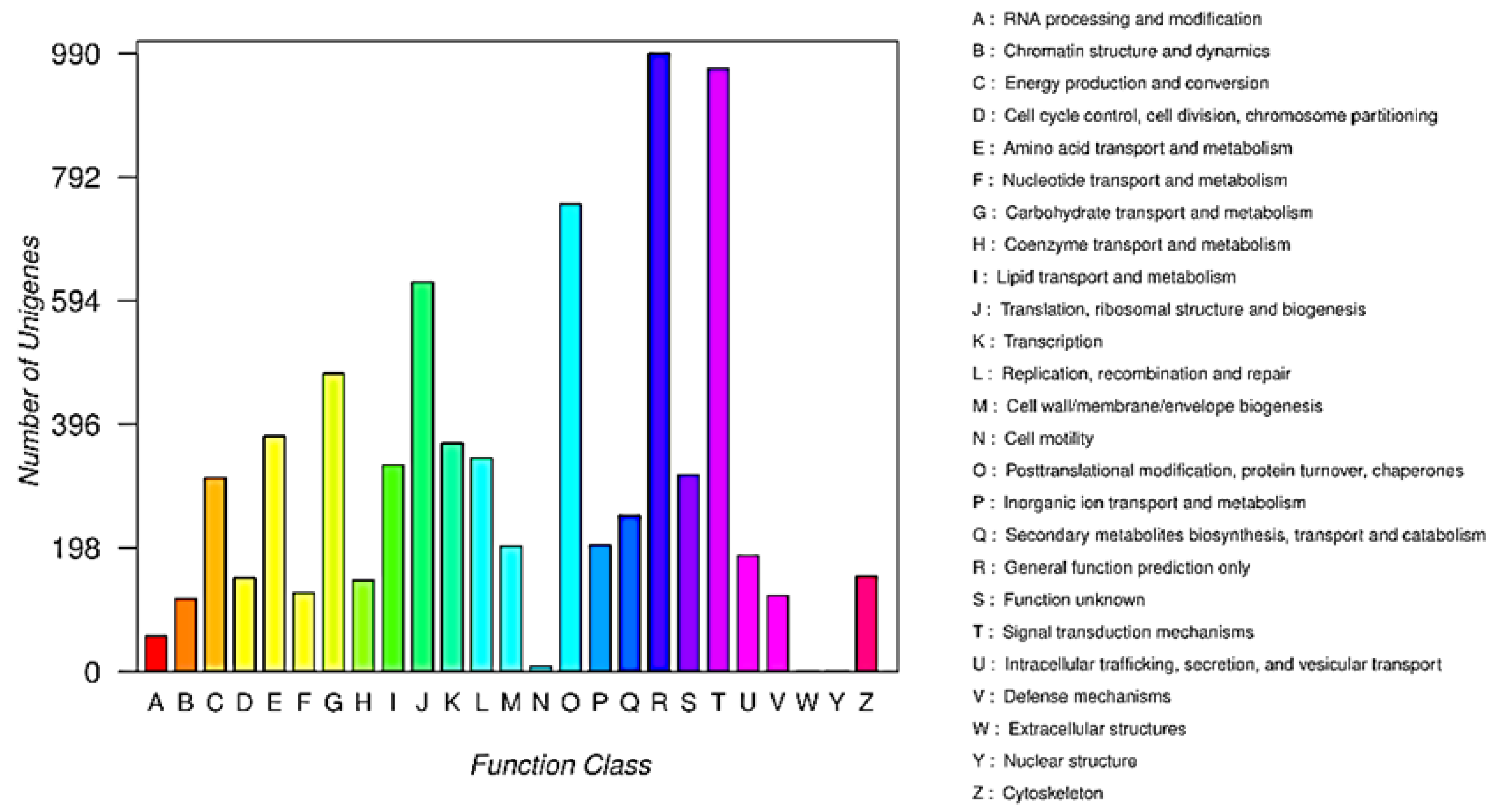
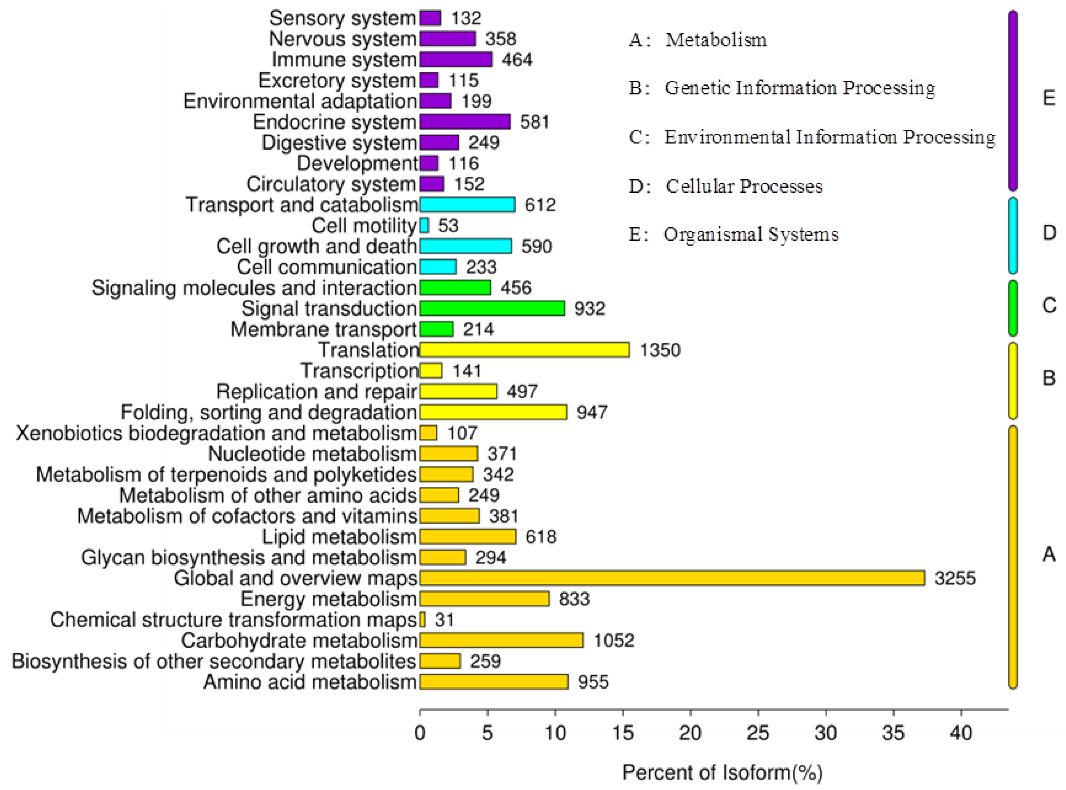
2.1. Establishment of a Transcriptome Library and Gene Annotation of X. sibiricum

The de novo transcriptome library of X. sibiricum included 5,989,562,311 nucleotides (nt), and the transcriptome Q20, N, and GC percentages were 97.57%, 0.01%, and 45.36%, respectively. After low-quality reads and filtering out those containing duplicates or junctions, 49,957,916 valid clean reads remained. The clean reads were assembled de novo using Trinity assembly software [23], and a total of 68,199 unigenes were obtained, with an average length of 639 nt and an N50 of 954 nt.
Protein function annotation information for all unigenes was obtained using BLAST [24]. Of the 68,199 unigenes, 19,129, 13,845, 24,721, 37,205, and 14,001 unigenes were successfully annotated with the Pfam, Kyoto Encyclopedia of Genes and Genome (KEGG), SwissProt, non-redundant protein sequence database (NR), and string libraries, respectively. A total of 13,619 unigenes from the transcriptome were successfully annotated to the Cluster of Orthologous Groups of proteins (COG) database, corresponding to the 25 functional categories. A total of 990 unigenes mainly focused on function prediction, with the highest percentage focusing on STLs, and a further 250 unigenes were annotated to secondary metabolite biosynthesis, transport, and catabolism (Appendix A, Figure A1).
A total of 13,845 unigenes were annotated to 128 metabolic pathways in the KEGG database, and 342 were annotated to “metabolism of terpenoids and polyketides” (Appendix A, Figure A2). Among these 342 unigenes, 89 were related to “terpenoid backbone biosynthesis”, and 22 were involved in “sesquiterpenoid and triterpenoid biosynthesis.”
In plants, the biosynthesis of STLs primarily occurs through the mevalonate (MVA) or methyl-D-erythritol phosphate (MEP) pathways, which synthesize DMAPP and IPP precursors [14]. KEGG pathway analysis showed that a total of 31 transcripts in X. sibiricum encoded six enzymes of the MVA pathway (acetyl-CoA C-acetyltransferase [ACCT, E.C.No:2.3.1.9], 3-hydroxy-3-methylglutaryl coenzyme A synthetase [HMGS, E.C.No:2.3.1.10], 3-hydroxy-3-methylglutaryl coenzyme A reductase [HMGR, E.C.No:1.1.3.34], mitogen-activated protein kinase/extracellular signal-regulated kinase [MEK, E.C.No:2.7.1.36], phosphomevalonate kinase [PMK, E.C.No:2.7.4.2], and pyrophospomevalonate decarboxylase [MVD, E.C.No:4.1.1.33]), and 21 transcripts encoded seven enzymes in the MEP pathway (1-deoxy-D-xylulose-5-phosphate synthase [DXS, E.C.No:2.2.1.7], 1-deoxy-D-xylulose 5-phosphate reductoisomerase [DXR, E.C.No:1.1.1.26], 2-C-methyl-D-erythritol 4-phosphate cytidyltransferase [MCT, E.C.No:2.7.7.60], 4-diphosphocytidyl-2-C-methyl-D-erythritol kinase [CMK, E.C.No:2.7.7.148], 2-C-methyl-D-erythritol 2,4-cyclodiphosphate synthase [MDS, E.C.No:4.6.1.12], 4-hydroxy-3-methylbut-2-en-1-yl diphosphate synthase [HDS, E.C.No:1.17.7.1], and 4-hydroxy-3-methylbut-2-enyl-diposphate reductase [HDR, E.C.No:1.17.1.2]).

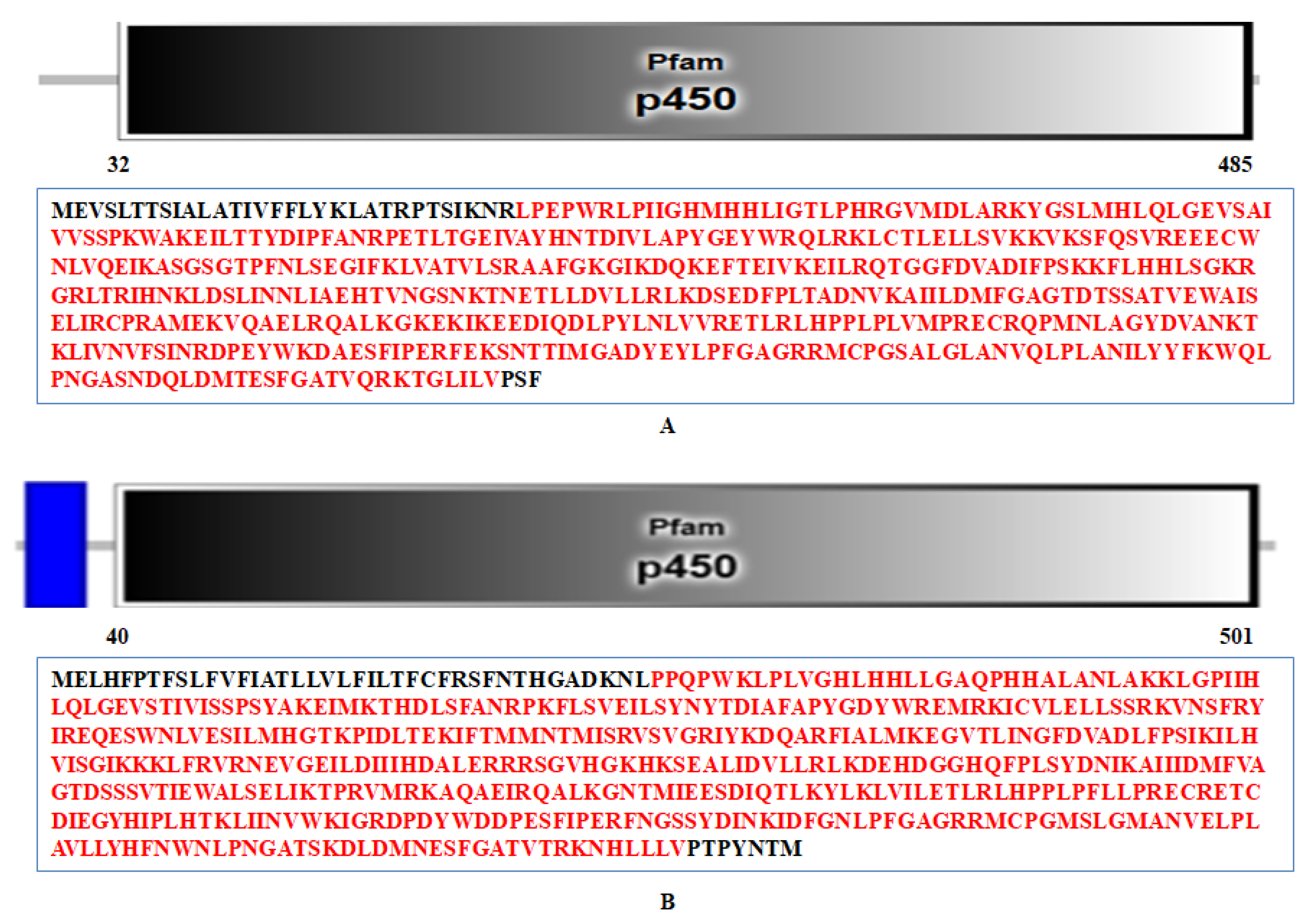
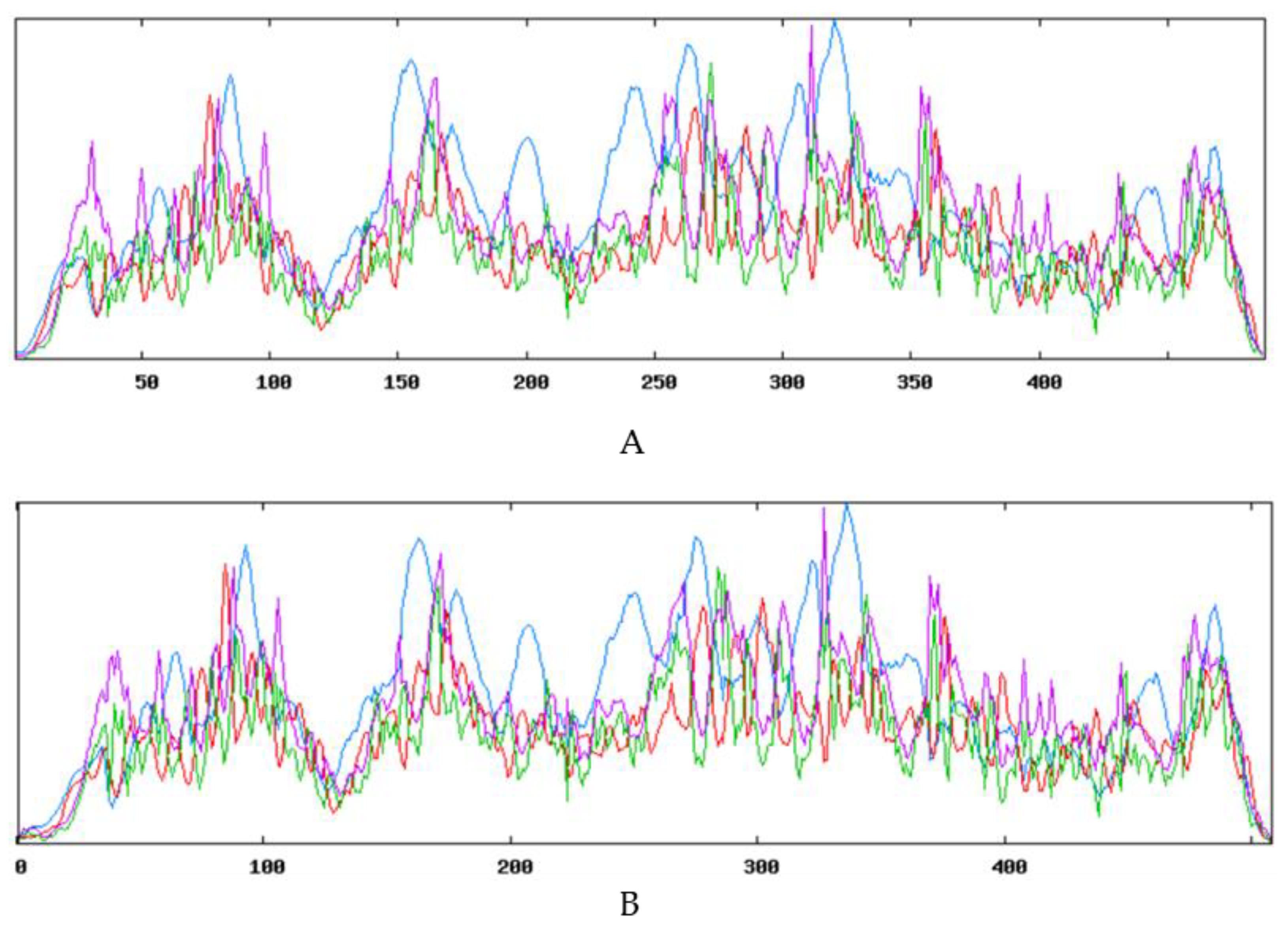
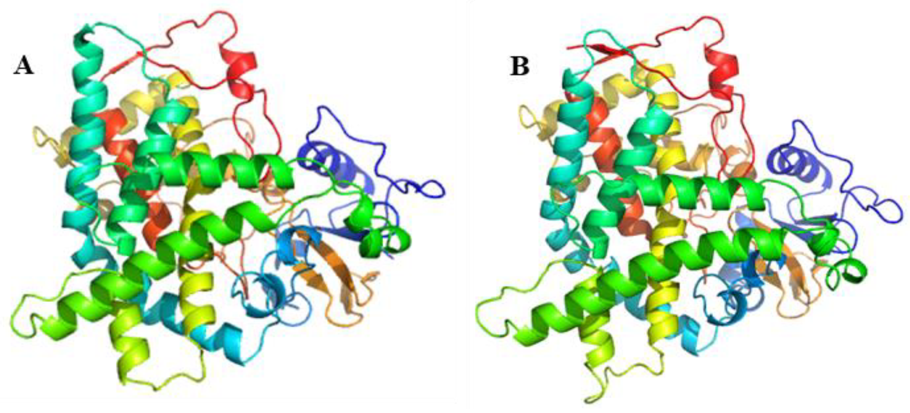
2.2. Cloning and Bioinformatics Analysis of XsGAO Genes

Two candidate GAO genes were designed using these sequences as templates (Appendix A, Table A1). The full lengths of XsGAO1 and XsGAO2 were 1467 and 1527 bp, encoding 488 and 508 amino acids, respectively. SMART (Simple Modular Architecture Research Tool) analysis showed that XsGAO1 and XsGAO2 encoded proteins with molecular weights of approximately 54.98 and 58.01 kDa, respectively, protein isoelectric points of approximately 8.72 and 8.40, respectively, and protein a pH value of 5.1. Protein domain analysis showed that XsGAO1 harbored a P450 domain comprising 454 amino acids (amino acid 42-485, Appendix A, Figure A3A,) and XsGAO2 harbored a P450 domain comprising 462 amino acids (amino acid 40-501, Appendix A, Figure A3B). The secondary structures of the XsGAO1 and XsGAO2 proteins were predicted using SOMPA online. The prediction of XsGAO1 indicated that 244 amino acid residues (50%) were involved in the formation of α-helix, 66 residues (13.52%) were involved in the extended chain, 27 residues (5.53%) were involved in β-turn, and 151 residues (30.94%) were involved in a random coil (Appendix A, Figure A4A). Prediction of XsGAO2 indicated that 253 amino acid residues (49.8%) were involved in the formation of α-helix, 62 residues (12.2%) were involved in the extended chain, 31 residues (6.1%) were involved in β-turn, and 162 residues (31.89%) were involved in a random coil (Appendix A, Figure A4B). To better characterize the XsGAO bioinformation, 3D-structure prediction of the XsGAO1- and XsGAO2-encoded proteins was performed, and spatial predictions showed high similarity with the P450 enzyme from Salvia miltiorrhiza (Appendix A, Figure A5).
BLAST analysis showed that XsGAO1 and XsGAO2 were highly similar to 17 GAO protein homologs from 10 species, which were obtained from the NCBI database (Appendix A, Table A3). Phylogenetic tree analysis indicated that two XsGAO genes were mainly conserved in Asteraceae; the amino acid sequence of XsGAO1 shared 94.06% sequence identity with HaGAO from Helianthus annuus (XP_022000663.1), while XsGAO2 was recovered on a distinct branch (Figure 3).

2.3. XsGAO1 and XsGAO2 Expression Patterns in X. sibiricum

Across the three X. sibiricum tissues tested (fruit, leaves, and stems), the expression patterns of XsGAO1 and XsGAO2 were similar: both genes were more strongly expressed in the fruits than the leaves or stems (Figure 4A,C).
To localize XsGAO1 and XsGAO2 in the cell, tobacco leaf transformation was performed. Confocal laser scanning microscopy (CLSM) examination of the transformed tobacco leaves identified XsGAO1 and XsGAO2 signals in the mitochondria (Figure 5). This was consistent with predictions based on their sequence features.

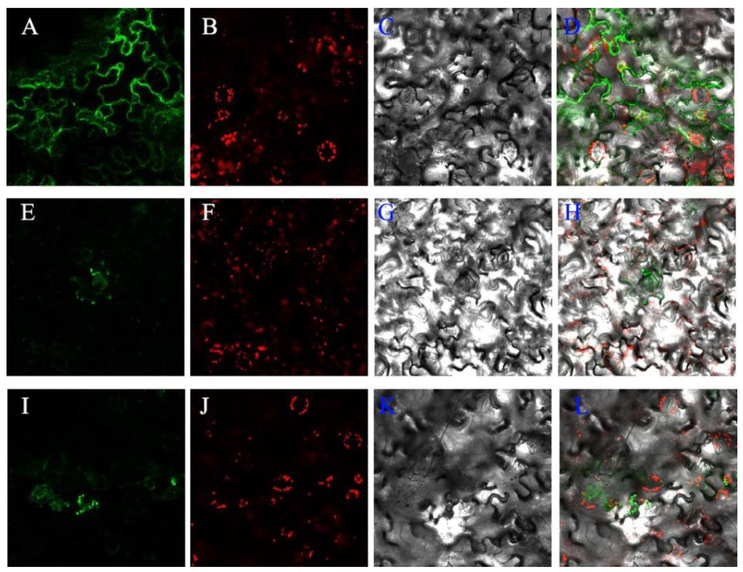
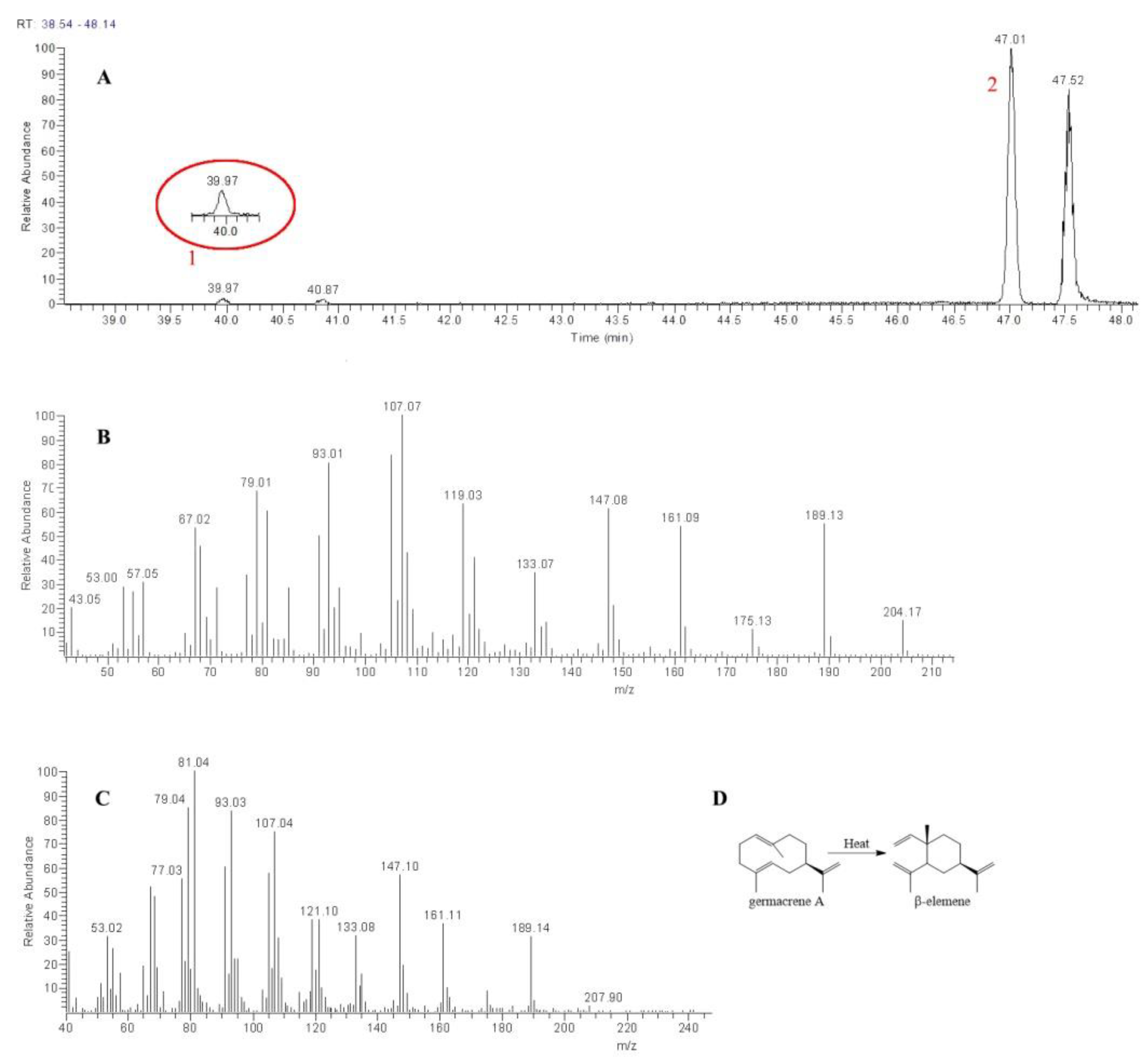
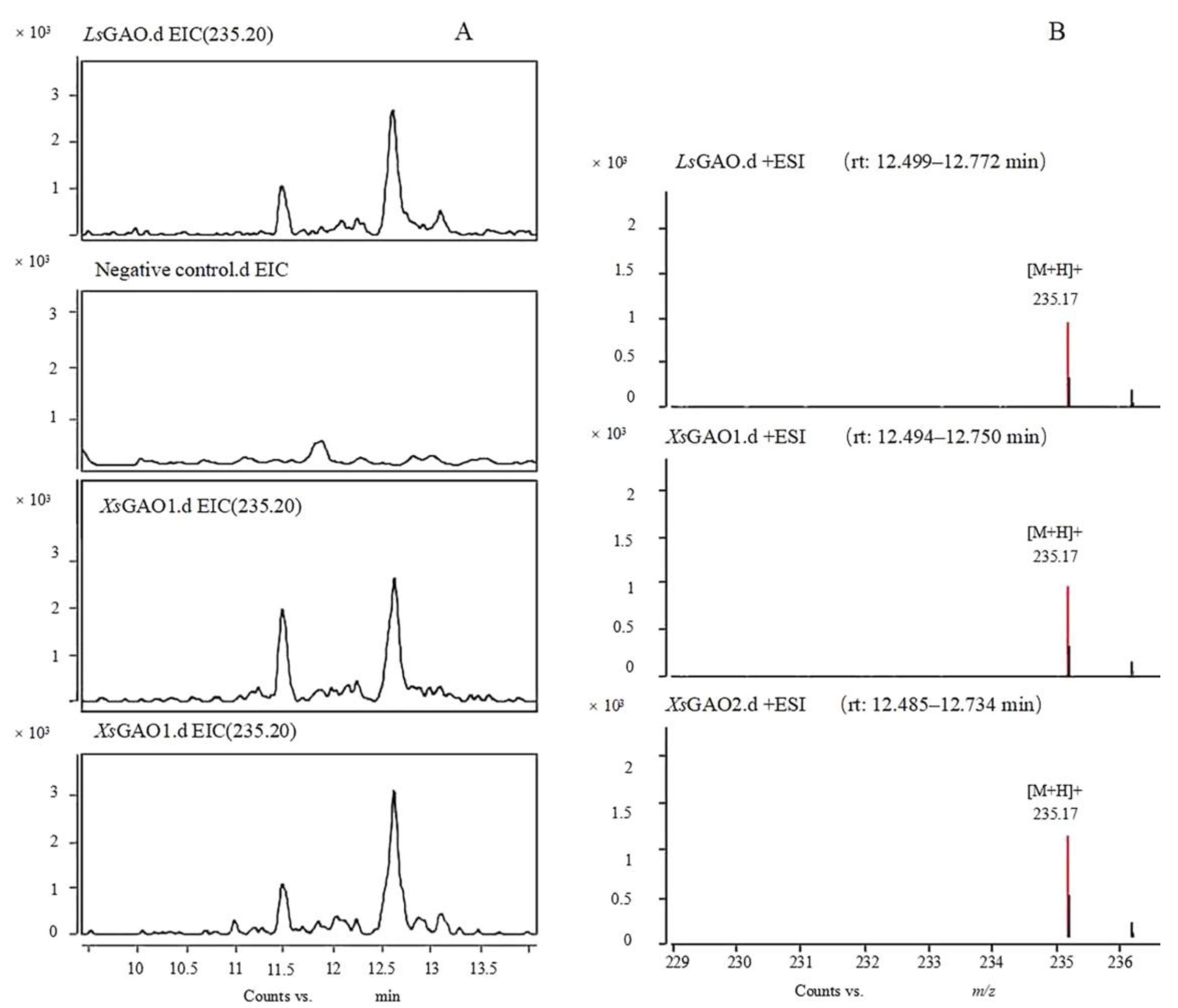
2.4. Functional Study of XsGAO1 and XsGAO2

The full-length XsGAO genes were cloned into the yeast expression vector pYeDP60 and co-transferred into S. cerevisiae WAT11. Compared with the expression of LsGAS alone or the control yeast bearing the empty vector, germacrene A was obtained through prokaryotic expression and recombinant protein enzyme activity assays and was used as the substrate for XsGAO1 and XsGAO2 in the enzymatic activity reaction. Additionally, the LsGAO gene was cloned from Lactuca sativa, and the microsomal protein expressed in this gene was used as a positive control. The inactivated microsomes were used as a negative control to verify whether the microsomal protein expressed in XsGAO1 and XsGAO2 showed catalytic activity through a two-step enzyme activity catalytic assay (Appendix A, Figure A6, Figure A7, Figure A8 and Figure A9).
The gas chromatography–mass spectrometry (GC-MS) chromatogram of enzymatic activity experiments on the LsGAS recombinant protein showed two distinct peaks (Figure 6A). The second peak exhibited fragment ion peaks at m/z 53, 67, 79, 93, 107, 119, 133, 147, 161, 175, 189, and 204 (Figure 6B), which corresponded to the characteristic ions of germacrene A. However, the first peak exhibited fragment ion peaks at m/z 77, 81, 93, 107, 121, 133, 147, 161, and 189, which corresponded to the characteristic ions of β-elemene (Figure 6C) [10,25]. Therefore, we assumed that peak 1 corresponded to β-elemene and peak 2 corresponded to germacrene A.
An analysis of the enzymatic activity of XsGAO1 and XsGAO2 in microsomes identified peaks in both liquid chromatography–mass spectrometry (LC-MS) chromatograms with the same retention time as the positive control LsGAO (tR = 12.54 min; Figure 7A). Further analysis showed that the peaks produced in all three assays (i.e., XsGAO1, XsGAO2, and LsGAO) had the same m/z in the positive ion mode ([M+H]+ = 235.17; Figure 7B), suggesting that this peak corresponded to GAA. The results indicated that the XsGAO1 and XsGAO2 proteins both catalyzed the production of GAA from germacrene A in yeast microsomes.

3. Discussion

To investigate plant gene expression and analyze its function, transcriptome sequencing is an important molecular method that can provide genetic information in the absence of genomic data [26]. The de novo assembly platform greatly contributes to finding new genes, providing databases of sesquiterpene synthases and cytochrome P450s for cloning in X. strumarium [27]. X. sibiricum is a traditional plant containing unique secondary metabolites, of which STLs have various pharmaceutical properties. Although sesquiterpene synthase (STP) has only been cloned from X. strumarium glandular trichomes [28], transcriptome databases established from fruits of X. sibiricum provided cDNAs of two GAO genes that were cloned accurately in this study.
As an enzyme that produces an important precursor substance for the synthesis of STLs, the GAO gene is conserved in Asteraceae [25,29]. To investigate the function of this gene, the full-length cDNA sequences of XsGAO1 and XsGAO2 were successfully cloned. Phylogenetic analysis indicated that XsGAO1 may have a similar function to the GAO in H. annuus. However, XsGAO2 was distinct from other GAOs, representing a separate branch that needs further investigation. Multiple comparisons showed that the predicted XsGAO1 protein had high homology with other redox-like proteins, and its protein sequence contained conserved amino acid residues that are expected in the cytochrome P450 enzyme family [30]. Analysis of the 3D structure predicted that XsGAO1 and XsGAO2 had functions similar to those corresponding with ferruginol synthase (CYP71 family).
The expression patterns of XsGAO1 and XsGAO2 in X. sibiricum leaves differed over time, with the highest expression level observed in young leaves, and expression levels decreasing with maturation. XsGAO1 and XsGAO2 were also differentially expressed among fruits and stems, presumably related to their functions. The expression of the two XsGAOs was the highest in fruits, which explains why fruits with higher contents of STLs are used in the traditional Chinese medicine Cang Er Zi.
XsGAO1 and XsGAO2 cDNAs with the correct sequence were successfully inserted into an expression vector and used in transient transformation assays of Nicotiana benthamiana. However, no fluorescence was observed in the protoplasts. This may be because the accumulated concentration of the product was below the detection limit of the instrument or because the GAA generated was intermediately transient in N. benthamiana [31]. Both XsGAO1 and XsGAO2 were localized in the mitochondria, which was consistent with terpene synthase in tomatoes (also localized in the mitochondria) [32,33].
GAO isolated from L. sativa was expressed in an engineered yeast to synthesize GAA de novo, and the classical GAO activity involved three-step oxidation of germacrene A (GA) to yield GAA and 12,6-guaianolides [25,34], similar to N. benthamiana [35]. Meanwhile, an XsGAO from X. strumarium catalyzed only one-step conversion of germacrene A to germacrene alcohol [36], but this study clearly shows that XsGAO1 and XsGAO2 catalyzed a second step of oxidation of the non-natural substrate germacrene A to germacrene A acid, which was not observed in yeast with a different GAO. Apparently, XsGAO2 is a unique enzyme, a functional adaption of SLTs’ biosynthetic pathway diversification. Of course, a structural analysis of the XsGAO2 biochemical function, such as to identify the active center and crystal structure of oxidase, would help to examine whether it has unique GAO activity. In addition, with the advent of CRISPR-Cas genome editing, CRISPR-Cas-mediated gene knockout in tomatoes and the medicinal plant Salvia miltiorrhiza has been successfully performed [37,38]. This approach could be applied to verify the function of XsGAO2 in the future.
Beyond this, X. sibiricum contains a variety of biologically active STLs, mainly xanthane-STLs with anti-tumor activities [39,40]. As such, studying the genes in the STL synthesis pathway could provide new ideas for the investigation of xanthane-STL biosynthesis pathways.

4. Materials and Methods

4.1. Establishment of a Transcriptome Library and Gene Annotation

The fresh samples (fruit, leaf, and stem) of X. sibiricum used for the RNA extraction were collected from Chaoyang District (N: 40.0031, E: 116.5468, H: 113 m, Beijing, China) in August, wrapped in tinfoil, and frozen immediately in liquid nitrogen for storage at −80 °C. Species verification was performed by Professor Dongmei Xie at the School of Pharmacy (Anhui University of Chinese Medicine). The total ribonucleic acid (RNA) was isolated using a TransZol Up Plus RNA kit (TansGen Biotech, Beijing, China), according to the manufacturer’s protocol. The RNA extract was reverse-transcribed and then sequenced on an Illumina HiSeq 3000 platform at Shanghai Majorbio Bio-pharm Technology Corporation. After high-throughput sequencing, unigenes were assembled de novo from the clean reads obtained from the raw sequencing reads. To predict the biological function, all unigenes were annotated via a similarity search against the public databases, which contained Pfam, NR (http://0-www-ncbi-nlm-nih-gov.brum.beds.ac.uk/) (accessed on 5 May 2018), SwissProt (http://www.uniprot.org/) (accessed on 6 May 2018), KEGG (http://www.genome.jp/kegg/) (accessed on 8 May 2018), GO (Gene Ontology, http://www.geneontology.org/) (accessed on 10 May 2018), and BlastX (E value < 1 × 10⁻⁵) [24,26].

4.2. Cloning and Bioinformatics Analysis of XsGAO1 and XsGAO2

GAO gene sequences were searched for in the transcriptome database of X. sibiricum using local BLAST, with seed sequences downloaded from the NCBI. The full-length cDNA of two candidate genes, XsGAO1 and XsGAO2, was then cloned using reverse transcription polymerase chain reaction (RT-PCR) (primers are listed in Appendix A, Table A1).
The nucleotide sequences and their encoded amino acid sequences were analyzed using bioinformatics software, and the physicochemical properties of the encoding proteins were predicted using vector NTI, open reading frames (ORF), and amino acid sequence translation through Expasy Translate (http://web.expasy.org/translate/) (accessed on 1 March 2020). Gene domain analysis was performed using SMART (http://smart.embl-heidelberg.de/) (accessed on 5 March 2020) [41].
Phylogenetic relationships were constructed using the amino acid sequences of XsGAO1 and XsGAO2 with different reported GAO sequences. Nineteen sequences were aligned using ClustalX2, and the alignment was used to construct a phylogenetic tree using MEGA5.0 software [42].
Secondary protein structures were obtained using SOMPA online analysis software (http://npsa-pbil.ibcp.fr/cgi-bin/npsa_automat.pl?page=npsa_sopma.html) (accessed on 20 March 2020), and 3D structures and peptide-sequence fragments of XsGAO proteins were predicted using the SWISS-MODEL web server (https://swissmodel.expasy.org/interactive) (accessed on 20 March 2020) and PyMOL software [43].

4.3. Examination of the Expression Patterns of the XsGAO1 and XsGAO2 Genes

To further understand the distribution characteristics of GAO genes in X. sibiricum, qRT-PCR (primers are listed in Appendix A, Table A2) was employed to determine the expression patterns of two GAO genes in different organs (leaf, stem, and fruit) at different stages of X. sibiricum. RNA was extracted according to protocol provided for the TRlzol reagent (Invitrogen), and this was then converted into cDNA via reverse transcription with the TransScriptor First-Strand cDNA Synthesis Supermix kit. Subcellular localization of the protein was observed using CLSM, and the consistency with prediction was determined using SLP-Local (https://sunflower.kuicr.kyoto-u.ac.jp/~smatsuda/slplocal.html) (accessed on 25 March 2018) online [27,43].

4.4. Functional Study of XsGAO1 and XsGAO2 Genes in Yeast

The ORFs of XsGAO1 and XsGAO1 were PCR-amplified (primers are listed in Appendix A, Table A2), and the amplicons were digested with BamHI/EcoRI and cloned into the respective sites in pYeDP60-XsGAO (pYeDp60 plasmid provided by the Department of Pharmacology, Second Military Medical University). To supply the substrate for XsGAOs, the germacrene A synthase gene (LsGAS; AF489965) from L. sativa (provided by the Department of Pharmacognosy, Second Military Medical University) was inserted into the E. coli expression vector pet28a-LsGAS at EcoRI-SacI sites. Germacrene A was produced by LsGAS, which was expressed through Transetta (DE3), and then germacrene A was catalyzed by the XsGAO gene expressed in S. cerevisiae WAT11 (WAT11 provided by the China Academy of Chinese Medical Sciences). For comparison, we used a classical GAO from L. sativa (LsGAO; GU198171) that is known to oxidize germacrene A in a three-step oxidation process. Transgenic yeast cells were cultivated in appropriate dropout media, and the expression of the transferred genes was induced by 2% galactose [25,34,41].

4.5. GC-MS and LC-MS Analyses

GC-MS analysis was performed [25] using a Shimadzu GC-MS TRACE GC Ultra/DSQ II instrument (Thermo Fisher Scientific, Waltham, MA, USA). A sample volume of 1 μL was injected at an inlet temperature of 150 °C, and compounds were separated using a DB-5MS column (30 m × 250 μm × 0.1 μm) with helium as a carrier gas at a flow rate of 2 mL/min. The GC oven temperature program was as follows: 45 °C for 4 min, 45–170 °C for 67 min, and 170 °C for 72 min. The electron impact ionization of the mass spectrometric detector was tuned to 70 eV and operated at 40–400 Da in full scan mode. LC-MS/MS analysis was performed [41] using a UHPLC-Q-TOF-MS system (Agilent Technologies, Santa Clara, CA, USA) equipped with an XBridgeTM C18 column (2.1 × 100 mm; Waters Corporation, Milford, MA, USA), with a mobile phase of 0.1% formic acid aqueous solution (A) and 0.1% acetonitrile (B) at a flow rate of 0.4 mL/min, injection volume of 1 μL, and column temperature of 40 °C. Mass spectrometry data were collected by Electrospray ion sources in the positive mode, and the collection range was 100–1700 m/z.

Author Contributions

M.-J.Q., D.-M.X., and C.-B.L. conceived and designed the experiments; Q.Z. and G.-K.W. performed the experiments and analyzed the data; G.-K.W. and D.-M.X. contributed the reagents, materials, and analysis tools; L.-K.X. and D.-M.X. wrote the paper. All authors have read and agreed to the published version of the manuscript.

Funding

The research was funded by the National Natural Science Foundation of China, grant number 81503190, and the National Natural Science Foundation of Anhui University of Chinese Medicine, grant number 2021hxts22.

Data Availability Statement

The data presented in this study are available in the article.

Conflicts of Interest

The authors declare no conflict of interest.

Sample Availability

Samples of the compounds GA and GAA are available from the authors.

Appendix A

The appendix provides data supplemental to the main text.
Table A1. Primers used in GAO gene cloning.
PrimerPrimer Sequence
XsGAO1FATGGAAGTCTCCCTCACCACTTC
XsGAO1RTTAAAAACTTGGTACCAATATCAACCC
XsGAO2FATGGAACTCCATTTTCCCAC
XsGAO2RTCCAAATATCACTATCCTTCG
M13FCAGGAAACAGCTATGAC
M13RGTAAAACGACGGCCAGT
Table A2. Primers used in RT-PCR, qRT-PCR, and expression in yeast.
PrimerPrimer Sequence
QXsGAO1FCTAATAAGGTGTCCGAGAG
QXsGAO1RGGCAGGTCTTGAATATCT
QXsGAO2FTCTCAACCATAGTAATCTCA
QXsGAO2RCGATGTCTGTGTAATTGTAT
ActinFTACTACAACGGCAGAACGGGAAA
ActinRTCATAGACGGCTGGAACAAAACC
GFP-XsGAO1FacgggggactcttgaccatggATGGAAGTCTCCCTCACCACTTC
GFP-XsGAO1RgcccttgctcaccatactagtAAAACTTGGTACCAATATCAACCCA
GFP-XsGAO2FacgggggactcttgaccatggATGGAACTCCATTTTCCCACC
GFP-XsGAO2RgcccttgctcaccatactagtCATTGTGTTGTAAGGTGTTGGGA
LsGASFATGGCAGCAGTTGACACTAATG
LsGASRTTACATGGATACAGAACCAAC
LsGAOFATGGAGCTTTCAATAACCACC
LsGAORCTAAAAACTCGGTACGAGTAACAAC
pYeDP60-XsGAO1FacacactaaattaccggatccATGGAAGTCTCCCTCACCACTTC
pYeDP60-XsGAO1RgggagatcccccgcggaattcTTAAAAACTTGGTACCAATATCAACCC
pYeDP60-XsGAO2FacacactaaattaccggatccATGGAACTCCATTTTCCCACC
pYeDP60-XsGAO2RgggagatcccccgcggaattcTCACATTGTGTTGTAAGGTGTTGG
pYeDP60-LsGAOFacacactaaattaccggatccATGGAGCTTTCAATAACCACCTCC
pYeDP60-LsGAORgggagatcccccgcggaattcCTAAAAACTCGGTACGAGTAACAACTC
Table A3. Reference sequences used for phylogeny construction.
NameSpeciesGene Bank Accession No.
CcVsGAO1Cynara cardunculus var. scolymusAIA09035.1
CcVsGAO2XP_024977750.1
CcVsGAO3AIA09037.1
CcVs-GAOlikeXP_024977969.1
TpGAOTanacetum partheniumAHN62855.1
LsGAO1Lactuca sativaXP_023734551.1
LsGAO2ADF32078.1
LsGAO3AIX97103.1
BsGAOBarnadesia spinosaADF43083.1
HaGAOHelianthus annuusADF43082.1
ScGAOSaussurea costusADF43081.1
CiGAOCichorium intybusADF43080.1
TcGAOTanacetum cinerariifoliumAGO03789.1
CeGAO1Cichorium endiviaAZI95573.1
CeGAO2AZI95575.1
CeGAO3AZI95574.1
OsJGGAOOryza sativa Japonica GroupXP_015624875.1
Figure A1. COG classification of X. sibiricum unigenes.
Molecules 27 03322 g0a1
Figure A2. Pathway analysis of associated X. sibiricum unigenes in KEGG.
Molecules 27 03322 g0a2
Figure A3. Domain prediction and amino acid sequences of XsGAO1 (A) and XsGAO2 (B). (Function domains shown in red).
Molecules 27 03322 g0a3
Figure A4. Secondary structures of (A) XsGAO1 and (B) XsGAO2. α-helixes are indicated in blue, β-sheets are indicated in green, and random coils are indicated in red.
Molecules 27 03322 g0a4
Figure A5. Predicted 3D structures of (A) XsGAO1 and (B) XsGAO2.
Molecules 27 03322 g0a5
Figure A6. Map of the vector used for subcellular localization of XsGAO1 and XsGAO2 created with SnapGene. BspDI* and ClaI* are isoschizomers.
Molecules 27 03322 g0a6
Figure A7. Map of the vector used in the yeast expression experiment created with SnapGene. Different protein showed with distinguished color.
Molecules 27 03322 g0a7
Figure A8. Map of the vector used for function validation of XsGAO1 and XsGAO2 in tobacco created with SnapGene. BclI* is methylation sensitive restriction endonuclease. Different protein showed with distinguished color.
Molecules 27 03322 g0a8
Figure A9. Map of the vector used for prokaryotic expression created with SnapGene. Different protein showed with distinguished color.
Molecules 27 03322 g0a9

References

  1. Guo, L.M.; Lv, J.L.; Zhang, L.B. Research progress on anti-inflammatory mechanism of natural sesquiterpenoids. China J. Chin. Mater. Med. 2018, 43, 3989–3999. [Google Scholar]
  2. Xu, D.; Liu, D.; Zhang, C.H.; Zhang, R.F.; Wang, Q.L.; Zou, K.; Wang, H. Antitumor constituents from roots of Ligularia veitchiana. J. Cent. China Norm. Univ. (Nat. Sci.) 2021, 55, 72–75. [Google Scholar]
  3. Yan, Z.P.; Xu, T.T.; An, Z.T.; Hu, Y.; Chen, W.Z.; Ma, J.X.; Shao, C.L.; Zhu, F.S. Costunolide induces mitochondria-mediated apoptosis in human gastric adenocarcinoma BGC-823 cells. BMC Complementary Altern. Med. 2019, 19, 151–162. [Google Scholar] [CrossRef] [PubMed] [Green Version]
  4. Qiao, W.; Wang, X.; Li, X.; Li, J.; Li, X. Advances in broad-spectrum antitumor effect of four sesquiterpenoids. Chin. J. Biochem. Pharm. 2016, 36, 5–8. [Google Scholar]
  5. Jiang, Y.Q.; Dong, Y.J.; Zhou, F.J.; Chen, J.P.; Zhou, Y.T.; Tian, C.W.; Chen, C.Q. Research progress on artemisinin and its derviatives. Chin. Tradit. Herb. Drugs 2022, 53, 599–609. [Google Scholar]
  6. Ramírez-Erosa, I.; Huang, Y.G.; Hickie, R.A.; Sutherland, R.G.; Barl, B. Xanthatin and xanthinosin from the burs of Xanthium strumarium L. as potential anticancer agents. Can. J. Physiol. Phamacol. 2007, 85, 1160–1172. [Google Scholar] [CrossRef]
  7. Zhang, L.; Ruan, J.S.; Yan, L.G.; Li, W.D.; Wu, Y.; Tao, L.; Zhang, F.; Zheng, S.Z.; Wang, A.Y.; Lu, Y. Xanthatin Induces Cell Cycle Arrest at G2/M Checkpoint and Apoptosis via Disrupting NF-κB Pathway in A549 Non-Small-Cell Lung Cancer Cells. Molecules 2012, 17, 3736–3750. [Google Scholar] [CrossRef] [Green Version]
  8. Aranjani, J.M.; Manuel, A.; Rao, C.M.; Udupa, N.; Rao, J.V.; Joy, A.M.; Gandhi, P.; Radhakrishnan, E.K. Preliminary evaluation of in vitro cytotoxicity and in vivo antitumor activity of Xanthium strumarium in transplantable tumors in mice. Am. J. Chin. Med. 2013, 41, 145–162. [Google Scholar] [CrossRef]
  9. Wang, L.J.; Fang, X.; Yang, C.Q.; Li, J.X.; Chen, X.Y. Biosynthesis and regulation of secondary terpenoid metabolism in plants. Scienta Sinica Vitae. 2013, 43, 1030–1046. [Google Scholar]
  10. Gou, J.B.; Hao, F.H.; Huang, C.Y.; Kwon, M.; Chen, F.F.; Li, C.F.; Liu, C.Y.; Ro, D.; Tang, H.R.; Zhang, Y.S. Discovery of a non-stereoselective cytochrome P450 catalyzing either 8α- or 8β-hydroxylation of germacrene A acid from the Chinese medicinal plant, Inula hupehensis. Plant J. 2018, 93, 92–106. [Google Scholar] [CrossRef] [Green Version]
  11. Liu, Q.; Kashkooli, A.B.; Manzano, D.; Pateraki, I.; Richard, L.; Kolkman, P.; Lucas, L.F.; Guallar, V.; Vos, R.C.; Franssen, M.C.R.; et al. Kauniolide synthase is a P450 with unusual hydroxylation and cyclization-elimination activity. Nat. Commun. 2018, 9, 4657. [Google Scholar] [CrossRef] [PubMed]
  12. Liao, P.; Hemmerlin, A.; Bach, T.J.; Chye, M.L. The potential of the mevalonate pathway for enhanced isoprenoid production. Biotechnol. Adv. 2016, 34, 697–713. [Google Scholar] [CrossRef] [PubMed]
  13. Zong, Z.; Cheng, L.; Chen, Z.J.; Wang, L.; Wang, C.; Qi, Y.G.; Liu, A.J. Research progress on biosynthesis of terpenoids for food. China Brew. 2018, 37, 22–27. [Google Scholar]
  14. Shi, M.; Wang, Y.; Zhou, W.; Hua, Q.; Kai, G.Y. Research progress in terms of the biosynthesis and regulation of terpenoids from medicinal plants. Sci. Sin. Vitae 2018, 48, 352–364. [Google Scholar]
  15. Xiao, L.; Chen, R.B.; Wu, Y.; Zhang, L. Research progress on effect of AP2/ERF transcription factors in regulating secondary metabolite biosynthesis. China J. Chin. Mater. Med. 2020, 45, 5412–5420. [Google Scholar]
  16. Little, D.B.; Croteau, R.B. Alteration of product formation by directed mutagenesis and truncation of the multiple-product sesquiterpene synthases δ-selinene synthase and γ-humulene synthase. Arch. Biochem. Biophys. 2002, 402, 120–135. [Google Scholar] [CrossRef]
  17. Yoshikuni, Y.; Martin, V.J.; Ferrin, T.E.; Keasling, J.D. Engineering cotton (+)-δ-cadinene synthase to an altered function: Germacrene D-4-ol synthase. Chem. Biol. 2006, 13, 91–98. [Google Scholar] [CrossRef] [Green Version]
  18. Frey, M.; Schmauder, K.; Paterki, I.; Spring, O. Biosynthesis of Eupatolide-A metabolic route for sesquiterpene lactone formation involving the P450 enzyme CYP71DD6. ACS Chem. Biol. 2018, 13, 1536–1543. [Google Scholar] [CrossRef]
  19. Wallaart, T.E.; Bouwmeester, H.J.; Hille, J.; Poppinga, L.; Maijers, N.C. Amorpha-4,11-diene synthase: Cloning and functional expression of a key enzyme in the biosynthetic pathway of the novel antimalarial drug artemisinin. Planta 2001, 212, 460–465. [Google Scholar] [CrossRef] [Green Version]
  20. Kraker, J.M.; Franssen, M.C.; Joerink, M.; Groot, A.D.; Bouwmeester, H.J. Biosynthesis of costunolide, dihydrocostunolide, and leucodin. Demonstration of cytochrome P450-catalyzed formation of the lactone ring present in sesquiterpene lactones of chicory. Plant Physiol. 2002, 129, 257–268. [Google Scholar] [CrossRef] [Green Version]
  21. Liu, Q.; Manzano, D.; Tanic, N.; Pesic, M.; Bankovic, J.; Pateraki, I.; Ricard, L.; Ferrer, A.; Vos, R.D.; Krol, S.V.D.; et al. Elucidation and in planta reconstitution of the parthenolide biosynthetic pathway. Metab. Eng. 2014, 23, 145–153. [Google Scholar] [CrossRef] [PubMed]
  22. Li, Y.J.; Chen, F.F.; Li, Z.Q.; Zhang, Y.S. Identification and functional characterization of sesquiterpene synthases from Xanthium strumarium. Plant Cell Physiol. 2016, 57, 630–641. [Google Scholar] [CrossRef] [PubMed] [Green Version]
  23. Grabherr, M.G.; Haas, B.J.; Yassour, M.; Levin, J.Z.; Thompson, D.A.; Amit, I.; Adiconis, X.; Lin, F.; Raychowdhury, R.; Zeng, Q.D.; et al. Full-length transcriptome assembly from RNA-Seq data without a reference genome. Nat. Biotechnol. 2011, 29, 644–652. [Google Scholar] [CrossRef] [PubMed] [Green Version]
  24. Camacho, C.; Coulouris, G.; Avagyan, V.; Ma, N.; Papadopoulos, J.; Bealer, K.; Madden, T.L. BLAST+: Architecture and applications. BMC Bioinform. 2009, 10, 421. [Google Scholar] [CrossRef] [Green Version]
  25. Nguyen, D.T.; Gopfert, J.C.; Ikezawa, N.; MacNevin, G.; Kathiresan, M.; Gonrad, J.; Spring, O.; Ro, D.K. Biochemical conservation and evolution of germacrene a oxidase in Asteraceae. J. Biol. Chem. 2010, 285, 16588–16598. [Google Scholar] [CrossRef] [Green Version]
  26. Hui, F.; Liu, X.Y.; Li, Z.Y.; Liu, F.S.; Yang, S.H. Application of transcriptome sequencing in study of medicinal plants. Chin. Tradit. Herb. Drugs 2019, 50, 6149–6155. [Google Scholar]
  27. Li, Y.J. Cloning and Functional Analysis of Genes Involved in the Biosynthesis Sesquiterpene Lactones in Xanthium strumarium L. Ph.D. Thesis, Chinese Academy of Sciences, Beijing, China, 2017. [Google Scholar]
  28. Chen, F.F.; Hao, F.H.; Li, C.F.; Gou, J.B.; Lu, D.Y.; Gong, F.J.; Tang, H.R.; Zhang, Y.S. Identifying three ecological chemotypes of Xanthium strumarium glandular trichomes using a combined NMR and LC-MS method. PLoS ONE 2013, 8, 10. [Google Scholar] [CrossRef]
  29. Nguyen, T.D.; Kwon, M.; Kim, S.U.; Fischer, C.; Ro, D.K. Catalytic plasticity of germacrene A oxidase underlies sesquiterpene lactone diversification. Plant Physiol. 2019, 181, 945–960. [Google Scholar] [CrossRef] [Green Version]
  30. Hamberger, B.; Bak, S. Plant P450s as versatile drivers for evolution of species-specific chemical diversity. Philos. Trans. R. Soc. B 2013, 368, 20120426. [Google Scholar] [CrossRef] [Green Version]
  31. Kraker, J.W.D.; Franssen, M.C.R.; Groot, A.D.; Shibata, T.; Bouwmeester, H.J. Germacrenes from fresh costus roots. Phytochemistry 2011, 58, 481–487. [Google Scholar] [CrossRef]
  32. Falara, V.; Akhtar, T.A.; Nguyen, T.T.; Spyropoulou, E.A.; Bleeker, P.M.; Schauvinhold, I.; Matsuba, Y.; Bonini, M.E.; Schilmiller, A.L.; Last, A.L.; et al. The tomato terpene synthase gene family. Plant Physiology. 2011, 157, 770–789. [Google Scholar] [CrossRef] [PubMed] [Green Version]
  33. Kappers, I.F.; Aharoni, A.; Herpen, T.W.; Luckerhoff, L.L.; Dicke, M. Genetic engineering of terpenoid metabolism attracts bodyguards to Arabidopsis. Science 2005, 309, 2070–2072. [Google Scholar] [CrossRef] [PubMed]
  34. Bennett, M.H.; Mansfield, J.W.; Lewis, M.J.; Beale, M.H. Cloning and expression of sesquiterpene synthase genes from lettuce (Lactuca satica L.). Phytochemistry 2002, 60, 255–261. [Google Scholar] [CrossRef]
  35. Liu, Q.; Majdi, M.; Cankar, K.; Goedbloed, M.; Charnikhova, T.; Verstappen, F.W.A.; Vos, R.C.H.D.; Beekwilder, J.; Krol, S.V.D.; Bouwmeester, H.J. Reconstitution of the costunolide biosynthetic pathway in yeast and Nicotiana benthamiana. PLoS ONE 2011, 6, e23255. [Google Scholar] [CrossRef]
  36. Zheng, L.F.; Chen, F.F.; Li, Y.J.; Li, C.F. Identification of a unique germacrene A oxidase from Xanthium strumarium. Phyton-Int. J. Exp. Bot. 2022, 91, 989–998. [Google Scholar] [CrossRef]
  37. Swinnen, G.; Jacobs, T.; Pauwels, L.; Goossens, A. CRISPR-Cas-Mediated Gene Knockout in Tomato. Methods Mol. Biol. 2020, 2083, 321–341. [Google Scholar]
  38. Li, B.; Cui, G.; Shen, G.; Zhan, Z.; Huang, L.; Chen, J.; Qi, X. Targeted mutagenesis in the medicinal plant Salvia miltiorrhiza. Sci. Rep. 2017, 7, 43320. [Google Scholar] [CrossRef] [Green Version]
  39. Shi, T.L.; Zhang, L.; Cheng, Q.Y.; Yu, J.S.; Liu, J.; Shen, Y.J. Xanthatin induces apoptosis by activating endoplasmic reticulum stress in hepatoma cells. Eur. J. Pharmacol. 2019, 843, 1–11. [Google Scholar] [CrossRef]
  40. Wang, L.; Wang, J.; Li, F.; Liu, X.; Chen, B.; Tang, Y.X.; Wang, M.K. Cytotoxic sesquiterpene lactones from aerial parts of Xanthium sibiricum. Planta Med. 2013, 79, 661–665. [Google Scholar] [CrossRef] [Green Version]
  41. Yao, S.B.; Wang, W.Z.; Li, Y.J.; Wang, Y.S.; Li, Y.J.; Gao, L.P.; Xia, T. The gene cloning and expression analysis of C4H in tea plant (Camellia sinensis). J. Tea Sci. 2015, 1, 35–44. [Google Scholar]
  42. Larkin, M.A.; Blackshields, G.; Brown, N.P.; Chenna, R.; Mcgettigan, P.A.; Mcwilliam, H.; Valentin, F.; Wallace, I.; Wilm, A.; Lppez, R. Clustal W and clustal X version 2.0. Bioinformatics 2007, 23, 2947–2948. [Google Scholar] [CrossRef] [PubMed] [Green Version]
  43. Chen, L.N.; Wang, Q.Y.; Deng, J.; Ming, S.F.; Gong, L.; Yu, K.; Xu, J.Z.; Wu, N. Cloning and analysis of two HMGR genes (AIHMGR) in Atractylodes lancea. Genom. Appl. Biol. 2020, 4, 1732–1740. [Google Scholar]
Figure 1. Overview of STL synthesis in X. stramonium. Solid lines are actual biosynthetic steps, and the dashed line indicates the hypothesized synthetic step.
Molecules 27 03322 g001
Figure 2. Hypothesized STL biosynthetic pathway in X. sibiricum.
Molecules 27 03322 g002
Figure 3. Phylogenetic tree analysis of GAOs is shown. OsJGGAO used as outgroups, which is from Oryza sativa Japonica Group. Bootstrap value were shown in percentage values from 1000 replicates.
Molecules 27 03322 g003
Figure 4. Expression of XsGAO1 and XsGAO2 in different organizations and periods ((A,C) are the expressions in different organs; (B,D) are the expressions at different times).
Molecules 27 03322 g004
Figure 5. Subcellular localization of XsGAO1 and XsGAO2 fusion proteins in tobacco leaves. Images show tobacco leaves transformed with (AD) the 1300-GFP empty plasmid, (EH) the 35S: GAO1-GFP plasmid, and (IL) the 35S: GAO2-GFP plasmid under various lights. Green signals correspond to the target gene fused with green fluorescent protein (GFP) after excitation at 488 nm; red signals correspond to the chloroplasts’ autofluorescence after excitation at 488 nm.
Molecules 27 03322 g005
Figure 6. LsGAS gene catalyzed the production of GA from FPP. (A) A GC-MS ion chromatogram, in which peak 1 corresponds to β-elemene and peak 2 corresponds to GA. (B) Fragment ion of GA. (C) Fragment ion of β-elemene. (D) Rearrangement of germacrene A to β-elemene through the application of heat.
Molecules 27 03322 g006
Figure 7. LsGAO, XsGAO1, and XsGAO2 catalyzed germacrene A. (A) Renal chromatogram and (B) mass spectrum.
Molecules 27 03322 g007
Publisher’s Note: MDPI stays neutral with regard to jurisdictional claims in published maps and institutional affiliations.

