Review

Phytochemistry, Bioactivities and Traditional Uses of Michelia × alba

by
Kian-Kai Cheng
1,2,
Muhammad Helmi Nadri
1,2,
Nor Zalina Othman
1,2,
Siti Nor Azlina Abd Rashid
1,2,
Ying-Chin Lim
3 and
Hong-Yeng Leong
1,2,*
1
Innovation Centre in Agritechnology, Universiti Teknologi Malaysia, Muar 84600, Malaysia
2
Faculty of Engineering, School of Chemical and Energy Engineering, Universiti Teknologi Malaysia, Skudai 81300, Malaysia
3
Faculty of Applied Sciences, Universiti Teknologi MARA, Shah Alam 40450, Malaysia
*
Author to whom correspondence should be addressed.
Submission received: 25 April 2022 / Revised: 17 May 2022 / Accepted: 23 May 2022 / Published: 26 May 2022
(This article belongs to the Special Issue Phytochemistry and Biological Properties of Medicinal Plants)

Abstract

:
Michelia × alba (M. alba) is a flowering tree best known for its essential oil, which has long been used as a fragrance ingredient for perfume and cosmetics. In addition, the plant has been used in traditional medicine in Asia and dates back hundreds of years. To date, there is a limited number of publications on the bioactivities of M. alba, which focused on its tyrosinase inhibition, antimicrobial, antidiabetic, anti-inflammatory, and antioxidant activities. Nevertheless, M. alba may have additional unexplored bioactivities associated with its bioactive compounds such as linalool (72.8% in flower oil and 80.1% in leaf oil), α-terpineol (6.04% flower oil), phenylethyl alcohol (2.58% flower oil), β-pinene (2.39% flower oil), and geraniol (1.23% flower oil). Notably, these compounds have previously been reported to exhibit therapeutic activities such as anti-cancer, anti-inflammation, anti-depression, anti-ulcer, anti-hypertriglyceridemia, and anti-hypertensive activities. In this review paper, we examine and discuss the scientific evidence on the phytochemistry, bioactivities, and traditional uses of M. alba. Here, we report a total of 168 M. alba biological compounds and highlight the therapeutic potential of its key bioactive compounds. This review may provide insights into the therapeutic potential of M. alba and its biologically active components for the prevention and treatment of diseases and management of human health and wellness.

1. Introduction

Michelia × alba (M. alba), also known as Magnolia × alba (D.C.) Figlar, is a perennial plant commonly found in tropical regions including Thailand, Indonesia, Malaysia, and China. The plant is well known for its flower essential oil, which is commonly produced through steam distillation [1]. Its scent is often described as sugary, floral, champagne like with a slight herbal scent. M. alba essential oil is registered under the category of GRAS “generally recognized as safe” (FEMA Number: 3950, CAS: 92457-18-6) under Section 201(s) of the Federal Food, Drug, and Cosmetic Act by Flavor and Extract Manufacturers Association (FEMA). The essential oil is a common fragrance ingredient found in skin care, perfume, and cosmetics. In addition, it is also widely used as a flavoring agent in baked goods, beverages, condiments, frozen dairy, gelatins/pudding, meat products, and soft candy.
To date, there are about 200 patents reported on M. alba essential oil. In addition, there is an increasing trend in the number of M. alba research publications since 2001 (https://link.lens.org/3k1hpsF0pmc, accessed on 12 January 2022). There are accumulating studies reporting its potential bioactivities including tyrosinase inhibition, photoprotection, anti-stress, anti-diabetic, antioxidant, anti-gout, and antimicrobial activities. Some of the studies focused on bioactive ingredients found in M. alba such as linalool (72.8% in flower oil and 80.1% in leaf oil), α-terpineol (6.04% in flower oil), phenylethyl alcohol, β-pinene (2.39% in flower oil), and geraniol (1.23% of flower oil) [2]. Notably, linalool is the primary component found in M. alba which is also found in lavender and jasmine oils [2] Numerous studies on linalool have reported its anti-cancer, anti-inflammatory, neuroprotective activity, anti-hypertensive activity, anti-ulcer, anti-hypertriglyceridemia, anti-psoriasis, antidepressant, and anti-diarrheal activities. In 2018, a new compound named Michelaine (C16H9NO4) was discovered from the flower of M. alba [3]. However, the bioactivities of Michelaine that are uniquely found in M. alba are not established. Taken together, the pharmacological potential of M. alba has not been fully uncovered. Therefore, the current review aimed to compile the important findings of M. alba and its bioactive compounds and further highlights the potential of M. alba as a drug discovery candidate for the future.

2. Botanical Description

2.1. Taxonomic Classification and Nomenclature

M. alba is a hybrid of Magnolia champaca (L.) Baill. ex Pierre and Magnolia montana (Blume) Figlar (International Plant Names Index (IPNI) Life Sciences Identifier (LSID): urn:lsid:ipni.org:names:20011680-1) [4]. The taxonomic classification and nomenclature of Michelia × alba are as follows:
Kingdom: Plantae
Division: Magnoliophyta
Class: Magnoliopsida
Order: Magnoliales
Family: Magnoliaceae
Genus: Michelia
Species: Michelia × alba

2.2. Botanical Name

2.2.1. Synonyms

Michelia × alba, Michelia alba D.C., Figlar, Magnolia (D.C.) Figlar × alba, Magnolia champaca × Magnolia montana, Magnolia longifolia Blume, Verh. Bat. Gen., Magnolia longifolia var. racemosa Blume, Fl. Java Magnol., Magnolia champaca auct. non Linne, Sampacca × longifolia (Blume) Kuntze.

2.2.2. Common Name

Bailan (白兰), White Sandalwood, White Champaca, White Chempaka, Cempaka Putih, Chempaka Puteh, Cempaka Gading, Chempak, Chempaka, Pecari Putih, White Jade Orchid Tree, tjempaka bodas, tjampaka momero, tjempaka mawuro, tjampaka pote, tjempaka putih, Djeumpa gadeng, petjari putih, sampaka kulo, s. mopoesi, bunga edga kebo, patene, bunga edja mapute, tjapaka bobudo, tjapaka bobulo.

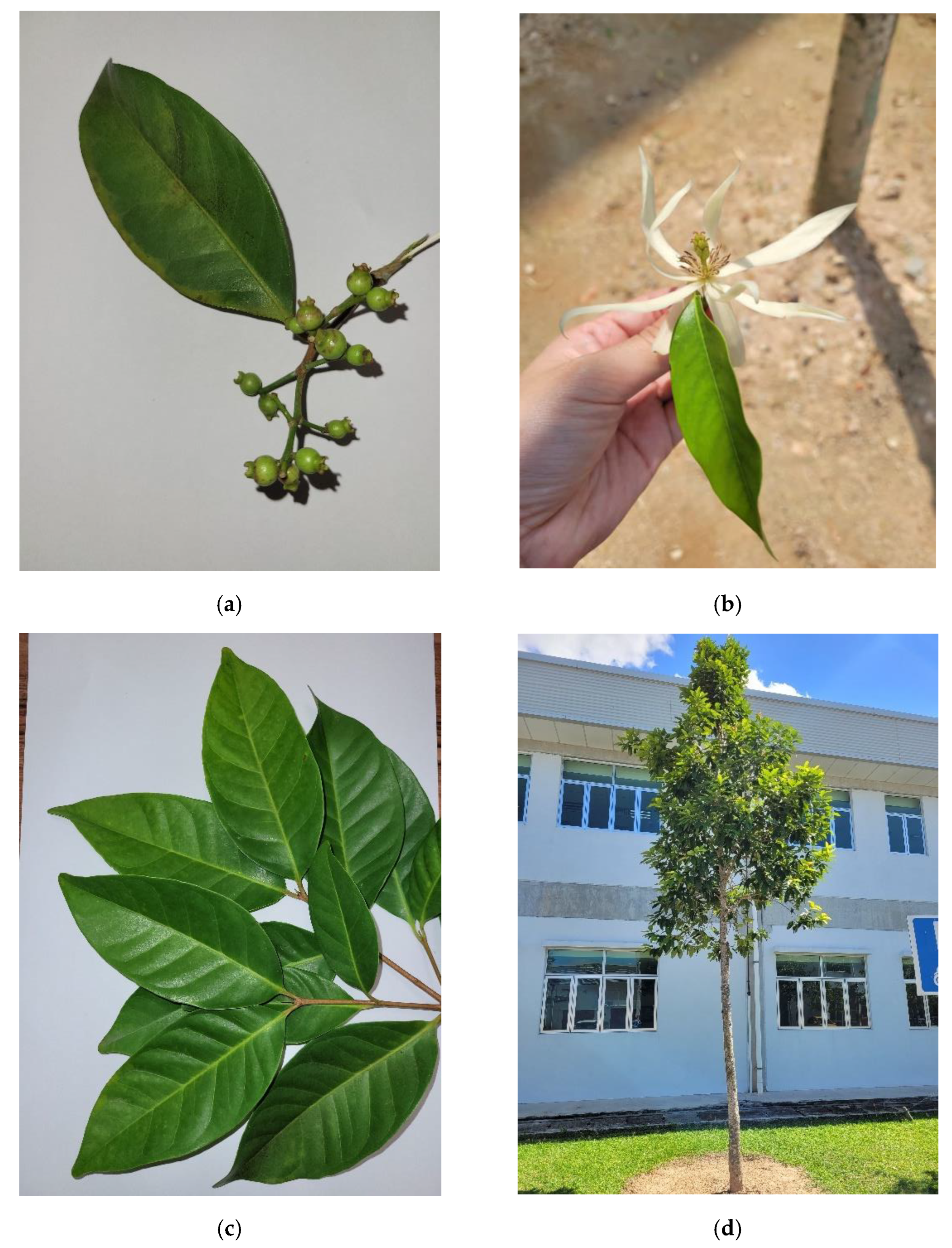
2.3. Distribution and Plant Morphology
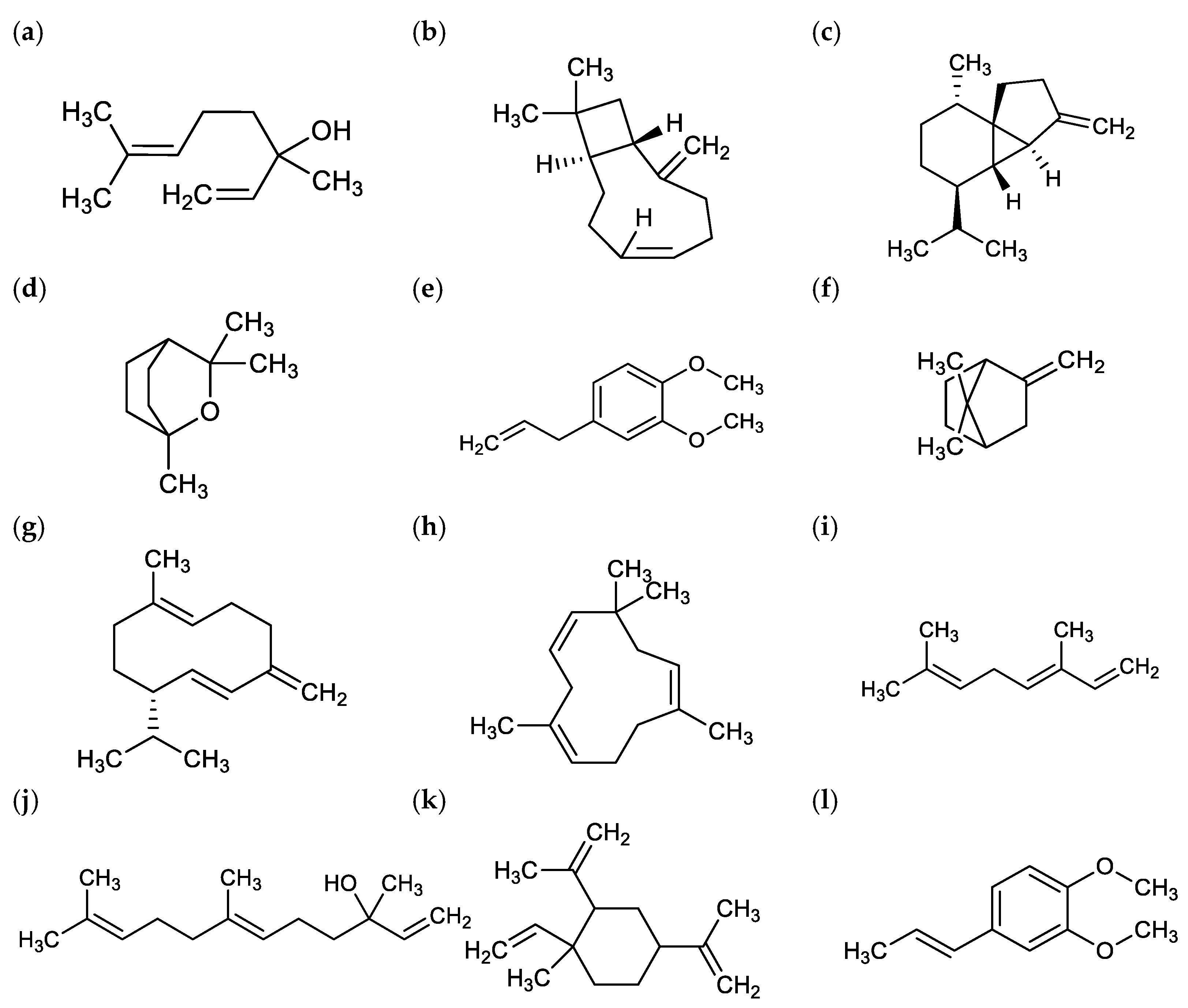
M. alba belongs to the genus Michelia (Magnoliaceae) which consists of about 30 species [5]. M. alba is commonly cultivated in tropical and sub-tropical regions such as Southeast Asia, and it is widely cultivated in China, especially in southern regions such as Fujian, Guangdong, Hainan, Guangxi, and Yunnan [5,6,7]. The flowering plant is also native to Thailand, Indonesia, and Malaysia. In these countries, M. alba is widely cultivated as an ornamental plant [1].
M. alba is an annual flowering plant that can grow as high as 20 m in high humidity regions [5]. The morphological features and pictures of the M. alba plant (Figure 1) are shown in Table 1. The plant starts to produce flowers at a height ranging from 10 to 15 m and the flowering time usually begins in the evening (8–9 pm). The flower scent is said to spread quickly and widely which will be faded in the afternoon. Therefore, the harvesting activities for M. alba flowers are usually conducted in the evening and at dawn [1,5].

3. Phytochemistry

M. alba is best known for its essential oil. Essential oils are first obtained by steam distillation in the Middle Ages by Arabs with characteristics of intense aroma. Its volatile compounds are formed by secondary metabolites extracted from aromatic plants [10]. Essential oil can be extracted from the flowers, leaves, or stems of M. alba [11]. Solvent extraction, steam distillation, and enfleurage are some of the common extraction methods for M. alba essential oil. Steam distillation is commonly used in commercial M. alba essential oil extraction because the processing method is convenient and economical [1,12].
One of the earliest studies in 1982 reported 24 constituents in M. alba flower essential oil namely, β-bisabolene, δ-cadinene, Δ~3-carene, camphene, cis-caryophyllene, α-cubebene, β-cubebene, ο-cymene, trans-carveol, eugenol, isoaristolene, limonene, linalool, cis-linalool oxide, trans-linalool oxide, methyl, methyl 2-methylbutyrate, methyl isoeugenol, β-myrcene, ocimene, α-phellandrene, β-pinene, β-selinene, and α-ylangene [13]. The findings, together with two other comprehensive studies by [14,15] on the chemical constituents of essential oil extracted from M. alba’s flower (F), leaves (L), and stem (S) have identified a total of 168 constituents in which 102 constituents were from the flower (F), 101 from leaves (L), and 77 from the stem (S) (Supplementary Data S1).
Terpenoids are secondary metabolites essential for plant growth and development which contribute to the aroma, flavor, and color of the plant. They protect the plant from environmental stress, pests, and microorganism [16]. Most of the chemical constituents identified are from monoterpene and sesquiterpene. There are differences in the chemical constituents of extracted M. alba oils in published reports. However, the main constituents are similar, i.e., linalool, caryophyllene, β-cubebene, eucalyptol, eugenol methyl ether, α-fenchene, germacrene D, α-humulene, (E)-ocimene, nerolidol, 2,4-diisopropenyl-1-methyl-1-vinylcyclohexane, and isoeugenyl methyl ether (Figure 2) [11,14,15,16]. M. alba contains relatively high levels of linalool in various plant parts. It was reported that the linalool concentration in leaves, flowers, and young twigs was about 0.21 to 0.65%, 1.63 to 4.89%, and 0.43%, respectively [7]. Linalool is especially high in juvenile M. alba flowers, which was reported to be ten-fold higher than in leaves and stems. In addition, the fallen leaves of M. alba also have high concentrations of linalool [7].
In addition to extensive studies on the chemical constituents of the M. alba essential oil, the molecular structures of the constituents isolated from different plant parts were also studied with the aim to understand the chemotaxonomy and identification of the medicinal properties of non-volatile compounds. The constituents from M. alba flower, leaves, and stem were extracted with methanol and analyzed with chromatography while compound structures were characterized and identified with spectral analyses. A total of 42 constituents were identified from the extracts of different plant parts of M. alba (Supplementary Data S2). Out of the total 42 constituents, 19 constituents were isolated from the flower (F), 21 isolated from leaves (L), and 20 from the stem (S) [3,17,18]. Different chemical constituents have been identified from various parts of M. alba extracts. Some of the major compounds discovered include aporphines, amide, benzenoids, and steroids. In 2018, a study by [3] discovered a new compound named Michelaine (C16H9NO4) in the flower of M. alba.

4. Bioactivities M. alba Extracts

To date, there are a number of reported bioactivities of M. alba extracts, including tyrosinase inhibition and photoprotective activities, antimicrobial, antidiabetic, anti-inflammatory, and antioxidant activities.

4.1. Tyrosinase Inhibition and Photoprotective Activities

Tyrosinase is an enzyme that catalyzes the production of melanin. Overexpression of tyrosinase can cause various dermatologic disorders including post-inflammatory hyperpigmentation [19]. This condition is not only aesthetically undesirable, but it may affect patients’ emotions and quality of life [20]. It has been reported that (−)-N-formylanonaine, a purified compound isolated from M. alba inhibits in vitro mushroom tyrosinase activity in a dose-dependent manner with an IC50 value of 74.3 µM [21]. This inhibition activity is comparable to an established tyrosinase inhibitor, kojic acid with a recorded IC50 value of 69.4 µM. In addition, a molecular docking study suggests the tyrosinase inhibitory effect of (−)-N-formylanonaine may be due to its ability to chelate two copper ions in the active site of tyrosinase [21]. In an epidermal melanocytes cell culture study, (−)-N-formylanonaine was found to inhibit human tyrosinase activity at concentration ranges of 10–200 µM. Consequently, melanin content was also found reduced in cells treated with this compound at the same concentrations with an EC50 value of 90 µM [21]. On the other hand, a cell culture study showed the potential of α-terpineol as a skin whitening agent. Treatment of α-terpineol (at 100 and 200 µM) was reported to reduce melanin content and tyrosinase activity in B16 cells stimulated with α-melanocyte-stimulating hormone (α-MSH) [22]. Importantly, α-terpineol at concentrations of 100 and 200 µM did not affect B16 cell viability. In the same cell model, α-terpineol also prevented oxidative stress by reducing cellular malondialdehyde and increased cellular GSH levels. Tyrosinase inhibition activity of phenylethyl alcohol has been reported by [23] using in vitro mushroom tyrosinase assay. This compound was isolated from Rosa rugosa Thunb. var. plena Regal tea. Phenylethyl alcohol inhibits mushroom tyrosinase activity in a dose-dependent manner with an IC50 value of 315 ± 13 μg/mL. However, kojic acid (positive control) showed more potent inhibitory activity with an IC50 value of 80 ± 17 μg/mL.
Exposure to solar ultraviolet (UV) radiation on the skin leads to photoaging. This condition is characterized by the degradation of extracellular matrix (ECM) proteins which include type 1 collagen, elastin, proteoglycans, and fibronectin. This will then damage the connective tissue and reduce the elasticity of the dermis [24]. Irradiation of UV promotes the formation of reactive oxygen species, induces the expression of the mitogen-activated (MAP) kinase signaling pathway, and upregulates the expression of matrix metalloproteinase (MMP)-1, MMP-3, and MMP-9 [24]. M. alba extract inhibits the expression of the three matrix metalloproteinases in UVB-induced activation of p-JNK and p-ERK on cultured human fibroblasts cells and consequently restores total collagen synthesis [24].

4.2. Antimicrobial Activity

Natural products from microorganisms, plants, animals, and algae may serve as a good source of novel antimicrobial compounds [25]. A number of phytochemical extracts from flowers (including M. alba) or their essential oils have been reported to have potential antimicrobial activities for treating various diseases [26,27,28].

4.2.1. Antibacterial and Anti-Fungal Activities

The antimicrobial activity of the Magnolia family may be due to the presence of various bioactive constituents extracted from different parts of the plants. M. alba is rich in carbohydrates, alkaloids, terpenoids, flavonoids, tannins, steroids, and phenols. It has been used not only in traditional medicine but also as a potential antiseptic for the prevention and treatment of microbial infections [29]. M. champaca seed and flower extracts were reported to inhibit the microbial growth of Aeromonas hydrophila, E. coli, Edwardsiella tarda, Flavobacterium spp., Klebsiella pneumonia, Salmonella typhi, Vibrio alginolyticus, V. parahaemolyticus, V. cholerae, Pseudomonas aeruginosa, Staphylococcus aureus, Bacillus subtilis, and Shigella dysenteriae [28,30,31].
M. alba and M. champaca exhibited comparable effects on antibacterial inhibition of S. aureus, E. coli, and Psedumonas aeuroginosa (Table 1). Notably, the antimicrobial activity of leaf oil was found stronger than that of stem oil on growth inhibition of S. aureus ATCC 13709; E. coli ATCC 25922; Candida albican ATCC 10231 [32]. In addition, [33] reported the M. alba dichloromethane leaf extract with 76.6% linalool gave a better inhibitory effect on the growth of Psedumonas aeuroginosa, C. albican, and Fusarium oxysporium compared with the n-pentane flower extract (PF) with 63.2% linalool. The dichloromethane leaf extract was an efficient C. albicans growth inhibitor, while F. oxysporium was more susceptible to the dichloromethane flower extract [33].
The methanol extract of M. alba bark was reported to inhibit the growth of C. Verruculosa, which causes leaf spot disease on rice [34]. It was found that the antifungal activity of M. alba essential oil was strongly correlated with linalool and caryophyllene which are known to inhibit the growth of Aspergillus flavus [11]. In addition, the antifungal activity of M. alba oil against the growth of Aspergillus niger, Aspergillus flavus, Penicillium sp., Rhizopus sp., Fusarium sp., and Cladosporium sp. was demonstrated through the application of the oil to the surface of bamboo paper packaging boxes [35].

4.2.2. Antiparasitics

Anti-parasitic agents have various applications including organic or conventional livestock production systems. Domestic animals such as cattle, pigs, dogs, and cats carry harmful parasites such as Trypanosoma cruzi [36]. T. cruzi can easily infest livestock animals and becomes an endemic that causes a devastating impact on the livestock industry worldwide. The trypanocidal constituents from the ethanol extract of the bark of M. alba (Table 2) showed good antiparasitic activity against T. cruzi [37]. In addition, the pharmacological activities of −anonaine from M. alba have been reviewed by Li and colleagues [38] which showed that the compound gives a significant inhibitory effect against Plasmodium falciparum that causes malaria in humans. The compound also protected red blood cells against P. falciparum. As the compound shows low cytotoxicity in the Chinese Ovarian cell line, it may be a potential phytochemical compound for the treatment of malaria (The Pharmacological Ac.).

4.3. Anti-Diabetic Activity

The anti-diabetic potential of M. alba essential oil was demonstrated through the inhibition of α-amylase, a digestive enzyme found in saliva and pancreatic juice. This enzyme digests complex carbohydrates into oligosaccharides and disaccharides. α-amylase inhibitors delay the hydrolysis of carbohydrates in the intestines [41]. Therefore, inhibition of α-amylase may serve as a therapeutic target for the prevention and medical treatment of diabetes [42]. The essential oil from M. alba inhibits α-amylase activity with an IC50 value of 0.67 mg/mL. The inhibition activity is lower than the positive control, acarbose, which showed an IC50 value of 0.06 mg/mL. GC-MS analysis of essential oil indicated the presence of β-linalool (65.03%) as its major compound [43]. A molecular docking study suggests the β-linalool forms hydrogen bonds with His-299 and Asp-300 residues of α-amylase with a binding energy of − 5.20 kcal/mol [43]. On the other hand, aldose reductase is an enzyme that converts glucose into sorbitol in the presence of nicotinamide adenine dinucleotide phosphate (NADPH). Accumulation of sorbitol in the cells has been associated with the development of diabetic neuropathy. Aldose reductase inhibitor can be used as a target to reduce the concentration of sorbitol in the cells. Lee et al. [44] reported that M. alba flower extract dose-dependently inhibits aldose reductase activity with an IC50 value of 1.98 µg/mL.

4.4. Anti-Inflammatory Activity

Gout is an inflammatory arthritis characterized by the accumulation of uric acid in the blood and further deposited within visceral tissues and joints. Xanthine oxidase catalyzes the oxidation of hypoxanthine to xanthine and its further conversion to uric acid. A number of plant extracts and their metabolites showed inhibition against xanthine oxidase [45]. Leaves extract of M. alba inhibits in vitro xanthine oxidase activity by 22.49% at a concentration of 100 µg/mL. The observed inhibition activity is higher than Gliricidia sepium which showed 6.94% at the same concentration. However, the inhibition activity of M. alba extract was found lower than several medicinal plants such as Antegonon leptopus (59%), Mimosa pudica (62.36%), and Vitex negundo (38.4%) at 100 µg/mL [46].

4.5. Antioxidant Activity

Oxidative stress has been recognized as one of the classical risk factors for human diseases such as cardiovascular diseases, cancers, and neurodegenerative diseases [47]. In biological systems, macromolecules such as lipids, proteins, and nucleic acids are prone to oxidation upon exposure to free radicals. Excessive production of free radicals and a low antioxidant level collectively contribute to oxidative stress leading to a negative impact on physiological function.
In 2018, Zheng and colleagues reported antioxidant activity and phenolics profile of 65 edible flowers in China [48]). In the study, the M. alba flower was extracted using a mixture of acetone/water/acetic acid (70:29.5:0.5, v/v/v). Its 2,2-diphenyl-1-picrylhydrazyl (DPPH) results showed that the extract recorded 58.22 µmol Trolox equivalents (TE)/g sample) on a dry weight basis, higher than several other edible flowers including Panax pseudoginseng (15.18 µmol TE/g sample), Prunella vulgaris (21.39 µmol TE/g sample), and Siraitia grosvenorii (21.03 µmol TE/g sample) [48]. In the 2,2′-azinobis-(3-ethylbenzothiazoline-6-sulfonate) (ABTS) and ferric reducing antioxidant power (FRAP) assays, the extract showed 111.54 µmol TE/g of dry weight sample and 15.51 mmol of Fe2+E/100 g sample, respectively [48]. In another study, petroleum ether extract of M. alba flower showed DPPH free radical scavenging activity with an IC50 value of 0.7155 mg/mL. This inhibition activity is higher than in several other aromatic plants such as Plumeria alba and Cananga odorata [49].

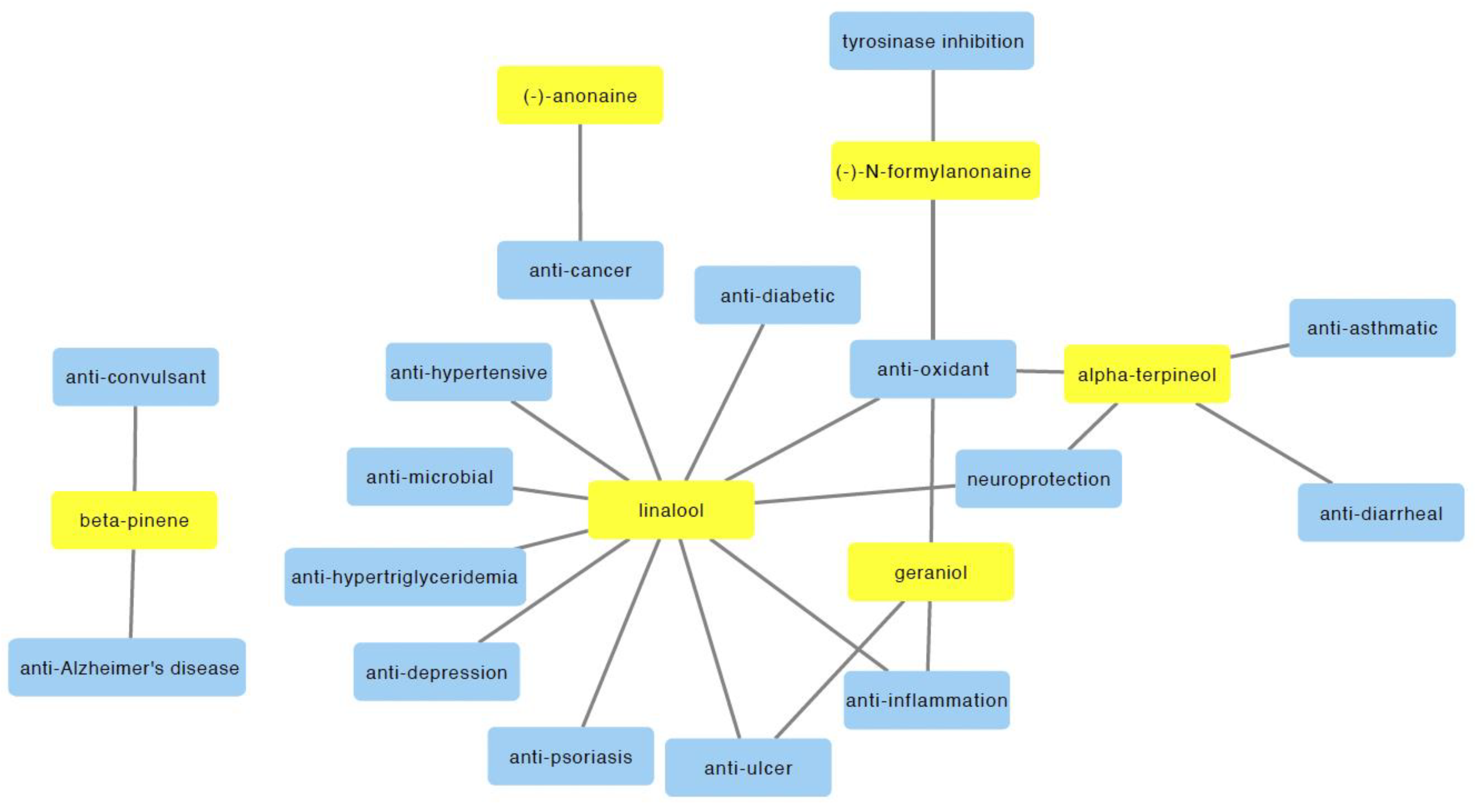
5. Therapeutic Potential of M. alba Metabolites

Despite the limited number of reported bioactivities of M. alba, its bioactive compounds have been associated with numerous therapeutic effects, suggesting the additional unexplored bioactivities of M. alba. In this section, we discuss the bioactivities of the major compounds (including linalool, α-terpineol, and β-pinene, (−)-anonaine, geraniol, (−)-N-formylanonaine, and geraniol) found in M. alba which may provide insight into the additional therapeutic potential of M. alba.

5.1. Anti-Cancer Activity

The potential anti-cancer activities of M. alba may be attributed to two of its bioactive compounds, including linalool and (−)-anonaine. Linalool is the most abundant component in the flower oil (72.8%) and leaf oil (80.1%) of M. alba [2]. It was reported to exhibit anticancer properties and induce apoptosis in the U937 leukemia cells and HeLa cervical cancer cells [50]. The anticancer activity may be associated with the expression of cyclin-dependent kinase inhibitors (CDKIs) as well as the non-expression of cyclin-dependent kinases (CDK) activity [50]. The results are consistent with a study by Gu and colleagues which showed that linalool is an activator of the p53/CDKI axis and inhibited the growth of a number of leukemia cells with wild-type p53 (including Kasumi-1, HL-60, Molt-4, and Raji cells) [51].
In another in vitro study, linalool treatment led to the selective inhibition of the growth of RPMI 7931 human melanoma cells, but not the NCTC 2544 normal keratinocytes cell lines [52]. In the study, the anti-cancer activity was attributed to the enhanced caspase 3 activity in the linalool-treated melanoma cells. Furthermore, linalool also showed anti-cancer properties on HCT 116 human colon cancer cell line and inhibited the growth of a human xenografted tumor mouse model with no observable side effects [53]. In the study, its anti-proliferative effect was linked to the induction of hydroxyl radical generation [53]. Using an in vitro cell culture assay, Cherng and colleagues also demonstrated anti-proliferative activity of linalool towards bladder (J82), lung (H661 and H520), cervix (HeLa), stomach (AGS), skin (BCC-1/KMC), bone (U2OS), mouth (OSCC-1/KMC), and kidney (RTCC-1/KMC) carcinoma cell lines [54]. Furthermore, linalool treatment was also found able to suppress the growth of breast (T-47D), colorectal (SW620), liver (Hep G2), lung (A549), and prostate (DU145, 22Rv1) cancer cell lines [55,56,57]. In addition to M. alba, linalool is also found present in several other plants. A coriander seed oil extract that contained 56.5% linalool was reported to decrease the viability of the HepG2 human liver cancer cell line, together with an increase in reactive oxygen species (ROS) and perturbation in mitochondrial activities [58]. In addition, treatment with linalool and lavender angustifolia essential oil (30.5% linalool) significantly inhibited the proliferation of DU145 and PC-3 human prostate cancer cells and suppressed tumor growth in a xenograft model with PC-3 cell transplantation [59].
On the other hand, (−)-anonaine is a bioactive compound found in the leaves of M. alba with reported anti-cancer activity [38]. Previously, Chen and colleagues showed that (−)-anonaine treatment significantly inhibited cell growth and resulted in anti-migratory and DNA-damaging effects on the H1299 human lung carcinoma cells [60]. Furthermore, (−)-anonaine was found to induce DNA damage and reduce viability in HeLa cells but not in noncancerous Madin-Darby canine kidney (MDCK) and Vero cells [61]. The anti-cancer property of (−)-anonaine on HeLa cells may be linked with induced nitric oxide and ROS, loss of mitochondrial membrane potential, activation of caspase 3, 7, 8, and 9, as well as increased expression of apoptosis-related proteins (including Bax, Bcl-2, and p53), and increased poly (ADP-ribose) polymerase cleavage [61].
Taken together, these findings highlight the anti-cancer properties of linalool and (−)-anonaine, the key bioactive components of M. alba, which may have the potential to be developed into therapeutic bioproducts. Notably, Han and colleagues developed linalool-incorporated nanoparticles which demonstrated anti-cancer properties in cell lines (including HeyA8, A2780, and SKOV3ip1) and patient-derived xenograft mouse models of epithelial ovarian cancer [62].

5.2. Anti-Inflammatory Activity

Previously, the anti-inflammatory potential of (−)-linalool, a major component in M. alba oil, has been demonstrated in a rat model of inflammation. In the study, (−)-linalool significantly inhibited carrageenin-induced edema in rats [63]. The anti-inflammatory effect was found more profound than (±) linalool [63]. In a further study, Peana and colleagues showed that (−)-linalool inhibited the production and release of nitric oxide (NO) in lipopolysaccharide-stimulated J774.A1 macrophages [64]. In addition, treatment with lavender essential oil which contained 32.52% linalool was found effective in reducing inflammatory response induced by carrageenan and by croton oil in a rat model [65].
Psoriasis is a common skin problem and is considered a chronic inflammatory disease. The hallmark feature of psoriasis includes hyperproliferation and abnormal differentiation of keratinocytes [66]. Psoriasis leads to negative physical and emotional impacts on the patients. Recently, Rai and colleagues demonstrated the anti-psoriasis potential of linalool. Using a cell-based assay, linalool at concentrations of 0.01 and 0.1% significantly reduced pro-inflammatory cytokines including TNF-α and IL-6 in LPS-activated murine macrophage cells [67]. In addition, an in silico study also showed that linalool may have good binding against several psoriasis targets such as IL-17, IL-22, IL-23, IL-17a, IL-17b, IL-17f, IL-22α1, and IL-22α2.
Furthermore, the anti-psoriasis activity of linalool was investigated in vivo using Imiquimod (IMQ) induced psoriasis-like skin inflammation in a BALB/c mouse model. Administration of linalool at concentrations of 1 and 2% improved IMQ-induced psoriasis mice with improved body weight and decreased ear thickness. Psoriasis area and severity index (PASI) is a gold standard for quantitative assessment of the severity and extent of psoriasis. PASI scoring is calculated based on parameters including thickness, scaling, and erythema of the skin. The recovery percentage of >50 in PASI scores can be considered a significant improvement in psoriasis [68]. Topical application of linalool at 1 and 2% showed 54.76 and 64.28% recovery, respectively, in PASI scores [67]. Linalool at a concentration of 2% also showed significant recovery in Th-1 cytokines (TNF-α and IL-1β) and Th-17 cytokines (IL-17 and IL-22). In the study, immunohistochemistry analysis also showed that linalool reduced the expression of NF-κB, IL-17, and CCR6 in the skin tissue sections. These results suggest the potential of linalool against psoriasis.
On the other hand, geraniol, an M. alba bioactive compound, was found able to improve osteoarthritis conditions in in vitro and in vivo assays. Treatment of geraniol (40 and 80 µM) attenuated the expression of proinflammatory markers (including iNOS, COX-2, PGE2, NO release, TNF-α, and IL-6) expression in IL-1β-activated mouse chondrocytes [69]. This inhibitory activity was found pronounced at gene and protein levels. Extracellular matrices compose of two essential components which are type II collagen and aggrecan. Exposure of IL-1β suppresses the expression of these components. Treatment of geraniol at concentrations of 40 and 80 µM significantly reduced the deleterious effect of IL-1β in mouse chondrocytes. A disintegrin and metalloproteinase with thrombospondin motifs 5 (ADAMTS-5) and matrix metalloproteinases-9 (MMP-9) are degradative enzymes that exhibit proteolytic activity on ECM, hence promoting osteoarthritis development. Exposure to IL-1β enhanced the expression of both enzymes. However, treatment of geraniol reduces the degradation of ECM induced by IL-1β. The mechanism of action by which geraniol exhibits such effect is known to involve inhibition of PI3K/Akt/NF-κB and MAPK activation. The study indicated the potential role of geraniol in inhibiting inflammatory markers that may be useful for the therapy of osteoarthritis.

5.3. Therapeutic Potential of M. alba on Mental Health Disorders

A few recent publications suggested the potential therapeutic effects of bioactive compounds in M. alba on mental health disorders such as depression, cognitive impairment, convulsion, Alzheimer’s disease, and stress. The bioactive compounds involved include linalool, α-terpineol, and β-pinene.
Depression is a common mental disorder that severely affects the quality of life. It has been reported that more than 264 million people suffer from this illness [70]. It is characterized by an inability to experience pleasure, fatigue, lack of appetite, loss of weight, insomnia, and loss of ability to think or concentrate. Previously, anti-depressant activity of linalool was demonstrated on a rat model using the splash test and forced swimming test. The splash test was used to identify depressive symptoms, as characterized by an increase in time for rats to initiate self-cleaning and decreased total self-cleaning time. Intraperitoneal (IP) injection of linalool at 30 mg/kg significantly decreased the latency of self-cleaning initiation and increased the total time of self-cleaning [71]. Linalool treatment showed a similar pattern to those rats that receive fluoxetine, an anti-depressant drug. In the forced swimming test, rats receiving an IP injection of linalool at a dose of 30 mg/kg showed significantly shorter immobility time as compared to the control [71]. These findings indicated the anti-depressant potential of linalool.
Recently, the neuroprotective activity of linalool had been demonstrated using a hemiparkinsonian rat model [72]. In the study, 6-hydroxydopamine (6-OHDA) was used to induce models of Parkinson’s disease (PD). Treatment of linalool at doses of 25, 50, and 100 mg/kg significantly improved the behavioral alteration in the 6-OHDA lesion group as compared to the untreated 6-OHDA group, as measured by apomorphine-induced rotations, open field, and force swimming tests. In addition, linalool treatment also attenuated the level of dopamine, 3,4-dihydroxyphenylacetic acid and homovanillic acid (HVA) compared to the untreated 6-OHDA lesion group. These data suggested the potential neuroprotection activity of linalool.
Cognitive impairment is one of the characteristics of a pathological condition of vascular dementia, Alzheimer’s disease, and ischemic stroke. In a transient cerebral ischemia rat model, α-terpineol has been reported to provide protection against impairment of hippocampal synaptic plasticity and spatial memory [73]. Treatment with α-terpineol at 100 mg/kg through IP injection significantly reduced the escape latency during training trials as evaluated by the Morris water maze method. A similar dose of α-terpineol also facilitated the induction of long-term potentiation in the hippocampus which was persistent over two hours as determined by electrophysiological recording. In addition, α-terpineol (at 100 and 200 mg/kg) also significantly reduced lipid peroxidation in the hippocampus of the rat model (p < 0.05) [73]. These results indicated the potential of this compound in improving cerebral ischemia cognitive impairment.
In another study, anti-convulsant activity of α- and β-pinene have been evaluated using male Swiss albino rats. To determine the lethal dose of both compounds, α- and β-pinene at a concentration of 2000 mg/kg were administered through IP injection to the rats and observed for a duration of 10, 30, 60, 120, 180, and 240 min, and for 14 consecutive days [74]. From the result, both compounds showed a mild sedation effect without any toxicity or death recorded. To determine the anti-convulsant effect, Swiss Albino rats were treated with α-pinene or β-pinene at concentrations of 100–400 mg/kg, with an equimolar combination of α- and β-pinene (400 mg/kg), reference drug diazepam (2 mg/kg), or vehicle solution (0.5% Tween 80 in saline). After 1 h of administration, 80 mg/kg pentylenetetrazol was injected through IP to stimulate seizures in the animal model. Animals were sacrificed at the end of the experiment. The results showed that compounds at a concentration of 400 mg/kg significantly reduced the seizure intensity as compared to the control group (p < 0.05). In addition, the equimolar mixture of both compounds (400 mg/kg) significantly enhanced the latency of the first convulsion. Furthermore, β-pinene (400 mg/kg) and mixture (400 mg/kg) also significantly increased the time of mortality of animals as compared to the controls (p < 0.05).
Acetylcholinesterase inhibitors have been used to treat Alzheimer’s disease. Recently, the potential acetylcholinesterase and butyrylcholinesterase inhibition activities of β-pinene have been demonstrated using a molecular docking technique. The results showed that β-pinene has a binding affinity of −6.4 kcal/mol towards acetylcholinesterase [75]. In the binding site, β-pinene interacts with hydrophobic amino acids such as Trp-83, Phe-329, Phe-330, and His-439. In butyrylcholinesterase, β-pinene showed a binding affinity of −5.6 kcal/mol with Tyr-329, Phe-326, Trp-79, and Ala-325 interactions. In addition, in silico ADMET (absorption, distribution, metabolism, and excretion toxicity) analysis predicted that β-pinene can be absorbed orally and passed through the blood–brain barrier without considerable side effects [75].
Analysis of electroencephalogram (EEG) showed that there are distinctive brain wave patterns associated with brain states and functions. Alpha waves are known for relaxed and passive attention brain state. In contrast, beta waves are observed during alertness, anxiety dominance, and attention [76,77]. A recent study using human subjects reported that inhalation of M. alba essential oil reduces beta waves and increases fast alpha wave activity [77]. The inhalation of linalool, the main component in the essential oil, also showed similar effects [77]. Collectively, M. alba essential oil and its key bioactive ingredient, linalool, may be useful as an anti-stress and sedative agent.

5.4. Antioxidant Activity

In addition to the reported antioxidant activity of M. alba extract, a number of studies had reported antioxidant activities of M. alba metabolites such as (−)-N-formylanonaine, α-terpineol, linalool, and geraniol. The (−)-N-formylanonaine was found to inhibit DPPH free radical scavenging with an IC50 value of 121.4 µM, as compared with ascorbic acid which showed a higher scavenging activity (IC50 value: 52.1 µM) [21]. In a metal chelating ability assay, (−)-N-formylanonaine recorded an IC50 value of 262.1 µM. The reducing power of (−)-N-formylanonaine (100 µM) was reported at 0.56, a moderate value compared with 1.28 for 3-tert-butyl-4-hydroxyanisole (BHA), a standard antioxidant [21]. In addition, Wang et al. [21] reported the isolation of several bioactive compounds including michephyll A from the leaves of M. alba [78]. Analysis of the antioxidant activity of michephyll A (100 µM) using ABTS free radical scavenging and metal chelating activities showed moderate antioxidant activity with 40.5 and 55.2% inhibitions, respectively [78].
On the other hand, Chaudhari and colleagues reported the antioxidant activity of α-terpineol using ABTS and DPPH free radical scavenging assays [79]. The study showed that α-terpineol has an IC50 value of 7.25 µL/mL for ABTS analysis and scavenges DPPH free radical with an IC50 value of 57.86 µL/mL. Encapsulation of α-terpineol with chitosan nanoemulsion enhanced DPPH free radical scavenging activity with an IC50 value of 39.57 µL/mL [79]. In addition to an in vitro assay, the antioxidant activity of α-terpineol has been reported in a rat model fed with a high-fat diet [80]. The intake of both R-(+)- and (–)-α-terpineol enantiomers at a dose of 50 mg/kg significantly reduced serum and liver thiobarbituric acid reactive substances (TBARS) in the rat model [80].
In a human study, the DPPH analysis of blood plasma showed that linalool inhalation increased antioxidant activity in healthy adults and patients with carpal tunnel syndrome (CTS) [81]. In addition, linalool affects CTS patients by reducing blood pressure and pulse rate which may serve as a useful intervention for CTS. In a cell culture assay, linalool acts as an antioxidant against UVB-induced ROS generation and prevents depletion of antioxidant enzymes [82]. It protects cultured skin cells from UVB-induced oxidative stress by modulating MAPK and NF-κB signaling. Furthermore, Jabir and colleagues showed that linalool (50 µg/mL) exhibits high scavenging activity (50.57%) in an in vitro DPPH assay and 56.36% scavenging activity in an H2O2 scavenging assay [83].
The antioxidant potential of geraniol has been demonstrated using cellular and animal models. In a DPPH assay, geraniol exhibits free radical scavenging activity with an IC50 value of 663 nmol [84]. Geraniol (100 nmol) also attenuated the effect of 2,2′-azobis(2-methylpropionamidine) (AAPH)-induced lipid peroxidation in rat brain tissue and sciatic nerve homogenates. In addition, geraniol at a concentration of 10 µM for 24 h reduced reactive oxygen species and hydroperoxides levels, but not GSH in glucose-induced oxidative stress cultured in SH-SY5Y human neuroblastoma cells [84].
Accumulation of aluminum in tissue can induce oxidative stress and toxicity. Exposure to aluminum chloride (AlCl3) (70 mg/kg, IP) in male Wistar rats increased malondialdehyde (MDA) activity and reduced total antioxidant and glutathione peroxidase activities [85]. The oxidative stress induced by AlCl3 in rats was found reversed by co-treatment of geraniol (100 mg/kg, orally) with AlCl3 as indicated by reduced MDA activity and increased total antioxidant and glutathione peroxidase activities. In another in vivo study, oral administration of geraniol (150 mg/kg) for 14 days increased total antioxidant activity, GSH levels, and normalizing MDA level in liver ischemia-reperfusion injury rats through activation of the Nrf2/HO-1 signaling pathway [86].

5.5. Potential Anti-Hypertensive, Anti-Diabetic and Anti-Hypertriglyceridemia Activities

Linalool, the primary bioactive compound in M. alba, was reported to exhibit potential anti-hypertensive, anti-diabetic, and anti-hypertriglyceridemia activities. Oral administration of linalool at a dose of 100 mg/kg showed anti-hypertensive activity in spontaneously hypertensive rats (SHR) [87]. Linalool at the same dose also improved cardiovascular parameters such as decreasing cardiac hypertrophy, enhancing vasodilation, and decreasing vasoconstriction. Cytokine IL-10 also decreased in the linalool-treated SHR model, indicating anti-inflammatory activity [87]. There are no symptoms of drug toxicity observed in treated rats. Interestingly, linalool complexed with β-cyclodextrin exhibited higher anti-hypertensive activity than a pure form [87]. This improvement may be due to the role of β-cyclodextrin in providing greater bioavailability as compared to uncomplexed linalool [87].
In addition, Jun and colleagues [88] reported that linalool reduced triglycerides levels in a dose-dependent manner. At 1 mM, linalool reduced 37% triglyceride level as compared to the control group in cultured hepatocytes. In gene and protein expression assays, linalool (1 mM) increased PPARα expression by 1.9- to 1.8-fold, respectively. Consequently, linalool also resulted in significant upregulation of lipid metabolism genes including FATP4, ACS1, ACOX 1, UPC2, and LPL expression as well as downregulation of APOC3 expression. Jun and colleagues [88] also performed in vivo experiments to study the effect of linalool on plasma triglycerides concentration. The result showed that oral administration of linalool (100 mg/kg body weight/day) on Western diet-fed C57BL/6J mice and human apo E2 mice for 3 weeks reduced plasma triglycerides concentration by 31 and 50%, respectively. Based on metabolomic analysis, linalool treatment reduced saturated fatty acids in mice [88]. These results indicated the potential of linalool in regulating the hepatic transcriptome and plasma metabolome.

5.6. Anti-Ulcer Activity

Peptic ulcer refers to the erosion of mucosa in the stomach (gastric) or small intestine (duodenal). It is well known that the use of NSAIDs and Helicobacter pylori infection are among the main causes of ulcers. Previous studies have demonstrated the therapeutic activity of natural products against peptic ulcers [89]. For example, the anti-ulcer activity of linalool was studied using ethanol-induced gastric ulcer in a mouse model. Treatment of 10, 20, and 40 mg/kg of linalool significantly reduced gastric lesion area by 85.50, 76.26, and 89.35%, respectively, compared to vehicle-treated mice [90]. In comparison, positive control mice receiving carbenoxolone at a concentration of 100 mg/kg exhibited reduced lesion area by 93.92%. In acetic acid-induced gastric ulcers, treatment of oral linalool (40 mg/kg) for 14 days also significantly reduced gastric lesion area by 48% without showing any sign of toxicity in mice. The incorporation of linalool with β-cyclodextrins further improved the gastroprotection effect of linalool alone against gastric ulcers [90].
In addition, the anti-ulcer activity of geraniol has been tested using an in vivo assay [91]. In the study, acetic acid injection and H. pylori inoculation were used to induce ulcers in adult male Sprague-Dawley rats. After induction of ulcers, the rats were administered with geraniol (at 15 and 30 mg/kg), vehicle, or a mixture of standard drugs (amoxicillin, 50 mg/kg; clarithromycin, 25 mg/kg; and omeprazole, 20 mg/kg) twice daily for a duration of 14 days. After treatments, the animals were sacrificed for evaluation of gastric ulcers. The results showed a significant reduction in ulcer index (UI) when rats were treated with geraniol at concentrations of 15 mg/kg (UI: 2.20) and 30 mg/kg (UI: 2.00) when compared to the control group with H. pylori (UI: 4.13). Geraniol also increased GSH levels and reduced MPO activity and gastric secretion in this rodent model. Histopathological evaluation revealed that treatment of geraniol (30 mg/kg) significantly reduced lesion score (score: 3.5) as compared to the H. pylori control group (9.0). Through Giemsa staining, geraniol at both concentrations reduced H. pylori load in the gastric mucosa as compared to controls with the H. pylori group. This study showed the gastro-protective effect of geraniol against gastric ulcers and H. pylori in rats.

5.7. Anti-Diarrheal Activity

Previously, a study showed that treatment of α-terpineol at all tested concentrations (6.25, 12.5, 25, and 50 mg/kg) significantly reduced castor oil-induced diarrheal in mice (47.11, 65.95, 55.62, and 10.33%, respectively) [92]. The same doses of α-terpineol also reduced stool total weight (55, 48, 44, and 24%, respectively). In addition, α-terpineol also reduced gastrointestinal transit (31%). Mice receiving 12.5 mg/kg α-terpineol also significantly reduced the effect of PGE2-induced diarrheal (39%). Furthermore, α-terpineol reduced fluid formation and loss of chloride ions by interacting with GM1 receptors and cholera toxin, which resulted in increased uptake of intestinal fluids, therefore showing anti-diarrheal activity of this compound in the mouse model [92].

5.8. Anti-Asthmatic Activity

Treatment of α-terpineol (0.75 and 1.25 mmol/L) was found to induce relaxation in the tracheal smooth muscle of a guinea pig model [93]. Several derivatives of α-terpineol, including 2-(4-methylcyclohex-3-enyl)propan-2-yl 2-(2-hydroxyethylthio)acetate, showed even higher relaxation activity than α-terpineol. The compound also showed anti-asthmatic activity in the ovalbumin-induced asthmatic rat model by decreasing airway resistance (RL) and enhancing the dynamic compliance of the airway. Together, the anti-asthmatic potential of α-terpineol and its derivatives warrant further study and development.

6. Traditional Uses and Potential Application of Michelia alba

6.1. Ethnomedicinal Uses

Michelia species have been used as a cancer treatment by natives in India and China. In India, M. champaca has been used to treat abdominal tumors, while in China, M. hypoleuca and M. officinalis were used for the treatment of carcinomatous sores and leukemia [17,94]. M. alba is well known for its wide variety of medicinal and traditional uses from the flower, barks, roots, and leaves. M. alba has a long history of safe use in traditional medicine. It has been used for the treatment of fever, syphilis, gonorrhea, malaria, and prevention of bronchitis, prostatitis, and leucorrhea, and the flower was used traditionally as an abortive agent in Asian countries [5,6,16,17]. In addition, the plant is also believed to be useful in treating abnormal vaginal discharge and irregular menstrual cycles [95]. The essential oil of M. alba is used to treat inflammatory conditions and cancer [11]. M. alba was also found to suppress cough and treat expectorant and bronchitis [1].
The flower of M. alba has a strong alluring and sweet floral scent. This property makes it a common garland in traditional ceremonies and religious offerings in Asia. In addition, the M. alba flower was often used in aromatherapy for mental disorders treatment and the dried flowers are also used in Thailand for heart and nerve care as well as motion sickness [1,96,97]. Besides medicinal use, M. alba leaves extraction was reported to be utilized as a repellent agent [98].
In Thailand, the flowers and leaves of M. alba were said to reduce mucus production, prevent bad breath, and break down kidney stones [34]. Furthermore, the bark of M. alba was reported to use for the treatment of malaria, syphilis, and gonorrhea, and to reduce fever symptoms and the white fragrant flower is used traditionally as an abortive agent [5]. In the Philippines, M. alba is used to treat inflammation disorders including gout, rheumatism, and asthma [46].

6.2. Food Additive and Preservative

Bioactive compounds (e.g., linalool) isolated from M. alba have been approved as a natural food additive and are deliberated to be generally recognized as safe (GRAS) by the FDA as a synthetic flavoring substance and adjuvant in food for human consumption including as an ingredient in animal drugs, feeds, and related products [99]. In 2017, Songsamoe and colleagues developed antifungal fragrant brown rice using the vapor phase of M. alba oil [39]. As bioactive compounds in M. alba such as linalool, caryophyllene and β-elements may affect the sensory quality leading to an anti-stress effect [11], the researchers demonstrated that the application of M. alba oil in the vapor phase is capable of improving consumer acceptance and preference for cooked brown rice while significantly controlling the growth of A. flavus, i.e., molds in brown rice [35]. In addition, the application of M. alba oil or isolated compounds such as linalool may help to protect natural packaging materials made from high carbon content materials from the growth of molds [35]. Taken together, M. alba is a potential natural food preservative and may enhance food flavor.

7. Conclusions

In summary, this review summarizes the published findings on the phytochemistry, bioactivities, and traditional uses of M. alba. A summary of its potential bioactivities is shown in Figure 3. However, some of the reported in vitro studies involved treatments with high concentrations of M. alba compounds. It should be noted that future studies are crucial to identify the precise therapeutic ranges of these compounds in humans based on different age groups. Notably, there is accumulating evidence showing the multiple therapeutic potential and applications of linalool, the primary bioactive compound in M. alba oil. Nevertheless, the pharmacological potential of M. alba has not been fully uncovered, which may be further studied by a combination of in vitro and in vivo bioactivity assays, network pharmacology, and in silico bioactivity prediction methods. In addition, further preclinical and clinical studies are required to validate the therapeutic potential of M. alba and its bioactive compounds, their interaction, safety, efficacy, and underlying mechanism of action for future clinical applications.

Supplementary Materials

The following supporting information can be downloaded at: https://0-www-mdpi-com.brum.beds.ac.uk/article/10.3390/molecules27113450/s1, Supplementary Data S1: The constituents identified from the essential oil (EO) extracted from flower (F), leaves (L), and stem (S) of Michelia alba and Supplementary Data S2: Constituents identified from the extracts of different plant part of M. alba.

Author Contributions

Writing (Section 1, Section 2, Section 3 and Section 6), review and editing, H.-Y.L.; writing (Section 4, Section 5 and Section 6) and review, K.-K.C.; writing (Section 4 and Section 5), M.H.N.; writing (Section 4 and Section 6), N.Z.O.; chemical drawing and validation, Y.-C.L. and S.N.A.A.R. All authors have read and agreed to the published version of the manuscript.

Funding

This work was supported by the Universiti Teknologi Malaysia IIIG Grant (Q.J130000.3609.02M35).

Institutional Review Board Statement

Not applicable.

Informed Consent Statement

Not applicable.

Data Availability Statement

Not applicable.

Conflicts of Interest

The authors declare that there is no conflict of interest regarding the publication of this paper.

References

  1. Pensuk, W.; Padumanonda, T.; Pichaensoonthon, C. Comparison of the chemical constituents in Michelia alba flower oil extracted by steam distillation, hexane extraction and enfleurage method. J. Thai Tradit. Altern. Med. 2007, 5, 30–39. [Google Scholar]
  2. Ueyama, Y.; Hashimoto, S.; Nii, H.; Furukawa, K. The chemical composition of the flower oil and the leaf oil of Michelia alba D.C. J. Essent. Oil Res. 1992, 4, 15–23. [Google Scholar] [CrossRef]
  3. Chen, C.; Kao, C.; Li, W.; Yeh, H.; Huang, S.; Li, H. Chemical Constituents of the Flowers of Michelia alba. Chem. Nat. Compd. 2018, 54, 512–514. [Google Scholar] [CrossRef]
  4. Royal Botanic Gardens, Kew—Plants of the World Online Herbarium Specimen: K000681585. Available online: https://powo.science.kew.org/taxon/20011680-1 (accessed on 14 April 2022).
  5. Lee, C.H.; Chen, H.L.; Li, H.T.; Chao, W.Y.; Chen, C.Y. Review on pharmacological activities of Michelia alba. Int. J. Pharm. Ther. 2014, 5, 289–292. [Google Scholar]
  6. Liang, C.B.; Nooteboom, H.P. Notes on Magnoliaceae III: The Magnoliaceae of China. Ann. Mo. Bot. Gard. 1993, 80, 999–1104. [Google Scholar] [CrossRef]
  7. Xia, E.Q.; Song, Y.; Ai, X.X.; Guo, Y.J.; Xu, X.R.; Li, H.B. A new high-performance liquid chromatographic method for the determination and distribution of linalool in Michelia alba. Molecules 2010, 15, 4890–4897. [Google Scholar] [CrossRef]
  8. Tan, S.; Abdullah, T.; Go, R. Effect of plant growth regulators on cutting propagation of cempaka putih (Magnolia alba) and cempaka kuning (Magnolia champaca). Plant Product. Environ. Conserv. 2018, 25, 124. [Google Scholar]
  9. Noteboom, H. Notes on Magnoliaceae with a revision of Pachylarnax and Elmerrillia and the Malasian species of Manglietia and Michelia. Blumea 1985, 31, 65–121. [Google Scholar]
  10. Bakkali, F.; Averbeck, S.; Averbeck, D.; Idaomar, M. Biological effects of essential oils–a review. Food Chem. Toxicol. 2008, 46, 446–475. [Google Scholar] [CrossRef]
  11. Songsamoe, S.; Matan, N.; Matan, N. Antifungal activity of Michelia alba oil in the vapor phase and the synergistic effect of major essential oil components against Aspergillus flavus on brown rice. Food Control 2017, 77, 150–157. [Google Scholar] [CrossRef]
  12. Fukushima, S.; Cohen, S.M.; Eisenbrand, G.; Gooderham, N.J.; Guengerich, F.P.; Hecht, S.S.; Rietjens, I.M.C.M.; Rosol, T.J.; Davidsen, J.M.; Harman, C.L.; et al. FEMA GRAS assessment of natural flavor complexes: Lavender, Guaiac Coriander-derived and related flavoring ingredients. Food Chem. Toxicol. 2020, 145, 111584. [Google Scholar] [CrossRef] [PubMed]
  13. Zhu, L.; Lu, B.; Xu, D. A preliminary study on the chemical constituents of the essential oil of Michelia Alba D.C. J. Integr. Plant Biol. 1982, 24, 355–359. [Google Scholar]
  14. Huang, X.; Yin, Y.; Huang, R.; Chen, M.; Ge, P.; Ma, Z.; Gui, H. Study on chemical constituents of essential oils from leaves and stems of Michelia alba D.C. Food Sci. 2009, 8, 241–244. [Google Scholar]
  15. Huang, X.; Yin, Y.; Liu, X.; Duan, L.; Liu, T.; Yang, Y.; Yu, M. Studies on chemical constituents of volatile oils from the flowers and leaves of Michelia alba D.C. in Yunnan. Chem. Ind. For. Prod. 2009, 29, 119–123. [Google Scholar]
  16. Kumar, D.; Kumar, S.; Taprial, S.; Kashyap, D.; Kumar, A.; Prakash, O. A review of chemical and biological profile of genus Michelia. J. Chin. Integr. Med. 2012, 10, 1336–1341. [Google Scholar] [CrossRef]
  17. Chen, C.; Huang, L.; Chen, L.; Lo, W.; Kuo, S.; Wang, Y.; Kuo, S.; Hsieh, T. Chemical constituents from the leaves of Michelia alba. Chem. Nat. Compd. 2008, 44, 137–139. [Google Scholar] [CrossRef]
  18. Lo, W.-L.; Huang, L.-Y.; Wang, H.-M.; Chen, C.-Y. Chemical constituents from the stems of Michelia alba. Chem. Nat. Compd. 2010, 46, 664–665. [Google Scholar] [CrossRef]
  19. Vashi, N.A.; Kundu, R.V. Facial hyperpigmentation: Causes and treatment. Br. J. Dermatol. 2013, 169, 41–56. [Google Scholar] [CrossRef]
  20. Darji, K.; Varade, R.; West, D.; Armbrecht, E.S.; Guo, M.A. Psychosocial impact of postinflammatory hyperpigmentation in patients with acne vulgaris. J. Clin. Aesthetic Dermatol. 2017, 10, 18. [Google Scholar]
  21. Wang, H.-M.; Chen, C.-Y.; Chen, C.-Y.; Ho, M.-L.; Chou, Y.-T.; Chang, H.-C.; Lee, C.-H.; Wang, C.-Z.; Chu, I.-M. (−)-N-Formylanonaine from Michelia alba as a human tyrosinase inhibitor and antioxidant. Bioorg. Med. Chem. 2010, 18, 5241–5247. [Google Scholar] [CrossRef]
  22. Chao, W.-W.; Su, C.-C.; Peng, H.-Y.; Chou, S.-T. Melaleuca quinquenervia essential oil inhibits α-melanocyte-stimulating hormone-induced melanin production and oxidative stress in B16 melanoma cells. Phytomedicine 2017, 34, 191–201. [Google Scholar] [CrossRef] [PubMed]
  23. Ren, G.; Xue, P.; Sun, X.; Zhao, G. Determination of the volatile and polyphenol constituents and the antimicrobial, antioxidant, and tyrosinase inhibitory activities of the bioactive compounds from the by-product of Rosa rugosa Thunb. var. plena Regal tea. BMC Complementary Altern. Med. 2018, 18, 307. [Google Scholar] [CrossRef] [PubMed] [Green Version]
  24. Chiang, H.-M.; Chen, H.-C.; Lin, T.-J.; Shih, I.-C.; Wen, K.-C. Michelia alba extract attenuates UVB-induced expression of matrix metalloproteinases via MAP kinase pathway in human dermal fibroblasts. Food Chem. Toxicol. 2012, 50, 4260–4269. [Google Scholar] [CrossRef] [PubMed]
  25. Abdel-Razek, A.S.; El-Naggar, M.E.; Allam, A.; Morsy, O.M.; Othman, S.I. Microbial natural products in drug discovery. Processes 2020, 8, 470. [Google Scholar] [CrossRef] [Green Version]
  26. Al-Shukaili, N.B.M.B.A.; Hossain, M.A. Antimicrobial and cytotoxic potential of seeds and flowers crude extracts of sunflower. Grain Oil Sci. Technol. 2019, 2, 103–108. [Google Scholar] [CrossRef]
  27. Pathania, R.; Khan, H.; Kaushik, R.; Khan, M.A. Essential oil nanoemulsions and their antimicrobial and food applications. Curr. Res. Nutr. Food Sci. 2018, 6, 626–643. [Google Scholar] [CrossRef]
  28. Wei, L.S.; Wee, W.; Siong, J.Y.F.; Syamsumir, D.F. Characterization of antimicrobial, antioxidant, anticancer property and chemical composition of Piper betle L. Leaf Extr. Arab. Gulf. J. Sci. Res. 2017, 35, 40–45. [Google Scholar]
  29. Elizabeth, K.M.; Jaya Lakshmi, Y.A.S. Antimicrobial activity of Michelia champaca. Asian J. Chem. 2006, 18, 196–200. [Google Scholar]
  30. Lavanya, T.; Ananthi, T. Evaluation of Preliminary Antibacterial Activity and Uv-Specroscopic Analysis of Michelia Champaca (L.). Eur. J. Pharm. Med. Res. 2017, 4, 430–434. [Google Scholar]
  31. Parimi, U.; Kolli, D. Antibacterial and free radical scavenging activity of Michelia champaca Linn. flower extracts. Free radic. antioxid. 2012, 2, 58–61. [Google Scholar] [CrossRef] [Green Version]
  32. Ha, C.T.T.; Thai, T.H.; Diep, L.N.; Thanh, T.X.; Thu Thuy, D.T.; Tra, N.T.; Thu Ha, N.T. Chemical composition and antimicrobial activity of the essential oils from stems and leaves of Michelia alba D.C. growing in Vietnam. Acad. J. Biol. 2018, 40. [Google Scholar]
  33. Abu Shah, N. Chemical Constituents and Biological Activities of Essential Oil from Chempaka (Michelia alba De Candolle). Ph.D. Thesis, Universiti Putra Malaysia, Selangor, Malaysia, 18 January 2013. [Google Scholar]
  34. Swantara, I.M.D.; Bawa, I.G.A.G.; Suprapta, D.N.; Agustina, K.K.; Temaja, I.G.R.M. Identification Michelia alba barks extract using Gas Chromatography-Mass Spectrometry (GC-MS) and its antifungal properties to inhibit microbial growth. Biodiversitas J. Biol. Divers. 2020, 21, 1541–1550. [Google Scholar] [CrossRef]
  35. Suhem, K.; Matan, N.; Matan, N.; Danworaphong, S.; Aewsiri, T. Enhanced antifungal activity of michelia oil on the surface of bamboo paper packaging boxes using helium-neon (HeNe) laser and its application to brown rice snack bar. Food Control 2017, 73, 939–945. [Google Scholar] [CrossRef]
  36. Gürtler, R.E.; Cecere, M.C.; Lauricella, M.A.; Cardinal, M.V.; Kitron, U.; Cohen, J.E. Domestic dogs and cats as sources of Trypanosoma cruzi infection in rural northwestern Argentina. Parasitology 2007, 134, 69–82. [Google Scholar] [CrossRef] [Green Version]
  37. Asaruddin, M.R.; Honda, G.; Tsubouchi, A.; Nakajima-Shimada, J.; Aoki, T.; Kiuchi, F. Trypanocidal constituents from Michelia alba. Nat. Med. 2003, 57, 61–63. [Google Scholar]
  38. Li, H.T.; Wu, H.M.; Chen, H.L.; Liu, C.M.; Chen, C.Y. The pharmacological activities of (−)-anonaine. Molecules 2013, 18, 8257–8263. [Google Scholar] [CrossRef] [Green Version]
  39. Songsamoe, S.; Koomhin, P.; Matan, N. The effects of Michelia alba oil against mould on brown rice and assessing the brain response using electroencephalogram (EEG). J. Food Sci. Technol. 2021, 58, 1776–1787. [Google Scholar] [CrossRef]
  40. Nasution, R.; Azwar, A.I.; Helwati, H.; Marianne. Antibacterial Activities of Perfume: Combination Flower Magnolia alba, Cananga odorata and Mimusops elengi L, Fixed with Pogostemon cablin Oil. Indones. J. Pharm. Clin. Res. 2019, 2, 19–23. [Google Scholar] [CrossRef]
  41. Kazeem, M.; Adamson, J.; Ogunwande, I. Modes of inhibition of α-amylase and α-glucosidase by aqueous extract of Morinda lucida Benth leaf. BioMed Res. Int. 2013, 2013, 527570. [Google Scholar] [CrossRef] [Green Version]
  42. Narita, Y.; Inouye, K. Chapter 84—Inhibition of Porcine Pancreas α-Amylase by Chlorogenic Acids from Green Coffee Beans and Cinnamic Acid Derivatives: A Focus on Kinetic. In Coffee in Health and Disease Prevention; Preedy, V.R., Ed.; Academic Press: San Diego, CA, USA, 2015; pp. 757–763. [Google Scholar]
  43. Khammee, T.; Jaratrungtawee, A.; Kuno, M. Gas Chromatography-Mass Spectrometry Analysis, In Vitro Activities, and In Silico Molecular Docking of Major Components of Michelia Alba D.C. Essential Oil and Scented Extracts. Asian J. Pharm. Clin. Res. 2018, 11, 499–504. [Google Scholar] [CrossRef]
  44. Lee, Y.M.; Kim, Y.S.; Bae, K.H.; Kim, J.H.; Kim, J.S. Screening of Chinese herbal medicines with inhibitory effect on aldose reductase (IV). Korean J. Pharmacogn. 2010, 41, 289–296. [Google Scholar]
  45. Abu Bakar, F.I.; Abu Bakar, M.F.; Rahmat, A.; Abdullah, N.; Sabran, S.F.; Endrini, S. Anti-gout potential of Malaysian medicinal plants. Front. Pharmacol. 2018, 9, 261. [Google Scholar] [CrossRef] [PubMed] [Green Version]
  46. Apaya, K.L.; Chichioco-Hern, C.L. Xanthine oxidase inhibition of selected Philippine medicinal plants. J. Med. Plants Res. 2011, 5, 289–292. [Google Scholar] [CrossRef]
  47. Yang, Q.; Cai, X.; Yan, A.; Tian, Y.; Du, M.; Wang, S. A specific antioxidant peptide: Its properties in controlling oxidation and possible action mechanism. Food Chem. 2020, 327, 126984. [Google Scholar] [CrossRef] [PubMed]
  48. Zheng, J.; Yu, X.; Maninder, M.; Xu, B. Total phenolics and antioxidants profiles of commonly consumed edible flowers in China. Int. J. Food Prop. 2018, 21, 1524–1540. [Google Scholar] [CrossRef] [Green Version]
  49. Leelapornpisid, P.; Chansakaow, S.; Chaiyasut, C.; Wongwattananukul, N. Antioxidant activity of some volatile oils and absolutes from Thai aromatic plants. Int. Workshop Med. Aromat. Plants 2007, 786, 61–66. [Google Scholar] [CrossRef]
  50. Chang, M.Y.; Shieh, D.E.; Chen, C.C.; Yeh, C.S.; Dong, H.P. Linalool induces cell cycle arrest and apoptosis in leukemia cells and cervical cancer cells through CDKIs. Int. J. Mol. Sci. 2015, 16, 28169–28179. [Google Scholar] [CrossRef] [Green Version]
  51. Gu, Y.; Ting, Z.; Qiu, X.; Zhang, X.; Gan, X.; Fang, Y.; Xu, X.; Xu, R. Linalool preferentially induces robust apoptosis of a variety of leukemia cells via upregulating p53 and cyclin-dependent kinase inhibitors. Toxicology 2010, 268, 19–24. [Google Scholar] [CrossRef]
  52. Cerchiara, T.; Straface, S.; Brunelli, E.; Tripepi, S.; Gallucci, M.C.; Chidichimo, G. Antiproliferative effect of linalool on RPMI 7932 human melanoma cell line: Ultrastructural studies. Nat. Prod. Commun. 2015, 10. [Google Scholar] [CrossRef] [Green Version]
  53. Iwasaki, K.; Zheng, Y.W.; Murata, S.; Ito, H.; Nakayama, K.; Kurokawa, T.; Sano, N.; Nowatari, T.; Villareal, M.O.; Nagano, Y.N.; et al. Anticancer effect of linalool via cancer-specific hydroxyl radical generation in human colon cancer. World J. Gastroenterol. 2016, 22, 9765. [Google Scholar] [CrossRef] [Green Version]
  54. Cherng, J.M.; Shieh, D.E.; Chiang, W.; Chang, M.Y.; Chiang, L.C. Chemopreventive effects of minor dietary constituents in common foods on human cancer cells. Biosci. Biotechnol. Biochem. 2007, 71, 1500–1504. [Google Scholar] [CrossRef] [PubMed]
  55. Chang, M.Y.; Shen, Y.L. Linalool exhibits cytotoxic effects by activating antitumor immunity. Molecules 2014, 19, 6694–6706. [Google Scholar] [CrossRef] [Green Version]
  56. Sun, X.B.; Wang, S.M.; Li, T.; Yang, Y.Q. Anticancer activity of linalool terpenoid: Apoptosis induction and cell cycle arrest in prostate cancer cells. Trop. J. Pharm. Res. 2015, 14, 619–625. [Google Scholar] [CrossRef] [Green Version]
  57. Zhao, Y.; Cheng, X.; Wang, G.; Liao, Y.; Qing, C. Linalool inhibits 22Rv1 prostate cancer cell proliferation and induces apoptosis. Oncol. Lett. 2020, 20, 289. [Google Scholar] [CrossRef] [PubMed]
  58. Usta, J.; Kreydiyyeh, S.; Knio, K.; Barnabe, P.; Bou-Moughlabay, Y.; Dagher, S. Linalool decreases HepG2 viability by inhibiting mitochondrial complexes I and II, increasing reactive oxygen species and decreasing ATP and GSH levels. Chem. Biol. Interact. 2009, 180, 39–46. [Google Scholar] [CrossRef] [PubMed]
  59. Zhao, Y.; Chen, R.; Wang, Y.; Qing, C.; Wang, W.; Yang, Y. In vitro and in vivo efficacy studies of Lavender angustifolia essential oil and its active constituents on the proliferation of human prostate cancer. Integr. Cancer Ther. 2017, 16, 215–226. [Google Scholar] [CrossRef] [PubMed] [Green Version]
  60. Chen, B.H.; Chang, H.W.; Huang, H.M.; Chong, I.W.; Chen, J.S.; Chen, C.Y.; Wang, H.M. (−)-Anonaine induces DNA damage and inhibits growth and migration of human lung carcinoma h1299 cells. J. Agric. Food Chem. 2011, 59, 2284–2290. [Google Scholar] [CrossRef]
  61. Chen, C.Y.; Liu, T.Z.; Tseng, W.C.; Lu, F.J.; Hung, R.P.; Chen, C.H.; Chen, C.H. (−)-Anonaine induces apoptosis through Bax-and caspase-dependent pathways in human cervical cancer (HeLa) cells. Food Chem. Toxicol. 2008, 46, 2694–2702. [Google Scholar] [CrossRef]
  62. Han, H.D.; Cho, Y.-J.; Cho, S.K.; Byeon, Y.; Jeon, H.N.; Kim, H.-S.; Kim, B.-G.; Bae, D.-S.; Lopez-Berestein, G.; Anil, K.; et al. Linalool-Incorporated Nanoparticles as a Novel Anticancer Agent for Epithelial Ovarian Carcinoma. Mol. Cancer Ther. 2016, 15, 618–627. [Google Scholar] [CrossRef] [Green Version]
  63. Peana, A.T.; D’Aquila, P.S.; Panin, F.; Serra, G.; Pippia, P.; Moretti, M.D.L. Anti-inflammatory activity of linalool and linalyl acetate constituents of essential oils. Phytomedicine 2002, 9, 721–726. [Google Scholar] [CrossRef]
  64. Peana, A.T.; Marzocco, S.; Popolo, A.; Pinto, A. (−)-Linalool inhibits in vitro NO formation: Probable involvement in the antinociceptive activity of this monoterpene compound. Life Sci. 2006, 78, 719–723. [Google Scholar] [CrossRef] [PubMed]
  65. Da Silva, G.L.; Luft, C.; Lunardelli, A.; Amaral, R.H.; da Silva Melo, D.A.; Donadio, M.V.F.; Nunes, F.B.; de Azambuja, M.S.; Santana, J.C.; Moraes, C.M.B.; et al. Antioxidant, analgesic and anti-inflammatory effects of lavender essential oil. An. Acad. Bras. Cienc. 2015, 87, 1397–1408. [Google Scholar] [CrossRef] [PubMed] [Green Version]
  66. Ronpirin, C.; Tencomnao, T. Dithranol downregulates expression of Id1 mRNA in human keratinocytes in vitro. Genet. Mol. Res. 2012, 11, 3290–3297. [Google Scholar] [CrossRef] [PubMed]
  67. Rai, V.K.; Sinha, P.; Yadav, K.S.; Shukla, A.; Saxena, A.; Bawankule, D.U.; Tandon, S.; Khan, F.; Chanotiya, C.S.; Yadav, N.P. Anti-psoriatic effect of Lavandula angustifolia essential oil and its major components linalool and linalyl acetate. J. Ethnopharmacol. 2020, 261, 113127. [Google Scholar] [CrossRef]
  68. Krueger, G.G.; Langley, R.G.; Leonardi, C.; Yeilding, N.; Guzzo, C.; Wang, Y.; Dooley, L.T.; Lebwohl, M. A human interleukin-12/23 monoclonal antibody for the treatment of psoriasis. N. Engl. J. Med. 2007, 356, 580–592. [Google Scholar] [CrossRef]
  69. Wu, Y.; Wang, Z.; Fu, X.; Lin, Z.; Yu, K. Geraniol-mediated osteoarthritis improvement by down-regulating PI3K/Akt/NF-κB and MAPK signals: In vivo and in vitro studies. Int. Immunopharmacol. 2020, 86, 106713. [Google Scholar] [CrossRef]
  70. James, S.L.; Abate, D.; Abate, K.H.; Abay, S.M.; Abbafati, C.; Abbasi, N.; Abbastabar, H.; Abd-Allah, F.; Abdela, J.; Abdelalim, A. Global, regional, and national incidence, prevalence, and years lived with disability for 354 diseases and injuries for 195 countries and territories, 1990–2017: A systematic analysis for the Global Burden of Disease Study 2017. Lancet 2018, 392, 1789–1858. [Google Scholar] [CrossRef] [Green Version]
  71. Dos Santos, É.R.Q.; Maia, C.S.F.; Junior, E.A.F.; Melo, A.S.; Pinheiro, B.G.; Maia, J.G.S. Linalool-rich essential oils from the Amazon display antidepressant-type effect in rodents. J. Ethnopharmacol. 2018, 212, 43–49. [Google Scholar] [CrossRef]
  72. de Lucena, J.D.; Gadelha-Filho, C.V.J.; da Costa, R.O.; de Araújo, D.P.; Lima, F.A.V.; Neves, K.R.T.; de Barros Viana, G.S. L-linalool exerts a neuroprotective action on hemiparkinsonian rats. Naunyn-Schmiedebergs Arch. Pharmacol. 2020, 393, 1077–1088. [Google Scholar] [CrossRef]
  73. Moghimi, M.; Parvardeh, S.; Zanjani, T.M.; Ghafghazi, S. Protective effect of α-terpineol against impairment of hippocampal synaptic plasticity and spatial memory following transient cerebral ischemia in rats. Iran. J. Basic Med. Sci. 2016, 19, 960. [Google Scholar]
  74. Felipe, C.F.B.; Albuquerque, A.M.S.; de Pontes, J.L.X.; de Melo, J.Í.V.; Rodrigues, T.C.M.L.; de Sousa, A.M.P.; Monteiro, Á.B.; Ribeiro, A.E.d.S.; Lopes, J.P.; de Menezes, I.R.A. Comparative study of alpha-and beta-pinene effect on PTZ-induced convulsions in mice. Fundam. Clin. Pharmacol. 2019, 33, 181–190. [Google Scholar] [CrossRef]
  75. Karimi, I.; Yousofvand, N.; Hussein, B.A. In vitro cholinesterase inhibitory action of Cannabis sativa L. Cannabaceae and in silico study of its selected phytocompounds. Silico Pharmacol. 2021, 9, 13. [Google Scholar] [CrossRef]
  76. Abhang, P.A.; Gawali, B.W.; Mehrotra, S.C. Technological basics of EEG recording and operation of apparatus. Introd. EEG-Speech-Based Emot. Recognit. 2016, 19–50. [Google Scholar] [CrossRef]
  77. Koomhin, P.; Sattayakhom, A.; Chandharakool, S.; Sinlapasorn, J.; Suanjan, S.; Palipoch, S.; Na-ek, P.; Punsawad, C.; Matan, N. Michelia Essential Oil Inhalation Increases Fast Alpha Wave Activity. Sci. Pharm. 2020, 88, 23. [Google Scholar] [CrossRef]
  78. Wang, H.-M.; Lo, W.-L.; Huang, L.-Y.; Wang, Y.-D.; Chen, C.-Y. Chemical constituents from the leaves of Michelia alba. Nat. Prod. Res. 2010, 24, 398–406. [Google Scholar] [CrossRef]
  79. Chaudhari, A.K.; Singh, A.; Singh, V.K.; Dwivedy, A.K.; Das, S.; Ramsdam, M.G.; Dkhar, M.S.; Kayang, H.; Dubey, N.K. Assessment of chitosan biopolymer encapsulated α-Terpineol against fungal, aflatoxin B1 (AFB1) and free radicals mediated deterioration of stored maize and possible mode of action. Food Chem. 2020, 311, 126010. [Google Scholar] [CrossRef] [PubMed]
  80. de Sousa, G.M.; Cazarin, C.B.B.; Junior, M.R.M.; de Almeida Lamas, C.; Quitete, V.H.A.C.; Pastore, G.M.; Bicas, J.L. The effect of α-terpineol enantiomers on biomarkers of rats fed a high-fat diet. Heliyon 2020, 6, e03752. [Google Scholar] [CrossRef]
  81. Seol, G.-H.; Kang, P.; Lee, H.S.; Seol, G.H. Antioxidant activity of linalool in patients with carpal tunnel syndrome. BMC Neurol. 2016, 16, 17. [Google Scholar] [CrossRef] [PubMed] [Green Version]
  82. Gunaseelan, S.; Balupillai, A.; Govindasamy, K.; Ramasamy, K.; Muthusamy, G.; Shanmugam, M.; Thangaiyan, R.; Robert, B.M.; Prasad Nagarajan, R.; Ponniresan, V.K. Linalool prevents oxidative stress activated protein kinases in single UVB-exposed human skin cells. PLoS ONE 2017, 12, e0176699. [Google Scholar] [CrossRef] [Green Version]
  83. Jabir, M.S.; Taha, A.A.; Sahib, U.I. Antioxidant activity of Linalool. J. Eng. Technol. 2018, 36, 64–67. [Google Scholar]
  84. Prasad, S.N.; Muralidhara, M. Analysis of the antioxidant activity of geraniol employing various in-vitro models: Relevance to neurodegeneration in diabetic neuropathy. Asian J. Pharm. Clin. Res. 2017, 10, 101–105. [Google Scholar] [CrossRef]
  85. Hosseini, S.M.; Hejazian, L.B.; Amani, R.; Badeli, N.S. Geraniol attenuates oxidative stress, bioaccumulation, serological and histopathological changes during aluminum chloride-hepatopancreatic toxicity in male Wistar rats. Environ. Sci. Pollut. Res. 2020, 27, 20076–20089. [Google Scholar] [CrossRef] [PubMed]
  86. El-Emam, S.Z.; Soubh, A.A.; Al-Mokaddem, A.K.; Abo El-Ella, D.M. Geraniol activates Nrf-2/HO-1 signaling pathway mediating protection against oxidative stress-induced apoptosis in hepatic ischemia-reperfusion injury. Naunyn-Schmiedebergs Arch. Pharmacol. 2020, 393, 1849–1858. [Google Scholar] [CrossRef] [PubMed]
  87. Camargo, S.B.; Simoes, L.O.; de Azevedo Medeiros, C.F.; de Melo Jesus, A.; Fregoneze, J.B.; Evangelista, A.; Villarreal, C.F.; de Souza Araujo, A.A.; Quintans-Junior, L.J.; Silva, D.F. Antihypertensive potential of linalool and linalool complexed with β-cyclodextrin: Effects of subchronic treatment on blood pressure and vascular reactivity. Biochem. Pharmacol. 2018, 151, 38–46. [Google Scholar] [CrossRef] [PubMed]
  88. Jun H-j Lee, J.H.; Kim, J.; Jia, Y.; Kim, K.H.; Hwang, K.Y.; Yun, E.J.; Do, K.-R.; Lee, S.-J. Linalool is a PPARα ligand that reduces plasma TG levels and rewires the hepatic transcriptome and plasma metabolome. J. Lipid Res. 2014, 55, 1098–1110. [Google Scholar] [CrossRef] [Green Version]
  89. Serafim, C.; Araruna, M.E.; Júnior, E.A.; Diniz, M.; Hiruma-Lima, C.; Batista, L. A Review of the Role of Flavonoids in Peptic Ulcer (2010–2020). Molecules 2020, 25, 5431. [Google Scholar] [CrossRef]
  90. da Silva, F.V.; de Barros Fernandes, H.; Oliveira, I.S.; Viana, A.F.S.C.; da Costa, D.S.; Lopes, M.T.P.; de Lira, K.L.; Quintans-Júnior, L.J.; de Sousa, A.A.; Oliveira, R.d.C.M. Beta-cyclodextrin enhanced gastroprotective effect of (−)-linalool, a monoterpene present in rosewood essential oil, in gastric lesion models. Naunyn-Schmiedebergs Arch. Pharmacol. 2016, 389, 1245–1251. [Google Scholar] [CrossRef]
  91. Bhattamisra, S.K.; Yan, V.L.Y.; Lee, C.K.; Kuean, C.H.; Candasamy, M.; Liew, Y.K.; Sahu, P.S. Protective activity of geraniol against acetic acid and Helicobacter pylori-induced gastric ulcers in rats. J. Tradit. Complementary Med. 2019, 9, 206–214. [Google Scholar] [CrossRef]
  92. dos Santos Negreiros, P.; da Costa, D.S.; da Silva, V.G.; de Carvalho Lima, I.B.; Nunes, D.B.; de Melo Sousa, F.B.; Araújo, T.d.S.L.; Medeiros, J.V.R.; Dos Santos, R.F.; Oliveira, R.d.C.M. Antidiarrheal activity of α-terpineol in mice. Biomed. Pharmacother. 2019, 110, 631–640. [Google Scholar] [CrossRef]
  93. Zhu, W.; Liu, X.; Wang, Y.; Tong, Y.; Hu, Y. Discovery of a novel series of α-terpineol derivatives as promising anti-asthmatic agents: Their design, synthesis, and biological evaluation. Eur. J. Med. Chem. 2018, 143, 419–425. [Google Scholar] [CrossRef]
  94. Noysang, C.; Mahringer, A.; Zeino, M.; Saeed, M.; Luanratana, O.; Fricker, G.; Bauer, R.; Efferth, T. Cytotoxicity and inhibition of P-glycoprotein by selected medicinal plants from Thailand. J. Ethnopharmacol. 2014, 155, 633–641. [Google Scholar] [CrossRef] [PubMed]
  95. Sinha, R.; Varma, R. Antioxidant activity in leaf extracts of Michelia champaca L. J. Adv. Pharm. Educ. Res. 2017, 7, 86–88. [Google Scholar]
  96. Punjee, P.; Dilokkunanant, U.; Sukkatta, U.; Vajrodaya, S.; Haruethaitanasan, V.; Pitpiangchan, P.; Rakthaworn, P. Scented extracts and essential oil extraction from Michelia alba D.C. Agric. Nat. Resour. 2009, 43, 197–203. [Google Scholar]
  97. Samakradhamrongthai, R.; Thakeow, P.; Kopermsub, P.; Utama-Ang, N. Microencapsulation of white champaca (Michelia alba D.C.) extract using octenyl succinic anhydride (OSA) starch for controlled release aroma. J. Microencapsul. 2016, 33, 773–784. [Google Scholar] [CrossRef] [PubMed]
  98. Qin, D.Q.; Huang, R.L.; Li, Z.H.; Wang, S.Y.; Cheng, D.M.; Zhang, Z.X. Volatile component analysis of michelia alba leaves and their effect on fumigation activity and worker behavior of solenopsis invicta. Sociobiology 2018, 65, 170–176. [Google Scholar] [CrossRef]
  99. Hongratanaworakit, T.; Buchbauer, G. Evaluation of the Harmonizing Effect of Ylang-Ylang Oil on Humans after Inhalation. Planta Med. 2004, 70, 632–636. [Google Scholar] [CrossRef]
Figure 1. Photos of M. alba. (a) carpels, (b) flower, (c) leaves, and (d) M. alba plant (photo taken on M. alba planted in Universiti Teknologi Malaysia Pagoh Campus).
Molecules 27 03450 g001
Figure 2. The main aroma constituents in the essential oil of flower, stem, and leaves of M. alba. (a) Linalool, (b) caryophyllene, (c) β-cubebene, (d) eucalyptol, (e) eugenol methyl ether, (f) α-fenchene, (g) germacrene D, (h) α-humulene, (i) (E)-ocimene, (j) nerolidol, (k) 2,4-diisopropenyl-1-methyl-1-vinylcyclohexane, (l) isoeugenyl methyl ether [11,14,15,16].
Molecules 27 03450 g002
Figure 3. A summary of bioactivities of selected bioactive compounds in M. alba.
Molecules 27 03450 g003
Table 1. Morphological features of Michelia alba’s leaves, stems, flowers, fruits, and seeds with photo [6,8,9].
Plant PartMorphological Features
TreeHeight: 10–30 m
Bark: Grey
LeavesColor: Green
Arrangement: coriaceous, glabrous above, sparsely pubescent below, elliptic to obovate-elliptic
Size: 15–35 cm × 5.5–11 cm
Apex: acuminate
Acumen: 0.7–30 mm
Shape: simple and elliptical
Twigs/petiolePetiole color: grayish
Petiole length: 15–50 mm
Arrangement: sparsely appressed puberulent to glabrous
FlowersOdor: aromatic, especially after dark
Diameter: 5 cm
Arrangement:
Tepals: 30–55 mm
Lanceolate: 3–5.5 × 0.3–0.5 mm
Stamens: 8–10 mm long
Filaments: 1–1.5 mm long
Color: white or cream
CarpelsThis plant does not produce fruit and it is propagated by grafting method
Carpels: sterile, most abortive with few ripening
Ripe carpels: ovoid to ellipsoid
Length: 5 mm
Table 2. Antimicrobial activities screening from different part of Michelia x alba plant.
Plant PartTypes of ExtractTypes of Antimicrobial Assay and Pathogens TestReferences
Antibacterial and antifungal
FlowerEssential oilWell diffusion—A. flavus[11,39]
Leaves and stemsEssential oilDisc diffusion—S. aureus ATCC 13709; E. coli ATCC 25922; Candida albican ATCC 10231[32]
BarkCrude methanol extractWell diffusion—Curvularia verruculosa[34]
LeafEssential oil extract in dichloromethaneDisc diffusion and in vitro assay—Psedumonas aeuroginosa and C. albican; disc diffusion and in vitro assay—F. oxysporium[33]
FlowerExtract
-Essential oilIn vitro assay: A. niger, A. flavus, Penicillium sp., Rhizopus sp., Fusarium sp. and Cladosporium sp.[35]
-Essential oilAgar plate of spore and mycellium of A. flavus WU 1511[11]
FlowerEssential oilDisc diffusion: S. aureus and E. coli[40]
Antiparasitics
BarkCaryophyllene oxide, costunolide, dihydrocostunolide, parthenolide, dihydroparthenolide, 11,13-dehydrolanuginolide, santamarine, and dehydrolinalool oxideTrypanosoma cruzi[37]
-Individual compound isolated from M. alba: (−)-anonaine Plasmodium falciparum[38]
Publisher’s Note: MDPI stays neutral with regard to jurisdictional claims in published maps and institutional affiliations.

