Review

The Dual Role of Smad7 in the Control of Cancer Growth and Metastasis

Via Montpellier 1, Department of Systems Medicine, University of Tor Vergata, Rome 00133, Italy
*
Authors to whom correspondence should be addressed.
Int. J. Mol. Sci. 2013, 14(12), 23774-23790; https://0-doi-org.brum.beds.ac.uk/10.3390/ijms141223774
Submission received: 8 October 2013 / Revised: 25 November 2013 / Accepted: 25 November 2013 / Published: 5 December 2013
(This article belongs to the Section Biochemistry)

Abstract

:
Smad7 was initially identified as an inhibitor of Transforming growth factor (TGF)-β due mainly to its ability to bind TGF-β receptor type I and prevent TGF-β-associated Smad signaling. More recently, it has been demonstrated that Smad7 can interact with other intracellular proteins and regulate also TGF-β-independent signaling pathways thus making a valid contribution to the neoplastic processes in various organs. In particular, data emerging from experimental studies indicate that Smad7 may differently modulate the course of various tumors depending on the context analyzed. These observations, together with the demonstration that Smad7 expression is deregulated in many cancers, suggest that therapeutic interventions around Smad7 can help interfere with the development/progression of human cancers. In this article we review and discuss the available data supporting the role of Smad7 in the modulation of cancer growth and progression.

1. Introduction

Cancer development is a complex process that involves different stages (i.e., initiation, promotion, progression). Cancer initiation and promotion are characterized by accumulation of genetic events and multiple host-tumor interactions that ultimately result in uncontrolled cell growth and clonal tumor development [1]. Tumor cell dissemination is a critical feature of cancer progression and involves multiple processes (e.g., decreased cell-cell adhesion, increased motility and invasive properties) that allow cancer cells to detach from the primary tumor, invade the surrounding tissue and generate metastasis at distant sites [2,3].
By regulating a variety of cellular processes such as cell growth, differentiation, apoptosis, migration, cell adhesion, and immune response, the transforming growth factor (TGF)-β signaling pathway controls numerous steps in the development/progression of cancers. In the early stages of tumorigenesis, TGF-β exerts tumor-suppressive action by restricting the growth of epithelial cells and maintaining their differentiation state. On the other hand, in more advanced stages of epithelial tumors, TGF-β acts as a potent driver of cancer progression and metastasis by increasing angiogenesis and inducing epithelial-mesenchymal transition (EMT) [4].
TGF-β signaling is initiated by interaction of the cytokine with a complex of heterodimeric transmembrane serine/threonine kinases, consisting of type I (TGF-β RI) and type II (TGF-β RII) receptors, which in turn propagates the signal to a family of intracellular signal mediators known as Smads [5]. Smad proteins are grouped into three functional classes: receptor-activated Smads (R-Smads, including Smad1, Smad2, Smad3, Smad5 and Smad8), common-mediator Smad (i.e., Smad4) and inhibitory Smads (i.e., Smad6 and Smad7). Once activated through phosphorylation by TGF-β RI, R-Smads form an oligomeric complex with Smad4, which translocates to the nucleus where it modulates the transcription of specific target genes [6]. TGF-β signaling is tightly controlled at multiple levels and negative regulators of this pathway have been implicated in the control of cancer growth and progression (Figure 1).
Extensive phosphorylation of R-Smads by endogenous kinases, including extracellular receptor kinases (ERKs) at specific sites in the region linking the DNA-binding domain and the transcriptional activation domain (i.e., linker region), attenuate the nuclear accumulation of these proteins and their ability to mediate TGF-β antiproliferative responses [7]. Ras activation induces the phosphorylation of ERK sites in the linker region of Smad2 and Smad3, and this could explain the loss of growth inhibition by TGF-β in the case of Ras hyperactivation by oncogenic mutations, a common event in several human cancers [7]. Moreover, mutations into the ERK phosphorylation sites of Smad3 resulted in a Ras-resistant form that could rescue the growth inhibitory response of TGF-β in Ras-transformed cells [7].
At the nuclear level, the Smad-binding transcriptional co-repressor Ski and its related protein Ski-related novel gene (SnoN) can interact with Smad2, Smad3 and Smad4 and repress the ability of Smad complexes to regulate expression of target genes [8,9].
While Ski and SnoN are highly expressed in many human cancer cells and tissues where they have been reported to exert pro-oncogenic action, emerging evidence suggests also a tumor suppressor activity for both [10], which could depend on the capacity of these proteins to modulate additional intracellular pathways involved in cancer cell growth.
Smad7, also known as mothers against decapentaplegic homolog 7 (MADH7) is located in the chromosome 18 in both human (i.e., 18q21.1) and mouse (i.e., 18 51.06 cM) and codifies a protein with 426 aa residues. Smad7 protein structure consists of an N-terminal MAD homology 1 (MH1) domain lacking the DNA-binding domain present in Smad4 and most of R-Smads, followed by a non-conserved region called linker and a highly conserved C-terminal MAD-Homology 2 (MH2) domain that lacks the SSXS phosphorylation motif present in R-Smads, identified as the target of receptor-dependent phosphorylation (please see refs. [11,12] for more detailed information).
Smad7 antagonizes TGF-β signaling through multiple mechanisms both in the cytoplasm and in the nucleus. For example, Smad7 blocks R-Smad phosphorylation by occupying the catalytic domain of TGF-β RI [13,14]. Smad7 also induces degradation of TGF-β RI through recruitment of Smurf1/2 or Nedd4-2, some of the E3 ubiquitin ligases that target activated TGF-β receptor complexes for degradation via proteasome [1517]. Moreover, Smad7 interacts with growth arrest and DNA damage protein (GADD34), a regulatory subunit of the protein phosphatase 1 (PP1) holoenzyme, thereby leading to TGF-β RI inactivation by dephosphorylation [18]. At nuclear level, Smad7 can exert its inhibitory activity by disrupting the formation of functional R-Smad/Smad4 complexes as well as their binding to DNA [19].
In addition to its role in the negative regulation of TGF-β signaling, Smad7 modulates other intracellular pathways in both TGF-β-dependent and -independent manner [11]. For instance, Smad7 promotes tumor necrosis factor (TNF)-induced apoptosis through the inhibition of expression of antiapoptotic NF-κB target genes [20]. Smad7 plays an important role in TGF-β-induced negative regulation of Interleukin-1/Toll-like receptor (IL-1R/TLR) signaling through binding to Pellino-1, an adaptor protein of interleukin-1 receptor associated kinase 1 (IRAK1). Smad7-Pellino-1 interaction blocks the formation of the IRAK1-mediated IL-1R/TLR signaling complex thus abrogating NF-κB activity and reducing the expression of pro-inflammatory genes [21]. Moreover, Smad7 affects NF-κB activity by regulating either directly or indirectly TGF-β-activated kinase 1 (TAK1) activation [22,23]. Smad7 can antagonize Wnt signaling by forming complexes with β-catenin and Smurf2 thereby promoting β-catenin degradation via proteasome [24]. The association of Smad7 with β-catenin also plays a pivotal role in the regulation of some transcription factor involved in tumorigenesis (e.g., c-myc) [25].
Due to these abilities, Smad7 affects numerous processes which are important for cell homeostasis and observations derived from Smad7 transgenic mice suggest that Smad7 is involved in the modulation of immune responses [26], embryo cardiac development and cardiac function [27], early development and organogenesis [28], and skeletal muscle cell differentiation [29,30].
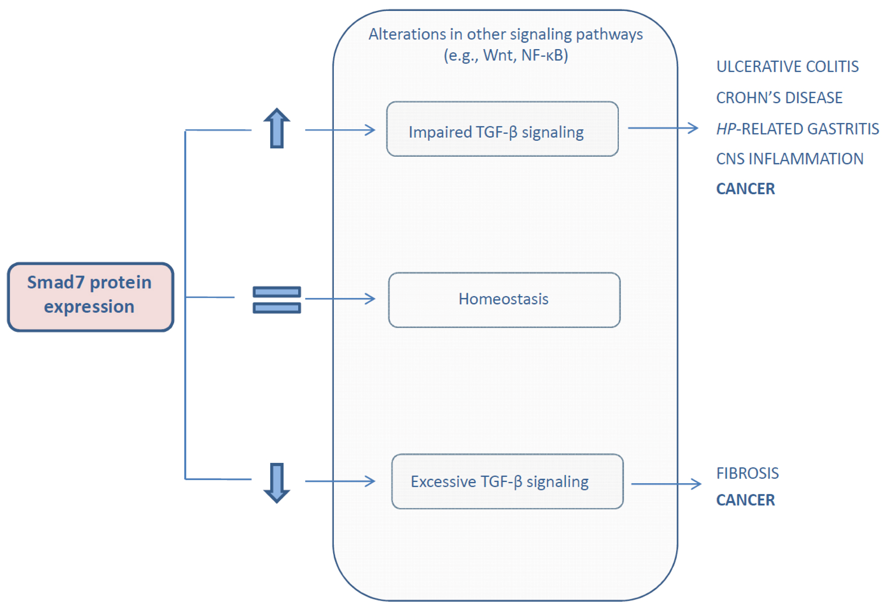
Interestingly, a deregulated Smad7 protein expression has been documented and supposed to play a pathogenic role in a variety of human disorders (Figure 2).
For example, Smad7 expression is down-regulated in fibrotic tissues and implicated in the progression of fibrosis in different organs such as pancreas, liver, lung, skin, and kidney [11] in line with the profibrotic actions of the TGF-β pathway.
On the other hand, Smad7 is over-expressed and limits TGF-β-mediated anti-inflammatory signals during inflammation of the central nervous system [31], in inflammatory bowel diseases (IBD), such as ulcerative colitis and Crohn’s disease, as well as in Helicobacter-pylori-related gastritis [32,33].
Increasing evidence indicates that Smad7 is differently expressed in human cancers [3436], and it could either sustain or restrain cancer cell growth [11]. Here, we review the data supporting the dual role of Smad7 in the control of carcinogenesis.

2. Expression and Role of Smad7 in Cancer

Smad7 can be transcriptionally regulated by TGF-β, epidermal growth factor (EGF) and inflammatory cytokines, such as TNF-α, IL-1β and IFN-γ in different cell lines [3740]. In contrast, little is known about its regulation in human tissues. Studies in patients with IBD have shown that Smad7 can be also regulated at post-transcriptional level by mechanisms that enhance acetylation on lysine residues thus reducing ubiquitination-mediated proteasomal degradation [41]. High Smad7 is seen in several human malignancies and there is preliminary evidence that Smad7 expression correlates with the clinical prognosis of cancer patients. For instance, Smad7 up-regulation was associated with poor survival rate in esophageal squamous cell carcinoma and a shorter time to recurrence in endometrial carcinoma [42,43]. However, a different scenario has emerged when Smad7 has been investigated in other tumors. In particular, it was shown that ectopic expression or deletion of Smad7 in cancer cells can differently regulate tumorigenesis depending on the cell context analyzed.
The pro- and anti-tumorigenic effects of Smad7 in different cancer types are summarized in the Table 1 and discussed below.

2.1. Colorectal Cancer

Smad7 gene variants have been extensively analyzed in patients with colorectal cancer (CRC). Boulay et al., found that CRC patients with deletion of Smad7 had a favorable clinical outcome compared with patients with Smad7 amplification [44]. More recently, further genetic variants within Smad7 gene have been linked to colorectal carcinogenesis in two genome-wide association studies (GWAS) [65,66]. In both studies, a highly significant association with CRC was found for two single nucleotide polymorphisms (SNPs) in Smad7 (i.e., rs4939827, rs12953717). The association of these SNPs with CRC was then confirmed by two other GWAS [67,68].
To clarify the role of Smad7 in CRC cell growth, Halder et al., overexpressed Smad7 in FET, a CRC cell line, and showed that Smad7 enhanced anchorage-independent cell growth, favored the formation of colonies on soft agar and increased resistance against apoptosis through a mechanism dependent on suppression of TGF-β signaling. Smad7-overexpressing FET cells showed an increased tumorigenicity when injected subcutaneously into immunodeficient nude mice [45] as compared to control FET cells. The same group assessed the role of Smad7 in colon cancer metastasis and demonstrated that injection of Smad7-overexpressing FET cells in the spleen of athymic nude mice favored the development of liver metastasis [46]. The pro-metastatic role of Smad7 was associated with increased expression of junctional proteins (e.g., E-cadherin, Claudin-1, Claudin-4) at distant sites. These findings are in line with our recent studies showing that Smad7 is over-expressed in human CRC and inhibition of Smad7 with a specific Smad7 antisense oligonucleotide reduces CRC cell growth both in vitro and in vivo in mice (personal unpublished observations). The factors/mechanisms underlying Smad7 upregulation in CRC cells are not yet known. It has been recently reported that microRNA-25 (miR-25), whose expression is down-regulated in CRC tissue, is a negative regulator of Smad7, raising the possibility that in CRC cells high Smad7 can be linked to the low content of miR-25 [69].
As pointed out above Smad7 is also over-expressed by immune cells in IBD tissue and studies in experimental models have shown that over-expression of Smad7 in T cells associates with severe colitis and reduced growth of colitis-associated CRC, thus highlighting the opposing role of Smad7 in the control of sporadic and colitis-associated CRC [47].

2.2. Pancreatic Cancer

An early study by Kleeff and colleagues showed that Smad7 RNA transcripts were increased in human pancreatic cancer as compared to the normal pancreas [34]. To determine the role of Smad7 in pancreatic cancer cells, COLO-357 cells were stably transfected with a full-length Smad7 expression vector. Smad7 overexpressing COLO-357 cells were resistant to the TGF-β-mediated growth inhibition in vitro and exhibited a marked capacity to form colonies in soft agar and tumors in nude mice [34]. Studies investigating the mechanisms underlying the pro-tumorigenic effects of Smad7 identified thioredoxin-1 and retinoblastoma as key molecules involved in the Smad7-dependent aggressiveness of pancreatic cancer cells [70,71].
More recently, Kuang and co-workers provided in vivo evidence that Smad7 is implicated in the early stages of pancreatic cancer. Using a transgenic mouse with pancreas specific Smad7 overexpression, these authors reported that Smad7 blocked TGF-β signaling in the pancreas and induced premalignant ductal lesions with the characteristics of pancreatic intraepithelial neoplasia (PanIN), the precursor stage to pancreatic carcinoma [48]. However, Wang and co-workers showed that high expression of Smad7 in pancreatic cancer associated with a more favorable prognosis compared with patients with lower levels of Smad7 who exhibited increased incidence of lymph node metastasis and liver metastasis after surgery [49]. The reason for this apparent difference is not yet known even though it is conceivable that such a discrepancy could be at least in part due to the ability of Smad7 to interfere with the opposing role of TGF-β in pancreatic tumor initiation and progression.

2.3. Gastric Cancer

The expression of Smad7 in gastric cancer progression and its prognostic significance was initially investigated by Kim and colleagues. By immunohistochemistry, it was demonstrated that 98 out of 304 patients (32.2%) who had undergone gastrectomy expressed Smad7 in gastric cancer tissues whereas no expression was detected in normal tissues [51]. This later result is however surprising as we had detected Smad7 in normal gastric mucosa by Western blotting and real-time PCR [33] and Leng and co-workers documented a constitutive expression of Smad7 in the normal gastric mucosa [50]. This group showed also up-regulation of Smad7 in gastric cancer and peri-tumoral area, particularly in poorly differentiated tumors and in those with lymphatic metastasis [50]. It has been also reported that gastric cancer patients with elevated levels of Smad7 had a poor prognosis independently of other well-established clinical prognostic factors, such as tumor size, depth of invasion and lymph node metastasis [51]. Consistent with this is the demonstration that ectopic Smad7 expression increases the survival of SGC7901 gastric cancer cells [50].
Altogether, these findings highlight the involvement of Smad7 in gastric tumorigenesis.

2.4. Skin Cancer

Human papilloma and squamous cell carcinoma (SCC) express elevated levels of Smad7 as compared to normal epidermis [36]. Using a mouse model of chemically-induced skin carcinogenesis, Liu et al., reported that Smad7 overexpression in H-ras-transduced keratinocytes determined the conversion of benign to malignant epithelial cells and a rapid progression to squamous cell carcinoma [52]. This effect was associated with a marked increase in cell proliferation, inhibition of TGF-β signaling and induction of EGF family members, which regulate various signals associated with tumor growth and metastasis [72]. Moreover, using a xenograft model in which primary keratinocytes mixed with dermal fibroblasts are grafted into nude mice, the same authors reported that H-ras/Smad7, but not H-ras, keratinocytes progressed to SCC [52].
In contrast, two studies by Mauviel’s group reported a TGF-β-dependent tumor-suppressive role of Smad7 in metastatic melanoma cells [53,54]. Stable over-expression of Smad7 in 1205Lu cells reduced production of tissue-degrading proteases and hence the invasive capacity and the in vitro anchorage-independent growth as well as tumor formation following subcutaneous injection in nude mice [53]. In a model of bone metastases induced by inoculation of tumor cells into the left cardiac ventricle of nude mice, Javelaud et al., showed that animals injected with Smad7-transfected 1205Lu cells had significantly less osteolytic metastases and longer survival compared with mice injected with parental and mock-transfected 1205Lu cells [54]. These changes were accompanied by a reduced secretion of gelatinases and diminished expression of metastasis-related molecules (e.g., interleukin-11, CXCR4, osteopontin). DiVito and co-workers, using an in vivo human skin grafting system, showed that Smad7-expressing 1205Lu cells positioned themselves proximal to the dermal-epidermal junction and failed to form tumors, while control cells invaded the dermis and formed tumors [55]. Mechanistically, it was proposed that Smad7 promoted heterotypic cell-cell interactions through the redistribution of cell adhesion proteins to the cell surface thereby mitigating tumor invasion [55]. Altogether these data suggest that Smad7 can be both pro- and anti-tumorigenic in the skin.

2.5. Breast Cancer

Theohari and colleagues investigated the expression of Smad7 in 150 invasive breast carcinoma specimens and showed that Smad7 levels positively correlate with tumor size, stage, matrix metalloproteinase (MMP)-9 and MMP-14 expression thus resulting in an aggressive phenotype [56]. However, experimental evidence suggests that Smad7 can regulate either positively or negatively breast carcinogenesis. In a model of breast cancer metastasis, Azuma et al., reported a decrease in lung and liver metastasis and longer survival when mice were intravenously injected with Smad7-transfected mouse mammary carcinoma JygMC(A) cells compared to mice injected with mock-transfected JygMC(A) cells. These effects were suggested to rely on the increased expression of major components of adherent and tight junctions (e.g., E-cadherin) and decreased expression of promigratory cadherins (e.g., N-cadherin) in JygMC(A) cells that expressed exogenous Smad7 [57]. Hong and colleagues showed that over-expression of Smad7 sensitized MCF7 breast cancer cells to TNF-induced cell death and associated this effect with inhibition of expression of antiapoptotic NF-κB target genes [20]. Finally, Smad7 was suggested to negatively regulate the EGF signaling pathway in breast cancer cells as ectopic Smad7 expression in SKBR3 cells completely abrogated EGF-induced MMP-9 expression [40].
In contrast with the above findings, two recent studies suggested that Smad7 is an inhibitor of EMT and cell invasion in breast carcinogenesis. Papageorgis and co-workers delineated a link between Smad7 and the maintenance of epigenetic silencing of epithelial genes during EMT of breast cancer cells. Using breast cancer cell lines derived from a common genetic background (i.e., MCF10A) which accumulated distinct genetic/epigenetic alterations in vivo thus acquiring properties associated with gradual progression from nontumorigenic to carcinogenic state, these authors showed that Smad7 overexpression suppresses migration and invasion of mesenchymal-like MCF10CA1h cells, a malignant variant of Ras-transformed MCF10A cells, by reversing the DNA methylation status of specific epithelial markers (i.e., E-cadherin, γ-catenin, and β-catenin) thus inducing their re-expression [58].
Along the same line is the work by Smith et al., who showed that the miR-106b-25 cluster negatively regulates Smad7 expression thereby activating TGF-β signaling and inducing EMT in MCF7 cells [59].

2.6. Liver Cancer

Park et al., evaluated Smad7 in the different phases of hepatocellular carcinoma (HCC) development and documented Smad7-expressing tumor cells in 25 out of 41 (61%) advanced tumors whereas no Smad7-positive cells were detected in dysplastic nodules and early HCCs [73]. Using a mouse model of HCC induced by diethylnitrosamine (DEN), Wang and colleagues showed that Smad7-deficient mice had higher tumor incidence and multiplicity than wild-type mice. Moreover, tumor cells from Smad7 KO mice displayed increased proliferation, diminished apoptosis and higher colony formation compared with those from wild-type littermates. Deletion of Smad7 increased cell growth of primary HCC cells while ectopic expression of Smad7 in HCC cell lines markedly suppressed cell growth and colony formation. These effects were associated with the Smad7-mediated inhibition of the G1-S phase transition and induction of apoptosis through attenuation of NF-κB and TGF-β signaling [60].
Xia et al., confirmed the decreased expression of Smad7 in HCC samples, particularly in patients with early recurrence and poor prognosis [61]. The same group also showed that, in HCC, Smad7 is inversely related to miR-216a/217 cluster, a negative regulator of Smad7, which controls EMT and cell migration of HCC cells [61]. Altogether, these observations suggest that Smad7 may act as a tumor suppressor in HCC.

2.7. Prostate Cancer

Evidence linking Smad7 and prostate cancer comes from the work of Landstrom’s group. Initial studies showed that Smad7 was expressed in rat prostate cancer cells undergoing apoptosis [74] and ectopic Smad7 expression induced apoptosis of PC-3U human prostate cancer cells [62]. Further investigation revealed that Smad7 acted as a scaffold protein to facilitate TGF-β-induced activation of p38 and subsequent apoptosis [75] and was required for induction of apoptosis by the anti-cancer agent 2-Methoxyestradiol [63]. More recently, Ekman et al., showed that Smad7 forms a complex with APC in a p38-dependent fashion and facilitates TGF-β-induced accumulation of β-catenin, thereby promoting migratory responses in prostate cancer cells [64].

3. Conclusions

The data described in this article indicate that Smad7 can have both pro- and anti-tumor actions depending on the cancer type analyzed.
Given the role of Smad7 in inhibiting TGF-β signaling, a possible explanation of these opposite effects could be due to the different functionality of this pathway among distinct cancer types. Indeed, while mutations in TGF-β signaling machinery are rare in most cancers, frequent genetic alterations in Smad components characterize gastrointestinal carcinomas (e.g., pancreatic, colorectal) [7678] and suggest a tumor suppressive and anti-metastatic role of TGF-β pathway in a context dependent manner [7982].
Moreover, even in the same tumor the function of Smad7 can switch from tumor-suppressive to tumor-promoting depending on the tumor stage (i.e., early versus advanced). These apparently contradictory functions are not surprising, given the opposite role of TGF-β signaling pathway in early versus advanced tumor stages and the interaction of Smad7 with a vast array of functionally heterogeneous molecules that may be differently expressed during the carcinogenetic process.
Further work is needed to delineate the mechanisms by which Smad7 exerts its distinct functions on tumorigenesis and to clarify which cancer-related pathways are predominantly affected by Smad7 in the different contexts. It would be also useful to study the role of Smad7 in experimental models of metastasis deriving from primary tumors that evolve in situ and more closely resemble the human disease as well as the effect of Smad7 inhibition in cancer cells that constitutively express this protein. This information will ultimately help us to understand the complex role of Smad7 in the different phases of carcinogenesis and eventually pave the way for the development of therapeutic strategies which, through modulation of Smad7 function, can contribute to attenuate/halt the course of these diseases.

Acknowledgments

This work received support from the “Fondazione Umberto di Mario ONLUS”, Rome, AIRC (MFAG-12108 to CS and IG-13049 to GM), and Giuliani SpA, Milan, Italy.

Conflicts of Interest

The authors declare no conflict of interest.

References

  1. Fearon, E.R.; Vogelstein, B. A genetic model for colorectal tumorigenesis. Cell 1990, 61, 759–767. [Google Scholar]
  2. Liotta, L.A.; Stetler-Stevenson, W.G. Tumor invasion and metastasis: An imbalance of positive and negative regulation. Cancer Res 1991, 51, 5054s–5059s. [Google Scholar]
  3. Oft, M.; Akhurst, R.J.; Balmain, A. Metastasis is driven by sequential elevation of H-ras and Smad2 levels. Nat. Cell Biol 2002, 4, 487–494. [Google Scholar]
  4. Katsuno, Y.; Lamouille, S.; Derynck, R. Tgf-beta signaling and epithelial-mesenchymal transition in cancer progression. Curr. Opin. Oncol 2013, 25, 76–84. [Google Scholar]
  5. Piek, E.; Heldin, C.H.; Ten Dijke, P. Specificity, diversity, and regulation in TGF-β superfamily signaling. FASEB J 1999, 13, 2105–2124. [Google Scholar]
  6. Massague, J.; Seoane, J.; Wotton, D. Smad transcription factors. Genes Dev 2005, 19, 2783–2810. [Google Scholar]
  7. Kretzschmar, M.; Doody, J.; Timokhina, I.; Massague, J. A mechanism of repression of TGFβ/Smad signaling by oncogenic ras. Genes Dev 1999, 13, 804–816. [Google Scholar]
  8. Luo, K.; Stroschein, S.L.; Wang, W.; Chen, D.; Martens, E.; Zhou, S.; Zhou, Q. The Ski oncoprotein interacts with the Smad proteins to repress tgfbeta signaling. Genes Dev 1999, 13, 2196–2206. [Google Scholar]
  9. Stroschein, S.L.; Wang, W.; Zhou, S.; Zhou, Q.; Luo, K. Negative feedback regulation of TGF-β signaling by the snon oncoprotein. Science 1999, 286, 771–774. [Google Scholar]
  10. Deheuninck, J.; Luo, K. Ski and snon, potent negative regulators of tgf-beta signaling. Cell Res 2009, 19, 47–57. [Google Scholar]
  11. Briones-Orta, M.A.; Tecalco-Cruz, A.C.; Sosa-Garrocho, M.; Caligaris, C.; Macias-Silva, M. Inhibitory Smad7: Emerging roles in health and disease. Curr. Mol. Pharmacol 2011, 4, 141–153. [Google Scholar]
  12. Yan, X.; Chen, Y.G. Smad7: Not only a regulator, but also a cross-talk mediator of TGF-β signalling. Biochem. J 2011, 434, 1–10. [Google Scholar]
  13. Nakao, A.; Afrakhte, M.; Moren, A.; Nakayama, T.; Christian, J.L.; Heuchel, R.; Itoh, S.; Kawabata, M.; Heldin, N.E.; Heldin, C.H.; et al. Identification of Smad7, a tgfbeta-inducible antagonist of tgf-beta signalling. Nature 1997, 389, 631–635. [Google Scholar]
  14. Hayashi, H.; Abdollah, S.; Qiu, Y.; Cai, J.; Xu, Y.Y.; Grinnell, B.W.; Richardson, M.A.; Topper, J.N.; Gimbrone, M.A., Jr.; Wrana, J.L.; et al. The Mad-Related protein Smad7 associates with the tgfbeta receptor and functions as an antagonist of tgfbeta signaling. Cell 1997, 89, 1165–1173. [Google Scholar]
  15. Ebisawa, T.; Fukuchi, M.; Murakami, G.; Chiba, T.; Tanaka, K.; Imamura, T.; Miyazono, K. Smurf1 interacts with transforming growth factor-beta type i receptor through Smad7 and induces receptor degradation. J. Biol. Chem 2001, 276, 12477–12480. [Google Scholar]
  16. Kavsak, P.; Rasmussen, R.K.; Causing, C.G.; Bonni, S.; Zhu, H.; Thomsen, G.H.; Wrana, J.L. Smad7 binds to smurf2 to form an e3 ubiquitin ligase that targets the tgf beta receptor for degradation. Mol. Cell 2000, 6, 1365–1375. [Google Scholar]
  17. Kuratomi, G.; Komuro, A.; Goto, K.; Shinozaki, M.; Miyazawa, K.; Miyazono, K.; Imamura, T. Nedd4-2 (neural precursor cell expressed, developmentally down-regulated 4-2) negatively regulates tgf-beta (transforming growth factor-beta) signalling by inducing ubiquitin-mediated degradation of Smad2 and tgf-beta type i receptor. Biochem. J 2005, 386, 461–470. [Google Scholar]
  18. Shi, W.; Sun, C.; He, B.; Xiong, W.; Shi, X.; Yao, D.; Cao, X. Gadd34-pp1c recruited by Smad7 dephosphorylates tgfbeta type i receptor. J. Cell Biol 2004, 164, 291–300. [Google Scholar]
  19. Zhang, S.; Fei, T.; Zhang, L.; Zhang, R.; Chen, F.; Ning, Y.; Han, Y.; Feng, X.H.; Meng, A.; Chen, Y.G. Smad7 antagonizes transforming growth factor beta signaling in the nucleus by interfering with functional Smad-DNA complex formation. Mol. Cell Biol 2007, 27, 4488–4499. [Google Scholar]
  20. Hong, S.; Lee, C.; Kim, S.J. Smad7 sensitizes tumor necrosis factor induced apoptosis through the inhibition of antiapoptotic gene expression by suppressing activation of the nuclear factor-κb pathway. Cancer Res 2007, 67, 9577–9583. [Google Scholar]
  21. Lee, Y.S.; Kim, J.H.; Kim, S.T.; Kwon, J.Y.; Hong, S.; Kim, S.J.; Park, S.H. Smad7 and Smad6 bind to discrete regions of pellino-1 via their mh2 domains to mediate TGF-β1-induced negative regulation of IL-1R/TLR signaling. Biochem. Biophys. Res. Commun 2010, 393, 836–843. [Google Scholar]
  22. Hoffmann, A.; Preobrazhenska, O.; Wodarczyk, C.; Medler, Y.; Winkel, A.; Shahab, S.; Huylebroeck, D.; Gross, G.; Verschueren, K. Transforming growth factor-β-activated kinase-1 (TAK1), a MAP3K, interacts with Smad proteins and interferes with osteogenesis in murine mesenchymal progenitors. J. Biol. Chem 2005, 280, 27271–27283. [Google Scholar]
  23. Hong, S.; Lim, S.; Li, A.G.; Lee, C.; Lee, Y.S.; Lee, E.K.; Park, S.H.; Wang, X.J.; Kim, S.J. Smad7 binds to the adaptors TAB2 and TAB3 to block recruitment of the kinase TAK1 to the adaptor TRAF2. Nat. Immunol 2007, 8, 504–513. [Google Scholar]
  24. Han, G.; Li, A.G.; Liang, Y.Y.; Owens, P.; He, W.; Lu, S.; Yoshimatsu, Y.; Wang, D.; Ten Dijke, P.; Lin, X.; et al. Smad7-induced β-catenin degradation alters epidermal appendage development. Dev. Cell 2006, 11, 301–312. [Google Scholar]
  25. Millar, S.E. Smad7: Licensed to kill β-catenin. Dev. Cell 2006, 11, 274–276. [Google Scholar]
  26. Li, R.; Rosendahl, A.; Brodin, G.; Cheng, A.M.; Ahgren, A.; Sundquist, C.; Kulkarni, S.; Pawson, T.; Heldin, C.H.; Heuchel, R.L. Deletion of exon i of Smad7 in mice results in altered b cell responses. J. Immunol 2006, 176, 6777–6784. [Google Scholar]
  27. Chen, Q.; Chen, H.; Zheng, D.; Kuang, C.; Fang, H.; Zou, B.; Zhu, W.; Bu, G.; Jin, T.; Wang, Z.; et al. Smad7 is required for the development and function of the heart. J. Biol. Chem 2009, 284, 292–300. [Google Scholar]
  28. Liu, X.; Chen, Q.; Kuang, C.; Zhang, M.; Ruan, Y.; Xu, Z.C.; Wang, Z.; Chen, Y. A 4.3 kb Smad7 promoter is able to specify gene expression during mouse development. Biochim. Biophys. Acta 2007, 1769, 149–152. [Google Scholar]
  29. Kollias, H.D.; Perry, R.L.; Miyake, T.; Aziz, A.; McDermott, J.C. Smad7 promotes and enhances skeletal muscle differentiation. Mol. Cell Biol 2006, 26, 6248–6260. [Google Scholar]
  30. Miyake, T.; Alli, N.S.; McDermott, J.C. Nuclear function of Smad7 promotes myogenesis. Mol. Cell Biol 2010, 30, 722–735. [Google Scholar]
  31. Kleiter, I.; Pedre, X.; Mueller, A.M.; Poeschl, P.; Couillard-Despres, S.; Spruss, T.; Bogdahn, U.; Giegerich, G.; Steinbrecher, A. Inhibition of Smad7, a negative regulator of TGF-β signaling, suppresses autoimmune encephalomyelitis. J. Neuroimmunol 2007, 187, 61–73. [Google Scholar]
  32. Monteleone, G.; Kumberova, A.; Croft, N.M.; McKenzie, C.; Steer, H.W.; MacDonald, T.T. Blocking Smad7 restores TGF-β1 signaling in chronic inflammatory bowel disease. J. Clin. Investig 2001, 108, 601–609. [Google Scholar]
  33. Monteleone, G.; Del Vecchio Blanco, G.; Palmieri, G.; Vavassori, P.; Monteleone, I.; Colantoni, A.; Battista, S.; Spagnoli, L.G.; Romano, M.; Borrelli, M.; et al. Induction and regulation of Smad7 in the gastric mucosa of patients with helicobacter pylori infection. Gastroenterology 2004, 126, 674–682. [Google Scholar]
  34. Kleeff, J.; Ishiwata, T.; Maruyama, H.; Friess, H.; Truong, P.; Buchler, M.W.; Falb, D.; Korc, M. The TGF-β signaling inhibitor Smad7 enhances tumorigenicity in pancreatic cancer. Oncogene 1999, 18, 5363–5372. [Google Scholar]
  35. Boulay, J.L.; Mild, G.; Reuter, J.; Lagrange, M.; Terracciano, L.; Lowy, A.; Laffer, U.; Orth, B.; Metzger, U.; Stamm, B.; et al. Combined copy status of 18q21 genes in colorectal cancer shows frequent retention of Smad7. Genes Chromosomes Cancer 2001, 31, 240–247. [Google Scholar]
  36. He, W.; Cao, T.; Smith, D.A.; Myers, T.E.; Wang, X.J. Smads mediate signaling of the TGFβ superfamily in normal keratinocytes but are lost during skin chemical carcinogenesis. Oncogene 2001, 20, 471–483. [Google Scholar]
  37. Afrakhte, M.; Moren, A.; Jossan, S.; Itoh, S.; Sampath, K.; Westermark, B.; Heldin, C.H.; Heldin, N.E.; ten Dijke, P. Induction of inhibitory Smad6 and Smad7 mrna by TGF-β family members. Biochem. Biophys. Res. Commun 1998, 249, 505–511. [Google Scholar]
  38. Bitzer, M.; von Gersdorff, G.; Liang, D.; Dominguez-Rosales, A.; Beg, A.A.; Rojkind, M.; Bottinger, E.P. A mechanism of suppression of TGF-β/Smad signaling by NF-κb/RelA. Genes Devel 2000, 14, 187–197. [Google Scholar]
  39. Ulloa, L.; Doody, J.; Massague, J. Inhibition of transforming growth factor-β/Smad signalling by the interferon-γ/STAT pathway. Nature 1999, 397, 710–713. [Google Scholar]
  40. Kim, S.; Han, J.; Lee, S.K.; Koo, M.; Cho, D.H.; Bae, S.Y.; Choi, M.Y.; Kim, J.S.; Kim, J.H.; Choe, J.H.; et al. Smad7 acts as a negative regulator of the epidermal growth factor (EGF) signaling pathway in breast cancer cells. Cancer Lett 2012, 314, 147–154. [Google Scholar]
  41. Monteleone, G.; Del Vecchio Blanco, G.; Monteleone, I.; Fina, D.; Caruso, R.; Gioia, V.; Ballerini, S.; Federici, G.; Bernardini, S.; Pallone, F.; et al. Post-transcriptional regulation of Smad7 in the gut of patients with inflammatory bowel disease. Gastroenterology 2005, 129, 1420–1429. [Google Scholar]
  42. Dowdy, S.C.; Mariani, A.; Reinholz, M.M.; Keeney, G.L.; Spelsberg, T.C.; Podratz, K.C.; Janknecht, R. Overexpression of the tgf-beta antagonist Smad7 in endometrial cancer. Gynecol. Oncol 2005, 96, 368–373. [Google Scholar]
  43. Osawa, H.; Nakajima, M.; Kato, H.; Fukuchi, M.; Kuwano, H. Prognostic value of the expression of Smad6 and Smad7, as inhibitory Smads of the tgf-beta superfamily, in esophageal squamous cell carcinoma. Anticancer Res 2004, 24, 3703–3709. [Google Scholar]
  44. Boulay, J.L.; Mild, G.; Lowy, A.; Reuter, J.; Lagrange, M.; Terracciano, L.; Laffer, U.; Herrmann, R.; Rochlitz, C. Smad7 is a prognostic marker in patients with colorectal cancer. Int. J. Cancer. J. Int. Du Cancer 2003, 104, 446–449. [Google Scholar]
  45. Halder, S.K.; Beauchamp, R.D.; Datta, P.K. Smad7 induces tumorigenicity by blocking TGF-β-induced growth inhibition and apoptosis. Experi. Cell Res 2005, 307, 231–246. [Google Scholar]
  46. Halder, S.K.; Rachakonda, G.; Deane, N.G.; Datta, P.K. Smad7 induces hepatic metastasis in colorectal cancer. Br. J. Cancer 2008, 99, 957–965. [Google Scholar]
  47. Rizzo, A.; Waldner, M.J.; Stolfi, C.; Sarra, M.; Fina, D.; Becker, C.; Neurath, M.F.; Macdonald, T.T.; Pallone, F.; Monteleone, G.; et al. Smad7 expression in T cells prevents colitis-associated cancer. Cancer Res 2011, 71, 7423–7432. [Google Scholar]
  48. Kuang, C.; Xiao, Y.; Liu, X.; Stringfield, T.M.; Zhang, S.; Wang, Z.; Chen, Y. In vivo disruption of TGF-β signaling by Smad7 leads to premalignant ductal lesions in the pancreas. Proc. Natl. Acad. Sci. USA 2006, 103, 1858–1863. [Google Scholar]
  49. Wang, P.; Fan, J.; Chen, Z.; Meng, Z.Q.; Luo, J.M.; Lin, J.H.; Zhou, Z.H.; Chen, H.; Wang, K.; Xu, Z.D.; et al. Low-level expression of Smad7 correlates with lymph node metastasis and poor prognosis in patients with pancreatic cancer. Ann. Surg. Oncol 2009, 16, 826–835. [Google Scholar]
  50. Leng, A.; Liu, T.; He, Y.; Li, Q.; Zhang, G. Smad4/Smad7 balance: A role of tumorigenesis in gastric cancer. Experi. Mol. Pathol 2009, 87, 48–53. [Google Scholar]
  51. Kim, Y.H.; Lee, H.S.; Lee, H.J.; Hur, K.; Kim, W.H.; Bang, Y.J.; Kim, S.J.; Lee, K.U.; Choe, K.J.; Yang, H.K. Prognostic significance of the expression of Smad4 and Smad7 in human gastric carcinomas. Ann. Oncol 2004, 15, 574–580. [Google Scholar]
  52. Liu, X.; Lee, J.; Cooley, M.; Bhogte, E.; Hartley, S.; Glick, A. Smad7 but not Smad6 cooperates with oncogenic ras to cause malignant conversion in a mouse model for squamous cell carcinoma. Cancer Res 2003, 63, 7760–7768. [Google Scholar]
  53. Javelaud, D.; Delmas, V.; Moller, M.; Sextius, P.; Andre, J.; Menashi, S.; Larue, L.; Mauviel, A. Stable overexpression of Smad7 in human melanoma cells inhibits their tumorigenicity in vitro and in vivo. Oncogene 2005, 24, 7624–7629. [Google Scholar]
  54. Javelaud, D.; Mohammad, K.S.; McKenna, C.R.; Fournier, P.; Luciani, F.; Niewolna, M.; Andre, J.; Delmas, V.; Larue, L.; Guise, T.A.; et al. Stable overexpression of Smad7 in human melanoma cells impairs bone metastasis. Cancer Res 2007, 67, 2317–2324. [Google Scholar]
  55. DiVito, K.A.; Trabosh, V.A.; Chen, Y.S.; Chen, Y.; Albanese, C.; Javelaud, D.; Mauviel, A.; Simbulan-Rosenthal, C.M.; Rosenthal, D.S. Smad7 restricts melanoma invasion by restoring n-cadherin expression and establishing heterotypic cell-cell interactions in vivo. Pigment Cell Mel. Res. 2010, 23, 795–808. [Google Scholar]
  56. Theohari, I.; Giannopoulou, I.; Magkou, C.; Nomikos, A.; Melissaris, S.; Nakopoulou, L. Differential effect of the expression of TGF-β pathway inhibitors, Smad-7 and Ski, on invasive breast carcinomas: Relation to biologic behavior. APMIS 2012, 120, 92–100. [Google Scholar]
  57. Azuma, H.; Ehata, S.; Miyazaki, H.; Watabe, T.; Maruyama, O.; Imamura, T.; Sakamoto, T.; Kiyama, S.; Kiyama, Y.; Ubai, T.; et al. Effect of Smad7 expression on metastasis of mouse mammary carcinoma JygMC(A) cells. J. Nat. Cancer Inst 2005, 97, 1734–1746. [Google Scholar]
  58. Papageorgis, P.; Lambert, A.W.; Ozturk, S.; Gao, F.; Pan, H.; Manne, U.; Alekseyev, Y.O.; Thiagalingam, A.; Abdolmaleky, H.M.; Lenburg, M.; et al. Smad signaling is required to maintain epigenetic silencing during breast cancer progression. Cancer Res 2010, 70, 968–978. [Google Scholar]
  59. Smith, A.L.; Iwanaga, R.; Drasin, D.J.; Micalizzi, D.S.; Vartuli, R.L.; Tan, A.C.; Ford, H.L. The miR-106b-25 cluster targets Smad7, activates TGF-β signaling, and induces EMT and tumor initiating cell characteristics downstream of Six1 in human breast cancer. Oncogene 2012, 31, 5162–5171. [Google Scholar]
  60. Wang, J.; Zhao, J.; Chu, E.S.; Mok, M.T.; Go, M.Y.; Man, K.; Heuchel, R.; Lan, H.Y.; Chang, Z.; Sung, J.J.; et al. Inhibitory role of Smad7 in hepatocarcinogenesis in mice and in vitro. J. Pathol. 2013, 230, 441–452. [Google Scholar]
  61. Xia, H.; Ooi, L.L.; Hui, K.M. MicroRNA-216a/217-induced epithelial-mesenchymal transition targets pten and Smad7 to promote drug resistance and recurrence of liver cancer. Hepatology 2013, 58, 629–641. [Google Scholar]
  62. Landstrom, M.; Heldin, N.E.; Bu, S.; Hermansson, A.; Itoh, S.; ten Dijke, P.; Heldin, C.H. Smad7 mediates apoptosis induced by transforming growth factor β in prostatic carcinoma cells. Curr. Biol 2000, 10, 535–538. [Google Scholar]
  63. Davoodpour, P.; Landstrom, M. 2-Methoxyestradiol-Induced apoptosis in prostate cancer cells requires Smad7. J. Biol. Chem 2005, 280, 14773–14779. [Google Scholar]
  64. Ekman, M.; Mu, Y.; Lee, S.Y.; Edlund, S.; Kozakai, T.; Thakur, N.; Tran, H.; Qian, J.; Groeden, J.; Heldin, C.H.; et al. APC and Smad7 link TGFβ type I receptors to the microtubule system to promote cell migration. Mol. Biol. Cell 2012, 23, 2109–2121. [Google Scholar]
  65. Broderick, P.; Carvajal-Carmona, L.; Pittman, A.M.; Webb, E.; Howarth, K.; Rowan, A.; Lubbe, S.; Spain, S.; Sullivan, K.; Fielding, S.; et al. A Genome-wide association study shows that common alleles of Smad7 influence colorectal cancer risk. Nature Genet 2007, 39, 1315–1317. [Google Scholar]
  66. Tenesa, A.; Farrington, S.M.; Prendergast, J.G.; Porteous, M.E.; Walker, M.; Haq, N.; Barnetson, R.A.; Theodoratou, E.; Cetnarskyj, R.; Cartwright, N.; et al. Genome-wide association scan identifies a colorectal cancer susceptibility locus on 11q23 and replicates risk loci at 8q24 and 18q21. Nat. Genet 2008, 40, 631–637. [Google Scholar]
  67. Slattery, M.L.; Herrick, J.; Curtin, K.; Samowitz, W.; Wolff, R.K.; Caan, B.J.; Duggan, D.; Potter, J.D.; Peters, U. Increased risk of colon cancer associated with a genetic polymorphism of Smad7. Cancer Res 2010, 70, 1479–1485. [Google Scholar]
  68. Thompson, C.L.; Plummer, S.J.; Acheson, L.S.; Tucker, T.C.; Casey, G.; Li, L. Association of common genetic variants in Smad7 and risk of colon cancer. Carcinogenesis 2009, 30, 982–986. [Google Scholar]
  69. Li, Q.; Zou, C.; Han, Z.; Xiao, H.; Wei, H.; Wang, W.; Zhang, L.; Zhang, X.; Tang, Q.; Zhang, C.; et al. MicroRNA-25 functions as a potential tumor suppressor in colon cancer by targeting Smad7. Cancer Lett 2013, 335, 168–174. [Google Scholar]
  70. Arnold, N.B.; Ketterer, K.; Kleeff, J.; Friess, H.; Buchler, M.W.; Korc, M. Thioredoxin is downstream of Smad7 in a pathway that promotes growth and suppresses cisplatin-induced apoptosis in pancreatic cancer. Cancer Res 2004, 64, 3599–3606. [Google Scholar]
  71. Boyer Arnold, N.; Korc, M. Smad7 abrogates transforming growth factor-β1-mediated growth inhibition in COLO-357 cells through functional inactivation of the retinoblastoma protein. J. Biol. Chem 2005, 280, 21858–21866. [Google Scholar]
  72. Saloman, D.S.; Bianco, C.; Ebert, A.D.; Khan, N.I.; De Santis, M.; Normanno, N.; Wechselberger, C.; Seno, M.; Williams, K.; Sanicola, M.; et al. The EGF-CFC family: Novel epidermal growth factor-related proteins in development and cancer. Endocr. Relat. Cancer 2000, 7, 199–226. [Google Scholar]
  73. Park, Y.N.; Chae, K.J.; Oh, B.K.; Choi, J.; Choi, K.S.; Park, C. Expression of Smad7 in hepatocellular carcinoma and dysplastic nodules: Resistance mechanism to transforming growth factor-β. Hepato-Gastroenterology 2004, 51, 396–400. [Google Scholar]
  74. Brodin, G.; ten Dijke, P.; Funa, K.; Heldin, C.H.; Landstrom, M. Increased Smad expression and activation are associated with apoptosis in normal and malignant prostate after castration. Cancer Res 1999, 59, 2731–2738. [Google Scholar]
  75. Edlund, S.; Bu, S.; Schuster, N.; Aspenstrom, P.; Heuchel, R.; Heldin, N.E.; ten Dijke, P.; Heldin, C.H.; Landstrom, M. Transforming growth factor-β1 (TGF-β)-induced apoptosis of prostate cancer cells involves Smad7-dependent activation of p38 by TGF-β-activated kinase 1 and mitogen-activated protein kinase kinase 3. Mol. Biol. Cell 2003, 14, 529–544. [Google Scholar]
  76. Riggins, G.J.; Kinzler, K.W.; Vogelstein, B.; Thiagalingam, S. Frequency of Smad gene mutations in human cancers. Cancer Res 1997, 57, 2578–2580. [Google Scholar]
  77. Hahn, S.A.; Hoque, A.T.; Moskaluk, C.A.; da Costa, L.T.; Schutte, M.; Rozenblum, E.; Seymour, A.B.; Weinstein, C.L.; Yeo, C.J.; Hruban, R.H.; et al. Homozygous deletion map at 18q21.1 in pancreatic cancer. Cancer Res 1996, 56, 490–494. [Google Scholar]
  78. Massague, J. Tgf-beta signal transduction. Annu. Rev. Biochem 1998, 67, 753–791. [Google Scholar]
  79. Zhang, B.; Halder, S.K.; Kashikar, N.D.; Cho, Y.J.; Datta, A.; Gorden, D.L.; Datta, P.K. Antimetastatic role of Smad4 signaling in colorectal cancer. Gastroenterology 2010, 138. [Google Scholar]
  80. Papageorgis, P.; Cheng, K.; Ozturk, S.; Gong, Y.; Lambert, A.W.; Abdolmaleky, H.M.; Zhou, J.R.; Thiagalingam, S. Smad4 inactivation promotes malignancy and drug resistance of colon cancer. Cancer Res 2011, 71, 998–1008. [Google Scholar]
  81. Zhu, Y.; Richardson, J.A.; Parada, L.F.; Graff, J.M. Smad3 mutant mice develop metastatic colorectal cancer. Cell 1998, 94, 703–714. [Google Scholar]
  82. Takaku, K.; Oshima, M.; Miyoshi, H.; Matsui, M.; Seldin, M.F.; Taketo, M.M. Intestinal tumorigenesis in compound mutant mice of both dpc4 (Smad4) and apc genes. Cell 1998, 92, 645–656. [Google Scholar]
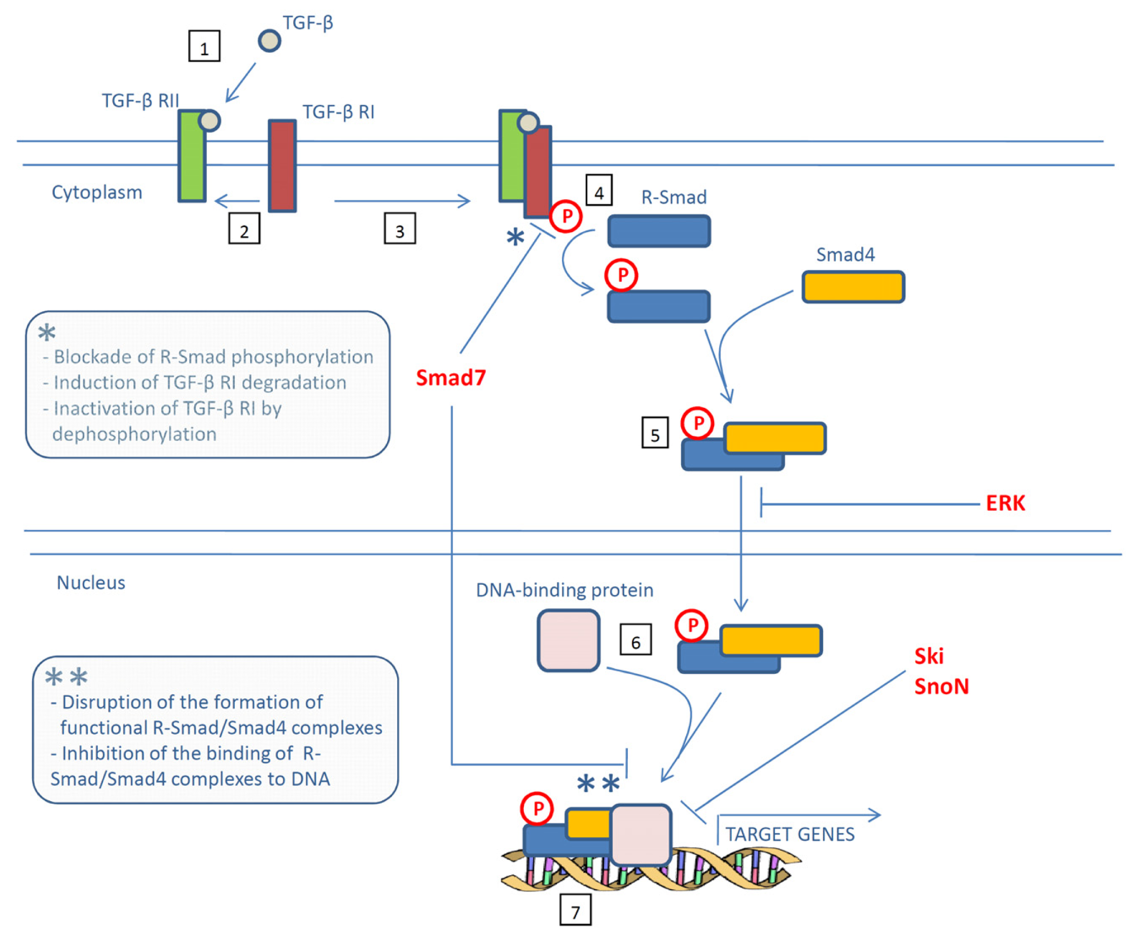
Figure 1. Schematic overview of the transforming growth factor (TGF)-β signaling pathway. Binding of TGF-β to its type II receptor (TGF-β RII) (1) attracts the TGF-β type I receptor (TGF-β RI) (2) and leads to formation of a receptor complex (3) and phosphorylation of TGF-β RI (4). Thus activated, TGF-β RI in turn phosphorylates a receptor-activated Smad (R-Smad) (4), allowing this protein to associate with Smad4 and move into the nucleus (5). Once in the nucleus, this Smad complex associates with DNA-binding proteins (6) to activate the transcription of specific target genes (7). Negative regulators of this signaling pathway are indicated in red. * and ** indicate mechanisms by which Smad7 antagonizes TGF-β signaling in the cytoplasm and in the nucleus respectively.
Ijms 14 23774f1
Figure 2. Schematic overview depicting the pathogenic roles of a deregulated Smad7 protein expression. Abbreviations: CNS, central nervous system; HP, Helicobacter-pylori.
Ijms 14 23774f2
Table 1. Pro- and anti-tumor effects of Smad7.
Cancer typeMethodObservationRef.
EndometrialObservational studySmad7 upregulation associates with poor survival rate[42]
EsophagealObservational studySmad7 upregulation associates with shorter time to recurrence[43]
ColorectalObservational studyCRC patients with deletion of Smad7 have a favorable clinical outcome compared with patients with Smad7 amplification[44]
ColorectalColony formation assay, xenografts induced by FET cells in immunodeficient miceSmad7-overexpressing FET cells show aggressive colony formation on soft agar and increased tumorigenicity in vivo compared with control FET cells[45]
ColorectalMetastasis induced by the injection of FET cells in the spleen of immunodeficient miceInjection of Smad7-overexpressing FET cells results in the development of liver metastasis[46]
ColorectalAOM + DSS-driven colitis associated CRCOver-expression of Smad7 in T cells associates with severe colitis and reduces the growth of colitis-associated CRC [47]
PancreaticColony formation assay, xenografts induced by FET cells in immunodeficient miceSmad7 overexpressing COLO-357 cells are resistant to the TGF-β-driven growth inhibition in vitro and exhibit a marked increase in their capacity to form colonies in soft agar and tumors in nude mice[34]
PancreaticTransgenic mouse with pancreatic overexpression of Smad7Smad7 blocks TGF-β signaling in the pancreas and induces premalignant ductal lesions with the characteristics of pancreatic intraepithelial neoplasia[48]
PancreaticObservational studyExpression of Smad7 associates with a more favorable prognosis compared with patients with lower levels of Smad7 who exhibited increased incidence of lymph node metastasis and liver metastasis after surgery [49]
GastricObservational studyElevated Smad7 levels in tumors with lymphatic metastasis[50]
GastricObservational studyPatients bearing tumors with positive Smad7 expression have a poor prognosis[51]
GastricCell cultureEctopic Smad7 expression increased the survival of SGC7901 gastric cancer cells[50]
SkinMouse model of chemically-induced skin carcinogenesisSmad7 overexpression in H-ras-transduced keratinocytes determines the conversion of benign to malignant epithelial cells and a rapid progression to squamous cell carcinoma[52]
SkinXenograft model in which primary keratinocytes mixed with dermal fibroblasts are grafted into nude miceH-ras/Smad7 but not H-ras keratinocytes progresses to SCC[52]
SkinColony formation assay, xenografts induced by 1205Lu cells into immunodeficient miceStable over-expression of Smad7 in 1205Lu cells reduces MMP-2 and MMP-9 production, invasive capacity and anchorage-independent growth in vitro as well as subcutaneous tumor formation in nude mice [53]
SkinModel of bone metastases in which tumor cells are inoculated into the left cardiac ventricle of nude miceAnimals injected with Smad7-transfected 1205Lu cells have significantly less osteolytic metastases and longer survival compared with mice injected with parental and mock-transfected 1205Lu cells [54]
SkinIn vivo human skin grafting systemSmad7-expressing 1205Lu cells position proximal to the dermal-epidermal junction and fail to form tumors, while control cells form tumors within the dermis [55]
BreastObservational studySmad7 expression correlates with a poor prognosis in patients with invasive breast carcinoma [56]
BreastBreast cancer metastasis induced by intravenous injection of mouse mammary carcinoma JygMC(A) cellsMice injected with Smad7-transfected JygMC(A) cells show fewer lung and liver metastasis and longer survival than mice injected with mock-transfected JygMC(A) cells [57]
BreastCell cultureSmad7 sensitizes MCF7 breast cancer cells to TNF-induced cell death [20]
BreastCell cultureEctopic Smad7 expression in SKBR3 cells completely abrogates EGF-induced MMP-9 expression [40]
BreastCell cultureSmad7 overexpression suppresses migration and invasion of mesenchymal-like MCF10CA1h cells[58]
BreastCell culturemiR-106b-25 cluster negatively regulates Smad7 expression thereby activating TGF-β signaling and inducing EMT in MCF7 cells[59]
LiverMouse model of HCC induced by DENSmad7-deficient mice have higher tumor incidence and multiplicity than wild-type mice [60]
LiverObservational studyLow Smad7 expression in HCC samples associates with better disease free survival [61]
LiverCell cultureSmad7 restrains EMT and cell migration of HCC cells [61]
ProstateCell cultureEctopic Smad7 expression induces apoptosis in PC-3U human prostate cancer cells [62]
ProstateCell cultureSmad7 is required for the induction of apoptosis by the anti-cancer agent 2-Methoxyestradiol in PC-3U cells [63]
ProstateCell cultureSmad7 promotes migratory responses in PC-3U cells[64]
White background = pro-tumorigenic effect; grey background = anti-tumorigenic effect; Abbreviations: AOM, azoxymethane; CRC, colorectal cancer; DEN, diethylnitrosamine; DSS, dextran sodium sulfate; EGF, epidermal growth factor; EMT, epithelial-mesenchymal transition; HCC, hepatocellular carcinoma; MMP, metalloproteinase; TGF, transforming growth factor; TNF, tumor necrosis factor.