Review

Low Oxygen Response Mechanisms in Green Organisms

1
PlantLab, Institute of Life Sciences, Scuola Superiore Sant'Anna, Via Mariscoglio 34, Pisa 56124, Italy
2
Institute of Agricultural Biology and Biotechnology, National Research Council, Via Moruzzi 1, Pisa 56100, Italy
3
Institute of Plant Biochemistry and Biotechnology, University of Münster, Schlossplatz 8, Münster 48143, Germany
*
Author to whom correspondence should be addressed.
Int. J. Mol. Sci. 2013, 14(3), 4734-4761; https://0-doi-org.brum.beds.ac.uk/10.3390/ijms14034734
Submission received: 29 January 2013 / Revised: 20 February 2013 / Accepted: 21 February 2013 / Published: 27 February 2013
(This article belongs to the Special Issue Abiotic and Biotic Stress Tolerance Mechanisms in Plants)

Abstract

:
Low oxygen stress often occurs during the life of green organisms, mostly due to the environmental conditions affecting oxygen availability. Both plants and algae respond to low oxygen by resetting their metabolism. The shift from mitochondrial respiration to fermentation is the hallmark of anaerobic metabolism in most organisms. This involves a modified carbohydrate metabolism coupled with glycolysis and fermentation. For a coordinated response to low oxygen, plants exploit various molecular mechanisms to sense when oxygen is either absent or in limited amounts. In Arabidopsis thaliana, a direct oxygen sensing system has recently been discovered, where a conserved N-terminal motif on some ethylene responsive factors (ERFs), targets the fate of the protein under normoxia/hypoxia. In Oryza sativa, this same group of ERFs drives physiological and anatomical modifications that vary in relation to the genotype studied. The microalga Chlamydomonas reinhardtii responses to low oxygen seem to have evolved independently of higher plants, posing questions on how the fermentative metabolism is modulated. In this review, we summarize the most recent findings related to these topics, highlighting promising developments for the future.

1. Plant Metabolic Regulations under Low Oxygen

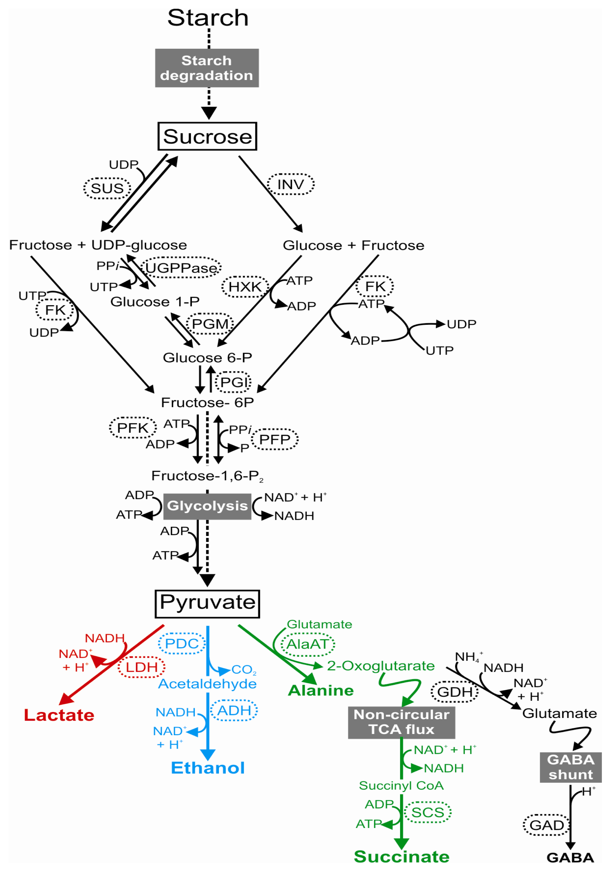
1.1. A Shift towards the Fermentation Pathway

Plants and algae rely on aerobic respiration to produce energy. Oxygen availability is thus required and changes in its accessibility lead to drastic metabolic rearrangements. Ultimately, aerobic organisms die if the absence of oxygen is prolonged. A lower level of oxygen can result from environmental conditions, but also anatomical and tissue constraints. During their life, plants and algae are exposed to a range of oxygen concentrations that can vary from a fully aerobic state (normoxia) to oxygen deficiency (hypoxia) or the total absence of oxygen (anoxia) [1]. Excessive rainfall can lead to soil waterlogging or even to the complete submergence of plants, with dramatic consequences for crops. In the context of climate change, an increased frequency of flooding has been observed worldwide [2]. Under hypoxia or anoxia, a series of rapid and profound molecular and metabolic responses are activated to endure such stress.
Oxygen deprivation compromises ATP production and energy supply. This is because oxygen is the final acceptor of electrons in the mitochondrial oxidative phosphorylation occurring during respiration [3,4]. Under submergence oxygen availability decreases drastically and oxygen production by photosynthesis is reduced by water turbidity. Reduced light availability in plants that are submerged also compromises carbohydrates production by the Calvin cycle. Fermentation is required because the mitochondrial production of ATP is hampered in the absence of oxygen. Under these conditions, plants have to rely on the small amount of ATP that can still be produced through glycolysis. A lack of the mitochondrial re-oxidation of NADH puts glycolysis at risk of stopping soon after the low oxygen conditions have been established, unless an alternative mechanism for NADH re-oxidation is activated, i.e., fermentation [5,6]. In some cases of oxygen absence, the “Pasteur effect” may occur, which accelerates the carbohydrate consumption with a 2 to 3 fold increase in the glycolytic flux with respect to the aerobic control [7]. Under oxygen deficiency, the plant metabolism uses pyruvate as a starting substrate to switch on two main pathways: ethanolic and lactic fermentations [8].
In almost all plant species, a rapid activation of lactate dehydrogenase (LDH), which catalyzes the reduction of pyruvate to lactate (Figure 1), has been observed under low oxygen conditions [912]. The production of lactate, however, exerts a negative effect on cytoplasmic pH, and therefore the production of lactate is only transient and replaced by ethanolic fermentation. The interaction between lactic and ethanolic fermentation leads to the tight control of the cytosolic pH of cells [13]. In fact, lactic fermentation acidifies cytoplasm in order to obtain an optimal value for PDC activity, with the consequent activation of ethanol production to avoid any deleterious cytoplasmic acidification [13,14]. Studies of several species, such as tomato, potato, Arabidopsis and rice, have shown that lactate production plays a minor role in low oxygen responses, because lactate is generally produced only during the first hours of stress and is easily expelled from the cell, in order to prevent an excessive accumulation of this compound in the cytosol [15]. Some proteins, such as the hypoxia-inducible Nodulin Intrinsic Protein (NIP2; 1) [16], have been indicated as members of the lactate extrusion process. However, an increase in root tolerance to hypoxia has been reported when LDH was over-expressed in Arabidopsis plants [17]. This resulted in a significantly higher activity of PDC, suggesting that the lactic and ethanolic fermentation pathways are inter-dependent and that the lactate metabolism is involved in the enhancement of ethanol production [17].
Ethanolic fermentation consists of the conversion of pyruvate to ethanol through the coupled reactions of pyruvate decarboxylase (PDC) and alcohol dehydrogenase (ADH) (Figure 1). The significant role of both ADH and PDC for hypoxia survival has been demonstrated in many plant species, which differ in their degree of low oxygen tolerance, such as maize, rice, Rumex palustris and Arabidopsis thaliana[1823]. Although the conversion of pyruvate to acetaldehyde, catalyzed by PDC, is considered to be rate-limiting in ethanol fermentation, the regulatory role of PDC has not yet been fully described. In several plant species, PDC controls anaerobic sugar catabolism and its over-expression in transgenic tobacco leaves has been shown to produce 10 to 20 fold more ethanol than wild-type plants under anoxia [24]. In Arabidopsis, there are four genes encoding PDC, and microarray datasets related to low oxygen conditions have shown that PDC1 and PDC2 genes are induced when oxygen deprivation occurs [25,26], whereas PDC3 and PDC4 mRNA levels are not markedly induced by anoxia [26].
Intriguingly, the overexpression of PDC1 and PDC2 in Arabidopsis plants has been shown to improve ethanol production and survival under low oxygen conditions [27]. Furthermore, it has been reported that pdc1 null mutants are less tolerant to anoxia, suggesting that the PDC1 gene is needed in order for the plant to survive the stress [25]. A recent detailed investigation of pdc1 and pdc2 mutants also indicated that PDC1 plays a predominant role in roots, while PDC2 is leaf-specific [26].
In ADH knock-out plants, a general reduction in low oxygen tolerance has been observed [20,27]. The Arabidopsis adh1 null mutants showed a lower survival rate under hypoxia [28]. In rice, rad (reduced adh activity) mutants displayed a reduction in the ADH protein due to a point mutation in the ADH1 gene, and, consequently, a higher sensitivity to submergence stress (i.e., reduced coleoptile elongation) [29,30]. In maize, the adh1 mutant, affected in the biosynthesis of ADH, has been found to be very susceptible to anoxia [31]. Conversely, an increased tolerance to hypoxia, together with an enhanced root growth, has been obtained when ADH was over-expressed in Arabidopsis hairy roots [32], but this had no effect on the ability of Arabidopsis transgenic plants to survive flooding [27].
Finally, Ala, γ-aminobutyric acid (GABA) and succinate have been suggested as additional products of the anaerobic metabolism [15,33,34]. Under anaerobic conditions, Ala is accumulated by the transfer of an amino group from glutamate to pyruvate, generating 2-oxoglutarate (2-OG) as a co-product, thus causing a decrease in glutamate content (Figure 1) [35]. This reaction is mediated by the Ala aminotransferase enzyme (AlaAT) and may contribute efficiently to tolerance, although how is still unknown, since Ala accumulation is not toxic for the cells [36,37]. Upon anoxia, the 2-OG can be metabolized via two different shunts [2,15]. In fact, 2-OG can again be oxidized by glutamate dehydrogenase (GDH), with the incorporation of NH4+ and the generation of NAD+, to glutamate, which is then decarboxylated to GABA by glutamate decarboxylase (GAD), a process that is stimulated in acidic conditions. The production of GABA and the next transamination to succinic semialdehyde may help to stabilize the cytosolic pH [38] and ensure an additional conversion of pyruvate to Ala [39]. Alternatively, 2-OG can be used as a substrate in the non-circular TCA flux and thereafter metabolized to succinate via the succinyl CoA ligase enzyme (SCS) [34,40]. To fuel this process, NADH is oxidized by the reduction of oxaloacetate by malate dehydrogenase (MDH) to malate, which is further converted to fumarate and succinate.
Overall, this evidence has shown that both ADH and PDC play a key role in low oxygen tolerance. Other products of the anaerobic metabolism can increase plant tolerance, however their regulation is still poorly understood and needs investigating further. For decades, plants metabolic mechanisms have been studied because of their significance in low oxygen tolerance, and it is clear that this will remain an exciting field of study. In the future, it will be important to deepen the current knowledge on the transcriptional regulation of metabolic genes during low oxygen responses, also in relation to the recent identification of the oxygen-sensing mechanism that seems to be at the basis of ethanolic fermentation activation [41,42] (see section 2.1), which represents a challenging starting point.

1.2. Sugar Metabolism under Low Oxygen

Sugars metabolism can be regulated in response to many environmental signals, which directly or indirectly influence the synthesis, concentration, transport and storage of sugars [43,44]. Under low oxygen conditions, the shift from aerobic to fermentative metabolism requires constant carbohydrate supplementation. Indeed, maintaining adequate levels of readily fermentable sugars in hypoxic or anoxic roots is one of the key adaptive mechanisms to an oxygen-deficient environment [45,46]. Several studies have found a close association between carbohydrate utilization and tolerance to low oxygen conditions [4749]. Anoxia or submergence tolerance has in most cases been associated with a slower use of carbohydrates, rather than with their relative abundance. Studies of the mechanism behind the tolerance of riparian plant species, such as Salix variegata, Arundinella anomala and Althernanthera philoxeroides, have revealed that the carbohydrate metabolism plays a very important role [46]. The ability to survive a long-term oxygen deficiency was explained by the capacity of these riparian species to preserve carbohydrate reserves, in contrast to the large decrease in carbohydrate concentrations observed in intolerant species [50]. The maintenance of adequate carbohydrate availability after prolonged anoxia or submergence is considered a direct consequence of a well-modulated fermentative metabolic activity, which is a strategy of some tolerant species [51]. In storage organs such as grass grains, potato tubers and Acorus calamus rhizomes, the supply of hexoses for glycolysis is guaranteed through an efficient starch mobilization via starch-degrading enzymes, such as endo-amylases, exo-amylases, debranching enzymes and starch phosphorylase [18,33,52,53]. Exogenous sugars supply can affect the ability to survive under low oxygen. The supplementation of sucrose, but not glucose, improves survival under anoxia [23,54]. In addition, the role played by sucrose in affecting anoxia responses has been investigated in terms of the transcriptome profile, revealing that this disaccharide modulates the anoxia-driven induction of a plethora of genes, different from the typical anaerobic ones as well. In order to provide hexose 6 phosphate for glycolysis, sucrose degradation takes place through two distinct pathways in plant cells: the sucrose synthase (SUS) bi-directional pathway, favoured in the catabolic direction, and the unidirectional invertase (INV) pathway (Figure 1). Furthermore, SUS (rather than INV) is reported to be the preferential pathway for sucrose degradation when oxygen is limited [55]. The SUS pathway is favoured under low oxygen because it partially preserves the energy of the glucose-fructose bound. While the INV pathway cleaves the sucrose molecule into glucose and fructose, the SUS reaction releases fructose and UDP-glucose, which can be subsequently utilized by UDP-glucose pyrophosphorylase (UGPPase), producing UTP and glucose-1P. UTP is then utilized by fructokinase (FK). The conversion of sucrose into phosphorylated hexoses, does not therefore require ATP, whereas the INV pathway requires 2 mol of ATP per mole of sucrose [56,57]. Studies on maize roots have shown that both sucrose synthase genes SUS1 and Sh1 were upregulated by low oxygen [58]. In Arabidopsis, SUS1 and SUS4 transcripts, as well as their relative proteins, increase significantly upon oxygen deprivation both in roots and shoots [59]. This leads to the hypothesis that SUS plays a central role in the sucrose metabolism under conditions of oxygen deprivation, contributing to the degradation of the disaccharides, and thus to the availability of substrates for energy production through glycolysis and fermentation. Indeed, Bieniawska and colleagues [60] showed that double mutant plants (sus1 × sus4) are less tolerant to 4-day root submergence compared to wild types.
Glucose and fructose need to be phosphorylated by hexokinases (HXK) to be channeled to the glycolytic pathway. In rice seedlings, the induction of the fructokinase OsFK2 has been shown, together with the hexokinase OsHXK7[61], to be likely induced via hypoxia-driven sucrose starvation [62,63].
Other glycolytic reactions may utilize the PPi available instead of ATP under oxygen deprivation, improving the net yield of ATP per mol of sucrose catabolized. The phosphorylation of fructose-6P to fructose-1,6P2 can be catalyzed by the unidirectional phosphofructokinase (PFK), which uses ATP as a phosphate group donor, or via the bidirectional PFK-PPi (PFP) which uses PPi. PFP is activated under anoxia in rice [64,65] and, together with PDC, it can regulate the carbon flux during anaerobiosis in different plant species [7,65,66]. Since the PFK step can be limiting under oxygen deprivation, the PPi supply may be increased by a substrate cycle coupled with pyruvate kinase (PK) and pyruvate orthophosphate dikinase (PPDK), in order to finally accelerate glycolysis. Interestingly, PPDK is up-regulated by low oxygen in rice, both at the gene and protein levels [63,67].
Many studies suggest that submergence tolerance in several plant species may be associated with the ability to mobilize carbohydrate reserves, and also that the most important enzymes, which are involved in the degradation of carbohydrates, seem to play a very important role.

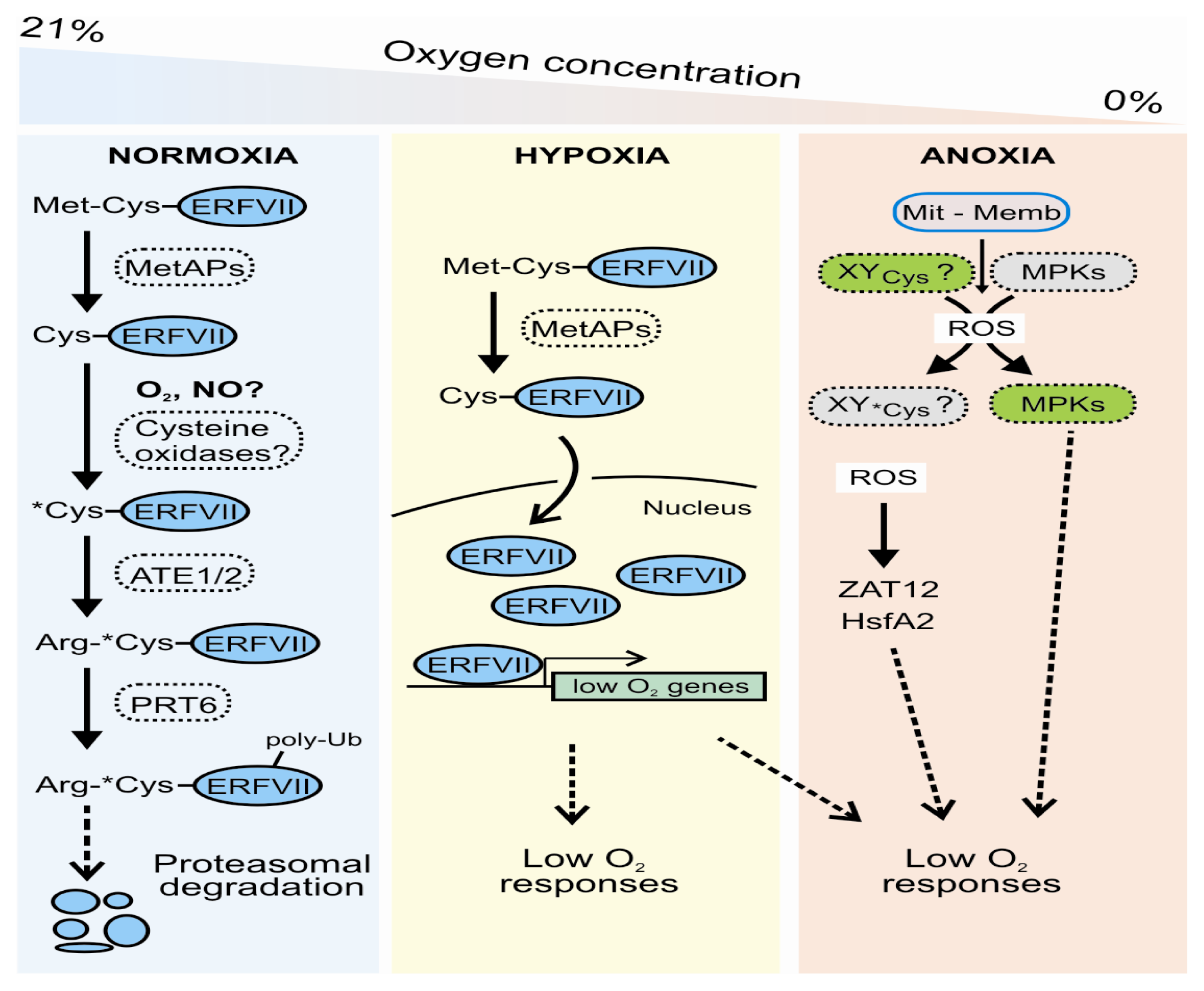
2. Low Oxygen Sensing and Signaling in Arabidopsis thaliana: The N-End Rule Pathway (NERP) and Side Defense Mechanisms

2.1. Oxygen Sensing in Plants

Metabolic alterations under low oxygen might in theory be exploited by plant cells to sense hypoxia and generate adaptive responses. Indirect oxygen-sensing mechanisms could be based on low energy status, variations in carbohydrate availability, reactive oxygen species (ROS) signals, nitric oxide (NO)-related responses, NAD/NADH ratio, calcium fluxes and homeostatic reactions to intracellular pH changes [15]. However, the oxygen status might also be directly measured by the cell, for example through sensor proteins or transcription factors whose activity is directly affected by oxygen levels. Indeed, direct oxygen-sensing systems have been described for metazoans, yeasts and microorganisms. In all animal taxa studied so far, the perception of hypoxia relies on the stabilization of the heterodimeric factor HIF, a protein belonging to the bHLH-PAS domain family of eukaryotic transcription factors, which has been recognized as the unique activator of the whole set of hypoxia-inducible genes. In fact low oxygen prevents prolyl hydroxylation of the animal HIFα subunit, which under normoxic conditions leads to the ubiquitination and consequent degradation of the protein [68,69]. Plant genomes encode both PAS domain proteins and prolyl hydroxylases (PHDs), some of which are low oxygen and/or redox regulated. Therefore, the presence of HIF-like signal transduction pathways has also been hypothesized in plants.
However, it has been suggested that plant PAS and PHD proteins have functionally diverged from animal proteins, acquiring exclusive roles in the transduction of light signals and the modification of the cell wall, respectively [70]. In algal cells, proline hydroxylation occurs in cell wall proteins, and several PHDs have been found to be induced in Chlamydomonas under anaerobiosis [71]. However, it is unlikely that this process occurs under anoxia, since it requires oxygen as a substrate [71]. Thus, the existence of on oxygen-dependent post-translational modulation of pivotal TFs needs further investigation. This confirmation has been obtained recently in plants, when two independent studies showed that oxygen availability directly impacts on the stability of the transcriptional activator RAP2.12, through the N-End Rule Pathway (NERP) for protein degradation [41,42].
NERP is an ubiquitin-dependent proteolytic system that links the half life of a peptide to its N-terminal amino acidic sequence [72]. N-terminal residues can be classified as stabilizing or destabilizing, depending on their ability to prevent or promote, respectively, the recognition of the protein by the components of the proteolytic machinery. In eukaryotic cells, amino-terminal degradation signals (N-degrons), corresponding to N-terminal destabilizing residues, can be specifically bound by E3 Ubiquitin (Ub) ligases, therefore called N-recognins, which, after recruiting Ub-activating (E1) and Ub-conjugating (E2) enzymes, target short-lived proteins to the 26S proteasome for degradation [73]. Structural determinants for a functional N-degron include an N-terminal destabilizing residue, a neighbouring Lys residue for ubiquitin conjugation, and the vicinity of a flexible region facilitating its exposure [72,74]. An N-degron can be created through the endoproteolytic cleavage of proteins, the removal of the N-terminal signal or transit peptides, or the elimination of NH2-Met [73,75].
In both eukaryotic and prokaryotic organisms, the NERP displays a common hierarchical organization, in which primary N-terminal destabilizing residues, directly bound by the N-recognins, can be produced by a sequential modification of secondary or tertiary destabilizing residues [76]. Plants and mammals share identical sets of destabilizing residues [77]. Primary residues consist of basic (Arg, Lys and His, “type I” residues) or bulky hydrophobic amino acids (Phe, Trp, Tyr, Leu and Ile, “type II” residues). Secondary residues are Asp, Glu or oxidized Cys, all of which, in the N-arginylation branch of the NERP, are substrates for the conjugation of Arg by Arg-tRNA transferase (ATE) enzymes. Secondary residues can be generated by N-terminal amidohydrolases (NtN- and NtQ-amidases), through the deamidation of Asn and Gln, or by oxidation of Cys, which therefore act as tertiary destabilizing residues. Proteins are generally synthesized with an N-terminal stabilizing Met residue, which however can be removed by the action of Met-aminopeptidase enzymes (MetAPs), provided that the following amino acid is characterized by a short side chain. Of such residues, Cys is the only one whose exposure can further lead to the creation of an eukaryotic N-degron [78].
After the disclosure of the NERP mechanism and the acknowledgment of its conserved organization across evolution, enzymatic players in plants were rapidly identified by forward genetic and sequence similarities, revealing a certain degree of divergence from the animal kingdom [79]. However, the physiological significance of the pathway in plants has started to be elucidated only recently and is mainly derived from the genetic analysis of components of the N-arginylation branch, which targets only a subset of all possible substrates to degradation. The Arabidopsis ATE1, which was first reported to be involved in leaf senescence [80], has also been demonstrated to work redundantly with its homolog ATE2 in rosette development and leaf shape determination [81]. Of the two plant N-recognins identified to date, named PRT1 and PRT6, only the latter is associated with a recognizable mutant phenotype in Arabidopsis, mainly characterized by a stronger seed after-ripening dormancy, which was found to correlate with hypersensitivity to ABA [82].
The finding that plant oxygen sensing relies on the N-terminal mechanism for protein destabilization thus provides the first evidence of the involvement of the NERP in plant stress responses. A conserved N-degron, whose consensus sequence is MCGGAI/L, was discovered in group VII ERF transcription factor proteins, and recognized as the key for the group VII ERF-driven activation of the downstream transcriptional events that are the signature of plant anaerobic responses. Members of this subfamily of the plant-specific ERF family [83], such as Arabidopsis Hypoxia Responsive ERF (HREs) and RAP2.2/2.12 and rice SUB1 and SK factors, have long been related to plant transcriptional regulation under low oxygen [8487] and postulated to work as oxygen-modulated transcriptional switches for acclimation responses.
In Arabidopsis, newly synthesized RAP2.12 proteins undergo a constitutive cleavage of the conserved NH2-Met-Cys sequence by MetAPs, which leads to exposure of a tertiary N-terminal destabilizing Cys residue. In the presence of oxygen, the Cys2 residue can be oxidized to Cys-sulfinic acid (CysO2H) or further to Cys-sulfonic acid (CysO3H) to trigger the conjugation of the primary destabilizing residue Arg by ATE1/2, the binding of the PRT6 N-recognin and the possible targeting of the protein to the proteasome (Figure 2). Under low oxygen conditions, on the other hand, the impairment of Cys oxidation directly translates into RAP2.12 stabilization, thereby allowing the TF to promote the transcription of low oxygen-responsive genes (Figure 2). In line with this, transcriptome analysis of the ate1ate2 and prt6 mutants showed a higher level of basal expression of hypoxia marker genes, such as ADH1, SUS4 and PDC1[41].
Constitutive degradation of RAP2.12 under normoxia can be prevented, provided that its N-terminus is protected from enzymatic modifications. The use of a RAP2.12:GFP fusion construct enabled Licausi and colleagues [42] to observe the sequestration of the protein at the plasma membrane in normal conditions, which turned out to depend on the ability of RAP2.12 to interact with the transmembrane acyl-CoA-binding proteins ACBP1/2. Constitutive availability of “inactive” RAP2.12 bound to the cell membrane is likely to be crucial in making RAP2.12 a true molecular switch for the early response to hypoxia. This is supported by the observation of immediate RAP2.12:GFP displacement from the plasma membrane to the nucleus at the onset of low oxygen conditions. Interestingly, despite all being NERP substrates and sharing overlapping sets of gene targets, RAP2.12 and HRE1/2 factors might diverge functionally. While RAP2.12 mediates the quick activation of the low oxygen response, HRE1/2 could be involved in the long-term acclimation of plants to prolonged oxygen deprivation, as it is accumulated during hypoxia and not constitutively present like RAP2.12 is [42].
It can thus be speculated that the oxygen-dependent NERP mechanism for RAP2.12 turnover is very significant both in preventing mislocalized cytosolic RAP2.12 from reaching the nucleus under aerobic conditions and, more importantly, in driving the degradation of the TF upon reoxygenation of the cell. The restoration of normoxia was consistently followed by the rapid and complete disappearance of the RAP2.12:GFP signal. It is still unknown whether Cys oxidation is spontaneous or enzymatic, however it has been reported to require previous Cys nitrosylation by NO in order to take place [88]. Some cross-talk between oxygen-dependent NERP and NO in oxygen-deprived plant cells is not unlikely, given the known role of NO during hypoxia (see section 2.2).
The importance of the N-end rule for plant physiological responses to low oxygen is corroborated by the observation that mutants for the Cys2-dependent NERP branch display altered tolerance to low oxygen stresses. Arabidopsis ate1/2 and prt6 mutants, in fact, survived submergence better than wild type plants [41]. However, acclimation of true submergence tolerant species, such as rice, is likely to involve divergence in group VII ERF regulation, as proved by the escape of the SUB1A protein from the NERP regulation (see section 3.1).

2.2. A Role for Reactive Oxygen and Nitrogen Species (ROS/NOS)

One of the most controversial aspects related to the molecular response to low oxygen is the presence of oxidative stress. This may seem counterintuitive since oxygen is needed for the production of ROS. However, many different microarray analyses carried out under different low oxygen conditions and on different plants have reported the activation of ROS-regulated genes or those associated with ROS.
Heat Shock Proteins (HSPs) are the most recurrent of ROS-related proteins that are also induced by anoxia [89,90]. HSPs expression has been shown to be associated with the presence of hydrogen peroxide (H2O2) in different kingdoms [91]. HSPs production has been observed predominantly under anoxia [90], probably as a mechanism needed to activate a defense that overlaps with heat stress [92] (see section 2.3). Some TFs associated with ROS production have also been shown to be regulated by low oxygen, i.e., HsfA2 and ZAT12[90]. Results from yeast and mammalian cells have suggested heat shock transcription factors (HSFs) as sensors of H2O2[93], and ZAT12 has been shown to be regulated by several stresses where the oxidative signal seems to be involved [94]. These genes have a lower induction in the rbohD Arabidopsis mutant under low oxygen stress, suggesting a possible role for this membrane NADPH oxidase superoxide (O2) producing enzyme in the mechanism of ROS generation [90].
The ROS produced by the membrane NADPH oxidase was proposed as being part of low oxygen signaling through a ROP rheostat, regulating ADH expression and activity [95]. A more recent report, on the other hand, suggested mitochondria imbalance to be the ROS generator mechanism under oxygen deprivation, in a system supporting survival mediated by mitogen-activated protein kinases (MPKs) activation [96]. Whatever the mechanism, it seems to be transcriptionally independent of the NERP, since under control conditions Arabidopsis mutants associated with this pathway activate anaerobic but not ROS-related genes [90]. An oxidative burst probably orchestrates an alternate pathway of response which, together with the NERP, contributes to tolerance [97]. This is also supported by the finding that plants over-expressing HsfA2 tolerate low oxygen stress better by activating heat-stress related genes but not the fermentation pathway [92].
One of the most interesting aspects of ROS production under low oxygen is the possible indirect interaction with the NERP. Indeed, this mechanism includes, after an initial Met cleavage at the N-end of target proteins, the oxidation of Cys, through a mechanism which may or may not be enzymatic [98] (see section 2.1). Interestingly, Cys residues are also considered to be targets for ROS, and increasing evidence suggests the pivotal role of Cys in ROS perception and network signaling.
Thiol moieties (–SH) on the side chain of Cys are very sensitive to oxidation and can form disulfide bonds in a reversible way, changing the protein state. In parallel, thiol moiety (–SH) on the side chain of Cys can also be subsequently oxidized to a sulfonyl moiety (–SO3H), which cannot be reduced under normal intracellular environmental conditions [99]. Indeed, the Cys position in a protein is an important prerequisite for its reactivity towards oxidant or reducing agents, since most thiol groups are either structurally not accessible to redox agents, or the microenvironment is strongly buffered by antioxidants. The Cys pKa value is strictly related to the Cys position, and is a good indicator to establish how prone it is to redox modification. The Cys as thiolate anion (Cys-S) is more readily oxidized than in its thiol form -SH. Since in non-stress conditions the majority of the Cys groups are in a thiol state, few proteins would be expected to be readily susceptible to oxidation. However, Cys may exist as thiolate anions at neutral pH values. Protein Tyr phosphatases (PTP) are examples of proteins with low pKa Cys residues (pKa = 4.7/5.4), thus reactive as Cys-S at neutral pH and sensitive to oxidation by various oxidants, including H2O2[100]. PTP Tyr (de) phosphorylation is involved in the regulation of MPKs activities in plant, fungi and animal cells [101]. Gupta and colleagues [101] showed that AtPTP1 phosphatase activity is inhibited by H2O2in vitro, suggesting that H2O2 induces a rapid and reversible Cys-dependent conformation change that leads to inactivation, as demonstrated in vivo in animals [102]. Interestingly, H2O2 treatments have been shown to activate MPKs in plant cells, known to be negatively regulated by PTPs. In particular, AtPTP1 inactivation has been shown to be correlated to AtMPK6 activation by H2O2. In this context, transient AtMPK6 activation, probably due to ROS of mitochondrial origin, has been observed in response to oxygen deprivation and re-oxygenation and is associated with improved plant survival [96].
The role of NO as a signaling molecule under low oxygen conditions is very promising. NO has been found to be required in the oxidation of the N-terminal Cys before arginylation by ATE in mammalian cells in vivo[88]. In addition, under low oxygen conditions, nitrite (NO2) acts as an alternative electron acceptor and its reduction to NO, conversion to nitrate (NO3) by the non-symbiotic hemoglobin Hb1, and then reduction to NO2 by nitrate reductase gives rise to a cycle that produces a certain amount of ATP [103,104]. Moreover, recent studies on animals have demonstrated that HIF stabilization and transcriptional activity is achieved also through S-nitrosylation of HIF pathway components [105,106].
Since the NERP regulates several different processes in plants other than low oxygen, such as growth and development [98], one interesting hypothesis is that the mechanisms behind Cys stability, and thus the fate of the protein, may be different depending on the situation, either directly regulated by oxygen availability or cellular changes in cytosolic pH or ROS and reactive nitrogen species (NOS) balance. Each of these actors could be involved in the regulation of NERP side mechanisms which, all passing through the modification of Cys located in different positions, could lead to the activation of different downstream events and thus contribute to stress tolerance (Figure 2).

2.3. Low Oxygen and Heat Stress Convergence

An intimate overlapping in the activation of a subgroup of Heat Shock Genes (HSGs) has been observed when comparing the transcriptomes of Arabidopsis seedlings under heat, anoxia and heat followed by anoxia, [23,92,107]. A high portion of these genes are strictly related to heat shock (HSPs) and oxidative stress-related proteins (ZAT12 and AOX1a) [92,108110]. A mild heat pre-treatment, able to induce thermotolerance in plants when subsequently exposed to lethal temperatures [111], can also confer a higher tolerance to subsequent anoxia [23,92]. This suggests that two apparently unrelated stress conditions share a mechanism for tolerance.
Of the heat-related genes up-regulated by both heat and anoxia, the heat shock transcription factor HsfA2 is one of the most strongly modulated [92]. HsfA2 is involved in several abiotic stress conditions [112114] and its molecular mechanism of action has recently been described in Arabidopsis [113,115117]. This transcription factor is assumed to cooperate with other factors to activate the downstream signaling pathway both during the stress period and the recovery phase [113,116,117], and is believed to coordinate the ROS-mediated cross-talk among cellular compartments and organelles [113,117,118]. In addition, a possible direct or indirect involvement of this TF in intracellular pH homeostasis cannot be ruled out, since one of its putative target genes (ROF2) plays a pivotal role in this mechanism [119]. Interestingly, HsfA2 confers increased tolerance to anoxia when over-expressed in Arabidopsis [92]. HsfA2 over-expression has a positive effect on the expression of small HSPs, such as HSP25.3-P and HSP18.2-CI, and also antioxidant genes, such as APX2, GolS1 and GolS2[92,112114]. HsfA2 does not affect the expression of any anaerobic genes, suggesting that HsfA2 does not control the anaerobic pathway [92]. Conversely, the anoxia-driven up-regulation of heat-related genes is independent of the NERP-dependent mechanism of oxygen sensing [41,42]. This suggests that activation of heat-related genes by an anoxic treatment is driven by an independent sensing mechanism that may rely on a ROS-dependent sensing mechanism [90,92,95,120]. Heat-induced tolerance to anoxia is possibly a consequence of the induction of HSPs as well as some antioxidants and ROS-related genes (ZAT12, AOX1a, APX2). These genes may be required during the post-anoxic recovery phase, to protect cells during reoxygenation, which is an oxidative-prone event [121,122].

3. Low Oxygen Tolerance Mechanisms in Oryza sativa

3.1. Rice Germination under Low Oxygen

In terms of tolerance to low oxygen, rice is considered one of the most interesting species. Unlike the majority of higher plants, it is able to germinate under submergence using the starchy reserves of the seeds to fuel the anaerobic metabolism [33]. During early seed germination under submergence, rice growth is restricted to the coleoptile, with both root and primary leaf elongation inhibited [123]. Rice coleoptile elongation under low oxygen enables successful crop establishment in submerged fields, since the long coleoptile can reach the water surface and thus make contact with the normoxic atmosphere [124].
Rice is able to use starch under low oxygen because of the presence of specific enzymes even under anoxia. Of these, α-amylases have a major role in degrading native starch granules [125127] and catalyzing starch hydrolysis to sugars, which are subsequently translocated to the embryonic axis to fuel glycolysis in order to produce energy for growth. These enzymes are not present in cereals, which are unable to germinate under anoxia or submergence [18,128,129]. Among the various rice isoforms of α-amylases, Ramy3D plays a major role for low oxygen survival since it is highly induced under low oxygen [63,130] and does not require gibberellins to be induced [131,132]. Indeed, the synthesis of gibberellins requires oxygen, and the GA-dependent pathway for α-amylase induction is inactive under low oxygen. Ramy3D is regulated by a sugar starvation-dependent pathway [133135]. There is a link between the reduction in soluble sugar content and low oxygen conditions [132136]. The regulation of Ramy3D requires the Calcineurin B-like Interacting Protein Kinase 15 (CIPK15) and the Snf1-Related protein Kinase 1A (SnRK1A), thereby activating the transcriptional regulator MYBS1 that upregulates Ramy3D[135,137139]. A rice mutant defective in CIPK15 (cipk15) hardly germinates at all when submerged [135].
The growth of the rice coleoptile under submergence is due to cell expansion, in which expansins are likely to play a role [140,141]. Although the length of coleoptiles under low oxygen does not correlate with expansin expression levels [142], EXPA7 and EXPB12 are strongly upregulated in the submerged rice coleoptile and represent good candidates as players in anoxic coleoptile growth [63]. Additionally, the altered expression of EXPA4 affects coleoptile elongation under submergence in the japonica variety Taipei 109 [143].

3.2. Strategies to Survive Low Oxygen in Adult Rice Plants

Adult rice plants display an array of adaptive mechanisms for cultivation in a large range of hydrological regimes, from deepwater to flash flooding-prone areas. A considerable variation in submergence tolerance exists among rice genotypes.
The wide number of ecological situations in which rice varieties, landraces and wild genotypes have been grown for centuries, enabled submergence tolerance characters to be selected that are effective in specific habitats. There are two main strategies to enable rice to survive different flooding regimes [84,86] and are also found in distinct wild progenitors [86,144,145]. One of these mechanisms was originally identified in Flooding Resistant 13A (FR13A) [146] a lowland rice landrace able to tolerate several weeks of flooding [147]. Molecular mapping revealed that tolerance was correlated with the SUBMERGENCE1 (SUB1) QTL, located on chromosome 9 [148]. This locus contains up to three ERF genes, including SUB1A, which in its allelic form SUB1A-1, was recently demonstrated to be the master regulator of submergence tolerance [84]. This gene is activated by ethylene [149]. The near isogenic line M202(SUB1) obtained through the introgression of the SUB1 locus from the tolerant FR13A into the intolerant background M202, revealed that the SUB1A-1 allele induces the fermentative metabolism under submergence [149] and represses growth through the GA repressors Slender Rice-1 (SLR1) and SLR1 Like-1 (SLRL1) [150]. SUB1A, therefore, induces a “quiescence” status in the submerged rice plants, enabling them to preserve carbohydrates for use during the recovery phase. SUB1 marker-assisted backcrossing (MABC) with productive varieties enabled the transfer of the submergence tolerance trait to highly productive mega-varieties [151,152].
Unlike rice varieties that possess the SUB1A gene, deepwater rice (DPR) varieties can elongate very rapidly under submergence. This allows DPR to escape the oxygen-limiting conditions that are associated with flooding by keeping at least the leaf tips above the water surface [86,147]. Stem elongation is driven by two ERF genes, named SNORKEL1 (SK1) and SNORKEL2 (SK2), which are driven by ethylene and promote gibberellin-dependent elongation.
SUB1A-1 and SKs belong to the VII ERF group similarly to the Arabidopsis ERFs that are targets of the NERP (RAP2.2, RAP2.12, HRE1 and HRE2) [41]. However, SUB1A-1 does not seem to be a substrate of the NERP [42], probably because the Met-Cys N-degron is made not functional by the absence of a crucial downstream Lys residue or by its structural conformation. In addition, SKs factors harbour variant N-termini that deviate from the NERP consensus. The fact that SUB1A-1 and SKs are not targets of the NERP implies that their stability is not affected by oxygen and suggests that these ERFs evolved to take full advantage of ethylene as signaling molecules which accumulate in plants that are submerged [2,97].

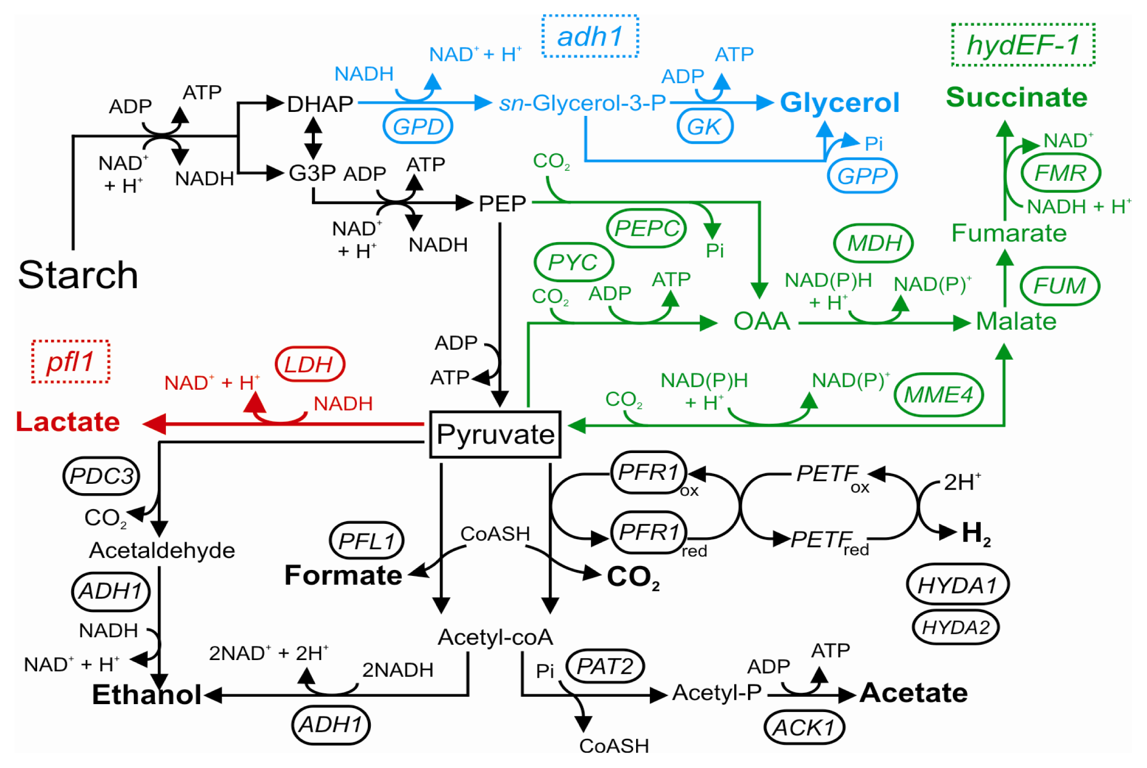
4. Anaerobic Metabolism in Photosynthetic Microorganisms: The case of Chlamydomonas reinhardtii

Photosynthetic microorganisms that inhabit the soil are periodically subjected to hypoxic and anoxic conditions, mainly as a result of day/night fluctuations, high microbial respiration and low rates of oxygen exchange [153]. The photosynthetic soil-dwelling alga Chlamydomonas reinhardtii has recently become a good model system for understanding how microalgae adapt to low oxygen levels. In addition to the availability of the nuclear genome sequence [154] and many molecular tools [155157], knock-out mutants and miRNA-lines have recently opened up new scenarios for green alga [158161]. Chlamydomonas shares many metabolic features with vascular plants, but it also offers a particular example of eukaryote encoding for proteins usually associated with strict anaerobes such as two oxygen-sensitive [FeFe]-hydrogenase enzymes (HYDA1 and HYDA2, the former being the primarily active hydrogenase isoform [162165] and the corresponding [FeFe]-hydrogenase maturation proteins (HYDG and HYDEF) [166,167]. Furthermore, pyruvate:ferredoxin oxidoreductase (PFR1), pyruvate formate lyase (PFL1), acetaldehyde/alcohol dehydrogenase (ADH1), phosphoacetyltransferase (PTA1 and PTA2), and acetate kinase (ACK1 and ACK2), typically present in prokaryotes and a few anaerobic eukaryotes [168], are all encoded in the Chlamydomonas genome and involved in the anaerobic metabolism [71,153,169173].
Interestingly, most of these oxygen-sensitive enzymes are located in the chloroplast [153,169,171], suggesting a tight control of fermentation over photosynthetic rates. Activities of these fermentative circuits result in the secretion of organic acids (formate, lactate, malate, acetate and succinate) and alcohols (ethanol and glycerol), with the concomitant evolution of small amounts of CO2 and H2, the latter being a promising carrier of renewable energy (Figure 3) [71,172179]. Pyruvate represents the central metabolite of this fermentative network, and differences in the ratio of the fermentative products can occur depending on the culturing conditions and Chlamydomonas strain [71].
Although the precise physiological function of hydrogenase and H2 evolution in Chlamydomonas has still not been completely resolved, H2 production is likely to have evolved as a component of photoprotection functioning as an electron valve for redox poising and fermentative energy production [153]. The metabolic consequences of a lack in functional HYDAs have been reported for the hydEF-1 mutant strain which does not have the maturation protein necessary for the correct assembly of the HYDA enzymes [179]. In this mutant the reverse TCA cycle (rTCA) is activated to dispose the excess reductants, resulting in high concentrations of extracellular succinate, a metabolite not detected in the parental strain (Figure 3) [179].
Given the extreme sensitivity of the [FeFe]-hydrogenase enzymes, H2 evolution in Chlamydomonas can only occur when cells are in micro-oxic or anoxic conditions (i.e., during fermentation in the dark or when cells are deprived of sulfur in the light) [180].
The disruption of specific fermentative branches has been suggested as a promising approach for rerouting the fermentative electron flow towards the PFR1-dependent H2 production pathway under dark anoxia. Recently, the effects of lesions in PFL1 [170,172] and ADH1 [173] enzymes have been reported. In spite of contradictory results in terms of H2 evolution, down-regulation of in vitro hydrogenase activity and lower HYDAs and PFR1 transcripts and protein levels in pfl1 mutants suggest that the H2 producing pathway cannot compensate for a loss of PFL1 activity. Instead of competing for the same substrate, PFL1 and PFR1 may actually work together to satisfy the physiological needs of the cell. ADH1 is the only enzyme responsible for ethanol production under dark anoxia in Chlamydomonas[173]. Intriguingly, the adh1 mutant showed no increase in H2 evolution, but high levels of extracellular and intracellular glycerol. Synthesis of glycerol at the level of dihydroxyacetonphosphate (DHAP) would allow for NAD+ recycling in the mutant strain, thus maintaining viability (Figure 3) [173]. Notably, the disruption of cyclic photosynthetic electron transfer (CEF)—which is induced under low oxygen in the alga—by the deletion of PGRL1 leads to a significant increase in H2 production [181]. The manipulation of electron transfer decisions made at the reducing side of photosystem I is therefore a valid instrument to enhance H2 evolution from the alga. Moreover, changes in the efficiency of CEF interfere with the overall induction of the anaerobic response in Chlamydomonas[182].
The metabolic restructuring observed for insertional mutants suggests that perturbation of the anaerobic metabolism activates unknown pathways that were set in place to regenerate reduced pyridine nucleotides and sustain survival. Additional work aimed at understanding how Chlamydomonas readjusts its metabolism to cope with blocks in specific reactions is clearly needed. Indeed, little is known about how photosynthetic microorganisms sense oxic conditions and induce transcription of their fermentative genes. Possible candidates have been proposed, such as the reduction state of the plastoquinone (PQ) pool [183] and the production and detoxification of ROS [184186]. Prolyl 4-hydroxylases, which directly sense oxygen and are involved in controlling responses to anoxia in animals [184], are also highly up regulated by anoxia in Chlamydomonas[71]; their putative role in the green alga, however, is still unknown.
Several fermentative genes follow precise day-night fluctuations in their expression levels, which are not necessarily linked to the photosynthetic oxygen dissolved in the medium [187]. Interestingly, the accumulation of ADH1 and HYDA2 transcripts has been shown to be independent of oxygen levels but is instead regulated by the circadian clock and/or cell cycles. PFR1 and HYDA1 expressions, on the other hand, are strongly influenced by oxygen tensions [187,188]. These observations raise the question of how and why different branches of the fermentative metabolism are differentially regulated in Chlamydomonas.
A comparative transcriptomic reconfiguration study by low oxygen stress suggests the independent evolution of the regulatory networks between Chlamydomonas and higher plants [89]. While in general no specific oxygen-responsive transcription factors are known for this alga, the Copper Response Regulator 1 (CRR1) transcription factor is responsible for differential transcription of HYDA1 and Ferredoxin 5 (FDX5) under copper (Cu) deficiency and hypoxia [189193]. Transcript levels encoding for typical anaerobic proteins, such as HYDG, HYDEF and PFR1, also increase in response to Cu limitation [194], suggesting the involvement of common regulatory pathways.
CRR1 binds the GTAC motif which is the core of Cu response elements (CuREs) [189,195], and two GTAC motifs have also been identified in HYDA1[193]. In spite of the overlap between Cuand oxygen-deficiency responses, promoter studies suggest that additional factors specific for each environmental condition influence HYDA1 transcription, by tuning protein levels to metabolic needs [193]. The identification of these unknown factor(s) is currently a hot topic in Chlamydomonas research and will help us in understanding how the fermentative metabolism is modulated and restructured in response to different environmental conditions.

5. Concluding Remarks

Global climate change has a deep impact on the frequency and magnitude of precipitations. This causes severe events such as drought and floods [2] that have a dramatically negative impact on crop production. As a consequence of these changes, contrasting environmental conditions, once limited to different climatic areas, can now often occur sequentially in the same geographic area and during a single crop cycle. The impact of flooding on crops is dramatic and causes huge economic losses worldwide [2]. Even crops that are relatively flood-tolerant, such as rice, suffer from prolonged flooding. The recent discovery of SUB1A as the gene able to confer enhanced tolerance to submergence in rice resulted in the development of new rice varieties that display an exceptional tolerance to submergence coupled to high yield [2]. Crops different from rice are usually extremely flood-intolerant. The recent discovery of the oxygen sensing mechanism in plants [41,42] opens new perspectives for the development of crop varieties that can withstand floods. In the context of the responses of algae to low oxygen, the importance of Chlamydomonas as hydrogen-producing organisms makes the studies of this organism important, because Chlamydomonas are able to produce hydrogen only under anoxia [153]. The development of Chlamydomonas strains that can better tolerate anoxia would contribute to the efficiency of hydrogen production by this organism. These examples highlight the importance of research on plant and algae responses to low-oxygen. Several aspects of the molecular responses to hypoxia are still incomplete. These include the role of reactive oxygen species, the possible cross-talk and partially overlapping signaling pathways among different abiotic stresses, and, most importantly, the development of a set of new plant varieties that can guarantee elevated yields even under critical environmental conditions such as those resulting from waterlogging and flooding.

Acknowledgments

This work was supported by the Scuola Superiore Sant’Anna (Pisa, Italy). The authors would like to thank Francesco Licausi for critically reading the manuscript. L.M. acknowledges support from the Alexander von Humboldt Stiftung during his stay at the Institute of Plant Biochemistry and Biotechnology (University of Münster) and critical comments by Michael Hippler on the Chlamydomonas section.

References

  1. Drew, M.C. Oxygen deficiency and root metabolism: Injury and acclimation under hypoxia and anoxia. Annu. Rev. Plant Physiol. Plant Mol. Biol 1997, 48, 223–250. [Google Scholar]
  2. Bailey-Serres, J.; Fukao, T.; Gibbs, D.J.; Holdsworth, M.J.; Lee, S.C.; Licausi, F.; Perata, P.; Voesenek, L.A.C.J.; van Dongen, J.T. Making sense of low oxygen sensing. Trends Plant Sci 2012, 17, 129–138. [Google Scholar]
  3. Gupta, K.J.; Zabalza, A.; van Dongen, J.T. Regulation of respiration when the oxygen availability changes. Physiol. Plantarum 2009, 137, 383–391. [Google Scholar]
  4. Zabalza, A.; van Dongen, J.T.; Froehlich, A.; Oliver, S.N.; Faix, B.; Gupta, K.J.; Schmalzlin, E.; Igal, M.; Orcaray, L.; Royuela, M.; et al. Regulation of respiration and fermentation to control the plant internal oxygen concentration. Plant Physiol 2009, 149, 1087–1098. [Google Scholar]
  5. Kennedy, R.A.; Rumpho, M.E.; Fox, T.C. Anaerobic metabolism in plants. Plant Physiol 1992, 100, 1–6. [Google Scholar]
  6. Ricard, B.; Couee, I.; Raymond, P.; Saglio, P.H.; Saint-Ges, V.; Pradet, A. Plant metabolism under hypoxia and anoxia. Plant Physiol. Biochem 1994, 32, 1–10. [Google Scholar]
  7. Gibbs, J.; Morrell, S.; Valdez, A.; Setter, T.L.; Greenway, H. Regulation of alcoholic fermentation in coleoptiles of two rice cultivars differing in tolerance to anoxia. J. Exp. Bot 2000, 51, 758–796. [Google Scholar]
  8. Geigenberger, P. Response of plant metabolism to too little oxygen. Curr. Opin. Plant Biol 2003, 6, 247–256. [Google Scholar]
  9. Rivoal, J.; Ricard, B.; Pradet, A. Lactate dehydrogenase in Oryza sativa L. seedlings and roots: Identification and partial characterization. Plant Physiol 1991, 95, 682–686. [Google Scholar]
  10. Rivoal, J.; Hanson, A. Metabolic control of anaerobic glycolysis: Overexpression of lactate dehydrogenase in transgenic tomato roots supports the Davies-Roberts hypothesis and points to a critical role for lactate secretion. Plant Physiol 1994, 106, 1179–1185. [Google Scholar]
  11. Christopher, M.E.; Good, A.G. Characterization of hypoxically inducible lactate dehydrogenase in maize. Plant Physiol 1996, 112, 1015–1022. [Google Scholar]
  12. Sweetlove, L.J.; Dunford, R.; Ratcliffe, R.G.; Kruger, N.J. Lactate metabolism in potato tubers deficient in lactate dehydrogenase activity. Plant Cell Environ 2000, 23, 873–881. [Google Scholar]
  13. Roberts, J.K.; Callis, J.; Jardetzky, O.; Walbot, V.; Freeling, M. Cytoplasmic acidosis as a determinant of flooding intolerance in plants. Proc. Natl. Acad. Sci. USA 1984, 81, 6029–6033. [Google Scholar]
  14. Felle, H.H. pH regulation in anoxic plants. Ann. Bot 2005, 96, 519–532. [Google Scholar]
  15. Licausi, F.; Perata, P. Low oxygen signaling and tolerance in plants. Adv. Bot. Res 2009, 50, 139–198. [Google Scholar]
  16. Choi, W.G.; Roberts, D.M. Arabidopsis NIP2;1, a major intrinsic protein transporter of lactic acid induced by anoxic stress. J. Biol. Chem 2007, 282, 24209–24218. [Google Scholar]
  17. Dolferus, R.; Wolansky, M.; Carroll, R.R.; Miyashita, R.Y.; Ismond, K.K.; Good, A. Functional analysis of lactate dehydrogenase during hypoxic stress in Arabidopsis. Funct. Plant Biol 2008, 35, 131–140. [Google Scholar]
  18. Perata, P.; Alpi, A. Plant responses to anaerobiosis. Plant Sci 1993, 93, 1–17. [Google Scholar]
  19. Dolferus, R.; Ellis, M.; de Bruxelles, G.; Trevaskis, B.; Hoeren, F.; Dennis, E.S.; Peacock, W.J. Strategies of gene action in Arabidopsis during hypoxia. Ann. Bot 1997, 79, 21–31. [Google Scholar]
  20. Ellis, M.H.; Dennis, E.S.; Peacock, W.J. Arabidopsis roots and shoots have different mechanisms for hypoxic stress tolerance. Plant Physiol 1999, 119, 57–64. [Google Scholar]
  21. Dolferus, R.; Klok, E.J.; Delessert, C.; Wilson, S.; Ismond, K.P.; Good, A.G.; Peacock, W.J.; Dennis, E.S. Enhancing the anaerobic response. Ann. Bot 2003, 91, 111–117. [Google Scholar]
  22. Visser, E.J.W.; Voesenek, L.A.C.J.; Vartapetian, B.B.; Jackson, M.B. Flooding and plant growth. Ann. Bot 2003, 91, 107–109. [Google Scholar]
  23. Loreti, E.; Poggi, A.; Novi, G.; Alpi, A.; Perata, P. A genome-wide analysis of the effects of sucrose on gene expression in Arabidopsis seedlings under anoxia. Plant Physiol 2005, 137, 1130–1138. [Google Scholar]
  24. Bucher, M.; Brädle, R.; Khuhlemeier, C. Ethanolic fermentation in transgenic tobacco expressing Zymomonas mobilis pyruvate decarboxylase. EMBO J 1994, 13, 2755–2763. [Google Scholar]
  25. Kürsteiner, O.; Dupuis, I.; Kuhlemeier, C. The pyruvate decarboxylase1 gene of Arabidopsis is required during anoxia but not other environmental stresses. Plant Physiol 2003, 132, 968–978. [Google Scholar]
  26. Mithran, M.; Paparelli, E.; Novi, G.; Perata, P.; Loreti, E. Analysis of the role of the pyruvate decarboxylase gene family in Arabidopsis thaliana under low oxygen conditions. Plant Biol. 2013. [Google Scholar] [CrossRef] [Green Version]
  27. Ismond, K.P.; Dolferus, R.; de Pauw, M.; Dennis, E.S.; Good, A.G. Enhanced low oxygen survival in Arabidopsis through increased metabolic flux in the fermentative pathway. Plant Physiol 2003, 132, 1292–1302. [Google Scholar]
  28. Jacobs, M.; Dolferus, R.; van Den Bosshe, V.B. Isolation and biochemical analysis of ethyl methyl sulfonate induced alcohol dehydrogenase null mutants of Arabidopsis thaliana (L.) Heynh. Biochem. Genet 1988, 26, 102–112. [Google Scholar]
  29. Matsumura, H.; Takano, T.; Yoshida, K.T.; Takeda, G. A rice mutant lacking alcohol dehydorogenase. Breeding Sci 1995, 45, 365–367. [Google Scholar]
  30. Matsumura, H.; Takano, T.; Takeda, G.; Uchimiya, H. Adh1 is transcriptionally active but its translational product is reduced in a rad mutant of rice (Oryza sativa L.), which is vulnerable to submergence stress. Theor. Appl. Genet 1998, 97, 1197–1203. [Google Scholar]
  31. Johnson, J.R.; Cobb, B.G.; Drew, M.C. Hypoxic induction of anoxia tolerance in roots of Adh null Zea mays. Plant Physiol 1994, 105, 61–67. [Google Scholar]
  32. Shiao, T.; Ellis, M.H.; Dolferus, R.; Dennis, E.S.; Doran, P.M. Overexpression of alcohol dehydrogenase or pyruvate decarboxylase improves the growth of hairy roots under hypoxia. Biotechnol. Bioeng 2002, 77, 455–461. [Google Scholar]
  33. Magneschi, L.; Perata, P. Rice germination and seedling growth in the absence of oxygen. Ann. Bot 2009, 103, 181–196. [Google Scholar]
  34. Rocha, M.; Licausi, F.; Araújo, W.L.; Nunes-Nesi, A.; Sodek, L.; Fernie, A.R.; van Dongen, J.T. Glycolysis and the tricarboxylic acid cycle are linked by alanine aminotransferase during hypoxia induced by waterlogging of Lotus japonicus. Plant Physiol 2010, 152, 1501–1513. [Google Scholar]
  35. Branco-Price, C.; Kaiser, K.A.; Jang, C.J.; Larive, C.K.; Bailey-Serres, J. Selective mRNA translation coordinates energetic and metabolic adjustments to cellular oxygen deprivation and reoxygenation in Arabidopsis thaliana. Plant J 2008, 56, 743–755. [Google Scholar]
  36. Ricoult, C.; Cliquet, J.B.; Limami, A.M. Stimulation of alanine amino transferase (AlaAT) gene expression and alanine accumulation in embryo axis of the model legume Medicago truncatula contribute to anoxia stress tolerance. Physiol. Plantarum 2005, 123, 30–39. [Google Scholar]
  37. Miyashita, Y.; Dolferus, R.; Ismond, K.P.; Good, A.G. Alanine aminotransferase catalyses the breakdown of alanine after hypoxia in Arabidopsis thaliana. Plant J 2007, 49, 1108–1121. [Google Scholar]
  38. Aurisano, N.; Bertani, A.; Reggiani, R. Involvement of calcium and calmodulin in protein and amino acid metabolism in rice roots under anoxia. Plant Cell Physiol 1995, 36, 1525–1529. [Google Scholar]
  39. Breitkreuz, K.E.; Allan, W.L.; van Cauwenberghe, O.R.; Jakobs, C.; Talibi, D.; Andre, B.; Shelp, B.J. Anovel γ-hydroxybutyrate dehydrogenase: Identification and expression of an Arabidopsis cDNA and potential role under oxygen deficiency. J. Biol. Chem 2003, 278, 41552–41556. [Google Scholar]
  40. Sweetlove, L.J.; Beard, K.F.M.; Nunes-Nesi, A.; Fernie, A.R.; Ratcliffe, R.G. Not just a circle: Flux modes in the plant TCA cycle. Trends Plant Sci 2010, 15, 462–470. [Google Scholar]
  41. Gibbs, D.J.; Lee, S.C.; Isa, N.M.; Gramuglia, S.; Fukao, T.; Bassel, G.W.; Correia, C.S.; Corbineau, F.; Theodoulou, F.L.; Bailey-Serres, J.; et al. Homeostatic response to hypoxia is regulated by the N-end rule pathway in plants. Nature 2011, 479, 415–418. [Google Scholar]
  42. Licausi, F.; Kosmacz, M.; Weits, D.A.; Giuntoli, B.; Giorgi, F.M.; Voesenek, L.A.C.J.; Perata, P.; van Dongen, J.T. Oxygen sensing in plants is mediated by an N-end rule pathway for protein destabilization. Nature 2011, 479, 419–422. [Google Scholar]
  43. Roitsch, T. Source-sink regulation by sugar and stress. Curr. Opin. Plant Biol 1999, 2, 198–206. [Google Scholar]
  44. Blasing, O.E.; Gibon, Y.; Gunther, M.; Hohne, M.; Morcuende, R. Sugars and circadian regulation make major contributions to the global regulation of diurnal gene expression in Arabidopsis. Plant Cell 2005, 17, 3257–3281. [Google Scholar]
  45. Xia, J.H.; Saglio, P.H. Lactic Acid efflux as a mechanism of hypoxic acclimation of maize root tips to anoxia. Plant Physiol 1992, 100, 40–46. [Google Scholar]
  46. Sairam, R.K.; Dharmar, K.; Chinnusamy, V.; Meena, R.C. Waterlogging-induced increase in sugar mobilization, fermentation, and related gene expression in the roots of mung bean (Vigna radiata). J. Plant Physiol 2009, 166, 602–616. [Google Scholar]
  47. Vartapetian, B.B.; Jackson, M.B. Plant adaptation to anaerobic stress. Ann. Bot 1997, 79, 3–20. [Google Scholar]
  48. Sturm, A.; Hess, D.; Lee, H.S.; Lienhard, S. Neutral invertase is a novel type of sucrose–cleaving enzyme. Physiol. Plant 1999, 107, 159–165. [Google Scholar]
  49. Bailey-Serres, J.; Voesenek, L.A.C.J. Flooding stress: Acclimations and genetic diversity. Annu. Rev. Plant Biol 2008, 59, 313–339. [Google Scholar]
  50. Barclay, A.M.; Crawford, R.M. The effect of anaerobiosis on carbohydrate levels in storage tissues of wetland plants. Ann. Bot 1983, 51, 255–259. [Google Scholar]
  51. Bertrand, A.; Castonguay, Y.; Nadeau, P.; Laberge, S.; Michaud, R.; Bélanger, G.; Rochette, P. Oxygen deficiency affects carbohydrate reserves in overwintering forage crops. J. Exp. Bot 2003, 54, 1721–1730. [Google Scholar]
  52. Stitt, M. Fructose-2,6-bisphosphate as a regulatory molecule in plants. Trends Plant Sci 1990, 41, 153–185. [Google Scholar]
  53. Smith, A.M.; Zeeman, S.C.; Smith, S.M. Starch degradation. Ann. Rev. Plant Biol 2005, 56, 73–98. [Google Scholar]
  54. Germain, V.; Raymond, P.; Ricard, B. Differential expression of two tomato lactate dehydrogenase genes in response to oxygen deficit. Plant Mol. Biol 1997, 35, 711–721. [Google Scholar]
  55. Zeng, Y.; Wu, Y.; Avigne, W.T.; Koch, K.E. Rapid repression of maize invertases by low oxygen. Invertase/sucrose synthase balance, sugar signaling potential, and seedling survival. Plant Physiol 1999, 121, 599–608. [Google Scholar]
  56. Van Dongen, J.T.; Roeb, G.W.; Dautzenberg, M.; Froehlich, A.; Vigeolas, H.; Minchin, P.E.H.; Geigenberger, P. Phloem import and storage metabolism are highly coordinated by the low oxygen concentrations within developing wheat seeds. Plant Physiol 2004, 135, 1809–1821. [Google Scholar]
  57. Mustroph, A.; Albrecht, G.; Hajirezaei, M.; Grimm, B.; Biemelt, S. Low levels of pyrophosphate in transgenic potato plants expressing E. coli pyrophosphatase lead to decreased vitality under oxygen deficiency. Ann. Bot 2005, 96, 717–726. [Google Scholar]
  58. Zeng, Y.; Wu, Y.; Avigne, W.T.; Koch, K.E. Differential regulation of sugar-sensitive sucrose synthases by hypoxia and anoxia indicate complementary transcriptional and posttranscriptional responses. Plant Physiol 1998, 116, 1573–1583. [Google Scholar]
  59. Baud, S.; Vaultier, M.N.; Rochat, C. Structure and expression profile of the sucrose synthase multigene family in Arabidopsis. J. Exp. Bot 2004, 55, 397–409. [Google Scholar]
  60. Bieniawska, Z.; Paul Barratt, D.H.; Garlick, A.P.; Thole, V.; Kruger, N.J. Analysis of the sucrose synthase gene family in Arabidopsis. Plant J 2007, 49, 810–828. [Google Scholar]
  61. Guglielminetti, L.; Morita, A.; Yamaguchi, J.; Loreti, E.; Perata, P.; Alpi, A. Differential expression of two fructokinases in Oryza sativa seedlings grown under aerobic and anaerobic conditions. J. Plant Res 2006, 119, 351–356. [Google Scholar]
  62. Cho, J.I.; Ryoo, N.; Ko, S.; Lee, S.K.; Lee, J.; Jung, K.H.; Lee, Y.H.; Bhoo, S.; Winderickx, J.; An, G.; et al. Structure, expression, and functional analysis of the hexokinase gene family in rice (Oryza sativa L.). Planta 2006, 224, 598–611. [Google Scholar]
  63. Lasanthi-Kudahettige, R.; Magneschi, L.; Loreti, E.; Gonzali, S.; Licausi, F.; Novi, G.; Beretta, O.; Vitulli, F.; Alpi, A.; Perata, P. Transcript profiling of the anoxic rice coleoptile. Plant Physiol 2007, 144, 218–231. [Google Scholar]
  64. Mertens, E.; Larondelle, Y.; Hers, H.G. Induction of pyrophosphate: Fructose 6-phosphate 1-phosphotransferase by anoxia in rice seedlings. Plant Physiol 1990, 93, 584–587. [Google Scholar]
  65. Mohanty, B.; Wilson, P.M.; ap Rees, T. Effects of anoxia on growth and carbohydrate metabolism in suspension cultures of soybean and rice. Phytochemistry 1993, 34, 75–82. [Google Scholar]
  66. Faiz-Ur-Rahman, A.T.M.; Trewavas, A.J.; Davies, D.D. The Pasteur effect in carrot root tissue. Planta 1974, 118, 195–210. [Google Scholar]
  67. Moons, A.; Valcke, R.; van Montagu, M. Low-oxygen stress and water deficit induce cytosolic pyruvate orthophosphate dikinase (PPDK) expression in roots of rice, a C3 plant. Plant J 1998, 15, 89–98. [Google Scholar]
  68. Kaelin, W.G., Jr; Ratcliffe, P.J. Oxygen sensing by metazoans: The central role of the HIF hydroxylase pathway. Mol. Cell 2008, 30, 393–402. [Google Scholar]
  69. Loenarz, C.; Coleman, M.L.; Boleininger, A.; Schierwater, B.; Holland, P.W.H.; Ratcliffe, P.J.; Schofield, C.J. The hypoxia-inducible transcription factor pathway regulates oxygen sensing in the simplest animal, Trichoplax. adhaerens. EMBO Rep 2011, 12, 63–70. [Google Scholar]
  70. Sasidharan, R.; Mustroph, A. Plant oxygen sensing is mediated by the N-end rule pathway: A milestone in plant anaerobiosis. Plant Cell 2011, 23, 4173–4183. [Google Scholar]
  71. Mus, F.; Dubini, A.; Seibert, M.; Posewitz, M.C.; Grossman, A.R. Anaerobic acclimation in Chlamydomonas reinhardtii, anoxic gene expression, hydrogenase induction, and metabolic pathways. J. Biol. Chem 2007, 282, 25475–25486. [Google Scholar]
  72. Bachmair, A.; Finley, D.; Varshavsky, A. In vivo half-life of a protein is a function of its amino-terminal residue. Science 1986, 234, 179–186. [Google Scholar]
  73. Varshavsky, A. The N-end rule pathway and regulation by proteolysis. Protein Sci 2011, 20, 1298–1345. [Google Scholar]
  74. Prakash, S.; Tian, L.; Ratliff, K.S.; Lehotzky, R.E.; Matouschek, A. An unstructured initiation site is required for efficient proteasome-mediated degradation. Nat. Struct. Mol. Biol 2004, 11, 830–837. [Google Scholar]
  75. Ditzel, M.; Wilson, R.; Tenev, T.; Zachariou, A.; Paul, A.; Deas, E.; Meier, P. Degradation of DIAP1 by the N-end rule pathway is essential for regulating apoptosis. Nat. Cell Biol 2003, 5, 467–473. [Google Scholar]
  76. Mogk, A.; Schmidt, R.; Bukau, B. The N-end rule pathway for regulated proteolysis: Prokaryotic and eukaryotic strategies. Trends Cell Biol 2007, 17, 165–172. [Google Scholar]
  77. Graciet, E.; Mesiti, F.; Wellmer, F. Structure and evolutionary conservation of the plant N-end rule pathway. Plant J 2010, 61, 741–751. [Google Scholar]
  78. Tasaki, T.; Kwon, Y.T. The mammalian N-end rule pathway: New insights into its components and physiological roles. Trends Biochem. Sci 2007, 32, 520–528. [Google Scholar]
  79. Graciet, E.; Wellmer, F. The plant N-end rule pathway: Structure and functions. Trends Plant Sci 2010, 15, 447–453. [Google Scholar]
  80. Yoshida, S.; Ito, M.; Callis, J.; Nishida, I.; Watanabe, A. A delayed leaf senescence mutant is defective in arginyl-tRNA: Proteinarginyltransferase, a component of the N-end rule pathway in Arabidopsis. Plant J 2002, 32, 129–137. [Google Scholar]
  81. Graciet, E.; Walter, F.; Maoileidigh, D.O.; Pollmann, S.; Meyerowitz, E.M.; Varshavsky, A.; Wellmer, F. The N-end rule pathway controls multiple functions during Arabidopsis shoot and leaf development. Proc. Natl. Acad. Sci. USA 2009, 106, 13618–13623. [Google Scholar]
  82. Holman, T.J.; Jones, P.D.; Russell, L.; Medhurst, A.; Ubeda, T.S.; Talloji, P.; Marquez, J.; Schmuths, H.; Tung, S.A.; Taylor, I.; et al. The N-end rule pathway promotes seed germination and establishment through removal of ABA sensitivity in Arabidopsis. Proc. Natl. Acad. Sci. USA 2009, 106, 4549–4554. [Google Scholar]
  83. Nakano, T.; Suzuki, K.; Fujimura, T.; Shinshi, H. Genome-wide analysis of the ERF gene family in Arabidopsis and rice. Plant Physiol 2006, 140, 411–432. [Google Scholar]
  84. Xu, K.; Xu, X.; Fukao, T.; Canlas, P.; Maghirang-Rodriguez, R.; Heuer, S.; Ismail, A.M.; Bailey-Serres, J.; Ronald, P.C.; Mackill, D.J. Sub1A is an ethylene-response-factor-like gene that confers submergence tolerance to rice. Nature 2006, 442, 705–708. [Google Scholar]
  85. Papdi, C.; Abraham, E.; Joseph, M.P.; Popescu, C.; Koncz, C.; Szabados, L. Functional identification of Arabidopsis stress regulatory genes using the controlled cDNA overexpression system. Plant Physiol. 2008, 147, 528–542. [Google Scholar]
  86. Hattori, Y.; Nagai, K.; Furukawa, S.; Song, X.-J.; Kawano, R.; Sakakibara, H.; Wu, J.; Matsumoto, T.; Yoshimura, A.; Kitano, H.; et al. The ethylene response factors SNORKEL1 and SNORKEL2 allow rice to adapt to deep water. Nature 2009, 460, 1026–1030. [Google Scholar]
  87. Licausi, F.; van Dongen, J.T.; Giuntoli, B.; Novi, G.; Santaniello, A.; Geigenberger, P.; Perata, P. HRE1 and HRE2, two hypoxia-inducible ethylene response factors, affect anaerobic responses in Arabidopsis thaliana. Plant J 2010, 62, 302–315. [Google Scholar]
  88. Hu, R.-G.; Sheng, J.; Qi, X.; Xu, Z.; Takahashi, T.T.; Varshavsky, A. The N-end rule pathway as a nitric oxide sensor controlling the levels of multiple regulators. Nature 2005, 437, 981–986. [Google Scholar]
  89. Mustroph, A.; Lee, S.C.; Oosumi, T.; Zanetti, M.E.; Yang, H.; Ma, K.; Yaghoubi-Masihi, A.; Fukao, T.; Bailey-Serres, J. Cross-kingdom comparison of transcriptomic adjustments to low oxygen stress highlights conserved and plant-specific responses. Plant Physiol 2010, 152, 1484–1500. [Google Scholar]
  90. Pucciariello, C.; Parlanti, S.; Banti, V.; Novi, G.; Perata, P. Reactive oxygen species-driven transcription in Arabidopsis under oxygen deprivation. Plant Physiol 2012, 159, 184–196. [Google Scholar]
  91. Vandenbroucke, K.; Robbens, S.; Vandepoele, K.; Inzè, D.; van de Peer, Y.; van Breusegem, F. Hydrogen peroxide-induced gene expression across kingdoms: A comparative analysis. Mol. Biol. Evol 2008, 25, 507–516. [Google Scholar]
  92. Banti, V.; Mafessoni, F.; Loreti, E.; Alpi, A.; Perata, P. The heat-inducible transcription factor HsfA2 enhances anoxia tolerance in Arabidopsis. Plant Physiol 2010, 152, 1471–1483. [Google Scholar]
  93. Mittler, R.; Vanderauwera, S.; Gollery, M.; van Breusegem, F. Reactive oxygen gene network of plants. Trends Plant Sci 2004, 9, 490–498. [Google Scholar]
  94. Davletova, S.; Schlauch, K.; Coutu, J.; Mittler, R. The zinc-finger protein Zat12 plays a central role in reactive oxygen and abiotic stress signaling in Arabidopsis. Plant Physiol 2005, 133, 847–856. [Google Scholar]
  95. Baxter-Burrell, A.; Yang, Z.; Springer, P.S.; Bailey-Serres, J. RopGAP4-dependent Rop GTPase rheostat control of Arabidopsis oxygen deprivation tolerance. Science 2002, 296, 2026–2028. [Google Scholar]
  96. Chang, R.; Jang, C.J.H.; Branco-Price, C.; Nghiem, P.; Bailey-Serres, J. Transient MPK6 activation in response to oxygen deprivation and reoxygenation is mediated by mitochondria and aids seedling survival in Arabidopsis. Plant Mol. Biol 2012, 78, 109–122. [Google Scholar]
  97. Pucciariello, C.; Perata, P. How plants sense low oxygen. Plant Signal. Behav 2012, 7, 1–4. [Google Scholar]
  98. Licausi, F.; Pucciariello, C.; Perata, P. New role for an old rule: N-end rule mediated degradation of ERF proteins governs low oxygen response in plants. J. Integr. Plant Biol 2012, 55, 31–39. [Google Scholar]
  99. Miki, H.; Funato, Y. Regulation of intracellular signalling through cysteine oxidation by reactive oxygen species. J. Biochem 2012, 151, 255–261. [Google Scholar]
  100. Rhee, S.G.; Chae, H.Z.; Kim, K. Peroxiredoxins: A historical overview and speculative preview of novel mechanisms and emerging concepts in cell signaling. Free Radic. Biol. Med 2005, 38, 1543–1552. [Google Scholar]
  101. Gupta, R.; Luan, S. Redox control of protein tyrosine phosphatases and mitogen-activated protein kinases in plants. Plant Physiol 2003, 132, 1149–1152. [Google Scholar]
  102. Blanchetot, C.; Tertoolen, L.G.J.; Hertog, J.D. Regulation of receptor protein-tyrosine phosphatase a by oxidative stress. EMBO J 2002, 21, 493–503. [Google Scholar]
  103. Igamberdiev, A.U.; Bykova, N.V.; Shah, J.K.; Hill, R.D. Anoxic nitric oxide cycling in plants: Participating reactions and possible mechanisms. Physiol. Plantarum 2010, 138, 393–404. [Google Scholar]
  104. Gupta, K.J.; Igamberdiev, A.U.; Manjunatha, G.; Segu, S.; Moran, J.F.; Neelawarne, B.; Bauwe, H.; Kaiser, W.M. The emerging roles of nitric oxide (NO) in plant mitochondria. Plant Sci 2011, 181, 520–526. [Google Scholar]
  105. Li, F.; Sonveaux, P.; Rabbani, Z.N.; Liu, S.; Yan, B.; Huang, Q.; Vujaskovic, Z.; Dewhirst, M.W.; Li, C.Y. Regulation of HIF-1 alpha stability through S-nitrosylation. Mol. Cell 2007, 26, 63–74. [Google Scholar]
  106. Ho, J.J.D.; Man, H.S.J.; Marsden, P.A. Nitric oxide signaling in hypoxia. J. Mol. Med 2012, 90, 217–231. [Google Scholar]
  107. Banti, V.; Loreti, E.; Novi, G.; Santaniello, A.; Alpi, A.; Perata, P. Heat acclimation and cross tolerance against anoxia in Arabidopsis. Plant Cell Environ 2008, 31, 1029–1037. [Google Scholar]
  108. Kim, H.S.; Snesrud, E.C.; Moy, L.P.; Haas, B.J.; Nierman, W.C.; Quackenbush, J. Transcriptional divergence of the duplicated oxidative stress-responsive genes in the Arabidopsis genome. Plant J 2005, 41, 212–220. [Google Scholar]
  109. Gadjev, I.; Vanderauwera, S.; Gechev, T.S.; Laloi, C.; Minkov, I.N.; Shulaev, V.; Apel, K.; Inzè, D.; Mittler, R.; van Breusegem, F. Transcriptomic footprints disclose specificity of reactive oxygen species signalling in Arabidopsis. Plant Physiol 2006, 141, 436–445. [Google Scholar]
  110. Van Dongen, J.T.; Fröhlic, A.; Ramìrez-Aguilar, S.J.; Schauer, N.; Fernie, A.R.; Erban, A.; Kopka, J.; Clark, J.; Langer, A.; Geigenberger, P. Transcript and metabolic profiling of the adaptive response to mild decreases in oxygen concentration in the roots of Arabidopsis plants. Ann. Bot. Lond 2008, 103, 269–280. [Google Scholar]
  111. Vierling, E. The role of heat shock proteins in plants. Ann. Rev. Plant Physiol 1991, 42, 579–620. [Google Scholar]
  112. Nishizawa, A.; Yabuta, Y.; Yoshida, E.; Maruta, T.; Yoshimura, K.; Shigeoka, S. Arabidopsis heat shock transcription factor A2 as a key regulator in response to several types of environmental stress. Plant J 2006, 48, 535–547. [Google Scholar]
  113. Schramm, F.; Ganguli, A.; Kiehlmann, E.; Englich, G.; Walch, D.; von Koskull-Döring, P. The heat stress transcription factor HsfA2 serves as a regulatory amplifier of a subset of genes in the heat stress response of Arabidopsis. Plant Mol. Biol 2006, 60, 759–772. [Google Scholar]
  114. Ogawa, D.; Yamaguchi, K.; Nishiuchi, T. High-level overexpression of the Arabidopsis HsfA2 gene confers not only increased themotolerance but also salt/osmotic stress tolerance and enhanced callus growth. J. Exp. Bot 2007, 58, 3373–3383. [Google Scholar]
  115. Nishizawa-Yokoi, A.; Tainaka, H.; Yoshida, E.; Tamoi, M.; Yabuta, Y.; Shigeoka, S. The 26S proteasome function and Hsp90 activity involved in the regulation of HsfA2 expression in response to oxidative stress. Plant Cell Physiol 2010, 51, 486–496. [Google Scholar]
  116. Nishizawa, A.; Nosaka, Y.R.; Hayashi, H.; Tainaka, H.; Maruta, T.; Tamoi, M.; Ikeda, M.; Takagi, M.O.; Yoshimura, K.; Yabuta, Y.; et al. HsfA1d and HsfA1e involved in the transcriptional regulation of HsfA2 function as key regulators for the Hsf signaling network in response to environmental stress. Plant Cell Physiol 2011, 52, 933–945. [Google Scholar]
  117. Yu, H.D.; Yang, X.F.; Chen, S.T.; Wang, Y.T.; Li, J.K.; Shen, Q.; Liu, X.L.; Guo, F.Q. Downregulation of chloroplast RPS1 negatively modulates nuclear heat-responsive expression of HsfA2 and its target genes in Arabidopsis. PLoS Genet 2012, 8, e1002669. [Google Scholar]
  118. Zhang, L.; Li, Y.; Xing, D.; Gao, C. Characterization of mitochondrial dynamics and subcellular localization of ROS reveal that HsfA2 alleviates oxidative damage caused by heat stress in Arabidopsis. J. Exp. Bot 2009, 60, 2073–2091. [Google Scholar]
  119. Bissoli, G.; Niñoles, R.; Fresquet, S.; Palombieri, S.; Bueso, E.; Rubio, L.; García-Sánchez, M.J.; Fernández, J.A.; Mulet, J.M.; Serrano, R. Peptidyl-prolyl cis-trans isomerase ROF2 modulates intracellular pH homeostasis in Arabidopsis. Plant J 2012, 70, 704–716. [Google Scholar]
  120. Pucciariello, C.; Banti, V.; Perata, P. ROS signaling as common element in low oxygen and heat stresses. Plant Physiol. Bioch 2012, 59, 3–10. [Google Scholar] [Green Version]
  121. Blokhina, O.B.; Chirkova, T.V.; Fagerstedt, K.V. Anoxic stress leads to hydrogen peroxide formation in plant cells. J. Exp. Bot 2001, 52, 1179–1190. [Google Scholar]
  122. Fukao, T.; Bailey-Serres, J. Plant responses to hypoxia—Is survival a balancing act? Trends Plant Sci 2004, 9, 449–456. [Google Scholar]
  123. Tsuji, H. Growth and metabolism in plants under anaerobic conditions. Environ. Control. Biol 1973, 11, 79–84. [Google Scholar]
  124. Huang, S.B.; Greenway, H.; Colmer, T.D. Anoxia tolerance in rice seedlings: Exogenous glucose improves growth of an anoxia-“intolerant”, but not of a “tolerant” genotype. J. Exp. Bot 2003, 54, 2363–2373. [Google Scholar]
  125. Murata, T.; Akazawa, T.; Fukuchi, S. Enzymic mechanism of starch breakdown in germinating rice seeds: 1. An analytical study. Plant Physiol 1968, 43, 1899–1905. [Google Scholar]
  126. Dunn, G. A model for starch breakdown in higher plants. Phytochemistry 1974, 13, 1341–1346. [Google Scholar]
  127. Sun, Z.; Henson, C.A. A quantitative assessment of the importance of barley seed α-amylase, debranching enzyme, and α-glucosidase in starch degradation. Arch. Biochem. Biophys 1991, 284, 298–305. [Google Scholar]
  128. Perata, P.; Pozuetaromero, J.; Akazawa, T.; Yamaguchi, J. Effect of anoxia on starch breakdown in rice and wheat seeds. Planta 1992, 188, 611–618. [Google Scholar]
  129. Guglielminetti, L.; Yamaguchi, J.; Perata, P.; Alpi, A. Amylolytic activities in cereal seeds under aerobic and anaerobic conditions. Plant Physiol 1995, 109, 1069–1076. [Google Scholar]
  130. Perata, P.; Matsukura, C.; Vernieri, P.; Yamaguchi, J. Sugar repression of a gibberellin-dependent signaling pathway in barley embryos. Plant Cell 1997, 9, 2197–2208. [Google Scholar]
  131. Morita, A.; Umemura, T.; Kuroyanagi, M.; Futsuhara, Y.; Perata, P.; Yamaguchi, J. Functional dissection of a sugar-repressed α-amylase gene (RAmy1A) promoter in rice embryos. FEBS Lett 1998, 423, 81–85. [Google Scholar]
  132. Loreti, E.; Yamaguchi, J.; Alpi, A.; Perata, P. Gibberellins are not required for rice germination under anoxia. Plant Soil 2003, 253, 137–143. [Google Scholar]
  133. Lu, C.A.; Ho, T.H.D.; Ho, S.L.; Yu, S.M. Three novel MYB proteins with one DNA binding repeat mediate sugar and hormone regulation of α-amylase gene expression. Plant Cell 2002, 14, 1963–1980. [Google Scholar]
  134. Lu, C.-A.; Lin, C.-C.; Lee, K.-W.; Chen, J.-L.; Huang, L.-F.; Ho, S.-L.; Liu, H.-J.; Hsing, Y.-I.; Yu, S.-M. The SnRK1A protein kinase plays a key role in sugar signaling during germination and seedling growth of rice. Plant Cell 2007, 19, 2484–2499. [Google Scholar]
  135. Lee, K.-W.; Chen, P.-W.; Lu, C.-A.; Chen, S.; Ho, T.-H.D.; Yu, S.-M. Coordinated responses to oxygen and sugar deficiency allow rice seedlings to tolerate flooding. Sci. Signal. 2009, 2, ra61. [Google Scholar]
  136. Kudahettige, N.P.; Pucciariello, C.; Parlanti, S.; Alpi, A.; Perata, P. Regulatory interplay of the Sub1A and CIPK15 pathways in the regulation of α-amylase production in flooded rice plants. Plant Biol 2011, 13, 611–619. [Google Scholar]
  137. Lu, C.-A.; Lim, E.-K.; Yu, S.-M. Sugar response sequence in the promoter of a rice α-amylase gene serves as a transcriptional enhancer. J. Biol. Chem 1998, 273, 10120–10131. [Google Scholar]
  138. Toyofuku, K.; Umemura, T; Yamaguchi, J. Promoter elements required for sugar-repression of the RAmy3D gene for α-amylase in rice. FEBS Lett. 1998, 428, 275–280. [Google Scholar]
  139. Chen, P.-W.; Lu, C.-A.; Yu, T.-S.; Tseng, T.-H.; Wang, C.-S.; Yu, S.-M. Rice α-amylase transcriptional enhancers direct multiple mode regulation of promoters in transgenic rice. J. Biol. Chem 2002, 277, 13641–13649. [Google Scholar]
  140. Cosgrove, D.J. Enzymes and other agents that enhance cell wall extensibility. Annu. Rev. Plant Physiol. Plant Mol. Biol 1999, 50, 391–417. [Google Scholar]
  141. Huang, J.; Takano, T.; Akita, S. Expression of α-expansin genes in young seedlings of rice (Oryza sativa L.). Planta 2000, 211, 467–473. [Google Scholar]
  142. Magneschi, L.; Kudahettige, R.L.; Alpi, A.; Perata, P. Expansin gene expression and anoxic coleoptile elongation in rice cultivars. J. Plant Physiol 2009, 166, 1576–1580. [Google Scholar] [Green Version]
  143. Choi, D.S.; Lee, Y.; Cho, H.T.; Kende, H. Regulation of expansin gene expression affects growth and development in transgenic rice plants. Plant Cell 2003, 15, 1386–1398. [Google Scholar]
  144. Li, Z.-X.; Septiningsih, E.M.; Quilloy-Mercado, S.M.; McNally, K.L.; Mackill, D.J. Identification of SUB1A alleles from wild rice Oryza rufipogon Griff. Genet. Resour. Crop. Ev 2011, 58, 1237–1242. [Google Scholar]
  145. Niroula, R.K.; Pucciariello, C.; Ho, V.T.; Novi, G.; Fukao, T.; Perata, P. SUB1A-dependent and -independent mechanisms are involved in the flooding tolerance of wild rice species. Plant J 2012, 72, 282–293. [Google Scholar]
  146. Vergara, B.S.; Mazaredo, A. Screening for resistance to submergence under greenhouse conditions. In Proceedings of the International Seminar on Deepwater Rice; Bangladesh Rice Research Institute: Dhaka, Bangladesh, 1975; pp. 67–70. [Google Scholar]
  147. Nagai, K.; Hattori, Y.; Ashikari, M. Stunt or elongate? Two opposite strategies by which rice adapts to floods. J. Plant Res 2010, 123, 303–309. [Google Scholar]
  148. Xu, K.; Mackill, D.J. A major locus for submergence tolerance mapped on rice chromosome 9. Mol. Breed 1996, 2, 219–224. [Google Scholar]
  149. Fukao, T.; Xu, K.; Ronald, P.C.; Bailey-Serres, J. A variable cluster of ethylene response factor- like genes regulates metabolic and developmental acclimation responses to submergence in rice. Plant Cell 2006, 18, 2021–2034. [Google Scholar]
  150. Fukao, T.; Bailey-Serres, J. Submergence tolerance conferred by Sub1A is mediated by SLR1 and SLRL1 restriction of gibberellin responses in rice. Proc. Natl. Acad. Sci. USA 2008, 105, 16814–16819. [Google Scholar]
  151. Septiningsih, E.M.; Pamplona, A.M.; Sanchez, D.L.; Neeraja, C.N.; Vergara, G.V.; Heuer, S.; Ismail, A.M.; Mackill, D.J. Development of submergence-tolerant rice cultivars: The Sub1 locus and beyond. Ann. Bot 2009, 103, 151–160. [Google Scholar]
  152. Singh, N.; Dang, T.; Vergara, G.; Pandey, D.; Sanchez, D.; Neeraja, C.; Septiningsih, E.; Mendioro, M.; Tecson-Mendoza, R.; Ismal, A.; et al. Molecular marker survey and expression analyses of the rice submergence-tolerance genes SUB1A and SUB1C. Theor. Appl. Genet 2010, 121, 1441–1453. [Google Scholar]
  153. Grossman, A.R.; Catalanotti, C.; Yang, W.; Dubini, A.; Magneschi, L.; Subramanian, V.; Posewitz, M.C.; Seibert, M. Multiple facets of anoxic metabolism and hydrogen production in the unicellular green alga Chlamydomonas reinhardtii. New Phytol 2011, 190, 279–288. [Google Scholar]
  154. Merchant, S.S.; Prochnik, S.E.; Vallon, O.; Harris, E.H.; Karpowicz, S.J.; Witman, G.B.; Terry, A.; Salamov, A.; Fritz-Laylin, L.K.; Maréchal-Drouard, L.; et al. The Chlamydomonas genome reveals the evolution of key animal and plant functions. Science 2007, 318, 245–250. [Google Scholar]
  155. Harris, E.H. Chlamydomonas as a model organism. Annu. Rev. Plant Physiol. Plant Mol. Biol 2001, 52, 363–406. [Google Scholar]
  156. Grossman, A.R. In the grip of algal genomics. Adv. Exp. Med. Biol 2007, 616, 54–76. [Google Scholar]
  157. Purton, S. Tools and techniques for chloroplast transformation of Chlamydomonas Adv. Exp. Med. Biol 2007, 616, 34–45. [Google Scholar]
  158. Molnar, A.; Bassett, A.; Thuenemann, E.; Schwach, F.; Karkare, S.; Ossowski, S.; Weigel, D.; Baulcombe, D. Highly specific gene silencing by artificial microRNAs in the unicellular alga Chlamydomonas reinhardtii. Plant J 2009, 58, 165–174. [Google Scholar]
  159. Gonzalez-Ballester, D.; Pootakham, W.; Mus, F.; Yang, W.; Catalanotti, C.; Magneschi, L.; de Montaigu, A.; Higuera, J.J.; Prior, M.; Galván, A.; et al. Reverse genetics in Chlamydomonas, a platform for isolating insertional mutants. Plant Methods 2011, 7, 24. [Google Scholar]
  160. Pootakham, W.; Gonzalez-Ballester, D.; Grossman, A.R. Identification and regulation of plasma membrane sulfate transporters in Chlamydomonas. Plant Physiol 2010, 153, 1653–1668. [Google Scholar]
  161. Schmollinger, S.; Strenkert, D.; Schroda, M. An inducible artificial microRNA system for Chlamydomonas reinhardtii confirms a key role for heat shock factor 1 in regulating thermotolerance. Curr. Genet 2010, 56, 383–389. [Google Scholar]
  162. Godman, J.E.; Molnár, A.; Baulcombe, D.C.; Balk, J. RNA silencing of hydrogenase(-like) genes and investigation of their physiological roles in the green alga Chlamydomonas reinhardtii. Biochem. J 2010, 431, 345–351. [Google Scholar]
  163. Meuser, J.E.; D’Adamo, S.; Jinkerson, R.E.; Mus, F.; Yang, W.; Ghirardi, M.L.; Seibert, M.; Grossman, A.R.; Posewitz, M.C. Genetic disruption of both Chlamydomonas reinhardtii [FeFe]-hydrogenases, insight into the role of HYDA2 in H2 production. Biochem. Biophys. Res. Commun 2012, 417, 704–709. [Google Scholar]
  164. Forestier, M.; King, P.; Zhang, L.; Posewitz, M.; Schwarzer, S.; Happe, T.; Ghirardi, M.L.; Seibert, M. Expression of two [Fe]-hydrogenases in Chlamydomonas reinhardtii under anaerobic conditions. Eur. J. Biochem 2003, 270, 2750–2758. [Google Scholar]
  165. Happe, T.; Kaminski, A. Differential regulation of the Fe-hydrogenase during anaerobic adaptation in the green alga Chlamydomonas reinhardtii. Eur. J. Biochem 2002, 269, 1022–1032. [Google Scholar]
  166. Posewitz, M.C.; King, P.W.; Smolinski, S.L.; Zhang, L.; Seibert, M.; Ghirardi, M.L. Discovery of two novel radical S-adenosylmethionine proteins required for the assembly of an active [Fe]-hydrogenase. J. Biol. Chem 2004, 279, 25711–25720. [Google Scholar]
  167. Posewitz, M.C.; King, P.W.; Smolinski, S.L.; Smith, R.D.; Ginley, A.R.; Ghirardi, M.L.; Seibert, M. Identification of genes required for hydrogenase activity in Chlamydomonas reinhardtii. Biochem. Soc. Trans 2005, 33, 102–104. [Google Scholar]
  168. Atteia, A.; van Lis, R.; Gelius-Dietrich, G.; Adrait, A.; Garin, J.; Joyard, J.; Rolland, N.; Martin, W. Pyruvate formate-lyase and a novel route of eukaryotic ATP synthesis in Chlamydomonas mitochondria. J. Biol. Chem 2006, 281, 9909–9918. [Google Scholar]
  169. Terashima, M.; Specht, M.; Naumann, B.; Hippler, M. Characterizing the anaerobic response of Chlamydomonas reinhardtii by quantitative proteomics. Mol. Cell Proteomics 2010, 9, 1514–1532. [Google Scholar]
  170. Philipps, G.; Krawietz, D.; Hemschemeier, A.; Happe, T. A pyruvate formate lyase-deficient Chlamydomonas reinhardtii strain provides evidence for a link between fermentation and hydrogen production in green algae. Plant J 2011, 66, 330–340. [Google Scholar]
  171. Terashima, M.; Specht, M.; Hippler, M. The chloroplast proteome, a survey from the Chlamydomonas reinhardtii perspective with a focus on distinctive features. Curr. Genet 2011, 57, 151–168. [Google Scholar]
  172. Catalanotti, C.; Dubini, A.; Subramanian, V.; Yang, W.; Magneschi, L.; Mus, F.; Seibert, M.; Posewitz, M.C.; Grossman, A.R. Altered fermentative metabolism in Chlamydomonas reinhardtii mutants lacking pyruvate formate lyase and both pyruvate formate lyase and alcohol dehydrogenase. Plant Cell 2012, 24, 692–707. [Google Scholar]
  173. Magneschi, L.; Catalanotti, C.; Subramanian, V.; Dubini, A.; Yang, W.; Mus, F.; Posewitz, M.C.; Seibert, M.; Perata, P.; Grossman, A.R. A mutant in the ADH1 gene of Chlamydomonas reinhardtii elicits metabolic restructuring during anaerobiosis. Plant Physiol 2012, 158, 1293–1305. [Google Scholar]
  174. Gfeller, R.P.; Gibbs, M. Fermentative metabolism of Chlamydomonas reinhardtii, I. Analysis of fermentative products from starch in dark and light. Plant Physiol 1984, 75, 212–218. [Google Scholar]
  175. Kreuzberg, K. Starch fermentation via formate producing pathway in Chlamydomonas reinhardtii; Chlorogonium elongatum and Chlorella fusca. Physiol. Plantarum 1984, 61, 87–94. [Google Scholar]
  176. Ohta, S.; Miyamoto, K.; Miura, Y. Hydrogen evolution as a consumption mode of reducing equivalents in green algal fermentation. Plant Physiol 1987, 83, 1022–1026. [Google Scholar]
  177. Tsygankov, A.A.; Kosourov, S.; Seibert, M.; Ghirardi, M.L. Hydrogen photoproduction under continuous illumination by sulfur-deprived, synchronous Chlamydomonas reinhardtii cultures. Int. J. Hydrog. Energ 2002, 27, 1239–1244. [Google Scholar]
  178. Kosourov, S.; Seibert, M.; Ghirardi, M.L. Effects of extracellular pH on the metabolic pathways in sulfur-deprived; H2-producing Chlamydomonas reinhardtii cultures. Plant Cell Physiol 2003, 44, 146–155. [Google Scholar]
  179. Dubini, A.; Mus, F.; Seibert, M.; Grossman, A.R.; Posewitz, M.C. Flexibility in anaerobic metabolism as revealed in a mutant of Chlamydomonas reinhardtii lacking hydrogenase activity. J. Biol. Chem 2009, 284, 7201–7213. [Google Scholar]
  180. Melis, A.; Zhang, L.; Forestier, M.; Ghirardi, M.L.; Seibert, M. Sustained photobiological hydrogen gas production upon reversible inactivation of oxygen evolution in the green alga Chlamydomonas reinhardtii. Plant Physiol 2000, 122, 127–136. [Google Scholar]
  181. Tolleter, D.; Ghysels, B.; Alric, J.; Petroutsos, D.; Tolstygina, I.; Krawietz, D.; Happe, T.; Auroy, P.; Adriano, JM.; Beyly, A.; et al. Plant Cell 2011, 23, 2619–2630.
  182. Terashima, M.; Petroutsos, D.; Hüdig, M.; Tolstygina, I.; Trompelt, K.; Gäbelein, P.; Fufezan, C.; Kudla, J.; Weinl, S.; Finazzi, G.; et al. Calcium-dependent regulation of cyclic photosynthetic electron transfer by a CAS, ANR1, and PGRL1 complex. Proc. Natl. Acad. Sci. USA 2012, 109, 17717–17722. [Google Scholar]
  183. Antal, T.K.; Krendeleva, T.E.; Laurinavichene, T.V.; Makarova, V.V.; Ghirardi, M.L.; Rubin, A.B.; Tsygankov, A.A.; Seibert, M. The dependence of algal H2 production on Photosystem II and O2 consumption activities in sulfur-deprived Chlamydomonas reinhardtii cells. BBA 2003, 1607, 153–160. [Google Scholar]
  184. Bailey-Serres, J.; Chang, R. Sensing and signalling in response to oxygen deprivation in plants and other organisms. Ann. Bot 2005, 96, 519–532. [Google Scholar]
  185. Guzy, R.D.; Schumacker, P.T. Oxygen sensing by mitochondria at complex III, the paradox of increased reactive oxygen species during hypoxia. Exp. Physiol 2006, 91, 807–819. [Google Scholar]
  186. Bell, E.L.; Chandel, N.S. Mitochondrial oxygen sensing, regulation of hypoxia-inducible factor by mitochondrial generated reactive oxygen species. Essays Biochem 2007, 43, 17–27. [Google Scholar]
  187. Whitney, L.A.; Loreti, E.; Alpi, A.; Perata, P. Alcohol dehydrogenase and hydrogenase transcript fluctuations during a day-night cycle in Chlamydomonas reinhardtii, the role of anoxia. New Phytol 2011, 190, 488–498. [Google Scholar]
  188. Whitney, L.A.; Novi, G.; Perata, P.; Loreti, E. Distinct mechanisms regulating gene expression coexist within the fermentative pathways in Chlamydomonas reinhardtii. ScientificWorldJournal 2012. [Google Scholar] [CrossRef]
  189. Quinn, J.M.; Eriksson, M.; Moseley, J.L.; Merchant, S. Oxygen deficiency responsive gene expression in Chlamydomonas reinhardtii through a copper-sensing signal transduction pathway. Plant Physiol 2002, 128, 463–471. [Google Scholar]
  190. Eriksson, M.; Moseley, J.L.; Tottey, S.; Del Campo, J.A.; Quinn, J.; Kim, Y.; Merchant, S. Genetic dissection of nutritional copper signaling in Chlamydomonas distinguishes regulatory and target genes. Genetics 2004, 168, 795–807. [Google Scholar]
  191. Kropat, J.; Tottey, S.; Birkenbihl, R.P.; Depège, N.; Huijser, P.; Merchant, S. A regulator of nutritional copper signaling in Chlamydomonas is an SBP domain protein that recognizes the GTAC core of copper response element. Proc. Natl. Acad. Sci. USA 2005, 102, 18730–18735. [Google Scholar]
  192. Lambertz, C.; Hemschemeier, A.; Happe, T. Anaerobic expression of the ferredoxin-encoding FDX5 gene of Chlamydomonas reinhardtii is regulated by the Crr1 transcription factor. Eukaryot. Cell 2010, 9, 1747–1754. [Google Scholar]
  193. Pape, M.; Lambertz, C.; Happe, T.; Hemschemeier, A. Differential expression of the Chlamydomonas [FeFe]-hydrogenase-encoding HYDA1 gene is regulated by the copper response regulator 1. Plant Physiol 2012, 159, 1700–1712. [Google Scholar]
  194. Castruita, M.; Casero, D.; Karpowicz, S.J.; Kropat, J.; Vieler, A.; Hsieh, S.I.; Yan, W.; Cokus, S.; Loo, J.A.; Benning, C.; et al. Systems biology approach in Chlamydomonas reveals connections between copper nutrition and multiple metabolic steps. Plant Cell 2011, 23, 1273–1292. [Google Scholar]
  195. Quinn, J.M.; Barraco, P.; Eriksson, M.; Merchant, S. Coordinate copper- and oxygen-responsive Cyc6 and Cpx1 expression in Chlamydomonas is mediated by the same element. J. Biol. Chem 2000, 275, 6080–6089. [Google Scholar]
Figure 1. Under low oxygen stress, plants reconfigurate their metabolism to maximize ATP production. Three fermentative pathways are activated using the pyruvate as a starting substrate, deriving from sugar catabolism and glycolysis. The direct products of these pathways and other metabolites involved in this mechanism are indicated in bold. Abbreviations: AlaAT, alanine aminotransferase; ADH, alcohol dehydrogenase; GABA, γ-aminobutyric acid; GAD, glutamic acid decarboxylase; FK, fructokinase; GDH, glutamate dehydrogenase; HXK, hexokinases; INV, invertase; LDH, lactate dehydrogenase; PDC, pyruvate decarboxylase; PFK phosphofructokinase ATP-dependent; PFP, phosphofructokinase PPi-dependent; PGI, glucose-6-phosphate isomerase; PGM, phosphoglucomutase; SCS, succinyl CoA; SUS, sucrose synthase; TCA, tricarboxylic acid; UGPPase, uridine diphosphate glucose pyrophosphatase; UDP-glucose pyrophosphorylase.
Ijms 14 04734f1
Figure 2. Low oxygen sensing mechanisms in plants are mediated by the N-end rule pathway and ROS. Group VII ERF transcription factors bear a conserved Met-Cys N-degron that, under normoxia, targets them to constitutive proteasomal degradation, through a branch of the N-end rule pathway that relies on the production of oxydized cysteine (*Cys) as an intermediate. Oxygen limiting conditions lead, instead, to stabilization of the Met-Cys N-degron, thereby allowing ERFVII to migrate into the nucleus and activate transcription of low oxygen-responsive genes. Low oxygen responses under anoxia also depend on the accumulation of reactive oxygen species (ROS), which triggers the accumulation of ROS-responsive transcription factors and may regulate cysteine-containing enzymes, contributing to the activation of MPKs. This triggers a signaling cascade leading to enhanced plant tolerance. Abbreviations: MetAPs, Met-aminopeptidases; ATE1/2, Arg-tRNA transferase 1/2; PRT6, Proteolysis 6; poly-Ub, poly-ubiquitin chain; MPKs, MAP kinases.
Ijms 14 04734f2
Figure 3. Fermentative pathways of Chlamydomonas reinhardtii. Under dark anoxia starch reserves are metabolized to pyruvate through glycolysis, thus generating ATP and reducing power (NADH). Pyruvate is therefore reduced by several fermentative circuits to enable NAD+ re-cycling. When specific branches of this fermentation network are impaired (e.g., by artificially knocking out key genes), electron re-routing is observed as highlighted by novel pathways leading to succinate in the hydEF-1 mutant (green), increased lactate and ethanol in the pfl1 mutant (red), and glycerol in the adh1 mutant (light blue). Modified from [153,172,173].
Ijms 14 04734f3

