Article

Molecular Cloning, Characterization and mRNA Expression of a Chitin Synthase 2 Gene from the Oriental Fruit Fly, Bactrocera dorsalis (Diptera: Tephritidae)

Key Laboratory of Entomology and Pest Control Engineering, College of Plant Protection, Southwest University, Chongqing 400716, China
*
Author to whom correspondence should be addressed.
Int. J. Mol. Sci. 2013, 14(8), 17055-17072; https://0-doi-org.brum.beds.ac.uk/10.3390/ijms140817055
Submission received: 14 May 2013 / Revised: 20 July 2013 / Accepted: 7 August 2013 / Published: 19 August 2013
(This article belongs to the Section Biochemistry)

Abstract

:
Chitin synthase (CHS), a potential target for eco-friendly insecticides, plays an essential role in chitin formation in insects. In this study, a full-length cDNA encoding chitin synthase 2 (BdCHS2) was cloned and characterized in the oriental fruit fly, Bactrocera dorsalis. The BdCHS2 cDNA had 4417 nucleotides, containing an open reading frame of 4122 nucleotides, which encoded 1373 amino acid residues with a predicted molecular weight of 158.5 kDa. Phylogenetic analysis with other insect CHSs suggested that BdCHS2 belongs to insect CHS2. The BdCHS2 transcript was predominately found in midgut but was detected at low levels in fat body, Malpighian tubules, integument, and trachea. Moreover, BdCHS2 was expressed in all developmental stages, and highly expressed in the feeding stages. There was a positive relationship between BdCHS2 expression and total chitin content during development. Furthermore, both the gene expression and chitin content in midgut decreased when the insect was fed for 24 h, then starved for 24 h, while they increased dramatically and rapidly under the condition of starvation for 24 h then feeding for 24 h. These results suggest that BdCHS2 may play an important role in regulating chitin content of the midgut, and subsequently affect the growth and development of B. dorsalis.

1. Introduction

The oriental fruit fly, Bactrocera dorsalis (Hendel), is one of the most damaging horticultural pests in Asian and Pacific countries [1], causing enormous losses in a wide variety of fruits and vegetables [2]. In recent years, it has become an especially troublesome pest because of its ability to develop resistance to various insecticides [3,4]. Therefore, more potential and powerful approaches are urgently needed for B. dorsalis control.
Chitin, widely distributed in fungi, nematodes and arthropods, is an especially abundant natural biopolymer, second only to cellulose. It is an important structural component of the insect trachea, cuticle, cuticular lining of the foregut, hindgut, and peritrophic membrane (PM) that lines the lumen of the midgut [5,6]. Chitin is a linear polymer of β-(1,4)-N-acetyl-d-glucosamine (GlcNAc), which plays a key role in protecting insects against external invasion of microorganisms, and the abrasion of food [7]. Based on the site of synthesis, the PM has two types: type I PM is only formed in response to feeding and the type of meal ingested which delaminated from the entire midgut epithelium (e.g., Coleoptera, Orthoptera, and larval Lepidoptera); type II PM presented throughout the life cycle is produced by a specialized tissue at the anterior midgut (e.g., Dermaptera, Isoptera, and larvae of Diptera) [6]. The presence of the chitin in the insect cuticle and the PM as well as the absence of chitin in plants and animals make chitin a potential selective target for insect control.
Chitin synthase (CHS) is a critical enzyme for synthesis of chitin and thus for subsequent growth and development in insects. It belongs to a large family of glycosyltransferases that catalyze the transfer of sugar moieties from activated sugar donors to specific acceptors resulting in a glycosidic bond [5,7]. Insect chitin synthases can be classified into two different types: CHS1 and CHS2. These two chitin synthases are very close to each other and have some basic properties in common. In the catalytic center, the two chitin synthases share some conserved motifs such as “DXD”, “EDR”, “CATMWHXT” and “QRRRW” which contribute to divalent cation binding, catalysis, and substrate binding, respectively [7]. During insect growth and development, CHS1 and CHS2 have different functions. CHS1 is predominantly expressed in the epidermis and tracheal cells that are responsible for chitin synthesis in cuticle and trachea [8]. CHS2 is mainly expressed in the midgut and is presumably responsible for synthesizing the chitin in the PM at the feeding stage [9,10]. However, a recent study showed that both enzymes were detected in newly formed compound eyes of A. gambiae pupae by using immunohistochemical analysis [11]. Moreover, CHS2 has no alternative splicing variants, whereas CHS1 is known to have alternative exons, producing two splicing variants. To date, the genes encoding CHS2 protein have been characterized in several insect species, including Aedes aegypti [12], Drosophila melanogaster [13], Tribolium castaneum [14], Manduca sexta [15], Spodoptera exigua [10], Ostrinia furnacalis [16], Spodoptera frugiperda [9], Locusta migratoria [17], and Anopheles gambiae [11]. The insect CHSs have received much attention and represent potential targets for developing selective insecticides.
A few studies showed that feed-mediated conditions played a role for gut CHS in controlling chitin-content, including the expression level of the CHS2 gene; chitin contents were changed by insect feeding or not [18,19]. If this gene is involved in the nutrient processing in midgut, the PM will be a candidate target site in pest management for disrupting the function to decrease the efficiency of the digestive process [18]. The part of chitin in the old cuticle needs to be digested followed by the synthesis of chitin for the formation of new cuticle during molting. Inhibition of CHS2 activity will result in insect death due to starvation [20].
In this study, we reported cloning and characterization of a chitin synthase 2 gene (BdCHS2) from B. dorsalis. The expression patterns of BdCHS2 at various developmental stages and in different tissues of the third instar larvae were examined. Moreover, feeding-mediated changes in transcription levels of BdCHS2 were also investigated, and correlations of BdCHS2 expression and chitin content in the midgut of B. dorsalis were analyzed.

2. Results and Discussion

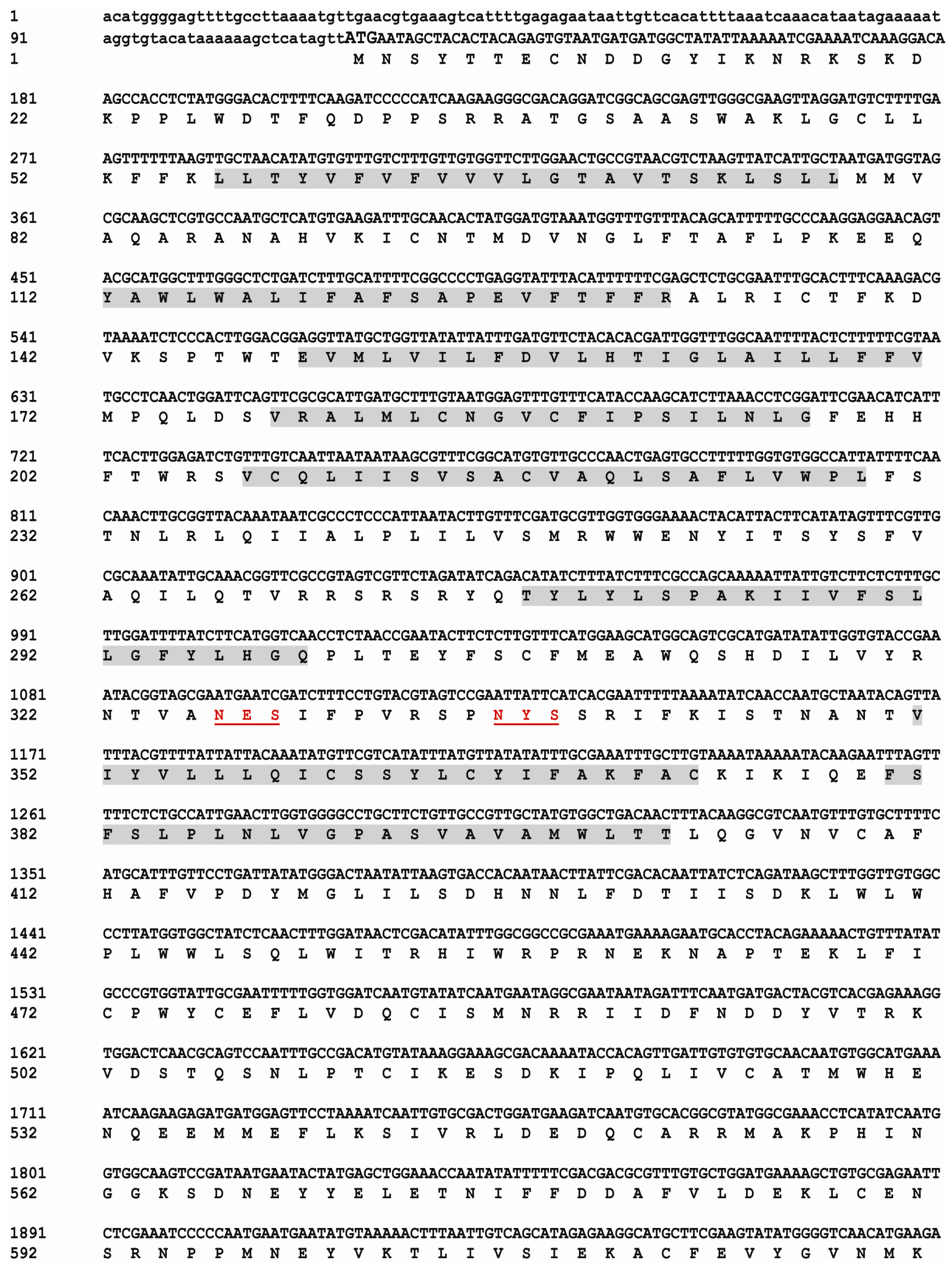
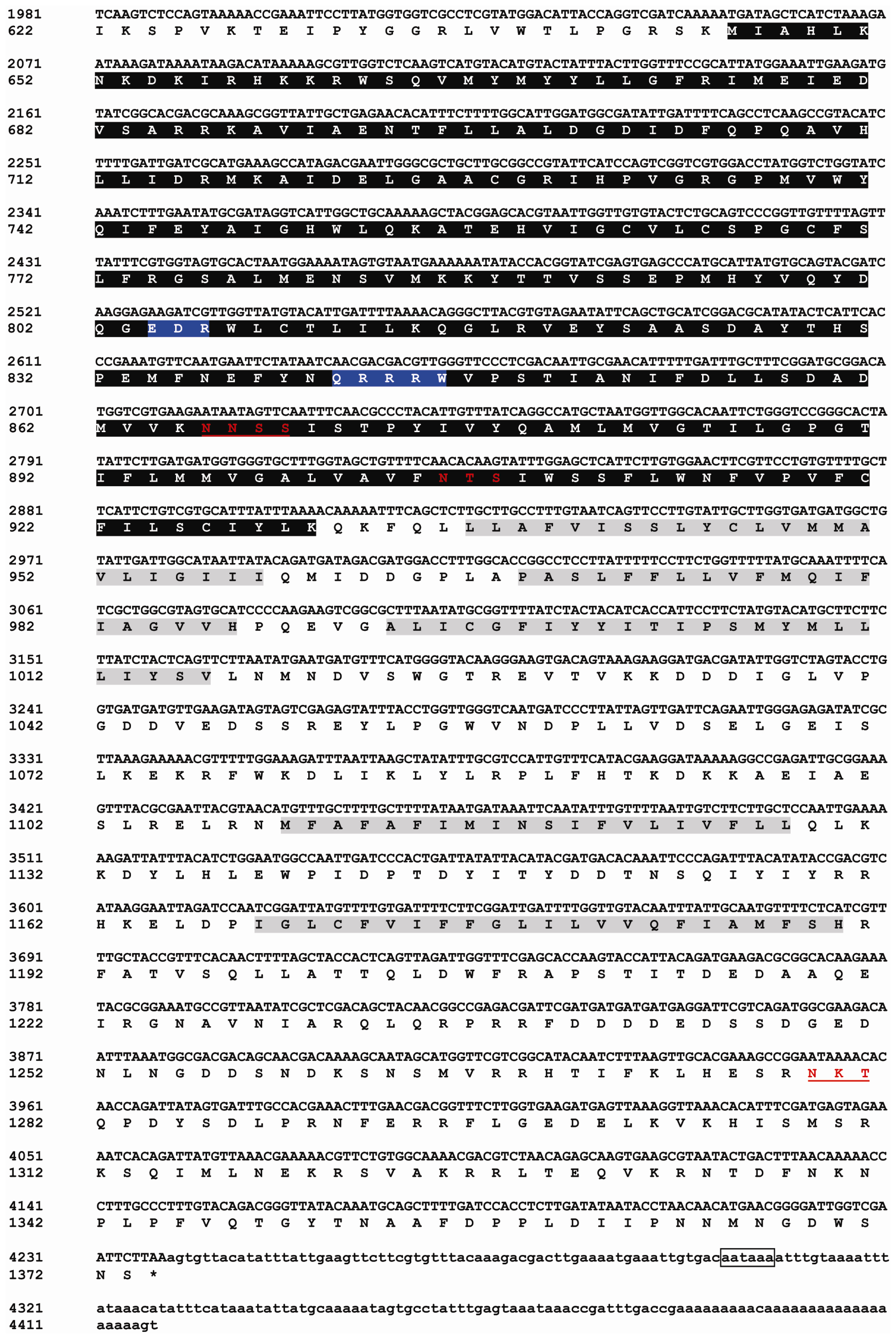
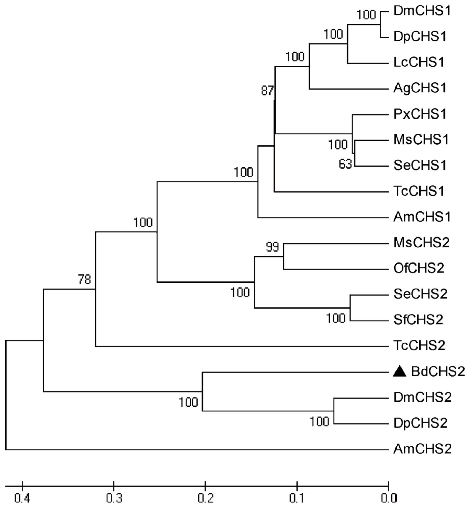
2.1. Identification and Characterization of BdCHS2

The full-length cDNA sequence of BdCHS2 was obtained by PCR (Polymerase Chain Reaction) and 5′ and 3′ RACE. The complete cDNA of the BdCHS2 (GenBank ID: KC354694) consisted of 4417 nucleotides with an open reading frame (ORF) of 4122 nucleotides encoding 1373 amino acids. The cDNA included a 5′-untranslated region (UTR) located 116 nucleotides upstream of the start codon (ATG) and a 3′ UTR of 179 nucleotides ending in a poly (A) tail. The complete nucleotide and deduced amino acid sequences of BdCHS2 were shown in Figure 1. A possible consensus signal sequence for polyadenylation (AATAAA) was located 79 nucleotides upstream of the poly (A) tail. The theoretical molecular weight of BdCHS2 based on the deduced amino acid sequence was calculated to be 158.5 kDa, with an isoelectric point of 6.83.
BdCHS2 was predicted to have three domains: an N-terminal domain (residues 1–645) with eight transmembrane helices; a catalytic domain (residues 646–930); and a C-terminal domain (residues 931–1373) with an additional five transmembrane helices. The signature sequence “QRRRW”, “WGTRE”, and “EDR” for chitin synthases were also found in BdCHS2. Five potential N-glycosylation sites was predicted using NetNGLyc 1.0 software (Technical University of Denmark, Copenhagen, Denmark), suggesting that the protein was glycosylated. However, no signal peptide was found.
Multiple protein alignments showed that BdCHS2 protein had homology to the known and predicted CHS2 in other insect species. For instance, the BdCHS2 protein shares 87% identity with the CHS2 of Drosophila mojavensis (XP_002008568), 85% identity with the CHS2 of D. persimilis (XP_002027231), 84% identity with the CHS2 of D. melanogaster (NP_001137997), and 81% identity with the CHS2 of Culex quinquefasciatus (XP_001864594). A phylogenetic tree was constructed based on the neighbor-joining method using complete CHSs proteins deposited in NCBI by MEGA 5.04 (Figure 2). The tree showed that BdCHS2 was classified into the CHS2 family, and was most closely related to DmCHS2 and DpCHS2 with these three genes clustering together.

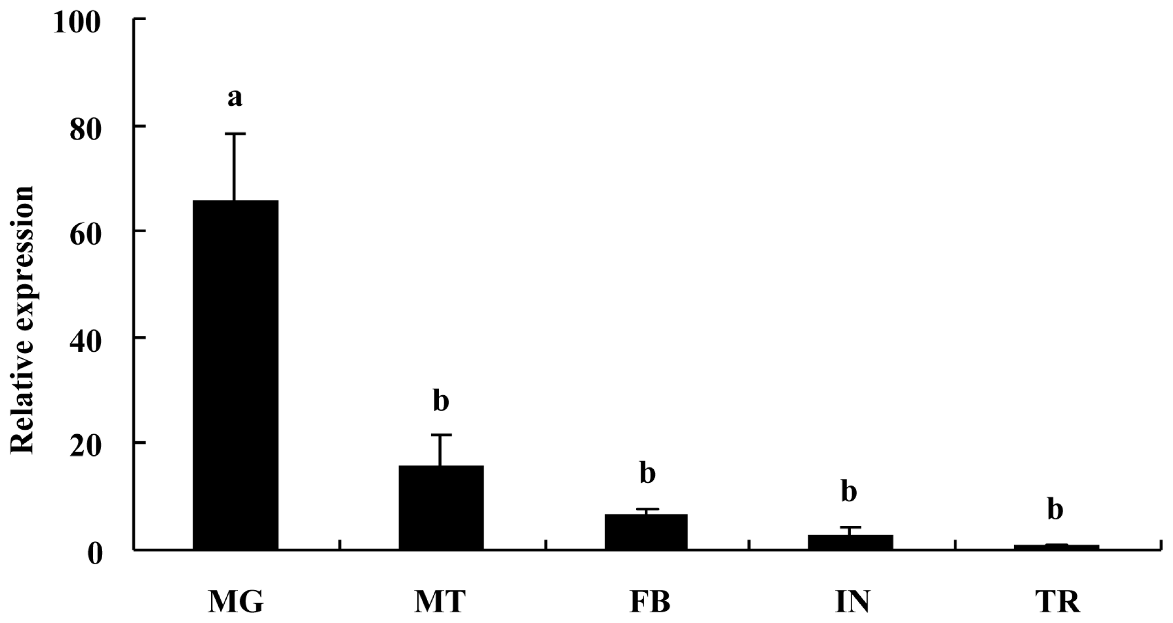
2.2. Tissue-Specific Expression Pattern of BdCHS2

The expression of BdCHS2 mRNA was investigated in various tissues in the third instar larvae of B. dorsalis (Figure 3). BdCHS2 was highly expressed in the midgut, but detected at low levels in fat body, Malpighian tubules, integument, or trachea. The relative expression level of BdCHS2 was the highest in midgut among the five tissues, and it was 66-, 16-, 7- and 3-fold higher in midgut, Malpighian tubule, fat body, and integument, respectively, than that in trachea.

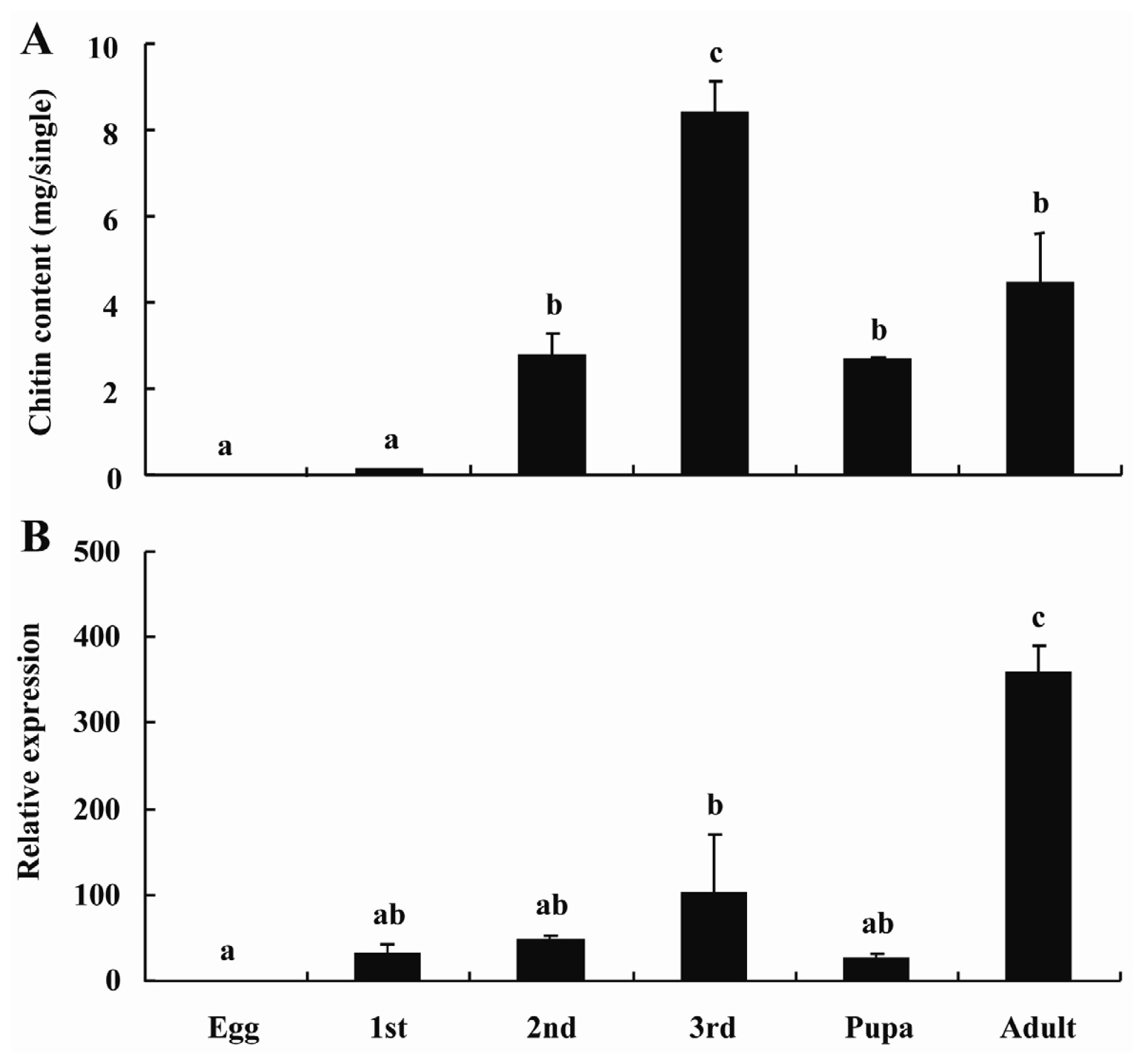
2.3. BdCHS2 Expression and Total Chitin Content during Development

To understand the function of BdCHS2, its expression patterns during development from egg to adult were examined (Figure 4). The results showed that BdCHS2 was expressed at all stages, indicating that it has a role throughout the entire life cycle. The highest mRNA level was found in the adult stage, and the relative expression levels of BdCHS2 was 31-, 47-, 102-, 26- and 358-fold higher in the first, second and third instar larvae, pupa, and adult than in the egg, respectively. Subsequently, the relative expression level of BdCHS2 in egg, the first, second and third instar larvae, and pupa were significantly lower from that in the adult (p < 0.05). There was an increasing expression level of BdCHS2 during the developmental period from the egg to the third instar larvae.
The chitin content was detected from the whole bodies of B. dorsalis during the developmental stages. The results showed that the highest chitin content was observed in the third instar larvae and the lowest content was in the egg (Figure 4). There was a positive relationship between BdCHS2 expression level and the total chitin contents during development.

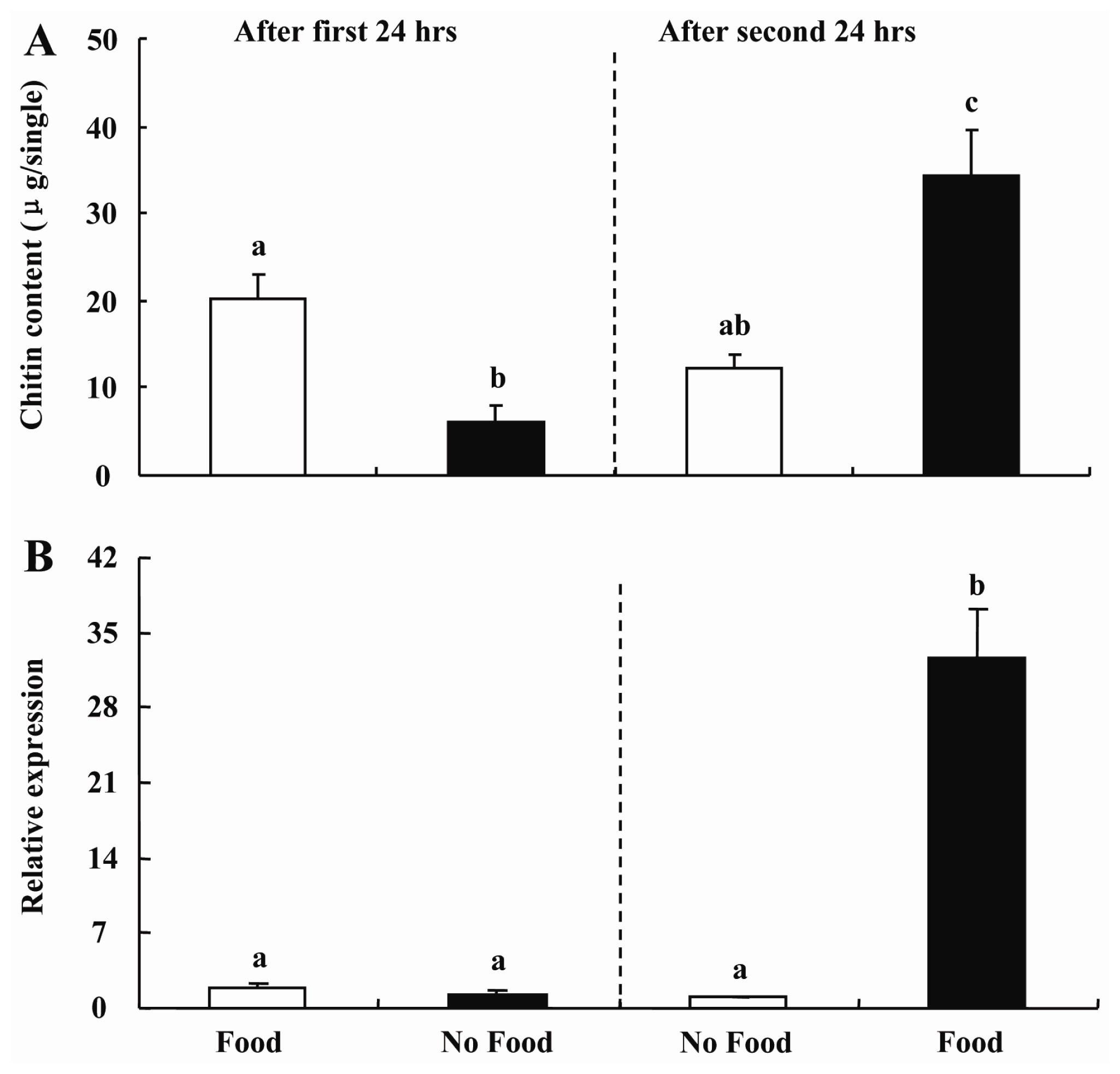
2.4. Feeding-Mediated Changes in Transcript Levels of BdCHS2 and Chitin Content in Midgut

Furthermore, to test the hypothesis that midgut chitin content was regulated during feeding, presumably to alter the porosity of the peritrophic membrane to facilitate food digestion, we examined the changes in transcript levels of BdCHS2 and chitin content in the midgut of larvae B. dorsalis with or without food. When the larvae were maintained with food for the first 24 h, the transcript levels of BdCHS2 in the midgut were 1.5-fold higher than that for larvae maintained with no food (p < 0.05). However, when the larvae maintained on food were transferred to a container with no food for another 24 h, the transcript level of BdCHS2 decreased by 20% (p < 0.05). In contrast, when the larvae were maintained with no food for the first 24 h, then were transferred to a container with food for the next 24 h, the transcript level increased by 24.6-fold (p < 0.05) (Figure 5).
When the larvae maintained on the food were transferred to a container without food for another 24 h, the chitin content decreased by 40% (p < 0.05). In contrast, when the larvae maintained with no food for the first 24 h, then were transferred to a container with food for the next 24 h, the chitin content level increased by 4.5-fold (p < 0.05) (Figure 5). Moreover, there was a positive relationship between BdCHS2 expression level and chitin content in the midgut.

2.5. Discussion

Tellam and his colleagues first isolated the complete cDNA sequence of putative chitin synthase in arthropod [21]. Two distinct CHS genes have been studied through molecular cloning and functional analyses in several orders in insects, such as Diptera, Orthoptera, Coleoptera, Lepidoptera, and Hymenoptera [11]. CHS was mainly responsible for the chitin synthesis in cuticular exoskeleton, tracheae and the PM in midgut. Recently, much more information about the CHS1 gene has been studied including B. dorsalis [22] while relatively little information is available about the gene CHS2 being involved in the midgut chitin synthesis in insects. In the present work, via molecular bioinformatics including sequence similarity analysis, unique signature sequences and phylogenetic analysis, it was confirmed that the sequence we cloned from the B. dorsalis was another chitin synthase gene BdCHS2. The isolation of BdCHS2 cDNA provided us an opportunity to study the expression patterns and biological functions of this gene in B. dorsalis.
Furthermore, the expression profiles of BdCHS2 in five different tissues were investigated. The results indicated that the BdCHS2 was expressed highest in midgut which was consistent with the expression pattern of CHS2 in other insects, including D. melanogaster [13], A. gambiae [11], T. castaneum [14], M. sexta [15], S. exigua [10], O. furnacali [16], S. Frugiperda [9], L. migratoria [17], and A. aegypti [19]. This result was also consistent with the hypothesis that CHS2 was responsible for biosynthesis of the chitin in midgut. BdCHS2 was expressed at a low level in integument and trachea which might be associated with CHS1 of its chitin biosynthesis [22,2326]. However, in A. gambiae, CHS2 protein was detected not only in the midgut, but also in newly formed compound eyes and abdominal inter-segmental regions of the pupae [11]. In A. aegypti, CHS2 localized to the periphery of the epithelial cells facing the midgut lumen [12]. Equally, the anterior midgut may play an important role in chitin biosynthesis more than the rest of the midgut in L. migratoria [17]. In summary, the CHS2 gene is mainly expressed in midgut and much more function of this gene is necessary for further research.
The chitin content and the BdCHS2 expression level were investigated in this study, and a similar trend was found during development except for the adult stage. This result was consistent with a recent study, i.e., the expression of LmCHS2 gradually increased from first to fifth-instar nymphs, and reached the highest in the first day of adults in L. migratoria [17]. In S. exigua, the expression level in different developmental stages also showed a similar trend to that found in our present study [10]. In S. frugiperda, SfCHS2 expressed in the midgut during the feeding stages [9] was also consistent with our results. High expression levels of BdCHS2 during the feeding stage indicated that BdCHS2 protein plays an important role in the production of the chitin-rich PM. The insect needs this structure to protect the gut lining cells and increase the efficiency of nutrient digestion during feeding stages [27,28]. Additionally, the trend of gene expression level had a positive correlation with that of total chitin content during development stages, indicating that this gene may play an important role in total body chitin synthesis.
Furthermore, we examined the changes in transcript levels of BdCHS2 and midgut chitin content in larvae of B. dorsalis fed on the artificial diet or starvation. Our results suggested that the expression level of the BdCHS2 was affected by feeding and this was in agreement with the report in blood-fed insects A. gambiae [29] and L. longipalpis [30]. In contrast, in Ostrinia nubilalis, expression level changes of CHS2 had a completely opposite result and chitinase had a similar result [18]. It might be due to the significant differences in the biological habits of these two insects, which belong to different Orders. Furthermore, their type of PM belong to two different types, O. nubilalis belongs to type I while B. dorsalis to type II PM [6]. In the present study, the chitin content of the midgut dissected from the larvae showed positive proof of a consistent correlation with change in gene expression level. From expression profiles of tissue and developmental stages, we can infer that BdCHS2 was mainly expressed in midgut and had a gradually increased expression level from the second instar to the third instar larvae. However, the expression level of BdCHS2 and the chitin content of the midgut decreased after treatment with food for 24 h then starvation for 24 h, indicating that starvation had a strong influence on expression of this gene in the midgut. On the other hand, Chironomid larvae only break down newly assimilated food for energy during starvation [31]. Therefore, the reason why the chitin content decreased after 24 h starvation may be that BdCHS2 was expressed at a low level of mRNA in midgut; additionally, the midgut chitin might be degraded to survive during the period of starvation. As expected, under the condition of feeding for 24 h after starvation for the first 24 h, gene expression and the chitin content level increased rapidly. It may be that the body needs much more digested food to grow into later developmental stages along with the increased midgut chitin and the mRNAs of BdCHS2 after the starvation for 24 h. The hypothesis that the midgut chitin content level is regulated during feeding, presumably to facilitate food digestion, was confirmed. In brief, the change under the feeding and starvation conditions suggested that BdCHS2 plays important roles in the regulation of chitin contents in the midgut. By using RNAi methodologies, it has been shown that the insect ceased feeding, shrinked in larval size, decreased in midgut chitin content [20], exhibited a high mortality [17], and disrupted formation of the peritrophic matrix [19] after CHS2 gene knockdown. Moreover, transgenic plants synthesized hairpin dsRNAs as a protective measure against damaging herbivorous insects [32]. Based on the results of RNAi in other insects and the results in this study, BdCHS2 might be a good candidate gene for B. dorsalis control by transgenic plants due to the ability to suppress a gene critical for insect survival, providing a new approach to block a significant pest using environmentally friendly and effective principles.

3. Experimental Section

3.1. Test Insect

The colony of B. dorsalis was kept in laboratory cages at 27 ± 1 °C, 70% ± 5% relative humidity and a photoperiod cycle of 14 h·Light/10 h·Dark. The insects were reared on an artificial diet as described previously [33]. The developmental stages were synchronized at each egg incubation. Fat body, integument, Malpighian tubules, midgut, and trachea were dissected from the third instar larvae in phosphate buffered saline (PBS) under a stereomicroscope (Olympus SZX12, Tokyo, Japan) and stored at −80 °C prior to use.

3.2. cDNA Cloning of BdCHS2 and Sequence Analysis

3.2.1. RNA Extraction and cDNA Synthesis

Total RNA was extracted from the midgut of the third instar larvae of B. dorsalis with TRIzol Reagent (Invitrogen, Carlsbad, CA, USA) according to the manufacturer’s instructions, and used in the amplification of cDNA fragments and rapid amplification of cDNA ends (RACE). The total RNA was treated with DNase (TaKaRa, Dalian, China) and dissolved in 30 μL DEPC treated water. The purity and quantity of extracted RNA was quantified by the ratio of OD260/OD280 with an ultraviolet spectrometer. First-strand cDNA was synthesized from 2 μg of DNase-treated RNA by PrimeScript® 1st Strand cDNA synthesis Kit (TaKaRa, Ohtsu, Japan) with oligo (dT)18 primers, and used as a template for PCR.

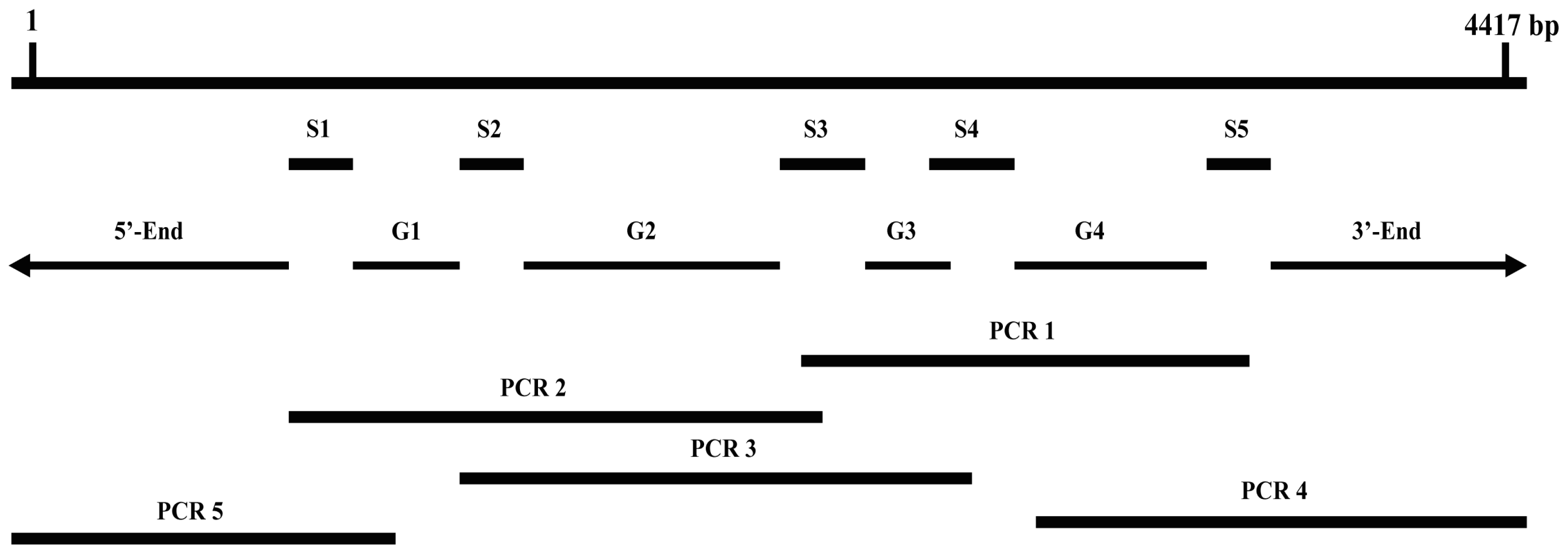
3.2.2. Obtaining Full-Length of BdCHS2 cDNA

Based on the transcriptome sequencing data of B. dorsalis [34], five cDNA fragments encoding BdCHS2 (S1–S5) were identified (Table 1). In order to generate a larger cDNA fragment, three pairs of primers (Table 2) were designed to amplify the three gaps among the assembled fragments of BdCHS2 (PCR1 to PCR3, Figure 6). 3′- and 5′-RACE ends (PCR4 and PCR5) were amplified according to the instructions of SMARTer™ RACE cDNA Amplification Kit (Clontech, Palo Alto, CA, USA). PCR amplifications were carried out in a total volume of 25 μL mixture, containing 2.5 μL Mg2+ (2.5 mM), 2 μL dNTPs (2.5 mM), 2.5 μL 10× PCR Buffer (Mg2+ free), 1 μL each primer (10 mM), 1 μL cDNA, and 0.25 μL rTaq™ polymerase (TaKaRa), and 15 μL ddH2O. Thermal cycling conditions were 95 °C for 5 min followed by 34 cycles of 95 °C for 30 s, 58 °C for 30 s and 72 °C for 1 min. The last cycle was followed by final extension at 72 °C for 10 min. The amplified products were analyzed on 1.0% agarose gel, which contained GoodView™ (SBS Genetech, Beijing, China). The target band of products was purified using the Gel Extraction Mini Kit (Watson Biotechnologies, Shanghai, China). Purified DNA was ligated into pGEM®-T Easy vector (Promega, Madison, WI, USA). The ligation reactions were transformed into Trans-T1 competent cells (Transgen, Beijing, China). By using standard ampicillin selection, successful clones were picked out and then PCR with gene-specific primers, and further sequenced in both directions with an ABI Model 3100 automated sequencer (BGI, Shenzhen, China).

3.2.3. Sequence Analysis and Phylogenetic Tree Construction

Searching for similar sequences was performed using BlastP in the non-redundant protein sequences (nr) database of the NCBI website [35]. The open reading frame (ORF) finder tool at the NCBI was used to identify the ORF of BdCHS2. Sequences were edited with DNAMAN 5.2.2 (Lynnon BioSoft, Quebec, Canada). ExPASy Proteomics Server [36] was used to compute isoelectric point and molecular weight of the deduced protein sequences. NetNGlyc 1.0 Server [37] was used to analyze the N-glycosylation sites. Cellular localization was conducted with the web site [38]. The signal peptide was predicted by SignalP 3.0 [39], and transmembrane helices were analyzed using TMHMM v.2.0 [40]. The neighbor-joining method was applied to construct a phylogenetic tree with 1000 replications as the bootstrap value using MEGA 5.04 [41].

3.3. Tissue-Specific Expression of BdCHS2 Using Quantitative Real-Time PCR

Tissue-specific expression of BdCHS2 was examined by quantitative real-time PCR (qPCR). Total RNA was isolated from fat body, integument, Malpighian tubules, midgut, and trachea of the third instar larvae, using RNeasy® Plus Micro Kit (with gDNA Elimator spin columns, Qiagen, Valencia, CA, USA). First strand cDNA was synthesized in a 10 μL reaction mixture using random hexamers by PrimeScript® RT reagent Kit (TaKaRa). The qPCR was conducted on Mx3000P thermal cycler (Stratagene, La Jolla, CA, USA) using SYBR Green detection system (iQ™ SYBR® Green Supermix, BIO-RAD, Hercules, CA, USA) and gene-specific primers CHS2-Q-F and CHS2-Q-R (Table 2). The PCR amplifications were performed in 20 μL reaction systems, including 7 μL ddH2O, 10 μL SYBR Green Supermix, 1 μL of template cDNA and 1 μL of each primer (0.2 mM) under the following conditions: pre-denaturation at 95 °C for 2 min, 40 cycles of denaturation at 95 °C for 15 s, annealing at 60 °C for 30 s, and elongation at 72 °C for 30 s. After reaction, a melting curve analysis from 60 to 95 °C was applied to all reactions to ensure consistency and specificity of the amplified. The qPCR analysis had three times of biological duplication. The data were normalized to the stable reference gene α-Tubulin (GU269902) (Table 2) based on our previous evaluations, and was calculated using 2−ΔΔCT method [42].

3.4. Developmental Stages-Specific Expression of BdCHS2 and Total Chitin Content

Eggs, the first, second, and third instar larvae, pupae, and adults were used for total RNA isolation using RNeasy® Plus Micro Kit (with gDNA Elimator spin columns, Qiagen, Valencia, CA, USA) (e.g., egg, the first instar larvae) or TRIzol reagent and treated with DNase (TaKaRa) for DNA digestion (e.g., the second, and third instar larvae, pupae, and adults). The stage-specific expression was examined using qPCR as pre-mentioned method. Furthermore, the chitin content in different developmental stages was assayed based on the previous described method [4345]. Briefly, the sample (30 individuals for each sample) was homogenized with 1.0 mL of distilled water by grinding in a cold mortar. Then, the chitin was isolated from the sample after treated by centrifuged and 3% SDS (sodium dodecyl sulfate). To deacetylate chitin, it was re-suspended in 0.3 mL of 14 M KOH and incubated in drying oven at 130 °C for 1 h. The insoluble chitosan was obtained after purified by different concentrations of alcohol. 100 μL of the chitosan solution was mixed with 100 μL of 10% NaNO2 and 100 μL of 10% KHSO4 to depolymerize the chitosan and deaminate the glucosamine residues from the chitosan. After treated by 12.5% NH4SO3NH2 (Sigma-Aldrich, St. Louis, MO, USA), the sample was added to MBTH (3-methyl-2-benzothiazolone hydrazone hydrochloride hydrate, Sigma-Aldrich) (50 mg/10 mL) and 0.83% FeCl3. Finally, 100 μL of each sample was transferred to a 96-well microplate and then colorimetric assay under 650 nm in a microplate reader (Sigma Laborzentrifugen GmbH, Ostrode, Germany). According to a standard curve constructed by using known concentrations of glucosamine (Sigma-Aldrich), chitin content was calculated as a glucosamine equivalent. Three biological replications, each with two technical replications, were used in this analysis.

3.5. Gene Expression Profiles and Chitin Content Assay under Feeding and Starvation Conditions

The 1-day-old third instar larvae were used for this experiment. Eight Petri dishes (diameter = 4 cm) were divided into two groups, each with four Petri dishes. The insects in the first group were maintained with the artificial diet (designated as with food) for 24 h and then with no food for next 24 h, while the insects in the second group were maintained with no food for 24 h and then with food for the next 24 h. Total RNA was isolated from the dissected midguts of the two groups after 24 and 48 h treatment. The transcript levels were measured using qPCR as mentioned above. Furthermore, the chitin content in the midguts of the above treated larvae was assayed.

4. Conclusions

In conclusion, a full-length cDNA encoding chitin synthase 2 was obtained from B. dorsalis. BdCHS2 was mainly expressed in midgut. Further, it expressed in all developmental stages, while highly in the feeding stages (larval and adult stage), and also had a positive relation to the total chitin content of the insect. In addition, the feeding and starvation had a very important effect on this gene expression. In sum, BdCHS2 is involved in the regulation of the midgut chitin and subsequently affects the growth and development of B. dorsalis.

Acknowledgments

This research was supported in part by the National Basic Research Program of China (2009CB125903), Natural Science Foundation of Chongqing (CSTC, JJA80020; 2013jjB0176), the Program for Innovative Research Team in University (IRT0976), the Fundamental Research Funds for the Central Universities (XDJK2013A017), and the Earmarked Fund for Modern Agro-industry (Citrus) Technology Research System of China.

Conflicts of Interest

The authors declare no conflict of interest.

References

  1. Stephens, A.; Kriticos, D.; Leriche, A. The current and future potential geographical distribution of the oriental fruit fly, Bactrocera dorsalis (Diptera: Tephritidae). Bull. Entomol. Res 2007, 97, 369–378. [Google Scholar]
  2. Clarke, A.R.; Armstrong, K.F.; Carmichael, A.E.; Milne, J.R.; Raghu, S.; Roderick, G.K.; Yeates, D.K. Invasive phytophagous pests arising through a recent tropical evolutionary radiation: The Bactrocera dorsalis complex of fruit flies. Annu. Rev. Entomol 2005, 50, 293–319. [Google Scholar]
  3. Hsu, J.C.; Feng, H.T.; Wu, W.J.; Geib, S.; Mao, C.H.; Vontas, J. Truncated transcripts of nicotinic acetylcholine subunit gene Bdα6 are associated with spinosad resistance in Bactrocera dorsalis. Insect Biochem. Mol. Biol 2012, 42, 805–816. [Google Scholar]
  4. Jin, T.; Zeng, L.; Lin, Y.; Lu, Y.; Liang, G. Insecticide resistance of the oriental fruit fly, Bactrocera dorsalis (Hendel) (Diptera: Tephritidae), in mainland China. Pest Manag. Sci 2011, 67, 370–376. [Google Scholar]
  5. Kramer, K.; Muthukrishnan, S. Chitin Metabolism in Insects. In Comprehensive Molecular Insect Science; Gilbert, L.I., Iatrou, K., Gill, S., Eds.; Elsevier Press: Oxford, England, UK, 2005; Volume 4, pp. 111–144. [Google Scholar]
  6. Lehane, M. Peritrophic matrix structure and function. Annu. Rev. Entomol 1997, 42, 525–550. [Google Scholar]
  7. Merzendorfer, H. Insect chitin synthases: A review. J. Comp. Physiol. B Biochem. Syst. Environ. Physiol 2006, 176, 1–15. [Google Scholar]
  8. Arakane, Y.; Specht, C.A.; Kramer, K.J.; Muthukrishnan, S.; Beeman, R.W. Chitin synthases are required for survival, fecundity and egg hatch in the red flour beetle, Tribolium castaneum. Insect Biochem. Mol. Biol 2008, 38, 959–962. [Google Scholar]
  9. Bolognesi, R.; Arakane, Y.; Muthukrishnan, S.; Kramer, K.J.; Terra, W.R.; Ferreira, C. Sequences of cDNAs and expression of genes encoding chitin synthase and chitinase in the midgut of Spodoptera frugiperda. Insect Biochem. Mol. Biol 2005, 35, 1249–1259. [Google Scholar]
  10. Kumar, N.S.; Tang, B.; Chen, X.; Tian, H.; Zhang, W. Molecular cloning, expression pattern and comparative analysis of chitin synthase gene B in Spodoptera exigua. J. Comp. Physiol. B 2008, 149, 447–453. [Google Scholar]
  11. Zhang, X.; Zhang, J.; Park, Y.; Zhu, K.Y. Identification and characterization of two chitin synthase genes in African malaria mosquito, Anopheles gambiae. Insect Biochem. Mol. Biol 2012, 42, 674–682. [Google Scholar]
  12. Ibrahim, G.H.; Smartt, C.T.; Kiley, L.M.; Christensen, B.M. Cloning and characterization of a chitin synthase cDNA from the mosquito Aedes aegypti. Insect Biochem. Mol. Biol 2000, 30, 1213–1222. [Google Scholar]
  13. Gagou, M.E.; Kapsetaki, M.; Turberg, A.; Kafetzopoulos, D. Stage-specific expression of the chitin synthase DmeChSA and DmeChSB genes during the onset of Drosophila metamorphosis. Insect Biochem. Mol. Biol 2002, 32, 141–146. [Google Scholar]
  14. Arakane, Y.; Hogenkamp, D.G.; Zhu, Y.C.; Kramer, K.J.; Specht, C.A.; Beeman, R.W.; Kanost, M.R.; Muthukrishnan, S. Characterization of two chitin synthase genes of the red flour beetle, Tribolium castaneum, and alternate exon usage in one of the genes during development. Insect Biochem. Mol. Biol 2004, 34, 291–304. [Google Scholar]
  15. Hogenkamp, D.G.; Arakane, Y.; Zimoch, L.; Merzendorfer, H.; Kramer, K.J.; Beeman, R.W.; Kanost, M.R.; Specht, C.A.; Muthukrishnan, S. Chitin synthase genes in Manduca sexta: Characterization of a gut-specific transcript and differential tissue expression of alternately spliced mRNAs during development. Insect Biochem. Mol. Biol 2005, 35, 529–540. [Google Scholar]
  16. Qu, M.; Liu, T.; Yang, J.; Yang, Q. The gene, expression pattern and subcellular localization of chitin synthase B from the insect Ostrinia furnacalis. Biochem. Biophys. Res. Commun 2011, 404, 302–307. [Google Scholar]
  17. Liu, X.; Zhang, H.; Li, S.; Zhu, K.Y.; Ma, E.; Zhang, J. Characterization of a midgut-specific chitin synthase gene (LmCHS2) responsible for biosynthesis of chitin of peritrophic matrix in Locusta migratoria. Insect Biochem. Mol. Biol 2012, 42, 902–910. [Google Scholar]
  18. Khajuria, C.; Buschman, L.L.; Chen, M.S.; Muthukrishnan, S.; Zhu, K.Y. A gut-specific chitinase gene essential for regulation of chitin content of peritrophic matrix and growth of Ostrinia nubilalis larvae. Insect Biochem. Mol. Biol 2010, 40, 621–629. [Google Scholar]
  19. Kato, N.; Mueller, C.R.; Fuchs, J.F.; Wessely, V.; Lan, Q.; Christensen, B.M. Regulatory mechanisms of chitin biosynthesis and roles of chitin in peritrophic matrix formation in the midgut of adult Aedes aegypti. Insect Biochem. Mol. Biol 2006, 36, 1–9. [Google Scholar]
  20. Arakane, Y.; Muthukrishnan, S.; Kramer, K.J.; Specht, C.A.; Tomoyasu, Y.; Lorenzen, M.D.; Kanost, M.; Beeman, R.W. The Tribolium chitin synthase genes TcCHS1 and TcCHS2 are specialized for synthesis of epidermal cuticle and midgut peritrophic matrix. Insect Biochem. Mol. Biol 2005, 14, 453–463. [Google Scholar]
  21. Tellam, R.L.; Vuocolo, T.; Johnson, S.E.; Jarmey, J.; Pearson, R.D. Insect chitin synthase-cDNA sequence, gene organization and expression. Eur. J. Biochem 2000, 267, 6025–6042. [Google Scholar]
  22. Yang, W.J.; Xu, K.K.; Cong, L.; Wang, J.J. Identification, mRNA expression, and functional analysis of chitin synthase 1 gene and its two alternative splicing variants in oriental fruit fly, Bactrocera dorsalis. Int. J. Biol. Sci 2013, 9, 331–342. [Google Scholar]
  23. Wang, Y.; Fan, H.W.; Huang, H.J.; Xue, J.; Wu, W.J.; Bao, Y.Y.; Xu, H.J.; Zhu, Z.R.; Cheng, J.A.; Zhang, C.X. Chitin synthase 1 gene and its two alternative splicing variants from two sap-sucking insects, Nilaparvatalugens and Laodelphaxstriatellus (Hemiptera: Delphacidae). Insect Biochem. Mol. Biol 2012, 42, 637–646. [Google Scholar]
  24. Qu, M.; Yang, Q. A novel alternative splicing site of class A chitin synthase from the insect Ostrinia furnacalis-Gene organization, expression pattern and physiological significance. Insect Biochem. Mol. Biol 2011, 41, 923–931. [Google Scholar]
  25. Ampasala, D.R.; Zheng, S.; Zhang, D.; Ladd, T.; Doucet, D.; Krell, P.J.; Retnakaran, A.; Feng, Q. An epidermis-specific chitin synthase cDNA in Choristoneura fumiferana: Cloning, characterization, developmental and hormonal-regulated expression. Arch. Insect Biochem. Physiol 2011, 76, 83–96. [Google Scholar]
  26. Zhang, J.; Liu, X.; Li, D.; Sun, Y.; Guo, Y.; Ma, E.; Zhu, K.Y. Silencing of two alternative splicing-derived mRNA variants of chitin synthase 1 gene by RNAi is lethal to the oriental migratory locust, Locusta migratoria manilensis (Meyen). Insect Biochem. Mol. Biol 2010, 40, 824–833. [Google Scholar]
  27. Terra, W.R. The origin and functions of the insect peritrophic membrane and peritrophic gel. Arch. Insect Biochem. Physiol 2001, 47, 47–61. [Google Scholar]
  28. Terra, W.; Ferreira, C. Biochemistry of digestion. Compr. Mol. Insect Sci 2005, 4, 171–224. [Google Scholar]
  29. Shen, Z.; Jacobs-Lorena, M. Characterization of a novel gut-specific chitinase gene from the human malaria vector Anopheles gambiae. J. Biol. Chem 1997, 272, 28895–28900. [Google Scholar]
  30. Ramalho-Ortigao, J.; Traub-Csekö, Y. Molecular characterization of Llchit1, a midgut chitinase cDNA from the leishmaniasis vector Lutzomyia longipalpis. Insect Biochem. Mol. Biol 2003, 33, 279–287. [Google Scholar]
  31. Kikuchi, E.; Takagi, S.; Shikano, S. Changes in carbon and nitrogen stable isotopes of chironomid larvae during growth, starvation and metamorphosis. Rapid Commun. Mass Spectrom 2007, 21, 997–1002. [Google Scholar]
  32. Baum, J.A.; Bogaert, T.; Clinton, W.; Heck, G.R.; Feldmann, P.; Ilagan, O.; Johnson, S.; Plaetinck, G.; Munyikwa, T.; Pleau, M. Control of coleopteran insect pests through RNA interference. Nat. Biotechnol 2007, 25, 1322–1326. [Google Scholar]
  33. Cong, L.; Yang, W.J.; Shen, G.M.; Dou, W.; Wang, J.J. Molecular characterization of the cDNA encoding ecdysone receptor isoform B1 and its expression in the oriental fruit fly, Bactrocera dorsalis (Diptera: Tephritidae). Fla. Entomol 2012, 95, 650–658. [Google Scholar]
  34. Shen, G.M.; Dou, W.; Niu, J.Z.; Jiang, H.B.; Yang, W.J.; Jia, F.X.; Hu, F.; Cong, L.; Wang, J.J. Transcriptome analysis of the oriental fruit fly (Bactrocera dorsalis). PLoS One 2011, 6, e29127. [Google Scholar]
  35. National Center for Biotechnology Information. Available online: http://0-www-ncbi-nlm-nih-gov.brum.beds.ac.uk/ (accessed on 15 September 2012).
  36. Compute pI/Mw Tool. Available online: http://cn.expasy.org/tools/pi_tool.html (accessed on 15 September 2012).
  37. NetNGlyc 1.0 Server. Available online: http://www.cbs.dtu.dk/services/NetNGlyc/ (accessed on 15 September 2012).
  38. PSORT WWW Server. http://psort.nibb.ac.jp/ (accessed on 15 September 2012).
  39. SignalP 3.0 Server. http://www.cbs.dtu.dk/services/SignalP-3.0/ (accessed on 15 September 2012).
  40. TMHMM Server 2.0. http://www.cbs.dtu.dk/services/TMHMM-2.0/ (accessed on 15 September 2012).
  41. Tamura, K.; Peterson, D.; Peterson, N.; Stecher, G.; Nei, M.; Kumar, S. MEGA5: Molecular evolutionary genetics analysis using maximum likelihood, evolutionary distance, and maximum parsimony methods. Mol. Biol. Evol 2011, 28, 2731–2739. [Google Scholar]
  42. Schmittgen, T.D.; Livak, K.J. Analyzing real-time PCR data by the comparative CT method. Nat. Protoc 2008, 3, 1101–1108. [Google Scholar]
  43. Lehmann, P.F.; White, L.O. Chitin assay used to demonstrate renal localization and cortisone-enhanced growth of Aspergillus fumigatus Mycelium in mice. Am. Soc. Microbiol 1975, 12, 987–992. [Google Scholar]
  44. Zhang, J.; Zhu, K.Y. Characterization of a chitin synthase cDNA and its increased mRNA level associated with decreased chitin synthesis in Anopheles quadrimaculatus exposed to diflubenzuron. Insect Biochem. Mol. Biol 2006, 36, 712–725. [Google Scholar]
  45. Ride, J.; Drysdale, R. A rapid method for the chemical estimation of filamentous fungi in plant tissue. Physiol. Plant Pathol 1972, 2, 7–15. [Google Scholar]
Figure 1. Nucleotide and deduced amino acid sequences of BdCHS2 cDNA from Bactrocera dorsalis (KC354694). The start codon is indicated in bold and the stop codon in bold with an asterisk. The putative polyadenylation signal (AATAA) is boxed. The putative transmembrane regions are shaded. The five potential N-glycosylation sites are double underlined. The amino acid sequence of the putative catalytic domain is in gray with black background. The signature sequences (EDR and QRRRW) are in white with a wavy line.
Ijms 14 17055f1aIjms 14 17055f1b
Figure 2. Evolutionary relationships of deduced amino acid sequence of BdCHS2 with other insect chitin synthases constructed using the neighbor-joining method. Bootstrap values with 1000 trials are indicated on branches. The scale bar represents the number of substitutions per site. The following insect chitin synthases sequence were used: Anopheles gambiae (Ag), Apis mellifera (Am), Bactrocera dorsalis (Bd), Drosophila melanogaster (Dm), Drosophila pseudoobscura (Dp), Lucilia cuprina (Lc), Manduca sexta (Ms), Ostrinia furnacalis (Of), Plutella xylostella (Px), Spodoptera exigua (Se), Spodoptera frugiperda (Sf), Tribolium castaneum (Tc). GenBank accession numbers are as follows: AgCHS1 (XP_321336), AmCHS1 (XP_395677), AmCHS2 (XP_001121152), BdCHS2 (KC354694), DmCHS1 (NP524233), DmCHS2 (NP_001137997), DpCHS1 (XP_001359390), DpCHS2 (XP_001352881), LcCHS1 (AF221067), MsCHS1 (AY062175), MsCHS2 (AY82156), OfCHS2 (AB_B97082), PxCHS1 (BAF47974), SeCHS1 (DQ062153), SeCHS2 (DQ912929), SfCHS2 (AY525599), TcCHS1 (AY291475), and TcCHS2 (AY291477).
Ijms 14 17055f2
Figure 3. Expression profiles of the BdCHS2 in different tissues of larval Bactrocera dorsalis. The tissues include integument (IN), fat body (FB), midgut (MG), Malpighian tubules (MT), and trachea (TR). α-Tubulin was used as an internal reference gene. The relative expression was calculated based on the value of the lowest expression, which was ascribed an arbitrary value of 1. Data are means ± SE of three biological replications. Different letters above each bar indicate statistically significant difference by ANOVA followed by the Duncan’s multiple range test (p < 0.05).
Ijms 14 17055f3
Figure 4. (A) Developmental changes of total chitin content and (B) mRNA levels of BdCHS2 in Bactrocera dorsalis. α-Tubulin was used as an internal reference gene. The relative expression was calculated based on the value of the lowest expression, which was ascribed an arbitrary value of 1. Data are means ± SE of three biological replications. Different letters above each bar indicate statistically significant difference by ANOVA followed by the Duncan’s multiple range test (p < 0.05).
Ijms 14 17055f4
Figure 5. (A) Changes of chitin content and (B) mRNA levels of BdCHS2 in the midgut of third instar larvae of Bactrocera dorsalis under food or no food conditions. Larvae in set 1 (empty bars) were fed for 24 h and then maintained with no food for the next 24 h, whereas larvae in set 2 (black bars) were maintained with no food for 24 h and then fed for the next 24 h. α-Tubulin was used as an internal reference gene. Data are means ± SE of four biological replications, each with two technical replications. Different letters above each bar indicate statistically significant difference by ANOVA followed by the Duncan’s multiple range test (p < 0.05).
Ijms 14 17055f5
Figure 6. PCR amplification and cloning of the full-length BdCHS2 cDNA in Bactrocera dorsalis. Five PCR fragments (S1–S5) were generated from a transcriptome sequencing data of B. dorsalis. Based on S1–S5 sequences, four gaps (G1–G4) were amplified. The 3′- and 5′-end fragments were obtained through 3′- and 5′-RACE respectively. PCR1–PCR5 fragments were amplified with specific primers designed according to the assembled full-length cDNA sequences of BdCHS2.
Ijms 14 17055f6
Table 1. The cDNA fragments of BdCHS2 extracted from a transcriptome sequencing data of B. dorsalis.
cDNA fragmentLength (bp)Position in the coding area of BdCHS2 (bp)
S1284770–1,053
S21831,353–1,535
S33192,177–2,495
S42522,643–2,894
S52433,718–3,960
Table 2. Primers used in this study.
Application of primerscDNA fragmentPrimer namePrimer sequence(5′-3′)cDNA position in the coding area (bp)
cDNA cloningPCR 1CHS2-1TACTCTGCAGTCCCGGTTGTT2404–2424
CHS2-2CTTGTGCCGCGTCTTCATCTG3757–3777

PCR 2CHS2-3TAGTCGTTCTAGATATCAGAC926–946
CHS2-4AGCAGCGCCCAATTCGTCTATG2273–2294

PCR 3CHS2-5GGATAACTCGACATATTTGGC1465–1485
CHS2-6TGTAGGGCGTTGAAATTGAACTA2717–2739

PCR 4 (3′-RACE)CHS2-7GGAAGTGACAGTAAAGAAGGATG3197–3219
CHS2-8TAAATGGCGACGACAGCAACG3874–3894

PCR 5 (5′-RACE)CHS2-9CCACATAGCAACGGCAACAGAAGC1290–1313
CHS2-10TAATGGGAGGGCGATTATTTGTAAC821–845

UPMCTAATACGACTCACTATAGGGC
NUPAAGCAGTGGTATCAACGCAGAGT

qPCR analysisCHS2CHS2-Q-FATTTTCAGCCTCAAGCCGTA2227–2246
CHS2-Q-RCGGGACTGCAGAGTACACAA2399–2418

A-tubulinα-tub-FCGCATTCATGGTTGATAACG
α-tub-RGGGCACCAAGTTAGTCTGGA

