An Overview of Poly(lactic-co-glycolic) Acid (PLGA)-Based Biomaterials for Bone Tissue Engineering


Abstract
:1. Introduction
2. Chemistry
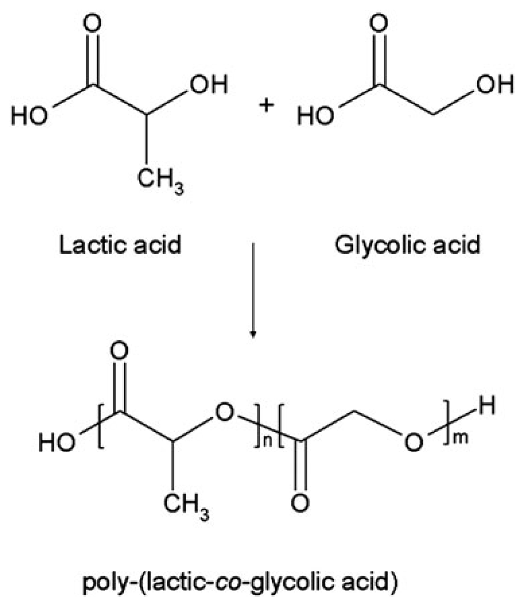
2.1. Synthesis
2.2. Physico-Chemical Properties
3. Applications in Bone Tissue Engineering
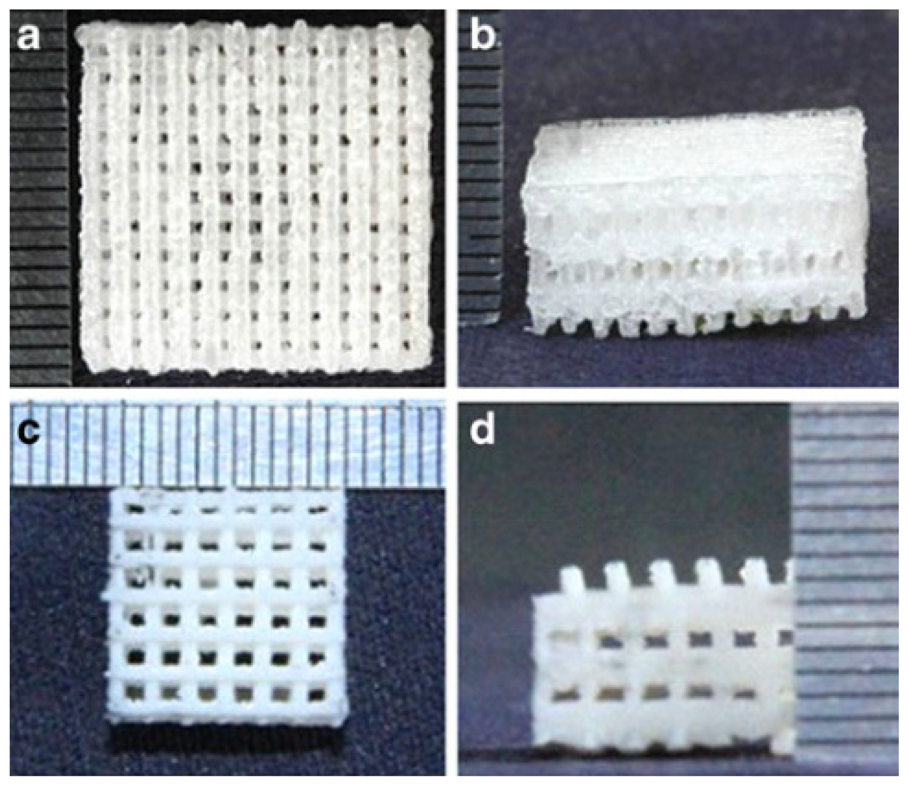
3.1. Porous PLGA-HA Scaffolds
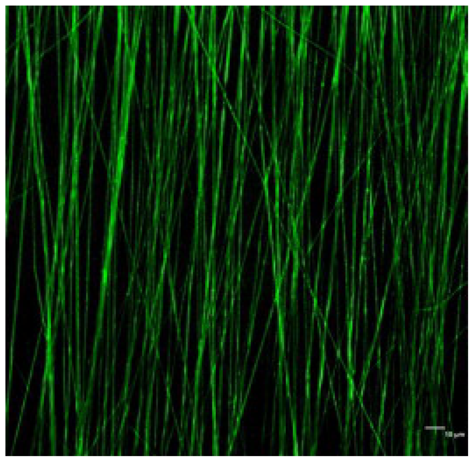
3.2. Fibrous Scaffolds
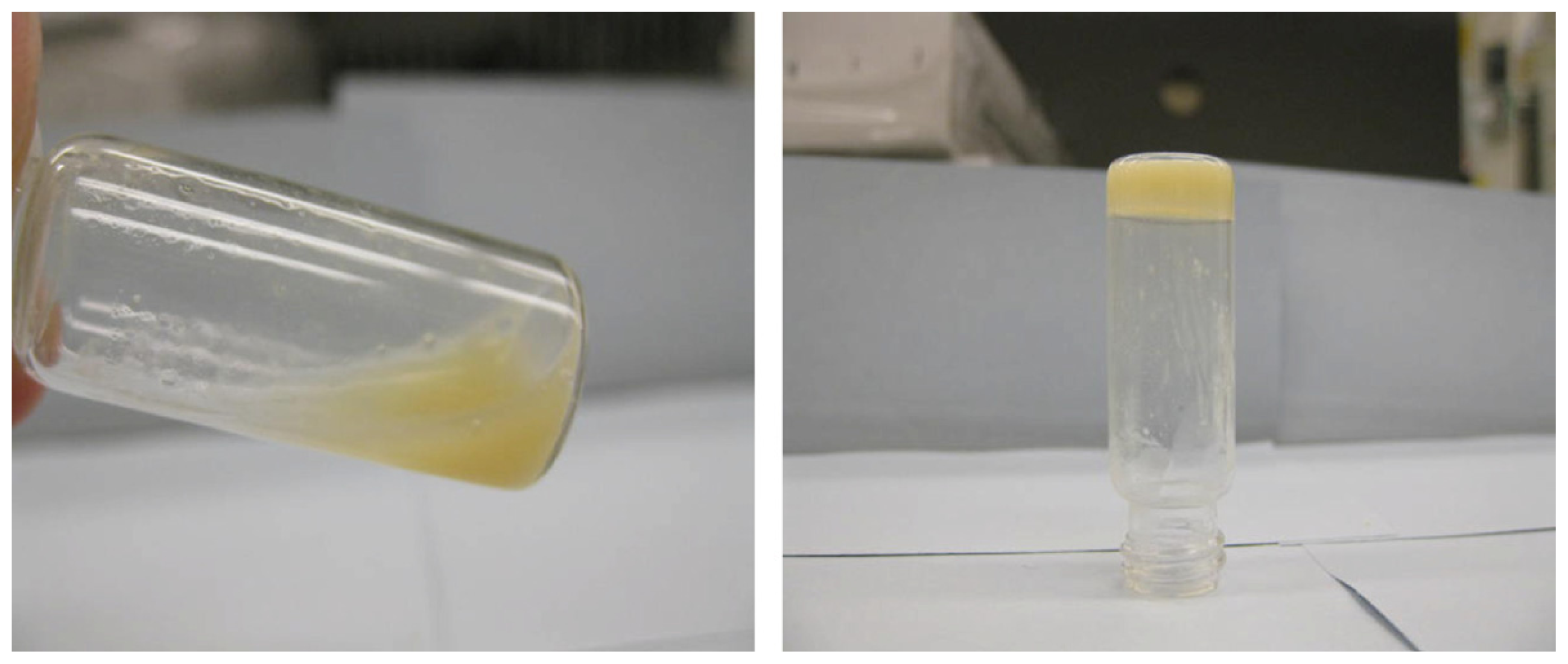
3.3. Hydrogels
3.4. Injectable Microspheres
4. Current Trends in the Development of Functionalized PLGA Constructs
5. Conclusions and Future Prospects
Acknowledgments
Conflicts of Interest
References
- Amini, A.R.; Laurencin, C.T.; Nukavarapu, S.P. Bone tissue engineering: Recent advances and challenges. Crit. Rev. Biomed. Eng. 2012, 40, 363–408. [Google Scholar]
- Ferrone, M.L.; Raut, C.P. Modern surgical therapy: Limb salvage and the role of amputation for extremity soft-tissue sarcomas. Surg. Oncol. Clin. N. Am. 2012, 21, 201–213. [Google Scholar]
- Dimitriou, R.; Jones, E.; McGonagle, D.; Giannoudis, P.V. Bone regeneration: Current concepts and future directions. BMC Med. 2011, 9, 66. [Google Scholar]
- Martou, G.; Antonyshyn, O.M. Advances in surgical approaches to the upper facial skeleton. Curr. Opin. Otolaryngol. Head Neck Surg. 2011, 19, 242–247. [Google Scholar]
- Stevens, M.M. Biomaterials for bone tissue engineering. Mater. Today 2008, 11, 18–25. [Google Scholar]
- Li, X.M.; Wang, L.; Fan, Y.B.; Feng, Q.L.; Cui, F.Z.; Watari, F. Nanostructured scaffolds for bone tissue engineering. J. Biomed. Mater. Res. Part A 2013, 101A, 2424–2435. [Google Scholar]
- Cordonnier, T.; Sohier, J.; Rosset, P.; Layrolle, P. Biomimetic materials for bone tissue engineering—State of the art and future trends. Adv. Eng. Mater. 2011, 13, B135–B150. [Google Scholar]
- Salgado, A.J.; Coutinho, O.P.; Reis, R.L. Bone tissue engineering: State of the art and future trends. Macromol. Biosci. 2004, 4, 743–765. [Google Scholar]
- Dhandayuthapani, B.; Yoshida, Y.; Maekawa, T.; Kumar, D.S. Polymeric scaffolds in tissue engineering application: A review. Int. J. Polym. Sci. 2011. [Google Scholar] [CrossRef]
- Gunatillake, P.A.; Adhikari, R. Biodegradable synthetic polymers for tissue engineering. Eur. Cells Mater. 2003, 5, 1–16; discussion 16. [Google Scholar]
- Mano, J.F.; Sousa, R.A.; Boesel, L.F.; Neves, N.M.; Reis, R.L. Bloinert biodegradable and injectable polymeric matrix composites for hard tissue replacement: State of the art and recent developments. Compos. Sci. Technol. 2004, 64, 789–817. [Google Scholar]
- Lin, H.R.; Kuo, C.J.; Yang, C.Y.; Shaw, S.Y.; Wu, Y.J. Preparation of macroporous biodegradable PLGA scaffolds for cell attachment with the use of mixed salts as porogen additives. J. Biomed. Mater. Res. A 2002, 63, 271–279. [Google Scholar]
- Jagur-Grodzinski, J. Biomedical application of functional polymers. React. Funct. Polym. 1999, 39, 99–138. [Google Scholar]
- Lanao, R.P.F.; Jonker, A.M.; Wolke, J.G.C.; Jansen, J.A.; van Hest, J.C.M.; Leeuwenburgh, S.C.G. Physicochemical properties and applications of poly(lactic-co-glycolic acid) for use in bone regeneration. Tissue Eng. Part B Rev. 2013, 19, 380–390. [Google Scholar]
- Pan, Z.; Ding, J.D. Poly(lactide-co-glycolide) porous scaffolds for tissue engineering and regenerative medicine. Interface Focus 2012, 2, 366–377. [Google Scholar]
- Zhang, L.J.; Webster, T.J. Nanotechnology and nanomaterials: Promises for improved tissue regeneration. Nano Today 2009, 4, 66–80. [Google Scholar]
- Zhou, S.B.; Deng, X.M.; Li, X.H.; Jia, W.X.; Liu, L. Synthesis and characterization of biodegradable low molecular weight aliphatic polyesters and their use in protein-delivery systems. J. Appl. Polym. Sci. 2004, 91, 1848–1856. [Google Scholar]
- Wang, Z.Y.; Zhao, Y.M.; Wang, F.; Wang, J. Syntheses of poly(lactic acid-co-glycolic acid) serial biodegradable polymer materials via direct melt polycondensation and their characterization. J. Appl. Polym. Sci. 2006, 99, 244–252. [Google Scholar]
- Ajioka, M.; Enomoto, K.; Suzuki, K.; Yamaguchi, A. The basic properties of poly(lactic acid) produced by the direct condensation polymerization of lactic-acid. J. Environ. Polym. Degrad. 1995, 3, 225–234. [Google Scholar]
- Ajioka, M.; Suizu, H.; Higuchi, C.; Kashima, T. Aliphatic polyesters and their copolymers synthesized through direct condensation polymerization. Polym. Degrad. Stab. 1998, 59, 137–143. [Google Scholar]
- Moon, S.I.; Lee, C.W.; Taniguchi, I.; Miyamoto, M.; Kimura, Y. Melt/solid polycondensation of l-lactic acid: An alternative route to poly(l-lactic acid) with high molecular weight. Polymer 2001, 42, 5059–5062. [Google Scholar]
- Takahashi, K.; Taniguchi, I.; Miyamoto, M.; Kimura, Y. Melt/solid polycondensation of glycolic acid to obtain high-molecular-weight poly(glycolic acid). Polymer 2000, 41, 8725–8728. [Google Scholar]
- Kricheldorf, H.R.; Boettcher, C.; Tonnes, K.U. Polylactones: 23 Polymerization of racemic and mesO dl-lactide with various organotin catalysts stereochemical aspects. Polymer 1992, 33, 2817–2824. [Google Scholar]
- Kowalski, A.; Duda, A.; Penczek, S. Mechanism of cyclic ester polymerization initiated with tin(II) octoate 2 Macromolecules fitted with tin(II) alkoxide species observed directly in maldi-tof spectra. Macromolecules 2000, 33, 689–695. [Google Scholar]
- Duval, C.; Nouvel, C.; Six, J.-L. Is bismuth subsalicylate an effective nontoxic catalyst for plga synthesis? J. Polym. Sci. Part A 2014. [Google Scholar] [CrossRef]
- Dechy-Cabaret, O.; Martin-Vaca, B.; Bourissou, D. Controlled ring-opening polymerization of lactide and glycolide. Chem. Rev. 2004, 104, 6147–6176. [Google Scholar]
- Li, J.; Stayshich, R.M.; Meyer, T.Y. Exploiting sequence to control the hydrolysis behavior of biodegradable plga copolymers. J. Am. Chem. Soc. 2011, 133, 6910–6913. [Google Scholar]
- Makadia, H.K.; Siegel, S.J. Poly lactic-co-glycolic acid (plga) as biodegradable controlled drug delivery carrier. Polymers-Basel 2011, 3, 1377–1397. [Google Scholar]
- Houchin, M.L.; Topp, E.M. Physical properties of plga films during polymer degradation. J. Appl. Polym. Sci. 2009, 114, 2848–2854. [Google Scholar]
- Engineer, C.; Parikh, J.; Raval, A. Review on hydrolytic degradation behavior of biodegradable polymers from controlled drug delivery system. Trends Biomater. Artif. Organs 2011, 25, 79–85. [Google Scholar]
- Baino, F. Biomaterials and implants for orbital floor repair. Acta Biomater. 2011, 7, 3248–3266. [Google Scholar]
- You, Y.; Min, B.M.; Lee, S.J.; Lee, T.S.; Park, W.H. In vitro degradation behavior of electrospun polyglycolide polylactide and poly(lactide-co-glycolide). J. Appl. Polym. Sci. 2005, 95, 193–200. [Google Scholar]
- You, Y.; Lee, S.W.; Youk, J.H.; Min, B.M.; Lee, S.J.; Park, W.H. In vitro degradation behaviour of non-porous ultra-fine poly(glycolic acid)/poly(l-lactic acid) fibres and porous ultra-fine poly(glycolic acid) fibres. Polym. Degrad. Stab. 2005, 90, 441–448. [Google Scholar]
- Holland, S.J.; Jolly, A.M.; Yasin, M.; Tighe, B.J. Polymers for biodegradable medical devices: II Hydroxybutyrate-hydroxyvalerate copolymers: Hydrolytic degradation studies. Biomaterials 1987, 8, 289–295. [Google Scholar]
- Bergsma, J.E.; Debruijn, W.C.; Rozema, F.R.; Bos, R.R.M.; Boering, G. Late degradation tissue-response to poly(l-lactide) bone plates and screws. Biomaterials 1995, 16, 25–31. [Google Scholar]
- Agrawal, C.M.; Ray, R.B. Biodegradable polymeric scaffolds for musculoskeletal tissue engineering. J. Biomed. Mater. Res. 2001, 55, 141–150. [Google Scholar]
- Vert, M.; Li, S.; Garreau, H.; Mauduit, J.; Boustta, M.; Schwach, G.; Engel, R.; Coudane, J. Complexity of the hydrolytic degradation of aliphatic polyesters. Angew. Makromol. Chem. 1997, 247, 239–253. [Google Scholar]
- Sarazin, P.; Roy, X.; Favis, B.D. Controlled preparation and properties of porous poly(l-lactide) obtained from a co-continuous blend of two biodegradable polymers. Biomaterials 2004, 25, 5965–5978. [Google Scholar]
- Borden, M.; Attawia, M.; Khan, Y.; Laurencin, C.T. Tissue engineered microsphere-based matrices for bone repair: Design and evaluation. Biomaterials 2002, 23, 551–559. [Google Scholar]
- Athanasiou, K.A.; Niederauer, G.G.; Agrawal, C.M. Sterilization toxicity biocompatibility and clinical applications of polylactic acid polyglycolic acid copolymers. Biomaterials 1996, 17, 93–102. [Google Scholar]
- Wu, X.S.; Wang, N. Synthesis characterization biodegradation and drug delivery application of biodegradable lactic/glycolic acid polymers Part II: Biodegradation. J. Biomater. Sci. Polym. Ed. 2001, 12, 21–34. [Google Scholar]
- Lu, L.C.; Peter, S.J.; Lyman, M.D.; Lai, H.L.; Leite, S.M.; Tamada, J.A.; Vacanti, J.P.; Langer, R.; Mikos, A.G. In vitro degradation of porous poly(l-lactic acid) foams. Biomaterials 2000, 21, 1595–1605. [Google Scholar]
- Holy, C.E.; Dang, S.M.; Davies, J.E.; Shoichet, M.S. In vitro degradation of a novel poly(lactide-co-glycolide) 75/25 foam. Biomaterials 1999, 20, 1177–1185. [Google Scholar]
- Park, P.I.P.; Jonnalagadda, S. Predictors of glass transition in the biodegradable polylactide and poly-lactide-co-glycolide polymers. J. Appl. Polym. Sci. 2006, 100, 1983–1987. [Google Scholar]
- Mikos, A.G.; Thorsen, A.J.; Czerwonka, L.A.; Bao, Y.; Langer, R.; Winslow, D.N.; Vacanti, J.P. Preparation and characterization of poly(l-lactic acid) foams. Polymer 1994, 35, 1068–1077. [Google Scholar]
- Harris, L.D.; Kim, B.S.; Mooney, D.J. Open pore biodegradable matrices formed with gas foaming. J. Biomed. Mater. Res. 1998, 42, 396–402. [Google Scholar]
- Zhang, R.; Ma, P.X. Poly(alpha-hydroxyl acids)/hydroxyapatite porous composites for bone-tissue engineering I Preparation and morphology. J. Biomed. Mater. Res. 1999, 44, 446–455. [Google Scholar]
- Zein, I.; Hutmacher, D.W.; Tan, K.C.; Teoh, S.H. Fused deposition modeling of novel scaffold architectures for tissue engineering applications. Biomaterials 2002, 23, 1169–1185. [Google Scholar]
- Zhang, P.; Wu, H.; Lu, Z.; Deng, C.; Hong, Z.; Jing, X.; Chen, X. Rgd-conjugated copolymer incorporated into composite of poly(lactide-co-glycotide) and poly(l-lactide)-grafted nanohydroxyapatite for bone tissue engineering. Biomacromolecules 2011, 12, 2667–2680. [Google Scholar]
- McMahon, R.E.; Wang, L.; Skoracki, R.; Mathur, A.B. Development of nanomaterials for bone repair and regeneration. J. Biomed. Mater. Res. B 2013, 101, 387–397. [Google Scholar]
- Kumar, S.K.; Krishnamoorti, R. Nanocomposites: Structure phase behavior and properties. Annu. Rev. Chem. Biomol. Eng. 2010, 1, 37–58. [Google Scholar]
- Kim, S.S.; Park, M.S.; Jeon, O.; Choi, C.Y.; Kim, B.S. Poly(lactide-co-glycolide)/hydroxyapatite composite scaffolds for bone tissue engineering. Biomaterials 2006, 27, 1399–1409. [Google Scholar]
- Cui, Y.; Liu, Y.; Cui, Y.; Jing, X.B.; Zhang, P.B.A.; Chen, X.S. The nanocomposite scaffold of poly(lactide-co-glycolide) and hydroxyapatite surface-grafted with l-lactic acid oligomer for bone repair. Acta Biomater. 2009, 5, 2680–2692. [Google Scholar]
- Okamoto, M.; John, B. Synthetic biopolymer nanocomposites for tissue engineering scaffolds. Prog. Polym. Sci. 2013, 38, 1487–1503. [Google Scholar]
- Forgacs, C.; Sun, W. Biofabrication: Micro- and Nano-Fabrication, Printing, Patterning, and Assemblies; William Andrew Publishing: Norwich, NY, USA, 2013; pp. 1–265. [Google Scholar]
- Ebrahimian-Hosseinabadi, M.; Ashrafizadeh, F.; Etemadifar, M.; Venkatraman, S.S. Evaluating and modeling the mechanical properties of the prepared PLGA/nano-BCP composite scaffolds for bone tissue engineering. J. Mater. Sci. Technol. 2011, 27, 1105–1112. [Google Scholar]
- Shim, J.H.; Moon, T.S.; Yun, M.J.; Jeon, Y.C.; Jeong, C.M.; Cho, D.W.; Huh, J.B. Stimulation of healing within a rabbit calvarial defect by a PCL/PLGA scaffold blended with TCP using solid freeform fabrication technology. J. Mater. Sci. Mater. Med. 2012, 23, 2993–3002. [Google Scholar]
- Park, J.K.; Shim, J.H.; Kang, K.S.; Yeom, J.; Jung, H.S.; Kim, J.Y.; Lee, K.H.; Kim, T.H.; Kim, S.Y.; Cho, D.W.; et al. Solid free-form fabrication of tissue-engineering scaffolds with a poly(lactic-co-glycolic acid) grafted hyaluronic acid conjugate encapsulating an intact bone morphogenetic protein-2/poly(ethylene glycol) complex. Adv. Funct. Mater. 2011, 21, 2906–2912. [Google Scholar]
- Xia, Y.; Zhou, P.; Cheng, X.; Xie, Y.; Liang, C.; Li, C.; Xu, S. Selective laser sintering fabrication of nano-hydroxyapatite/poly-epsilon-caprolactone scaffolds for bone tissue engineering applications. Int. J. Nanomed. 2013, 8, 4197–4213. [Google Scholar]
- Shuai, C.J.; Yang, B.; Peng, S.P.; Li, Z. Development of composite porous scaffolds based on poly(lactide-co-glycolide)/nano-hydroxyapatite via selective laser sintering. Int. J. Adv. Manuf. Technol. 2013, 69, 51–57. [Google Scholar]
- Puppi, D.; Piras, A.M.; Chiellini, F.; Chiellini, E.; Martins, A.; Leonor, I.B.; Neves, N.; Reis, R. Optimized electro- and wet-spinning techniques for the production of polymeric fibrous scaffolds loaded with bisphosphonate and hydroxyapatite. J. Tissue Eng. Regen. Med. 2011, 5, 253–263. [Google Scholar] [Green Version]
- Morgan, S.M.; Tilley, S.; Perera, S.; Ellis, M.J.; Kanczler, J.; Chaudhuri, J.B.; Oreffo, R.O. Expansion of human bone marrow stromal cells on poly-(dl-lactide-co-glycolide) (PDL LGA) hollow fibres designed for use in skeletal tissue engineering. Biomaterials 2007, 28, 5332–5343. [Google Scholar]
- Huang, Z.M.; Zhang, Y.Z.; Kotaki, M.; Ramakrishna, S. A review on polymer nanofibers by electrospinning and their applications in nanocomposites. Compos. Sci. Technol. 2003, 63, 2223–2253. [Google Scholar]
- Wang, X.Y.; Drew, C.; Lee, S.H.; Senecal, K.J.; Kumar, J.; Sarnuelson, L.A. Electrospun nanofibrous membranes for highly sensitive optical sensors. Nano Lett. 2002, 2, 1273–1275. [Google Scholar]
- Nie, H.M.; Wang, C.H. Fabrication and characterization of plga/hap scaffolds for delivery of BMP-2 plasmid composite DNA. J. Control. Release 2007, 120, 111–121. [Google Scholar]
- Mouthuy, P.A.; Ye, H.; Triffitt, J.; Oommen, G.; Cui, Z. Physico-chemical characterization of functional electrospun scaffolds for bone and cartilage tissue engineering. Proc. Inst. Mech. Eng. H 2010, 224, 1401–1414. [Google Scholar]
- Peng, F.; Yu, X.H.; Wei, M. In vitro cell performance on hydroxyapatite particles/poly(l-lactic acid) nanofibrous scaffolds with an excellent particle along nanofiber orientation. Acta Biomater. 2011, 7, 2585–2592. [Google Scholar]
- Shin, S.H.; Purevdorj, O.; Castano, O.; Planell, J.A.; Kim, H.W. A short review: Recent advances in electrospinning for bone tissue regeneration. J. Tissue Eng. 2012, 3. [Google Scholar] [CrossRef]
- Teo, W.E.; Ramakrishna, S. A review on electrospinning design and nanofibre assemblies. Nanotechnology 2006, 17, R89–R106. [Google Scholar]
- Talwar, S.; Krishnan, A.S.; Hinestroza, J.P.; Pourdeyhimi, B.; Khan, S.A. Electrospun nanofibers with associative polymer-surfactant systems. Macromolecules 2010, 43, 7650–7656. [Google Scholar]
- Kriegel, C.; Kit, K.M.; McClements, D.J.; Weiss, J. Influence of surfactant type and concentration on electrospinning of chitosan-poly(ethylene oxide) blend nanofibers. Food Biophys. 2009, 4, 213–228. [Google Scholar]
- Jose, M.V.; Thomas, V.; Johnson, K.T.; Dean, D.R.; Nyairo, E. Aligned PLGA/HA nanofibrous nanocomposite scaffolds for bone tissue engineering. Acta Biomater. 2009, 5, 305–315. [Google Scholar]
- Yun, Y.P.; Kim, S.E.; Lee, J.B.; Heo, D.N.; Bae, M.S.; Shin, D.R.; Lim, S.B.; Choi, K.K.; Park, S.J.; Kwon, I.K. Comparison of osteogenic differentiation from adipose-derived stem cells mesenchymal stem cells and pulp cells on plga/hydroxyapatite nanofiber. Tissue Eng. Regen. Med. 2009, 6, 336–345. [Google Scholar]
- Bergman, K.; Engstrand, T.; Hilborn, J.; Ossipov, D.; Piskounova, S.; Bowden, T. Injectable cell-free template for bone-tissue formation. J. Biomed. Mater. Res. Part A 2009, 91A, 1111–1118. [Google Scholar]
- Dhillon, A.; Schneider, P.; Kuhn, G.; Reinwald, Y.; White, L.J.; Levchuk, A.; Rose, F.R.A.J.; Muller, R.; Shakesheff, K.M.; Rahman, C.V. Analysis of sintered polymer scaffolds using concomitant synchrotron computed tomography and in situ mechanical testing. J. Mater. Sci. Mater. Med. 2011, 22, 2599–2605. [Google Scholar]
- Rahman, C.V.; Ben-David, D.; Dhillon, A.; Kuhn, G.; Gould, T.W.A.; Muller, R.; Rose, F.R.A.J.; Shakesheff, K.M.; Livne, E. Controlled release of bmp-2 from a sintered polymer scaffold enhances bone repair in a mouse calvarial defect model. J. Tissue Eng. Regen. Med. 2014, 8, 59–66. [Google Scholar]
- Eyrich, D.; Brandl, F.; Appel, B.; Wiese, H.; Maier, G.; Wenzel, M.; Staudenmaier, R.; Goepferich, A.; Blunk, T. Long-term stable fibrin gels for cartilage engineering. Biomaterials 2007, 28, 55–65. [Google Scholar]
- D’Este, M.; Eglin, D. Hydrogels in calcium phosphate moldable and injectable bone substitutes: Sticky excipients or advanced 3-D carriers? Acta Biomater. 2013, 9, 5421–5430. [Google Scholar]
- Lin, G.; Cosimbescu, L.; Karin, N.J.; Tarasevich, B.J. Injectable and thermosensitive PLGA-G-PEG hydrogels containing hydroxyapatite: Preparation characterization and in vitro release behavior. Biomed. Mater. 2012, 7, 024107. [Google Scholar]
- Habraken, W.J.; Wolke, J.G.; Mikos, A.G.; Jansen, J.A. Injectable plga microsphere/calcium phosphate cements: Physical properties and degradation characteristics. J. Biomater. Sci. Polym. Ed. 2006, 17, 1057–1074. [Google Scholar]
- Wang, Q.; Gu, Z.; Jamal, S.; Detamore, M.S.; Berkland, C. Hybrid hydroxyapatite nanoparticle colloidal gels are injectable fillers for bone tissue engineering. Tissue Eng. Part A 2013, 19, 2586–2593. [Google Scholar]
- Kang, S.W.; Yang, H.S.; Seo, S.W.; Han, D.K.; Kim, B.S. Apatite-coated poly(lactic-co-glycolic acid) microspheres as an injectable scaffold for bone tissue engineering. J. Biomed. Mater. Res. Part A 2008, 85A, 747–756. [Google Scholar]
- Shi, X.T.; Wang, Y.J.; Ren, L.; Gong, Y.H.; Wang, D.A. Enhancing alendronate release from a novel PLGA/hydroxyapatite microspheric system for bone repairing applications. Pharm. Res. 2009, 26, 422–430. [Google Scholar]
- Kawasaki, K.; Aihara, M.; Honmo, J.; Sakurai, S.; Fujimaki, Y.; Sakamoto, K.; Fujimaki, E.; Wozney, J.M.; Yamaguchi, A. Effects of recombinant human bone morphogenetic protein-2 on differentiation of cells isolated from human bone muscle and skin. Bone 1998, 23, 223–231. [Google Scholar]
- Meng, Z.X.; Li, H.F.; Sun, Z.Z.; Zheng, W.; Zheng, Y.F. Fabrication of mineralized electrospun plga and plga/gelatin nanofibers and their potential in bone tissue engineering. Mater. Sci. Eng: C 2013, 33, 699–706. [Google Scholar]
- Chiono, V.; Gentile, P.; Boccafoschi, F.; Carmagnola, I.; Ninov, M.; Georgieva, V.; Georgiev, G.; Ciardelli, G. Photoactive chitosan switching on bone-like apatite deposition. Biomacromolecules 2010, 11, 309–315. [Google Scholar]
- Li, F.; Feng, Q.L.; Cui, F.Z.; Li, H.D.; Schubert, H. A simple biomimetic method for calcium phosphate coating. Surf. Coat. Technol. 2002, 154, 88–93. [Google Scholar]
- Chen, G.; Xia, Y.; Lu, X.L.; Zhou, X.F.; Zhang, F.M.; Gu, N. Effects of surface functionalization of plga membranes for guided bone regeneration on proliferation and behavior of osteoblasts. J. Biomed. Mater. Res. Part A 2013, 101A, 44–53. [Google Scholar]
- Wan, Y.; Qu, X.; Lu, J.; Zhu, C.; Wan, L.; Yang, J.; Bei, J.; Wang, S. Characterization of surface property of poly(lactide-co-glycolide) after oxygen plasma treatment. Biomaterials 2004, 25, 4777–4783. [Google Scholar]
- Qu, X.; Cui, W.; Yang, F.; Min, C.; Shen, H.; Bei, J.; Wang, S. The effect of oxygen plasma pretreatment and incubation in modified simulated body fluids on the formation of bone-like apatite on poly(lactide-co-glycolide) (70/30). Biomaterials 2007, 28, 9–18. [Google Scholar]
- Ito, Y. Covalently immobilized biosignal molecule materials for tissue engineering. Soft Matter 2008, 4, 46–56. [Google Scholar]
- Pierschbacher, M.D.; Ruoslahti, E. Cell attachment activity of fibronectin can be duplicated by small synthetic fragments of the molecule. Nature 1984, 309, 30–33. [Google Scholar]
- Yoon, J.J.; Song, S.H.; Lee, D.S.; Park, T.G. Immobilization of cell adhesive RGD peptide onto the surface of highly porous biodegradable polymer scaffolds fabricated by a gas foaming/salt leaching method. Biomaterials 2004, 25, 5613–5620. [Google Scholar]
- Huang, Y.; Ren, J.; Ren, T.; Gu, S.; Tan, Q.; Zhang, L.; Lv, K.; Pan, K.; Jiang, X. Bone marrow stromal cells cultured on poly (lactide-co-glycolide)/nano-hydroxyapatite composites with chemical immobilization of Arg-Gly-Asp peptide and preliminary bone regeneration of mandibular defect thereof. J. Biomed. Mater. Res. A 2010, 95, 993–1003. [Google Scholar]
- Saxena, S.; Ray, A.R.; Kapil, A.; Pavon-Djavid, G.; Letourneur, D.; Gupta, B.; Meddahi-Pelle, A. Development of a new polypropylene-based suture: Plasma grafting surface treatment characterization and biocompatibility studies. Macromol. Biosci. 2011, 11, 373–382. [Google Scholar]
- Lee, H.; Dellatore, S.M.; Miller, W.M.; Messersmith, P.B. Mussel-inspired surface chemistry for multifunctional coatings. Science 2007, 318, 426–430. [Google Scholar]
- Ku, S.H.; Lee, J.S.; Park, C.B. Spatial control of cell adhesion and patterning through mussel-inspired surface modification by polydopamine. Langmuir 2010, 26, 15104–15108. [Google Scholar]
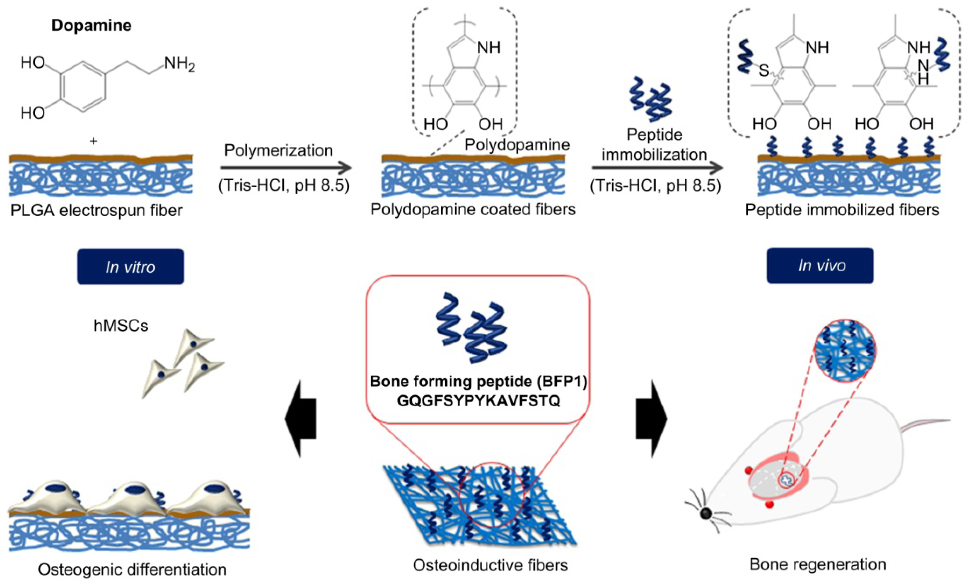
- Lee, Y.J.; Lee, J.H.; Cho, H.J.; Kim, H.K.; Yoon, T.R.; Shin, H. Electrospun fibers immobilized with bone forming peptide-1 derived from bmp7 for guided bone regeneration. Biomaterials 2013, 34, 5059–5069. [Google Scholar]


| Polymer | Modulus (GPa) | Elongation (%) | Solvent | Cristallinity (%) | Degradation Time (Weeks) | Applications | Reference |
|---|---|---|---|---|---|---|---|
| Polyglycolide/Polyglactine | 7.0 | 15–20 | Hexafluoroispropanol | 45–55 | 6–12 | Suture anchors, meniscus repair, medical devices, drug delivery, orbital floor | [31–33] |
| Poly(l-lactide) | 2.7 | - | Benzene, THF, dioxane | 37 | 12–18 | Fracture fixation, interference screws, suture anchors, meniscus repair | [33–35] |
| Poly(d,l-lactide) | - | 3–10 | Methanol, DMF | Amorphous | 11–15 | Orthopaedic implants, drug delivery | [32,33,36] |
| Poly(d,l-lactide-co-glycolide) 85/15 | 2.0 | 3–10 | Ethyl acetate, chloroform, acetone, THF | Amorphous | 5–6 | Interference screws, suture anchors, ACL reconstruction | [33,34,37,38] |
| Poly(d,l-lactide-co-glycolide) 75/25 | 2.0 | 3–10 | Ethyl acetate, chloroform, acetone, DMF, THF | Amorphous | 4–5 | Plates, mesh, screws, tack, drug delivery | [32,33,36] |
| Poly(d,l-lactide-co-glycolide) 50/50 | 2.0 | 3–10 | Ethyl acetate, chloroform, acetone, DMF, THF | Amorphous | 1–2 | Orthopaedic implants, drug delivery | [36,38] |
| Poly (l-lactide-co-glycolide) 10/90 | - | [39,40] |
© 2014 by the authors; licensee MDPI, Basel, Switzerland This article is an open access article distributed under the terms and conditions of the Creative Commons Attribution license (http://creativecommons.org/licenses/by/3.0/).
Share and Cite
Gentile, P.; Chiono, V.; Carmagnola, I.; Hatton, P.V. An Overview of Poly(lactic-co-glycolic) Acid (PLGA)-Based Biomaterials for Bone Tissue Engineering. Int. J. Mol. Sci. 2014, 15, 3640-3659. https://0-doi-org.brum.beds.ac.uk/10.3390/ijms15033640
Gentile P, Chiono V, Carmagnola I, Hatton PV. An Overview of Poly(lactic-co-glycolic) Acid (PLGA)-Based Biomaterials for Bone Tissue Engineering. International Journal of Molecular Sciences. 2014; 15(3):3640-3659. https://0-doi-org.brum.beds.ac.uk/10.3390/ijms15033640
Chicago/Turabian StyleGentile, Piergiorgio, Valeria Chiono, Irene Carmagnola, and Paul V. Hatton. 2014. "An Overview of Poly(lactic-co-glycolic) Acid (PLGA)-Based Biomaterials for Bone Tissue Engineering" International Journal of Molecular Sciences 15, no. 3: 3640-3659. https://0-doi-org.brum.beds.ac.uk/10.3390/ijms15033640