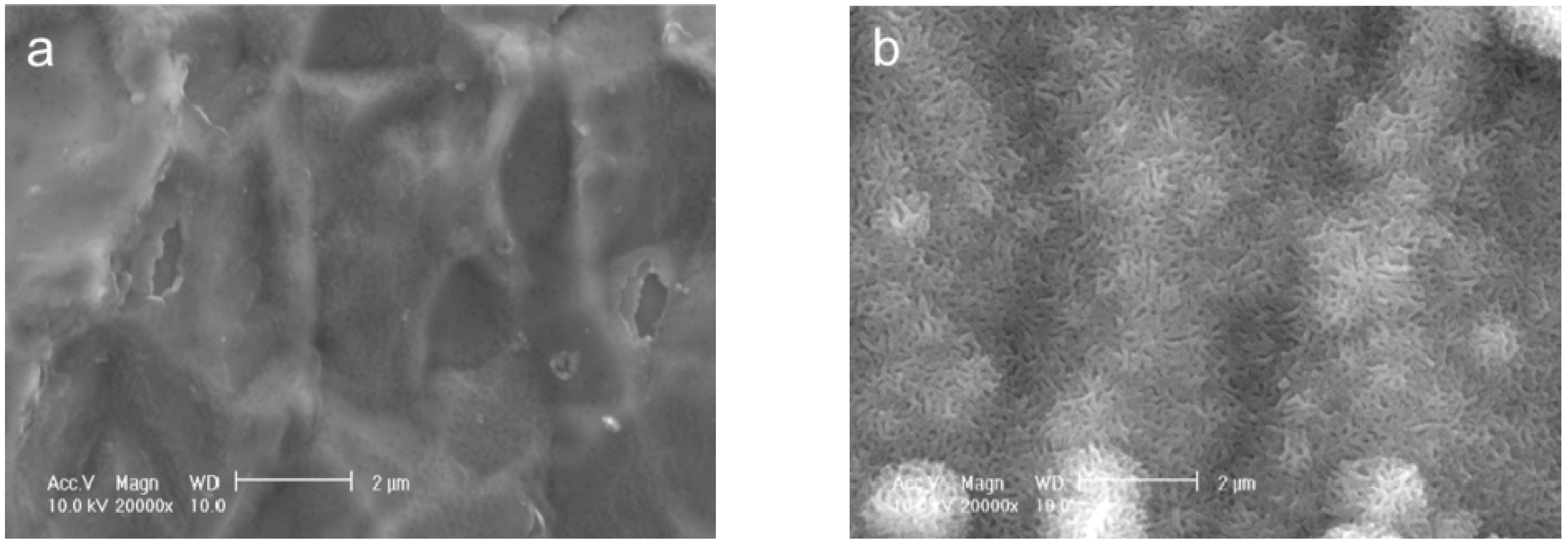
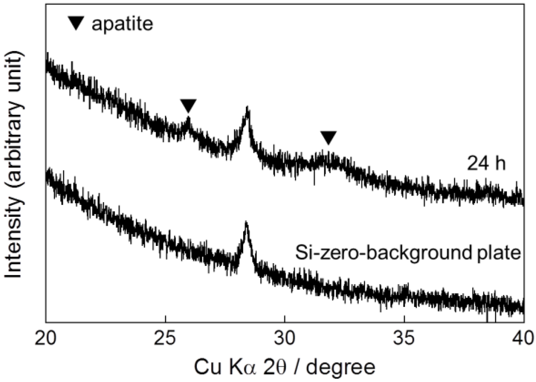
Correction: Mutsuzaki, H., et al. Improved Bonding of Partially Osteomyelitic Bone to Titanium Pins Owing to Biomimetic Coating of Apatite. Int. J. Mol. Sci. 2013, 14, 24366–24379.
| Location | Original Version | Correction |
|---|---|---|
| Abstract, Page 24366 Line 4 | In the present study, an Ap layer was also successfully formed using a one-step method at 25 °C for 48 h in an infusion fluid-based supersaturated calcium phosphate solution, which is clinically useful due to the immersion temperature. | In the present study, an Ap layer was also successfully formed using a one-step method at 25 °C for 24 h in an infusion fluid-based supersaturated calcium phosphate solution, which is clinically useful due to the immersion temperature. |
| 2. Results 2.1. One-Step Formation of Apatite Coating on Ti Pins at 25 °C; Page 24368 Lines 2–3 | In a one-step procedure, increasing the supersaturation of the infusion fluid-based supersaturated CaP solution effectively caused an apatite layer to form on the surface of Ti pins under conditions of 25 °C for 48 h. | In a one-step procedure, increasing the supersaturation of the infusion fluid-based supersaturated CaP solution effectively caused an apatite layer to form on the surface of Ti pins under conditions of 25 °C for 24 h. |
| Figure legend: Figure 10; Page 24372 Line 2 | (a) Averaged values of extraction torque for the UN and Ap groups. Apatite layer was formed at 25 °C for 48 h. | (a) Averaged values of extraction torque for the UN and Ap groups. Apatite layer was formed at 25 °C for 24 h. |
| 3. Discussion; Page 24374 Line 13 | An apatite layer was wholly and homogeneously formed on the Ti screw even at room temperature (25 °C) within 48 h without pretreatment of the Ti pin if it is performed in a CaP solution using increased concentrations of calcium and phosphate ions compared with the previous conditions [15,16]. | An apatite layer was wholly and homogeneously formed on the Ti screw even at room temperature (25 °C) within 24 h without pretreatment of the Ti pin if it is performed in a CaP solution using increased concentrations of calcium and phosphate ions compared with the previous conditions [15,16]. |
| 4. Materials and Methods; 4.2. Immersion of Ti Pins in the Supersaturated CaP Solution; Page 24375 Line 4 | Each Ti pin was immersed in 10 mL of the infusion fluid-based supersaturated CaP solution at 25 °C for 48 h followed by immersion in 2 mL of distilled water for injection (Wasser “Fuso”; Fuso Pharmaceuticals Industries, Osaka, Japan) twice for rinsing. | Each Ti pin was immersed in 10 mL of the infusion fluid-based supersaturated CaP solution at 25 °C for 24 h followed by immersion in 2 mL of distilled water for injection (Wasser “Fuso”; Fuso Pharmaceuticals Industries, Osaka, Japan) twice for rinsing. |
| 5. Conclusions; Page 24377 Line 2 | An apatite layer was formed on Ti pins using a clinically useful method: The Ti pins were immersed in an infusion fluid-based supersaturated CaP solution at 25 °C for 48 h. | An apatite layer was formed on Ti pins using a clinically useful method: The Ti pins were immersed in an infusion fluid-based supersaturated CaP solution at 25 °C for 24 h. |


Supplementary Files
Reference
- Mutsuzaki, H.; Sogo, Y.; Oyane, A.; Ito, A. Improved bonding of partially osteomyelitic bone to titanium pins owing to biomimetic coating of apatite. Int. J. Mol. Sci. 2013, 14, 24366–24379. [Google Scholar]
© 2014 by the authors; licensee MDPI, Basel, Switzerland. This article is an open access article distributed under the terms and conditions of the Creative Commons Attribution license (http://creativecommons.org/licenses/by/3.0/).
Share and Cite
Mutsuzaki, H.; Sogo, Y.; Oyane, A.; Ito, A. Correction: Mutsuzaki, H., et al. Improved Bonding of Partially Osteomyelitic Bone to Titanium Pins Owing to Biomimetic Coating of Apatite. Int. J. Mol. Sci. 2013, 14, 24366–24379. Int. J. Mol. Sci. 2014, 15, 9789-9792. https://0-doi-org.brum.beds.ac.uk/10.3390/ijms15069789
Mutsuzaki H, Sogo Y, Oyane A, Ito A. Correction: Mutsuzaki, H., et al. Improved Bonding of Partially Osteomyelitic Bone to Titanium Pins Owing to Biomimetic Coating of Apatite. Int. J. Mol. Sci. 2013, 14, 24366–24379. International Journal of Molecular Sciences. 2014; 15(6):9789-9792. https://0-doi-org.brum.beds.ac.uk/10.3390/ijms15069789
Chicago/Turabian StyleMutsuzaki, Hirotaka, Yu Sogo, Ayako Oyane, and Atsuo Ito. 2014. "Correction: Mutsuzaki, H., et al. Improved Bonding of Partially Osteomyelitic Bone to Titanium Pins Owing to Biomimetic Coating of Apatite. Int. J. Mol. Sci. 2013, 14, 24366–24379." International Journal of Molecular Sciences 15, no. 6: 9789-9792. https://0-doi-org.brum.beds.ac.uk/10.3390/ijms15069789


