Article

Activity Analysis and Preliminary Inducer Screening of the Chicken DAZL Gene Promoter

College of Animal Science and Technology, Yangzhou University, Yangzhou 225009, Jiangsu, China
*
Author to whom correspondence should be addressed.
These authors contributed equally to this work.
Int. J. Mol. Sci. 2015, 16(3), 6595-6605; https://0-doi-org.brum.beds.ac.uk/10.3390/ijms16036595
Submission received: 24 November 2014 / Revised: 16 January 2015 / Accepted: 5 March 2015 / Published: 23 March 2015
(This article belongs to the Section Biochemistry)

Abstract

:
This study was aimed at identifying the active control area of chicken DAZL gene core promoter, to screen optimum inducers of the DAZL gene, thus to enhance the differentiation of embryonic stem cells into spermatogonial stem cells. Fragments of chicken DAZL gene promoter were cloned into fluorescent reporter plasmids and transfected into DF-1 cells. Then Dual-Luciferase® Reporter Assay System was used to identify the activity of the DAZL gene under different inducers. Our studies showed that the DAZL core promoter region for the Suqin yellow chicken was −383 to −39 bp. The dual-luciferase® reporter showed that all-trans retinoic acid (ATRA), a retinoic acid receptor alpha agonist (tamibarotene/Am80), or estradiol (E2) could significantly enhance DAZL transcription. The in vitro inductive culture of chicken ESCs demonstrated that, with ATRA treatment, DAZL transcription peaked at 6 days and then decreased slowly; whereas, DAZL transcription was continuous and peaked at 10 days with Am80 treatment. E2 treatment significantly increased DAZL expression after 8 days. All three treatments were associated with the appearance of male germ cell (MGC)-like cells on day 10. These results provide the optimum inducer screening of the DAZL gene and lay the foundation for further screening of compounds that can induce the differentiation of ESCs into MGCs in vitro.

1. Introduction

The autosomal DAZL gene is an important member of the Deleted in Azoospermia (DAZ) family (including DAZ, DAZL and BOULE), which encode RNA-binding proteins of the RNA recognition motif (RRM) type and are involved in translation of polysomal mRNA [1]. DAZL, which contains the conserved RRM repeat motif structural domain DAZ, was first discovered in the Drosophila testis [2]. Mouse Dazl expression starts during the early stage of germ cell development, persists into meiosis, and is expressed both in the adult testis and ovaries [3,4]. The RNA-binding protein encoded by Dazl is required for the completion of spermatocyte meiosis in the mouse [5]. Houston et al. [6] found that Xenopus DAZL was critically involved in primordial germ cell (PGC) development. Vogel et al. [7] determined that Xenopus DAZL could only substitute for the murine homologue with respect to the early functions in the establishment of the germ cells. Reynolds et al. [8] found that in Dazl knockout mice, meiotic prophase was arrested at the zygotene stage. Together these results indicate that DAZL has a conserved role in germ cell development, regulation of germ cell differentiation, and is one of the important regulatory factors in the process of gamete formation.
Currently, many research groups are focused on germ cell gene regulation and inductive differentiation of PGCs from embryonic stem cells (ESCs) in vitro. Yu [9] induced mouse ESCs to differentiate into sperm-like cells and oocyte-like cells by overexpression of Dazl. Haston [10] had shown that Dazl plays an important role in the process of differentiation from to germ cell in vitro. Using overexpression of Dazl Kee et al. [11] found that PGC formation could be promoted. Thus far, there have been a number of reports on mouse, human, and Drosophila DAZL and identification of a large number of inducers of its expression; however, the reports on the chicken ortholog are not comprehensive enough. Our team has been committed for decades to studying the inductive differentiation of chicken male germ cells (MGCs) in vitro. Unfortunately, the induction efficiency is low. To improve the induction efficiency, selecting the optimum inducers for chicken DAZL gene expression is crucial.
Chicken, as a classic model anima of developmental biology, displays a unique process of embryonic development. As an experimental model system, chickens provide sufficient material for the study of embryonic germ cells and have more permissive rules than mammalian models. The 5' flanking region of the DAZL gene promoter was cloned upstream of a fluorescent reporter and transfected into DF-1 cells. The Dual-Luciferase® Reporter Assay System allowed quantification of the activity of the chicken DAZL promoter fragments and identification of the gene’s core promoter. The level of DAZL gene promoter activity was detected under different inducer treatments. Candidate optimal inducers were screened for their functional capacity in vitro to induce chicken ESCs to differentiate into MGCs, to provide the optimum inducer screening of the DAZL gene, and to lay the foundation for in vitro screening for inducers of ESC to MGC differentiation in the chicken.

2. Results and Discussion

2.1. The Qualitative Analysis of DAZL Gene Promoter Activity

To test whether the fragment of −932 to −39 bp has promoter activity, pDAZL-EGFP, pEGFP-N1, and pLinker-EGFP plasmids were transfected into DF-1 cells (Figure 1A). The cells transfected with pDAZL-EGFP vector could express green fluorescent protein (GFP), but its fluorescence intensity was weaker than positive control plasmid pEGFP-N1, and pLinker-EGFP transfected cells had no GFP expression.
Four recombinant plasmids: pGL-P1 (−932 to −39 bp), pGL-P2 (−647 to −39 bp), pGL-P3 (−383 to −39 bp), and pGL-P4 (−186 to −39 bp) were used to transfect DF-1 cells with pRL-SV40. pGL-basic and pRL-SV40 were used to transfect DF-1 cells as the negative control group. After 48 h of transfection, the activity of the four promoter segments was detected and different segments of the Suqin yellow chicken DAZL promoter had varying expression (Figure 1B). There was almost no promoter activity in the −186 to −36 bp fragment. The promoter activity in the −383 to −39 bp was the highest.
Figure 1. (A) The green fluorescent protein (GFP) detection of promoter activity of the chicken Deleted in Azoospermia (DAZL) long promoter fragment in DF-1 cells transfected with positive control pDAZL-EGFP, pEGFP-N1, and negative control pLinker-EGFP (40× magnification); (B) The activity of different promoter regions of the chicken DAZL gene in DF-1 cells; (C) The effect of different inducers on the activity of the chicken DAZL core gene promoter in mouse DF-1 cells. * represents p < 0.05, ** represents p < 0.01.
Ijms 16 06595 g001

2.2. The Influence of Different Inducers on Chicken DAZL Gene Promoter Activity

According to the above result, pGL3/334 GC-1 cells had the highest DAZL transcriptional activity. pGL3/334 DF-1 cells were treated with RA (10−5 mol/L), ATRA (10−5 mol/L), Am80 (10−6 mol/L), BMP4 (40 ng/mL), FSH (0.1 IU/mL), E2 (1 μg/mL), testosterone (15 ng/mL), or testicular extract. After 48 h of induction, treatment with BMP4 or testicular extracts had no obvious effect on the activity of the DAZL gene promoter. RA, FSH, and testosterone increased the activity but not significantly. ATRA, Am80, and E2 significantly increased the activity of the DAZL gene promoter (Figure 1C).

2.3. In Vitro Induction Validation

Based on their induction of DAZL transcription ATRA, Am80, and E2 were screened as the optimal inducers of the DAZL gene. Morphological changes were induced in chicken ESCs in vitro by ATRA, AM80, and E2. qRT-PCR and immunohistochemistry were used to detect the kinetics of DAZL gene expression during differentiation. The results showed that cell morphology gradually changed under a different culture system (Figure 2). Under basic medium, control cells grew slowly. A few cell colonies appeared at 2 days, and the cells differentiated at 6 days. With ATRA or Am80 treatment, embryonic bodies gradually formed and increased at 2 days, began to collapse at 6 days, and MGC-like cells were observed at 10 days. With E2 treatment, embryonic bodies gradually formed and enlarged at 2 days, began to collapse at 6 days, MGC-like cells were observed at 8 days, and the number increased at 10 days.
Figure 2. Differentiation of chicken embryonic stem cells (ESCs) induced by different DAZL inducers. (A) chicken ESCs with DMEM; (B) chicken ESCs with 10−5 mol/L l-trans retinoic acid (ATRA) induction; (C) chicken ESCs with 10−6 mol/L Am800 induction; (D) chicken ESCs with 1 μg/mL E2 induction. Arrows represent spermatogonial stem cell-like (SSC-like) cells. (400× magnification).
Ijms 16 06595 g002
qRT-PCR results showed that, compared with the control group, DAZL gene expression achieved the highest level at 6 days of ATRA treatment, and then showed a trend of slow decline. DAZL gene showed continuous expression after Am80 treatment, achieving the highest level at 10 days. DAZL gene expression was significantly increased at 8 days of E2 treatment (Figure 3). Immunohistochemistry also showed that with ATRA, Am80, or E2 treatment, the cells expressed DAZL protein (green), which was absent in the control cells (Figure 4).
Figure 3. DAZL mRNA expression under ATRA, Am80, and E2 induction compared with control chicken ESCs. The results are presented as a mean ± SEM of three duplicate runs. Error bars in charts represent the corresponding standard deviations.
Ijms 16 06595 g003
Figure 4. Immunohistochemical detection of DAZL protein expression in chicken ESCs treated with various inducers of germ cell differentiation. (A) chicken ESCs with DMEM; (B) chicken ESCs with 10−5 mol/L ATRA induction; (C) chicken ESCs with 10−6 mol/L Am80 induction; (D) chicken ESCs with 1 μg/mL E2 induction. (400× magnification).
Ijms 16 06595 g004

3. Experimental Section

3.1. Materials and Reagents

Procedures involving animals and their care conformed to the U.S. National Institute of Health guidelines (NIH Pub. No. 85-23, revised 1996) and were approved by the laboratory-animal management and experimental-animal ethics committee of Yanzhou University.
Fertilized eggs of Suqin yellow chickens (Gallus gallus domesticus) were purchased from the Chinese Academy of Agricultural Sciences Experimental Poultry Farm (Yangzhou, China). ESCs were collected from stage X embryos as described previously [12].
The pEGFP-N1 vector is maintained by our laboratory. E. coli DH5α competent cells, gel extraction kits, and miniprep kits were purchased from Tiangen Biotech (Beijing, China). DL5000 DNA Marker Prime, STAR Max DNA Polymerase, T4 DNA ligase, and restriction endonucleases were purchased from Takara (Shiga, Japan). Expression vectors pGL3.0-Basic, pRL-SV40, and the dual-luciferase assay kit, Dual-Luciferase Reporter Assay System, were purchased from Promega Corporation (Madison, WI, USA). LipofectamineTM2000 was purchased from Invitrogen Life Technologies (Carlsbad, CA, USA). Retinoic acid (RA), all-trans retinoic acid (ATRA), tamibarotene (Am80), estrogen (E2), and testosterone were purchased from SIGMA-Aldrich (St. Louis, MO, USA). Mouse bone morphogenetic protein 4 (BMP4) and follicle stimulating hormone (FSH) were purchased from ProSpec-Tany TechnoGene Ltd. (Rehovot, Israel). The mouse spermatogonial cell line (GC-1) and the chicken embryo fibroblast cell line (DF-1) were purchased from ATCC (Manassas, VA, USA). The remaining reagents were imported or domestic analytical grade. Primer synthesis and sequencing were conducted by Invitrogen.

3.2. Extraction of Genomic DNA

Genomic DNA was extracted from chicken peripheral blood samples using a Puregene DNA isolation kit (Gentra, Minneapolis, MN, USA) according to the manufacturer’s instructions.

3.3. Genomic Clone

Four fragments of the chicken DAZL gene promoter were amplified by polymerase chain reaction (PCR), forward primers (positions relative to the transcription start site, corresponding to the genomic positions chr2:34953772-34954681 for the longest amplicon) were: pEGFP-P0 (−932 to −39 bp): 5'-CGCATTAATTATGCACTCCAGTTGATCAGTTTAA-3', pGL-P1 (−932 to −39 bp): 5'-GGGGTACCTATGCACTCCAGTTGATCAGTTTAA-3', pGL-P2 (−647 to −39 bp): 5'-GGGTACCCCAGCCTGCTCCAGAGTATCCA-3', pGL-P3 (−383 to −39 bp): 5'-GGGGTACCCACACATCGCCCTTCGTCTT-3', pGL-P4 (−186 to −39 bp): 5'-GGGGTACCAAGAACTGCCTCTTTCGCAC-3', and common reverse primer: 5'-CCCTCGAGCAAACGAGGCCTTCAAGACAA-3'.
The PCR consisted of 35 cycles of gene amplification as follows: 98 °C for 10 s followed by 35 cycles composed of a denaturation step at 95 °C for 30 s, 56–62 °C for 30 s, and an elongation step at 72 °C for 40 s. The PCR products were resolved by electrophoresis on 1% agarose gels, and the appropriate band was excised and purified for the subsequent steps of cloning or sequence determination. Sequence analysis was performed with an automatic sequencer (ABI 377, Applied Biosystems/PE/Life Technologies, Grand Island, NY, USA).

3.4. Vector Construction

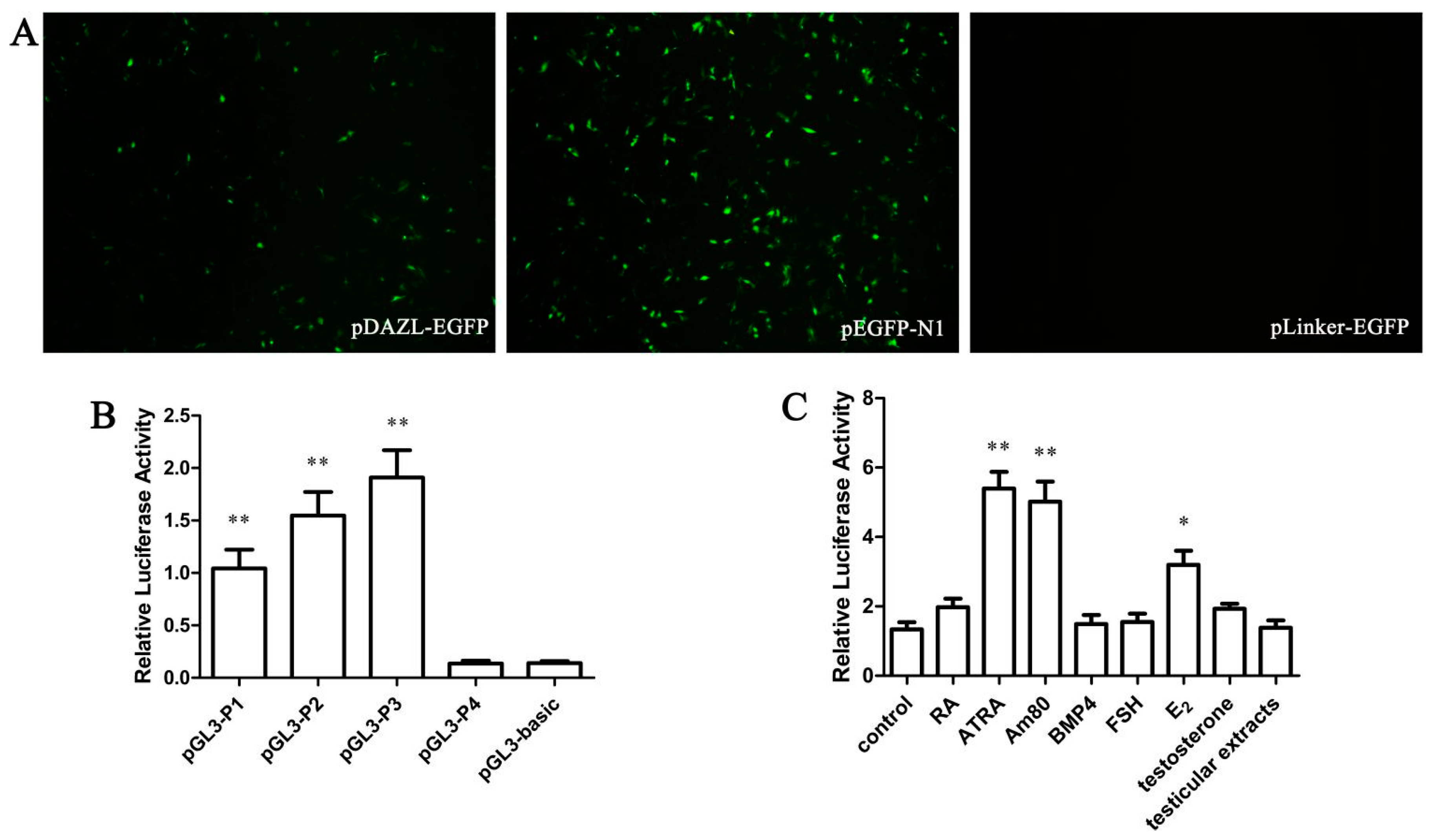
The DAZL gene 5' flanking area −932 to −39 bp was inserted into the pEGFP-N1 vector between AseI and XhoI restriction enzyme cutting sites and named pDAZL-EGFP. A negative control carrier was also built as pLinker-EGFP. Four fragments of the DAZL gene promoter region cloned previously were inserted into the dual-luciferase reporter gene carrier pGL3-Basic. All vectors were verified by AseI and XhoI restriction enzyme digestion and sequence analysis.

3.5. Qualitative Analysis of DAZL Gene Promoter

DF-1/GC-1 cells were inoculated into 12 wells, approximately 5 × 10⁵ cells per well. According the manufacturer’s instructions for LipofectamineTM2000, pEGFP-N1/893 was used for transfection of DF-1/GC-1 cells, and a positive (pEGFP-N1 transfected cells) and negative control group (pEGFP-N1-CMV transfected cells) were created. The results for each group represent three replications. After 48 h of transfection, fluorescence was observed under a Nikon Eclipse microscopy (Nikon, Tokyo, Japan).

3.6. Cell Transfection

DF-1/GC-1 cells were inoculated into 24 wells, approximately 2.5 × 10⁵ cells per well. According to the manufacturer’s instructions for LipofectamineTM2000, all recombinant plasmids and a reference plasmid pRL-SV40 (35:1) were transfected into DF-1/GC-1 cells. DF-1/GC-1 cells transfected with pGL3-basic and pRL-SV40 were selected as a negative group. The results from each group represent three replications. After 48 h, the Dual-Luciferase® Reporter Assay was performed.
After 6 h of transfection, cells were treated with RA (10−5 mol/L), ATRA (10−5 mol/L), Am80 (10−6 mol/L), BMP4 (40 ng/mL), FSH (0.1 IU/mL), E2 (1 μg/mL), testosterone (15 ng/mL), or testicular extract. The negative group was treated with the same volume of phosphate-buffered saline. After treatment for 48 h, cells were collected. The results of each group represent three replications.

3.7. Dual-Luciferase Activity Detection

Cells were collected in 70 μL PBS and the suspension was transferred to a 96-well plate. A Dual-Luciferase® Reporter Assay was performed to detect the activity of the DAZL gene promoter constructs. The relative luciferase activity representing the promoter activity is reported as the ratio of firefly luciferase value/renilla luciferase.

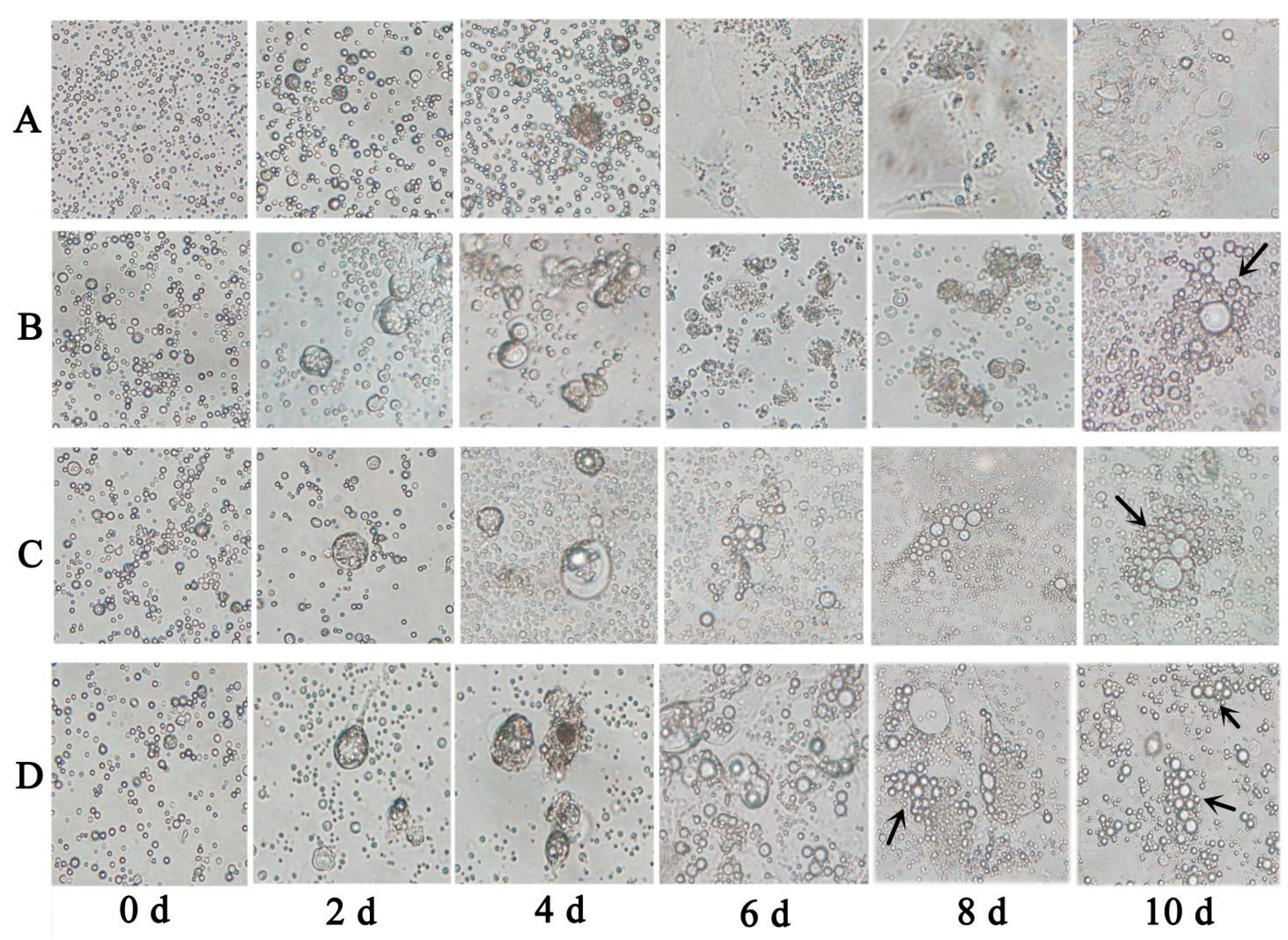
3.8. In Vitro Induction Experiment

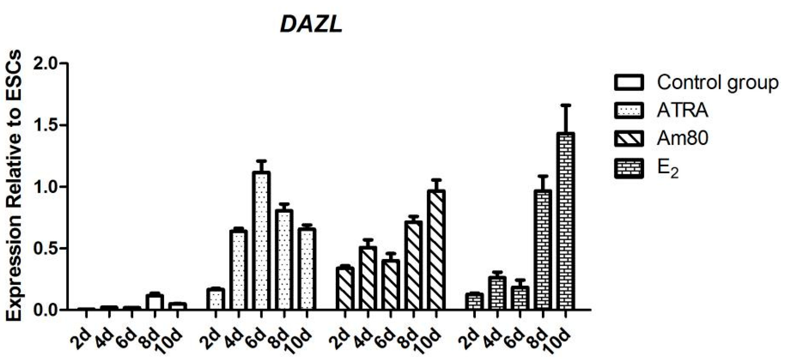
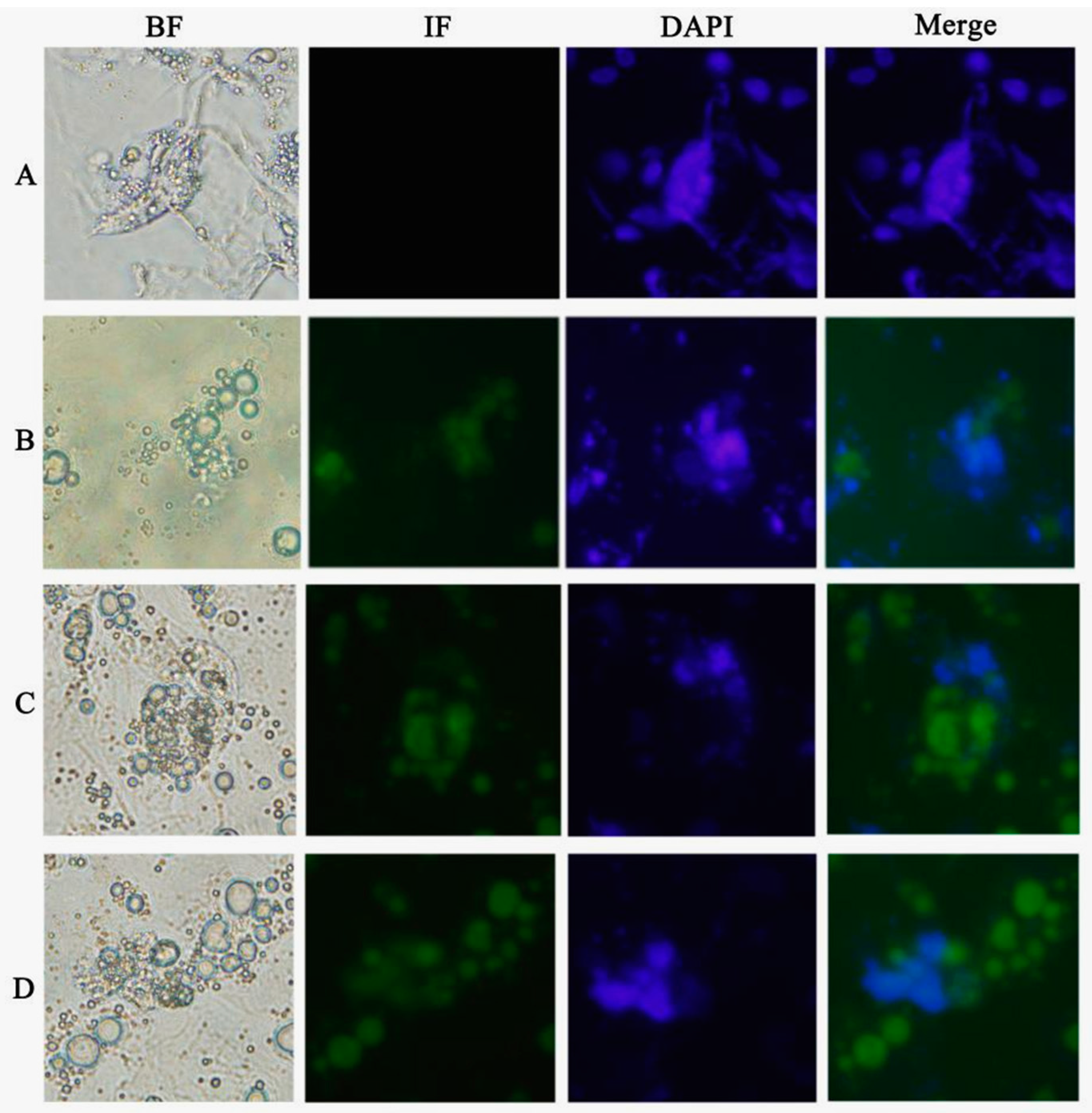
Chicken ESCs were inoculated into 24 wells, approximately 2 × 10⁵ cells per well. ATRA (10−5 mol/L), Am80 (10−6 mol/L), and E2 (1 μg/mL) were used to induce chicken ESC differentiation in vitro. The cells were collected every two days after inducer treatment. The results of each group consisted of three replications.

3.9. Quantitative Real-Time Reverse Transcriptase PCR (qRT-PCR)

Total RNA was extracted using the RNeasy kit (Qiagen, Suzhou, China). Reverse transcription into cDNA was performed by Reverse Transcription System (Qiagen) to serve as a template for qRT-PCR. qRT-PCR of the cDNA was performed according to the instructions provided in the fluorescence quantitative PCR kit (Qiagen), using SYBR as the fluorescent reagent and 7500 System fluorescence quantitative instrument (Applied Biosystems, Foster City, CA, USA). Each group had three replications. The data were analyzed in Microsoft Excel using the 2−ΔΔCt relative quantitative method.

4. Discussion

DAZL is one of the DAZ family members encoding an RNA-binding protein, which is also an indispensable control factor during the animal gametogenesis process. To explore the role of the DAZL gene in the specific regulatory mechanism of male reproductive cells, different lengths of the chicken DAZL gene promoter attached to a fluorescent reporter were constructed, and were transfected into DF-1. A Dual-Luciferase® Reporter Assay was performed, showing that the pGL-P3 promoter has the highest activity of the DAZL gene promoter fragments in DF-1 cells. This indicates that there are important regulatory elements in the −382 to −39 bp region of the chicken DAZL gene promoter.
Zhuo et al. [13] found that with ectopic expression of the Dazl gene, both motile-tailed sperm and oocytes could be induced from mouse ESCs. Several other groups [14,15,16,17] also verified that Dazl was a master gene controlling germ cell differentiation. However, DAZL gene research is not comprehensive in chickens, and there is doubt whether specific inducers of the mouse Dazl gene could be used for directional induction by the chicken ortholog in vitro. To evaluate the conservation of DAZL gene function, combining previous research results [18,19,20], RA, ATRA, Am80, BMP4, FSH, E2, testosterone, and testicular extracts evaluated for induction of pGL3/334 GC-1 cells.
BMP4 is a multifunctional cytokine, which can induce ESCs to differentiate into MGCs in vitro. Shi et al. [20] found that the expression of Dazl, Stra8, Integrin α6, and c-kit was enhanced with every 2 days of BMP4 treatment in chicken. In our study, BMP4 treatment did not enhance the activity of the core chicken DAZL gene promoter. Considering the short treatment time of BMP4 in our study (48 h), the lack of an effect might be because BMP4 does not directly affect the chicken DAZL gene, but indirectly causes the expression of DAZL mRNA through interaction with multiple genes. Neither testosterone nor testicular extract had an effect on the chicken DAZL gene promoter, which is consistent with the results by Sun and colleagues [12]. Pan et al. [19] found that E2 and FSH could induce mouse ESCs to differentiate into MGCs with high-level expression of mouse Dazl mRNA on day 7. Our study is consistent with their earlier findings. Additionally E2 had a more significant effect than FSH in our hands. Retinoic acids, including ATRA and RA, are fat-soluble small molecule metabolites of vitamin A. RA plays an important role in biological development and normal physiological conditions. The compound can mediate cell differentiation, proliferation, apoptosis, and regulate immune function. Kerkis and colleagues found that, 10−5 mol/L of RA could induce mouse ESCs to differentiate into MGCs positive for Dazl gene expression. The single factor induction by ATRA and RA in our study showed that both can enhance the expression of the chicken DAZL gene. Am80 is a new synthetic retinoic acid analogue and specific receptor agonist of RARα. Am80 has a higher induced efficiency than ATRA or RA.
Combining with the prediction of regulatory elements binding sites of chicken DAZL gene (Figure S3), a RAR-beta (RARβ) binding site presented in the gene’s core promotor region. This indicated that ATRA and RA could probably promoting chicken DAZL gene transcription by directly binding this interval. The absence of ER (estrogen receptor) and RARalpha (RARα) binding site indicating that E2 or Am80 might play its role through other bound transcription factors or by signaling. We also found two AR (androgen receptor) binding sites in this region, ATRA, Am80 or E2 could also directly binding this interval to paly the role in chicken DAZL gene transcriptional regulation. We are investigating further about the specific molecular mechanism of chicken DAZL gene inducer.
To further validate whether the inducers we screened from chicken DF-1 cells could be applied to other species, we also used mouse GC-1 cells for the same test (Figure S1). The results showed that ATRA, Am80, and E2 could also significantly increased the activity of the DAZL gene promoter in GC-1 cells (Figure S2C). ATRA, Am80, and E2 can significantly enhance the activity of the chicken DAZL gene promoter and expression of the encoded DAZL protein. The formation of MGC-like cells was also observed during the induction, indicating that these factors can regulate the minimal chicken DAZL promoter similar to its mammalian counterparts.

5. Conclusions

In this paper, we identified the core promoter region of the DAZL gene for the Suqin yellow chicken, and compared the induction efficiency of different inducers. ATRA, Am80, and E2 were screened as optimal inducers of the chicken DAZL gene, and in vitro induction validation was performed. This study provides a starting point to design the optimal culture system to induce chicken ESCs to differentiate into MGCs in vitro, in part, by significantly raising DAZL gene expression.

Supplementary Materials

Acknowledgments

The author thanks the Poultry Institute of the Chinese Academy of Agricultural Sciences Experimental Poultry Farm for providing the experimental materials.

Author Contributions

Bichun Li conceived and designed the experiments; Lei Zhang, Rui Zhu, Qisheng Zuo and Dong Li performed the experiments; Lei Zhang analyzed the data; Lei Zhang and Rui Zhu wrote the paper; and Lei Zhang, Chao Lian, Beibei Tang, Tianrong Xiao and Yani Zhang edited it; Lei Zhang and Rui Zhu worked as the same in this study.

Conflicts of Interest

The authors declare no conflict of interest.

References

  1. Xu, E.Y.; Moore, F.L.; Pera, R.A.R. A gene family required for human germ cell development evolved from an ancient meiotic gene conserved in metazoans. Proc. Natl. Acad. Sci. USA 2001, 98, 7414–7419. [Google Scholar] [CrossRef] [PubMed]
  2. Eberhart, C.G.; Maines, J.Z.; Wasserman, S.A. Meiotic cell cycle requirement for a fly homologue of human Deleted in Azoospermia. Nature 1996, 381, 783–785. [Google Scholar] [CrossRef] [PubMed]
  3. Ruggiu, M.; Cooke, H.J. In vivo and in vitro analysis of homodimerization activity of the mouse Dazl1 protein. Gene 2000, 252, 119–126. [Google Scholar] [CrossRef] [PubMed]
  4. Yen, P.H. Putative biological functions of the DAZ family. Int. J. Androl. 2004, 27, 125–129. [Google Scholar] [CrossRef] [PubMed]
  5. Lin, Y.M.; Chen, C.W.; Sun, H.S.; Tsai, S.J.; Hsu, C.C.; Teng, Y.N.; Lin, J.S.; Kuo, P.L. Expression patterns and transcript concentrations of the autosomal DAZL gene in testes of azoospermic men. Mol. Hum. Reprod. 2001, 7, 1015–1022. [Google Scholar] [CrossRef] [PubMed]
  6. Houston, D.W.; King, M.L. A critical role for Xdazl, a germ plasm-localized RNA, in the differentiation of primordial germ cells in Xenopus. Development 2000, 127, 447–456. [Google Scholar] [PubMed]
  7. Vogel, T.; Speed, R.M.; Ross, A.; Cooke, H.J. Partial rescue of the Dazl knockout mouse by the human DAZL gene. Mol. Hum. Reprod. 2002, 8, 797–804. [Google Scholar] [CrossRef] [PubMed]
  8. Nicola, R.; Brian, C.; Victoria, B.; Nicola, K.G.; Howard, J.C. Translation of the synaptonemal complex component Sycp3 is enhanced in vivo by the germ cell specific regulator Dazl. RNA 2007, 13, 974–981. [Google Scholar] [CrossRef] [PubMed]
  9. Yu, Z.; Ji, P.; Cao, J.; Zhu, S.; Li, Y.; Zheng, L.; Chen, X.; Feng, L. Dazl promotes germ cell differentiation from embryonic stem cells. J. Mol. Cell Biol. 2010, 2, 170. [Google Scholar] [CrossRef]
  10. Haston, K.M.; Tung, J.Y.; Pera, R.A.R. Dazl functions in maintenance of pluripotency and genetic and epigenetic programs of differentiation in mouse primordial germ cells in vivo and in vitro. PLoS One 2009, 4, e5654. [Google Scholar] [CrossRef] [PubMed]
  11. Kehkooi, K.; Vanessa, T.A.; Martha, F.; Ha, N.N.; Renee, A.R.P. Human DAZL, DAZ and BOULE genes modulate primordial germ-cell and haploid gamete formation. Nature 2009, 462, 222–225. [Google Scholar] [CrossRef] [PubMed]
  12. Sun, M. Study on Male Germ Cell Derived from Chicken Embryonic Stem Cell and the Generation of Transgenic Chicken. Ph.D. Dissertation, Yangzhou University, Yangzhou, China, 2012. [Google Scholar]
  13. Tung, J.Y.; Rosen, M.P.; Nelson, L.M.; Turek, P.J.; Witte, J.S.; Cramer, D.W.; Cedars, M.I.; Reijo-Pera, R.A. Novel missense mutations of the Deleted-in-AZoospermia-Like (DAZL) gene in infertile women and men. Reprod. Biol. Endocrinol. 2006, 4, 40. [Google Scholar] [CrossRef] [PubMed]
  14. Zhang, Q.B.; Li, Q.F.; Li, J.H.; Li, X.F; Liu, Z.S.; Pan, Z.X.; Song, D.W.; Xie, Z. The cow b-DAZL spermatogenesis related new gene cloning and bioinformatics analysis and expression study. Prog. Natl. Sci. 2008, 18, 493–504. [Google Scholar]
  15. Linher, K.; Cheung, Q.; Baker, P.; Bedecarrats, G.; Shiota, K.; Li, J. An epigenetic mechanism regulates germ cell-specific expression of the porcine Deleted in Azoospermia-Like (DAZL) gene. Differentiation 2009, 77, 335–349. [Google Scholar] [CrossRef] [PubMed]
  16. Wang, H.; Zhang, L.; Hou, M.; Wang, C.-F.; Wang, L.-L.; Sun, T.; He, H.-B.; Zhong, J.-F. Cloning and activity analysis of bovine natural resistance associated macrophage protein 1 (Nramp1) Gene Promoter. Sci. Agric. Sin. 2011, 44, 1022–1028. [Google Scholar]
  17. Teng, Y.N.; Chang, Y.P.; Tseng, J.T.; Kuo, P.H.; Lee, I.W.; Lee, M.S.; Kuo, P.L. A single-nucleotide polymorphism of the DAZL gene promoter confers susceptibility to spermatogenic failure in the Taiwanese Han. Hum. Reprod. 2012, 27, 2857–2865. [Google Scholar] [CrossRef] [PubMed]
  18. Lin, Y.; Gill, M.E.; Koubova, J.; Page, D.C. Germ cell-intrinsic and-extrinsic factors govern meiotic initiation in mouse embryos. Science 2008, 322, 1685–1687. [Google Scholar] [CrossRef] [PubMed]
  19. Pan, S. Study on Mouse Embryonic Stem Cells Differentiate into Male Germ Cells. Ph.D. Dissertation, Northwest Agriculture & Forestry University, Yangling, China, 2010. [Google Scholar]
  20. Shi, Q.; Zhang, Z.; Li, P.; Zheng, M.-M.; Wang, D.; Huang, X.-M.; Zhang, Y.-N.; Li, B.-C. Study on differentiation of chicken embryonic stem cells to male germ cells by BMP4. Acta Vet. Zootech. Sin. 2013, 44, 1749–1757. [Google Scholar]

