Article

Altered Phenotypes in Saccharomyces cerevisiae by Heterologous Expression of Basidiomycete Moniliophthora perniciosa SOD2 Gene

by
Sônia C. Melo
1,
Regineide X. Santos
2,
Ana C. Melgaço
1,
Alanna C. F. Pereira
1,
Cristina Pungartnik
1,* and
Martin Brendel
1
1
Departamento de Ciências Biológicas, Laboratório de Biologia de Fungos, Centro de Biotecnologia e Genética, Universidade Estadual de Santa Cruz (UESC), Rodovia Jorge Amado, km 16, Ilhéus, Bahia CEP 45662-900, Brazil
2
Departamento de Ciências Naturais, Universidade Estadual do Sudoeste da Bahia (UESB), Estrada do Bem Querer, km 4, Vitória da Conquista, Bahia CEP 45083-900, Brazil
*
Author to whom correspondence should be addressed.
Int. J. Mol. Sci. 2015, 16(6), 12324-12344; https://0-doi-org.brum.beds.ac.uk/10.3390/ijms160612324
Submission received: 20 December 2014 / Accepted: 8 April 2015 / Published: 1 June 2015
(This article belongs to the Section Biochemistry)

Abstract

:
Heterologous expression of a putative manganese superoxide dismutase gene (SOD2) of the basidiomycete Moniliophthora perniciosa complemented the phenotypes of a Saccharomyces cerevisiae sod2Δ mutant. Sequence analysis of the cloned M. perniciosa cDNA revealed an open reading frame (ORF) coding for a 176 amino acid polypeptide with the typical metal-binding motifs of a SOD2 gene, named MpSOD2. Phylogenetic comparison with known manganese superoxide dismutases (MnSODs) located the protein of M. perniciosa (MpSod2p) in a clade with the basidiomycete fungi Coprinopsis cinerea and Laccaria bicolor. Haploid wild-type yeast transformants containing a single copy of MpSOD2 showed increased resistance phenotypes against oxidative stress-inducing hydrogen peroxide and paraquat, but had unaltered phenotype against ultraviolet–C (UVC) radiation. The same transformants exhibited high sensitivity against treatment with the pro-mutagen diethylnitrosamine (DEN) that requires oxidation to become an active mutagen/carcinogen. Absence of MpSOD2 in the yeast sod2Δ mutant led to DEN hyper-resistance while introduction of a single copy of this gene restored the yeast wild-type phenotype. The haploid yeast wild-type transformant containing two SOD2 gene copies, one from M. perniciosa and one from its own, exhibited DEN super-sensitivity. This transformant also showed enhanced growth at 37 °C on the non-fermentable carbon source lactate, indicating functional expression of MpSod2p. The pro-mutagen dihydroethidium (DHE)-based fluorescence assay monitored basal level of yeast cell oxidative stress. Compared to the wild type, the yeast sod2Δ mutant had a much higher level of intrinsic oxidative stress, which was reduced to wild type (WT) level by introduction of one copy of the MpSOD2 gene. Taken together our data indicates functional expression of MpSod2 protein in the yeast S. cerevisiae.

1. Introduction

Aerobic organisms use oxygen (O2) as the final electron acceptor in their carbohydrate metabolism. During respiration, some of the O2 is only partially reduced, forming reactive oxygen species (ROS) such as anion superoxide (O2·), hydrogen peroxide (H2O2) and hydroxyl radical (OH·). Mitochondria convert 1%–2% of the oxygen consumed into O2· [1]. As these ROS can cause significant cellular stress and damage, antioxidant protection is essential for survival in an aerobic environment. The balance between ROS production and cellular anti-ROS defenses, therefore, determines the degree of oxidative stress [2], which results from the imbalance between oxidants and anti-oxidants in favor of the former [3]. Apart from the endogenous production of ROS during respiration in aerobic organisms, oxidative stress may also be induced by a wide range of environmental factors including UV-light, pathogen invasion, herbicide action or shortage of O2 [4].
Cells protect themselves against oxidative damage by different defense mechanisms. These include enzymes (such as peroxidases, catalases and superoxide dismutases (SOD)), anti-oxidants, such as glutathione, vitamins A, C and E [5]; and non-protein complexes of manganese as a back up of ROS-scavenging systems for handling O2· and related ROS [6]. The first defense against O2 toxicity involves at least one form of the SOD enzymes [7,8] that promote the conversion of two molecules of O2· into O2 and H2O2; the latter in turn is further degraded by catalase into O2 and H2O [9].
SOD enzymes (EC1.15.1.1) are characterized in many different organisms and may employ different co-factors to carry out the dismutation of O2·, like copper (CuSOD), manganese (MnSOD), iron (FeSOD) or nickel (NiSOD) or combinations like copper and zinc (Cu/ZnSOD) [10]. The Sod1p (Cu/ZnSOD) and Sod2p (MnSOD) are important to improve survival, but so far are only well characterized in yeasts and mammals [11,12].
A few SOD genes from fungi have been characterized. In Neurospora crassa sod-1 encodes a major Cu/ZnSOD, and mutant strains lacking this enzyme were sensitive to paraquat and elevated O2 concentrations, and exhibited an increased spontaneous mutation rate [13]; evidence was provided for autoreactivity to the human MnSOD in allergic persons sensitized to an environmental allergen from Aspergillus fumigatus, which shares a high degree of sequence homology to the corresponding human enzyme [14]; Colletotrichum graminicola predicted MnSOD protein did not appear to contain a signal peptide that would target it to the mitochondria, and the expression was associated with differentiation of both oval and falcate conidia [15]; a homologue of yeast and human SOD1, MoSod1 from Magnaporthe oryzae was identified as Cu/ZnSOD and regulated by MoSir2 to alleviate MoSOD1 transcript repression and detoxify host ROS [16]. In S. cerevisiae, the SOD1 gene (encoding a Cu/ZnSOD protein) is predominantly located in the cytosol, while the SOD2 gene encodes MnSOD, which is located in the mitochondria. Although located in different cellular compartments, both proteins share the same main role in ROS protection by dismutating O2· into O2 and H2O2 [17,18,19]. In yeast and many other eukaryotes Sod2p (MnSOD) is synthesized by 80S ribosomes and imported into the mitochondrial matrix [20] where it plays an essential role in oxidative stress protection. Apart from its role in dismutation of O2· MnSOD, especially at high activity, may cause cell growth inhibition due to increased production of H2O2 [21]. Elevated MnSOD activity may change the mitochondrial redox state and thus influence coordination of physiological and biochemical events in cellular compartments [22]. Beauveria bassiana, a filamentous entomopathogen, has five distinct SODs, which were proved to contribute to intracellular SOD activity and additively acted in antioxidation and virulence. Subcellular localization of mitochondrial FeSOD (Sod4) and cell wall-anchored Cu/ZnSOD (Sod5) were characterized in this fungus [23].
SODs were found to be an important virulence factor in nearly all pathogenic fungi [24]. Candida albicans Cu/ZnSOD is required for the protection against oxidative stresses and for expression of full virulence in human cells. Upon encountering superoxide stress, such as generation of nicotinamide adenine dinucleotide phosphate-oxidase (NADPH oxidase)-mediated O2· species, predominant antioxidant proteins named SOD4 and SOD5 rapidly break down O2· on cell surfaces [25]. Also, SOD influences the virulence of Cryptococcus neoformans by affecting its growth within macrophages [26]. However, A. fumigatus SODs are intracellular and do not neutralize extracellular ROS in spite of the high sensitivity of this fungus to intracellular ROS generators, thus suggesting that in this case SODs are not putative fungal virulence factors [27].
Apart from modulating virulence of pathogenic fungi, SOD also may have other functions. In the case of Candida glabrata the absence of both SODs leads to auxotrophy for lysine, a high rate of spontaneous mutation and reduced chronological lifespan. In a more general context SODs also play an important role in metabolism, acting in biosynthesis, DNA protection and aging [28]. In the yeast S. cerevisiae, SOD enzymes play a substantial role in preserving the genomic integrity and their absence leads to shorter life span by allowing increased DNA fragmentation [29]. Also, by triggering production of organic acids, SOD2 has the potential to promote cell population growth under nutrient deprivation stress [30]. In the yeast Schizosaccharomyces pombe, Sod2p is the major salt tolerance plasma membrane protein as it functions in removing excess intracellular sodium (or lithium) in exchange for protons [31].
After successful invasion of the host, phytopathogens, such as Moniliophthora perniciosa, can elicit in planta either a localized response that is often associated with an oxidative burst or a more generalized systemic response mediated by signaling molecules, or a combination of both [32]. The oxidative burst generates ROS, i.e., O2·, OH· and H2O2, that form a toxic barrier to pathogen invasion [33]. In order to survive the produced ROS, parasites and phytopathogens rely on their anti-ROS defenses for survival under these conditions [34,35,36].
Witches’ broom disease (WBD) of cacao (Theobroma cacao), caused by the hemibiotrophic basidiomycete M. perniciosa, exhibits a succession of symptoms that are caused during the monokaryotic, biotrophic phase of the fungus that precedes the dikaryotic, necrotrophic phase in T. cacao. WBD begins when wind-borne monokaryotic basidiospores infect young meristematic tissues through stomatal openings and form intercellular monokaryotic hyphae, which cause hypertrophy and hyperplasia of the tissues, loss of apical dominance and proliferation of auxiliary shoots, known as “green brooms”. After 3–6 weeks of infection, the homothallic fungus undergoes sexual differentiation, produces clamp connections and forms a dikaryotic mycelium; this marks the transformation from biotrophic to necrotrophic growth phase characterized by the change from inter- to intra-cellular growth; this in turn causes necrosis and death of infected tissues, known as “dry brooms” [37,38,39,40]. Infected tissues (green brooms) present high levels of glycerol and increased accumulation of H2O2 [40].
The mycelium of M. perniciosa in necrotrophic growth phase, either in vivo or in vitro, is mainly a dikaryon with two nuclei per cell [41,42]. Expression of MpSOD2 (M. perniciosa homologue of fungal SOD2 genes) has been monitored in M. perniciosa dikaryotic cells. MpSod2p was shown to have constant basal expression when grown either in glycerol or glucose, and was induced after H2O2 exposure in glycerol grown cells [35,43]. Therefore, this gene is supposed to be an important antioxidant defense of this fungus [35,43] against intense oxidative stress generated by the invaded plant host [39,44,45].
Since Sod2p seems essential in protecting against mitochondria-induced oxidative stress we have transferred its encoding gene MpSOD2 into the S. cerevisiae mutant sod2Δ (yeast mutant allele in which most of the open reading frame (ORF) has been deleted) in order to verify its role in oxidative stress protection via heterologous expression.

2. Results and Discussion

In this study we identified via sequence homology and functional heterologous expression the M. perniciosa Sod2p-encoding gene. To date, apart from Saccharomyces and Candida [46], four other MnSODs from filamentous fungi have been characterized which are from Aspergillus [14], Penicillum [47], Ganoderma [48], Colletotrichum [15]. But to our knowledge this is the only one indicating that its heterologous expression in S. cerevisiae enhances resistance to ROS and activates the pro-mutagen diethylnitrosamine (DEN).

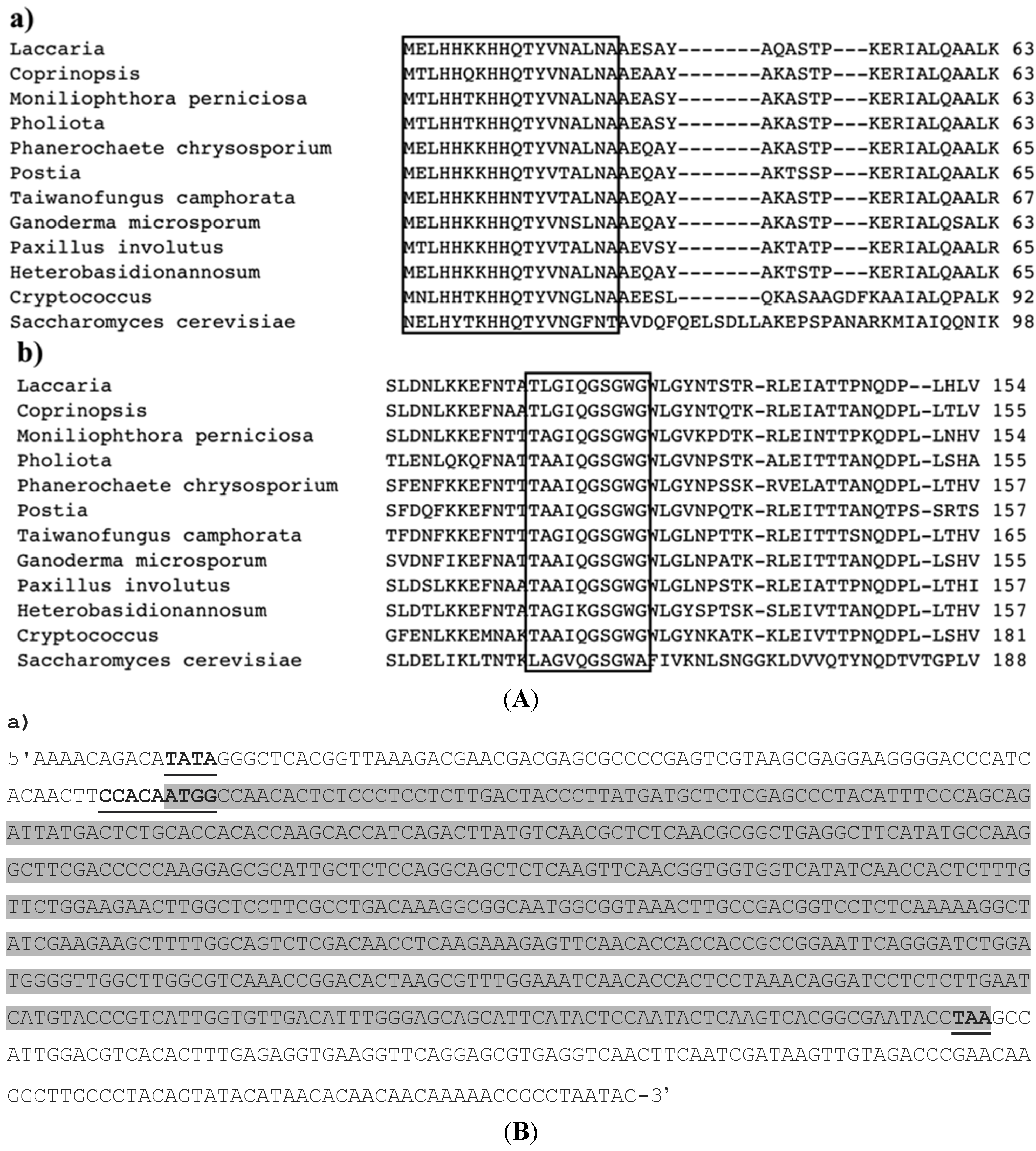
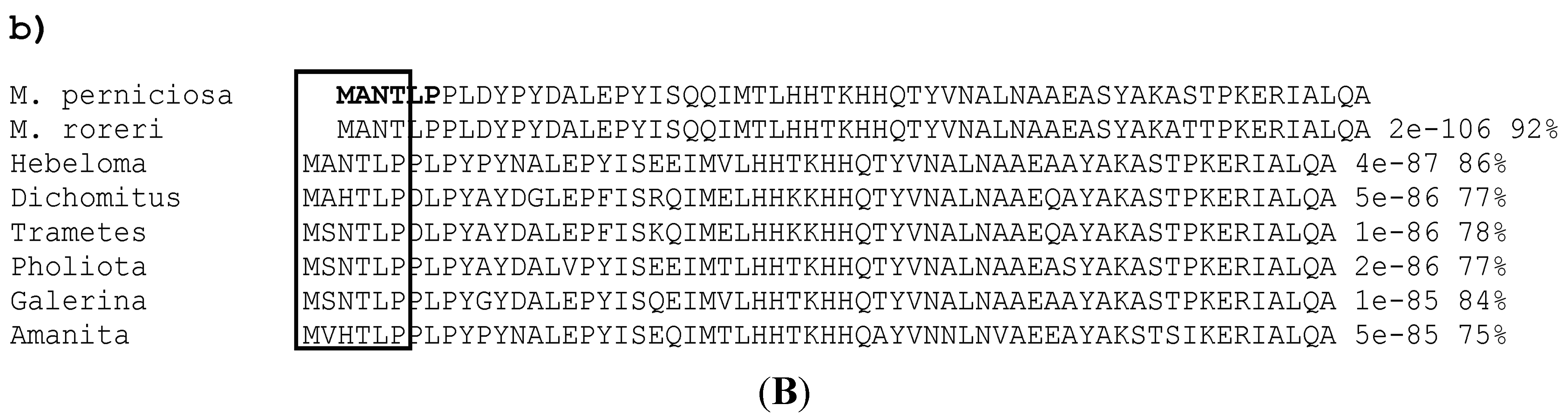
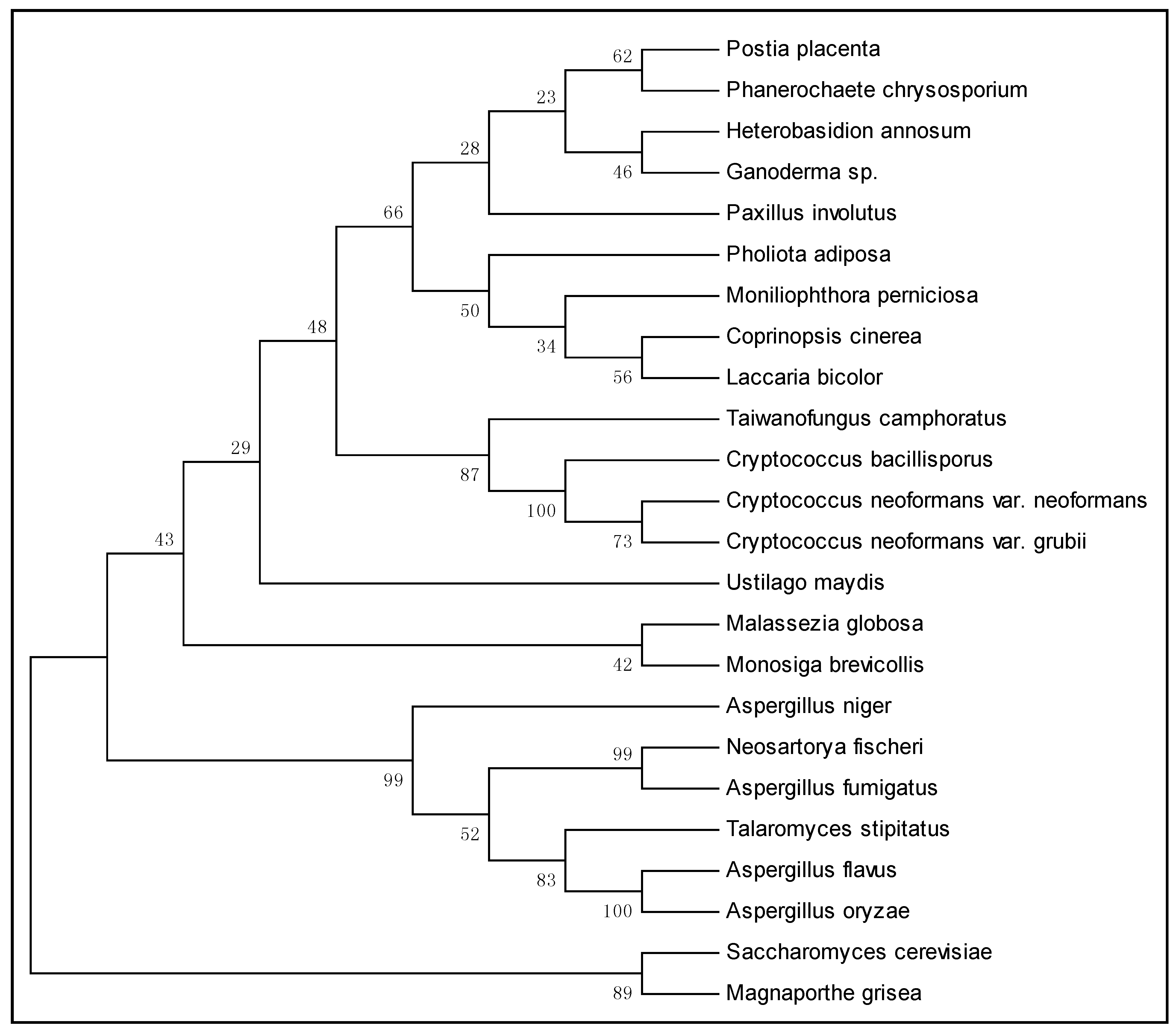
2.1. Characterization of M. perniciosa Manganese Superoxide Dismutase (MpSOD2) Gene and Its Predicted Product

Sequence analysis of a putative MpSOD2 clone revealed that it contained a 748 bp insert with an ORF encoding a predicted polypeptide of 176 amino acids (aa) (Genbank accession No. XM2395448). Searches of GenBank using the BLASTP algorithm indicated that the predicted protein was similar to several proteins within the family of manganese-type superoxide dismutases (MnSODs, Figure 1A). Accordingly, the M. perniciosa gene was named MpSOD2. A Kozak consensus sequence [49] surrounds the putative SOD2 start codon (Figure 1B). There is an upstream consensus TATA box motif, but a 3ʹ AT-rich region (a region rich in residues of adenine and timine) is missing. The MpSOD2 cDNA contains a 132 bp 3ʹ-untranslated region. A consensus polyadenylation signal (AATAAA; [15]) is absent.
Figure 1. (A) Alignment of the MpSOD2 deduced amino acid sequence with MnSOD2 polypeptides from other organisms; Conserved residues are boxed in black frame. Fe/MnSod2p (a) N-terminal α hairpin domain; (b) C-terminal domain. Protein domains are in a black frame, full alignment not shown; (B) Nucleotide sequence of gene MpSOD2 and its protein sequence. (a) Highlighted are upstream a region defined as sequence of thymine-adenine-thymine-adenine TATA box (marked in bold letters), Kozak motif with start and stop codon (marked in bold letters). The highlighted gray sequence is the CDS (codon DNA sequence); (b) Highlighted are upstream region with the following sequence of aminoacids MANTLP (methionine, adenine, asparagine, threonine, leucine, proline) in basidiomycete fungi marked in bold letters from M. perniciosa and in a box frame from other organism.
Ijms 16 12324 g001aIjms 16 12324 g001b
The predicted Sod2p has four conserved aa residues, His-31, His-32, Asp-149, and Pro-155 that are known to coordinate an epitope for metal ligand binding in the MnSOD gene family [50,51,52]. The presence of Gly-69 and Gly-70 instead of Ala-69 and Gln-70 and the absence of a conserved Trp-71 that occurs in proteins of the FeSOD family [53,54] confirm that MpSod2p is affiliated with the MnSOD rather than the FeSOD family. In eukaryotes MnSOD is usually targeted to mitochondria and contains a typical signal peptide rich in basic and hydrophilic aa [55]. The MpSOD2 sequence lacks this information and is thus similar to Sod2p of A. fumigatus [14], Penicillium chrysogenum [46] and Ganoderma microsporum [47]. The N-terminal aa sequence of MpSod2p was determined and compared with those of Sod2p from other organisms that have the typical 6 aa sequence of M-A-N-T-L-P (Figure 1B). This sequence is part of a conserved domain of iron/manganese superoxide dismutase and, therefore, this specific sequence could be used to identify the SOD2 gene in basidiomycetes.
Cluster analysis is represented graphically and consists of nodes and branches that summarize evolutionary relationships among particular taxa [56]. By using the neighbor-joining method [57] we could construct a dendogram based on SOD2 gene DNA sequences of 24 species. That placed M. perniciosa close to the basidiomycota Coprinopsis cinerea and Laccaria bicolor (Figure 2) and, due to low similarity, far from species of ascomycota (A. fumigatus, M. grisea) and deuteromycota (A. niger). M. perniciosa, C. cinerea and L. bicolor form a clade with limited statistical support of 34% and is neighbor clade of the filamentous fungus Pholiota adiposa (statistical support of 50%).
Figure 2. Neighbor-joining tree based on SOD2 gene DNA sequences of 24 species. Bootstrap values (1000 replicates) are shown under the branches.
Ijms 16 12324 g002
According to Blastx (NCBI), gene MpSOD2 showed similarity with P. adiposa (6 × 10⁻¹⁷), with C. cinerea (1 × 10⁻¹⁹) and L. bicolor (1 × 10⁻¹⁷). Fourteen species of basidiomycetes formed a clade that was separated from the ascomycetes A. fumigates and S. cerevisiae, with limited statistical support of 43%.

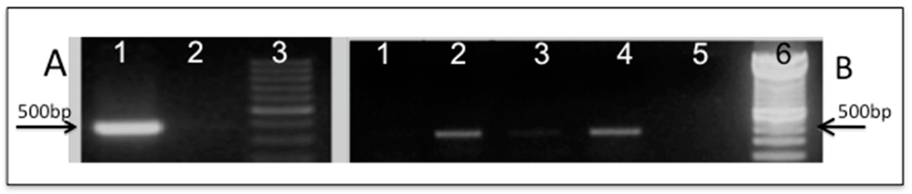
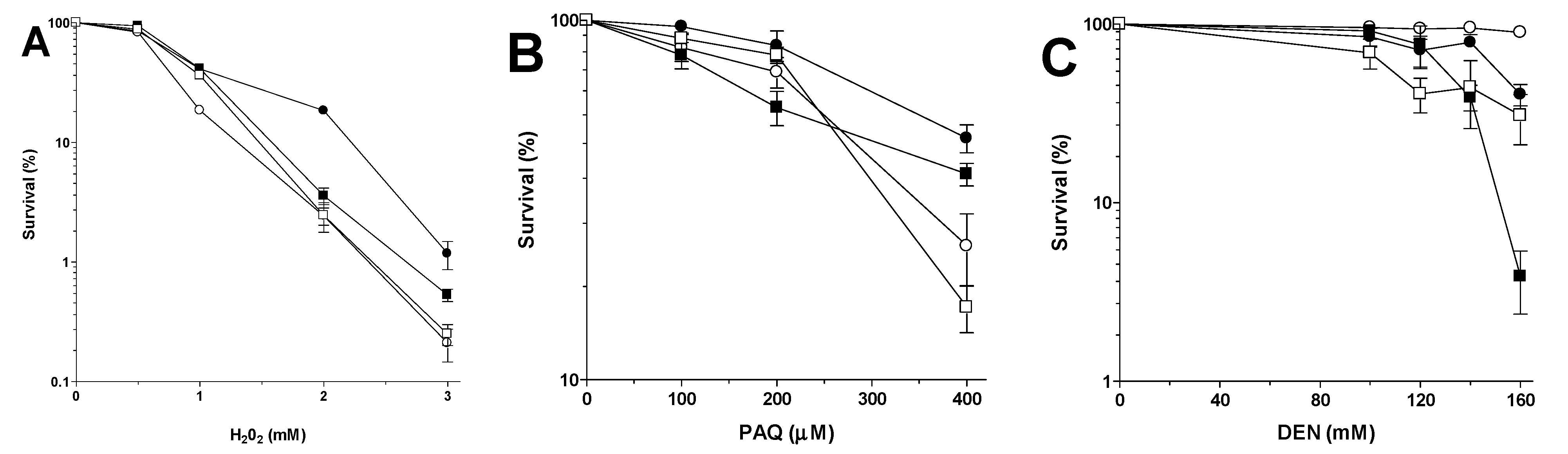
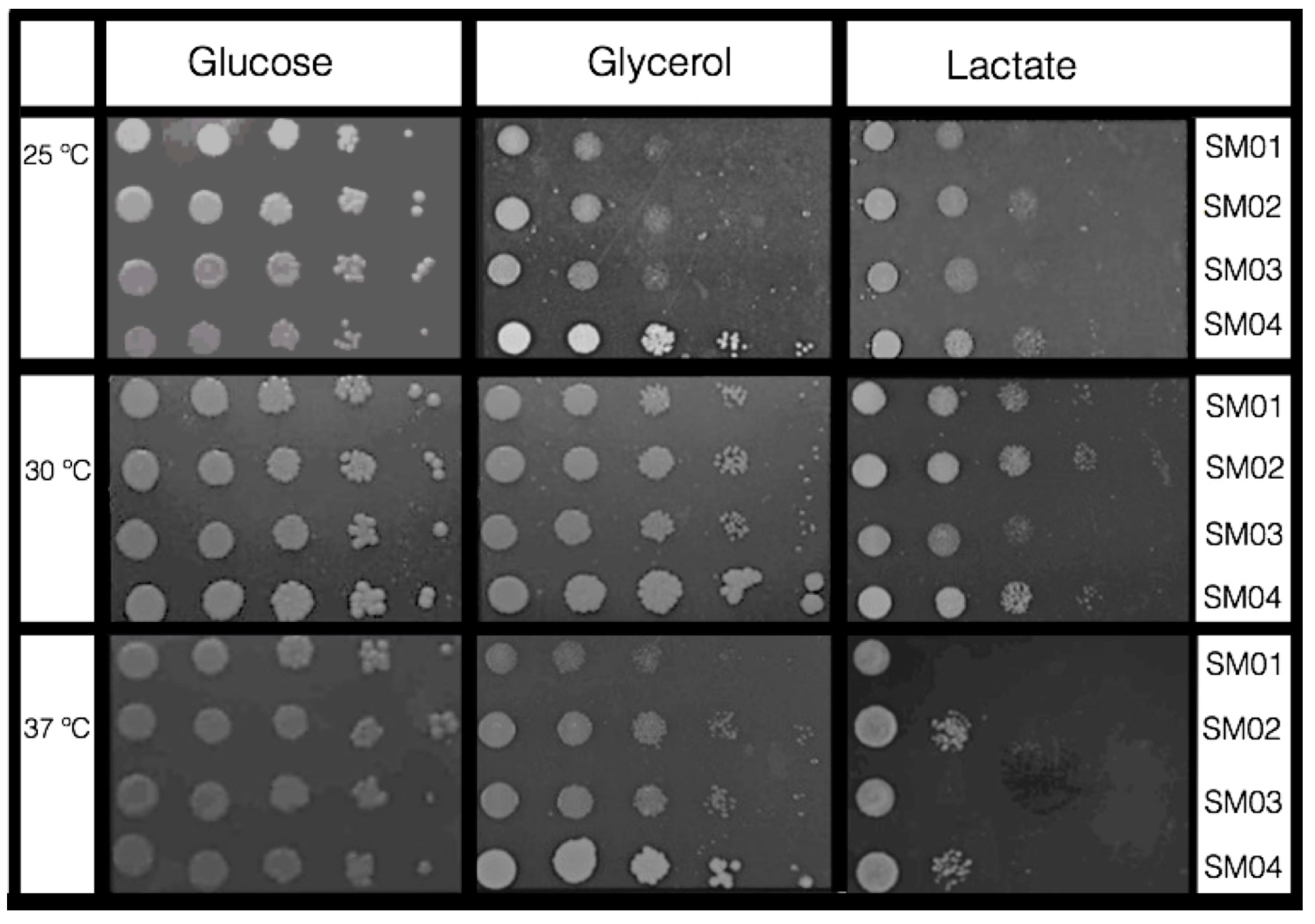
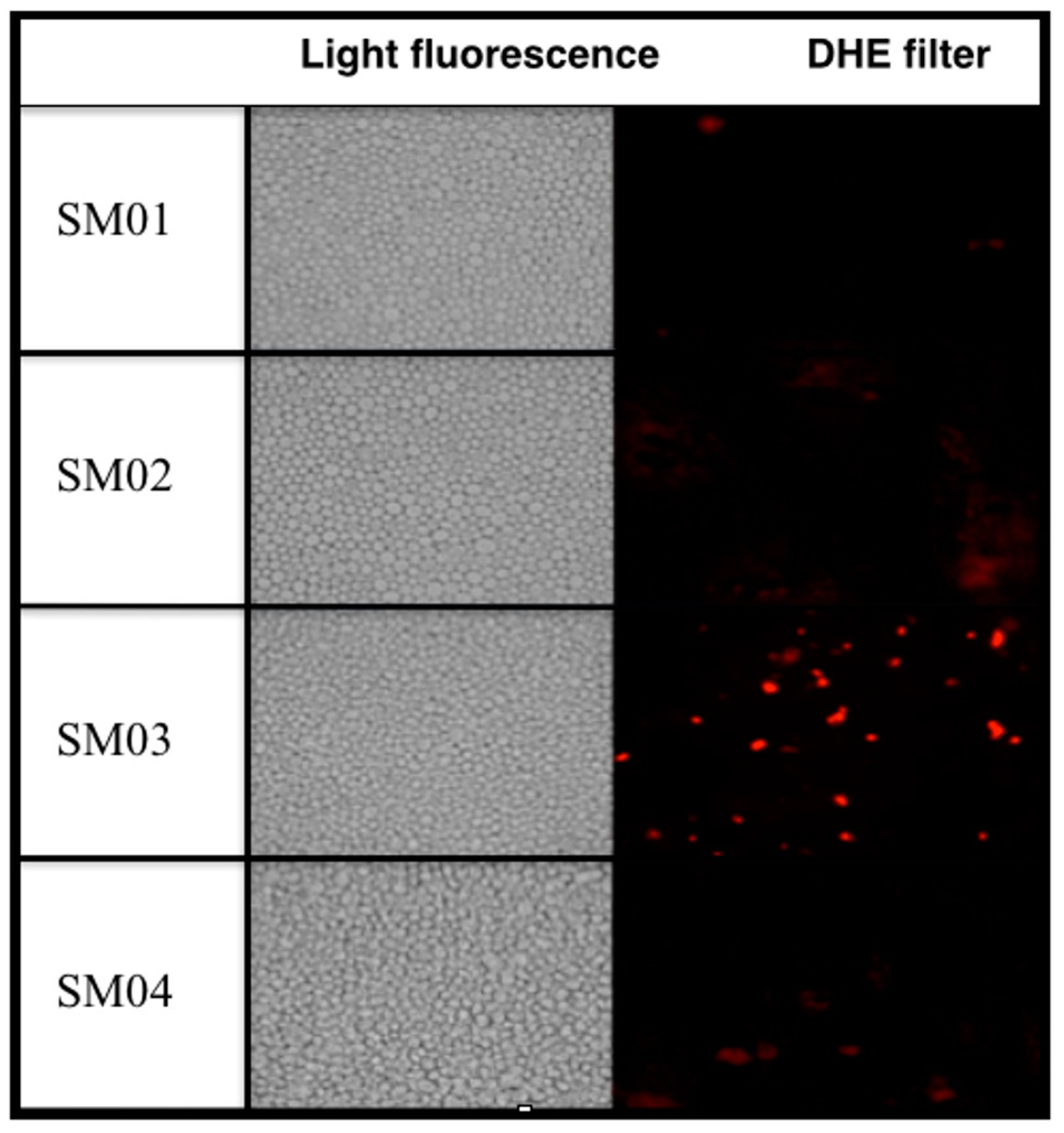
2.2. Functional Expression of MpSOD2 in S. cerevisiae sod2Δ Mutant

The putative MpSOD2 ORF was shown to be correctly expressed and the homology function confirmed by phenotypic complementation of the yeast sod2Δ mutant. Construction of the plasmid named pLBF01 containing putative the putative MpSOD2 gene followed by transformation of yeast mutant (Scsod2Δ) and its isogenic WT with both empty (pRS313) and MpSOD2-containing (pLBF01) plasmids (Figure 3A,B) yielded transformants named SM01, SM02, SM03 and SM04, respectively.
Figure 3. Molecular confirmation of plasmid constructions and yeast transformation by PCR: (A) Plasmid pLBF01 containing (1) MpSOD2; (2) pRS313; (3) molecular DNA marker (100 bp ladder); (B) Confirmation of yeast transformants containing either pRS313 or pLBF01 (PCR of DNA from each yeast clone): (1) SM01 (WT/pRS313); (2) SM02 (WT/pLBF01); (3) SM03 (Scsod2Δ/pRS313); (4) SM04 (Scsod2Δ/pLBF01); (5) no DNA added to reaction; (6) molecular DNA marker (100 bp ladder).
Ijms 16 12324 g003
Heterologous expression of genes in yeast has the advantage that the probably genetically best-studied eukaryotic organism is used for this purpose [58]. Two genes of Zygosaccharomyces rouxii (Z-SOD22 and Z-SOD2) were functionally expressed in a S. cerevisiae salt-sensitive mutant and the latter was shown to contribute to the halotolerance in Z. rouxii [59]. In addition, a S. cerevisiae sod1 mutant deficient in Cu/ZnSOD could be complemented by a MnSOD gene from Bacillus stearothermophilus [60].
Exposure of the four transformants to different mutagens allowed checking for phenotype complementation (Figure 4). The presence of MpSOD2 had no significant influence on the WT sensitivity phenotype (SM02) except when exposing the transformant to Paraquat (PAQ). Although the Scsod2Δ mutant is only slightly sensitive to H2O2 when compared to the WT (SM03 vs. SM01) the change of mutagen-sensitivity phenotype of pLBF01-transformed Scsod2Δ mutant (SM04) was always highly significant, except for UVC (Figure 4D, two-way analyses of variance—ANOVA). The MpSOD2-containing WT transformant had a significantly altered phenotype after exposure to PAQ and DEN (Figure 4B). WT and sod2Δ mutant transformed with empty pRS313 (SM01 and SM03) had the same phenotypes as non-transformed cells to all tested mutagens.
Figure 4. Sensitivity of stationary phase (STAT) cultures of yeast transformants after exposure to (A) H2O2; (B) Paraquat (PAQ); (C) DEN; and (D) UVC. Symbol legend: □ = WT(pRS313); ■ = WT(pLBF01); ○ = Scsod2Δ(pRS313); ● = Scsod2Δ(pLBF01). Statistical analyses of the influence of MpSOD2 on the sensitivity phenotype of (E) WT and Scsod2Δ mutant compared amongst themselves in each treatment; (F) WT compared to Scsod2Δ mutant in each treatment. ns = not significant; * p < 0.04; *** p < 0.001.
Ijms 16 12324 g004aIjms 16 12324 g004b
MpSOD2 containing yeast transformants SM02 and SM04 exhibited higher resistance to H2O2 and PAQ when compared to the non MpSOD2-containing strains SM01 and SM03. When exposed to H2O2 or PAQ non-MpSOD2-containing yeast transformants, SM01 and SM03 were as resistant as the non-transformed WT (Figure 4A). A similar response of H2O2 sensitivity is known for S. pombe where a sod2 deletion mutant also exhibits no clear H2O2 sensitivity phenotype [61]. However, when the WT and sod2Δ mutant were transformed with MpSOD2 (SM02 and SM04) they were slightly more resistant to H2O2 or PAQ than transformants harboring the empty vector (Figure 4A,B,E). Although yeast Sod2p is not directly involved in detoxification of H2O2 [20] introduction of a single copy of the MpSOD2 in the yeast mutant sod2Δ led to a significantly higher H2O2 and PAQ resistance as compared to that of the WT (Figure 4F).
This could be due to alterations in expression of ROS and to the level of anti-oxidant enzymes induced by an extra copy of MpSOD2. Guo et al. [62] proposed that over-expression of MnSOD in mammalian cells results in redox alterations with subsequent expression of stress-responsive nuclear genes. In addition, over-expression of human MnSOD cDNA led to transient increases in mRNA levels of endogenous sod2 and txn2 genes in mouse NIH/3T3 cells and smaller increases in MnSOD and thioredoxin 2 protein content [63]. When constitutive gene expression led to over expression of MnSOD, the respective clones not only had higher MnSOD levels but often also showed alterations of other anti-oxidant enzyme profiles. These changes were considered adaptive phenotypes of MnSOD-over-expressing cells, which had to accommodate the elevated H2O2 production due to increased MnSOD activity [63,64]. Yan et al. [65] also observed increased expression of the endogenous SOD2 gene in human lung fibroblasts following constitutive over-expression of exogenous SOD2 cDNA. In addition, high-level expression of MnSOD from multi-copy plasmids rendered S. pombe sod2 deletion mutant cells more resistant than the WT to superoxide-generating agents [61]. Therefore, the observed hyper-resistance phenotypes of yeast WT transformants induced by a single-copy MpSOD2 were expected.
Using the pro-mutagen DEN (that needs to be activated by redox cycling) we expected that either homologous or heterologous over expression of SOD2 should induce a DEN sensitivity phenotype [66]. Nitrosamines are considered highly mutagenic and carcinogenic through in vivo generated β-oxidized metabolites, which lead mainly to α-carbon derivatives that are incorporated into DNA as a chain-shortened methyl group [67]. β-hydroxynitrosamines undergo a chemical retro-aldol-like, base-induced cleavage to methylalkynitrosamine [68]. Oxidizing enzymes play a possible role in the chain shortening, thus producing an alkoxy radical at the 2-carbon of the nitrosamine chain, which in turn fragments into aldehydes and ketones and shorter alkyl radicals. The production of the latter could be mediated by an appropriate metallo-enzyme [68,69]. Absence of MnSOD in yeast mutant sod2Δ (SM03) led to a DEN hyper-resistance phenotype as compared to the respective WT (SM01) (Figure 4C) whereas one copy of MpSOD2 (SM04) rendered the sod2Δ transformants WT-like (SM01) and two copies of SOD2 genes in SM02, one from yeast, the other from M. perniciosa, conferred a DEN super-sensitivity phenotype (Figure 4C). Clearly, the presence of MnSOD, be it from yeast or from M. perniciosa, increased the DEN-induced cell damage which led to lower survival. In our Scsod2Δ transformant SM04, this metallo-enzyme apparently is MpSod2p and the diminished survival in the WT transformant SM02 could be due to increased DNA alkylation by α-carboxy anions.
Both transformed and non-transformed yeast cells showed no significant phenotypic differences after UVC irradiation (Figure 4D), indicating that the small amount of ROS produced by this radiation [70] does not need Sod2p for removal.
Another typical phenotype of Scsod2Δ is its sensitivity to lactate at different temperatures. Medium with glycerol or lactate shows that respiration on non-fermentable carbon sources generates superoxide radicals that inhibit the growth of cells lacking mitochondrial dismutase [71]. When we exposed the four yeast transformants to lactate, glucose and glycerol containing media at different growth temperatures (Figure 5), survival of the transformed yeast strains is influenced by the carbon source in the growth medium and the temperature.
Cell growth in 1% lactate was poor at all three incubation temperatures, especially at 37 °C. According to Krasowska et al. [71], lack of mitochondrial Sod2p in metabolism that relies on respiration of non-fermentable carbon sources generates growth-inhibiting superoxide radicals, especially in cells unable to cope with this oxidative stress. Clearly, the presence of functional MpSod2p in both SM02 and SM04 transformants enabled better growth and survival on lactate medium.
Cells grew well in glycerol (GLY) medium at 37 °C, especially the MpSOD2-containing SM02 and SM04 transformants (Figure 5). According to Whittaker [72] the mechanism of metal binding of MnSOD and FeSOD is strongly temperature-dependent: while incubation of the purified MnSOD apoprotein with metal salts at ambient temperatures did not restore SOD activity, re-activation could be achieved by heating the protein with Mn salts. MnSOD deficiency in S. pombe causes both osmotic and heat sensitivity, suggesting that in this organism MnSod2p plays a general role in protection against multiple stresses [61]. We may thus speculate that MpSod2p may have a general role in protection against heat sensitivity as well (Figure 4 and Figure 5; [22,28]), since functional over-expression of Z-SOD2 genes (in multi-copy plasmid) of Z. rouxii improved salt resistance of some S. cerevisiae WT under acidic conditions [73].
Figure 5. Temperature and carbon source-dependent growth of S. cerevisiae transformants harboring either plasmid pRS313 or pLBF01. Lines (fiveserial 1:10 dilutions): (1) SM01; (2) SM02; (3) SM03; (4) SM04.
Ijms 16 12324 g005
Survival strategies of fungal plant pathogens in hosts are conserved and ROS-protection mechanisms may be one of the primary lines of fungal defense, guaranteeing a successful infection [74]. SOD enzymes are amongst the most important antioxidant metallo-enzymes protecting cells against oxidative stress arising from ROS produced by aerobic metabolism [9,20,52,72]. In M. perniciosa, a hemibiotrofic phytopathogen of T. cacao, general acquired resistance of dikaryotic cells pre-grown in GLY or shifted from glucose (GLU) to glycerol (GLY) media to the same set of mutagens (except DEN) has been reported [35]. This phenomenon could be explained by a pre-conditioned cellular response to oxidative stress generated by the non-fermentable carbon source GLY [43] and up-regulated expression of MpSOD2 may have a biological role in resistance of this fungus during infection of T. cacao.
The phenotypic changes introduced by heterologous expression of MpSod2p in the sod2Δ yeast mutant are a strong indication that the MpSOD2 gene has the same biological function in M. perniciosa. Further biochemical analysis is however necessary to prove conserved enzyme function.
As expected transformants SM01, SM02 and SM04 did not demonstrate physiological alteration in the presence of the pro-mutagen dihydroethidium (DHE), which indicates that there is low oxidative stress since the experiment was performed at basal level. However, SM03 presented strong coloration demonstrating a high level of oxidative stress (Figure 6).
Figure 6. Visualization of oxidative stress via fluorescence microscopy of dihydroethidium (DHE)-stained strains.
Ijms 16 12324 g006

3. Experimental Section

3.1. Strains and Growth Conditions

Yeast and bacterial strains, as well as plasmids used in this study are listed in Table 1. Stock cultures were usually grown in YPD media (2% glucose, 2% peptone, 1% yeast extract; 2% agar added for solid medium) at 30 °C, 180 rpm, in a gyratory water-bath shaker (New Brunswick Scientific®, Enfield, CT, USA) for 2 days to stationary phase of growth (STAT) with a cell titer of approximately 2 × 10⁸/mL. Selective growth of transformants was in SynCo medium (0.16% yeast nitrogen base (United States Biological, Swampscott, MA, USA), 2% glucose, 0.5% ammonium sulfate) supplemented with the appropriate essential amino acids and bases (40 μg/mL). To ascertain respiratory competence and for elimination of accumulated petites, all strains were pre-grown on YPG media (1% yeast extract, 2% peptone, 2% glycerol) before being grown in liquid YPD (1% yeast extract, 2% peptone, 2% glucose). Media, solutions and buffers were prepared according to [75].
Table 1. Bacterial and yeast strains used in this study.
StrainsGenotypeSource
DH5αF-φ80lacZΔM15 Δ(lacZYA-argF)U169 deoR recA1 endA1 hsdR17(rk−, mk+) phoA supE44 thi-1 gyrA96 relA1 λInvitrogen
BY4742 (WT)MATα his3Δ1 leu2Δ0 lys2Δ0 ura3Δ0EUROSCARF
BY4741 ( sod2Δ)MATα his3Δ1 leu2Δ0 met15Δ0 ura3Δ0 SOD2: KanMX4EUROSCARF
SM01Same as BY4742 containing pRS313This study
SM02Same as BY4742 containing MpSD2This study
SM03Same as BY4741 containing pRS313This study
SM04Same as BY4741 containing MpSOD2This study
Plasmid NameRelevant Sequence IdentificationSource
pDNR-LibCLONTECH containing MpSOD2Acassia BL Pires
pRS313Single copy plasmid, HIS1protrotrophy[76]
pLBF01pRS313 MpSOD2This work

3.2. Identification of an MpSOD2 cDNA Clone and Sequence Analysis

MpSOD2 cDNA was identified from a previously constructed mycelial cDNA library in the pDNR-LIB plasmid using DB SMART Creator cDNA (Creator SMART cDNA Library Construction Kit, Clontech, Palo Alto, CA, USA) that had been derived from primordia and mature basidiomata [77] (plate identity: CP02-EC-001-002-C06-UE.F, clone C06). Based on its similarity to known SOD2 genes an open reading frame (ORF) analysis was performed (ORFinder program, Lasergene, Madison, WI, USA) followed by homology search with BLAST [78] against sequences in the NCBI database. ClustalW was used for multiple sequence alignment of amino acid sequence [79].
Phylogenetic analysis was by Neighbor-Joining method [58] using program MEGA4 [80]. The bootstrap consensus tree was inferred from 1000 replicates [81]. Evolutionary distances were estimated using the Poisson correction method [82]. We eliminated all positions containing gaps and missing data from the analysis (complete deletion option). There were 93 positions in the final dataset. Phylogenetic analyses were conducted in MEGA4. The sequence CDS was predicted by Augustus Gene Predictor v2.7 (Department of Bioinformatics, University of Göttingen, Göttingen, Alemanha) [83].

3.3. Amplification of MpSOD2 and Sub-Cloning Plasmid pRS313 (Yeast Centromere Vector with a HIS3 Marker and a Multiple Cloning Site)

Bacterial clone C06 containing MpSOD2 was grown overnight in LBC medium (NaCl 1%, tryptone 1%, yeast extract 0.5%; 34 μg/L chloramphenicol) and plasmid DNA was extracted via alkaline lysis [41]. The entire putative MpSOD2 gene was amplified with specific primers of pDNR-LIB (Primer sequence M13F: 5ʹ-CGCCAGGGTTTTCCCAGTCACGAC-3ʹ, M13R: 5ʹ-TCACACAGGAAACAGCTATGAC-3ʹ). DNA was amplified in a thermo cycler (Mastercycler Eppendorf®, Hamburg, Germany) at 94 °C for 5 min (1 cycle); 94 °C for 1 min, 58 °C for 1 min and 72 °C for 1 min (30 cycles) and 72 °C for 7 min (1 cycle).
Amplification products were run on 1% Tris-borate-EDTA (TBE)-agarose gel followed by fragment purification with the QIAquick Gel Extraction Kit, Qiagen (Venlo, Limburg, The Netherlands). Both amplified fragments of MpSOD2 and shuttle vector pRS313 were digested with restriction enzyme SmaI (Fermentas International, Vilnius, Lithuania, for 16 h at 30 °C), followed by a treatment for 4–12 h at 11 °C with Klenow enzyme (Applied Biosystems®, Life Technologies, Waltham, MA, USA) and incubated for 1 h at 16 °C with T4 DNA Ligase (Fermentas International) for blunt-end ligation. The entire reaction mixture was incubated with competent DH5α cells (Invitrogen™, Life Technologies, Waltham, MA, USA) and transformants were selected by blue/white screening. Putative clones were individually grown on LBA solid media and confirmed by a PCR run of each colony. Then each candidate was grown in LBA medium (NaCl 1%, tryptone 1%, yeast extract 0.5%; 50 μg/L ampicillin) for 13–18 h at 37 °C at 200 rpm agitation. DNA was extracted from each putative clone [84] and MpSOD2-containing clones confirmed via PCR (internal primers of sequence MpSOD2F: 5ʹ-TGCTCTCGAGCCCTACATTT-3ʹ, MpSOD2R: 5ʹ-AACGCTTAGTGTCCGGTTTG-3ʹ; conditions of PCR as described above). The confirmed construct of plasmid pRS313 (MpSOD2) was named pLBF01.

3.4. Cloning of pRS313 (MpSOD2) in S. cerevisiae Yeast Mutant

Yeast mutant Scsod2Δ and its isogenic WT were transformed with pLBF01 by the LiAc/SS-DNA/PEG method [85] and putative transformants were selected in SynCo medium lacking histidine (SC-His). Selected transformants were tested for their sensitivity to paraquat, H2O2, diethylnitrosamine, and UVC (conditions below).

3.5. Mutagen Exposure and Cell Survival

Sensitivity of S. cerevisiae/MpSOD2 transformants to different mutagens was determined as described by [86] on SC-His media supplemented (at the time of pouring the liquid agar media) with the following oxidative stress-inducing chemicals: paraquat (PAQ, 100 to 400 μM), hydrogen peroxide (H2O2, 1 to 3 mM), diethylnitrosamine (DEN, 100 to 160 mM). We determined sensitivity to UVC radiation by irradiation of agar-plated tranformant cells with exposure doses between 0 and 150 J/m2 (Spectrolinker, Spectronics Corp., Westburg, NY, USA). Results are expressed as the percentage of survival related to the untreated controls and are the mean of at least three independent experiments. The error bars represent standard deviation as calculated by the GraphPad Prism® program (GraphPad Software Inc., San Diego, CA, USA). Yeast transformants drop tests show cell growth in each 5 μL drops of a serial 1:10 dilution (107 to 103 cells/mL) placed on SC-His media containing one of the following carbon sources: glucose, glycerol or lactate (2%). Incubation was for 5 days at 3 different temperatures, 25, 30 and 37 °C. Photos represent one of at least three triplicates.

3.6. Fluorescence Assay

A stock solution (1 mg/mL) of the fluorogenic probe dihydroethidium (DHE, Sigma Aldrich, St. Louis, MO, USA) was prepared by dissolving it in dimethyl sulfoxide (Sigma Aldrich, St. Louis, MO, USA). One mL of cells in LOG phase was stained by addition of 1 μL of stock solution and mixed by inversion, then incubated for 30 min 35 °C, washed 3 times with saline, and finally resuspended in 100 μL. An aliquot was used to check oxidative/reductive stress of the cells. Cytosolic DHE when oxidized by ROS (singlet oxygen, hydroxyl radicals, superoxide, hydroperoxides and peroxides) yields ethidium, which intercalates with a cell’s DNA and fluoresces bright red (605 nm) [87] when observed under fluorescence microscope DMRA2 (Leica, Wetzlar, Germany) attached with DHE filter. Images were captured using a 40× objective under bright field as well as under fluorescent filters using the IM50 software (Leica). Photos represent cells from at least three independent experiments.

4. Conclusions

In this work, the putative MpSOD2 gene was cloned using standard molecular biology tools and the phenotypical complementation of the yeast mutant sod2Δ was demonstrated via classical genetics. The clone containing MpSOD2 gene, named SM04, showed increased resistance phenotypes against oxidative stress-inducing H2O2 and paraquat, and enhanced growth at 37 °C on the non-fermentable carbon source lactate. Surprisingly, the presence of the SOD2 gene either from yeast or from M. perniciosa, rendered higher sensitivity of the cells against treatment with the pro-mutagen diethylnitrosamine (DEN) that requires oxidation to become an active mutagen/carcinogen. Absence of MpSOD2 in the yeast sod2Δ mutant led to DEN hyper-resistance while introduction of a single copy of this gene restored the yeast wild-type phenotype. Fluorescence assay using DHE was performed to observe basal levels of yeast cell oxidative stress and results demonstrated that the sod2Δ mutant has a much higher level of intrinsic oxidative stress, which could be abolished by introducing MpSOD2. Taken together, this data indicates that MpSod2p is functionally expressed in yeast S. cerevisiae. This is the first report of in vivo functional expression of a M. perniciosa gene.

Acknowledgments

Research supported by grants from Conselho Nacional de Desenvolvimento Científico e Tecnológico (CNPq). We thank Acassia Pires Leal for access to her cDNA library, Marco Antonio Costa for help in phylogenetic analysis and Ramon Vidal for construction of Figure 2. Regineide X. Santos held a CNPq doctoral fellowship and Ana C. Melgaço was a CNPq scientific initiation fellow. Martin Brendel held a Fundação de Amparo à Pesquisa do Estado da Bahia (FAPESB) fellowship and Cristina Pungarntik a CNPq fellowship. We thank Ramon Vidal and Eduardo Mangabeira for their contribution to bioinformatics analyses.

Author Contributions

Sônia C. Melo, Regineide X. Santos, Ana C. Melgaço and Alanna C. F. Pereira performed the experiments and analyzed data; Cristina Pungartnik and Martin Brendel conceived and designed the experiments and wrote the paper.

Conflicts of Interest

The authors declare no conflict of interest.

References

  1. Boveris, A.; Chance, B. The mitochondrial generation of hydrogen peroxide. General properties and effect of hyperbaric oxygen. Biochem. J. 1973, 134, 707–716. [Google Scholar] [PubMed]
  2. França, M.B.; Panek, A.D.; Eleutherio, E.C.A. Oxidative stress and its effects during dehydration. Comp. Biochem. Physiol. 2007, 146, 621–631. [Google Scholar] [CrossRef] [PubMed]
  3. Fuchs-Tarlovsky, V. Role of antioxidants in cancer therapy. Nutrition 2012, 29, 15–21. [Google Scholar] [CrossRef] [PubMed]
  4. Blokhina, O.; Virolainen, E.; Fagerstedt, K.V. Antioxidants, oxidative damage and oxygen deprivation stress: A review. Ann. Bot. 2003, 91, 179–194. [Google Scholar] [CrossRef] [PubMed]
  5. Da Costa, L.A.; Badawi, A.; El-Sohemy, A. Nutrigenetics and modulation of oxidative stress. Ann. Nutr. Metab. 2012, 60, 27–36. [Google Scholar]
  6. Culotta, V.C.; Daly, M.J. Manganese complexes: Diverse metabolic routes to oxidative stress resistance in prokaryotes and yeast. Antioxid. Redox Signal. 2013, 19, 933–944. [Google Scholar] [CrossRef] [PubMed]
  7. Jamieson, D.J. Oxidative stress responses of the yeast Saccharomyces cerevisiae. Yeast 1998, 14, 1511–1527. [Google Scholar] [CrossRef]
  8. Pereira, M.D.; Herdeiro, R.S.; Fernandes, P.N.; Eleutherio, E.C.A.; Panek, A.D. Targets of oxidative stress in yeast sod mutants. Biochim. Biophys. Acta 2003, 1620, 245–251. [Google Scholar] [CrossRef]
  9. Zelko, I.N.; Mariani, T.J.; Folz, R.J. Superoxide dismutase multigene family: A comparison of the CuZn-SOD (SOD1), Mn-SOD (SOD2), and EC-SOD (SOD3) gene structures, evolution, and expression. Free Radic. Biol. Med. 2002, 33, 337–349. [Google Scholar] [CrossRef]
  10. Miller, A.F. Superoxide dismutases: Ancient enzymes and new insights. FEBS Lett. 2012, 586, 585–595. [Google Scholar] [CrossRef] [PubMed]
  11. Gessler, N.N.; Aver’yanov, A.A.; Belozerskaya, T.A. Reactive oxygen species in regulation of fungal development. Biochemistry (Mosc) 2007, 72, 1091–1109. [Google Scholar] [CrossRef] [PubMed]
  12. Ataya, F.S.; Fouad, D.; Al-Olayan, E.; Malik, A. Molecular cloning, characterization and predicted structure of a putative copper-zinc SOD from the camel, Camelus dromedaries. Int. J. Mol. Sci. 2012, 13, 879–900. [Google Scholar] [CrossRef] [PubMed]
  13. Chary, P.; Dillon, D.; Schroeder, A.L.; Natvig, D.O. Superoxide dismutase (sod-1) null mutants of Neurospora crassa: Oxidative stress sensitivity, spontaneous mutation rate and response to mutagens. Genetics 1994, 137, 723–730. [Google Scholar] [PubMed]
  14. Crameri, R.; Faith, A.; Hemmann, S.; Jaussi, R.; Ismail, C.; Menz, G.; Blaser, K. Humoral and cell-mediated autoimmunity in allergy to Aspergillus fumigatus. J. Exp. Med. 1996, 184, 265–270. [Google Scholar] [CrossRef] [PubMed]
  15. Fang, G.C.; Hanau, R.M.; Vaillancourt, L.J. The SOD2 gene, encoding a manganese-type superoxide dismutase, is up-regulated during conidiogenesis in the plant-pathogenic fungus Colletotrichum graminicola. Fungal Genet. Biol. 2002, 36, 155–165. [Google Scholar] [CrossRef]
  16. Fernandez, J.; Marroquin-guzman, M.; Nandakumar, R.; Shijo, S.; Cornwell, K.M.; Li, G.; Wilson, R.A. Plant defence suppression is mediated by a fungal sirtuin during rice infection by Magnaporthe oryzae. Mol. Microbiol. 2014, 94, 70–88. [Google Scholar] [CrossRef] [PubMed]
  17. Liu, X.F.; Elashvili, I.; Gralla, E.B.; Valentine, J.S.; Lapinskas, P.; Culotta, V.C. Yeast lacking superoxide dismutase: Isolation of genetic suppressors. J. Biol. Chem. 1992, 267, 18298–18302. [Google Scholar] [PubMed]
  18. Barnese, K.; Sheng, Y.; Stich, T.A.; Gralla, E.B.; David Britt, R.; Cabelli, D.E.; Valentine, J.S. Investigation of the highly active manganese superoxide dismutase from Saccharomyces cerevisiae. J. Am. Chem. Soc. 2010, 132, 12525–12527. [Google Scholar] [CrossRef] [PubMed]
  19. Weisiger, R.A.; Fridovich, I. Superoxide dismutase. Organelle specificity. J. Biol. Chem. 1973, 248, 3582–3592. [Google Scholar] [PubMed]
  20. Culotta, V.C.; Yang, M.; O’Halloran, T.V. Activation of superoxide dismutases: Putting the metal to the pedal. Biochim. Biophys. Acta 2006, 1763, 747–758. [Google Scholar] [CrossRef] [PubMed]
  21. Li, Y.; Copin, J.C.; Reola, L.F.; Calagui, B.; Gobbel, G.T.; Chen, S.F.; Sato, S.; Epstein, C.J.; Chan, P.H. Reduced mitochondrial manganese-superoxide dismutase activity exacerbates glutamate toxicity in cultured mouse cortical neurons. Brain Res. 1998, 814, 164–170. [Google Scholar] [CrossRef]
  22. Kim, J.; Choi, B.H.; Jang, K.L.; Min, D.S. Phospholipase D activity is elevated in hepatitis C virus core protein-transformed NIH3T3 mouse fibroblast cells. Exp. Mol. Med. 2004, 36, 454–460. [Google Scholar] [CrossRef] [PubMed]
  23. Li, F.; Shi, H.Q.; Ying, S.H.; Feng, M.G. Distinct contributions of one Fe- and two Cu/Zn-cofactored super oxide dismutases to antioxidation, UV tolerance and virulence of Beauveria bassiana. Fungal Genet. Biol. 2014. [Google Scholar] [CrossRef]
  24. Ding, C.; Festa, R.A.; Sun, T.S.; Wang, Z.Y. Iron and copper as virulence modulators in human fungal pathogens. Mol. Microbiol. 2014, 93, 10–23. [Google Scholar] [CrossRef] [PubMed]
  25. Hwang, C.S.; Rhie, G.E.; Oh, J.H.; Huh, W.K.; Yim, H.S.; Kang, S.O. Copper- and zinc-containing superoxide dismutase (Cu/ZnSOD) is required for the protection of Candida albicans against oxidative stresses and the expression of its full virulence. Microbiology 2002, 148, 3705–3713. [Google Scholar] [PubMed]
  26. Cox, G.M.; Harrison, T.S.; Mcdade, H.C.; Taborda, C.P.; Heinrich, G.; Casadevall, A.; Perfect, J.R. Superoxide dismutase in uences the virulence of. Infect. Immun. 2003, 71, 173–180. [Google Scholar] [CrossRef] [PubMed]
  27. Lambou, K.; Lamarre, C.; Beau, R.; Dufour, N.; Latge, J. Functional analysis of the superoxide dismutase family in Aspergillus fumigatus. Mol. Microbiol. 2010, 75, 910–923. [Google Scholar] [CrossRef] [PubMed]
  28. Briones-Martin-del-Campo, M.; Orta-Zavalza, E.; Cañas-Villamar, I.; Gutiérrez-Escobedo, G.; Juárez-Cepeda, J.; Robledo-Márquez, K.; Arroyo-Helguera, O.; Castaño, I.; de las Penas, A. The superoxide dismutases of Candida glabrata protect against oxidative damage and are required for lysine biosynthesis, DNA integrity and chronological life survival. Microbiology 2015, 161, 300–310. [Google Scholar] [CrossRef] [PubMed]
  29. Muid, K.A.; Karakaya, H.C.; Koc, A. Absence of superoxide dismutase activity causes nuclear DNA fragmentation during the aging process. Biochem. Biophys. Res. Commun. 2014, 444, 260–263. [Google Scholar] [CrossRef] [PubMed]
  30. Baron, J.A.; Laws, K.M.; Chen, J.S.; Culotta, V.C. Superoxide triggers an acid burst in Saccharomyces cerevisiae to condition the environment of glucose-starved cells. J. Biol. Chem. 2013, 288, 4557–4566. [Google Scholar] [CrossRef] [PubMed]
  31. Ullah, A.; El-Magd, R.A.; Fliegel, L. Functional role and analysis of cysteine residues of the salt tolerance protein Sod. Mol. Cell. Biochem. 2014, 386, 85–98. [Google Scholar] [CrossRef] [PubMed]
  32. Gesteira, A.S.; Micheli, F.; Carels, N.; da Silva, A.C.; Gramacho, K.P.; Schuster, I.; Macedo, J.N.; Pereira, G.A.G.; Cascardo, J.C.M. Comparative analysis of expressed genes from cacao meristems infected by Moniliophthora perniciosa. Ann. Bot. 2007, 100, 129–140. [Google Scholar] [CrossRef] [PubMed]
  33. Lamb, C.; Dixon, R.A. The oxidative burst in plant disease resistance. Annu. Rev. Plant Physiol. Plant Mol. Biol. 1997, 48, 251–275. [Google Scholar] [CrossRef] [PubMed]
  34. Campos, E.G.; Jesuino, R.S.; Dantas Ada, S.; Brigido Mde, M.; Felipe, M.S. Oxidative stress response in Paracoccidioides brasiliensis. Genet. Mol. Res. 2005, 4, 409–429. [Google Scholar] [PubMed]
  35. Santos, R.X.; Melo, S.C.O.; Cascardo, J.C.M.; Brendel, M.; Pungartnik, C. Carbon source-dependent variation of acquired mutagen resistance of Moniliophthora perniciosa: Similarities in natural and artificial systems. Fungal Genet. Biol. 2008, 45, 851–860. [Google Scholar] [CrossRef] [PubMed]
  36. Ramegowda, V.; Senthil-Kumar, M.; Ishiga, Y.; Kaundal, A.; Udayakumar, M.; Mysore, K.S. Drought stress acclimation imparts tolerance to Sclerotinia sclerotiorum and Pseudomonas syringae in Nicotiana benthamiana. Int. J. Mol. Sci. 2013, 14, 9497–9513. [Google Scholar] [CrossRef] [PubMed]
  37. Frias, G.A.; Purdy, L.H.; Schmidt, R.A. Infection biology of Crinipellis perniciosa on vegetative flushes of cacao. Plant Dis. 1991, 79, 787–791. [Google Scholar] [CrossRef]
  38. Kilaru, A.; Hasenstein, K.H. Development and Pathogenicity of the fungus Crinipellis perniciosa on interaction with cacao leaves. Phytopathology 2005, 95, 101–107. [Google Scholar] [CrossRef] [PubMed]
  39. Scarpari, L.M.; Meinhardt, L.W.; Mazzafera, P.; Pomella, A.W.; Schiavinato, M.A.; Cascardo, J.C.; Pereira, G.A. Biochemical changes during the development of witches broom: The most important diseases of cocoa in Brazil caused by Crinipellis perniciosa. J. Exp. Bot. 2005, 56, 413–865. [Google Scholar] [CrossRef] [PubMed]
  40. De Oliveira Ceita, G.; Macêdo, J.N.A.; Santos, T.B.; Alemanno, L.; da Silva Gesteira, A.; Micheli, F.; Mariano, A.C.; Gramacho, K.P.; da Costa Silva, D.; Meinhardt, L.; et al. Involvement of calcium oxalate degradation during programmed cell death in Theobroma cacao tissues triggered by the hemibiotrophic fungus Moniliophthora perniciosa. Plant Sci. 2007, 173, 106–117. [Google Scholar] [CrossRef]
  41. Evans, H.C. Pleomorphism in Crinipellis perniciosa, causal agent of witches’ broom disease of cocoa. Trans. Br. Mycol. Soc. 1980, 74, 515–523. [Google Scholar] [CrossRef]
  42. Calle, P.P.; Seagars, D.J.; McClave, C.; Senne, D.; House, C.; House, J.A. Viral and bacterial serology of six free-ranging bearded seals Erignathus barbatus. Dis. Aquat. Organ. 2008, 81, 77–80. [Google Scholar] [CrossRef] [PubMed]
  43. Pungartnik, C.; Melo, S.C.O.; Basso, T.S.; Macena, W.G.; Cascardo, J.C.M.; Brendel, M. Reactive oxygen species and autophagy play a role in survival and differentiation of the phytopathogen Moniliophthora perniciosa. Fungal Genet. Biol. 2009, 46, 461–472. [Google Scholar] [CrossRef] [PubMed]
  44. Gratão, P.L.; Polle, A.; Lea, P.J.; Azevedo, R.A. Making the life of heavy metal-stressed plants a little easier. Funct. Plant Biol. 2005, 32, 481–494. [Google Scholar] [CrossRef]
  45. Meinhardt, L.W.; Costa, G.G.L.; Thomazella, D.P.T.; Teixeira, P.J.P.L.; Carazzolle, M.F.; Schuster, S.C.; Carlson, J.E.; Guiltinan, M.J.; Mieczkowski, P.; Farmer, A.; et al. Genome and secretome analysis of the hemibiotrophic fungal pathogen, Moniliophthora roreri, which causes frosty pod rot disease of cacao: Mechanisms of the biotrophic and necrotrophic phases. BMC Genomics 2014, 15, 164. [Google Scholar] [CrossRef] [PubMed]
  46. Sheng, Y.; Stich, T.A.; Barnese, K.; Gralla, E.B.; Cascio, D.; Britt, R.D.; Cabelli, D.E.; Valentine, J.S. Comparison of two yeast mnsods: Mitochondrial Saccharomyces cerevisiae versus cytosolic Candida albicans. J. Am. Chem. Soc. 2011, 133, 20878–20889. [Google Scholar] [CrossRef] [PubMed]
  47. Díez, B.; Schleissner, C.; Moreno, M.A.; Rodríguez, M.; Collados, A.; Barredo, J.L. The manganese superoxide dismutase from the penicillin producer Penicillium chrysogenum. Curr. Genet. 1998, 33, 387–394. [Google Scholar] [PubMed]
  48. Pan, S.M.; Ye, J.S.; Hseu, R.S. Purification and characterization of manganese superoxide dismutase from Ganoderma microsporum. Biochem. Mol. Biol. Int. 1997, 42, 1035–1043. [Google Scholar] [CrossRef] [PubMed]
  49. Kozak, M. Point mutations define a sequence flanking the AUG initiator codon that modulates translation by eukaryotic ribosomes. Cell 1986, 44, 283–292. [Google Scholar] [CrossRef]
  50. Carlioz, A.; Ludwig, M.L.; Stallings, W.C.; Fee, J.A.; Steinman, H.M.; Touati, D. Iron superoxide dismutase. Nucleotide sequence of the gene from Escherichia coli K12 and correlations with crystal structures. J. Biol. Chem. 1988, 263, 1555–1562. [Google Scholar] [PubMed]
  51. Borgstahl, G.E.; Parge, H.E.; Hickey, M.J.; Beyer, W.F.; Hallewell, R.A.; Tainer, J.A. The structure of human mitochondrial manganese superoxide dismutase reveals a novel tetrameric interface of two 4-helix bundles. Cell 1992, 71, 107–118. [Google Scholar] [CrossRef]
  52. Landis, G.N.; Tower, J. Superoxide dismutase evolution and life span regulation. Mech. Ageing Dev. 2005, 126, 365–379. [Google Scholar] [CrossRef] [PubMed]
  53. Parker, M.W.; Blake, C.C. Iron- and manganese-containing superoxide dismutases can be distinguished by analysis of their primary structures. FEBS Lett. 1988, 229, 377–382. [Google Scholar] [CrossRef]
  54. Van Camp, W.; Bowler, C.; Villarroel, R.; Tsang, E.W.; van Montagu, M.; Inzé, D. Characterization of iron superoxide dismutase cDNAs from plants obtained by genetic complementation in Escherichia coli. Proc. Natl. Acad. Sci. USA 1990, 87, 9903–9907. [Google Scholar] [CrossRef] [PubMed]
  55. Hurt, E.; Vanloon, A. How proteins find mitochondria and intramitochondrial compartments. Trends Biochem. Sci. 1986, 11, 204–207. [Google Scholar] [CrossRef]
  56. Avise, J.C. Molecular Markers, Natural History, and Evolution; Sinauer: Sunderland, MA, USA, 2004; p. 684. [Google Scholar]
  57. Saitou, N.; Nei, M. The neighbor-joining method: A new method for reconstructing phylogenetic trees. Mol. Biol. Evol. 1987, 4, 406–425. [Google Scholar] [PubMed]
  58. Lushchak, V.I. Budding yeast Saccharomyces cerevisiae as a model to study oxidative modification of proteins in eukaryotes. Acta Biochim. Pol. 2006, 53, 679–684. [Google Scholar] [PubMed]
  59. Iwaki, T.; Higashida, Y.; Tsuji, H.; Tamai, Y.; Watanabe, Y. Characterization of a second gene (ZSOD22) of Na+/H+ antiporter from salt-tolerant yeast Zygosaccharomyces rouxii and functional expression of ZSOD2 and ZSOD22 in Saccharomyces cerevisiae. Yeast 1998, 14, 1167–1174. [Google Scholar] [CrossRef]
  60. Bowler, C.; van Kaer, L.; van Camp, W.; van Montagu, M.; Inze, D.; Dhaese, P. Characterization of the Bacillus stearothermophilus manganese superoxide dismutase gene and its ability to complement copper/zinc superoxide dismutase deficiency in Saccharomyces cerevisiae. J. Bacteriol. 1990, 172, 1539–1546. [Google Scholar] [PubMed]
  61. Jeong, J.H.; Kwon, E.S.; Roe, J.H. Characterization of the manganese-containing superoxide dismutase and its gene regulation in stress response of Schizosaccharomyces pombe. Biochem. Biophys. Res. Commun. 2001, 283, 908–914. [Google Scholar] [CrossRef] [PubMed]
  62. Guo, G.; Yan-Sanders, Y.; Lyn-Cook, B.D.; Wang, T.; Tamae, D.; Ogi, J.; Khaletskiy, A.; Li, Z.; Weydert, C.; Longmate, J.A.; et al. Manganese superoxide dismutase-mediated gene expression in radiation-induced adaptive responses. Mol. Cell. Biol. 2003, 23, 2362–2378. [Google Scholar] [CrossRef] [PubMed]
  63. Kim, A.; Murphy, M.P.; Oberley, T.D. Mitochondrial redox state regulates transcription of the nuclear-encoded mitochondrial protein manganese superoxide dismutase: A proposed adaptive response to mitochondrial redox imbalance. Free Radic. Biol. Med. 2005, 38, 644–654. [Google Scholar] [CrossRef] [PubMed]
  64. Li, N.; Oberley, T.D. Modulation of antioxidant enzymes, reactive oxygen species, and glutathione levels in manganese superoxide dismutase- overexpressing NIH/3T3 fibroblasts during the cell cycle. J. Cell. Physiol. 1998, 177, 148–160. [Google Scholar] [CrossRef]
  65. Yan, T.; Oberley, L.W.; Zhong, W.; St. Clair, D.K. Manganese-containing superoxide dismutase overexpression causes phenotypic reversion in SV40-transformed human lung fibroblasts. Cancer Res. 1996, 56, 2864–2871. [Google Scholar] [PubMed]
  66. Pungartnik, C.; Picada, J.; Brendel, M.; Henriques, J.A. Further phenotypic characterization of pso mutants of Saccharomyces cerevisiae with respect to DNA repair and response to oxidative stress. Genet. Mol. Res. 2002, 1, 79–89. [Google Scholar] [PubMed]
  67. Krueger, F.W. Metabolism of nitrosamines in vivo. I. Evidence for -oxydation of aliphatic di-N-alkylnitrosamines: The stimultaneous formation of 7-methylguanine besides 7-propyl- or 7-butylguanine after application of di-N-propyl- or di-N-butylnitrosamine. Z. Krebsforsch. Klin. Onkol. Cancer Res. Clin. Oncol. 1971, 76, 1945–1954. [Google Scholar]
  68. Loeppky, R.N.; Li, Y.E. Nitrosamine activation and detoxication through free radicals and their derived cations. IARC Sci. Publ. 1991, 105, 375–382. [Google Scholar] [PubMed]
  69. D’Ischia, M.; Napolitano, A.; Manini, P.; Panzella, L. Secondary targets of nitrite-derived reactive nitrogen species: Nitrosation/nitration pathways, antioxidant defense mechanisms and toxicological implications. Chem. Res. Toxicol. 2011, 24, 2071–2092. [Google Scholar] [CrossRef] [PubMed]
  70. Ewing, D.; Jones, S.R. Superoxide removal and radiation protection in bacteria. Arch. Biochem. Biophys. 1987, 254, 53–62. [Google Scholar] [CrossRef]
  71. Krasowska, A.; Dziadkowiec, D.; Lukaszewicz, M.; Wojtowicz, K.; Sigler, K. Effect of antioxidants on Saccharomyces cerevisiae mutants deficient in superoxide dismutases. Folia Microbiol. (Praha) 2003, 48, 754–760. [Google Scholar] [CrossRef] [PubMed]
  72. Whittaker, J.W. The irony of manganese superoxide dismutase. Biochem. Soc. Trans. 2003, 31, 1318–1321. [Google Scholar] [CrossRef] [PubMed]
  73. Kinclová, O.; Potier, S.; Sychrová, H. The Zygosaccharomyces rouxii strain CBS732 contains only one copy of the HOG1 and the SOD2 genes. J. Biotechnol. 2001, 88, 151–158. [Google Scholar] [CrossRef]
  74. Kotchoni, S.O.; Kuhns, C.; Ditzer, A.; Kirch, H.H.; Bartels, D. Over-expression of different aldehyde dehydrogenase genes in Arabidopsis thaliana confers tolerance to abiotic stress and protects plants against lipid peroxidation and oxidative stress. Plant Cell. Environ. 2006, 29, 1033–1048. [Google Scholar] [CrossRef] [PubMed]
  75. Amberg, D.C.; Burke, D.J.; Strathern, J.N. Methods in Yeast Genetics: A Cold Spring Harbor Laboratory Course Manual; Cold Spring Harbor New York, Ed.; Cold Spring Harbor Laboratory Press: New York, NY, USA, 2005; p. 205. [Google Scholar]
  76. Sikorski, R.S.; Hieter, P. A system of shuttle vectors and yeast host strains designed for efficient manipulation of DNA in Saccharomyces cerevisiae. Genetics 1989, 122, 19–27. [Google Scholar] [PubMed]
  77. Pires, A.B.L.; Gramacho, K.P.; Silva, D.C.; Góes-Neto, A.; Silva, M.M.; Muniz-Sobrinho, J.S.; Porto, R.F.; Villela-Dias, C.; Brendel, M.; Cascardo, J.C.M.; et al. Early development of Moniliophthora perniciosa basidiomata and developmentally regulated genes. BMC Microbiol. 2009, 9, 158. [Google Scholar] [CrossRef] [PubMed]
  78. Altschul, S.F.; Madden, T.L.; Schäffer, A.A.; Zhang, J.; Zhang, Z.; Miller, W.; Lipman, D.J. Gapped BLAST and PSI-BLAST: A new generation of protein database search programs. Nucleic Acids Res. 1997, 25, 3389–3402. [Google Scholar] [CrossRef] [PubMed]
  79. Thompson, J.D.; Higgins, D.G.; Gibson, T.J. CLUSTAL W: Improving the sensitivity of progressive multiple sequence alignment through sequence weighting, position-specific gap penalties and weight matrix choice. Nucleic Acids Res. 1994, 22, 4673–4680. [Google Scholar] [CrossRef] [PubMed]
  80. Tamura, K.; Dudley, J.; Nei, M.; Kumar, S. MEGA4: Molecular Evolutionary Genetics Analysis (MEGA) software version 4.0. Mol. Biol. Evol. 2007, 24, 1596–1599. [Google Scholar] [CrossRef] [PubMed]
  81. Felsenstein, J. Confidence limits on phylogenies: An approach using the bootstrap. Evolution (N. Y.) 1985, 39, 783–791. [Google Scholar] [CrossRef]
  82. Zuckerkandl, E.; Pauling, L. Evolutionary divergence and convergence in proteins. In Evolving Genes and Proteins; Academic Press: New York, NY, USA, 1965; pp. 97–166. [Google Scholar]
  83. Stanke, M.; Morgenstern, B. Augustus: A web server for gene prediction in eukaryotes that allows user defined constraints. Nucleic Acids Res. 2005, 33, 465–467. [Google Scholar] [CrossRef] [PubMed]
  84. Ausubel, F.M.; Brent, R.; Kingston, R.E.; Moore, D.D.; Seidman, J.G.; Smith, J.A.; Struhl, K. Current Protocols in Molecular Biology; Greene Publishing Associates and Wiley-Interscience: New York, NY, USA, 1994; Volume 1, p. 670. [Google Scholar]
  85. Gietz, R.D.; Woods, R.A. Transformation of yeast by lithium acetate/single-stranded carrier DNA/polyethylene glycol method. Methods Enzymol. 2002, 350, 87–96. [Google Scholar] [PubMed]
  86. Ruhland, A.; Haase, E.; Siede, W.; Brendel, M. Isolation of yeast mutants sensitive to the bifunctional alkylating agent nitrogen mustard. Mol. Gen. Genet. 1981, 181, 346–351. [Google Scholar] [CrossRef] [PubMed]
  87. Bradner, J.R.; Nevalainen, K.M.H. Metabolic activity in filamentous fungi can be analysed by flow cytometry. J. Microbiol. Methods 2003, 54, 193–201. [Google Scholar] [CrossRef]

