Article

High Genetic Diversity of Microbial Cellulase and Hemicellulase Genes in the Hindgut of Holotrichia parallela Larvae

1
State Key Laboratory of Agricultural Microbiology, Institute of Urban and Horticultural Pests, Hubei Insect Resources Utilization and Sustainable Pest Management Key Laboratory, College of Plant Science and Technology, Huazhong Agricultural University, Wuhan 430070, China
2
Institute of Biological Resources, Jiangxi Academy of Sciences, Nanchang 330096, China
3
Hubei Collaborative Innovation Center for the Characteristic Resources Exploitation of Dabie Mountains, Huanggang 438000, China
4
Innovative Farming Systems, AgResearch, Lincoln Research Centre, Christchurch 8140, New Zealand
*
Author to whom correspondence should be addressed.
Int. J. Mol. Sci. 2015, 16(7), 16545-16559; https://0-doi-org.brum.beds.ac.uk/10.3390/ijms160716545
Submission received: 25 May 2015 / Revised: 10 July 2015 / Accepted: 10 July 2015 / Published: 21 July 2015
(This article belongs to the Section Biochemistry)

Abstract

:
In this study, we used a culture-independent method based on library construction and sequencing to analyze the genetic diversity of the cellulase and hemicellulase genes of the bacterial community resident in the hindgut of Holotrichia parallela larvae. The results indicate that there is a large, diverse set of bacterial genes encoding lignocellulose hydrolysis enzymes in the hindgut of H. parallela. The total of 101 distinct gene fragments (similarity <95%) of glycosyl hydrolase families including GH2 (24 genes), GH8 (27 genes), GH10 (19 genes), GH11 (14 genes) and GH36 (17 genes) families was retrieved, and certain sequences of GH2 (10.61%), GH8 (3.33%), and GH11 (18.42%) families had <60% identities with known sequences in GenBank, indicating their novelty. Based on phylogenetic analysis, sequences from hemicellulase families were related to enzymes from Bacteroidetes and Firmicutes. Fragments from cellulase family were most associated with the phylum of Proteobacteria. Furthermore, a full-length endo-xylanase gene was obtained, and the enzyme exhibited activity over a broad range of pH levels. Our results indicate that there are large number of cellulolytic and xylanolytic bacteria in the hindgut of H. parallela larvae, and these symbiotic bacteria play an important role in the degradation of roots and other organic matter for the host insect.

Graphical Abstract

1. Introduction

The family Scarabaeidae is currently defined as comprising over 30,000 species of beetles, which are almost exclusively herbivorous or saprophagous [1]. Many scarab larvae live in the soil and feed on peanut, plant roots and other organic matters [2]. In China, the phytophagous scarab H. parallela larva is a severe peanut and sweet potato crops pest [3]. The hindgut of scarab larvae is similar to that of the wooding-feed termite: It is enlarged and houses diverse microbes [2,4]. Previous studies have shown that 25%–65% of the ingested pure cellulose or neutral detergent fibers in scarab larvae’s diet are degraded by scarab larvae and their intestinal bacteria [4,5]. Furthermore, cellulolytic and hemicellulolytic bacteria have been isolated from the hindgut of some scarab species [6,7]. The scarab gut may be a potential source of bioreactor activities for bio-fuel production [1].
Lignocellulose is the major component of biomass in nature, and it can be utilized to produce ethanol. Hemicellulose, cellulose and lignin are its three main components, and they constitute 20%–40%, 40%–60%, and 10%–25% of lignocellulosic biomass, respectively [8]. Enzymatic hydrolysis of lignocellulosic materials is of great interest to various industries because the process can be environmentally friendly, high efficiency, and have lower energy requirements than physical and chemical hydrolysis [9]. Enzymes such as cellulase and hemicellulase are used in the hydrolysis of lignocellulosic materials in bioethanol production. Endoglucanases, belonging to glycosyl hydrolase (GH) family 8 (EC 3.2.1.4), and endo-xylanases, belonging to glycosyl hydrolase families 10 (EC 3.2.1.8) and 11 (EC 3.2.1.8), are the most abundant family of endoglucanases and endo-xylanases and have been extensively studied [10,11].
Two strategies are usually used to explore glycosyl hydrolase genes from complicated samples, such as soil, insect guts, rumen, and so on: metagenomic library construction followed by clone screening or culture-independent molecular method based on PCR. Wang et al. [12] explored the genetic diversity of endo-xylanases in goat rumen contents by using the culture-independent method, which indicated that the method could be used to analyze the functional gene diversity in some other ecosystems.
In this study, we report on a phylogenetic analysis of bacterial cellulase and hemicellulase genes cloned from the hindgut of H. parallela larvae using degenerate primers. The objective was to shed further light on the diversity of these genes in these lignocellulose-metabolizing ecosystems and to complement studies on bacterial lignocellulose degradation system in H. parallela larvae.

2. Results

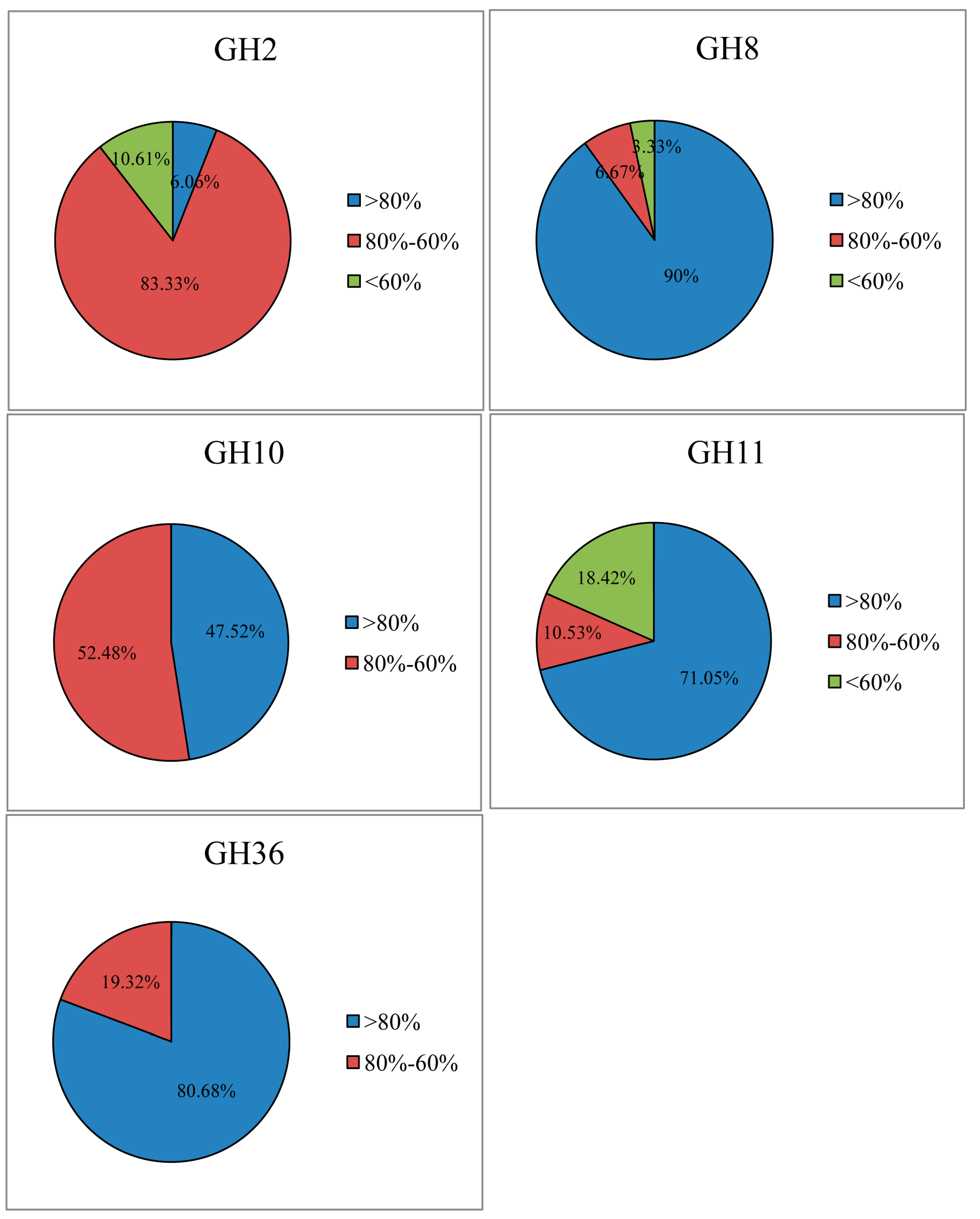
2.1. Cellulase and Hemicellulase Gene Cloning and Diversity Analysis

PCR with degenerate primers using metagenomic DNA from H. parallela larvae hindgut content yielded amplicons (with size between 154 and 344 bp) for β-galactosidase (GH2), β-1,4-endoglucanase (GH8), endo-xylanase (GH10, 11), α-galactosidase (GH36) enzymes. However, genes from the other β-1,4-endoglucanase (GH5, 45), cellobiohydrolase (GH48), β-glucosidase (GH3), and β-xylosidase (GH39, 52) enzyme families were not found in this study.
In the GH2, 8, 10, 11, and 36 families, 157, 170, 164, 160 and 170 clones showed 51%–99% amino acid identity with known microbial cellulases and hemicellulases based on BLAST analysis, and among them, 24, 27, 19, 14 and 17 clones showed sequence divergence (sharing <95% identity), respectively. Of these distinct fragments, 93.94%, 10%, 52.48%, 28.95% and 19.32% of GH2, GH8, GH10, GH11 and GH36 sequences were <80% identical with those of known GH enzymes, respectively. Moreover, 10.61%, 3.33% and 18.42% of GH2, GH8 and GH11 sequences had <60% identities with the available sequences in the NCBI database (Figure 1, Tables S1–S6). Therefore, it appears that a large number of cellulase and hemicellulase gene sequences discovered in this study originate from unknown micro-organisms. The rarefaction curves tended to approach the saturation plateau, indicating a sufficient sampling depth of all libraries (Figure 2).
Figure 1. Amino acid sequence identities of cellulase and hemicellulase gene fragments to known enzymes. Each sequence was analyzed with NCBI BLASTp against the GenBank database.
Ijms 16 16545 g001
Figure 2. Rarefaction curves corresponding to the cellulase and hemicellulase genes from the hindgut of Holotrichia parallela larvae. Operational taxonomic units (OTUs) were defined at the 3% level of sequence divergence.
Ijms 16 16545 g002
Figure 3. Maximum Likelihood phylogenetic tree of the GH8 amino acid sequences.
Ijms 16 16545 g003

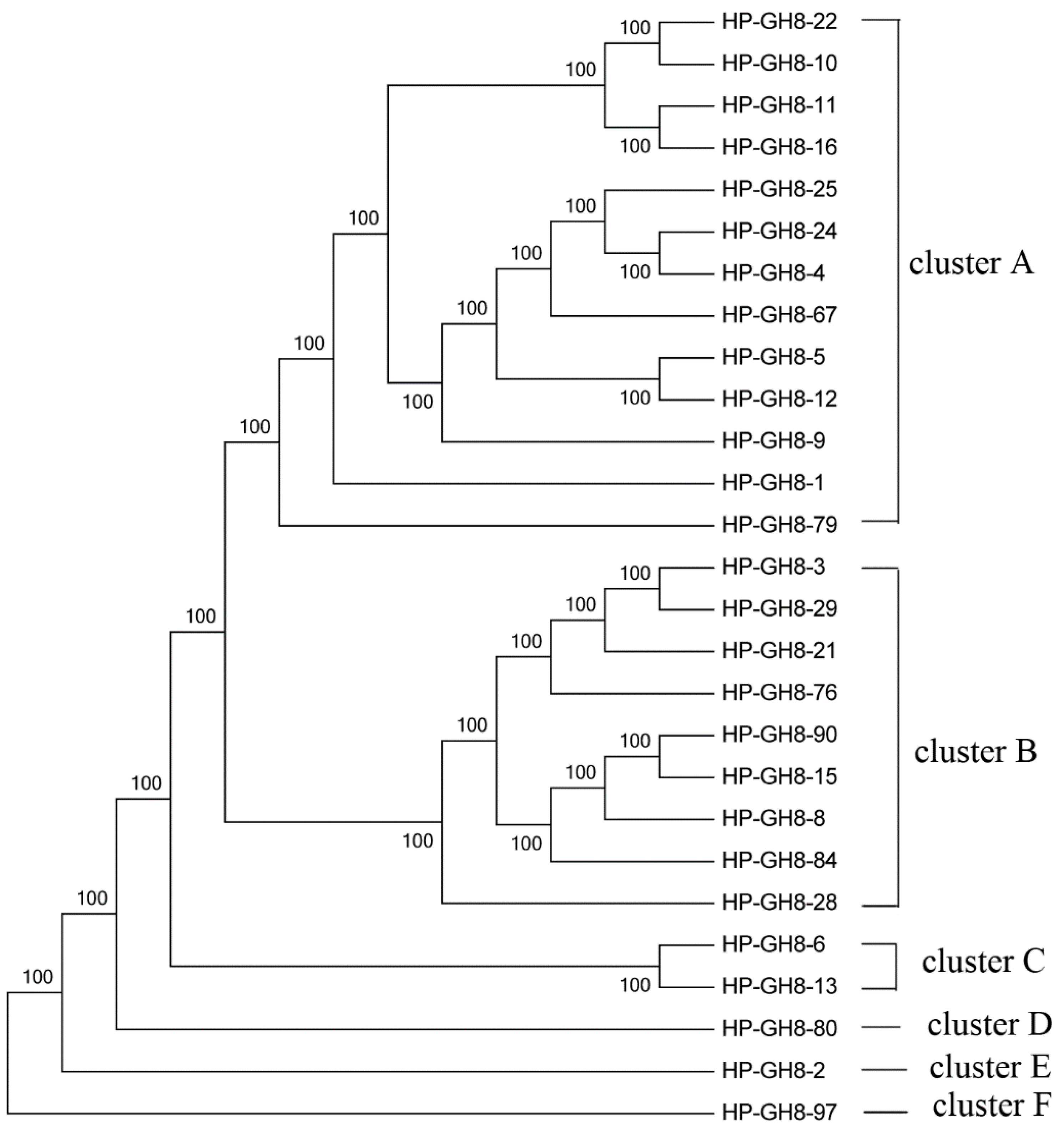
2.2. Phylogenetic Analysis of GH8 Endoglucanase Gene Fragments

The phylogenetic tree of the GH8 (β-1,4-endoglucanase) family was constructed using 27 distinct sequences. These sequences were devided into six clusters (Figure 3), indicating substantial diversity among GH8 endoglucanases in the scarab larval hindgut. From Figure 3, we found that most sequences grouped together except GH8-80, 2, and 97. Interestingly, 25 sequences were closed related to endoglucanases of Proteobacteria, and most of them belonged to the Enterobacter-like endoglucanase. The other two sequences were related to enzymes of Actinobacteria (Table S2). Our results indicated there is a rich genetic diversity of cellulolytic Proteobacteria species in the hindgut of H. parallela larvae.

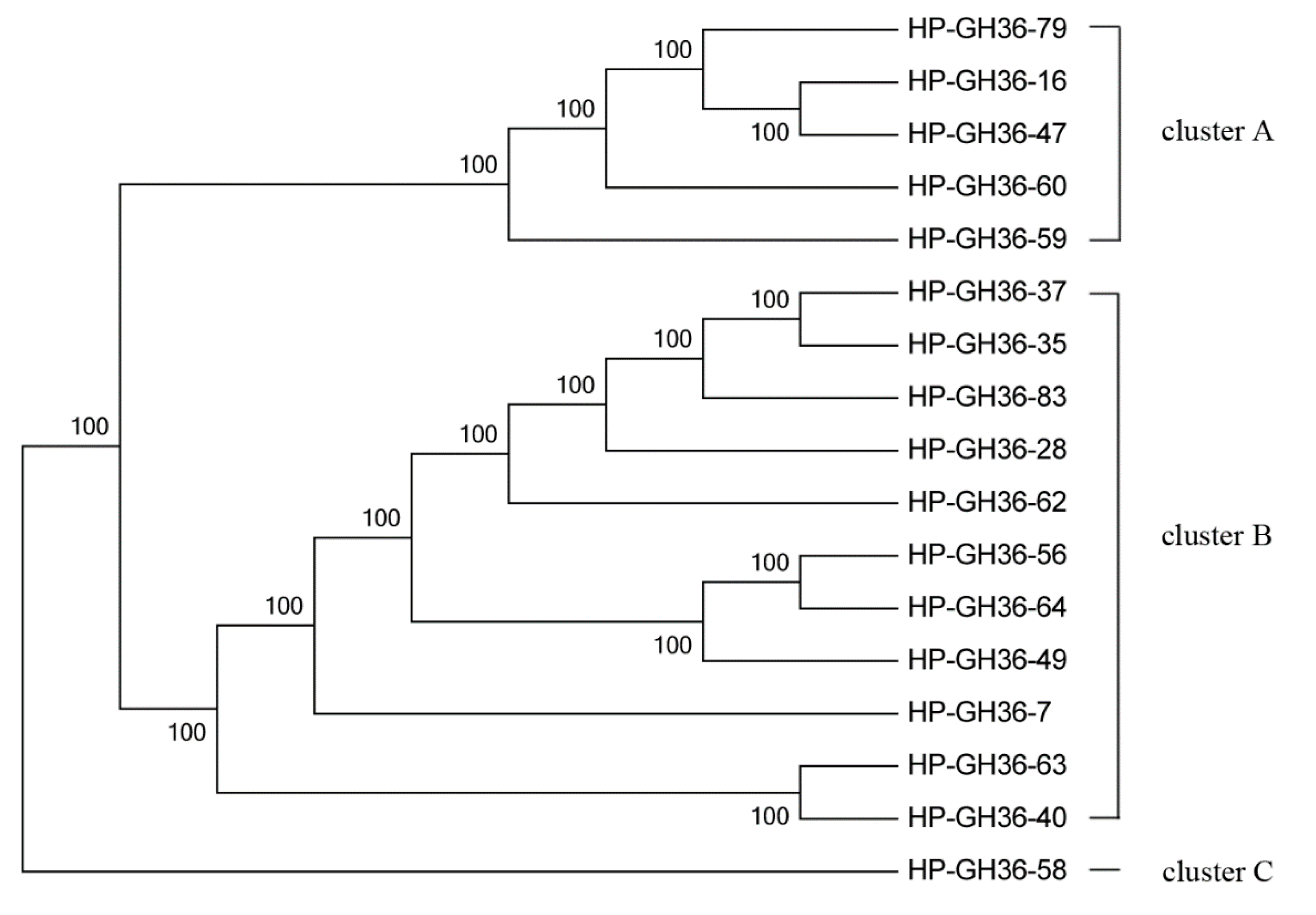
2.3. Phylogenetic Analysis of GH2 and GH36 Galactosidase Gene Fragments

The phylogenetic tree of the GH2 family was constructed using 24 distinct sequences. These sequences were devided into four clusters (Figure 4). They were closed related to galactosidase of Bacteroidetes, Firmicutes, Proteobacteria, Arthropoda, Planctomycetes and some other uncultured bacteria (Table S3).
Figure 4. Maximum Likelihood phylogenetic tree of the GH2 amino acid sequences.
Ijms 16 16545 g004
A total of 17 sequences from the GH36 α-galactosidase enzyme family were used to construct the phylogenetic tree. Three clusters were separated in this tree (Figure 5). Among these sequences, eight sequences were closely related to galactosidases of Bacteroidetes, most of them belonged to the Dysgonomonas-like galactosidases. Seven sequences were closely related to galactosidases of Firmicutes, only few sequences were related to enzymes of Acidobacteria and Proteobacteria (Table S4). These results indicate the high diversity of enzymes from Bacteroidetes and Firmicutes species in the scarab larval hindgut.
Figure 5. Maximum Likelihood phylogenetic tree of the GH36 amino acid sequences.
Ijms 16 16545 g005
Figure 6. Maximum Likelihood phylogenetic tree of the GH10 amino acid sequences.
Ijms 16 16545 g006

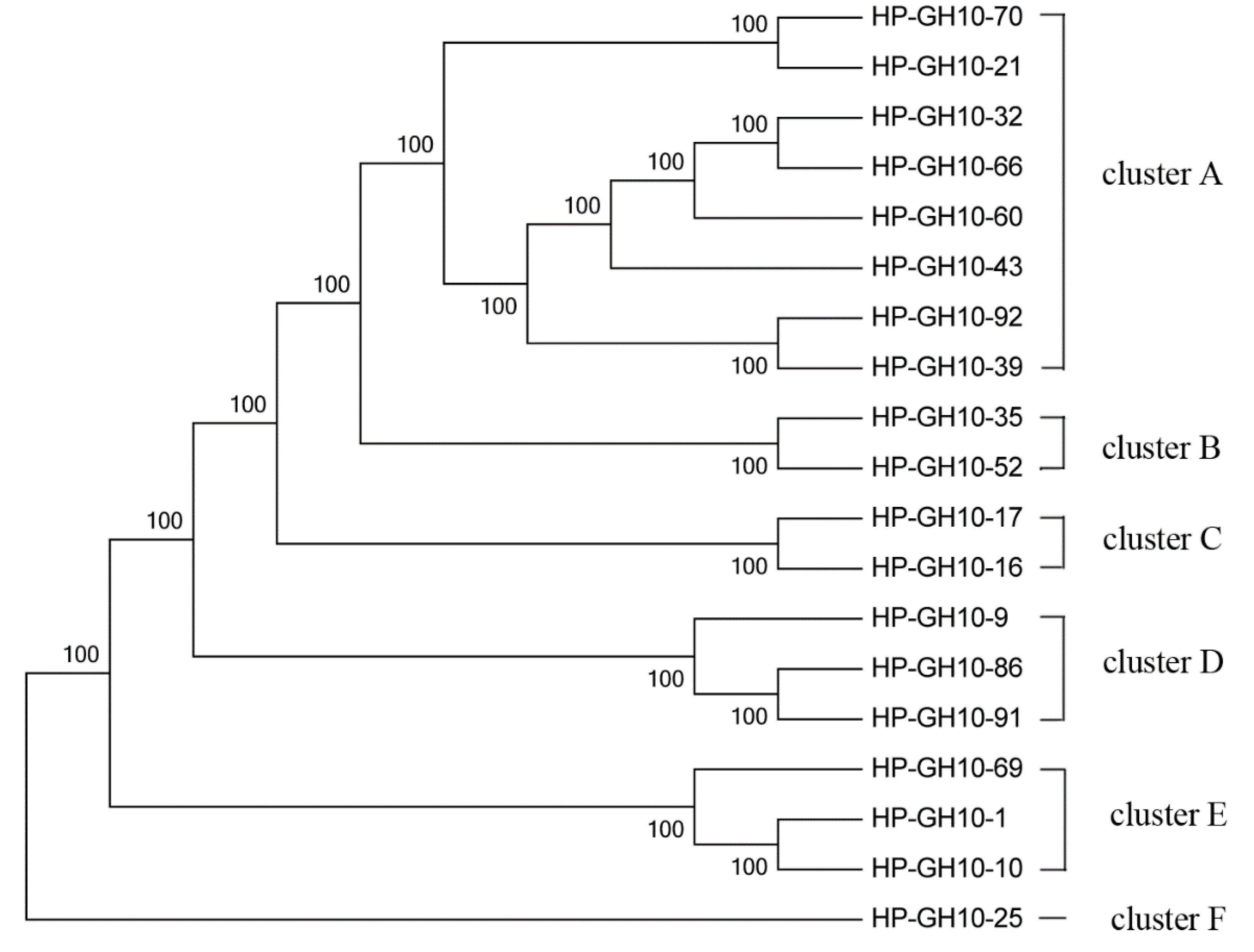
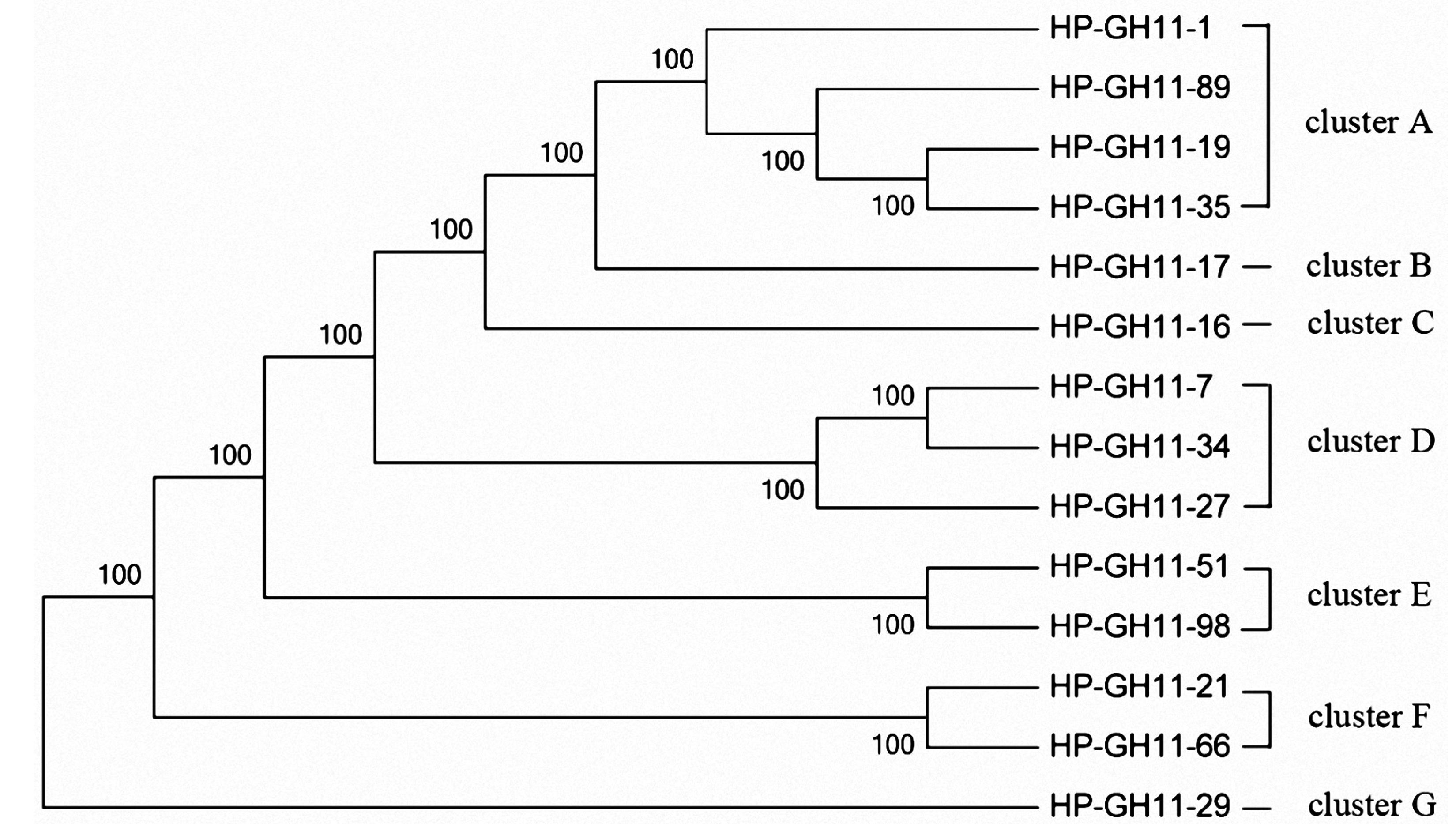
2.4. Phylogenetic Analysis of GH10 and GH11 Endo-Xylanase Gene Fragments

The 19 distinct partial sequences of GH10 endo-xylanase were used to construct a phylogenetic tree. The sequences from GH10 families were confined to six clusters (Figure 6). Most of them were closely related to endo-xylanases of Bacteroidetes, Firmicutes and Proteobacteria. Others were related to enzymes of uncultured bacteria (Table S5).
The phylogenetic tree of the GH11 family was constructed using 14 distinct sequences. These sequences were divided into seven clusters (Figure 7), indicating substantial diversity among GH11 endo-xylanases in the scarab larval hindgut. They were closely related to endo-xylanases of Firmicutes, Bacteroidetes, and Ascomycota. Among them, many sequences belonged to the Dysgonomonas-like endo-xylanases (Table S6).
Figure 7. Maximum Likelihood phylogenetic tree of the GH11 amino acid sequences.
Ijms 16 16545 g007

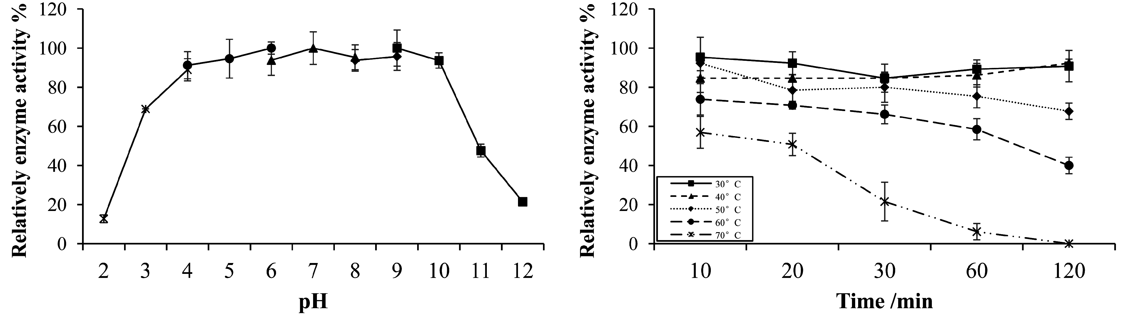
2.5. Gene Cloning, Expression, and Characterization of Endo-Xylanase

The partial endo-xylanase sequence GH11-7, which showed the lowest identity (53%) with known sequences, was selected for further study. The full-length xynGH11-7 endo-xylanase gene was directly cloned from the metagenomic DNA from the H. parallela larval hindgut. The ORF for xynGH11-7 is 711 bp, encoding 237 amino acids. The protein is predicted have a molecular mass of 26.7 kDa with no signal peptide. The predicted theoretical pI for xynGH11-7 was at pH 6.07, and it is predicted to be hydrophilic. Sequence analysis of the amplified xynGH11-7 fragment exhibited the highest amino acid sequence identity (50%) to an endo-xylanase gene in the GH 11 family from Bacillus sp. NCL (Figure S1).
The optimal pH and temperature for xynGH11-7 enzyme activities on xylan were observed at pH 6.0 and 30 °C, respectively (Figure 8). Furthermore, we found that the purified recombinant xynGH11-7 was stable at a pH range of 4.0 to 10.0, and approximately 88%–93% of the original enzymatic activity was still maintained after 1 h incubation at various pH levels (Figure 9). The thermal stability study shown in Figure 8 revealed that the enzyme was stable from 30 to 50 °C for approximately 2 h incubations, but was dramatically inactivated when temperatures reached 60 °C.
Figure 8. Optimal pH and temperature for the activities of recombinant xynGH11-7.
Ijms 16 16545 g008
Figure 9. Stability of recombinant xynGH11-7 at different pH and temperatures.
Ijms 16 16545 g009

3. Discussion

In this study, cellulase and hemicellulase genes originating from symbiotic bacteria residing in the phytophagous H. parallela larval hindgut were targeted for diversity analysis because of their synergistic roles in lignocelluloses degradation. Using culture-independent methods, 101 different endo-xylanase, galactosidase, and β-1,4-endoglucanase sequences were isolated from the H. parallela larval hindgut. The results are consistent with previous studies of gut bacterial communities in H. parallela larvae, in which rich collections of cellulolytic and xylanolytic bacteria were found in the hindgut of H. parallela larvae [1,2,6,7]. Similar phenomena have also been observed in the wood-feeding higher termite [13] and another scarab beetle, Pachnoda marginata [14]. A common feature of these insects is that they all eat high-fiber diets, and that likely explains why large, diverse sets of bacterial genes for cellulose and xylan hydrolysis were found in their hindguts. Recent data also support the idea that the gut symbiotic bacteria are important in lignocellulose hydrolysis for the host insects [13,15,16].
Enzyme fragments from GH2, 8, 10, 11, and 36 families were obtained, suggesting that the gut of H. parallela larvae harbor diverse cellulolytic and xylanolytic microbes and enzymes. β-Galactosidases and α-galactosidases from GH2 and GH36 are known to degrade β-d-galactosides and α-d-galactosides, respectively. Endoglucanases from GH8 can degrade cellulose, and endo-xylanases from GH10 and GH11 were reported to degrade xylan (http://www.cazy.org/Glycoside-Hydrolases.html). Furthermore, we did not find sequences from the GH3, 5, 39, 45, 48, and 52 enzyme families. A similar phenomenon was also observed in the termite hindgut, where a pyrosequenced hindgut microbiota metagenome analysis by Warnecke et al. showed a smaller number of GH39, GH45, and GH52 enzyme sequences, with gene fragments from the GH48 family cellobiohydrolase being completely absent [13].
Proteobacteria is the most abundant bacterial phylum in the hindgut of H. parallela larvae, and Actinobacteria is the second most abundant [6]. A 16S rDNA analysis of the hindgut microbiota of H. parallela larvae from different geographic regions by Huang et al. also showed that the Enterobacteriaceae family of the Proteobacteria phylum was commonly found in all of the natural populations, and constituted dominant and stable populations in the scarab gut [17]. Our current study showed that 25 distinct sequences from the GH8 β-1,4-endoglucanase family were closely related to this enzyme from Proteobacteria, comprising 92.59% (25/27) of the total distinct sequences, and the remaining sequences were related to enzymes from Actinobacteria (Table S2). Among them, 85.19% (23/27) of the total distinct sequences belonged to the family Enterobacteriaceae (Table S2). Consulting the collection of GH8 bacterial sequences in the CAZy database (http://www.cazy.org/), we also found that the phylum of Proteobacteria predominated, followed by Firmicutes. A similar result has also been reported by Elifantz (2008) in a study of endoglucanase diversity in marine bacteria [18]. Moreover, members of the Enterobacteriaceae family have been implicated in nitrogen and carbon metabolism in the fruit fly Ceratitis capitata, and they have an indirect contribution to host fitness [19]. Our results suggest a possible cellulose degradation function for these bacterial species in the host insect. However, the main endoglucanase enzyme in wood-feeding “higher” Nasutitermes hindgut was attributed to the GH5 family [13]; in the H. parallela larvae hindgut, it was assigned to the GH8 family, and no enzyme sequences belonging to the GH5 family were cloned. The species differences observed in cellulose degradation enzymes may be due to host insect differences, including diet [20,21].
Based on phylogenetic analysis, the GH10 and GH11 endo-xylanase sequences were closely related to the endo-xylanases from Bacteroides and Firmicutes (Tables S5 and S6). These results are similar to those of previous studies. Based on the collected endo-xylanase sequences in the Pfam database, Bacteroidetes and Firmicutes are the main microbial source of endo-xylanase production. Interestingly, in goat and sheep rumens and in some other herbivores (e.g., wallabies), endo-xylanase genes were also found to be mainly distributed between these two phyla [12,19]. However, the distribution of GH10 and GH11 endo-xylanases in the scarab hindgut differed from both that of tundra soil (in which bacterial endo-xylanase genes were mainly detected from Actinobacteria and Proteobacteria) [22] and the termite hindgut (in which endo-xylanases were mainly from the phyla of Fibrobacteres and Spirochaetes) [13]. Additionally, in comparison with endo-xylanase sequences of the GH11 family, the GH10 family possessed much more gene richness. This result was similar to that found in other microenvironments, such as the termite hindgut [13], and goat and cow rumen [12,23]. GH11 endo-xylanases have a lower catalytic versatility than GH10 endo-xylanases, and the products of their action can be further hydrolyzed by the GH10 enzymes [24,25]. Therefore, the difference in gene richnesses between these two families implies that they may have different roles in xylan degradation in the hindgut.
Furthermore, we cloned, expressed and biochemically characterized one endo-xylanase. To our knowledge, no GH 11 xylanase has been isolated from the hindgut of H. parallela larvae before. The enzyme showed a broad pH stability, indicating that this enzyme is promising for applications in many industries, such as the paper industry, which requires enzymes to exhibit pH stability over a wide range [26]. Some xylanases had also been cloned from the gut of other insects, such as Gryllotalpa orientalis [27,28], Batocera horsfieldi larvae [29], however, most of them showed a narrower pH stability. Another prominent feature of the xynGH11-7 enzyme is its low identity (50%) with known endo-xylanases in the NCBI database, further suggesting the novelty of this enzyme.
In conclusion, large numbers of cellulase (endoglucanase) and hemicellulase (galactosidase and endo-xylanase) fragments from the GH2, GH8, GH10, GH11 and GH36 families were cloned from the hindgut of H. parallela larvae using culture-independent molecular methods. Some of the sequences have low identities with known glycosyl hydrolases, suggesting the phylogenetic diversity and novelty of these enzyme genes. Enzyme fragments amplified in our work were mostly distributed to the bacterial species in Proteobacteria, Bacteroides and Firmicutes, which indicate potential contributors to lignocellulose degradation in the H. parallela larval hindgut. Full length cloning and heterologous expression of an endo-xylanase-like gene further indicated its function as an active endo-xylanase. Taken in combination with previous studies on H. parallela larvae, our work highlights the hindgut of the larvae as a reservoir of extensive and specific cellulase and hemicellulase sequence diversities.

4. Materials and Methods

4.1. Ethics Statement
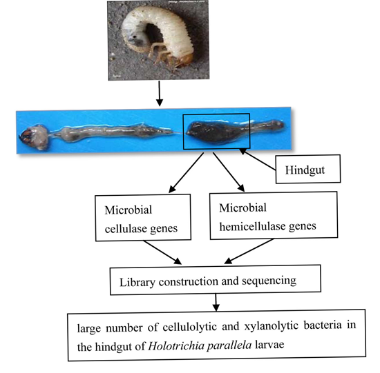
H. parallela has not been notified under any act or laws and rules thereof of the Government of China as an endangered or threatened species restricting or regulating its collection and observation. The peanut field was privately owned, and we have the land owner’s consent to access the land for collecting the larvae, there are no permits required for the land use or collection of the larvae.

4.2. Insect Samples

Healthy early third-instar grub larvae were collected from a peanut field (E: 112.199266°, N: 31.035516°) and then reared in a terrarium in the lab. The terrarium was filled with organic soil, held at a constant temperature of 27 ± 1 °C and light-dark photoperiod of 14:10 h, as described by Huang et al. [17]. Larvae were fed with peanut roots until they were used. Before dissection, larvae were separated and reared in a sterile container without food and soil for 2 days.

4.3. Insect Dissection and Total Bacterial DNA Extraction

Insect dissections were performed according to the method described by Zhang and Jackon [2]. Briefly, grub larvae were surface-sterilized with 75% ethanol and then washed in sterile distilled water. All further steps were performed under sterile conditions. Fifty entire hindguts were removed from abdomens and then homogenized. Total bacterial genomic DNA was extracted using a QIAamp stool DNA extraction kit (Qiagen, Hilden, Germany), and the genomic DNA was stored at −70 °C before use.

4.4. PCR Amplification, Library Construction and Sequencing

The degenerate primers used for amplification of cellulase and hemicellulase genes are listed in Table 1, they are according to the method described by Wang et al. [22]. The purified bacterial genomic DNA was used as a template and underwent a touchdown PCR. The PCR was performed using a thermal cycler in a 50 μL reaction mix. The detail amplification procedures for the targeted gene families are shown in Table 2. The PCR-amplified gene products were cloned using the TA cloning method of the pEASY-T1 cloning kit (Transgen, Beijing, China) according to the manufacturer’s instructions for sequencing. Clones from each enzyme family were selected for sequencing, which was conducted by Sangon Biotech (Shanghai) Co., Ltd.
Table 1. Degenerate primers designed to amplify cellulase and hemicellulase gene fragments.
Pfam FamilyEnzymeEC NumberPrimer (5ʹ–3ʹ)Length bp
GH2β-GalactosidaseEC3.2.1.23GH2F: GTGCGYACSWSBCAYTAYCC204–219
GH2R: CCAAATRAYRAYGCTYGGRTGRTT
GH3β-GlucosidaseEC3.2.1.21GH3F: GTKAAYCCRWSYGGIMRIYT183–200
GH3R: TAISWYAKICCRTRVCCRAA
GH5β-1,4-EndoglucanaseEC3.2.1.4GH5F: TWYGARYTIYTIAAYGARC195–258
GH5R: NGGRTTRTARWARTGRAA
GH8β-1,4-EndoglucanaseEC3.2.1.4GH8F: GAAGGYCWGGGYTWYGSVATG183–207
GH8R: AATMWSYWSATCRCCATCGSTSGC
GH10Endo-xylanaseEC3.2.1.8GH10F: GGYCAYACBCTNRTNTGGCA138–186
GH10R: YTCRTTNACNACRTCCCA
GH11Endo-xylanaseEC3.2.1.8GH11F: TAYMTGDSNSTBTAYGGBTGG336
GH11R: TRCCVCTVCTYTKRTAVCCYTC
GH36α-GalactosidaseEC3.2.1.22GH36F: GACATGTTCGTGATGGACGAYGGNTGGTT193
GH36R: CGGACTCTGGGTTCACCATYTCNGGYTC
GH39β-XylosidaseEC3.2.1.37GH39F: TTYGARGTNTGGAAYGARCC223–230
GH39R: GCRTGNCKISWIACRAARTC
GH45β-1,4-EndoglucanaseEC3.2.1.4GH45F: ACCMGITAYTGGGAYTGYTG377–413
GH45R: AAGRYICCNAVICCNCCICCNGG
GH48Cellobio-HydrolaseEC3.2.1.91GH48F: GARGCNCCNGAYYAYGGICA420
GH48R: CCNCGYTGRWAIGTRTTDA
GH52β-XylosidaseEC3.2.1.37GH52F: GARGGNGARTAYMGIATGATGAAYAC197–200
GH52R: GCVACNCCCATRTCRTGNGT
Table 2. Settings for amplification of cellulase and hemicellulase gene fragments.
StandardTouchdown PCR Settings
94 °C 5 min
94 °C 30 s, X °C 30 s (−0.5 °C/cycle), 72 °C 30 s; 30 cycles
94 °C 30 s, Y °C 30 s, 72 °C 30 s; 20 cycles
72 °C 10 min
4 °C infinity
Pfam Family The following PCR settings are same as the Standard:
GH2 (PF02836)X = 62Y = 52
GH3 (PF01915)X = 65Y = 50
GH5 (PF00150)X = 58Y = 52
GH8 (PF01270)X = 65Y = 55
GH10 (PF00331)X = 60Y = 48
GH11 (PF00457)X = 60Y = 48
GH36 (PF02065)X = 65Y = 58
GH39 (PF01229)X = 65Y = 50
GH45 (PF02015)X = 68Y = 55
GH48 (PF02011)X = 65Y = 50
GH52 (PF03512)X = 68Y = 57

4.5. Phylogenetic Analysis of Cellulase and Hemicellulase Gene Sequences

The cellulase and hemicellulase nucleotide sequences were translated using ExPASy tool (http://www.expasy.ch/tools/). All the deduced amino-acid sequences were aligned with ClustalX and the redundant sequences were removed with CD-HIT, using a sequence identity cutoff of 95% [30]. Gene abundance and rarefaction curves of each GH family was estimated using distance-based operational taxonomic unit and richness determination (DOTUR) software [31]. The coverage of the clone library was considered to be: C = 1 − (n1/N), where n1 is the number of single clones, and N is the total number of clones. All richness estimations and diversity indices were computed using EstimateS software (http://viceroy.eeb.uconn.edu/EstimateS). Phylogenetic analyses were conducted with the Maximum Likelihood (ML) method using the PHYLIP program [32].

4.6. Gene Cloning, Expression, and Characterization of Endo-Xylanase

Fragment xynGH11-7 was chosen for full-length gene cloning based on its low similarity (53%) to known endo-xylanase enzymes, which suggested potential for novelty, and was therefore chosen for full-length gene cloning. The full-length gene was obtained by thermal asymmetric interlaced-PCR (TAIL-PCR) using six nested, insertion-specific primers. The pPIC9K expression vector and Pichia pastoris were selected for enzyme expression, with the His6 tagged protein purified by chelation to a sepharose (Ni-NTA) resin matrix (Novagen, Merck, Kenilworth, NJ, USA). The details regarding oligonucleotide primers used for xynGH11-7 cloning and expression are provided in Table S1.
Enzyme activity was determined as described by Zhou [29], with several modifications: The release of reducing sugar from beech wood xylan was measured (1% w/v in acetate buffer, pH 6.0) at 37 °C for 10 min using a 3,5-dinitrosalicylic acid (DNSA) reagent. One unit (U) of endo-xylanase activity was defined as the amount of enzyme required to release 1 μmol of reducing sugars as xylose from xylan per minute under the reaction conditions.
The optimal pH was determined in a series of buffers with pH values ranging from 2.0 to 12.0, while the optimal temperature was determined over 10 °C intervals ranging from 20–70 °C. The buffers used were glycine-HCl (pH 2.0 to 3.0), acetate sodium-acetic acid (pH 4.0 to 6.0), dibasic sodium phosphate/sodium biphosphate (pH 6.0 to 8.0), and glycine-NaOH (pH 9.0 to 12.0). The pH stability of the enzyme activity was tested by pre-incubating the recombinant enzyme for 1 h at 37 °C and under pH conditions ranging from 2.0 to 12.0. The thermostability of the enzyme was determined by pre-incubating the enzyme for 1 h at 30–70 °C at a 10 °C intervals. The residual enzyme activities were assayed as described above.

4.7. Nucleotide Sequence Accession Numbers

The nucleotide sequences of the GH2, GH8, GH10, GH11 and GH36 gene fragments were deposited into the GenBank and DDBJ databases under accession numbers KF844174-KF844194, AB872357-AB872359, AB872360-AB872386, AB872387-AB872404, KF844195-KF844206, and AB872405-AB872421. The full-length endo-xylanase gene sequence has been submitted to the Genbank database with the accession number KF927127.

Supplementary Materials

Acknowledgments

This research was supported by the National Natural Science Foundation of China (No. 31272378), the Special Fund for Agro-scientific Research in the Public Interest (No. 201003025), and the earmarked fund for China Agriculture Research System (No. CARS-27).

Author Contributions

Conceived and designed the experiments: Hongyu Zhang, Ping Sheng; Performed the experiments: Ping Sheng, Yushan Li; Analyzed the data: Ping Sheng, Hongyu Zhang; Contributed reagents/materials/analysis tools: Hongyu Zhang; Wrote the paper: Ping Sheng, Hongyu Zhang, Sean D. G. Marshall.

Conflicts of Interest

The authors declare no conflict of interest.

References

  1. Huang, S.W.; Zhang, H.Y.; Marshall, S.; Jackson, T.A. The scarab gut: A potential bioreactor for bio-fuel production. Insect Sci. 2010, 17, 175–183. [Google Scholar] [CrossRef]
  2. Zhang, H.Y.; Jackson, T.A. Autochthonous bacterial flora indicated by PCR-DGGE of 16S rRNA gene fragments from the alimentary tract of Costelytra zealandica (Coleoptera: Scarabaeidae). J. Appl. Microbiol. 2008, 105, 1277–1285. [Google Scholar] [CrossRef] [PubMed]
  3. Wei, X.T.; Xu, X.D.; Deloach, C.J. Biological control of white grubs (Coleopera: Scarabaeidae) by larvae of Promachus yesonicus (Diptera: Asilidae) in China. Biol. Control 1995, 5, 290–296. [Google Scholar] [CrossRef]
  4. Cazemier, A.E.; Hackstein, J.H.P.; Op den Camp, H.J.M.; Rosenberg, J.; van der Drift, C. Bacteria in the intestinal tract of different species of arthropods. Microb. Ecol. 1997, 33, 189–197. [Google Scholar] [CrossRef] [PubMed]
  5. Bayon, C. Volatile fatty acids and methane production in relation to anaerobic carbohydrate fermentation in Oryctes nasicornis larvae (Coleoptera: Scarabaeidae). J. Insect Physiol. 1980, 26, 819–828. [Google Scholar] [CrossRef]
  6. Huang, S.W.; Sheng, P.; Zhang, H.Y. Isolation and identification of cellulolytic bacteria from the gut of Holotrichia parallela larvae (Coleoptera: Scarabaeidae). Int. J. Mol. Sci. 2012, 13, 2563–2577. [Google Scholar] [CrossRef] [PubMed]
  7. Sheng, P.; Huang, S.W.; Wang, Q.; Wang, A.L.; Zhang, H.Y. Isolation, screening, and optimization of the fermentation conditions of highly cellulolytic bacteria from the hindgut of Holotrichia parallela larvae (Coleoptera: Scarabaeidae). Appl. Biochem. Biotechnol. 2012, 167, 270–284. [Google Scholar] [CrossRef] [PubMed]
  8. McKendry, P. Energy production from biomass (part 1): Overview of biomass. Bioresour. Technol. 2002, 83, 27–46. [Google Scholar] [CrossRef]
  9. Badger, P.C. Ethanol from cellulose: A general review. In Trends in New Crops and New Uses; Janick, J., Whipkey, A., Eds.; ASHS Press: Alexandria, VA, USA, 2002. [Google Scholar]
  10. Collins, T.; Gerday, C.; Feller, G. Xylanases, xylanase families and extremophilic xylanases. FEMS Microbiol. Rev. 2006, 29, 3–23. [Google Scholar] [CrossRef] [PubMed]
  11. Henrissat, B.; Claeyssens, M.; Tomme, P.; Lemesle, L.; Mornon, J.P. Cellulase families revealed by hydrophobic cluster analysis. Gene 1989, 81, 83–95. [Google Scholar] [CrossRef]
  12. Wang, G.Z.; Luo, H.Y.; Wang, Y.R.; Huang, H.Q.; Shi, P.J.; Yang, P.L.; Meng, K.; Bai, Y.G.; Yao, B. A novel cold-active xylanase gene from the environmental DNA of goat rumen contents: Direct cloning, expression and enzyme characterization. Bioresour. Technol. 2011, 102, 3330–3336. [Google Scholar] [CrossRef] [PubMed]
  13. Warnecke, F.; Luginbühl, P.; Ivanova, N.; Ghassemian, M.; Richardson, T.H.; Stege, J.T.; Cayouette, M.; McHardy, A.C.; Djordjevic, G.; Aboushadi, N.; et al. Metagenomic and functional analysis of hindgut microbiota of a wood-feeding higher termite. Nature 2007, 450, 560–565. [Google Scholar] [CrossRef] [PubMed]
  14. Cazemier, A.E.; Verdoes, J.C.; Reubsaet, F.A.; Hackstein, J.H.; van der Drift, C.; Op den Camp, H.J. Promicromonospora pachnodae sp. nov., a member of the (hemi)cellulolytic hindgut flora of larvae of the scarab beetle Pachnoda marginata. Antonie Van Leeuwenhoek 2003, 83, 135–148. [Google Scholar] [CrossRef] [PubMed]
  15. Tokuda, G.; Watanabe, H. Hidden cellulases in termites: Revision of an old hypothesis. Biol. Lett. 2007, 3, 336–339. [Google Scholar] [CrossRef] [PubMed]
  16. Scharf, M.E.; Tartar, A. Termite digestomes as sources for novel lignocellulases. Biofuel Bioprod. Biorefin. 2008, 2, 540–552. [Google Scholar] [CrossRef]
  17. Huang, S.W.; Zhang, H.Y. The impact of environmental heterogeneity and life stage on the hindgut microbiota of Holotrichia parallela larvae (Coleoptera: Scarabaeidae). PLoS ONE 2013, 8, e57169. [Google Scholar] [CrossRef] [PubMed]
  18. Elifantz, H.; Waidner, LA.; Michelou, V.K.; Cottrell, M.T.; Kirchman, D.L. Diversity and abundance of glycosyl hydrolase family 5 in the North Atlantic Ocean. FEMS Microbiol. Ecol. 2008, 63, 316–327. [Google Scholar] [CrossRef] [PubMed]
  19. Behar, A.; Yuval, B.; Jurkevitch, E. Gut bacterial communities in the Mediterranean fruit fly (Ceratitis capitata) and their impact on host longevity. J. Insect Physiol. 2008, 54, 1377–1383. [Google Scholar] [CrossRef] [PubMed]
  20. Leser, T.D.; Lindecrona, R.H.; Jensen, T.K.; Jensen, B.B.; Moller, K. Changes in bacterial community structure in the colon of pigs fed different experimental diets and after infection with Brachyspira hyodysenteriae. Appl. Environ. Microbiol. 2000, 66, 3290–3296. [Google Scholar] [CrossRef] [PubMed]
  21. Shi, P.J.; Meng, K.; Zhou, Z.G.; Wang, Y.R.; Diao, Q.Y.; Yao, B. The host species affects the microbial community in the goat rumen. Lett. Appl. Microbiol. 2008, 46, 132–135. [Google Scholar] [CrossRef] [PubMed]
  22. Wang, G.Z.; Wang, Y.R.; Yang, P.L.; Luo, H.Y.; Huang, H.Q.; Shi, P.J.; Meng, K.; Yao, B. Molecular detection and diversity of xylanase genes in alpine tundra soil. Appl. Microbiol. Biotechnol. 2010, 87, 1383–1393. [Google Scholar] [CrossRef] [PubMed]
  23. Hess, M.; Sczyrba, A.; Egan, R.; Kim, T.W.; Chokhawala, H.; Schroth, G.; Luo, S.J.; Clark, D.S.; Chen, F.; Zhang, T.; et al. Metagenomic discovery of biomass-degrading genes and genomes from cow rumen. Science 2011, 331, 463–467. [Google Scholar] [CrossRef] [PubMed]
  24. Biely, P.; Kluepfel, D.; Morosoli, R.; Shareck, F. Mode of action of three endo-β-1,4-endo-xylanases of Streptomyces lividans. Biochim. Biophys. Acta 1993, 1162, 246–254. [Google Scholar] [CrossRef]
  25. Biely, P.; Vrsanska, M.; Tenkanen, M.; Kluepfel, D. Endo-β-1,4-endo-xylanase families: Differences in catalytic properties. J. Biotechnol. 1997, 57, 151–166. [Google Scholar] [CrossRef]
  26. Georis, J.; Giannotta, F.; de Buyl, E.; Granier, B.; Frere, J.M. Purification and properties of three endo-β-1,4-xylanases produced by Streptomyces sp. strain S38 which diver in their ability to enhance the bleaching of kraft pulps. Enzym. Microb. Technol. 2000, 26, 178–186. [Google Scholar] [CrossRef]
  27. Oh, H.W.; Heo, S.Y.; Kim, D.Y.; Park, D.S.; Bae, K.S.; Park, H.Y. Biochemical characterization and sequence analysis of a xylanase produced by an exo-symbiotic bacterium of Gryllotalpa orientalis, Cellulosimicrobium sp. HY-12. Antonie van Leeuwenhoek 2008, 93, 437–442. [Google Scholar] [CrossRef] [PubMed]
  28. Kim, D.Y.; Shin, D.H.; Jung, S.; Lee, J.S.; Cho, H.Y.; Bae, K.S.; Sung, C.K.; Rhee, Y.H.; Son, K.H.; Park, H.Y. Biocatalytic properties and substrate-binding ability of a modular GH10 β-1,4-xylanase from an insect-symbiotic bacterium, Streptomyces mexicanus HY-14. J. Microbiol. 2014, 52, 863–870. [Google Scholar] [CrossRef] [PubMed]
  29. Zhou, J.P.; Huang, H.Q.; Meng, K.; Shi, P.J.; Wang, Y.R.; Luo, H.Y.; Yang, P.L.; Bai, Y.G.; Zhou, Z.G.; Yao, B. Molecular and biochemical characterization of a novel xylanase from the symbiotic Sphingobacterium sp. TN19. Appl. Microbiol. Biotechnol. 2009, 85, 323–333. [Google Scholar] [CrossRef] [PubMed]
  30. Li, W.Z.; Godzik, A. Cd-hit: A fast program for clustering and comparing large sets of protein or nucleotide sequences. Bioinformatics 2006, 22, 1658–1659. [Google Scholar] [CrossRef] [PubMed]
  31. Schloss, P.D.; Handelsman, J. Introducing DOTUR, a computer program for defining operational taxonomic units and estimating species richness. Appl. Environ. Microbiol. 2005, 71, 1501–1506. [Google Scholar] [CrossRef] [PubMed]
  32. Retief, J.D. Phylogenetic analysis using PHYLIP. Bioinform. Methods Protoc. 1999, 132, 243–258. [Google Scholar]

