
Drosophotoxicology: An Emerging Research Area for Assessing Nanoparticles Interaction with Living Organisms

 ,
,
Abstract
:
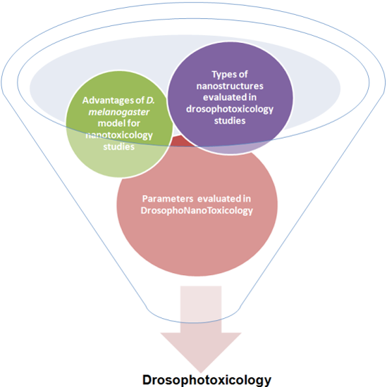
1. Introduction
2. Advantages of the D. melanogaster Experimental Model
3. Investigation of NPs Toxicity and Genotoxicity Using D. melanogaster Model
4. Use of D. melanogaster Model for the Study of NPs-Based Delivery Systems

| NPs | Concentrations of NPs | Developmental Stage at the Time of Exposure | Effects | References |
|---|---|---|---|---|
| C nanotubes | n/a | larval | tissue incorporation, no toxic effects | [33,34,35,56,57] |
| adult | affected grooming that resulted in impaired locomotor function and mortality | |||
| GaP nanowires | n/a | larval/adult | no incorporation | [36] |
| Fe3O4 | n/a | adult | compromised fecundity | [37] |
| compromised oogenesis | ||||
| ovarian defects | ||||
| developmental delay | ||||
| TiO2 | 0.002–2 mg/L | larval | increased pupation time | [41,42,45,46,58] |
| catalase and superoxide dismutase 2 down-regulation | ||||
| rare aberrant eye phenotype: “nanomaterial mutated” | ||||
| 80–1600 mg/L | larval/adult | cytotoxic effects on midgut and imaginal disc tissues | ||
| increased DNA damage | ||||
| Ag | n/a | larval | oxidative stress | [38,39,47,51,52] |
| Hsp70, p53, p-38, caspase-3, and caspase-9 down-regulation | ||||
| reduced larval and pupal survival | ||||
| affected larval climbing activity | ||||
| pigmentation d-efects in adults | ||||
| affected locomotor ability in adults | ||||
| reduced gut microbiota diversity | ||||
| 20 mg/L | larval | over 50% pupal lethality | ||
| up to 50 mg/L | adult | loss of melanin | ||
| compromised fertility | ||||
| affected vertical movement | ||||
| tyrosinase and superoxide dismutase decreased activity | ||||
| 0.1–1 mg/L | embryo to adult | decreased life-span | ||
| 5 mg/L | embryo to adult | compromised fertility | ||
| Au | 0.5–2 nM | larval | no toxic effects | [53,54,55] |
| 5 nM | larval | increased lipid anabolism | ||
| 2.5 mg/L | embryo to adult | aberrant eye phenotype: “nanomaterial mutated” | ||
| ZnO | n/a (high doses) | larval | increased DNA damage | [60,61,62] |
| affected Hsp70 and p53 expression | ||||
| increased mitotic recombination | ||||
| CuO | n/a | larval | increased DNA damage | [63,64,65] |
| cytotoxic effects | ||||
| slowed development | ||||
| reduced adult longevity | ||||
| decreased sperm competition | ||||
| CeO2 | n/a | larval | no toxic effects | [66,69] |
| Co | n/a | larval | cytotoxic effects | [70] |
| increased mitotic recombination | ||||
| Silica | n/a | larval | Hsp70, Hsp22, and caspase up-regulation | [71,72,73,74] |
| membrane destabilization | ||||
| mitochondrial membrane potential loss | ||||
| 0.1–0.5 mM | larval | reduced toxic effects | ||
| >5 mM | larval | increased DNA damage | ||
| Alumina | n/a | adult | decreased average frequencies of spontaneous rhythmic activities in the antennal lobe | [76] |
5. Conclusions
Acknowledgments
Author Contributions
Conflicts of Interest
References
- Donaldson, K.; Stone, V.; Tran, C.L.; Kreyling, W.; Borm, P.J.A. Nanotoxicology. Occup. Environ. Med. 2004, 61, 727–728. [Google Scholar] [CrossRef] [PubMed]
- Oberdörster, G.; Oberdörster, E.; Oberdörster, J. Nanotoxicology: An emerging discipline evolving from studies of ultrafine particles. Environ. Health Perspect. 2005, 113, 823–839. [Google Scholar]
- Xiaoke, H.; Wang, P.; Hwang, H. In vitro evaluation of cytotoxicity of engineering metal oxide NPs. Sci. Total Environ. 2009, 407, 3070–3072. [Google Scholar]
- Scown, T.M.; Johnston, B.D.; Gaiser, B.; Baalousha, M.; Mitov, S.; Lead, J.R.; Stone, V.; Fernandes, T.F.; Jepson, M.; van Aerle, R.; et al. Effects of aqueous exposure to silver NPs of different sizes in rainbow trout. Toxicol. Sci. 2010, 115, 521–534. [Google Scholar] [CrossRef] [PubMed]
- Wang, H.; Xing, B. Toxicity of nanoparticulate and bulk ZnO, Al2O3 and TiO2 to the nematode Caenorhabditis elegans. Environ. Pollut. 2009, 157, 1171–1177. [Google Scholar]
- Simon, K.H. Ecotoxic effect of photocatalytic active NPs (TiO2) on algae and daphnids. Environ. Sci. Pollut. Res. Int. 2004, 124, 225–232. [Google Scholar]
- Bouldin, J.L.; Sengupta, A.; Alexander, R.; Hannigan, R.; Buchanan, R.A. Aqueous toxicity and food chain transfer of quantum dots (TM) in freshwater algae and ceriodaphina dubia. Environ. Toxicol. Chem. 2008, 27, 1958–1963. [Google Scholar] [CrossRef] [PubMed]
- Oberdörster, G. Safety assessment for nanotechnology and nanomedicine: Concepts of nanotoxicology. J. Intern. Med. 2010, 267, 89–105. [Google Scholar] [CrossRef] [PubMed]
- Boverhof, D.R.; David, R.M. Nanomaterial characterization: Considerations and needs for hazard assessment and safety evaluation. Anal. Bioanal. Chem. 2010, 396, 953–961. [Google Scholar] [CrossRef] [PubMed]
- Vecchio, A.; Galeone, G.; Brunetti, V.; Maiorano, G.; Sabella, S.; Cingolani, R.; Pompa, P.P. Concentration-dependent, size-independent toxicity of citrate capped AuNPs in Drosophila melanogaster. PLoS ONE 2012, 7, e29980. [Google Scholar] [CrossRef] [PubMed]
- Barbara, H.J. Drosophila—A versatile model in biology & medicine. Mater. Today 2011, 14, 190–195. [Google Scholar]
- Rubin, G.M.; Lewis, E.B. A brief history of Drosophila's contributions to genome research. Science 2000, 287, 2216–2218. [Google Scholar] [CrossRef] [PubMed]
- Friedman, R.; Hughes, A.L. Pattern and timing of gene duplication in animal genomes. Genome Res. 2001, 11, 1842–1847. [Google Scholar] [PubMed]
- Bier, E. Drosophila, the golden bug, emerges as a tool for human genetics. Nat. Rev. Genet. 2005, 6, 9–23. [Google Scholar] [CrossRef] [PubMed]
- Pandey, U.B.; Nicholsm, C.D. Human Disease Models in Drosophila melanogaster and the role of the fly in therapeutic drug discovery. Pharmacol. Rev. 2011, 63, 411–436. [Google Scholar] [CrossRef] [PubMed]
- Posgai, R.T. Development of a Drosophila melanogaster Model System for Nanoparticle Toxicity Assessment. Ph.D. Thesis, The College of Arts and Sciences of the University Of Dayton, Dayton, OH, USA, December 2012. [Google Scholar]
- Rand, M.D. Drosophotoxicology: The growing potential for Drosophila in neurotoxicology. Neurotoxicol. Teratol. 2010, 32, 74–83. [Google Scholar] [CrossRef] [PubMed]
- Hsu, T.; Schulz, R.A. Sequence and functional properties of Ets genes in the model organism Drosophila. Oncogene 2000, 19, 6409–6416. [Google Scholar] [CrossRef] [PubMed]
- Posgai, R.; Ahamed, M.; Hussain, S.M.; Rowe, J.J.; Nielsen, M.G. Inhalation method for delivery of NPs to the Drosophila respiratory system for toxicity testing. Sci. Total Environ. 2009, 408, 439–443. [Google Scholar]
- Roeder, T.; Isermann, K.; Kallsen, K.; Uliczka, K.; Wagner, C. A Drosophila asthma model—what the fly tells us about inflammatory diseases of the lung. Adv. Exp. Med. Biol. 2012, 710, 37–47. [Google Scholar] [PubMed]
- Graf, U.l.; Würgler, F.E.; Katz, A.J.; Frei, H.; Juon, H.; Hall, C.B.; Kale, P.G. Somatic mutation and recombination test in Drosophila melanogaster. Environ. Mutagen. 1984, 6, 153–88. [Google Scholar] [CrossRef]
- Wodarz, A.; Nusse, R. Mechanisms of Wnt signaling in development. Annu. Rev. Cell Dev. Biol. 1998, 14, 59–88. [Google Scholar] [CrossRef] [PubMed]
- Raftery, L.A.; Sutherland, D.J. TGF-β Family signal transduction in Drosophila development: From mad to smads. Dev. Biol. 1999, 210, 251–268. [Google Scholar] [CrossRef] [PubMed]
- Tabata, T.; Eaton, S.; Kornberg, T.B. The Drosophila hedgehog gene is expressed specifically in posterior compartment cells and is a target of engrailed regulation. Genes Dev. 1992, 6, 2635–2645. [Google Scholar] [CrossRef] [PubMed]
- Shilo, B.Z. Signaling by the Drosophila epidermal growth factor receptor pathway during development. Exp. Cell Res. 2003, 284, 140–149. [Google Scholar] [CrossRef]
- Copf, T.; Goguel, V.; Lampin-Saint-Amaux, A.; Scaplehorn, N.; Preat, T. Cytokine signaling through the JAK/STAT pathway is required for long-term memory in Drosophila. Proc. Natl. Acad. Sci. USA 2011, 108, 8059–8064. [Google Scholar] [CrossRef] [PubMed]
- Bray, S. Notch signalling in Drosophila: Three ways to use a pathway. Semin. Cell Dev. Biol. 1998, 9, 591–597. [Google Scholar] [CrossRef] [PubMed]
- Branson, K.; Robiem, A.A.; Bender, J.; Perona, P.; Dickinson, M.H. Highthroughput ethomics in large groups of Drosophila. Nat. Methods 2009, 6, 451–457. [Google Scholar] [CrossRef] [PubMed]
- Dankert, H.; Wang, L.; Hoopfer, E.D.; Anderson, D.J.; Perona, P. Automated monitoring and analysis of social behavior in Drosophila. Nat. Methods 2009, 6, 297–303. [Google Scholar] [CrossRef] [PubMed]
- Pompa, P.P.; Vecchio, G.; Galeone, A.; Brunetti, V.; Maiorano, G.; Sabella, S.; Cingolani, R. Physical assessment of toxicology at nanoscale: Nano dose-metrics and toxicity factor. Nanoscale 2011, 3, 2889–2897. [Google Scholar] [CrossRef] [PubMed]
- Vecchio, G.; Galeone, A.; Malvindi, M.A.; Cingolani, R.; Pompa, P.P. Ranking the in vivo toxicity of nanomaterials in Drosophila melanogaster. J. Nanopart. Res. 2013, 15, 1936. [Google Scholar] [CrossRef]
- Vega-Alvarez, S.; Herrera, A.; Rinaldi, C.; Carrero-Martínez, F.A. Tissue-specific direct microtransfer of nanomaterials into Drosophila embryos as a versatile in vivo test bed for nanomaterial toxicity assessment. Int. J. Nanomed. 2014, 9, 2031–2041. [Google Scholar]
- Liu, X.; Vinson, D.; Abt, D.; Hurt, R.H.; Rand, D.M. Differential toxicity of carbon nanomaterials in Drosophila, larval dietary uptake is benign, but adult exposure causes locomotor impairment and mortality. Environ. Sci. Technol. 2009, 43, 6357–6363. [Google Scholar] [CrossRef] [PubMed]
- Lam, C.W.; James, J.T.; McCluskey, R.; Arepalli, S.; Hunter, R.L. A review of carbon nanotube toxicity and assessment of potential occupational and environmental health risks. Crit. Rev. Toxicol. 2006, 36, 189–217. [Google Scholar] [CrossRef] [PubMed]
- Leeuw, T.K.; Reith, R.M.; Simonette, R.A.; Harden, M.E.; Cherukuri, P.; Tsyboulski, D.A.; Beckingham, K.M.; Weisman, R.B. Single-walled carbon nanotubes in the intact organism: Near-IR imaging and biocompatibility studies in Drosophila. Nano Lett. 2007, 7, 2650–2654. [Google Scholar] [CrossRef] [PubMed]
- Adolfsson, K.; Schneider, M.; Hammarin, G.; Häcker, U.; Prinz, C.N. Ingestion of gallium phosphide nanowires has no adverse effect on Drosophila tissue function. Nanotechnology 2013, 24, 285101. [Google Scholar] [CrossRef] [PubMed]
- Chen, H.; Wang, B.; Feng, W.; Du, W.; Ouyang, H.; Chai, Z.; Bi, X. Oral magnetite nanoparticles disturb the development of Drosophila melanogaster from oogenesis to adult emergence. Nanotoxicology 2015, 9, 302–312. [Google Scholar] [CrossRef] [PubMed]
- Gorth, D.J.; Rand, D.M.; Webster, T.J. Silver nanoparticle toxicity in Drosophila, size does matter. Int. J. Nanomed. 2011, 6, 343–350. [Google Scholar]
- Key, C.S.; Reaves, D.; Turner, F.; Bang, J.J. Impacts of silver nanoparticle ingestion on pigmentation and developmental progression in Drosophila. Atlas J. Biol. 2011, 3, 52–61. [Google Scholar]
- Araj, S.-E.A.; Salem, N.M.; Ghabeish, I.H.; Awwad, A.M. Toxicity of Nanoparticles against Drosophila melanogaster (Diptera: Drosophilidae). J. Nanomater. 2015, 2015, 758132. [Google Scholar]
- Philbrook, N.A.; Winn, L.M.; Afrooz, A.R. The effect of TiO2 and Ag NPs on reproduction and development of Drosophila melanogaster and CD-1 mice. Toxicol. Appl. Pharm. 2011, 257, 429–436. [Google Scholar] [CrossRef] [PubMed]
- Posgai, R.; Cipolla-McCulloch, C.B.; Murphy, K.R.; Hussain, S.M.; Rowe, J.J.; Nielsen, M.G. Differential toxicity of silver and titanium dioxide NPs on Drosophila melanogaster development, reproductive effort, and viability, size, coatings and antioxidants matter. Chemosphere 2011, 85, 34–42. [Google Scholar] [CrossRef] [PubMed]
- Brunetti, V.; Chibli, H.; Fiammengo, R.; Galeone, A.; Malvindi, M.A.; Vecchio, G.; Cingolani, R.; Nadeau, J.L.; Pompa, P.P. InP/ZnS as a safer alternative to CdSe/ZnS core/shell quantum dots: In vitro and in vivo toxicity assessment. Nanoscale 2013, 5, 307–317. [Google Scholar] [CrossRef] [PubMed]
- Galeone, A.; Vecchio, G.; Malvindi, M.A.; Brunetti, V.; Cingolani, R.; Pompa, P.P. In vivo assessment of CdSe-ZnS quantum dots: Coating dependent bioaccumulation and genotoxicity. Nanoscale 2012, 4, 6401–6407. [Google Scholar] [CrossRef] [PubMed]
- Jovanović, B.; Cvetković, V.J.; Mitrović, T.L. Effects of human food grade titanium dioxide nanoparticle dietary exposure on Drosophila melanogaster survival, fecundity, pupation and expression of antioxidant genes. Chemosphere 2015, 144, 43–49. [Google Scholar] [CrossRef] [PubMed]
- Carmona, E.R.; Escobar, B.; Vales, G.; Marcos, R. Genotoxic testing of titanium dioxide anatase NPs using the wing-spot test and the comet assay in Drosophila. Mutat. Res. Genet. Toxicol. Environ. Mutagen. 2015, 778, 12–21. [Google Scholar] [CrossRef] [PubMed]
- Panacek, A.; Prucek, R.; Safarova, D.; Dittrich, M.; Richtrova, J.; Benickova, K.; Zboril, R.; Kvitek, L. Acute and chronic toxicity effects of silver NPs (NPs) on Drosophila melanogaster. Environ. Sci. Technol. 2011, 45, 4974–4979. [Google Scholar] [CrossRef] [PubMed]
- Benetti, F.; Bregoli, L.; Olivato, I.; Sabbioni, E. Effects of metal(loid)-based nanomaterials on essential element homeostasis: The central role of nanometallomics for nanotoxicology. Metallomics 2014, 6, 729–747. [Google Scholar] [CrossRef] [PubMed]
- Tian, H.; Eom, H.J.; Moon, S.; Lee, J.; Choi, J.; Chung, Y.D. Development of biomarker for detecting silver NPs exposure using a GAL4 enhancer trap screening in Drosophila. Environ. Toxicol. Pharmacol. 2013, 36, 548–556. [Google Scholar] [CrossRef] [PubMed]
- Armstrong, N.; Ramamoorthy, M.; Lyon, D.; Jones, K.; Duttaroy, A. Mechanism of silver NPs action on insect pigmentation reveals intervention of copper homeostasis. PLoS ONE 2013, 8, e53186. [Google Scholar] [CrossRef] [PubMed]
- Ávalos, A.; Haza, A.I.; Drosopoulou, E.; Mavragani-Tsipidou, P.; Morales, P. In vivo genotoxicity assessment of silver NPs of different sizes by the Somatic Mutation and Recombination Test (SMART) on Drosophila. Food Chem. Toxicol. 2015. [Google Scholar] [CrossRef] [PubMed]
- Ahamed, M.; Posgai, R.; Gorey, T.J.; Nielsen, M.; Hussain, S.M.; Rowe, J.J. Silver NPs induced heat shock protein 70, oxidative stress and apoptosis in Drosophila melanogaster. Toxicol. Appl. Pharmacol. 2010, 242, 263–269. [Google Scholar] [CrossRef] [PubMed]
- Hadrup, N.; Sharma, A.K.; Poulsen, M.; Nielsen, E. Toxicological risk assessment of elemental gold following oral exposure to sheets and NPs—A review. Regul. Toxicol. Pharmacol. 2015, 72, 216–221. [Google Scholar] [CrossRef] [PubMed]
- Wang, B.; Chen, N.; Wei, Y.; Li, J.; Sun, L.; Wu, J.; Huang, Q.; Liu, C.; Fan, C.; Song, H. Akt signalling-associated metabolic effects of dietary gold NPs in Drosophila. Sci. Rep. 2012, 2, 563. [Google Scholar] [CrossRef] [PubMed]
- Vecchio, G.; Galeone, A.; Brunetti, V.; Maiorano, G.; Rizzello, L.; Sabella, S.; Cingolani, R.; Pompa, P.P. Mutagenic effects of gold NPs induce aberrant phenotypes in Drosophila melanogaster. Int. J. Nanomed. 2014, 9, 2031–2041. [Google Scholar]
- Jia, G.; Wang, H.; Yan, L.; Wang, X.; Pei, R.; Yan, T.; Zhao, Y.; Guo, X. Cytotoxicity of carbon nanomaterials: Single-wall nanotube, multi-wall nanotube, and fullerene. Environ. Sci. Technol. 2005, 39, 1378–1383. [Google Scholar] [CrossRef] [PubMed]
- De Andrade, L.R.; Brito, A.S.; Melero, A.M.; Zanin, H.; Ceragioli, H.J.; Baranauskas, V.; Cunha, K.S.; Irazusta, S.P. Absence of mutagenic and recombinogenic activity of multi-walled carbon nanotubes in the Drosophila wing-spot test and Allium cepa test. Ecotoxicol. Environ. Saf. 2014, 99, 92–97. [Google Scholar] [CrossRef] [PubMed]
- Long, T.C.; Saleh, N.; Tilton, R.D.; Lowry, G.V.; Veronesi, B. Titanium dioxide (P25) produces reactive oxygen species in immortalized brain microglia (BV2): Implications for nanoparticle neurotoxicity. Environ. Sci. Technol. 2006, 40, 4346–4352. [Google Scholar] [CrossRef] [PubMed]
- Das, S.; Debnath, N.; Patra, P.; Datta, A.; Goswami, A. NPs influence on expression of cell cycle related genes in Drosophila: A microarray-based toxicogenomics study. Toxicol. Environ. Chem. 2012, 94, 952–957. [Google Scholar] [CrossRef]
- Carmona, E.R.; Inostroza-Blancheteau, C.; Rubio, L.; Marcos, R. Genotoxic and oxidative stress potential of nanosized and bulk zinc oxide particles in Drosophila melanogaster. Toxicol. Ind. Health 2015. [Google Scholar] [CrossRef] [PubMed]
- Alaraby, M.; Annangi, B.; Hernández, A.; Creus, A.; Marcos, R. A comprehensive study of the harmful effects of ZnO NPs using Drosophila melanogaster as an in vivo model. J. Hazard. Mater. 2015, 296, 166–174. [Google Scholar] [CrossRef] [PubMed]
- Alaraby, M.; Annangi, B.; Hernández, A.; Creus, A.; Marcos, R. Assessment of the genotoxic potential of two zinc oxide sources (amorphous and NPs) using the in vitro micronucleus test and the in vivo wing somatic mutation and recombination test. Food Chem. Toxicol. 2015, 84, 55–63. [Google Scholar]
- Carmona, E.R.; Inostroza-Blancheteau, C.; Obando, V.; Rubio, L.; Marcos, R. Genotoxicity of copper oxide NPs in Drosophila melanogaster. Mutat. Res. Genet. Toxicol. Environ. Mutagen. 2015, 791, 1–11. [Google Scholar] [CrossRef] [PubMed]
- Siddique, Y.H.; Haidari, M.; Khan, W.; Fatima, A.; Jyoti, S.; Khanam, S.; Naz, F.R.; Ali, F.; Singh, B.R.; Beg, T.M.; et al. Toxic potential of copper-doped ZnO NPs in Drosophila melanogaster (Oregon R). Toxicol. Mech. Methods 2015, 25, 425–432. [Google Scholar] [CrossRef] [PubMed]
- Han, X.; Geller, B.; Moniz, K.; Das, P.; Chippindale, A.K.; Walker, V.K. Monitoring the developmental impact of copper and silver nanoparticle exposure in Drosophila and their microbiomes. Sci. Total Environ. 2014, 487, 822–829. [Google Scholar] [CrossRef] [PubMed]
- Pop, C.S.; Hussien, M.D.; Popa, M.; Mares, A.; Grumezescu, A.M.; Grigore, R.; Lazar, V.; Chifiriuc, M.C.; Sakizlian, M.; Bezirtzoglou, E.; et al. Metallic-based micro and nanostructures with antimicrobial activity. Curr. Top. Med. Chem. 2015, 15, 1577–1582. [Google Scholar] [CrossRef] [PubMed]
- Tomkovich, S.; Jobin, C. Microbiota and host immune responses: A love-hate relationship. Immunology 2015. [Google Scholar] [CrossRef] [PubMed]
- Demir, E.; Turna, F.; Vales, G.; Kaya, B.; Creus, A.; Marcos, R. In vivo genotoxicity assessment of titanium, zirconium and aluminium NPs, and their microparticulated forms, in Drosophila. Chemosphere 2013, 93, 2304–2310. [Google Scholar] [CrossRef] [PubMed]
- Alaraby, M.; Hernández, A.; Annangi, B.; Demir, E.; Bach, J.; Rubio, L.; Creus, A.; Marcos, R. Antioxidant and antigenotoxic properties of CeO2 NPs and cerium sulphate: Studies with Drosophila melanogaster as a promising in vivo model. Nanotoxicology 2015, 9, 749–759. [Google Scholar] [CrossRef] [PubMed]
- Vales, G.; Demir, E.; Kaya, B.; Creus, A.; Marcos, R. Genotoxicity of cobalt NPs and ions in Drosophila. Nanotoxicology 2013, 7, 462–468. [Google Scholar] [CrossRef] [PubMed]
- Demir, E.; Aksakal, S.; Turna, F.; Kaya, B.; Marcos, R. In vivo genotoxic effects of four different nano-sizes forms of silica NPs in Drosophila melanogaster. J. Hazard. Mater. 2015, 283, 260–266. [Google Scholar] [CrossRef] [PubMed]
- Pandey, A.; Chandra, S.; Chauhan, L.K.; Narayan, G.; Chowdhuri, D.K. Cellular internalization and stress response of ingested amorphous silica NPs in the midgut of Drosophila melanogaster. Biochem. Biophys. Acta 2013, 1830, 2256–2266. [Google Scholar] [CrossRef] [PubMed]
- Kumar, R.; Roy, I.; Ohulchanskyy, T.Y.; Goswami, L.N.; Bonoiu, A.C.; Bergey, E.J.; Tramposch, K.M.; Maitra, A.; Prasad, P.N. Covalently dye-linked, surface-controlled, and bioconjugated organically modified silica NPs as targeted probes for optical imaging. ACS Nano 2008, 2, 449–456. [Google Scholar] [CrossRef] [PubMed]
- Barandeh, F.; Nguyen, P.L.; Kumar, R.; Iacobucci, G.J.; Kuznicki, M.L.; Kosterman, A.; Bergey, E.J.; Prasad, P.N.; Gunawardena, S. Organically modified silica NPs are biocompatible and can be targeted to neurons in vivo. PLoS ONE 2012, 7, e29424. [Google Scholar] [CrossRef] [PubMed]
- Pavlidis, P.; Tanouye, M.A. Seizures and failures in the giant fiber pathway of Drosophila bang-sensitive paralytic mutants. J. Neurosci. 1995, 8, 5810–5819. [Google Scholar]
- Huang, N.; Yan, Y.; Xu, Y.; Jin, Y.; Lei, J.; Zou, X.; Ran, D.; Zhang, H.; Luan, S.; Gu, H. Alumina NPs alter rhythmic activities of local interneurons in the antennal lobe of Drosophila. Nanotoxicology 2013, 7, 212–220. [Google Scholar] [CrossRef] [PubMed]
- Yadav, J.S.; Das, P.P.; Krishnan, A.; Mohapatra, D.K.; Bhadra, M.P.; Bhadra, U. 4-N-pyridine-2-yl benzamide nanotubes compatible with mouse stem cell and oral delivery in Drosophila. Nanotechnology 2010, 21, 155102. [Google Scholar] [CrossRef] [PubMed]
- Yadav, J.S.; Das, P.P.; Reddy, T.L.; Bag, I.; Lavanya, P.M.; Jagannadh, B.; Mohapatra, D.K.; Bhadra, M.P.; Bhadra, U. Sub-cellular internalization and organ specific oral delivery of PABA NPs by side chain variation. J. Nanobiotechnol. 2011, 9, 10. [Google Scholar] [CrossRef] [PubMed]
- Zhang, Y.; Swaminathan, S.; Tang, S.; Garcia-Amorós, J.; Boulina, M.; Captain, B.; Baker, J.D.; Raymo, F.M. Photoactivatable BODIPYs designed to monitor the dynamics of supramolecular nanocarriers. J. Am. Chem. Soc. 2015, 137, 4709–4719. [Google Scholar] [CrossRef] [PubMed]
- You, S.; Cai, Q.; Zheng, Y.; He, B.; Shen, J.; Yang, W.; Yin, M. Perylene-cored star-shaped polycations for fluorescent gene vectors and bioimaging. ACS Appl. Mater. Interfaces 2014, 6, 16327–16334. [Google Scholar] [CrossRef] [PubMed]
- Sonane, M.; Goyal, R.; Chowdhuri, D.K.; Ram, K.R.; Gupta, K.C. Enhanced efficiency of P-element mediated transgenesis in Drosophila: Microinjection of DNA complexed with nanomaterial. Sci. Rep. 2013, 3, 3408. [Google Scholar] [CrossRef] [PubMed]
- Biju, V.; Muraleedharan, D.; Nakayama, K.; Shinohara, Y.; Itoh, T.; Baba, Y.; Ishikawa, M. Quantum dot-insect neuropeptide conjugates for fluorescence imaging, transfection, and nucleus targeting of living cells. Langmuir 2007, 23, 10254–10261. [Google Scholar] [CrossRef] [PubMed]
© 2016 by the authors; licensee MDPI, Basel, Switzerland. This article is an open access article distributed under the terms and conditions of the Creative Commons by Attribution (CC-BY) license (http://creativecommons.org/licenses/by/4.0/).
Share and Cite
Chifiriuc, M.C.; Ratiu, A.C.; Popa, M.; Ecovoiu, A.A. Drosophotoxicology: An Emerging Research Area for Assessing Nanoparticles Interaction with Living Organisms. Int. J. Mol. Sci. 2016, 17, 36. https://0-doi-org.brum.beds.ac.uk/10.3390/ijms17020036
Chifiriuc MC, Ratiu AC, Popa M, Ecovoiu AA. Drosophotoxicology: An Emerging Research Area for Assessing Nanoparticles Interaction with Living Organisms. International Journal of Molecular Sciences. 2016; 17(2):36. https://0-doi-org.brum.beds.ac.uk/10.3390/ijms17020036
Chicago/Turabian StyleChifiriuc, Mariana Carmen, Attila Cristian Ratiu, Marcela Popa, and Alexandru Al. Ecovoiu. 2016. "Drosophotoxicology: An Emerging Research Area for Assessing Nanoparticles Interaction with Living Organisms" International Journal of Molecular Sciences 17, no. 2: 36. https://0-doi-org.brum.beds.ac.uk/10.3390/ijms17020036