
The Retentive Strength of Cemented Zirconium Oxide Crowns after Dentin Pretreatment with Desensitizing Paste Containing 8% Arginine and Calcium Carbonate


Abstract
:1. Introduction
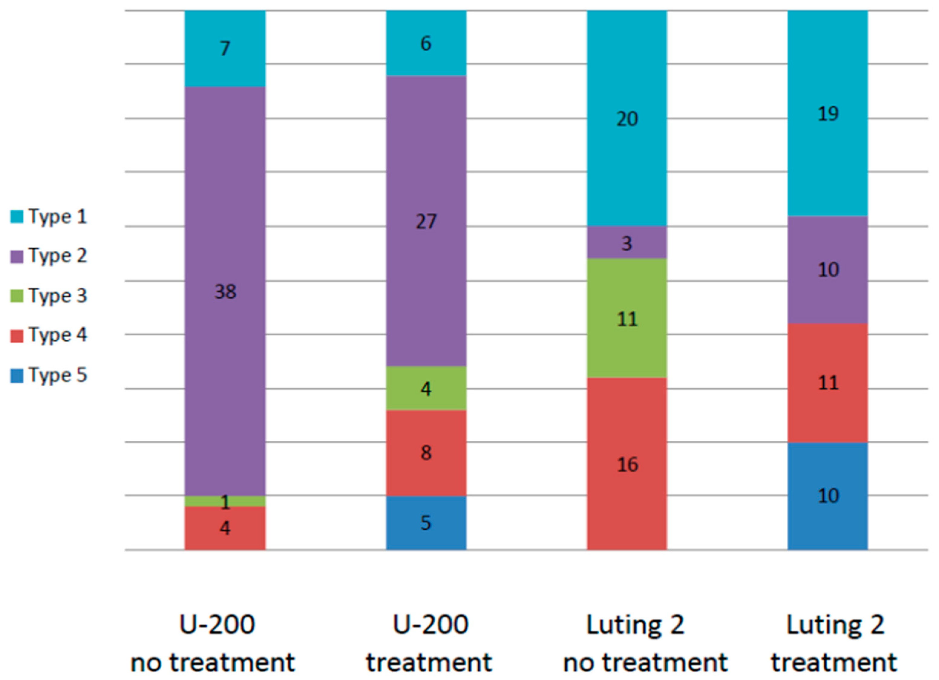
2. Results
3. Discussion
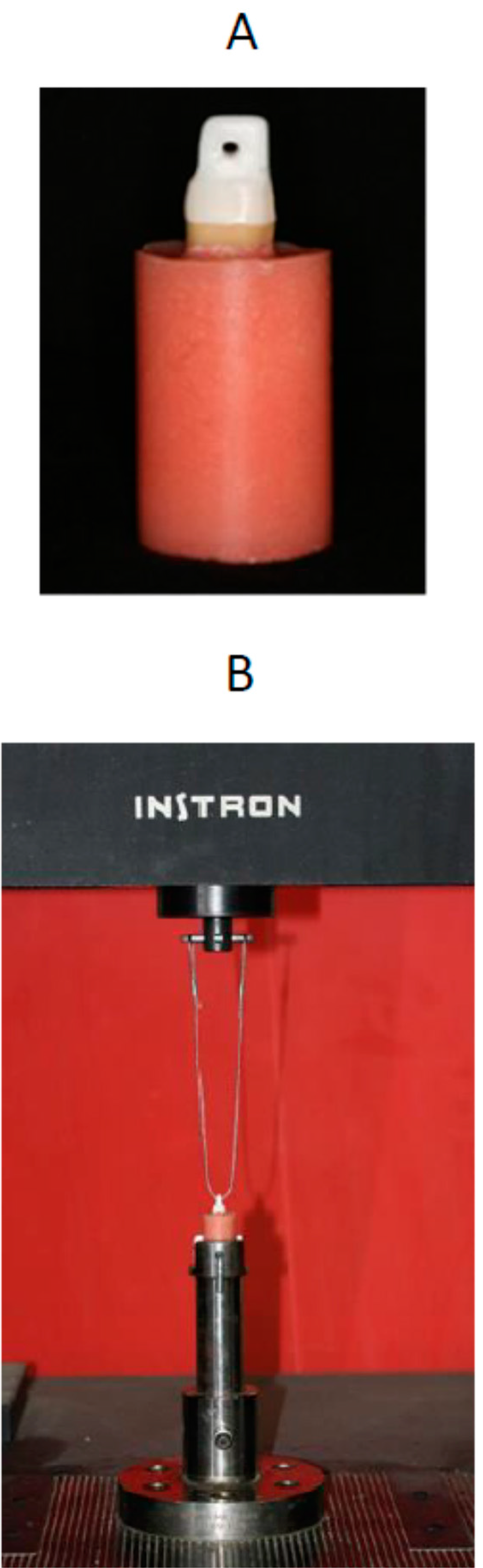
4. Experimental Section
Statistical Analysis
5. Conclusions
Author Contributions
Conflicts of Interest
References
- Rosenstiel, S.F.; Rashid, R.G. Postcementation hypersensitivity: Scientific data versus dentists’ perceptions. J. Prosthodont. 2003, 12, 73–81. [Google Scholar] [CrossRef]
- Hilton, T.; Hilton, D.; Randall, R.; Ferracane, J.L. A clinical comparison of two cements for levels of post-operative sensitivity in a practice-based setting. Oper. Dent. 2004, 29, 241–248. [Google Scholar] [PubMed]
- Magne, P. IDS: Immediate dentin sealing (IDS) for tooth preparations. J. Adhes. Dent. 2014, 16. [Google Scholar] [CrossRef]
- Gupta, N.; Reddy, U.N.; Vasundhar, P.L.; Ramarao, K.S.; Varma, K.P.; Vinod, V. Effectiveness of desensitizing agents in relieving the pre- and postcementation sensitivity for full coverage restorations: A clinical evaluation. J. Contemp. Dent. Pract. 2013, 14, 858–865. [Google Scholar] [CrossRef] [PubMed]
- Watanabe, T.; Sano, M.; Itoh, K.; Wakumoto, S. The effects of primers on the sensitivity of dentin. Dent. Mater. 1991, 7, 148–150. [Google Scholar] [CrossRef]
- Jalalian, E.; Meraji, N.; Mirzaei, M. A comparison of the efficacy of potassium nitrate and gluma desensitizer in the reduction of hypersensitivity in teeth with full-crown preparations. J. Contemp. Dent. Pract. 2009, 10, 66–73. [Google Scholar] [PubMed]
- Bavbek, A.B.; Goktas, B.; Cekic-Nagas, I.; Egilmez, F.; Ergun, G.; Eskitascioglu, G. Micro-shear bond strength of resin cement to dentin after application of desensitizing toothpastes. Acta Odontol. Scand. 2013, 71, 952–956. [Google Scholar] [CrossRef] [PubMed]
- Mausner, I.K.; Goldstein, G.R.; Georgescu, M. Effect of two dentinal desensitizing agents on retention of complete cast coping using four cements. J. Prosthet. Dent. 1996, 75, 129–134. [Google Scholar] [CrossRef]
- Johnson, G.H.; Lepe, X.; Bales, D.J. Crown retention with use of a 5% glutaraldehyde sealer on prepared dentin. J. Prosthet. Dent. 1998, 79, 671–676. [Google Scholar] [CrossRef]
- Yim, N.H.; Rueggeberg, F.A.; Caughman, W.F.; Gardner, F.M.; Pashley, D.H. Effect of dentin desensitizers and cementing agents on retention of full crowns using standardized crown preparations. J. Prosthet. Dent. 2000, 83, 459–465. [Google Scholar] [CrossRef]
- Johnson, G.H.; Hazelton, L.R.; Bales, D.J.; Lepe, X. The effect of a resin-based sealer on crown retention for three types of cement. J. Prosthet. Dent. 2004, 91, 428–435. [Google Scholar] [CrossRef] [PubMed]
- Palacios, R.P.; Johnson, G.H.; Phillips, K.M.; Raigrodski, A.J. Retention of zirconium oxide ceramic crowns with three types of cement. J. Prosthet. Dent. 2006, 96, 104–114. [Google Scholar] [CrossRef] [PubMed]
- Alves, M.; Campos, F.; Bergoli, C.D.; Bottino, M.A.; Ozcan, M.; Souza, R. Effect of adhesive cementation strategies on the bonding of Y-TZP to human dentin. Oper. Dent. 2015. [Google Scholar] [CrossRef] [PubMed]
- Hamlin, D.; Williams, K.P.; Delgado, E.; Zhang, Y.P.; DeVizio, W.; Mateo, L.R. Clinical evaluation of the efficacy of a desensitizing paste containing 8% arginine and calcium carbonate for the in-office relief of dentin hypersensitivity associated with dental prophylaxis. Am. J. Dent. 2009, 22, 16A–20A. [Google Scholar] [PubMed]
- Schiff, T.; Delgado, E.; Zhang, Y.P.; Cummins, D.; DeVizio, W.; Mateo, L.R. Clinical evaluation of the efficacy of an in-office desensitizing paste containing 8% arginine and calcium carbonate in providing instant and lasting relief of dentin hypersensitivity. Am. J. Dent. 2009, 22, 8A–15A. [Google Scholar] [PubMed]
- Ayad, F.; Ayad, N.; Zhang, Y.P.; DeVizio, W.; Cummins, D.; Mateo, L.R. Comparing the efficacy in reducing dentin hypersensitivity of a new toothpaste containing 8.0% arginine, calcium carbonate, and 1450 ppm fluoride to a commercial sensitive toothpaste containing 2% potassium ion: An eight-week clinical study on canadian adults. J. Clin. Dent. 2009, 20, 10–16. [Google Scholar] [PubMed]
- Docimo, R.; Montesani, L.; Maturo, P.; Costacurta, M.; Bartolino, M.; Zhang, Y.P.; DeVizio, W.; Delgado, E.; Cummins, D.; Dibart, S.; et al. Comparing the efficacy in reducing dentin hypersensitivity of a new toothpaste containing 8.0% arginine, calcium carbonate, and 1450 ppm fluoride to a benchmark commercial desensitizing toothpaste containing 2% potassium ion: An eight-week clinical study in Rome, Italy. J. Clin. Dent. 2009, 20, 137–143. [Google Scholar] [PubMed]
- Nathoo, S.; Delgado, E.; Zhang, Y.; DeVizio, W.; Cummins, D.; Mateo, L. Comparing the efficacy in providing instant relief of dentin hypersensitivity of a new toothpaste containing 8.0% arginine, calcium carbonate, and 1450 ppm fluoride relative to a benchmark desensitizing toothpaste containing 2% potassium ion and 1450 ppm fluoride, and to a control toothpaste with 1450 ppm fluoride: A three-day clinical study in New Jersey, USA. J. Clin. Dent. 2008, 20, 123–130. [Google Scholar]
- Garcia-Godoy, A.; Garcia-Godoy, F. Effect of an 8.0% arginine and calcium carbonate in-office desensitizing paste on the shear bond strength of composites to human dental enamel. Am. J. Dent. 2010, 23, 324–326. [Google Scholar] [PubMed]
- Wang, Y.; Liu, S.; Pei, D.; Du, X.; Ouyang, X.; Huang, C. Effect of an 8.0% arginine and calcium carbonate in-office desensitizing paste on the microtensile bond strength of self-etching dental adhesives to human dentin. Am. J. Dent. 2012, 25, 281–286. [Google Scholar] [PubMed]
- Canares, G.; Salgado, T.; Pines, M.S.; Wolff, M.S. Effect of an 8.0% arginine and calcium carbonate desensitizing toothpaste on shear dentin bond strength. J. Clin. Dent. 2012, 23, 68–70. [Google Scholar] [PubMed]
- Sailer, I.; Oendra, A.E.; Stawarczyk, B.; Hammerle, C.H. The effects of desensitizing resin, resin sealing, and provisional cement on the bond strength of dentin luted with self-adhesive and conventional resincements. J. Prosthet. Dent. 2012, 107, 252–260. [Google Scholar] [CrossRef] [Green Version]
- Stawarczyk, B.; Hartmann, R.; Hartmann, L.; Roos, M.; Ozcan, M.; Sailer, I.; Hammerle, C.H. The effect of dentin desensitizer on shear bond strength of conventional and self-adhesive resin luting cements after aging. Oper. Dent. 2011, 36, 492–501. [Google Scholar] [CrossRef] [PubMed]
- Stawarczyk, B.; Hartmann, L.; Hartmann, R.; Roos, M.; Ender, A.; Ozcan, M.; Sailer, I.; Hammerle, C.H. Impact of gluma desensitizer on the tensile strength of zirconia crowns bonded to dentin: An in vitro study. Clin. Oral Investig. 2012, 16, 201–213. [Google Scholar] [CrossRef] [PubMed] [Green Version]
- Vaz, R.R.; Hipolito, V.D.; D’Alpino, P.H.; Goes, M.F. Bond strength and interfacial micromorphology of etch-and-rinse and self-adhesive resin cements to dentin. J. Prosthodont. 2012, 21, 101–111. [Google Scholar] [CrossRef] [PubMed]
- De Mendonca, A.A.; de Oliveira, C.F.; Hebling, J.; Costa, C.A. Influence of thicknesses of smear layer on the transdentinal cytotoxicity and bond strength of a resin-modified glass-ionomer cement. Braz. Dent. J. 2012, 23, 379–386. [Google Scholar] [CrossRef] [PubMed]
- Jalandar, S.S.; Pandharinath, D.S.; Arun, K.; Smita, V. Comparison of effect of desensitizing agents on the retention of crowns cemented with luting agents: An in vitro study. J. Adv. Prosthodont. 2012, 4, 127–133. [Google Scholar] [CrossRef] [PubMed]
- De Munck, J.; Vargas, M.; van Landuyt, K.; Hikita, K.; Lambrechts, P.; van Meerbeek, B. Bonding of an auto-adhesive luting material to enamel and dentin. Dent. Mater. 2004, 20, 963–971. [Google Scholar] [CrossRef] [PubMed]
- Capa, N.; Ozkurt, Z.; Canpolat, C.; Kazazoglu, E. Shear bond strength of luting agents to fixed prosthodontic restorative core materials. Aust. Dent. J. 2009, 54, 334–340. [Google Scholar] [CrossRef] [PubMed]
- Sabatini, C.; Patel, M.; D’Silva, E. In vitro shear bond strength of three self-adhesive resin cements and a resin-modified glass ionomer cement to various prosthodontic substrates. Oper. Dent. 2013, 38, 186–196. [Google Scholar] [CrossRef] [PubMed]
- Da Silva, E.M.; Miragaya, L.; Sabrosa, C.E.; Maia, L.C. Stability of the bond between two resin cements and an yttria-stabilized zirconia ceramic after six months of aging in water. J. Prosthet. Dent. 2014, 112, 568–575. [Google Scholar] [CrossRef] [PubMed]
- Sanli, S.; Comlekoglu, M.D.; Comlekoglu, E.; Sonugelen, M.; Pamir, T.; Darvell, B.W. Influence of surface treatment on the resin-bonding of zirconia. Dent. Mater. 2015, 31, 657–668. [Google Scholar] [CrossRef] [PubMed]
- Pilo, R.; Lewinstein, I.; Ratzon, T.; Cardash, H.S.; Brosh, T. The influence of dentin and/or metal surface treatment on the retention of cemented crowns in teeth with an increased taper. Dent. Mater. 2008, 24, 1058–1064. [Google Scholar] [CrossRef] [PubMed]
- Gale, M.S.; Darvell, B.W. Thermal cycling procedures for laboratory testing of dental restorations. J. Dent. 1999, 27, 89–99. [Google Scholar] [CrossRef]


| Cement Type | Treatment | Sample No. | Mean Retentive Value (MPa) | Standard Deviation |
|---|---|---|---|---|
| RelyX U-200 | − | 10 | 2.29 | 0.55 |
| + | 10 | 2.27 | 0.64 | |
| Total | 20 | 2.28 | 0.58 | |
| RelyX Luting 2 | − | 10 | 3.16 | 0.73 |
| + | 10 | 2.92 | 0.84 | |
| Total | 20 | 3.04 | 0.77 | |
| Total | − | 20 | 2.72 | 0.77 |
| + | 20 | 2.60 | 0.80 | |
| Total | 40 | 2.66 | 0.78 |
| Classification | Description | Criteria |
|---|---|---|
| 1 | Cement principally on crown surface | Adhesive cement-dentin |
| 2 | Cement principally on dentin surface | Adhesive cement-crown |
| 3 | Cement equally distributed on dentin and crown surfaces | Cohesive cement |
| 4 | Mixed mode | Adhesive and cohesive cement |
| 5 | Fracture of the tooth | Cohesive dentin |
© 2016 by the authors; licensee MDPI, Basel, Switzerland. This article is an open access article distributed under the terms and conditions of the Creative Commons by Attribution (CC-BY) license (http://creativecommons.org/licenses/by/4.0/).
Share and Cite
Pilo, R.; Harel, N.; Nissan, J.; Levartovsky, S. The Retentive Strength of Cemented Zirconium Oxide Crowns after Dentin Pretreatment with Desensitizing Paste Containing 8% Arginine and Calcium Carbonate. Int. J. Mol. Sci. 2016, 17, 426. https://0-doi-org.brum.beds.ac.uk/10.3390/ijms17040426
Pilo R, Harel N, Nissan J, Levartovsky S. The Retentive Strength of Cemented Zirconium Oxide Crowns after Dentin Pretreatment with Desensitizing Paste Containing 8% Arginine and Calcium Carbonate. International Journal of Molecular Sciences. 2016; 17(4):426. https://0-doi-org.brum.beds.ac.uk/10.3390/ijms17040426
Chicago/Turabian StylePilo, Raphael, Noga Harel, Joseph Nissan, and Shifra Levartovsky. 2016. "The Retentive Strength of Cemented Zirconium Oxide Crowns after Dentin Pretreatment with Desensitizing Paste Containing 8% Arginine and Calcium Carbonate" International Journal of Molecular Sciences 17, no. 4: 426. https://0-doi-org.brum.beds.ac.uk/10.3390/ijms17040426