Article

Characterization of Virulence Properties of Aeromonas veronii Isolated from Diseased Gibel Carp (Carassius gibelio)

1
College of Animal Science and Technology, Yangzhou University, Yangzhou 225009, China
2
College of Ocean, Huaihai Institute of Technology, Lianyungang 222005, China
3
Bren School of Environmental Science and Management, University of California, Santa Barbara, CA 93106, USA
4
Department of Aquatic Animal Medicine, College of Fisheries, Huazhong Agricultural University, Wuhan 430070, China
*
Authors to whom correspondence should be addressed.
Int. J. Mol. Sci. 2016, 17(4), 496; https://0-doi-org.brum.beds.ac.uk/10.3390/ijms17040496
Submission received: 13 February 2016 / Revised: 16 March 2016 / Accepted: 29 March 2016 / Published: 1 April 2016
(This article belongs to the Special Issue Fish Molecular Biology)

Abstract

:
Aeromonas veronii is a kind of opportunistic pathogen to fish and humans, significantly impending aquaculture production. Recently, we isolated two A. veronii strains, named GYC1 and GYC2, from diseased Gibel carp (Carassius gibelio) in China. Based on gyrB (DNA gyrase B subunit) genes of GYC1 and GYC2, the constructed phylogenetic tree showed that the two strains were clustered with A. veronii. Sixteen virulence genes related to the pathogenicity of Aeromonas spp. were subjected to PCR assay. The genes of ompAI, ompAII, lafA, act, aer, fla, gcaT and acg were detected in the two strains, while genes of hly, ahp, lip, ast and alt were not detected. Additionally, genes eprCAI, ela and exu were only detected in the strain GYC1. Furthermore, the results of extracellular enzyme analysis revealed that the two isolates can produce hemolysin, caseinase, esterase, amylase and lecithinase, which were closely related to the pathogenicity of the two strains. However, the results showed that there was no gelatinase activity in either strain. According to the antibiotic resistant assay, the two strains were sensitive to cephalosporins and aminoglycosides, while they were resistant to penicillins and quinolones. Through this study, the virulence characteristics, including virulence genes and extracellular enzymes, the pathogenicity of A. veronii was clarified, enhancing the understanding about this pathogenic bacterium and providing the theoretical basis in disease control.

Graphical Abstract

1. Introduction

The species from the genus Aeromonas of family Aeromonadaceae are considered to be emerging pathogens and clinical attention surrounding them has risen in aquaculture as well as in avian and human health. A. hydrophila, A. veronii, A. sobria and A. caviae are the major pathogenic bacteria for Aeromoniasis [1]. Currently there are 25 species in the genus Aeromonas [2]. Among them, A. veronii, as an opportunistic pathogen, seems to have the broadest host range in virulence which has been reported to cause wound infections, diarrhea and septicemia in humans [3,4]. Additionally, A. veronii has been reported to be a digestive tract symbiont of zebra fish and medicinal leech [2,5,6,7,8,9].
It is widely known that the pathogenesis of Aeromonas infections is due to multiple virulence-related factors including biologically active substances, adhesion organs and extracellular factors such as enzymes and toxins [3,10]. These different toxins and enzymes include lipase (lip), serine protease (ser), temperature-sensitive protease (eprCAI), aerolysin (aer), ompAI, ompAII, collagenase (acg), elastase (ela), cytotonic enterotoxins (act, ast, alt), and glycerophospholipids such as cholesterol acyltransferase (gcaT), elastase (ahyB), DNases (exu), etc. These virulence-encoded genes have been widely used in determining the potential pathogenicity of Aeromonas species [11,12]. The extracellular enzymes which can cause host cell damage and degeneration would facilitate the pathogen in invading the host and establishing infection [13]. The pathogenicity of Aeromonas species is due to the combination of multiple virulence factors. Nowadays, it is hard to tell or define their role in the disease-causing process. That is to say, continuously surveying the presence of several accepted virulence-related factors in clinical Aeromonas isolates is essential to understanding the pathogenesis and epidemiology of Aeromonas [14,15,16].
Aeromonas species are ubiquitous Gram-negative bacilli found in aquatic environments. The involvement of A. veronii can cause the infected fish, with internal and surface bleeding accompanied by a high mortality rate. Gibel carp (Carassius gibelio), introduced into aquaculture as an important commercial fish species in China in the late 20th century, has been bred with a rapid growth rate. To reduce the influence of bacteria in this commercially important species is meaningful. In this study, we isolated two dominant A. veronii strains from diseased Gibel carp. Furthermore, the phylogenetic tree, virulence genes, extracellular enzymes and antibiotic sensitivity were characterized. The results will clarify molecular and phenotypic characteristics and, especially, the virulent traits of A. veronii to provide the theoretical basis in disease control.

2. Results

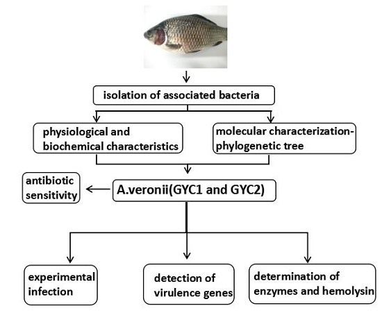
2.1. Diseased Fish and Gross Examination

Diseased Gibel carp exhibited dirty, swelling and congestion of the gill filament, and bleeding of jaw and operculum. Internally, diseased fish exhibited a distended gallbladder with some intestine and abdominal cavity effusion.

2.2. Physiological and Biochemical Characteristics

The results of the physiological and biochemical characteristics of the isolates are listed in Table 1. Additionally, the representative reactions such as H2S production, lactose, and the Voges–Proskauer (V–P) test and oxidation/fermentation (O–F) test were consistent with the data of A. veronii from Bergey’s Manual of Determinative Bacteriology [17], which were also listed in Table 1.

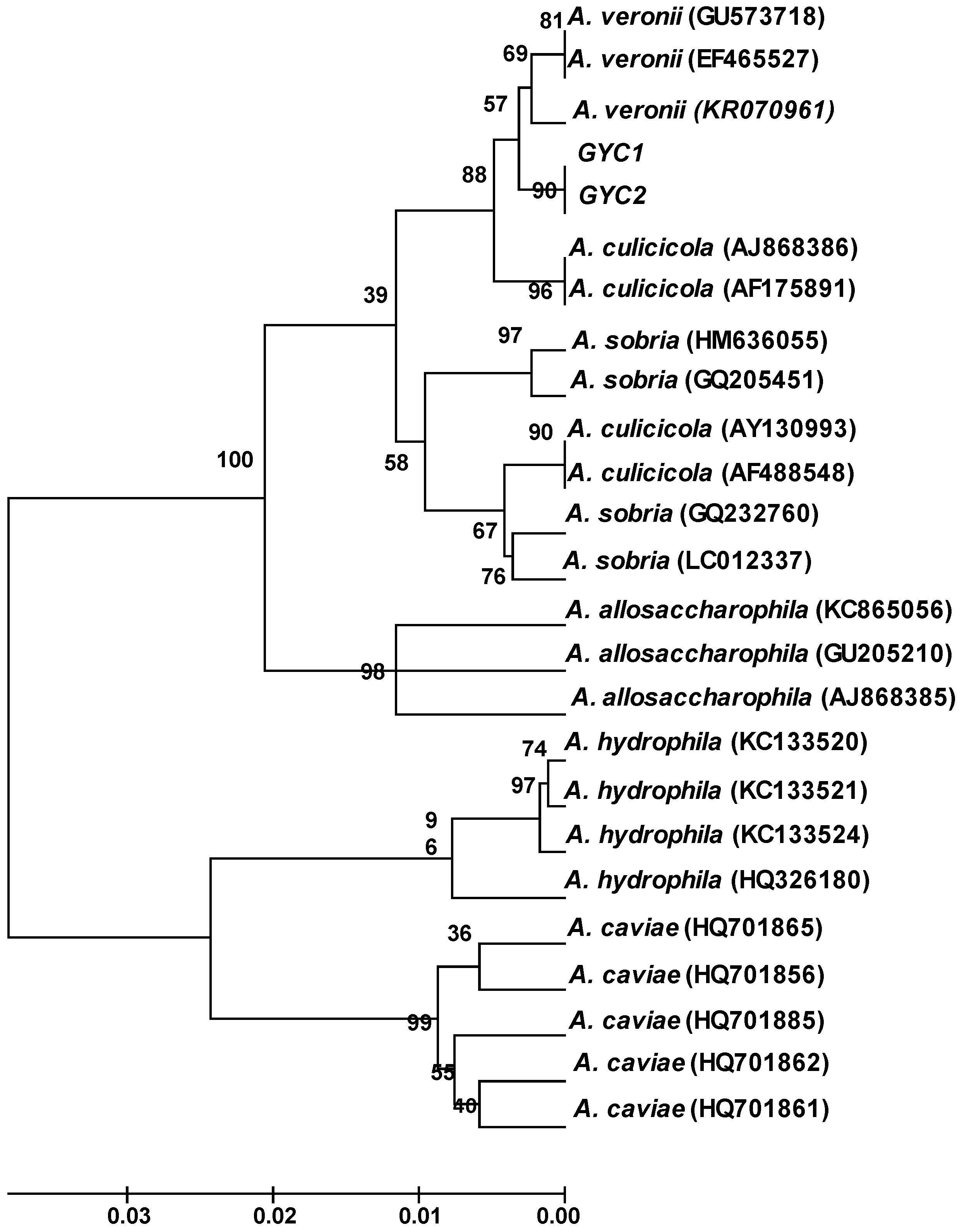
2.3. Molecular Characterization

In order to get the phylogenetic information about the studied strains GYC1 and GYC2, we determined the DNA sequences of gyrB genes from the isolates. Meanwhile, A. veronii, A. culicicola, A. sobria, A. allosaccharophila, A. hydrophila and A. caviae were chosen to build the phylogenetic tree. The results showed that GYC1 and GYC2 have been related to A. veronii, which is supported by a high bootstrap value (Figure 1). The phylogenetic tree was constructed according to Zhang et al. [18]

2.4. Experimental Infection

The fish experimentally infected with GYC1 and GYC2 showed identical symptoms as observed in the diseased Gibel carp during the disease outbreak in Ganyu County. Furthermore, A. veronii was re-isolated from the experimental infected fish, as confirmed by colonial morphology observation and the results of physiological and biochemical characteristics analysis. All fish injected with the isolates died from the fourth to seventh day post-injection. There were no clinical symptoms or death in the control groups. These results demonstrated that the isolated A. veronii was the pathogen of the Gibel carp.

2.5. Determination of Extracellular Enzymes and Hemolysin Activities

It is important to differentiate pathogenic and non-pathogenic A. veronii strains as it is only the pathogenic strains that can cause serious diseases in fish. In this study, we investigated hemolysin activity as well as lytic enzymes of the isolated strains, including caseinase, esterase, amylase, lecithinase and gelatinase, which are closely correlated with the bacterial virulence properties [19]. The results of extracellular enzymes and hemolysin activities were given in Table 2. Both strains have hemolysin activity and could produce caseinase, esterase, amylase and lecithinase. However, no gelatinase activity was detected.

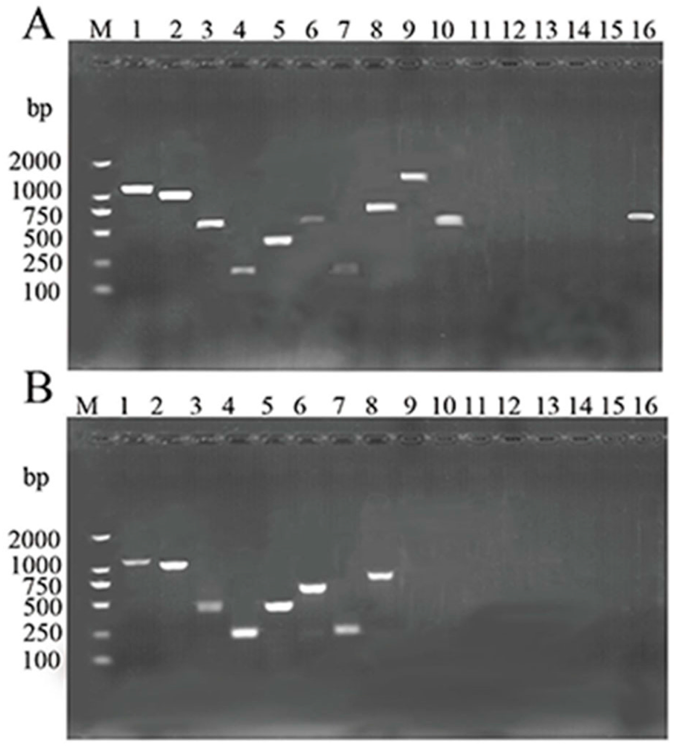
2.6. Detection of Virulence Genes

There would be either one or a couple of virulence-related genes in A. veronii isolates which participate in the development of diseases in fish. In this study, 16 virulence genes (ompAI, ompAII, lafA, act, aer, fla, gcaT, acg, eprCAI, ela, hly, ahp, lip, ast, alt, exu) were screened by PCR assay. The PCR profiles of the amplified virulence genes from the two A. veronii strains are presented in Figure 2. The genes ompAI, ompAII, lafA, act, aer, fla, gcaT and acg were detected in both the two strains, while the genes hly, ahp, lip, ast and alt were absent. However, the genes eprCAI, ela and exu were detected in the strain GYC1, but were absent in GYC2.

2.7. Antibiotic Sensitivity

The antibiotic resistance patterns of the A. veronii, valued by the size of the inhibition zones around each disc, showed that the two strains were sensitive to cephradine, cefoperazone, cefotaxime, cefuroxime, cefoxitin, cefepime, clarithromycin, gentamycin, tobramycin, streptomycin, streptomycin and chloramphenicol. However, they were resistant to oxacillin, penicillin G, cefazolin, levofloxacin, ofloxacin, ciprofloxacin, norfloxacin (Table 3).

3. Discussion

The genus Aeromonas comprises a group of Gram-negative, facultative anaerobic bacteria that are opportunistic pathogens to aquatic and terrestrial animals, including humans [20]. In this report, we isolated two A. veronii strains (GYC1, GYC2) which have caused serious diseases of Gibel carp cultured in Jiangsu Province, China. The 16S rRNA gene sequence is a commonly used index in the construction of the phylogenetic tree of bacterial genera. Nevertheless, difficulties often arise when using this technique for species identification within Aeromonas spp. due to its smaller discriminatory power [21]. Compared to 16S rRNA, the gene of gyrB is more suitable for distinguishing Aeromonas at the species level for its higher expression level. Therefore, in this report, the phylogenetic tree was built on the basis of the gyrB gene of the bacteria, and the results showed that GYC1 and GYC2 clearly have a high relatedness to A. veronii, supported by a high bootstrap value.
The determination of extracellular enzymes and hemolysin is a direct way to manifest the virulence of a bacterium. The results indicated that both strains produced caseinase, esterase, and amylase lecithinase, and they contained hemolysin activity, supporting the strong virulence of the two isolates in Gibel carp cultured in China. Furthermore, virulence factor genes are also good markers for identifying the pathogenicity of a given microorganism. In this study, we studied 16 virulence genes related to Aeromonas. The results showed that the virulence genes eprCAI, ela and exu were only detected in GYC1, indicating that even the pathogenic strains may vary in their degree of virulence. On the other hand, the ompAI, ompAII, lafA, act, aer, fla, gcaT and acg genes were present in both strains. These genes have been shown to be closely related with the pathogenicity of Aeromonas. Namba et al. [22] reported that ompA was an adhesion factor of A. veronii which was isolated from the carp intestinal tract. Gao et al. [23] found that mice that received ompA-hly antigen-loaded poly(lactic-co-glycolic) acid (PLGA) microspheres by intraperitoneal or intragastric administration mounted a strong and sustained IgG response. Lateral (laf) flagella are important in certain Aeromonas species for the adherence process and biofilm formation [24,25]. Kirov et al. [25] showed that, in general, all species of mutant Aeromonad defective in genes fla demonstrate a sharply decreased ability to form biofilms compared with the wild types. Additionally, A. caviae strains lacking lateral flagella resulted in a 60% decrease in adhesion to cells of the intestinal cell lines Henle 407 or Caco-2. In the research of Sen et al. [15], the swarming motility of tested strains corresponded with the presence of lafA in all A. hydrophila, A. caviae, and A. veronii. At the same time, only those strains that had one or more of the enterotoxins flaA, flaB, and either flaG or lafA, showed signs of being virulent. Han et al. [19] indicated the involvement of the collagenase gene in the pathogenesis of A. veronii. Adhesion and invasion abilities of the mutant strain on epithelioma papillosum of carp cells were only 56% of that of the wild-type strain, and the cytotoxicity was only 42%. It is noticeable that the genes hly, ahp, lip, ast and alt were absent in our two A. veronii strains. These results are consistent to some degree with Mohamed et al. [26], who found that in the 81 strains of A. veronii isolated from farm-raised catfish, none of the isolates contained the ast or alt gene. Based on our results, there may be an effective way to build an A. veronii detection method according to the mutual virulence genes, and there may even be an applicable way to construct a multivalent vaccine against the A. veronii infection.
The antibiotic sensitivity test demonstrated that both strains were sensitive to cephradine, cefoperazone, cefotaxime, cefuroxime, cefoxitin, cefepime, clarithromycin, gentamycin, tobramycin, streptomycin, streptomycin and chloramphenicol. Overall, the two strains were sensitive to cephalosporins and aminoglycosides. Antibiotics, no doubt, are an economical and effective option to fight against bacteria. However, the wide usage of antibiotics in clinical practice, veterinary medicine and agriculture has resulted in the release of large amounts of these pollutants to the environment [27]. Antibiotics, in addition to being chemical pollutants, exert a selective pressure retaining and spreading the various antibiotic resistance genes (ARG) among microbiota, which poses a risk to human health [28]. For example, the two isolated A. veronii strains already extended resistance to oxacillin, penicillin G, cefazolin, levofloxacin, ofloxacin, ciprofloxacin, and norfloxacin which were widely used in aquaculture. Instead of using antibiotics, the development of more effective strategies to fight bacterial infections is greatly expected in the future.

4. Materials and Methods

4.1. Diseased Fish and Gross Examination

The diseased Gibel carp were collected from Ganyu County, Jiangsu Province, China. The surfaces of each fish were examined for lesions and signs of physical damage. Post-mortem internal examinations were then conducted and wet mounts were taken from scrapes of the gills and skin and examined for the presence of protozoan and metazoan parasites.

4.2. Isolation of the Associated Bacteria

Abdominal cavities of diseased fish were opened up after surface sterilization with 70% ethanol. Samples were taken aseptically with scissors and tweezers from the liver, kidney and spleen tissues of freshly dead fish. The samples were streaked onto nutrient agar plate and incubated at 28 °C for 24 h. After incubation, bacterial colonies were selected based on the size, shape and color and purified before being stored at −80 °C in nutrient broth with 30% glycerol prior to identification.

4.3. Identification of the Isolates

4.3.1. Physiological and Biochemical Characteristics

Physiological and biochemical examinations were carried out using standard plate and tube tests (Hangzhou Tianhe Microorganism Reagent Co., Ltd., Hangzhou, China) referring to Bergey’s Manual of Systematic Bacteriology [17], including the following tests: arabitol, mannose, maltose, H2S production, tartrate utilization, V–P test, xylose, mannitol, sucrose, galactosidase, aesculin, dulcitol, lactose, inositol, mushroom sugar, nitrate reduction, acetate, α-methyl-d-glucoside, galactose, sorbitol, O–F test, erythrite, l-rhamnose. The two strains were tested in twice and a third experiment would be carried out to eliminate the discrepancies when the results were different. The reactions were compared with the results of A. veronii from Bergey’s Manual of Systematic Bacteriology.

4.3.2. Molecular Characterization

The chromosomal DNA from A. veronii was extracted using the EasyPure Genomic DNA Kit (Transgen Biotech, Beijing, China) in accordance with the manufacturer’s instructions. A partial 16S rRNA and gyrB genes sequence were amplified as described previously [18]. Briefly, universal PCR primers 27F (5′-AGAGTTTGATCMTGGCTCAG-3′) and 1492R (5′-TACGGMTACCTTGTTACGACTT-3′) were used for amplification of the 16S rRNA gene [29], while universal PCR primers UP1 (5′-GAAGTCATCATGACCGTTCTGCAYGCNGGNGGNAARTTYGA-3′) and UP2r (5′-AGCAGGGTACGGATGTGCGAGCCRTCNACRTCNGCRTCNGTCAT-3′) were used for amplification of the gyrB gene [30]. Polymerase chain reaction (PCR) amplification was performed in a total volume of 25 μL containing the appropriate reaction buffer and reagents: 2× EasyTaq PCR SuperMix (Transgen Biotech, Beijing, China) 12.5 μL, 1 μL (10 μM) forward and reverse primers, respectively, 9.5 μL ddH2O and 1 μL DNA as template. The PCR cycling protocol was as follows: a first denaturation at 95 °C for 5 min, followed by 30 cycles of denaturation at 95 °C for 1 min, annealing at 55 °C for 1 min and extension at 72 °C for 1 min. After a final extension at 72 °C for 10 min, the tubes were cooled to 4 °C. The amplification of 16S rRNA and gyrB genes and the construction of phylogenetic trees using MEGA4 were conducted according to Zhang et al. [18].

4.3.3. Nucleotide Accession Numbers

The partial nucleotide sequences of 16S rRNA and gyrB locus of A. veronii strains were submitted to the GeneBank. The accession number of 16S rRNA and gyrB genes for GYC1 was KU543614 and KU543616, respectively, while the accession number of 16S rRNA and gyrB genes for GYC2 was KU543615 and KU543617, respectively.

4.4. Evaluation the Virulence of Isolates

4.4.1. Experimental Infection

To confirm the pathogenicity of GYC1 and GYC2, experimental infections were conducted. Seventy healthy Gibel carp were purchased from fish farms located in Lianyungang City of Jiangsu Province, China, and have been maintained in still water supplemented with oxygen in 200 L aquaria at 20 °C for 10 days. Groups of 10 healthy Gibel carp (average 10–15 cm in length) were injected intraperitoneally with 0.1 mL live cells (containing 106, 107 or 108 CFU·mL−1, respectively) suspended in saline per fish, while the other 10 fish were similarly injected with 0.1 mL saline and were used as negative controls. All mortalities of inoculated fish kept at 20 °C were recorded lasting 10 days. Dead and moribund fish were removed for pathological examination and external and internal signs of disease were recorded. Samples of the liver, spleen and kidney were streaked onto 2216E marine agar plates and blood agar plates for bacteriological examination. The surviving fish were euthanized in the end of the experiment.

4.4.2. Determination of Extracellular Enzymes and Hemolysin Activities

The presence of extracellular enzymes and hemolysin of the isolates were determined according to the methods reported by Yang et al. [31]. Hemolytic activity of the bacteria were detected by streaking onto the rabbit blood nutrient agar plates and incubated at 28 °C for 24 h. The presence of a clear colorless zone surrounding the colonies indicated β-hemolytic activity. Likewise, incomplete transparent zone indicates α-hemolytic activity.
Casein hydrolysis, amylase, gelatinase, lipase activity and lecithin hydrolysis were tested on nutrient agar containing 10% skimmed milk, 1% starch, 8% gelatin, 1% Tween 80 and 1% egg yolk, respectively. The extracellular enzymes were assayed by streaking the cells onto the plates and incubating at 28 °C for 24 h. The presence of a transparent zone surrounding the colonies indicated the particular enzyme activity. All treatments were performed in triplicate.

4.4.3. Detection of Virulence Genes

The two A. veronii strains were subjected to PCR assays to detect the 16 genes encoding the toxins, including ompAI, ompAII, lateral flagella (lafA), cytotonic enterotoxins (act, ast, alt), aerolysin (aer), the structural gene flagellin (fla), glycerophospholipid: cholesterol acyltransferase (gcaT), collagenase (acg), temperature-sensitive protease (eprCAI), elastase (ela), hemolysin (hly), serine protease (ahp), lipase (lip), DNases (exu). Oligonucleotide primers, target genes, and amplicon sizes are shown in Table 4. The final concentrations in the PCR mixture were as follows: 2× EasyTaq PCR SuperMix 12.5, 1 μL (10 μM) forward and reverse primers, respectively, 9.5 μL ddH2O and 1 μL DNA as template. The thermocycling program was optimized as follows: a first denaturation at 95 °C for 5 min, then 30 cycles of denaturation at 95 °C for 1 min, annealing at 55 °C for 1 min and extension at 72 °C for 1 min. After a final extension at 72 °C for 10 min, the tubes were cooled to 4 °C. Aliquots from amplification reactions were analyzed by 1% agarose gel electrophoresis.

4.5. Antibiotic Sensitivity

The sensitivity of the two strains to 31 antibiotics listed in Table 3 was evaluated by disc diffusion method [36] on Muller-Hinton agar using commercial antibiotic discs (Hangzhou Tianhe Microorganism Reagent Co.). All the tests were performed in triplicate. Antibiotic sensitivity of the strains as resistant, intermediate or sensitive was measured by the size of the inhibition zones around each disc according to standards suggested by Hangzhou Tianhe Microorganism Reagent Co.

5. Conclusions

In this study, virulence properties of two bacterial strains (GYC1 and GYC2) from diseased Gibel carp in China were characterized. The physiological and biochemical characteristics of the isolates were consistant with the data of A. veronii from Bergey’s Manual of Determinative Bacteriology [17]. The constructed phylogenetic tree based on gyrB (DNA gyrase B subunit) genes of GYC1 and GYC2 showed that the two strains were clustered with A. veronii. Through PCR assay, 16 genes related to Aeromonas virulence were studied and the genes ompAI, ompAII, lafA, act, aer, fla, gcaT and acg were found in both strains. Furthermore, the results of extracellular enzymes analysis revealed that the two isolates could produce hemolysin, caseinase, esterase, amylase and lecithinase while no gelatinase activity was detected in either strain. Experimental infection showed that the mortality of fish injected with the isolated bacteria was 100% within 10 days and the control group was zero, furtherly confirming the pathogenicity of the isolates. The antibiotic resistant assay revealed that the two strains were sensitive to cephalosporins and aminoglycosides, while they were resistant to penicillins and quinolones.

Acknowledgments

This work was supported by the Science Research Foundation of the Education Ministry for Returned Chinese Scholars, the Major Natural Science Research Program of Jiangsu Higher Education Institutions (14KJA240001), and the Projects of Shui Chan San Xin of Jiangsu Province (Y2014-35, D2015-13-S3). The Fundamental Research Funds for the Central Universities (2013PY069, 2014PY035) and the Special funds for science and technology from Hubei Province (2015BBA228).

Author Contributions

Xiaojun Zhang designed the research. Xiaojun Zhang and Li Lin finalized the paper writing. Jingjing Sun, Xiaojian Gao, Qun Jiang, and Yi Wen performed the experiments, contributed to the data collection and statistical analysis.

Conflicts of Interest

The authors declare no conflict of interest.

References

  1. Wahli, T.; Burr, S.E.; Pugovkin, D.; Mueller, O.; Frey, J. Aeromonas Sobria, a causative agent of disease in farmed perch, Perca Fluviatilis L. J. Fish Dis. 2005, 28, 141–150. [Google Scholar] [CrossRef] [PubMed]
  2. Krishnan, S.; Philip, R.; Singh, I.S. Characterization and virulence potential of phenotypically diverse Aeromonas veronii isolates recovered from moribund freshwater ornamental fishes of Kerala, India. Antonie Leeuwenhoek 2013, 103, 53–67. [Google Scholar]
  3. Janda, J.M.; Abbott, S.L. The genus Aeromonas: Taxonomy, pathogenicity, and infection. Clin. Microbiol. Rev. 2010, 23, 35–73. [Google Scholar] [CrossRef] [PubMed]
  4. Silver, A.C.; Williams, D.; Faucher, J.; Horneman, A.J.; Gogarten, P.; Graf, J. Complex evolutionary history of the Aeromonas veronii group revealed by host interaction and DNA sequence data. PLoS ONE 2011, 6, e1675. [Google Scholar] [CrossRef] [PubMed]
  5. Rahman, M.; Navarro, C.P.; Kuhn, I.; Huys, G.; Swings, J.; Mollby, R. Identification and characterization of pathogenic Aeromonas veronii biovar sobria associated with epizootic ulcerative syndrome in fish in Bangladesh. Pak. J. Biol. Sci. 2002, 68, 650–655. [Google Scholar] [CrossRef]
  6. Bates, J.M.; Mittge, E.; Kuhlman, J.; Baden, K.N.; Cheesman, S.E. Distinct signals from the microbiota promote different aspects of zebrafish gut differentiation. Dev. Biol. 2006, 297, 374–386. [Google Scholar] [CrossRef] [PubMed]
  7. Graf, J.; Kikuchi, Y.; Rio, R.V. Leeches and their microbiota: Naturally simple symbiosis models. Trends Microbiol. 2006, 14, 365–371. [Google Scholar] [CrossRef] [PubMed]
  8. Hossain, M. Isolation of pathogenic bacteria from the skin ulcerous symptomatic gourami (Colisa lalia) through 16S rDNA analysis. Univ. J. Zool. Rajshahi Univ. 2008, 27, 21–24. [Google Scholar] [CrossRef]
  9. Ma, Z.; Yang, H.; Li, T.; Luo, L.; Gao, J. Isolation and identification of pathogenic Aeromonas veronii isolated from infected Siberian sturgeon (Acipenser baerii). J. Microbiol. 2009, 49, 1289–1294. [Google Scholar]
  10. Von Gravenitz, A. The role of Aeromonas in diarrhea: A review. Infection 2007, 35, 59–64. [Google Scholar] [CrossRef] [PubMed]
  11. Li, J.; Ni, X.D.; Liu, Y.J.; Lu, C.P. Detection of three virulence genes alt, ahp and aerA in Aeromonas hydrophila and their relationship with actual virulence to zebrafish. J. Appl. Microbiol. 2011, 110, 823–830. [Google Scholar] [CrossRef] [PubMed]
  12. Yi, S.W.; You, M.J.; Cho, H.S.; Lee, C.S.; Kwon, J.K.; Shin, G.W. Molecular characterization of Aeromonas species isolated from farmed eels (Anguilla Japonica). Vet. Microbiol. 2013, 164, 195–200. [Google Scholar] [CrossRef] [PubMed]
  13. Rodriguez, L.A.; Ellis, A.E.; Nieto, T.P. Purification and characterization of an extracellular metalloprotease, serine protease and haemolysin of Aeromonas hydrophila strain B32: All are lethal for fish. Microb. Pathog. 1992, 13, 17–24. [Google Scholar] [CrossRef]
  14. Chaco´n, M.R.; Figueras, M.J.; Castro-Escarpulli, G.; Soler, L.; Guarro, J. Distribution of virulence genes in clinical and environmental isolates of Aeromonas spp. Antonie Leeuwenhoek 2003, 84, 269–278. [Google Scholar] [CrossRef]
  15. Sen, K.; Rodgers, M. Distribution of six virulence factors in Aeromonas species isolated from U.S. drinking water utilities: A PCR identification. J. Appl. Microbiol. 2004, 97, 1077–1086. [Google Scholar] [CrossRef] [PubMed]
  16. Aguilera-Arreola, M.G.; Hernández-Rodríguez, C.; Zúñiga, G.; Figueras, M.J.; Castro-Escarpulli, G. Aeromonas hydrophila clinical and environmental ecotypes as revealed by genetic diversity and virulence genes. FEMS Microbiol. Lett. 2005, 242, 231–240. [Google Scholar] [CrossRef] [PubMed]
  17. Brenner, D.J.; Krieg, N.R.; Staley, J.T. Bergey’s Manual of Systematic Bacteriology, 2nd ed.; Springer: Berlin, Germany, 2008; Volume 2, pp. 546–551. [Google Scholar]
  18. Zhang, X.J.; Qin, G.M.; Bing, X.W.; Yan, B.L.; Liang, L.G. Molecular and phenotypic characterization of Vibrio aestuarianus, a pathogen of the cultured tongue sole, Cynoglossus semilaevis Gunther. J. Fish. Dis. 2011, 34, 57–64. [Google Scholar] [CrossRef] [PubMed]
  19. Hyun-Ja, H.; Tatsuo, T.; Hidehiro, K.; Ikuo, H.; Takashi, A. Pathogenic potential of a collagenase gene from Aeromonas veronii. Can. J. Microbiol. 2008, 54, 1–10. [Google Scholar]
  20. Van der Marel, M.; Schroers, V.; Neuhaus, H.; Steinhagen, D. Chemotaxis towards, adhesion to, and growth in carp gut mucus of two Aeromonas hydrophila strains with different pathogenicity for common carp, Cyprinus carpio L. J. Fish Dis. 2008, 31, 321–330. [Google Scholar] [CrossRef] [PubMed]
  21. Yánez, M.A.; Catalán, V.; Apráiz, D.; Figueras, M.J.; Martínez-Murcia, A.J. Phylogenetic analysis of members of the genus Aeromonas based on gyrB gene sequences. Int. J. Syst. Evol. Microbiol. 2003, 53, 875–883. [Google Scholar] [CrossRef] [PubMed]
  22. Namba, A.; Mano, N.; Takano, H.; Beppu, T.; Ueda, K.; Hirose, K. OmpA is an adhesion factor of Aeromonas veronii, an optimistic pathogen that habituates in carp intestinal tract. J. Appl. Microbiol. 2008, 105, 1441–1451. [Google Scholar] [CrossRef] [PubMed]
  23. Gao, S.S.; Zhao, N.; Amer, S.; Qian, M.M.; Lv, M.X.; Zhao, Y.L.; Su, X.; Cao, J.; He, H.X.; Zhao, B.H. Protective efficacy of PLGA microspheres loaded with divalent DNA vaccine encoding the ompA gene of Aeromonas veronii and the hly gene of Aeromonas hydrophila in mice. Vaccine 2013, 31, 5754–5762. [Google Scholar] [CrossRef] [PubMed]
  24. Gavin, R.; Rabaan, A.A.; Merino, S.; Tomas, J.M.; Gryllos, I.; Shaw, J.G. Lateral flagella of Aeromonas species are essential for epithelial cell adherence and biofilm formation. Mol. Microbiol. 2002, 43, 383–397. [Google Scholar] [CrossRef] [PubMed]
  25. Kirov, S.M.; Castrisios, M.; Shaw, J.G. Aeromonas flagella (polar and lateral) are enterocyte adhesins that contribute to biofilm formation on surfaces. Infect. Immun. 2004, 72, 1939–1945. [Google Scholar] [CrossRef] [PubMed]
  26. Nawaz, M.; Khan, S.A.; Khan, A.A.; Sung, K.; Tran, Q.; Kerdahi, K.; Steele, R. Detection and characterization of virulence genes and integrons in Aeromonas veronii isolated from catfish. Food Microbiol. 2010, 27, 327–331. [Google Scholar]
  27. Cantas, L.; Shah, S.Q.; Cavaco, L.M.; Manaia, C.M.; Walsh, F.; Popowska, M.; Garelick, H.; Bürgmann, H.; Sørum, H. A brief multidisciplinary review on antimicrobial resistance in medicine and its linkage to the global environmental microbiota. Front. Microbiol. 2013, 4. [Google Scholar] [CrossRef] [PubMed] [Green Version]
  28. Kemper, N. Veterinary antibiotics in the aquatic and terrestrial environment. Ecol. Indic. 2008, 8, 1–13. [Google Scholar] [CrossRef]
  29. Polz, M.F.; Cavanaugh, C.M. Bias is template to product ratios in multitemplate PCR. Appl. Environ. Microbial. 1998, 64, 3724–3730. [Google Scholar]
  30. Yamamoto, S.; Harayama, S. PCR amplification and direct sequencing of gyrB genes with universal primers and their application to the detection and taxonomic analysis of Pseudomonas putida strains. Appl. Environ. Microbiol. 1995, 61, 1104–1109. [Google Scholar] [PubMed]
  31. Yang, Z.S.; Fang, H. Human and Animal Pathogenic Bacteriology; Hebei Science and Technology Press: Shijiazhuan, China, 2003; pp. 1550–1610. [Google Scholar]
  32. Wang, H. Comparative Study between Biological Characteristics of the Different Animal Species Aeromonas veronii and Four Virulence Genes. Master’s Thesis, Agricultural University, Jilin, China, April 2014. [Google Scholar]
  33. Merino, S.; Gavin, R.; Vilches, S.; Shaw, J.G.; Tomas, J.M. A colonization factor (production of lateral flagella) of mesophilic Aeromonas spp. is inactive in Aeromonas salmonicida strains. Appl. Environ. Microbiol. 2003, 69, 663–667. [Google Scholar] [CrossRef] [PubMed]
  34. Ren, Y.; Lu, C.P.; Yao, H.C. Cloning, sequence analysis and detection of an extracellular temperaturelabile protease encoding gene (eprJ) from Aeromonas hydrophila. J. Fish Sci. China 2006, 6, 924–927. [Google Scholar]
  35. Wong, C.Y.F.; Heuzenroeder, M.W.; Flower, R.L.P. Inactivation of two haemolytic toxin genes in Aeromonas hydrophila attenuates virulence in a suckling mouse model. Microbiology UK 1998, 144, 291–298. [Google Scholar] [CrossRef] [PubMed]
  36. Igbinosa, I.H.; Chigor, V.N.; Igbinosa, E.O.; Obi, L.C.; Okoh, A.I. Antibiogram, adhesive characteristics, and incidence of class 1 integron in Aeromonas species isolated from two South African rivers. Biomed. Res. Int. 2013. [Google Scholar] [CrossRef] [PubMed]
Figure 1. Phylogenetic tree based on the partial gyrB gene sequences (numbers in tree are bootstrap values).
Ijms 17 00496 g001
Figure 2. Agarose gel electrophoresis of 1% agarose of the amplification products of isolates GYC1 and GYC2 isolated from Gibel carp. M, Trans2K DNA Marker; lane 1, ompAI; lane 2, ompAII; lane 3, lafA; lane 4, act; lane 5, aer; lane 6, fla; lane 7, gcaT; lane 8, acg; lane 9, eprCAI; lane 10, ela; lane 11, hly; lane 12, ahp; lane 13, lip; lane 14, ast; lane 15, alt; lane 16, exu; (A) GYC1; (B) GYC2.
Ijms 17 00496 g002
Table 1. The represented physiological and biochemical characteristics of the isolates.
ItemsIsolatesA. veronii
GYC1GYC2
Arabitol
Mannose+++
Maltose+++
H2S production
Tartrate utilization
V–P test+++
Xylose
Mannitol+++
Sucrose+++
Galactosidase++[+]
Aesculin+++
Dulcitol
Lactose
Inositol
Mushroom sugar+++
Nitrate reduction+++
Acetate[+]
α-methyl-d-glucoside[+]
Galactose+++
Sorbitol
O–F testFFF
Erythrite
l-Rhamnose
“+”, positive; “−”, negative; “F”, fermentative; ”[+]”, 76%–89% of the strains are positive; V–P test: Voges–Proskauer test; O–F test: oxidation/fermentation test.
Table 2. Production of extracellular enzymes and hemolysin activity of the isolated A. veronii.
IsolatesCaseinaseEsteraseAmylaseLecithinaseHemolysinGelatinase
GYC1++++β-hemolysis
GYC2++++β-hemolysis
“+”, positive; “"−”, negative.
Table 3. Antibiotic sensitivity of the two isolates.
GroupsChemicalsDisc Content (μg)Mean Inhibition Zone Diameter (mm)Sensitivity a
GYC1GYC2GYC1GYC2
PenicillinsOxacillin10.00.0RR
Piperacillin10026.00.0SR
Penicillin G1015.00.0RR
Ampicillin1013.00.0IR
CephalosporinsCephradine3025.024.0SS
Cefoperazone7526.022.0SS
Cefalotin3020.00.0SR
Cefotaxime3027.025.0SS
Cefuroxime3025.024.0SS
Cefoxitin3020.020.0SS
Cefepime3026.025.0SS
Cefazolin3014.014.0RR
MacrolidesMidecamycin3016.014.0II
Clarithromycin1525.020.0SS
Erythromycin1527.020.0SI
QuinolonesLevofloxacin50.011.0RR
Ofloxacin59.010.0RR
Ciprofloxacin59.00.0RR
Norfloxacin100.010.0RR
AminoglycosidesGentamycin1019.016.0SS
Tobramycin1018.020.0SS
Streptomycin1022.015.0SS
Streptomycin3020.018.0SS
Amikacin3019.016.5SI
Spectinomycin10027.00.0SR
LincomycinsClindamycin215.012.5IR
AmphenicolsChloramphenicol3022.022.0SS
PolymyxinPolymyxin B30014.00.0SR
NitrofuranMacrodantin30018.016.0SI
AztreonamAztreonam3026.010.0SR
GlycopeptidesVancomycin3011.010.0RR
a: R, resistance; S, sensitive; I, intermediate.
Table 4. PCR primers, targets, and amplicon sizes used for this study.
Target GeneProduct Size (bp)PCR Primers Sequence (5′-3′)Reference
ompAI1026F: GACGATATCATGATGAAAATGGCTCTTWang Hui [32]
R: GCGAAGCTTTTACTTCTGAACTTCTTG
ompAII1001F: GCTGAATTCATGAAACTCAAAATGGCTCWang Hui [32]
R: GCGAAGCTTTTACTGTTGTACTTGC
lafA550F: GGTCTGCGCATCCAACTCMerino et al. [33]
R: GCTCCAGACGGTTGATG
act232F: AGAAGGTGACCACCACCAAGAACAMohamed et al. [26]
R: AACTGACATCGGCCTTGAACTC
aer431F: CCTATGGCCTGAGCGAGAAGMohamed et al. [26]
R: CCAGTTCCAGTCCCACCACT
fla608F: TCCAACCGTYTGACCTCMohamed et al. [26]
R: GMYTGGTTGCGRATGGT
gcaT237F: CTCCTGGAATCCCAAGTATCAGMohamed et al. [26]
R: GGCAGGTTGAACAGCAGTATCT
acg761F: AACAAGCACCCGTTAAGCCACHan et al. [19]
R: ACGTAGTCGAGCCCCTTGAGG
eprCAI387F: GCTCGACGCCCAGCTCACCRen et al. [34]
R: GGCTCACCGCATTGGATTCG
ela513F: ACACGGTCAAGGAGATCAACSen and Rodgers [15]
R: CGCTGGTGTTGGCCAGCAGG
hly597F: GGCCGGTGGCCCGAAGATACGGGWong et al. [35]
R: GGCGGCGCCGGACGAGACGGG
ahp911F: ATTGGATCCCTGCCTALi et al. [11]
R: GCTAAGCTTGCATCCG
lip382F: ATCTTCTCCGACTGGTTCGGSen and Rodgers [15]
R: CCGTGCCAGGACTGGGTCTT
ast331F: TCTCCATGCTTCCCTTCCACTMohamed et al. [26]
R: GTGTAGGGATTGAAGAAGCCG
alt442F: TGACCCAGTCCTGGCACGGCYang and Fang [31]
R: GGTGATCGATCACCACCAGC
exu323F: AGACATG CACAACCTCTTCCYang and Fang [31]
R: GATTGGTATTGCCTTGCAAG

