
Plasma Lipoprotein-Associated Phospholipase A2 Levels Correlated with the Cardio-Ankle Vascular Index in Long-Term Type 2 Diabetes Mellitus Patients

Abstract
:1. Introduction
2. Methods
3. Results
4. Discussion
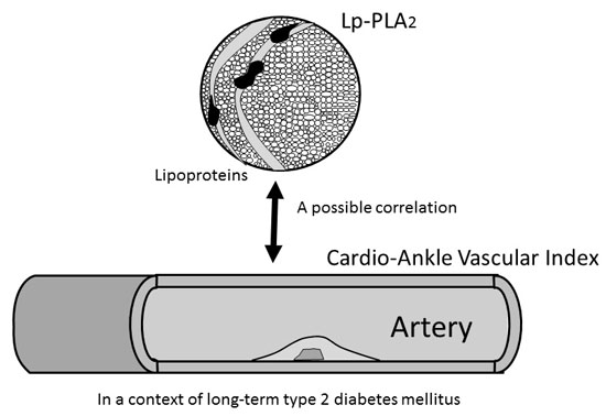
5. Conclusions
Acknowledgments
Author Contributions
Conflicts of Interest
References
- Stehouwer, C.D.; Henry, R.M.; Ferreira, I. Arterial stiffness in diabetes and the metabolic syndrome: A pathway to cardiovascular disease. Diabetologia 2008, 51, 527–539. [Google Scholar] [CrossRef] [PubMed]
- Christen, A.I.; Armentano, R.L.; Miranda, A.; Graf, S.; Santana, D.B.; Zócalo, Y.; Baglivo, H.P.; Sánchez, R.A. Arterial wall structure and dynamics in type 2 diabetes mellitus methodological aspects and pathophysiological findings. Curr. Diabetes Rev. 2010, 6, 367–377. [Google Scholar] [CrossRef] [PubMed]
- Lerman, A.; McConnell, J.P. Lipoprotein-associated phospholipase A2: A risk marker or a risk factor? Am. J. Cardiol. 2008, 101, 11F–22F. [Google Scholar] [CrossRef] [PubMed]
- Vittos, O.; Toana, B.; Vittos, A.; Moldoveanu, E. Lipoprotein-associated phospholipase A2 (Lp-PLA2): A review of its role and significance as a cardiovascular biomarker. Biomarkers 2012, 17, 289–302. [Google Scholar] [CrossRef] [PubMed]
- Lp-PLA2 Studies Collaboration; Thompson, A.; Gao, P.; Orfei, L.; Watson, S.; di Angelantonio, E.; Kaptoge, S.; Ballantyne, C.; Cannon, C.P.; Criqui, M.; et al. Lipoprotein-associated phospholipase A2 and risk of coronary disease, stroke, and mortality: Collaborative analysis of 32 prospective studies. Lancet 2010, 375, 1536–1544. [Google Scholar]
- Hatoum, I.J.; Hu, F.B.; Nelson, J.J.; Rimm, E.B. Lipoprotein-associated phospholipase A2 activity and incident coronary heart disease among men and women with type 2 diabetes. Diabetes 2010, 59, 1239–1243. [Google Scholar] [CrossRef] [PubMed]
- Onat, A.; Akbaş, T.; Yüksel, H. An inverse-to-anticipated relationship of Lp-PLA2 activity in diabetes: Reflection of underlying autoimmune activation. Eur. J. Intern. Med. 2015, 26, 72. [Google Scholar] [CrossRef] [PubMed]
- Davidson, M.H.; Corson, M.A.; Alberts, M.J.; Anderson, J.L.; Gorelick, P.B.; Jones, P.H.; Lerman, A.; McConnell, J.P.; Weintraub, H.S. Consensus panel recommendation for incorporating lipoprotein-associated phospholipase A2 testing into cardiovascular disease risk assessment guidelines. Am. J. Cardiol. 2008, 101, 51F–57F. [Google Scholar] [CrossRef] [PubMed]
- Maiolino, G.; Bisogni, V.; Rossitto, G.; Rossi, G.P. Lipoprotein-associated phospholipase A2 prognostic role in atherosclerotic complications. World J. Cardiol. 2015, 7, 609–620. [Google Scholar] [CrossRef] [PubMed]
- Talmud, P.J.; Holmes, M.V. Deciphering the causal role of sPLA2s and Lp-PLA2 in coronary heart disease. Arterioscler. Thromb. Vasc. Biol. 2015, 35, 2281–2289. [Google Scholar] [CrossRef] [PubMed]
- Gregson, J.; Stirnadel-Farrant, H.A.; Doobaree, I.U.; Koro, C. Variation of lipoprotein associated phospholipase A2 across demographic characteristics and cardiovascular risk factors: A systematic review of the literature. Atherosclerosis 2012, 225, 11–21. [Google Scholar] [CrossRef] [PubMed]
- Shirai, K.; Utino, J.; Otsuka, K.; Takata, M. A novel blood pressure-independent arterial wall stiffness parameter; cardio-ankle vascular index (CAVI). J. Atheroscler. Thromb. 2006, 13, 101–107. [Google Scholar] [CrossRef] [PubMed]
- Kotani, K.; Miyamoto, M.; Taniguchi, N. Clinical significance of the cardio-ankle vascular index (CAVI) in hypertension. Curr. Hypertens. Rev. 2010, 6, 251–253. [Google Scholar] [CrossRef]
- Shirai, K.; Hiruta, N.; Song, M.; Kurosu, T.; Suzuki, J.; Tomaru, T.; Miyashita, Y.; Saiki, A.; Takahashi, M.; Suzuki, K.; et al. Cardio-ankle vascular index (CAVI) as a novel indicator of arterial stiffness: Theory, evidence and perspectives. J. Atheroscler. Thromb. 2011, 18, 924–938. [Google Scholar] [CrossRef] [PubMed]
- Namekata, T.; Suzuki, K.; Ishizuka, N.; Shirai, K. Establishing baseline criteria of cardio-ankle vascular index as a new indicator of arteriosclerosis: A cross-sectional study. BMC Cardiovasc. Disord. 2011, 11, 51. [Google Scholar] [CrossRef] [PubMed]
- Kotani, K.; Remaley, A.T. Cardio-ankle vascular index (CAVI) and its potential clinical implications for cardiovascular disease. Cardiol. Pharmacol. 2013, 2, 108. [Google Scholar]
- Shirai, K.; Saiki, A.; Nagayama, D.; Tatsuno, I.; Shimizu, K.; Takahashi, M. The role of monitoring arterial stiffness with cardio-ankle vascular index in the control of lifestyle-related diseases. Pulse 2015, 3, 118–133. [Google Scholar] [CrossRef] [PubMed]
- Ikonomidis, I.; Kadoglou, N.N.; Tritakis, V.; Paraskevaidis, I.; Dimas, K.; Trivilou, P.; Papadakis, I.; Tzortzis, S.; Triantafyllidi, H.; Parissis, J.; et al. Association of Lp-PLA2 with digital reactive hyperemia, coronary flow reserve, carotid atherosclerosis and arterial stiffness in coronary artery disease. Atherosclerosis 2014, 23, 34–41. [Google Scholar] [CrossRef] [PubMed]
- Theocharidou, E.; Tellis, C.C.; Mavroudi, M.; Soufleris, K.; Gossios, T.D.; Giouleme, O.; Athyros, V.G.; Tselepis, A.D.; Karagiannis, A. Lipoprotein-associated phospholipase A2 and arterial stiffness evaluation in patients with inflammatory bowel diseases. J. Crohns Colitis 2014, 8, 936–944. [Google Scholar] [CrossRef] [PubMed]
- Fox, C.S.; Sullivan, L.; D’Agostino, R.B., Sr.; Wilson, P.W. Framingham Heart Study. The significant effect of diabetes duration on coronary heart disease mortality: The Framingham Heart Study. Diabetes Care 2004, 27, 704–708. [Google Scholar] [CrossRef] [PubMed]
- Lindsey, J.B.; House, J.A.; Kennedy, K.F.; Marso, S.P. Diabetes duration is associated with increased thin-cap fibroatheroma detected by intravascular ultrasound with virtual histology. Circ. Cardiovasc. Interv. 2009, 2, 543–548. [Google Scholar] [CrossRef] [PubMed]
- Estacio, R.O.; Dale, R.A.; Schrier, R.; Krantz, M.J. Relation of reduction in urinary albumin excretion to ten-year cardiovascular mortality in patients with type 2 diabetes and systemic hypertension. Am. J. Cardiol. 2012, 109, 1743–1748. [Google Scholar] [CrossRef] [PubMed]
- Thanopoulou, A.; Pectasides, D. Real life cancer comorbidity in Greek patients with diabetes mellitus followed up at a single diabetes center: An unappreciated new diabetes complication. J. Diabetes Res. 2014, 2014, 231425. [Google Scholar] [CrossRef] [PubMed]
- Calvo-Maroto, A.M.; Cerviño, A.; Perez-Cambrodí, R.J.; García-Lázaro, S.; Sanchis-Gimeno, J.A. Quantitative corneal anatomy: Evaluation of the effect of diabetes duration on the endothelial cell density and corneal thickness. Ophthal. Physiol. Opt. 2015, 35, 293–298. [Google Scholar] [CrossRef] [PubMed]
- The Examination Committee of Criteria for Metabolic Syndrome. The definition and criteria of metabolic syndrome. J. Jpn. Soc. Int. Med. 2005, 94, 794–809. [Google Scholar]
- Okada, K.; Osuga, J.; Kotani, K.; Yagyu, H.; Miyamoto, M.; Nagasaka, S.; Ishibashi, S. Current smoking status may be associated with overt albuminuria in female patients with type 1 diabetes mellitus: A cross-sectional study. Tob. Ind. Dis. 2012, 10, 12. [Google Scholar] [CrossRef] [PubMed]
- Wilkinson, C.P.; Ferris, F.L., III; Klein, R.E.; Lee, P.P.; Agardh, C.D.; Davis, M.; Dills, D.; Kampik, A.; Pararajasegaram, R.; Verdaguer, J.T.; et al. Proposed international clinical diabetic retinopathy and diabetic macular edema disease severity scales. Ophthalmology 2003, 110, 1677–1682. [Google Scholar] [CrossRef]
- Satoh, N.; Shimatsu, A.; Kato, Y.; Araki, R.; Koyama, K.; Okajima, T.; Tanabe, M.; Ooishi, M.; Kotani, K.; Ogawa, Y. Evaluation of the cardio-ankle vascular index, a new indicator of arterial stiffness independent of blood pressure, in obesity and metabolic syndrome. Hypertens. Res. 2008, 31, 1921–1930. [Google Scholar] [CrossRef] [PubMed]
- Wang, H.; Liu, J.; Wang, Q.; Zhao, H.; Shi, H.; Yu, X.; Fu, X. Descriptive study of possible link between cardioankle vascular index and homocysteine in vascular-related diseases. BMJ Open 2013, 3, e002483. [Google Scholar] [CrossRef] [PubMed]
- Philip, R.; Alpert, B.S.; Schwingshackl, A.; Huang, X.; Blakely, D.; Rovnaghi, C.R.; Tran, Q.T.; Velasquez, A.; Arevalo, A.; Anand, K.J. Inverse relationship between cardio-ankle vascular index and body mass index in healthy children. J. Pediatr. 2015, 167, 361–365. [Google Scholar] [CrossRef] [PubMed]
- Wakabayashi, I.; Masuda, H. Relationship between obesity and atherosclerotic risk in elderly type 2 diabetic patients. Nihon Ronen Igakkai Zasshi 2004, 41, 223–227. [Google Scholar] [CrossRef] [PubMed]

| Parameters | All (n = 65) | <10 Years (n = 40) | ≥10 Years (n = 25) | p |
|---|---|---|---|---|
| Age (years) | 62 ± 9 | 61 ± 9 | 63 ± 8 | 0.24 |
| Diabetes duration (years) | 8.6 ± 7.6 | 3.3 ± 3.1 | 16.5 ± 5.0 | <0.01 ** |
| Obesity (presence) | 31 (48%) | 20 (50%) | 11 (44%) | 0.64 |
| Hypertension (presence) | 40 (62%) | 21 (53%) | 19 (76%) | 0.06 |
| Mean blood pressure (mmHg) | 96 ± 11 | 95 ± 9 | 98 ± 13 | 0.25 |
| Anti-hypertensive drugs (use) | 32 (49%) | 16 (40%) | 16 (64%) | 0.06 |
| Hypercholesterolemia (presence) | 52 (80%) | 32 (80%) | 20 (80%) | 0.99 |
| Total cholesterol (mg/dL) | 196 ± 33 | 205 ± 32 | 182 ± 30 | <0.01 ** |
| Lipid-lowering drugs (use) | 34 (52%) | 17 (43%) | 17 (68%) | 0.045 * |
| Hemoglobin A1c (%) | 7.0 ± 1.3 | 6.8 ± 1.4 | 7.4 ± 1.1 | 0.06 |
| Hypoglycemic therapies (use) | 39 (60%) | 16 (40%) | 23 (92%) | <0.01 ** |
| Diabetic nephropathy (presence) | 21 (32%) | 9 (23%) | 12 (48%) | 0.03 * |
| Diabetic retinopathy (presence) | 23 (32%) | 3 (8%) | 12 (48%) | 0.01 ** |
| Lp-PLA2 (IU/mL) | 20.5 (13.3–26.1) | 20.2 (11.9–27.0) | 20.5 (14.8–25.1) | 0.96 |
| CAVI | 8.5 ± 1.2 | 8.1 ± 1.1 | 9.0 ± 1.1 | <0.01 ** |
| Parameters | <10 Years | ≥10 Years | ||
|---|---|---|---|---|
| r (p) | β (p) | r (p) | β (p) | |
| Age (years) | 0.55 (<0.01 **) | 0.48 (<0.01 **) | 0.50 (0.01 *) | NE |
| Diabetes duration (years) | 0.10 (0.55) | NE | 0.56 (<0.01 **) | 0.50 (<0.01 **) |
| Obesity (presence) | −0.31 (0.06) | −0.27 (0.07) | −0.34 (0.09) | −0.36 (0.02 *) |
| Hypertension (presence) | 0.01 (0.94) | NE | 0.10 (0.65) | NE |
| Hypercholesterolemia (presence) | 0.12 (0.46) | 0.18 (0.20) | 0.34 (0.10) | 0.17 (0.27) |
| Hemoglobin A1c (%) | −0.12 (0.46) | NE | −0.10 (0.62) | NE |
| Diabetic nephropathy (presence) | 0.18 (0.27) | NE | 0.23 (0.27) | NE |
| Diabetic retinopathy (presence) | 0.13 (0.44) | 0.23 (0.11) | −0.05 (0.81) | NE |
| Lp-PLA2 (IU/mL) | −0.03 (0.85) | NE | 0.34 (0.09) | 0.43 (<0.01 **) |
© 2016 by the author; licensee MDPI, Basel, Switzerland. This article is an open access article distributed under the terms and conditions of the Creative Commons Attribution (CC-BY) license (http://creativecommons.org/licenses/by/4.0/).
Share and Cite
Kotani, K. Plasma Lipoprotein-Associated Phospholipase A2 Levels Correlated with the Cardio-Ankle Vascular Index in Long-Term Type 2 Diabetes Mellitus Patients. Int. J. Mol. Sci. 2016, 17, 634. https://0-doi-org.brum.beds.ac.uk/10.3390/ijms17050634
Kotani K. Plasma Lipoprotein-Associated Phospholipase A2 Levels Correlated with the Cardio-Ankle Vascular Index in Long-Term Type 2 Diabetes Mellitus Patients. International Journal of Molecular Sciences. 2016; 17(5):634. https://0-doi-org.brum.beds.ac.uk/10.3390/ijms17050634
Chicago/Turabian StyleKotani, Kazuhiko. 2016. "Plasma Lipoprotein-Associated Phospholipase A2 Levels Correlated with the Cardio-Ankle Vascular Index in Long-Term Type 2 Diabetes Mellitus Patients" International Journal of Molecular Sciences 17, no. 5: 634. https://0-doi-org.brum.beds.ac.uk/10.3390/ijms17050634