Article

The Conserved Arginine Cluster in the Insert of the Third Cytoplasmic Loop of the Long Form of the D2 Dopamine Receptor (D2L-R) Acts as an Intracellular Retention Signal

1
Institute of Anatomy, Histology & Embryology, Veterinary Faculty, University of Ljubljana, Gerbičeva 60, SI-1000 Ljubljana, Slovenia
2
Department of Incretin & Islet Biology, Novo Nordisk A/S, DK-2760 Måløv, Denmark
3
Laboratory for Molecular Endocrinology-G Protein-Coupled Receptors, Western Australian Institute for Medical Research (WAIMR) and Centre for Medical Research, The University of Western Australia, WA 6009 Perth, Australia
*
Author to whom correspondence should be addressed.
These authors contributed equally to this work.
Present address: Institute of Biochemistry, Center for Functional Genomics and Bio-Chips, Medical Faculty, University of Ljubljana, SI-1000 Ljubljana, Slovenia
Int. J. Mol. Sci. 2016, 17(7), 1152; https://0-doi-org.brum.beds.ac.uk/10.3390/ijms17071152
Submission received: 17 March 2016 / Revised: 5 July 2016 / Accepted: 9 July 2016 / Published: 19 July 2016
(This article belongs to the Collection G Protein-Coupled Receptor Signaling and Regulation)

Abstract

:
This study examined whether the conserved arginine cluster present within the 29-amino acid insert of the long form of the D2 dopamine receptor (D2L-R) confers its predominant intracellular localization. We hypothesized that the conserved arginine cluster (RRR) located within the insert could act as an RXR-type endoplasmic reticulum (ER) retention signal. Arginine residues (R) within the cluster at positions 267, 268, and 269 were charge-reserved to glutamic acids (E), either individually or in clusters, thus generating single, double, and triple D2L-R mutants. Through analyses of cellular localization by confocal microscopy and enzyme-linked immunosorbent assay (ELISA), radioligand binding assay, bioluminescence resonance energy transfer (BRET2) β-arrestin 2 (βarr2) recruitment assay, and cAMP signaling, it was revealed that charge reversal of the R residues at all three positions within the motif impaired their colocalization with ER marker calnexin and led to significantly improved cell surface expression. Additionally, these data demonstrate that an R to glutamic acid (E) substitution at position 2 within the RXR motif is not functionally permissible. Furthermore, all generated D2L-R mutants preserved their functional integrity regarding ligand binding, agonist-induced βarr2 recruitment and Gαi-mediated signaling. In summary, our results show that the conserved arginine cluster within the 29-amino acid insert of third cytoplasmic loop (IC3) of the D2L-R appears to be the ER retention signal.

1. Introduction

The effects of the neurotransmitter dopamine are mediated by several types of dopamine receptors, which are G-protein-coupled receptors (GPCRs). Five types of dopamine receptors have been identified, and based on their structural, functional, and pharmacological properties, they are categorized as either D1 or D2 receptors, which stimulate and inhibit adenylyl cyclase (AC) activity, respectively. D2-like receptors (D2, D3, and D4) have long third cytoplasmic loops and short carboxyl-terminal tails and include the receptor variants generated by alternative splicing (D2 and D3) or polymorphic variation (D4) (reviewed by Beaulieu et al. [1]). Two alternatively-spliced variants of the D2 receptor were classified as short (D2S-R) and long (D2L-R) receptor isoforms. The presence of the 29-amino acid insert in the third cytoplasmic loop (IC3) of the D2L-R [2,3,4] has led to speculation that this insert might determine the functional differences between D2S-R and D2L-R in regards to G protein coupling [5,6,7], post-translational modification [8], and in vivo function, as D2S-R and D2L-R participate in presynaptic and postsynaptic dopaminergic transmission, respectively [9,10]. Data obtained with the D2-R knockout mice provided additional evidence for their different roles in motor and cognitive functions [11], responsiveness to cocaine [12], and therapeutic/side effects of antipsychotic agents [13]. D2-R isoforms also displayed differences in their intracellular localization and trafficking. Studies reported predominant intracellular localization of the D2L-R in the primate brain [14] and different cell lines [15,16,17], whereas D2S-R displayed predominant plasma membrane (PM) localization [16,17]. The primary intracellular localization site of the D2L-R in transiently transfected COS-7, HeLa, and HEK-293 cells was the endoplasmic reticulum (ER) [15], whereas retention in the perinuclear region and Golgi apparatus (GA) was observed in transfected NG108-15 cells [17]. Ligand-promoted recruitment of the D2L-R [18], D4-R [19], and other GPCRs, i.e., thrombin receptors (protease-activated receptor 1 (PAR1) and PAR2), D1-Rs, and δ-opioid receptors, to the PM provided further evidence for the existence of pre-existing, functional intracellular receptor stores (reviewed in [20,21]).
GPCR export from the ER and GA and their targeting to the PM is regulated by several factors, including (i) conserved motifs necessary for receptor export from the ER and GA; (ii) folding in the ER; (iii) ER-localized chaperone proteins; (iv) interacting proteins modulating anterograde transport; (v) homo-/heteromerization (reviewed by Dong et al. [22] and Milligan [23]); and (vi) chemical and pharmacological chaperones (reviewed by Babcock and Li [24]). On the other hand, retention in the ER could be due to the presence of specific ER retention signals [22] and their postulated interactions with the ER-resident gatekeeper proteins [25]. Three types of ER retention motifs were identified in the intracellular regions of various proteins, i.e., KDEL, KKXX, and RXR amino acid sequences, where K is for lysine, D/E is for aspartic/glutamic acid, L is for leucine, R is for arginine and X is for any amino acid. KDEL and KKXX are supposed to act as retrieval signals that recycle proteins from the GA back to the ER, whereas the RXR motif prevents their exit from the ER [22]. In the case of the prototypic γ-aminobutyric acid B1 receptor (GABAB1), the arginine-based RXR was identified in the C-terminal tail [26]. It was subsequently demonstrated that the RXR motif and the upstream di-leucine (LL) export motif [27] are both engaged in the interaction with the prenylated Rab acceptor family 2 (PRAF-2), thus preventing its PM trafficking in the absence of hetero-dimerization with GB2 [28]. RXR-type ER retention signals were also described within the carboxyl terminals of the Kir6.2 potassium channel [29], α-adrenergic receptor-type 2c (α2c-AR) [30], G-protein coupled receptor 15 (GPR15) [31], and in the intracellular loop of the kainate receptor subunit KA2 [32]. Considering that the only difference between the D2-R isoforms is the presence of an additional 29 amino acids in the IC3 of D2L-R, we hypothesized that the molecular determinants underlying its pronounced retention in the ER/intracellular compartment could be present within this insert. Comparisons of the alternatively-spliced exon amino acid sequences (Genbank database) showed high evolutionary conservation among species (Figure 1). There are two potential glycosylation sites in the 29-amino acid insert (N243 and N260), although their physiological importance is presently unknown. In the terminal end of the insert is a set of basic arginine residues (R267, R268 and R269, mammals), which could represent a conserved RXR-type ER retention motif (Figure 1).
To test our hypothesis, we mutated conserved basic arginine residues in the presumed ER retention motif into glutamic acid (E) and assessed the cellular localization and functional properties of D2L-R and its mutants using confocal microscopy, enzyme-linked immunosorbent assay (ELISA), radioligand binding assay, BRET2 β-arrestin 2 (βarr2) recruitment assay, and cAMP signaling. Our data suggest that the conserved arginine cluster in the insert of IC3 of the D2L-R acts as an ER retention signal.

2. Results

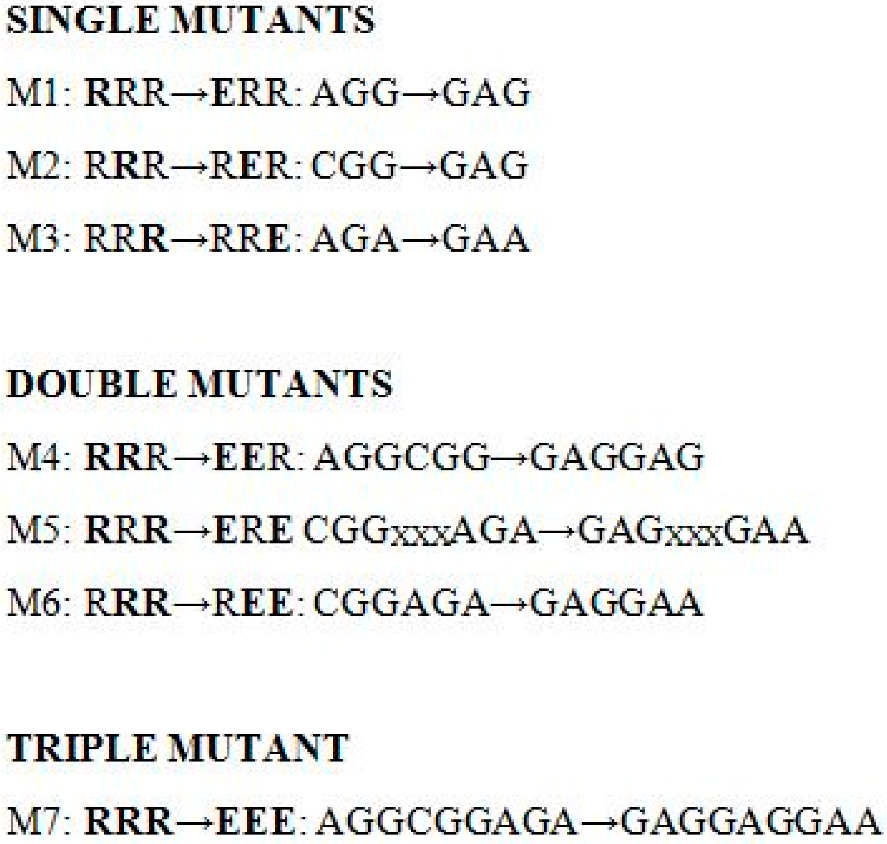
2.1. Characteristics of the Generated Long Form of the D2 Dopamine Receptor (D2L-R) Mutants

D2L-R and generated mutants were first pharmacologically characterized by radioligand binding assay using the selective, hydrophilic D2-Rs antagonist sulpiride, which only binds to membrane-localized receptors. In HEK-293 cells expressing D2L-R, sulpiride competed for binding against the [125I]-iodosulpride with an affinity of 0.82 ± 0.17 nM. This value is in good agreement with those reported for rat and rabbit striatal membranes [33] and human D2-Rs expressed in CHO cells [34]. The IC50 values obtained with the generated mutants did not significantly differ from those obtained for the D2L-R (p = 0.281) (Table 1). Data from the radioligand self-displacement assays were also used to assess surface receptor density (Bmax) (Table 1). All D2L-R mutants showed a trend toward an increased surface receptor density, but due to high inter-assay variations in their surface expression, this did not reach statistical significance (p = 0.587) (Table 1).

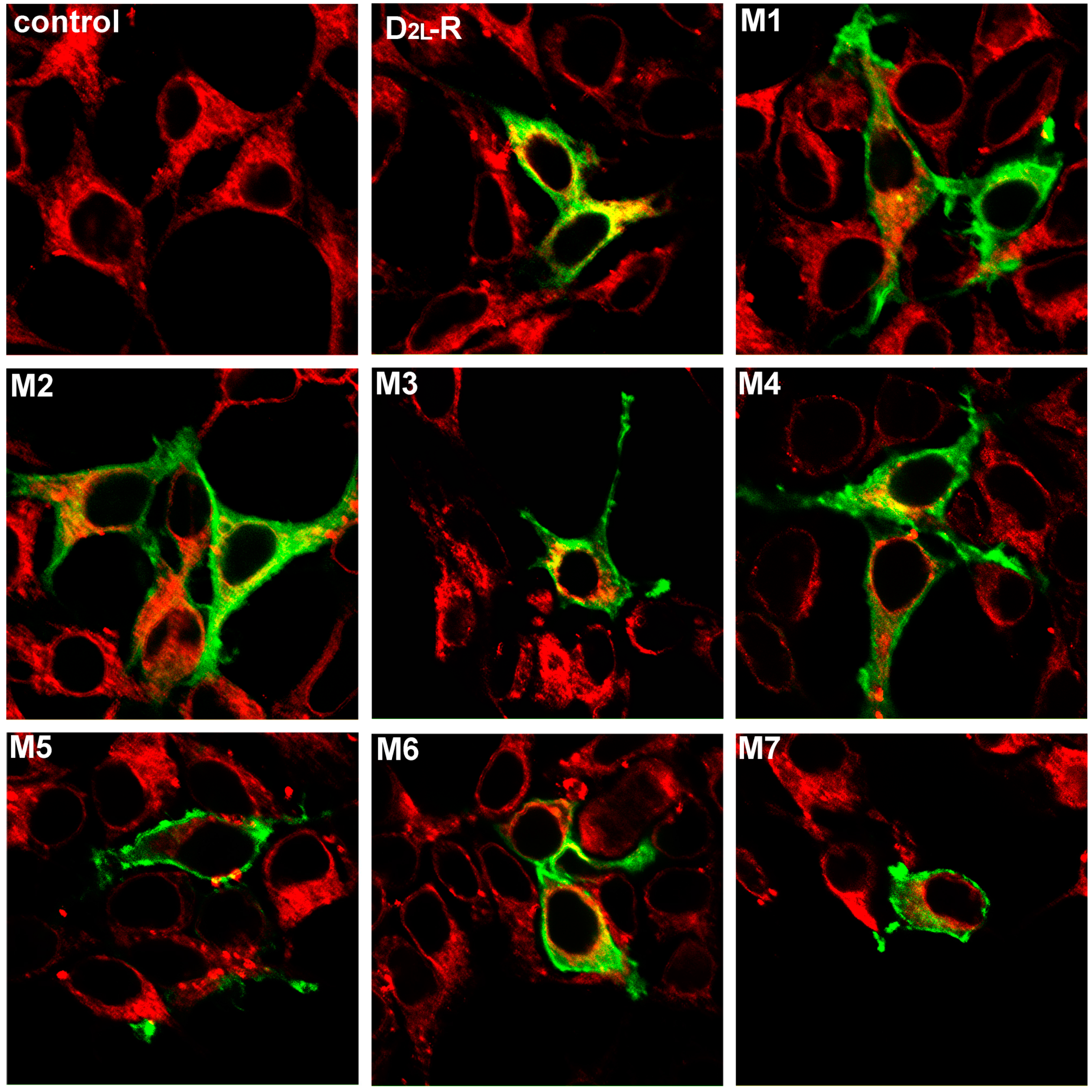
2.2. Visualization and Cellular Localization of the D2L-Rs—Colocalization Study

The cellular distributions of the D2L-R and generated mutants were examined by indirect immunofluorescent staining. Figure 2 shows the distribution of the D2L-R and the mutants thereof at a steady state (untreated cells; green signal) and their colocalization with the ER marker calnexin (red signal). Confocal fluorescence microscopy demonstrated a substantial proportion of the D2L-Rs in the intracellular compartment largely overlapped with calnexin (yellow/orange signal). The fluorescent signal obtained with the D2L-R mutants was confined mainly to the cell surface. In addition, a small intracellular receptor pool was detected with M2, M3, and M4 that partially co-localized with calnexin. The degree of colocalization between the D2L-R constructs and calnexin was quantitatively assessed by calculating Pearson’s correlation coefficient for images above thresholds (Rcoloc; Supplementary Material, Table S1). D2L-R and calnexin showed a high degree of colocalization (Rcoloc ~0.76), while lower Rcoloc (0.40 to 0.51) obtained for the D2L-R mutants suggest a substantial decrease of colocalization.

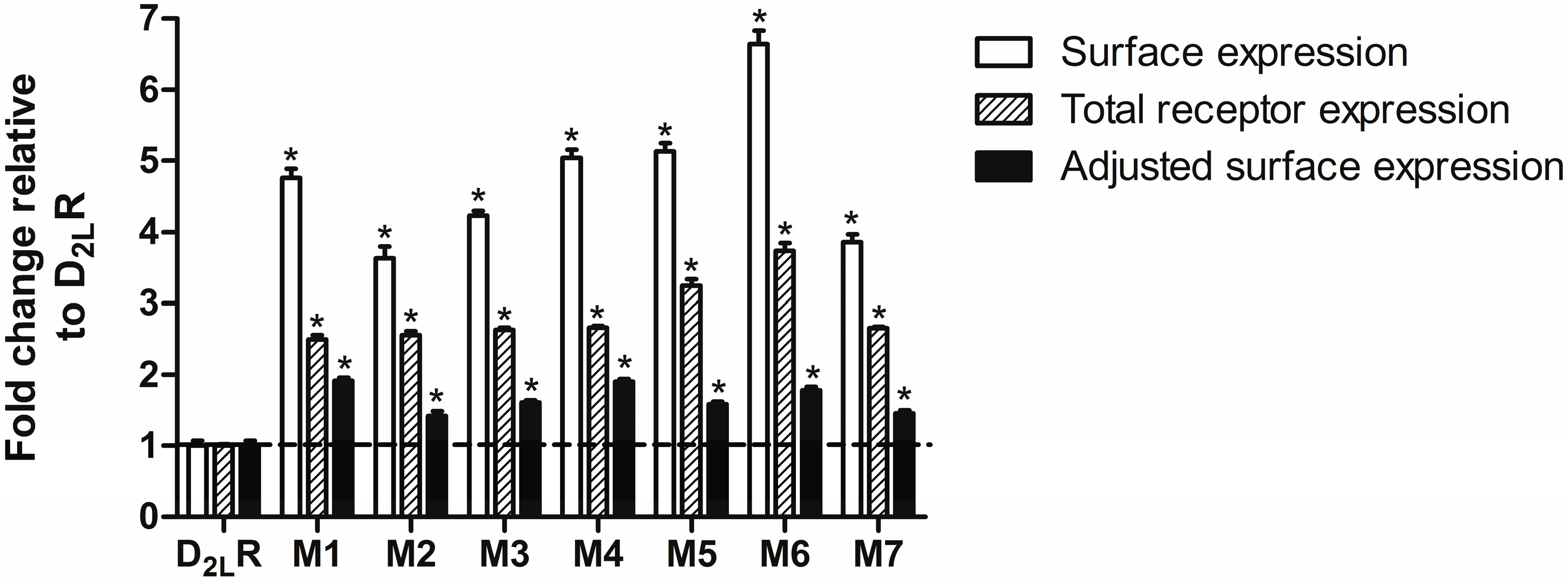
2.3. D2L-R Constructs Surface Expression—Enzyme-Linked Immunosorbent Assay (ELISA)

The effect of mutated arginine residues in the presumed RXR-type ER retention motif on D2L-R surface expression was then quantitatively assessed by ELISA. C-terminally RLuc8-tagged D2L-R constructs were used in ELISA and all subsequent experiments, as they allowed us to monitor the total (surface and intracellular) receptor expression and perform data adjustment to the total receptor expression. As shown in Figure 3, a significant and approximate four- to six-fold increase in the surface expression was observed in HEK-293 cells expressing D2L-R mutants compared with cells expressing D2L-R (open bars). Total luminescence measurements revealed that the expression levels of individual mutants were significantly higher than that of D2L-R (hatched bars), which could underlie the increased surface expression of the mutant forms of the D2L-R. The increases in the surface expression of D2L-R/RLuc8 mutants determined by ELISA were then adjusted by total expression levels using the RLuc8 signal quantified by total luminescence measurements. After adjustment of the surface expression data to the total receptor expression, the surface expression of individual mutants was lower, but still significantly higher (up to ~2-fold) compared to D2L-R (closed bars). To achieve comparable total expression levels, HEK-293 cells were also transfected with different amounts of cDNA encoding either D2L-R/Rluc8 or M1/RLuc8. In the case that comparable total expression of D2L-R/Rluc8 and M1/RLuc8 was achieved, i.e., 1.00 ± 0.01 and 0.89 ± 0.03, surface expression of M1/RLuc8 was significantly higher (1.00 ± 0.08 vs. 2.73 ± 0.14). The experimental M1/RLuc8 surface expression level was slightly higher, but it was still in a range comparable with that for the M1/RLuc8 adjusted surface expression (1.91 ± 0.05), thus providing the rationale for the total receptor expression adjustments.

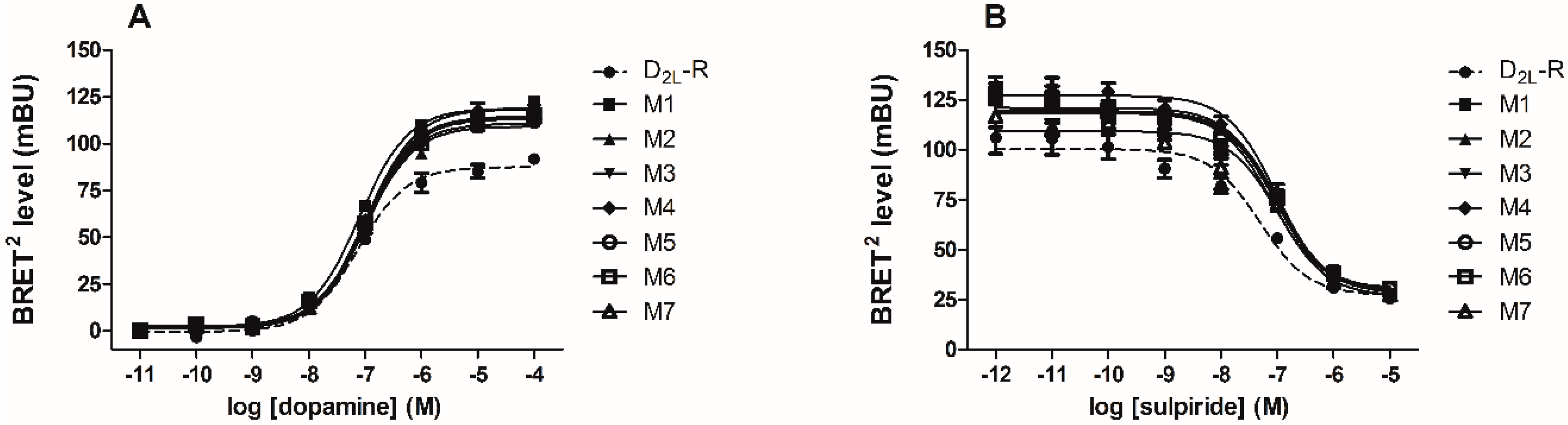
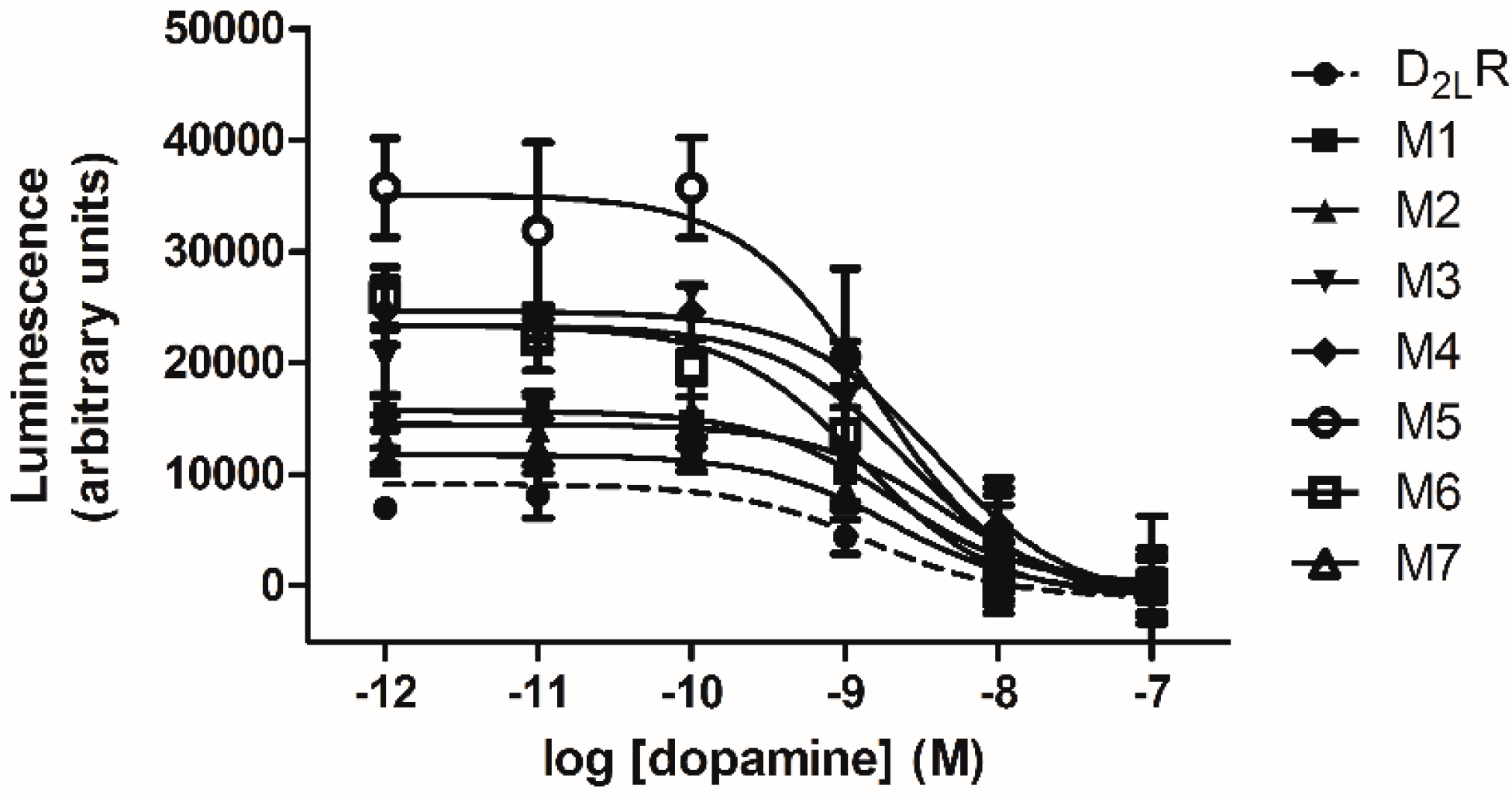
2.4. D2L-R Constructs Functional Characterization—Bioluminescence Resonance Energy Transfer (BRET2) and cAMP Assay

BRET2 assay was used to monitor the recruitment of green fluorescent protein 2 (GFP2)/βarr2 constructs to RLuc8-tagged D2L-R and D2L-R mutants. A more than two-fold increase in the maximal agonist-induced BRET signal (BRETmax) was obtained with the double GFP2/βarr2 R393E,R395E mutant compared to GFP2/βarr2, whereas the potency of dopamine was comparable, i.e., 262.8 ± 85 and 140.5 ± 31.6 nM for the GFP2/βarr2 and GFP2/βarr2 R393E,R395E, respectively. This is in agreement with our previous observations for other GPCRs [35,36] and also for the D2S-R [37]. The later study also reported that both D2S-R and D2L-R displayed comparable potencies in the BRET βarr2 recruitment assay. Due to a larger signal window, the GFP2/βarr2 R393E,R395E mutant was used in subsequent experiments. The constitutive BRET2 signal (BRETconst) generated by non-activated receptor interaction with the GFP2/βarr2 R393E,R395E mutant was comparable between D2L-R and individual mutants. Stimulation of the D2L-R/RLuc8 and D2L-R/RLuc8 mutants by dopamine produced a dose-dependent increase in the BRET signal (Figure 4A). BRET2 antagonist dose-response curves generated in the presence of increasing concentrations of the D2L-R antagonist sulpiride are shown in Figure 4B. The agonist-induced BRETmax signal and the half-maximal effective concentrations (EC50) and the half maximal inhibitory concentration (IC50) values from the BRET2 agonist and antagonist dose-response curves for wild type (WT) and D2L-R mutants are summarized in Table 2. The BRETmax obtained with M1–M6 was significantly higher than the agonist-induced BRETmax signal generated from D2L-R interactions with the βarr2 R393E,R395E, whereas there were no substantial changes in the tested ligand potencies.
As Gi/o protein-mediated cAMP inhibition is the best response for characterizing D2-Rs activation, the ability of D2L-R and individual D2L-R mutants to inhibit forskolin-mediated activation of adenylyl cyclase was also measured to assess their capacity to initiate signaling via Gi/o proteins. As shown in Figure 5, dopamine inhibited forskolin-stimulated cAMP accumulation in live HEK-293 cells expressing D2L-R or individual D2L-R mutants. Low concentrations of dopamine (up to 10−7 M) were used to generate dose-response curves, as it was previously shown that cAMP increases at higher concentrations due to endogenously-expressed D1-Rs in HEK-293 cells [38]. The maximal agonist-induced decrease in cAMP accumulation and the calculated EC50 values derived from the agonist dose-response curves for the WT and D2L-R mutants are summarized in Table 3. There were no substantial differences in the potency of dopamine to inhibit forskolin-stimulated cAMP accumulation, whereas the efficacy of M3, M4, M5, and M6 was significantly higher than that of the D2L-R.

3. Discussion

This study was focused on elucidating the mechanisms underlying predominant intracellular localization of the D2L-R that has been reported in different heterologous expression systems and native tissues [14,15,16,17] and could underlay differential ability of both isoforms to increase steady-state receptor concentration induced by prolonged agonist/antagonist treatment in different transfected cell lines [18,39,40,41]. Similarly, D4-R repeat variants also displayed differences in pharmacological chaperone-mediated upregulation, thus suggesting that the ligands may stabilize a specific receptor conformation and so improve their processing through the ER [19]. A regulated pool of intracellular receptors could also provide a mechanistic basis for behavioral supersensitivity to D2 agonists reported in animals exposed to indirect dopamine agonists [42,43] and for a sustained clinical efficiency of dopamine agonists during long term therapy in patients (reviewed in [21]). We hypothesized that this is an intrinsic property of the D2L-R and that the conserved arginine cluster located within the 29-amino acid insert could act as an intracellular retention signal. A triple arginine cluster (R367–R369) represents the prospective RXR-type ER retention motif, which, unlike the C-terminal–K(X)KXX ER-localization signal, is present in different cytosolic domains of integral membrane proteins [22,44]. The cluster position within the IC3, which is a certain distance from the PM, is also suggestive of its functionality, as many proteins that have the RXR motif near the cytoplasmic side of the PM are not retained in the ER [45,46]. Supporting the latter, a further set of four arginine residues (R217–R220) located toward the N-terminal region of IC3 close to the TM5 of D2-Rs was shown to be essential for efficient PM localization and heteromerization with the D1-R [47] and other GPCRs, including adenosine A2A receptor [48]. However, subsequent studies revealed that arginine residues adjacent to the insert (R374 and R375) are involved in forming heteromers with the D1-R [49] and D5-R [50]. Even though that arginine residues common to both D2-R isoforms were implicated as the possible heteromer interacting site, protein generated from the 29-amino acid insert of D2L-R disrupted the D1–D2 receptor heteromer and showed antidepressant-like effects in mice (reviewed in [51]).
It was initially observed that D2L-R mutants with charge-reversed basic R residues within the prospective ER retention motif showed increased cell surface expression and reduced colocalization with the ER marker calnexin, whereas their ligand-binding properties were not significantly affected. The study by Guiramand et al. [52], which also evaluated the importance of certain amino acids residues within the IC3 of the D2L-R, revealed that mutants with basic residues substituted with nonpolar valine within (K251, K258, R367, R368, and R369), and adjacent to, the insert (R374 and R375) displayed an unchanged or a slightly higher binding affinity for spiperone, but their expression was decreased. This would appear to contradict our observations, but it should be noted that a single mutation, i.e., K251V, already reduced the Bmax by more than 30% and that the mutants in which only R residues were in or adjacent to the insert were not generated. Additionally, mutation of a single lysine residue in the IC1 of α2A-adrenergic receptor (AR) (K65A) positioned next to leucine (L64) decreased the receptor cell surface expression [53]. Hurt et al. [54] proposed that identification of the potential ER motif requires special attention in regards to the membrane/total receptor expression ratio and the mutant functional properties. Therefore, we employed a previously described approach [55] and used double N-terminally HA- and C-terminally RLuc8-tagged receptor constructs that allowed us to monitor the surface and total receptor expression. ELISA and total luminescence measurements showed higher surface and total expression of all generated mutants. However, their adjusted surface expression normalized against the total receptor expression was significantly higher than that for the D2L-R. Together with the data obtained from the confocal colocalization study, these data suggest that mutation of the potential RXR-type ER retention motif interfere with the ability of D2L-R to maintain substantial intracellular receptor pool. The strength of the ER retention motif depends on the number of R residues, whereas the residue preceding RXR and the characteristics of the amino acids at position X modulates its efficacy (reviewed in [44]). The data obtained with single mutants showed that the charge reversal of either R residue rendered the motif inactive, with only a small difference in their efficacy. Additionally, no substantial additive effect on the receptor surface expression was observed with either double or triple mutants. The effect obtained with the single R368E (M2; RER) corroborates the observation that negatively charged residues at position X makes the motif inactive [56]. Functional characterization of the D2L-R and generated mutants was done by BRET-based βarr2 recruitment assay and cAMP signaling. All mutants showed some differences in their efficacies, but not in their tested ligand potencies. However, it should be emphasized that only PM-localized receptors were stimulated, as dopamine is membrane-impermeable. βarr2 recruitment assay was used as the IC3, which is the main site of interaction with β-arrestins for many GPCRs, including D2L-R [57], and the suitability of this assay for the D2-Rs was confirmed in previous studies [37,38,58]. The applicability of our previously described βarr2 R393E,R395 mutant, which shows an overall two-fold improvement in the BRET ratio [35], had also been demonstrated for D2S-Rs [37], which, similar to D2L-R, showed only small or no changes in the compound potency. Most of the tested ligands also displayed comparable efficacies and potencies in recruiting βarr2 to either D2S-R or D2L-R [37]. Moderate constitutive interactions of the D2L-R constructs with the βarr2 R393E,R395 are in agreement with a previous report obtained with the murine D2L-R/βarr2 pair expressed in HEK-293 cells [38]. The maximal agonist-induced BRET2 signal (BRETmax) obtained for the D2L-R mutant interaction with the βarr2 R393E,R395 was up to 1.5-fold higher than that for the D2L-R. This could be due to small changes in the orientation of the donor and acceptor molecules and/or fluctuations in their relative expression levels [59]. Most importantly, the D2L-R mutants displayed unchanged potency when tested in agonist and antagonist modes. This argues against the involvement of the arginine cluster within the IC3 in the interaction with βarr2. Actually, four residues at the N-terminal region of IC3 are common to both D2-R isoforms, i.e., IYIV212 comprise the βarr2 binding domain [60]. The cAMP data also provided evidence for the preserved functional integrity of D2L-R mutants. Similarly, substitution of the 29-amino acid insert with an equivalent length epitope had no functional consequences in regards to cAMP signaling [61]. The improved efficacy correlates with their higher surface expression, whereas the potency of dopamine in inhibiting forskolin-mediated activation of AC was comparable with that for the WT D2L-R and those previously reported for the rat D2L-R expressed in HEK-293 cells [60]. It was previously reported that the mutants S259A, S262A, and D249V located within the insert displayed increased potency in inhibiting AC, whereas this was not the case when basic residues were mutated [52]. The BRET and cAMP data, therefore, provide evidence that the D2L-R mutants have preserved functional properties in regards to Gαi-coupling and βarr2 recruitment, thus further pointing to the role of a conserved arginine cluster (R367–R369) as an ER retention motif.
Exit from the ER is a highly-regulated process critical for the anterograde transport of GPCRs through the GA to the PM and their surface expression [22]. Therefore, it is unlikely that only the RXR-type ER retention motif would be involved. Over the years, several underlying mechanisms have been proposed for the D2L-R intracellular retention, which have included (i) partial glycosylation in the post-ER compartment [8]; (ii) constitutive, clathrin- and dynamin-independent endocytosis of the D2L-R [62]; (iii) interaction with the intracellularly-located Gαi2 splice variant [63]; and (iv) interaction with a specific ER-resident gatekeeper protein, such as GTPase 6 interacting protein 5 (ARL6IP5) (previously named as GTRAP3-18, Yip6b or PRAF-3) [64]. The RXR motif is probably involved in the interaction with the ER-resident gatekeeper protein, and PRAF-3 could be a candidate interaction partner, as it has been shown to confine β2-AR and D2-R to the ER [64]. Such a role has recently been reported for the related ER-resident gatekeeper PRAF-2 in GABAB receptor cell surface export [28]. Studies have attempted to identify specific interacting partners for the IC3 of D2L-R, but were largely unsuccessful because most of the identified partners including Gαi2 splice variant bind to the IC3 of both isoforms [65,66]. So far, only one protein, i.e., heart-type fatty acid binding protein (H-FABP), has been identified that specifically interacts with the 29-amino acid insert region of D2L-R [17]. However, due to its involvement in the regulation of D2-R functions [67], it is unlikely to be involved in its anterograde trafficking to the PM. Additionally, a naturally-occurring synonymous mutation of human D2-R (C957T, Pro319Pro) postulated to correlate with the schizophrenia phenotype was shown to markedly change mRNA stability and reduce dopamine-induced upregulation of D2-R expression [68].
Taken together, the present study presents evidence that the evolutionary conserved arginine cluster located within the 29-amino acid insert could act as an ER motif and be a part of the intricate underlying mechanisms responsible for differentially-regulated anterograde trafficking of the D2-R isoforms and their PM availability.

4. Materials and Methods

4.1. Materials

Molecular biology reagents, tissue culture reagents, and media were from Sigma-Aldrich (St. Louis, MO, USA) and Gibco Invitrogen Corporation (Breda, The Netherlands). [125I]-iodosulpride was purchased from PerkinElmer (Boston, MA, USA). Dopamine and (S)-(−)-sulpiride were obtained from Sigma-Aldrich and coelenterazine 400a from Biotrend Chemikalien GmbH (Köln, Germany). Anti-hemagglutinin (HA) rat monoclonal and anti-calnexin rabbit polyclonal antibodies were from Roche (Basel, Switzerland) and Sigma-Aldrich, respectively. Anti-rat horseradish peroxidase (HRP)-conjugated, anti-rat fluorescein isothiocyanate (FITC)-conjugated and anti-rabbit tetramethylrhodamine (TRITC)-conjugated antibodies were purchased from Sigma-Aldrich.

4.2. Receptor and β-Arrestin 2 Constructs

Human D2L-R N-terminally-tagged with triple HA-tag in the vector pcDNA3.1(+) was obtained from the Missouri S and T cDNA Resource Center (University of Missouri-Rolla, Rolla, MO, USA) and is hereafter referred to as D2L-R. D2L-R C-terminally tagged with Renilla luciferase 8 (RLuc8) (D2L-R/RLuc8) was generated according to standard molecular biology techniques and verified by sequencing. Arginine (R) residues in the D2L-R and D2L-R/RLuc8 at positions 267, 268, and 269 were mutated to E, either individually or in clusters, thus generating single, double, and triple mutants (Figure 6) that are hereafter referred to as M1–M7 and M1/RLuc8–M7/RLuc8. The mutants were generated and verified by sequencing at Genscript USA Inc. (Piscataway, NJ, USA). Human β-arrestin 2 (βarr2) N-terminally tagged with GFP2 (GFP2/βarr2) (PerkinElmer BioSignal, Inc. (Montreal, ON, Canada)) was cloned into the vector pcDNA3.1(+) using the NheI/XbaI restriction sites. The GFP2/βarr2 R393E,R395E mutant was the same as that described previously [35]. Double GFP2/βarr2 R393E,R395E mutant is phosphorylation-independent and defective in their ability to interact with the components of the clathrin-coated vesicles, thus yielding an augmented and more stable BRET2 signal.

4.3. Cell Culture and Transfection

Human embryonic kidney (HEK)-293 cells (European Collection of Animal Cell Cultures (Salisbury, UK)) were routinely cultured in Dulbecco’s modified Eagle’s medium (DMEM) with 10% (v/v) heat-inactivated fetal bovine serum (FBS), 2 mM Glutamax-I, penicillin (100 U/mL), and streptomycin (100 μg/mL) at 37 °C in a humidified atmosphere of 5% (v/v) CO2. Transient transfections were performed following the Lipofectamine®-PlusReagent protocol when cells reached ~90% confluence.

4.4. Confocal Microscopyand Quantification of Co-Localization

The cellular distribution of D2L-R constructs in transiently-transfected HEK-293 and their possible co-localization with the ER marker calnexin was assessed by confocal microscopy using a previously-described protocol [69,70]. Briefly, HEK-293 cells transiently transfected with 1 μg of individual D2L-R constructs were grown on poly-d-lysine-coated glass coverslips. After 48 h, cells were fixed with 4% paraformaldehyde and then washed with phosphate-buffered saline (PBS), permeabilized with 0.01% Triton X-100 in PBS, then incubated in a blocking solution (1% bovine serum albumin (BSA) in PBS) to reduce the nonspecific binding. Next, cells were incubated overnight at 4 °C with a 1:100 dilution of rat anti-HA and 1:100 dilution of rabbit anti-calnexin antibodies. After extensive washing, cells were incubated for 60 min at room temperature in the dark with a 1:50 dilution of secondary anti-rat FITC- and anti-rabbit TRITC-conjugated antibodies. After washing, cells were mounted using an anti-fading ProLong®Gold reagent (Molecular Probes, Leiden, The Netherlands), sealed and examined under an oil immersion objective (Planapo 40×, numerical aperture (N.A.) = 1.25) using a Leica multispectral confocal laser microscope (Leica TCS NT, Heidelberg, Germany). Sequential images were collected in an eight-fold frame with an average resolution of 1024 × 1024 pixels. Representative sections corresponding to the middle of the cells are presented using Adobe Photoshop 7.0.
The degree of co-localization was quantified using ImageJ [71] and the Colocalization plugin [72] image analysis software. Pearson’s correlation coefficient for image above thresholds (Rcoloc) was calculated, which represents pixels where both channels (red and green) were above their respective threshold; its values range between −1.0 and 1.0, where −1, 0, and +1 indicate complete negative correlation, no significant correlation and perfect correlation, respectively. Five to seven individual cells showing co-localization between the individual D2L-R construct and calnexin were analyzed.

4.5. Radiolig and Binding Assay

Self-displacement radioligand binding assay performed on whole cells was based on a previously-described protocol [35,69,70]. The hydrophilic, membrane-impermeable ligand [125I]-iodosulpride was used to determine the number and binding properties of cell surface-expressed D2L-R and individual D2L-R mutants (M1–M7). After transfection with 4 μg of cDNA (75 cm2 flask) for D2L-R or individual D2L-R mutants (M1–M7), cells were plated onto 24-well plates at a density of ~1 × 10⁵ cells per well, and the assay was performed after 48 h. Cells were incubated with the [125I]-iodosulpride (30.000 cpm/well) and increasing concentrations (10−12 to 10−5 M) of unlabeled -/-sulpiride in assay buffer (HEPES-modified DMEM with 0.1% BSA) for 30 min at 25 °C. After washing in ice-cold PBS, cells were solubilized with 0.2 M NaOH and 1% sodium dodecyl sulfate (SDS) solution and the radioactivity levels determined using a γ counter (LKB Wallac, Turku, Finland). All treatments were performed in triplicate in at least three independent experiments. The binding parameters were obtained from self-displacement curves (GraphPad Prism 5.0) and the receptor density (Bmax) calculated as previously described by Ramsay et al. [73].

4.6. Luminescence and Fluorescence Measurements

Total luminescence and fluorescence measurements were performed as previously described [36] to deterimine the expression levels of RLuc8-tagged D2L-R receptor and GFP2-tagged β-arrestin 2 constructs. Signals were measured in a TriStar2 LB 942 microplate reader (Berthold Technologies, Bad Wildbad, Germany). Background values obtained with mock-transfected HEK-293 cells were subtracted in both measurements, and the mean values of triplicate wells/sample were calculated.

4.7. Enzyme-Linked Immunosorbent Assay (ELISA)

ELISA was used to establish the level of surface-expressed RLuc8-tagged D2L-R constructs, while the luminescence was measured to determine total receptor expression. ELISA was performed as previously described [69,70]. HEK-293 cells were transiently transfected with 1 μg of cDNA for D2L-R/RLuc8 or individual D2L-R/RLuc8 mutants (M1/RLuc8–M7/RLuc8). To achieve comparable expression levels, different amounts of cDNA were also used, ranging from 0.5 to 2.5 μg, and the total amount of cDNA was kept uniform by adding an empty pcDNA3.1 vector. After transfection, the cells were seeded in a 24-well plate (~1 × 10⁵ cells/well). ~3 × 10⁵ cells from each transfection were also seeded into a 60-mm dish for total luminescence measurements. After 48 h, cells were first serum-starved for 2-h and then fixed with 4% paraformaldehyde, washed in PBS, and blocked (1% BSA in PBS). Subsequently, cells were incubated at 4 °C overnight with a 1:600 dilution of anti-HA antibody. After three washes, cells were incubated with anti-rat HRP-conjugated antibody at a 1:1000 dilution and, finally, the 3,3′,5,5′-tetra-methylbenzidine (TMB) liquid substrate system (Sigma) was used to develop the reaction, which was stopped after 10 min at 37 °C by adding 0.2 N sulfuric acid solution. The absorbance was measured at 450 nm in a Rosys Anthos 2010 microplate reader (Anthos Labtec Instruments, Wals, Austria). HEK-293 cells transfected with empty vector pcDNA3.1 were used to determine the background, which was subtracted from all readings. Determinations were made in triplicate.

4.8. BRET-Based β-Arrestin 2 Recruitments Assay

The ability of agonist-activated D2L-R constructs to recruit βarr2 was determined utilizing a previously-described BRET-basedβarr2 recruitments assay [35,36]. Briefly, HEK-293 cells in a 75 cm2 flask were transiently co-transfected with constructs encoding individual D2L-R/RLuc8 constructs (0.4 μg) together with constructs encoding GFP2-βarr2 (5.6 μg) or double GFP2-βarr2 R393E,R395E mutant (5.6 μg). At 48 h after transfection, HEK-293 cells were trypsinized, washed once in Dulbecco’s phosphate-buffered saline (DPBS), and resuspended in DPBS supplemented with Ca2+/Mg2+, 1 g/L glucose, and 36 mg/L sodium pyruvate. Resuspended cells (~2 × 10⁵) were distributed in 96-well microplates (white Optiplate; Packard BioScience, Meriden, CT, USA) and treated with increasing concentrations (10−4 to 10−11 M) of dopamine and incubated for 5 min. For the antagonist assay, the antagonist sulpiride (10−5 to 10−12 M, final concentration) was added to the cells 10 min prior to the addition of the dopamine (1 μM, final concentration). The substrate coelenterazine 400a was then injected with a final concentration of 5 μM. The signals detected at 395 and 515 nm were measured sequentially, and the 515/395 ratios were calculated and expressed as a miliBRET level (mBU; BRET ratio × 1000). Expression levels of RLuc8- and GFP2-tagged constructs for each experiment were determined by total luminescence and fluorescence measurement.

4.9. cAMP Assay

The HitHunter® cAMP assay (DiscoveRx Corporation Ltd.) was used to determine the ability of D2L-R constructs to inhibit forskolin-stimulated cAMP accumulation and was performed according to the manufacturer’s instructions. HEK-293 cells in 60 mm dishes were transiently transfected with 1 μg of cDNA encoding individual D2L-R constructs. After 48 h, cells were trypsinized, resuspended in a cell assay buffer (PBS with 0.5 mM 3-isobutyl-1-methylxanthine (IBMX)), and seeded (5 × 10⁴ cells/well) onto 96-well plates (white, Optiplate). Cells were then treated for 60 min at room temperature with increasing concentrations (10−13 to 10−7 M) of dopamine diluted in PBS in the presence of forskolin (20 μM, final concentration). HitHunter cAMP reagents were then added sequentially, and the cells were incubated in the dark at room temperature. Total luminescence was read after 6 h in a TriStar LB 942 microplate reader at 1 s/well and data analysis performed using GraphPad Prism 5.0. Expression levels of RLuc8-tagged constructs in each experiment were monitored by total luminescence measurement.

4.10. Statistical Analysis

The results are expressed as the mean ± S.E.M. The statistical significance relative to the D2L-R (control) was determined by one-way ANOVA, followed by Dunnett’s post hoc test. Statistical analyses were conducted by SPSS 20.0 for Windows (SPSS Inc., Chicago, IL, USA). A p-value ≤0.05 was regarded as statistically significant.

Supplementary Materials

Supplementary materials can be found at https://0-www-mdpi-com.brum.beds.ac.uk/1422-0067/17/7/1152/s1.

Acknowledgments

The authors would like to thank American Journal Experts for their English language editing of this manuscript. We acknowledge funding from the Slovenian Research Agency program P4-0053. Milka Vrecl participates in the European COST Action CM1207 (GLISTEN).

Author Contributions

Karin A. Eidne and Milka Vrecl conceived and designed the experiments; Kaja Blagotinšek and Valentina Kubale performed the experiments; Valentina Kubale and Milka Vrecl analyzed the data; Jane Nøhr contributed materials/analysis tools; Kaja Blagotinšek, Jane Nøhr and Milka Vrecl wrote the paper.

Conflicts of Interest

The authors declare no conflict of interest. The funding sponsors had no role in the design of the study; in the collection, analyses, or interpretation of data; in the writing of the manuscript, and in the decision to publish the results.

References

  1. Beaulieu, J.M.; Espinoza, S.; Gainetdinov, R.R. Dopamine receptors—IUPHAR review 13. Br. J. Pharmacol. 2015, 172, 1–23. [Google Scholar] [CrossRef] [PubMed]
  2. Eidne, K.A.; Taylor, P.L.; Zabavnik, J.; Saunders, P.T.; Inglis, J.D. D2 receptor, a missing exon. Nature 1989, 342, 865. [Google Scholar] [CrossRef] [PubMed]
  3. Giros, B.; Sokoloff, P.; Martres, M.P.; Riou, J.F.; Emorine, L.J.; Schwartz, J.C. Alternative splicing directs the expression of two D2 dopamine receptor isoforms. Nature 1989, 342, 923–926. [Google Scholar] [CrossRef] [PubMed]
  4. Monsma, F.J.; Mcvittie, L.D.; Gerfen, C.R.; Mahan, L.C.; Sibley, D.R. Multiple D2 dopamine-receptors produced by alternative rna splicing. Nature 1989, 342, 926–929. [Google Scholar] [CrossRef] [PubMed]
  5. Grunewald, S.; Reilander, H.; Michel, H. In vivo reconstitution of dopamine D2S receptor-mediated G protein activation in baculovirus-infected insect cells: Preferred coupling to Gi1 versus Gi2. Biochemistry 1996, 35, 15162–15173. [Google Scholar] [CrossRef] [PubMed]
  6. Montmayeur, J.P.; Guiramand, J.; Borrelli, E. Preferential coupling between dopamine-D2 receptors and G-proteins. Mol. Endocrinol. 1993, 7, 161–170. [Google Scholar] [PubMed]
  7. Senogles, S.E. The D2 dopamine receptor isoforms signal through distinct G proteins to inhibit adenylyl cyclase. A study with site-directed mutant G proteins. J. Biol. Chem. 1994, 269, 23120–23127. [Google Scholar] [PubMed]
  8. Fishburn, C.S.; Elazar, Z.; Fuchs, S. Differential glycosylation and intracellular trafficking for the long and short isoforms of the D2 dopamine receptor. J. Biol. Chem. 1995, 270, 29819–29824. [Google Scholar] [PubMed]
  9. Lindgren, N.; Usiello, A.; Goiny, M.; Haycock, J.; Erbs, E.; Greengard, P.; Hokfelt, T.; Borrelli, E.; Fisone, G. Distinct roles of dopamine D2L and D2S receptor isoforms in the regulation of protein phosphorylation at presynaptic and postsynaptic sites. Proc. Natl. Acad. Sci. USA 2003, 100, 4305–4309. [Google Scholar] [CrossRef] [PubMed]
  10. Usiello, A.; Baik, J.H.; Rougé-Pont, F.; Picetti, R.; Dierich, A.; LeMeur, M.; Piazza, P.V.; Borrelli, E. Distinct functions of the two isoforms of dopamine D2 receptors. Nature 2000, 408, 199–203. [Google Scholar] [PubMed]
  11. Wang, Y.; Xu, R.; Sasaoka, T.; Tonegawa, S.; Kung, M.P.; Sankoorikal, E.B. Dopamine D2 long receptor-deficient mice display alterations in striatum-dependent functions. J. Neurosci. 2000, 20, 8305–8314. [Google Scholar] [PubMed]
  12. Welter, M.; Vallone, D.; Samad, T.A.; Meziane, H.; Usiello, A.; Borrelli, E. Absence of dopamine D2 receptors unmasks an inhibitory control over the brain circuitries activated by cocaine. Proc. Natl. Acad. Sci. USA 2007, 104, 6840–6845. [Google Scholar] [CrossRef] [PubMed]
  13. Xu, R.; Hranilovic, D.; Fetsko, L.A.; Bucan, M.; Wang, Y. Dopamine D2S and D2L receptors may differentially contribute to the actions of antipsychotic and psychotic agents in mice. Mol. Psychiatry 2002, 7, 1075–1082. [Google Scholar] [CrossRef] [PubMed]
  14. Khan, Z.U.; Mrzljak, L.; Gutierrez, A.; de la Calle, A.; Goldman-Rakic, P.S. Prominence of the dopamine D2 short isoform in dopaminergic pathways. Proc. Natl. Acad. Sci. USA 1998, 95, 7731–7736. [Google Scholar] [CrossRef] [PubMed]
  15. Prou, D.; Gu, W.J.; Le Crom, S.; Vincent, J.D.; Salamero, J.; Vernier, P. Intracellular retention of the two isoforms of the D2 dopamine receptor promotes endoplasmic reticulum disruption. J. Cell Sci. 2001, 114, 3517–3527. [Google Scholar] [PubMed]
  16. Sedaghat, K.; Nantel, M.F.; Ginsberg, S.; Lalonde, V.; Tiberi, M. Molecular characterization of dopamine D2 receptor isoforms tagged with green fluorescent protein. Mol. Biotechnol. 2006, 34, 1–14. [Google Scholar] [CrossRef]
  17. Takeuchi, Y.; Fukunaga, K. Differential subcellular localization of two dopamine D2 receptor isoforms in transfected NG108-15 cells. J. Neurochem. 2003, 85, 1064–1074. [Google Scholar] [CrossRef] [PubMed]
  18. Ng, G.Y.; Varghese, G.; Chung, H.T.; Trogadis, J.; Seeman, P.; O’Dowd, B.F.; George, S.R. Resistance of the dopamine D2L receptor to desensitization accompanies the up-regulation of receptors on to the surface of Sf9 cells. Endocrinology 1997, 138, 4199–4206. [Google Scholar] [CrossRef] [PubMed]
  19. Van Craenenbroeck, K.; Clark, S.D.; Cox, M.J.; Oak, J.N.; Liu, F.; van Tol, H.H. Folding efficiency is rate-limiting in dopamine D4 receptor biogenesis. J. Biol. Chem. 2005, 280, 19350–19357. [Google Scholar] [CrossRef] [PubMed]
  20. Achour, L.; Labbe-Jullie, C.; Scott, M.G.H.; Marullo, S. An escort for GPCRs: Implications for regulation of receptor density at the cell surface. Trends Pharmacol. Sci. 2008, 29, 528–535. [Google Scholar] [CrossRef] [PubMed] [Green Version]
  21. Shirvani, H.; Gata, G.; Marullo, S. Regulated GPCR trafficking to the plasma membrane: General issues and theCCR5 chemokine receptor example. Subcell. Biochem. 2012, 63, 97–111. [Google Scholar] [PubMed]
  22. Dong, C.M.; Filipeanu, C.M.; Duvernay, M.T.; Wu, G.Y. Regulation of G protein-coupled receptor export trafficking. Biochim. Biophys. Acta 2007, 1768, 853–870. [Google Scholar] [CrossRef] [PubMed]
  23. Milligan, G. G protein-coupled receptor hetero-dimerization: Contribution to pharmacology and function. Br. J. Pharmacol. 2009, 158, 5–14. [Google Scholar] [CrossRef] [PubMed]
  24. Babcock, J.J.; Li, M. Inside job: Ligand-receptor pharmacology beneath the plasma membrane. Acta Pharmacol. Sin. 2013, 34, 859–869. [Google Scholar] [CrossRef] [PubMed]
  25. Doly, S.; Marullo, S. Gatekeepers controlling GPCR export and function. Trends Pharmacol. Sci. 2015, 36, 636–644. [Google Scholar] [CrossRef] [PubMed]
  26. Margeta-Mitrovic, M.; Jan, Y.N.; Jan, L.Y. A trafficking checkpoint controls GABAB receptor heterodimerization. Neuron 2000, 27, 97–106. [Google Scholar] [CrossRef]
  27. Restituito, S.; Couve, A.; Bawagan, H.; Jourdain, S.; Pangalos, M.N.; Calver, A.R.; Freeman, K.B.; Moss, S.J. Multiple motifs regulate the trafficking of GABAB receptors at distinct checkpoints within the secretory pathway. Mol. Cell. Neurosci. 2005, 28, 747–756. [Google Scholar] [CrossRef] [PubMed]
  28. Doly, S.; Shirvani, H.; Gata, G.; Meye, F.J.; Emerit, M.B.; Enslen, H.; Achour, L.; Pardo-Lopez, L.; Yang, S.K.; Armand, V.; et al. GABAB receptor cell-surface export is controlled by an endoplasmic reticulum gatekeeper. Mol. Psychiatry 2016, 4, 480–490. [Google Scholar] [CrossRef] [PubMed] [Green Version]
  29. Zerangue, N.; Schwappach, B.; Jan, Y.N.; Jan, L.Y. A new ER trafficking signal regulates the subunit stoichiometry of plasma membrane KATP channels. Neuron 1999, 22, 537–548. [Google Scholar] [CrossRef]
  30. Ma, D.; Zerangue, N.; Lin, Y.F.; Collins, A.; Yu, M.; Jan, Y.N.; Jan, L.Y. Role of ER export signals in controlling surface potassium channel numbers. Science 2001, 291, 316–319. [Google Scholar] [CrossRef] [PubMed]
  31. Okamoto, Y.; Shikano, S. Phosphorylation-dependent C-terminal binding of 14-3-3 proteins promotes cell surface expression of HIV co-receptor GPR15. J. Biol. Chem. 2011, 286, 7171–7181. [Google Scholar] [CrossRef] [PubMed]
  32. Nasu-Nishimura, Y.; Hurtado, D.; Braud, S.; Tang, T.T.; Isaac, J.T.; Roche, K.W. Identification of an endoplasmic reticulum-retention motif in an intracellular loop of the kainate receptor subunit KA2. J. Neurosci. 2006, 26, 7014–7021. [Google Scholar] [CrossRef] [PubMed]
  33. Zahniser, N.R.; Dubocovich, M.L. Comparison of dopamine receptor sites labeled by [3H]-S-sulpiride and [3H]-spiperone in striatum. J. Pharmacol. Exp. Ther. 1983, 227, 592–599. [Google Scholar] [PubMed]
  34. Audinot, V.; Newman-Tancredi, A.; Gobert, A.; Rivet, J.M.; Brocco, M.; Lejeune, F.; Gluck, L.; Desposte, I.; Bervoets, K.; Dekeyne, A.; et al. A comparative in vitro and in vivo pharmacological characterization of the novel dopamine D3 receptor antagonists (+)-S 14297, nafadotride, GR 103,691 and U 99194. J. Pharmacol. Exp. Ther. 1998, 287, 187–197. [Google Scholar] [PubMed]
  35. Vrecl, M.; Jorgensen, R.; Pogacnik, A.; Heding, A.; Vrecl, M.; Jorgensen, R.; Pogacnik, A.; Heding, A. Development of a BRET2 screening assay using β-arrestin 2 mutants. J. Biomol. Screen. 2004, 9, 322–333. [Google Scholar] [CrossRef] [PubMed]
  36. Vrecl, M.; Norregaard, P.K.; Almholt, D.L.C.; Elster, L.; Pogacnik, A.; Heding, A. β-arrestin-based BRET2 screening assay for the “non”-β-arrestin binding CB1 receptor. J. Biomol. Screen. 2009, 14, 371–380. [Google Scholar] [CrossRef] [PubMed]
  37. Klewe, I.V.; Nielsen, S.M.; Tarpo, L.; Urizar, E.; Dipace, C.; Javitch, J.A.; Gether, U.; Egebjerg, J.; Christensen, K.V. Recruitment of β-arrestin2 to the dopamine D2 receptor: Insights into anti-psychotic and anti-parkinsonian drug receptor signaling. Neuropharmacology 2008, 54, 1215–1222. [Google Scholar] [CrossRef] [PubMed]
  38. Masri, B.; Salahpour, A.; Didriksen, M.; Ghisi, V.; Beaulieu, J.M.; Gainetdinov, R.R.; Caron, M.G. Antagonism of dopamine D2 receptor/β-arrestin 2 interaction is a common property of clinically effective antipsychotics. Proc. Natl. Acad. Sci. USA 2008, 105, 13656–13661. [Google Scholar] [CrossRef] [PubMed]
  39. Filtz, T.M.; Artymyshyn, R.P.; Guan, W.; Molinoff, P.B. Paradoxical regulation of dopamine receptors in transfected 293 cells. Mol. Pharmacol. 1993, 44, 371–379. [Google Scholar] [PubMed]
  40. Starr, S.; Kozell, L.B.; Neve, K.A. Drug-induced up-regulation of dopamine D2 receptors on cultured cells. J. Neurochem. 1995, 65, 569–577. [Google Scholar] [CrossRef] [PubMed]
  41. Zhang, L.J.; Lachowicz, J.E.; Sibley, D.R. The D2S and D2L dopamine receptor isoforms are differentially regulated in Chinese hamster ovary cells. Mol. Pharmacol. 1994, 45, 878–889. [Google Scholar] [PubMed]
  42. Bischoff, S.; Krauss, J.; Grunenwald, C.; Gunst, F.; Heinrich, M.; Schaub, M.; Stocklin, K.; Vassout, A.; Waldmeier, P.; Maitre, L. Endogenous dopamine (DA) modulates [3H]spiperone binding in vivo in rat brain. J. Recept. Res. 1991, 11, 163–175. [Google Scholar] [CrossRef] [PubMed]
  43. Rouillard, C.; Bedard, P.J.; Falardeau, P.; Dipaolo, T. Behavioral and biochemical evidence for a different effect of repeated administration of l-DOPA and bromocriptine on denervated versus non-denervated striatal dopamine receptors. Neuropharmacology 1987, 26, 1601–1606. [Google Scholar] [CrossRef]
  44. Michelsen, K.; Yuan, H.; Schwappach, B. Hide and run. EMBO Rep. 2005, 6, 717–722. [Google Scholar] [CrossRef] [PubMed]
  45. Gassmann, M.; Haller, C.; Stoll, Y.; Abdel Aziz, S.; Biermann, B.; Mosbacher, J.; Kaupmann, K.; Bettler, B. The RXR-type endoplasmic reticulum-retention/retrieval signal of GABAB1 requires distant spacing from the membrane to function. Mol. Pharmacol. 2005, 68, 137–144. [Google Scholar] [PubMed]
  46. Shikano, S.; Li, M. Membrane receptor trafficking: Evidence of proximal and distal zones conferred by two independent endoplasmic reticulum localization signals. Proc. Natl. Acad. Sci. USA 2003, 100, 5783–5788. [Google Scholar] [CrossRef] [PubMed]
  47. Lukasiewicz, S.; Faron-Gorecka, A.; Dobrucki, J.; Polit, A.; Dziedzicka-Wasylewska, M. Studies on the role of the receptor protein motifs possibly involved in electrostatic interactions on the dopamine D1 and D2 receptor oligomerization. FEBS J. 2009, 276, 760–775. [Google Scholar] [CrossRef] [PubMed]
  48. Fuxe, K.; Ferre, S.; Canals, M.; Torvinen, M.; Terasmaa, A.; Marcellino, D.; Goldberg, S.R.; Staines, W.; Jacobsen, K.X.; Lluis, C.; et al. Adenosine A2A and dopamine D2 heteromeric receptor complexes and their function. J. Mol. Neurosci. 2005, 26, 209–220. [Google Scholar] [CrossRef]
  49. O’Dowd, B.F.; Ji, X.; Nguyen, T.; George, S.R. Two amino acids in each of D1 and D2 dopamine receptor cytoplasmic regions are involved in D1–D2 heteromer formation. Biochem. Biophys. Res. Commun. 2012, 417, 23–28. [Google Scholar] [CrossRef] [PubMed]
  50. O’Dowd, B.F.; Nguyen, T.; Ji, X.; George, S.R. D5 dopamine receptor carboxyl tail involved in D5–D2 heteromer formation. Biochem. Biophys. Res. Commun. 2013, 431, 586–589. [Google Scholar] [CrossRef] [PubMed]
  51. Perreault, M.L.; Hasbi, A.; O’Dowd, B.F.; George, S.R. Heteromeric dopamine receptor signaling complexes: Emerging neurobiology and disease relevance. Neuropsychopharmacology 2014, 39, 156–168. [Google Scholar] [CrossRef] [PubMed]
  52. Guiramand, J.; Montmayeur, J.P.; Ceraline, J.; Bhatia, M.; Borrelli, E. Alternative splicing of the dopamine D2 receptor directs specificity of coupling to G-proteins. J. Biol. Chem. 1995, 270, 7354–7358. [Google Scholar] [PubMed]
  53. Fan, Y.; Li, C.; Guo, J.; Hu, G.; Wu, G. A single Lys residue on the first intracellular loop modulates the endoplasmic reticulum export and cell-surface expression of α2A-adrenergic receptor. PLoS ONE 2012, 7, e50416. [Google Scholar] [CrossRef] [PubMed]
  54. Hurt, C.M.; Ho, V.K.; Angelotti, T. Systematic and quantitative analysis of G protein-coupled receptor trafficking motifs. Methods Enzymol. 2013, 521, 171–187. [Google Scholar] [PubMed]
  55. Sauvageau, E.; Rochdi, M.D.; Oueslati, M.; Hamdan, F.F.; Percherancier, Y.; Simpson, J.C.; Pepperkok, R.; Bouvier, M. CNIH4 interacts with newly synthesized GPCR and controls their export from the endoplasmic reticulum. Traffic 2014, 15, 383–400. [Google Scholar] [CrossRef] [PubMed]
  56. Zerangue, N.; Malan, M.J.; Fried, S.R.; Dazin, P.F.; Jan, Y.N.; Jan, L.Y.; Schwappach, B. Analysis of endoplasmic reticulum trafficking signals by combinatorial screening in mammalian cells. Proc. Natl. Acad. Sci. USA 2001, 98, 2431–2436. [Google Scholar] [CrossRef] [PubMed]
  57. Macey, T.A.; Gurevich, V.V.; Neve, K.A. Preferential interaction between the dopamine D2 receptor and arrestin2 in neostriatal neurons. Mol. Pharmacol. 2004, 66, 1635–1642. [Google Scholar] [CrossRef] [PubMed]
  58. Free, R.B.; Chun, L.S.; Moritz, A.E.; Miller, B.N.; Doyle, T.B.; Conroy, J.L.; Padron, A.; Meade, J.A.; Xiao, J.; Hu, X.; et al. Discovery and characterization of a G protein-biased agonist that inhibits β-arrestin recruitment to the D2 dopamine receptor. Mol. Pharmacol. 2014, 86, 96–105. [Google Scholar] [CrossRef] [PubMed]
  59. Drinovec, L.; Kubale, V.; Nohr Larsen, J.; Vrecl, M. Mathematical models for quantitative assessment of bioluminescence resonance energy transfer: Application to seven transmembrane receptors oligomerization. Front. Endocrinol. 2012, 3, 104. [Google Scholar] [CrossRef] [PubMed]
  60. Lan, H.; Liu, Y.; Bell, M.I.; Gurevich, V.V.; Neve, K.A. A dopamine D2 receptor mutant capable of G protein-mediated signaling but deficient in arrestin binding. Mol. Pharmacol. 2009, 75, 113–123. [Google Scholar] [CrossRef] [PubMed]
  61. Kendall, R.T.; Senogles, S.E. Investigation of the alternatively spliced insert region of the D2L dopamine receptor by epitope substitution. Neurosci. Lett. 2006, 393, 155–159. [Google Scholar] [CrossRef] [PubMed]
  62. Vickery, R.G.; von Zastrow, M. Distinct dynamin-dependent and -independent mechanisms target structurally homologous dopamine receptors to different endocytic membranes. J. Cell Biol. 1999, 144, 31–43. [Google Scholar] [CrossRef] [PubMed]
  63. Lopez-Aranda, M.F.; Acevedo, M.J.; Gutierrez, A.; Koulen, P.; Khan, Z.U. Role of a Gαi2 protein splice variant in the formation of an intracellular dopamine D2 receptor pool. J. Cell Sci. 2007, 120, 2171–2178. [Google Scholar] [CrossRef] [PubMed]
  64. Ruggiero, A.M.; Liu, Y.; Vidensky, S.; Maier, S.; Jung, E.; Farhan, H.; Robinson, M.B.; Sitte, H.H.; Rothstein, J.D. The endoplasmic reticulum exit of glutamate transporter is regulated by the inducible mammalian Yip6b/GTRAP3–18 protein. J. Biol. Chem. 2008, 283, 6175–6183. [Google Scholar] [CrossRef] [PubMed]
  65. Kabbani, N.; Woll, M.P.; Nordman, J.C.; Levenson, R. Dopamine receptor interacting proteins: Targeting neuronal calcium sensor-1/D2 dopamine receptor interaction for antipsychotic drug development. Curr. Drug Targets 2012, 13, 72–79. [Google Scholar] [CrossRef] [PubMed]
  66. Tirotta, E.; Fontaine, V.; Picetti, R.; Lombardi, M.; Samad, T.A.; Oulad-Abdelghani, M.; Edwards, R.; Borrelli, E. Signaling by dopamine regulates D2 receptors trafficking at the membrane. Cell Cycle 2008, 7, 2241–2248. [Google Scholar] [CrossRef] [PubMed]
  67. Shioda, N.; Yamamoto, Y.; Watanabe, M.; Binas, B.; Owada, Y.; Fukunaga, K. Heart-type fatty acid binding protein regulates dopamine D2 receptor function in mouse brain. J. Neurosci. 2010, 30, 3146–3155. [Google Scholar] [CrossRef] [PubMed]
  68. Duan, J.; Wainwright, M.S.; Comeron, J.M.; Saitou, N.; Sanders, A.R.; Gelernter, J.; Gejman, P.V. Synonymous mutations in the human dopamine receptor D2 (DRD2) affect mRNA stability and synthesis of the receptor. Hum. Mol. Genet. 2003, 12, 205–216. [Google Scholar] [CrossRef] [PubMed]
  69. Vrecl, M.; Anderson, L.; Hanyaloglu, A.; McGregor, A.M.; Groarke, A.D.; Milligan, G.; Taylor, P.L.; Eidne, K.A. Agonist-induced endocytosis and recycling of the gonadotropin releasing hormone receptor: Effect of β-arrestin on internalization kinetics. Mol. Endocrinol. 1998, 12, 1818–1829. [Google Scholar] [CrossRef] [PubMed]
  70. Mandic, M.; Drinovec, L.; Glisic, S.; Veljkovic, N.; Nohr, J.; Vrecl, M. Demonstration of a direct interaction between β2-adrenergic receptor and insulin receptor by BRET and bioinformatics. PLoS ONE 2014, 9, e112664. [Google Scholar] [CrossRef] [PubMed]
  71. Rasband, W.S. ImageJ; U.S. National Institutes of Health: Bethesda, MD, USA, 1997–2016. Available online: http://imagej.nih.gov/ij/ (accessed on 30 June 2016).
  72. Schindelin, J.; Arganda-Carreras, I.; Frise, E.; Kaynig, V.; Longair, M.; Pietzsch, T.; Preibisch, S.; Rueden, C.; Saalfeld, S.; Schmid, B.; et al. Fiji: An open-source platform for biological-image analysis. Nat. Methods 2012, 9, 676–682. [Google Scholar] [CrossRef] [PubMed]
  73. Ramsay, D.; Kellett, E.; McVey, M.; Rees, S.; Milligan, G. Homo- and hetero-oligomeric interactions between G-protein-coupled receptors in living cells monitored by two variants of bioluminescence resonance energy transfer (BRET): Hetero-oligomers between receptor subtypes form more efficiently than between less closely related sequences. Biochem. J. 2002, 365, 429–440. [Google Scholar] [PubMed]
Figure 1. Sequence alignment of the alternatively-spliced exon in the long form of the D2 dopamine receptor (D2L-R). Comparisons of the amino acid sequence show its high conservation among species. Amino acids that differ from the sequence of the insert in humans are indicated in red. Potential N-glycosylation sites (N243 and N260) are marked in blue. The conserved sequence of three basic amino acids (arginine (R) or lysine (K)), which represent a potential endoplasmic reticulum (ER) retention motif, is written in bold.
Ijms 17 01152 g001
Figure 2. Visualization of the D2L-R and D2L-R mutants and their cellular localization and colocalization with calnexin in transiently transfected HEK-293 cells. D2L-R and D2L-R mutant (M1–M7) cellular localization was assessed by confocal microscopy in unstimulated cells. The green color indicates D2L-R and D2L-R mutants; red indicates calnexin, an ER marker; yellow/orange indicates colocalization of an individual D2L-R construct and calnexin. Note that all D2L-R mutants displayed reduced colocalization with calnexin and more apparent localization to the cell surface compared to the D2L-R. Objective 40× and zoom factor 4 apply for all images.
Ijms 17 01152 g002
Figure 3. Expression levels of D2L-R and D2L-R mutants in transiently transfected HEK-293 cells. Intact HEK-293 cells transiently transfected with D2L-R/RLuc8 or individual D2L-R/RLuc8 mutants were used in an Enzyme-Linked Immunosorbent Assay (ELISA). Receptor surface expression was assessed by measuring the absorbance at 450 nm; the background values obtained with untransfected HEK-293 were subtracted from all readings (open bars). The D2L-R/RLuc8 construct total expression i.e., surface and intracellular were determined by luminescence measurements as described in the Materials and Methods section (hatched bars). Closed bars represent adjusted surface expression normalized against the total receptor expression. Data are expressed as the mean ± standard error of the mean (S.E.M.) of three independent experiments performed in triplicate. Significance relative to the D2L-R was determined by one-way ANOVA with Dunnett’s post hoc test (*, p < 0.05).
Ijms 17 01152 g003
Figure 4. BRET2-based agonist and antagonist dose-response curves for the D2L-R and D2L-R mutants. HEK-293 cells were transfected transiently with the indicated RLuc8-tagged D2L-R construct and GFP2/βarr2 R393E,R395E at a 1:14 cDNA ratio. For the agonist dose-response curves (A); increasing concentrations (10−11 to 10−4 M) of dopamine were added to the cells. For antagonist dose-response curves (B); cells were first treated with increasing concentrations (10−12 to 10−5 M) of the antagonist sulpiride for 15 min. Subsequently, dopamine was added, resulting in a final concentration of 1 μM. BRET2 signals were measured as described in the Materials and Methods. The data are expressed as the mean ± standard deviation (S.D.) of triplicate observations from a single experiment and are representative of at least three measures.
Ijms 17 01152 g004
Figure 5. Inhibition of forskolin-stimulated cAMP accumulation by agonist-activated D2L-R and D2L-R mutants. Decreases in cAMP through D2L-R construct-induced Gi activation were determined in HEK-293 cells transiently transfected with D2L-R/RLuc8 or individual D2L-R/RLuc8 mutants (M1/Rluc8-M7/RLuc8). Shown in the figure are the dose-response curves of dopamine for inhibiting cAMP accumulation induced by 20 μM of forskolin in HEK-293 cells. Total expression of D2L-R/RLuc8 constructs was also determined by luminescence measurement as described in the Materials and Methods. A dopamine-induced signal reduction was calculated by subtracting the background signal (signal not displaced by the highest 10−7 M concentration of dopamine) from the maximal signal measured in the absence of agonist and then adjusting for the total receptor expression. The data shown are the mean ± standard deviation (S.D.) of quadruple observations from a single experiment representative of at least three.
Ijms 17 01152 g005
Figure 6. D2L-R mutants used in the study. Basic arginine (R) residues at positions 267, 268, and 269 in the insert of the D2L-R and D2L-R/RLuc8 were charge-reversed into glutamic acid (E), either individually or in clusters, to generate single, double, and triple mutants. Mutated residues are written in bold. “xxx”, unchanged nucleotide triplet.
Ijms 17 01152 g006
Table 1. The pharmacological properties and D2L-R and D2L-R mutant surface receptor density (Bmax) in transiently transfected HEK-293 cells. HEK-293 cells transiently transfected with D2L-R or individual D2L-R mutants were incubated with [125I]-iodosulpride and increasing concentrations (10−12 to 10−5 M) of sulpiride. The IC50 values were obtained by sigmoidal dose-response curve fit (GraphPad Prism 5.0, GraphPad Software, San Diego, CA, USA) and Bmax was calculated as described in the Materials and Methods section. The data are expressed as the mean ± standard error of the mean (S.E.M.) of three independent experiments performed in triplicate. D2L-R, long form of the D2 dopamine receptor; IC50, the half maximal inhibitory concentration.
ConstructIC50 (nM)Bmax (Fold Change)
D2L-R0.82 ± 0.171.00 ± 0.00
M11.84 ± 0.032.20 ± 0.54
M21.11 ± 0.191.84 ± 0.33
M31.74 ± 0.292.27 ± 0.36
M41.26 ± 0.032.15 ± 0.77
M51.83 ± 0.602.94 ± 0.48
M62.40 ± 0.023.72 ± 1.40
M72.93 ± 1.413.40 ± 2.03
Table 2. Maximal agonist-induced BRET2 (BRETmax) and pharmacological characterization of βarr2/receptor interaction. HEK-293 cells were transiently transfected with GFP2/βarr2 R393E,R395E mutant and the indicated RLuc8-tagged receptor construct. Cells were treated with an increasing concentration (10−11 to 10−4 M) of dopamine or were pretreated for 15 min with an increasing concentration (10−12 to 10−5 M) of sulpiride before dopamine was added (final concentration 1 μM). BRETmax is presented as the fold change relative to D2LR-RLuc8 and GFP2/βarr2 R393E,R395E-transfected cells. The EC50 and IC50 values were obtained by sigmoidal dose-response curve fit (GraphPad Prism). The data shown are the mean ± S.E.M. of triplicate observations from 3 to 5 independent experiments. The significance relative to the D2L-R was determined by one-way ANOVA with Dunnett’s post hoc test (*, p < 0.05). EC50, the half-maximal effective concentrations; IC50, the half maximal inhibitory concentration.
ConstructBRETmax (Fold Change)EC50 (nM)IC50 (nM)
D2L-R/RLuc81.00 ± 0.10140.5 ± 31.6149.7 ± 54.4
M1/RLuc81.41 ± 0.03 *232.1 ± 73.199.5 ± 10.1
M2/RLuc81.51 ± 0.10 *222.8 ± 62.2129.9 ± 16.8
M3/RLuc81.44 ± 0.07 *183.6 ± 28.1107.1 ± 24.1
M4/RLuc81.54 ± 0.09 *218.3 ± 40.798.8 ± 9.1
M5/RLuc81.51 ± 0.07 *208.8 ± 45.4100.7 ± 37.5
M6/RLuc81.41 ± 0.09 *318.8 ± 145.296.1 ± 30.5
M7/RLuc81.31 ± 0.05327.9 ± 119.475.8 ± 23.9
Table 3. Signaling properties of the D2L-R constructs in HEK-293 cells. The half-maximal effective concentrations (EC50) for the inhibition of forskolin-stimulated cAMP accumulation were determined for the D2L-R/Rluc8 and D2L-R/Rluc8 mutants that were transiently expressed in HEK-293 cells using HitHunter® cAMP assay (DiscoveRx Corporation Ltd., Fremont, CA, USA) as described in the Materials and Methods section. The data shown are the mean ± S.E.M. from three independent experiments. The EC50 values were obtained by sigmoidal dose response-curve fit (GraphPad Prism). The efficacy of the D2L-R constructs in inhibiting forskolin-induced cAMP accumulation is given as the fold change relative to D2L-R. The significance relative to the D2L-R was determined by one-way ANOVA with Dunnett’s post hoc test (*, p < 0.05).
ConstructEC50(nM)Inhibition of Forskolin—Induced cAMP Accumulation (Fold Change)
D2L-R/RLuc81.88 ± 0.571.00 ± 0.04
M1/RLuc83.68 ± 0.552.09 ± 0.21
M2/RLuc82.45 ± 0.871.74 ± 0.22
M3/RLuc82.82 ± 0.662.96 ± 0.35 *
M4/RLuc82.17 ± 0.962.91 ± 0.48 *
M5/RLuc81.15 ± 0.684.46 ± 0.81 *
M6/RLuc82.82 ± 0.892.85 ± 0.59 *
M7/RLuc81.91 ± 1.21.68 ± 0.22

