Article

H2O2 Is Involved in the Metallothionein-Mediated Rice Tolerance to Copper and Cadmium Toxicity

1
College of Agriculture, Henan University of Science and Technology, Luoyang 471023, China
2
College of Environment and Safety Engineering, Qingdao University of Science and Technology, Qingdao 266042, China
*
Authors to whom correspondence should be addressed.
Int. J. Mol. Sci. 2017, 18(10), 2083; https://0-doi-org.brum.beds.ac.uk/10.3390/ijms18102083
Submission received: 5 September 2017 / Revised: 27 September 2017 / Accepted: 27 September 2017 / Published: 1 October 2017
(This article belongs to the Section Molecular Plant Sciences)

Abstract

:
Cadmium (Cd) and excess copper (Cu) are toxic to plants, causing a wide range of deleterious effects including the formation of reactive oxygen species. Metallothioneins (MTs) may protect plant cells from heavy metal toxicity by chelating heavy metals via cysteine thiol groups. They may also function as antioxidants. The study investigated the relationship of H2O2 production and ricMT expression in rice radicles and rice suspension cells under Cu or Cd stress. The results showed that H2O2 production in the rice radicles increased before Cu-induced ricMT expression, and after Cd-induced ricMT expression. Rice suspension cells of sense- and antisense-ricMT transgenic lines were obtained by an Agrobacterium-mediated transformation. Overexpression of ricMT significantly decreased the death rate of rice cells, which was accompanied by blocked H2O2 accumulation in rice suspension cells subject to Cu and Cd stress. Our findings confirm that H2O2 is involved in the MT-mediated tolerance of Cu and Cd toxicity in rice.

Graphical Abstract

1. Introduction

Copper (Cu) is essential for plant growth and development. However, excessive levels of essential and non-essential metals, including cadmium (Cd), are toxic to plants, with a wide range of deleterious effects [1]. As a redox-active metal, Cu can catalyze the formation of reactive oxygen species (ROS) such as superoxide anion (O2•−), hydrogen peroxide (H2O2), and hydroxyl radical (HO·) via Fenton-type reactions. The O2•− generated is usually dismutated to H2O2 via superoxide dismutase (SOD) [2]. By contrast, Cd is not redox-sensitive and does not participate in Fenton-type reactions, however it can promote ROS production probably via the depletion of thiol compounds [3].
ROS are highly toxic and can oxidize biological macromolecules such as lipids, proteins, and nucleic acids, causing lipid peroxidation, membrane damage, and enzyme inactivation. To scavenge ROS and alleviate their deleterious effects, plants have evolved diverse protective mechanisms, including various enzymes and non-enzymatic systems, to adjust ROS levels [4]. However, ROS can serve as signaling molecules for the induction of plant responses to environmental stresses such as heavy metals [5]. Plants possess over 150 genes that encode different ROS-detoxifying or ROS-producing enzymes forming a well-organised ROS gene web [4]. Cho and Seo [6] reported that reduced H2O2 accumulation increased Cd tolerance in Arabidopsis seedlings. H2O2 supplied exogenously to rice seedlings increased the glutathione level and protected them against subsequent Cd stress [7]. The improved Cd tolerance in rice seedlings may be due to a stimulated antioxidant system and Cd sequestration [8]. Although many physiological and biochemical analyses have examined the responses of plants to metal toxicity, the role of H2O2 in regulating metal-responsive protein expression in plants is still not completely understood.
Metallothioneins (MTs) are a class of low molecular weight, cysteine (Cys)-rich, metal-binding proteins. In animals, MTs are involved in maintaining the homeostasis of essential metals and metal detoxification, and have been implicated in a range of other physiological processes, including ROS scavenging and regulating cell growth and proliferation [9]. Plant MTs may protect cells against the toxic effects of heavy metals by chelating them via Cys thiol groups, and they are also proposed to function as antioxidants [3,10]. Note that plant MTs are induced by a variety of environmental stimuli including peroxides, drought, cold, salt, and heavy metal toxicity, and these stimuli are accompanied by the production of ROS [11,12,13,14,15]. Consequently, the increased MT expression in stressed plants may be important for ROS scavenging or signaling [16,17]. Although studies have attempted to determine the functional action of MTs in plants [12,15,18,19,20], additional information of linking MTs to ROS in the response to heavy metal stress in plants is still needed.
Rice possesses more MT genes than other plant species that have been studied. The MT isoforms expressed in rice are classified into four types based on their Cys content and the organization of the Cys residues at their N- and C-termini [21]. Some rice MTs have been shown to be ROS scavengers [20,22]. In our previous study, proteomic evidence showed that a MT-like protein, called ricMT by Yu et al. [23] and OsMT2c or OsMT-I-2b by Zhou et al. [21], and a copper/zinc superoxide dismutase (CuZn-SOD) are Cu-responsive proteins in germinating rice seeds [24,25,26], and OsMT2c transcription was also induced in response to both Cu and H2O2 [18], which suggests that H2O2 and MTs are connected in rice under metal stress. To clarify the relationship between MTs and H2O2 in rice under Cu and Cd stress, we investigated the H2O2 production, ricMT and CuZn-SOD mRNA expression patterns in the radicles of germinating rice seeds under Cu and Cd stress, as well as Cd- and Cu-induced cell death and H2O2 production in rice suspension cells of the wild-type (WT), and transgenic lines overexpressing and under expressing ricMT.

2. Results

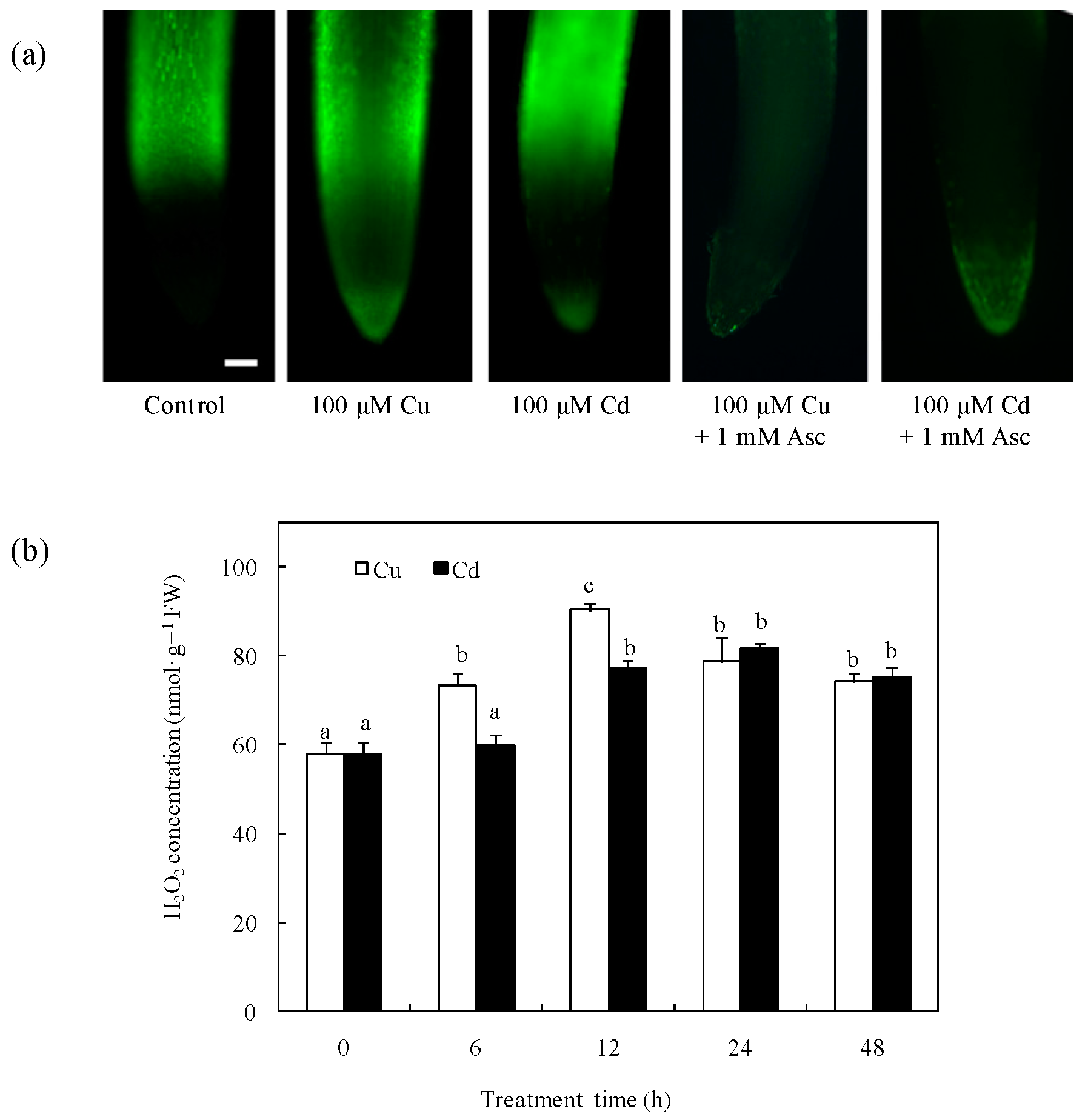
2.1. Effects of Cu and Cd on H2O2 Production in Rice Radicles

To understand the effects of Cu and Cd on H2O2 production in rice, we investigated the H2O2 production in rice radicles by 2′,7′-dichlorodihydrofluorescein diacetate (H2DCFDA) staining. Compared with the control and the treatment with the H2O2 scavenger, Asc, the 12-h treatments with 100 μM Cu or 100 μM Cd significantly increased the H2O2 production of radicles (Figure 1a). When the H2O2 concentrations were assayed spectrophotometrically, H2O2 gradually increased during the first 12 h of 100 µM Cu exposure and then decreased slightly but remained higher than that of the control; however, the Cd-induced H2O2 happened only after 12 h of 100 µM Cd exposure, which lagged behind that of Cu exposure (Figure 1b).

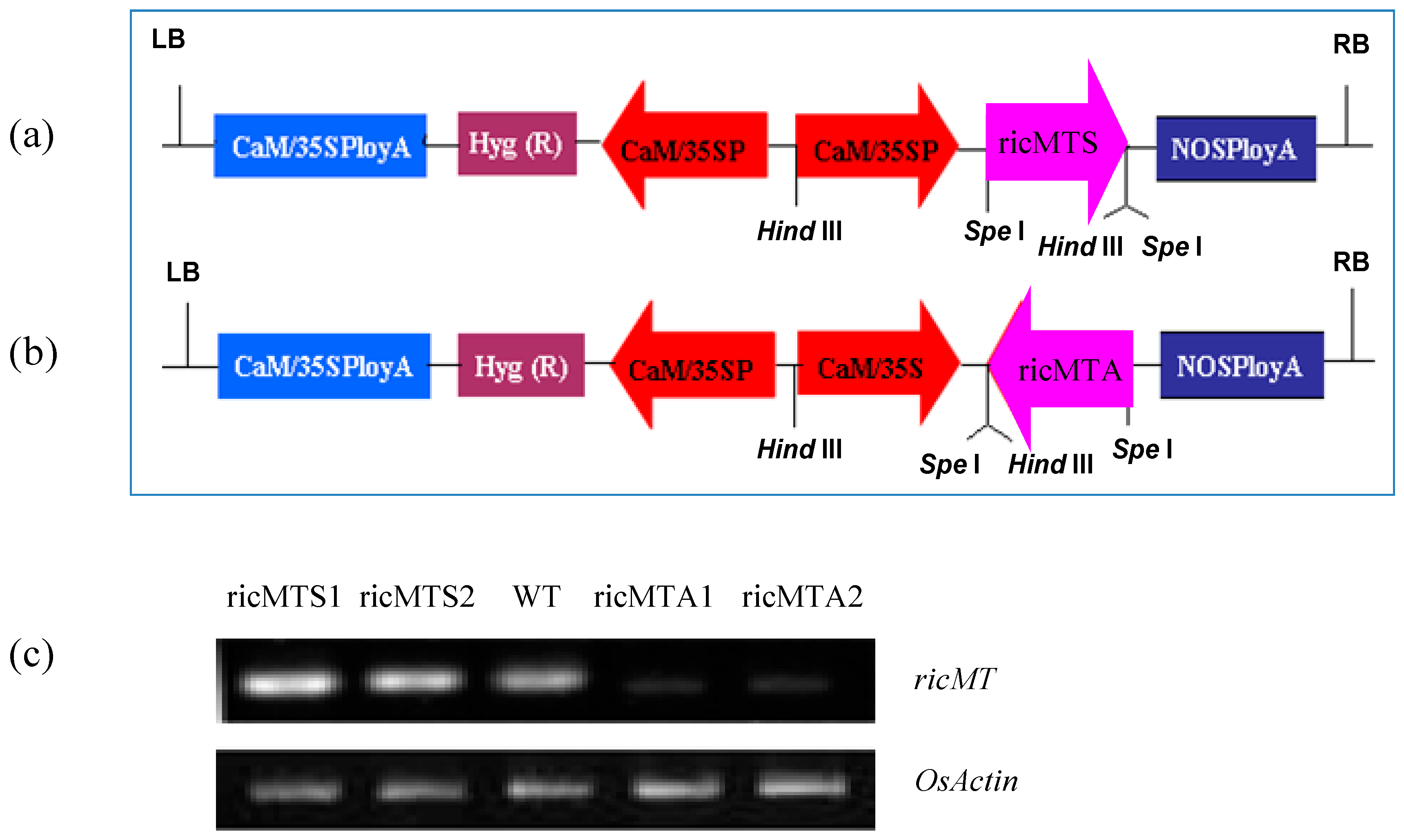
2.2. Cu and Cd Up-Regulate the ricMT and CuZn-SOD Gene Expression in Rice Radicles

The temporal changes in the gene expression of ricMT and CuZn-SOD were analyzed in rice radicles using quantitative RT-PCR. There was no significant difference in the expression level under the control medium within 48 h (Figure 2a). The expression of ricMT and CuZn-SOD mRNA was significantly higher in rice radicles treated with 100 µM Cu or 100 µM Cd for 24 and 48 h than in the control (Figure 2b,c). By contrast, Cd significantly up-regulated the mRNA levels of two proteins under 6 and 12 h treatment, while Cu did not.

2.3. ricMT Expression Improved Cu and Cd Tolerance of Rice Suspension Cells

To evaluate the roles of ricMT, we generated transgenic rice suspension cells expressing the full-length ricMT cDNA under control of the CaMV 35S promoter using Agrobacterium mediated transformation (Figure 3a,b). The expression of ricMT in transgenic lines (sense-ricMT lines ricMTS1 and ricMTS2, antisense-ricMT lines ricMTA1 and ricMTA2) and the wild-type (WT) was analyzed using semi-quantitative RT-PCR (Figure 3c). Rice cells of the sense-ricMT lines ricMTS2 and antisense-ricMT lines ricMTA2 were used for subsequent rice suspension cell experiments.
There was no significant difference in the growth rates of WT and transgenic cells in normal medium (Figure 4a). When rice cell cultured on medium supplemented with 100 µM Cu or 100 µM Cd, there was higher cell death rate than that of normal medium. In comparison with the WT, the sense-ricMT line (ricMTS2) had a decreased rate of cell death after 6 h Cu treatment or after 12 h Cd treatment; by contrast, the antisense-ricMT line (ricMTA2) showed an increased cell death rate at 12 and 24 h of Cu treatment or after 6 h of Cd treatment (Figure 4b,c).

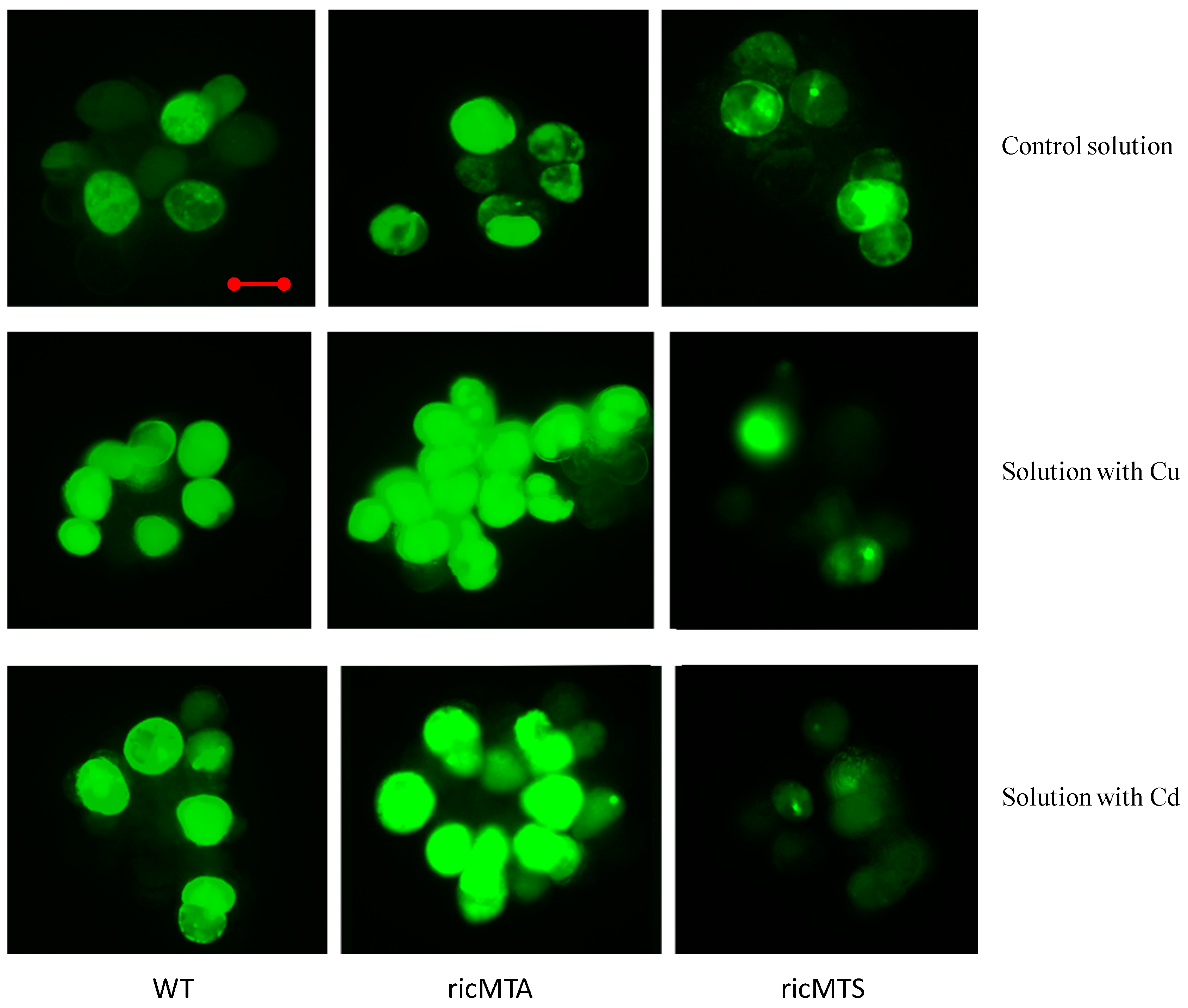
2.4. ricMT Expression Decreased H2O2 Production in Rice Suspension Cells under Cu and Cd Stress
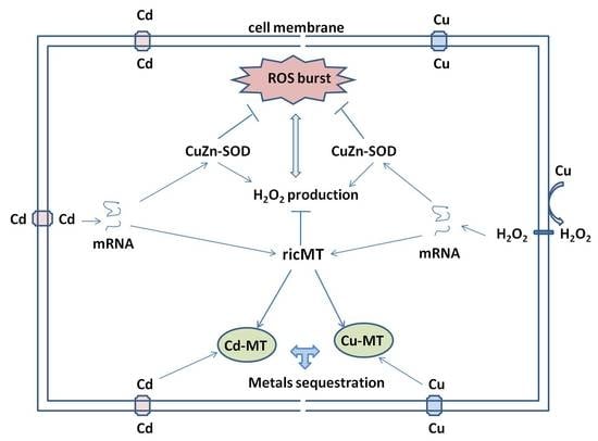
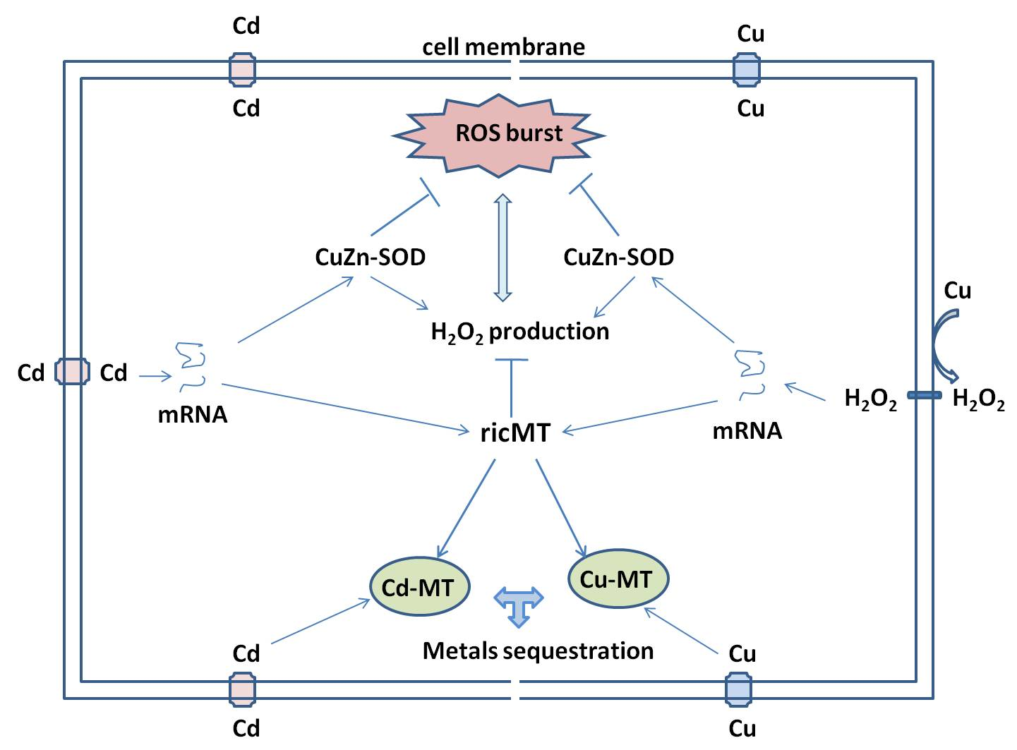
To understand the role of ricMT in antioxidant protection, H2O2 production in rice suspension cells was detected by H2DCFDA staining. There was no significant difference in H2O2 production between WT and transgenic cells in normal medium (Figure 5). In comparison with the control solution, when cultured in medium supplemented with 100 µM Cu or 100 µM Cd for 24 h, the H2O2 production increased significantly in the rice suspension cells. In comparison with the WT, when cultured in medium supplemented with 100 µM Cu or 100 µM Cd for 24 h, the sense-ricMT line (ricMTS2) showed decreased H2O2 production, while the antisense-ricMT line (ricMTA2) showed increased H2O2 production.

3. Discussion

Numerous studies have shown that heavy metals can induce the formation of ROS, including H2O2, and cause oxidative stress. Cu and Cd toxicity causes an oxidative burst with rapid H2O2 production and its release into the plant apoplast [27,28,29,30]. In this study, the formation of H2O2 was observed in Cu- or Cd-treated rice radicles and cells (Figure 1 and Figure 5). Since H2O2 is relatively stable and can diffuse through cell membranes, it can modulate gene expression and participate in various physiological processes [5,31].
SODs play a key role in the antioxidant defense system through the dismutation of O2•− to H2O2. Excess Cu or Cd treatment increased SOD expression [28,32] and activity [28,29,30], which also influenced the H2O2 production in plants. It was reported that rice MT transcription was induced in response to both Cu and H2O2 [18,20]. In this study, the treatment of Cu for 24 and 48 h or Cd for more than 6 h activated the transcription of ricMT and CuZn-SOD in rice radicles (Figure 2). The 6- and 12-h Cu treatments induced significant H2O2 production, which happened before the expression of CuZn-SOD and ricMT increased, therefore we guess that Cu-induced H2O2 production acts upstream from ricMT and CuZn-SOD expression in the induction of Cu stress. In comparison, the H2O2 induced by 12-h Cd was lower than the Cu-induced H2O2, which happened after increased expression of CuZn-SOD and ricMT, so we guess that it can act downstream from ricMT and CuZn-SOD expression and block H2O2 production in rice radicles (Figure 1 and Figure 2). Consistent with our results, the expression of H2O2-removing enzymes is reported to be up-regulated by excess Cu or elevated endogenous H2O2 [26,28], and increased H2O2-removing enzymes decrease H2O2 production [28,33]. However, overexpressing CuZn-SOD also showed increased H2O2 production in transgenic potato [34]. The co-regulation of CuZn-SOD and MT expression in yeast may protect against cell toxicity caused by excess Cu [35]. However, the overexpression of MT did not protect cultured motor neurons from mutant CuZn-SOD toxicity [36].
In addition to chelating extra metal ions in plant cells via their Cys thiol groups, MTs may also enhance plant tolerance to stress by up-regulating anti-oxidative enzymes to maintain the redox balance and thereby reduce ROS-induced injury [15,17,37,38]. To make clear the role of ricMT on oxidative damage, rice suspension cells of sense- and antisense-ricMT transgenic lines were obtained by an Agrobacterium-mediated transformation for the first time. In comparison with the WT, the sense-ricMT line (ricMTS2) had a decreased rate of cell death after 24 h Cu treatment or 48 h Cd treatment, and the antisense-ricMT line (ricMTA2) showed an increased cell death rate after 6 h of Cu treatment or 12 h of Cd treatment (Figure 4b,c). It is reported that MT-overexpressing plants [18,39] or yeast [40,41] have a more efficient antioxidant system with increased enzyme activity against stress conditions. Kumar et al. [11] found that ectopic expression of OsMT1e-P protected against oxidative stress primarily through efficient scavenging of ROS.
In the present study, ricMT expression blocked the production of H2O2 in rice suspension cells under Cu or Cd stress (Figure 5). Contrasting the WT, an increase of H2O2 was accompanied by high cell death rate in antisense-ricMT rice lines under Cu and Cd treatment, and a decrease of H2O2 was accompanied by low cell death rate in sense-ricMT lines (Figure 4 and Figure 5). Moreover, Cu, being a redox-active metal, causes higher accumulation of H2O2 in rice suspension cells than Cd (Figure 5), which is coincident with higher cell death under Cu treatment (Figure 4). Consistent with our results, heterologous expression of OsMT2c [18] or BcMT [19] in Arabidopsis provided increased tolerance against Cu or Cd stress and accumulated lower amounts of H2O2. In comparison, Cu alone (and not oxidative stress) was reported to induce MT expression in Neurospora crassa [42].
H2O2 production in rice radicles was increased before the expression of ricMT and CuZn-SOD induced by Cu, but not Cd, which suggests that Cu-induced H2O2 production acts upstream from ricMT and CuZn-SOD expression in the induction of Cu stress. The overexpression of ricMT significantly decreased the rice cell death rate, which was accompanied by lower H2O2 accumulation in rice cells in response to Cu and Cd stress. This indicates that H2O2 is involved in the ricMT-mediated rice tolerance to Cu and Cd toxicity.

4. Materials and Methods

4.1. Plant Materials

Rice (Oryza sativa L. cv. Wuyunjing No. 7) seeds were surface-sterilized with 5% (v/v) sodium hypochlorite (NaClO) for 15 min and washed thoroughly in distilled water. Then, the seeds were germinated on moist filter paper. Twenty seeds were randomly placed on filter paper in 90-mm Petri dishes and germinated in the dark at 25 °C with the distilled water renewed at 2-day intervals. After germinating for 4 days, 5 mL of freshly prepared 100 µM CuSO4 solution, 100 µM CdCl2 solution, or distilled water (control) was added to the Petri dishes for 0, 6, 12, 24, or 48 h; each treatment was performed in triplicate. The radicles were dissected from germinating rice seeds for quantitative RT-PCR and H2O2 determination.

4.2. Generation of Sense and Antisense ricMT Transgenic Rice

The full-length sequences of sense- and antisense-ricMT were obtained by PCR and inserted into the SpeI and HindIII restriction sites of the plant expression vector pCAMBIA1304 (Cambia, Canberra, Australia) under the control of the cauliflower mosaic virus (CaMV) 35S promoter. Then, pCAMBIA1304 vector harboring the sense- or antisense-ricMT was transformed into the rice cultivar Nipponbare by using Agrobacterium-mediated transformation [43].

4.3. Suspension Cell Cultures

Rice suspension cells were cultured in Chu (N6) medium containing 30 g·L−1 of sucrose, 2 mg·L−1 of 2,4-d-dicholorophenoxyacetic acid and 0.2 mg·L−1 kinetin. After autoclaving, 50 mg·L−1 of filter-sterilized hygromycin B was added for positive selection. The cells were maintained in 500-mL Erlenmeyer flasks containing 180 mL of fresh medium, and subcultured every week. For the flask experiments, a 100-mL flask was used, containing 30 mL of fresh medium which was inoculated with 3 g of fresh cells. The cultures were incubated in a gyratory shaking incubator at 28 °C and 120 rpm. For the treatments, cells were used 5 days after subculture, control medium, medium with 100 μM CuSO4 or CdCl2 were tested at concentrations of 100 µM for 0, 6, 12, 24, or 48 h.

4.4. Total RNA Isolation, cDNA Synthesis and Quantitative RT-PCR

Total RNA was extracted using the RNA simple Total RNA Kit (LifeFeng, Shanghai, China) according to the manufacturer’s instructions and then converted to cDNA after DNase I treatment using a PrimeScriptTM RT Master Mix (TaKaRa Bio, Tokyo, Japan). Real-time quantitative RT-PCR was performed on a MyiQ Real-Time PCR Detection System (Bio-Rad Hercules, Berkeley, CA, USA) using SYBR Premix Ex Taq (TaKaRa Bio, Tokyo, Japan). The primers for rice CuZn-SOD (AAA33917) mRNA were forward TCATTGGCAGAGCCGTCGTTGT and reverse AGTCCGATGATCCCGCAAGCAA, the primers for ricMT mRNA were forward CACCATGTCGTGCTGGGTGGCAA and reverse CTTCTAGTTGCAGTTGCAGCAGG, and the primers for the internal control OsActin were forward TTATGGTTGGGATGGGACA and reverse AGCACGGCTTGAATAGCG. The PCR protocol included an initial 7 min incubation at 95 °C for complete denaturation followed by 40 cycles at 94 °C for 30 s, 60 °C for 30 s, and 72 °C for 30 s. The specificity of the PCR amplification was examined based on a heat dissociation curve (65–95 °C) following the final cycle. Normalized relative expression was calculated using the 2−ΔΔCt (cycle threshold) method.

4.5. Hydrogen Peroxide Localization In Situ

The H2O2 production was detected by the infiltration of H2DCFDA, as reported by Ezaki et al. [44] with some modifications. Rice radicles or suspension cells were incubated in 20 μM H2DCFDA for 20 min. The excess dye had been removed by washing with distilled water for 1 min, the radicles or suspension cells were transferred to microscope slides and observed with a Zeiss Axio Imager A1 fluorescence microscope (Carl Zeiss, Jena, Germany) fitted with an AxioCam HRc camera to visualize the green fluorescence of the H2O2-oxidized probe.

4.6. H2O2 Determination in Extracts

The content of H2O2 in rice radicles from Cu-treated plants was measured by monitoring the A415 of the titanium-peroxide complex following the method described by Jiang et al. [45]. Absorbance values were calibrated to a standard curve established with 0.1–1.0 µM H2O2.

4.7. Evans Blue Assay for Suspension Cell Death

The death of suspension cells was monitored using Evans blue, which is excreted from intact viable cells and is used to estimate cell death spectrophotometrically, as described by Baker and Mock [46]. Briefly, aliquots of treated suspension cells were stained with Evans blue. The cells were washed to remove the excess stain, transferred to 1.5-mL Eppendorf tubes, ground with a micro-sample pestle in the presence of 0.5% SDS to release the trapped stain, and centrifuged to pellet the cellular debris. The A600 of the supernatant was used to quantify cell death.

4.8. Statistical Analysis

Data were analyzed using SPSS ver. 16.0 (Statistical Package for Social Science for Windows, SPSS, Inc., Chicago, IL, USA). All values reported in this paper are means ± SE (n = 3) of three separate experiments. Means denoted by the same letter did not significantly differ at p < 0.05 according to Duncan’s multiple range test.

Acknowledgments

This research project was partly supported by Innovation Team Foundation of Henan Universityof Science and Technology (2015TTD002), the Plan for Scientific Innovation Talent of Henan Province (154100510010), the Key projects of the Department of Education of Henan Province (16A180004) and the Development Program to National Natural Science Foundation of Henan University of Science and Technology (13000786).

Author Contributions

Hongxiao Zhang and Youjun Li conceived and designed the experiments. Hongxiao Zhang, Shufang Lv, and Huawei Xu performed the experiments. Hongxiao Zhang and Fayuan Wang analyzed the data and wrote the manuscript. Dianyun Hou also have contributed to the data interpretation and writing.

Abbreviations

Cyscysteine
ROSreactive oxygen species
H2DCFDA2′,7′-dichlorodihydrofluorescein diacetate
MTmetallothionein
SODsuperoxide dismutase
WTwild-type

References

  1. Marschner, H. Mineral Nutrition of Higher Plants; Academic Press: London, UK, 1995. [Google Scholar]
  2. Schützendübel, A.; Polle, A. Plant responses to abiotic stresses: Heavy metal-induced oxidative stress and protection by mycorrhization. J. Exp. Bot. 2002, 53, 1351–1365. [Google Scholar] [CrossRef] [PubMed]
  3. Cobbett, C.; Goldsbrough, P. Phytochelatins and metallothioneins: Roles in heavy metal detoxification and homeostasis. Ann. Rev. Plant Biol. 2002, 53, 159–182. [Google Scholar] [CrossRef] [PubMed]
  4. Mittler, R.; Vanderauwera, S.; Gollery, M.; van Breusegem, F. Reactive oxygen gene network of plants. Trends Plant Sci. 2004, 9, 490–498. [Google Scholar] [CrossRef] [PubMed]
  5. Ahmad, P.; Sarwat, M.; Sharma, S. Reactive oxygen species, antioxidants and signaling in plants. J. Plant Biol. 2008, 51, 167–173. [Google Scholar] [CrossRef]
  6. Cho, U.H.; Seo, N.H. Oxidative stress in arabidopsis thaliana exposed to cadmium is due to hydrogen peroxide accumulation. Plant Sci. 2005, 168, 113–120. [Google Scholar] [CrossRef]
  7. Chao, Y.Y.; Hsu, Y.T.; Kao, C.H. Involvement of glutathione in heat shock- and hydrogen peroxide-induced cadmium tolerance of rice (Oryza sativa L.) seedlings. Plant Soil 2009, 318, 37–45. [Google Scholar] [CrossRef]
  8. Hu, Y.; Ge, Y.; Zhang, C.; Ju, T.; Cheng, W. Cadmium toxicity and translocation in rice seedlings are reduced by hydrogen peroxide pretreatment. Plant Growth Regul. 2009, 59, 51–61. [Google Scholar] [CrossRef]
  9. Thirumoorthy, N.; Shyam Sunder, A.; Manisenthil Kumar, K.T.; Senthil kumar, M.; Ganesh, G.N.K.; Chatterjee, M. A review of metallothionein isoforms and their role in pathophysiology. World J. Surg. Oncol. 2011, 9, 54. [Google Scholar] [CrossRef] [PubMed]
  10. Mir, G.; Domenech, J.; Huguet, G.; Guo, W.J.; Goldsbrough, P.; Atrian, S.; Molinas, M. A plant type 2 metallothionein (MT) from cork tissue responds to oxidative stress. J. Exp. Bot. 2004, 55, 2483–2493. [Google Scholar] [CrossRef] [PubMed]
  11. Kumar, G.; Kushwaha, H.R.; Panjabi-Sabharwal, V.; Kumari, S.; Joshi, R.; Karan, R.; Mittal, S.; Pareek, S.L.S.; Pareek, A. Clustered metallothionein genes are co-regulated in rice and ectopic expression of OsMT1e-P confers multiple abiotic stress tolerance in tobacco via ROS scavenging. BMC Plant Biol. 2012, 12, 1–16. [Google Scholar] [CrossRef] [PubMed]
  12. Yang, Z.; Wu, Y.; Li, Y.; Ling, H.Q.; Chu, C. Osmt1a, a type 1 metallothionein, plays the pivotal role in zinc homeostasis and drought tolerance in rice. Plant Mol. Biol. 2009, 70, 219–229. [Google Scholar] [CrossRef] [PubMed]
  13. Zhu, W.; Zhao, D.X.; Miao, Q.; Xue, T.T.; Li, X.Z.; Zheng, C.C. Arabidopsis thaliana metallothionein, AtMT2a, mediates ROS balance during oxidative stress. J. Plant Biol. 2009, 52, 585–592. [Google Scholar] [CrossRef]
  14. Kim, S.H.; Lee, H.S.; Song, W.Y.; Choi, K.S.; Hur, Y. Chloroplast-targeted BrMT1 (Brassica rapa type-1 metallothionein) enhances resistance to cadmium and ROS in transgenic Arabidopsis plants. J. Plant Biol. 2007, 50, 1–7. [Google Scholar] [CrossRef]
  15. Xia, Y.; Qi, Y.; Yuan, Y.; Wang, G.; Cui, J.; Chen, Y.; Zhang, H.; Shen, Z. Overexpression of Elsholtzia haichowensis metallothionein 1 (EhMT1) in tobacco plants enhances copper tolerance and accumulation in root cytoplasm and decreases hydrogen peroxide production. J. Hazard. Mater. 2012, 233, 65–71. [Google Scholar] [CrossRef] [PubMed]
  16. Samardzic, J.T.; Nikolic, D.B.; Timotijevic, G.S.; Jovanovic, Z.S.; Milisavljevic, M.D.; Maksimovic, V.R. Tissue expression analysis of FeMT3, a drought and oxidative stress related metallothionein gene from buckwheat (Fagopyrum esculentum). J. Plant Physiol. 2010, 167, 1407–1411. [Google Scholar] [CrossRef] [PubMed]
  17. Zhang, J.; Zhang, M.; Tian, S.; Lu, L.; Shohag, M.J.I.; Yang, X. Metallothionein 2 (SaMT2) from Sedum alfredii hance confers increased Cd tolerance and accumulation in yeast and tobacco. PLoS ONE 2014, 9, e102750. [Google Scholar] [CrossRef] [PubMed]
  18. Liu, J.; Shi, X.; Qian, M.; Zheng, L.; Lian, C.; Xia, Y.; Shen, Z. Copper-induced hydrogen peroxide upregulation of a metallothionein gene, OsMT2c, from Oryza sativa L. confers copper tolerance in Arabidopsis thaliana. J. Hazard. Mater. 2015, 294, 99–108. [Google Scholar] [CrossRef] [PubMed]
  19. Lv, Y.; Deng, X.; Quan, L.; Xia, Y.; Shen, Z. Metallothioneins BcMT1 and BcMT2 from Brassica campestris enhance tolerance to cadmium and copper and decrease production of reactive oxygen species in Arabidopsis thaliana. Plant Soil 2012, 367, 507–519. [Google Scholar] [CrossRef]
  20. Wong, H.L.; Sakamoto, T.; Kawasaki, T.; Umemura, K.; Shimamoto, K. Down-regulation of metallothionein, a reactive oxygen scavenger, by the small GTPase OsRac1 in rice. Plant Physiol. 2004, 135, 1447–1456. [Google Scholar] [CrossRef] [PubMed]
  21. Zhou, G.; Xu, Y.; Li, J.; Yang, L.; Liu, J.Y. Molecular analyses of the metallothionein gene family in rice (Oryza sativa L.). J. Biochem. Mol. Biol. 2006, 39, 595–606. [Google Scholar] [CrossRef] [PubMed]
  22. Steffens, B.; Sauter, M. Epidermal cell death in rice is confined to cells with a distinct molecular identity and is mediated by ethylene and H2O2 through an autoamplified signal pathway. Plant Cell 2009, 21, 184–196. [Google Scholar] [CrossRef] [PubMed]
  23. Yu, L.H.; Umeda, M.; Liu, J.Y.; Zhao, N.M.; Uchimiya, H. A novel MT gene of rice plants is strongly expressed in the node portion of the stem. Gene 1998, 206, 29–35. [Google Scholar] [CrossRef]
  24. Zhang, H.; Lian, C.; Shen, Z. Proteomic identification of small, copper-responsive proteins in germinating embryos of Oryza sativa. Ann. Bot. 2009, 103, 923–930. [Google Scholar] [CrossRef] [PubMed]
  25. Chen, C.; Song, Y.; Zhuang, K.; Li, L.; Xia, Y.; Shen, Z. Proteomic analysis of copper-binding proteins in excess copper-stressed roots of two rice (Oryza sativa L.) varieties with different Cu tolerances. PLoS ONE 2015, 10, e0125367. [Google Scholar] [CrossRef] [PubMed]
  26. Zhang, H.X.; Xia, Y.; Chen, C.; Zhuang, K.; Song, Y.F.; Shen, Z.G. Analysis of copper-binding proteins in rice radicles exposed to excess copper and hydrogen peroxide stress. Front. Plant Sci. 2016, 7, 1216. [Google Scholar] [CrossRef] [PubMed]
  27. Zhang, H.; Xia, Y.; Wang, G.; Shen, Z. Excess copper induces accumulation of hydrogen peroxide and increases lipid peroxidation and total activity of copper-zinc superoxide dismutase in roots of Elsholtzia haichowensis. Planta 2008, 227, 465–475. [Google Scholar] [CrossRef] [PubMed]
  28. Zhang, H.; Zhang, F.; Xia, Y.; Wang, G.; Shen, Z. Excess copper induces production of hydrogen peroxide in the leaf of Elsholtzia haichowensis through apoplastic and symplastic CuZn-superoxide dismutase. J. Hazard. Mater. 2010, 178, 834–843. [Google Scholar] [CrossRef] [PubMed]
  29. Deng, X.; Xia, Y.; Hu, W.; Zhang, H.; Shen, Z. Cadmium-induced oxidative damage and protective effects of N-acetyl-l-cysteine against cadmium toxicity in Solanum nigrum L. J. Hazard. Mater. 2010, 180, 722–729. [Google Scholar] [CrossRef] [PubMed]
  30. Zhang, F.; Zhang, H.; Xia, Y.; Wang, G.; Xu, L.; Shen, Z. Exogenous application of salicylic acid alleviates cadmium toxicity and reduces hydrogen peroxide accumulation in root apoplasts of Phaseolus aureus and Vicia sativa. Plant Cell Rep. 2011, 30, 1475–1483. [Google Scholar] [CrossRef] [PubMed]
  31. Neill, S.; Desikan, R.; Hancock, J. Hydrogen peroxide signalling. Curr. Opin. Plant Biol. 2002, 5, 388–395. [Google Scholar] [CrossRef]
  32. Cohu, C.M.; Abdel-Ghany, S.E.; Reynolds, K.A.G.; Onofrio, A.M.; Bodecker, J.R.; Kimbrel, J.A.; Niyogi, K.K.; Pilon, M. Copper delivery by the copper chaperone for chloroplast and cytosolic copper/zinc-superoxide dismutases: Regulation and unexpected phenotypes in an Arabidopsis mutant. Mol. Plant 2009, 2, 1336–1350. [Google Scholar] [CrossRef] [PubMed]
  33. Vandenabeele, S.; van der Kelen, K.; Dat, J.; Gadjev, I.; Boonefaes, T.; Morsa, S.; Rottiers, P.; Slooten, L.; van Montagu, M.; Zabeau, M.; et al. A comprehensive analysis of hydrogen peroxide-induced gene expression in tobacco. Proc. Natl. Acad. Sci. USA 2003, 100, 16113–16118. [Google Scholar] [CrossRef] [PubMed]
  34. Yoon-Sik, K.; Hyun-Soon, K.; Yong-Hwa, L.; Mi-Sun, K.; Hyun-Woo, O.; Kyu-Woong, H.; Hyouk, J.; Jae-Heung, J. Elevated H2O2 production via overexpression of a chloroplastic Cu/Zn SOD gene of lily (Lilium oriental hybrid ‘marco polo’) triggers ethylene synthesis in transgenic potato. Plant Cell Rep. 2008, 27, 973–983. [Google Scholar]
  35. Carri, M.T.; Galiazzo, F.; Ciriolo, M.R.; Rotilio, G. Evidence for co-regulation of Cu,Zn superoxide dismutase and metallothionein gene expression in yeast through transcriptional control by copper via the ACE1 factor. FEBS Lett. 1991, 278, 263–266. [Google Scholar] [CrossRef]
  36. Taylor, D.M.; Minotti, S.; Agar, J.N.; Durham, H.D. Overexpression of metallothionein protects cultured motor neurons against oxidative stress, but not mutant Cu/Zn-superoxide dismutase toxicity. Neurotoxicology 2004, 25, 779–792. [Google Scholar] [CrossRef] [PubMed]
  37. Zhou, Y.; Chu, P.; Chen, H.; Li, Y.; Liu, J.; Ding, Y.; Tsang, E.W.T.; Jiang, L.; Wu, K.; Huang, S. Overexpression of Nelumbo nucifera metallothioneins 2a and 3 enhances seed germination vigor in arabidopsis. Planta 2011, 235, 523–537. [Google Scholar] [CrossRef] [PubMed]
  38. Zhou, B.; Yao, W.; Wang, S.; Wang, X.; Jiang, T. The metallothionein gene, TaMT3, from Tamarix androssowii confers Cd2+ tolerance in tobacco. Int. J. Mol. Sci. 2014, 15, 10398–10409. [Google Scholar] [CrossRef] [PubMed]
  39. Nezhad, R.M.; Shahpiri, A.; Mirlohi, A. Discrimination between two rice metallothionein isoforms belonging to type 1 and type 4 in metal-binding ability. Biotechnol. Appl. Biochem. 2013, 60, 275–282. [Google Scholar] [CrossRef] [PubMed]
  40. Xue, T.; Li, X.; Zhu, W.; Wu, C.; Yang, G.; Zheng, C. Cotton metallothionein GhMT3a, a reactive oxygen species scavenger, increased tolerance against abiotic stress in transgenic tobacco and yeast. J. Exp. Bot. 2009, 60, 339–349. [Google Scholar] [CrossRef] [PubMed]
  41. Yang, J.; Wang, Y.; Liu, G.; Yang, C.; Li, C. Tamarix hispida metallothionein-like ThMT3, a reactive oxygen species scavenger, increases tolerance against Cd2+, Zn2+, Cu2+, and NaCl in transgenic yeast. Mol. Biol. Rep. 2010, 38, 1567–1574. [Google Scholar] [CrossRef] [PubMed]
  42. Kumar, K.S.; Dayananda, S.; Subramanyam, C. Copper alone, but not oxidative stress, induces copper-metallothionein gene in Neurospora crassa. FEMS Microbiol. lett. 2005, 242, 45–50. [Google Scholar] [CrossRef] [PubMed]
  43. Nishimura, A.; Aichi, I.; Matsuoka, M. A protocol for Agrobacterium-mediated transformation in rice. Nat. Protoc. 2007, 1, 2796–2802. [Google Scholar] [CrossRef] [PubMed]
  44. Ezaki, B.; Gardner, R.C.; Ezaki, Y.; Matsumoto, H. Expression of aluminum-induced genes in transgenic arabidopsis plants can ameliorate aluminum stress and/or oxidative stress. Plant Physiol. 2000, 122, 657–666. [Google Scholar] [CrossRef] [PubMed]
  45. Jiang, M.Y.; Zhang, J.H. Effect of abscisic acid on active oxygen species, antioxidative defence system and oxidative damage in leaves of maize seedlings. Plant Cell Physiol. 2001, 42, 1265–1273. [Google Scholar] [CrossRef] [PubMed]
  46. Baker, C.J.; Mock, N.M. An improved method for monitoring cell death in cellsuspension and leaf disc assays using evans blue. Plant Cell Tiss. Org. 1994, 39, 7–12. [Google Scholar] [CrossRef]
Figure 1. Cu- and Cd-induced H2O2 accumulation in rice radicles. (a) Histochemical detection of H2O2 in rice radicles under different treatment for 12 h; (b) The total contents of H2O2 in rice radicles under varied Cu or Cd treatment time. Germinating rice embryos were treated with 1 mM ascorbic acid (Asc) solution for 12 h, or treated with 100 µM CuSO4 and 100 µM CdCl2 solution for 0, 3, 6, 12, 24, and 48 h. Subsequently radicles from germinating rice seeds were incubated in 20 μM H2DCFDA for 20 min or were homogenized and the H2O2 content assayed by spectrophotometry. Bar, 100 μm. Experiments were repeated at least three times with similar results. Values are means ± SE (n = 3) of three separate experiments. Means denoted by the same letter did not significantly differ at p < 0.05 according to Duncan’s multiple range test.
Ijms 18 02083 g001
Figure 2. Time course for Cu- and Cd-induced expression of ricMT and CuZn-SOD. (ac) Time course for control (a); Cu-induced (b) and Cd-induced (c) expression of ricMT and CuZn-SOD. Germinating rice embryos were treated with distilled water (control), 100 μM CuSO4 or 100 μM CdCl2 for various times (0, 6, 12, 24, and 48 h). Subsequently, radicles were isolated from the germinating seeds for gene analyses by quantitative RT-PCR. Values are means ± SE (n = 3) of three separate experiments. Means denoted by the same letter did not significantly differ at p < 0.05 according to Duncan’s multiple range test.
Ijms 18 02083 g002
Figure 3. Generation of transgenic rice. (a) Diagram of the pCAMBIA1304 vectors harboring sense-ricMT; (b) Diagram of the pCAMBIA1304 vectors harboring antisense-ricMT; (c) Semi-quantitative RT-PCR analysis of ricMT expression in wild-type and transgenic rice suspension cells. LB represents the left border; RB represents the right border; WT represents the wild-type rice suspension cells; ricMTS1 and ricMTS2 represent two independent sense-ricMT transgenic rice suspension cell lines; ricMTA1 and ricMTA2 represent two independent antisense-ricMT rice suspension cell lines. Experiments were repeated at least three times with similar results.
Ijms 18 02083 g003
Figure 4. Effect of treatment of Cu and Cd on cell death of rice suspension cells. (ac) Rice suspension cells were cultured with control medium (a), medium with 100 μM CuSO4 (b) or medium with 100 μM CdCl2 (c) for various times (0, 6, 12, 24, and 48 h). Subsequently, aliquots of the suspension cells were stained with Evans blue. Cells were then washed to remove excess stain, ground with a micro-sample pestle in the presence of 0.5% SDS to release trapped stain. The A600 of the supernatant was used to monitor cell death. WT represents the wild-type rice suspension cells; ricMTS and ricMTA respectively represent rice suspension cells of the sense-ricMT lines ricMTS2 and antisense-ricMT lines ricMTA2. Values are means ± SE (n = 3) of three separate experiments. Means denoted by the same letter did not significantly differ at p < 0.05 according to Duncan’s multiple range test.
Ijms 18 02083 g004
Figure 5. Effect of treatment of Cu or Cd on H2O2 production of rice suspension cells. Rice suspension cells were cultured with control medium, or medium with 100 μM CuSO4 or medium with 100 μM CdCl2 for 24 h. Subsequently, the suspension cells were incubated in 20 μM H2DCFDA for 20 min. WT represents the wild-type rice suspension cells; ricMTA and ricMTS respectively represent rice suspension cells of the antisense-ricMT line and sense-ricMT line. Bar, 20 μm. Experiments were repeated at least three times with similar results.
Ijms 18 02083 g005

