Bevacizumab for Patients with Recurrent Gliomas Presenting with a Gliomatosis Cerebri Growth Pattern

 , ,
, ,
Abstract
:
1. Introduction
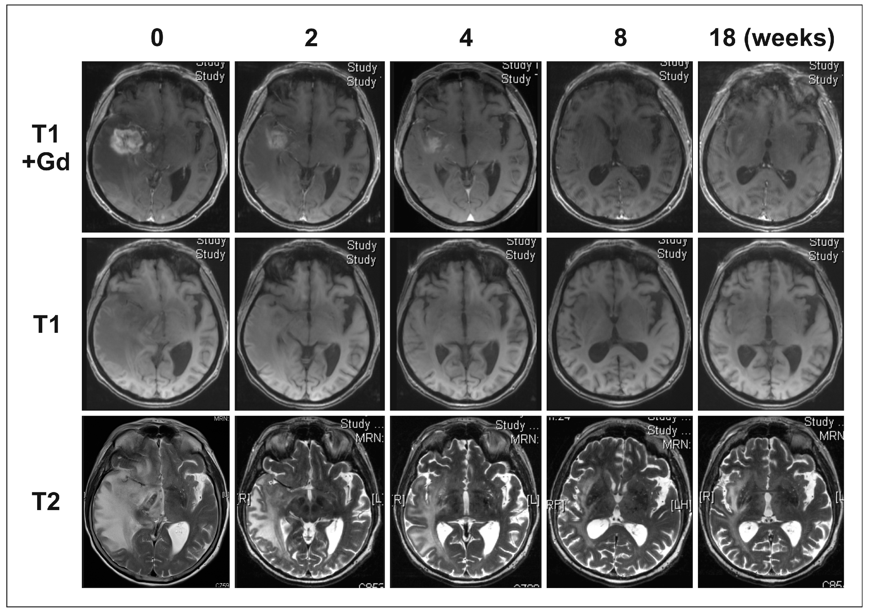
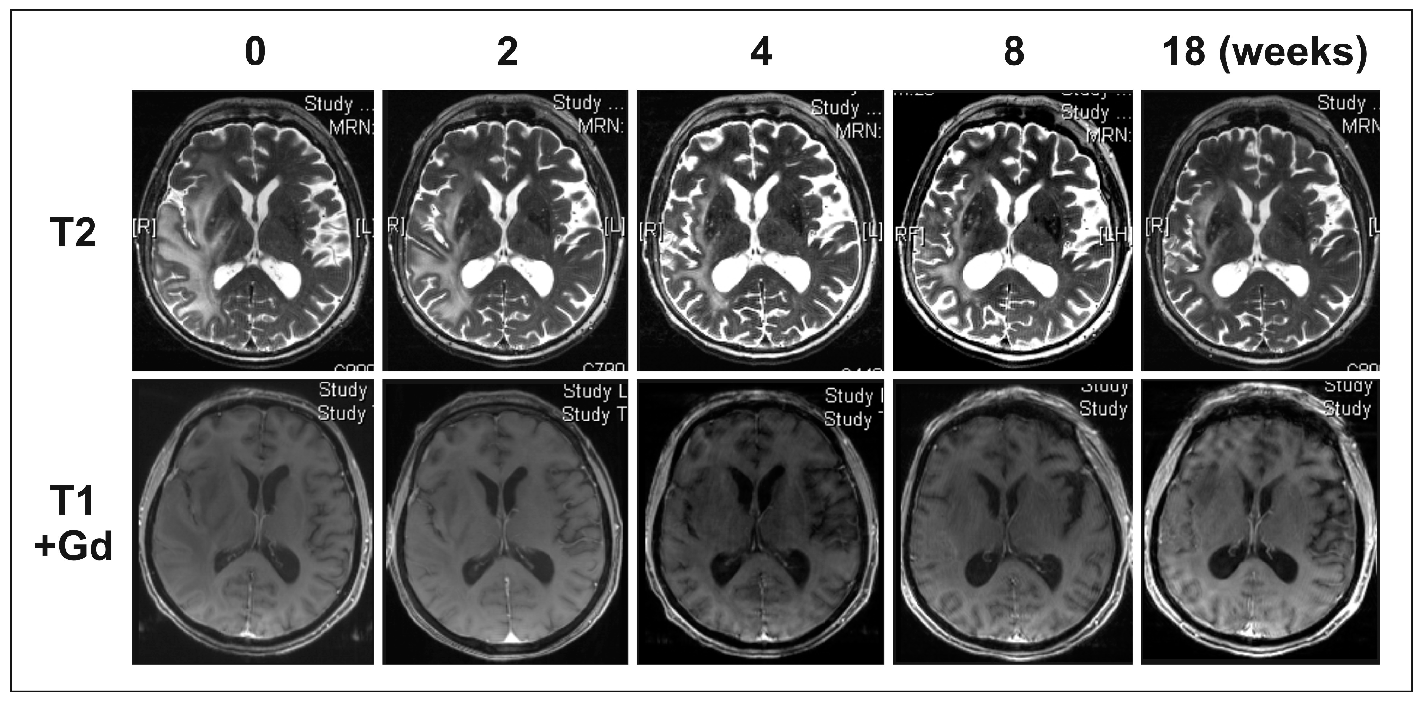
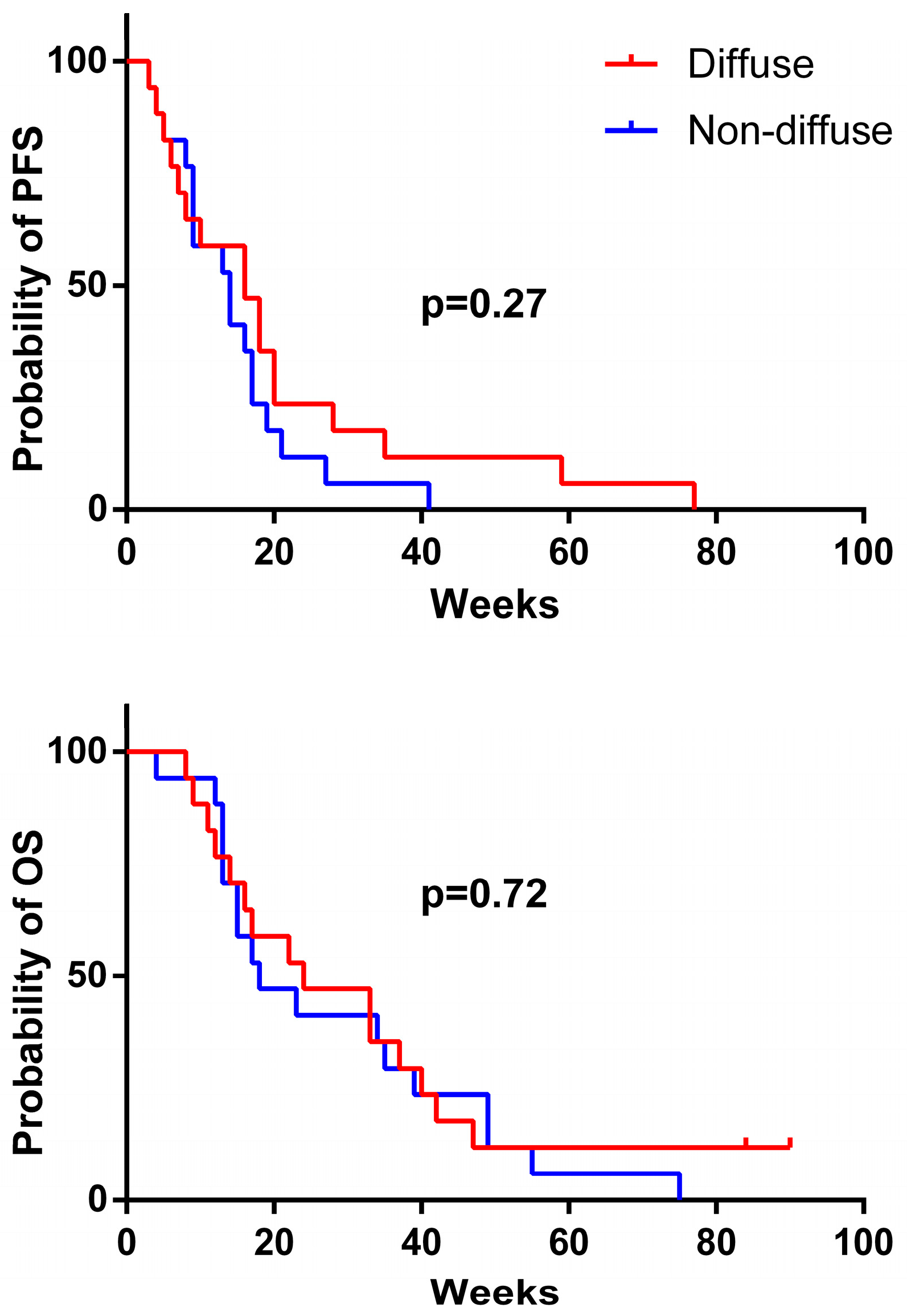
2. Results
3. Discussion
4. Patients and Methods
4.1. MGMT Promoter Methylation Status Assessment
4.2. Analysis of 1p and 19q Loss by Fluorescence In Situ Hybridization (FISH)
4.3. mIDH1R132H Immunohistochemistry
Acknowledgments
Author Contributions
Conflicts of Interest
References
- Tsai, J.C.; Goldman, C.K.; Gillespie, G.Y. Vascular endothelial growth factor in human glioma cell lines: Induced secretion by EGF, PDGF-BB, and bFGF. J. Neurosurg. 1995, 82, 864–873. [Google Scholar] [CrossRef] [PubMed]
- Bates, D.O. Vascular endothelial growth factors and vascular permeability. Cardiovasc. Res. 2010, 87, 262–271. [Google Scholar] [CrossRef] [PubMed]
- Gerstner, E.R.; Duda, D.G.; di Tomaso, E.; Ryg, P.A.; Loeffler, J.S.; Sorensen, A.G.; Ivy, P.; Jain, R.K.; Batchelor, T.T. VEGF inhibitors in the treatment of cerebral edema in patients with brain cancer. Nat. Rev. Clin. Oncol. 2009, 6, 229–236. [Google Scholar] [CrossRef] [PubMed]
- Batchelor, T.T.; Sorensen, A.G.; di Tomaso, E.; Zhang, W.T.; Duda, D.G.; Cohen, K.S.; Kozak, K.R.; Cahill, D.P.; Chen, P.J.; Zhu, M.; et al. AZD2171, a pan-VEGF receptor tyrosine kinase inhibitor, normalizes tumor vasculature and alleviates edema in glioblastoma patients. Cancer Cell 2007, 11, 83–95. [Google Scholar] [PubMed]
- Tong, R.T.; Boucher, Y.; Kozin, S.V.; Winkler, F.; Hicklin, D.J.; Jain, R.K. Vascular normalization by vascular endothelial growth factor receptor 2 blockade induces a pressure gradient across the vasculature and improves drug penetration in tumors. Cancer Res. 2004, 64, 3731–3736. [Google Scholar] [CrossRef] [PubMed]
- Takano, S.; Kimu, H.; Tsuda, K.; Osuka, S.; Nakai, K.; Yamamoto, T.; Ishikawa, E.; Akutsu, H.; Matsuda, M.; Matsumura, A. Decrease in the apparent diffusion coefficient in peritumoral edema for the assessment of recurrent glioblastoma treated by bevacizumab. Acta Neurochir. Suppl. 2013, 118, 185–189. [Google Scholar] [PubMed]
- Kickingereder, P.; Wiestler, B.; Burth, S.; Wick, A.; Nowosielski, M.; Heiland, S.; Schlemmer, H.P.; Wick, W.; Bendszus, M.; Radbruch, A. Relative cerebral blood volume is a potential predictive imaging biomarker of bevacizumab efficacy in recurrent glioblastoma. Neuro Oncol. 2015, 17, 1139–1147. [Google Scholar] [CrossRef] [PubMed]
- Jain, R.K.; di Tomaso, E.; Duda, D.G.; Loeffler, J.S.; Sorensen, A.G.; Batchelor, T.T. Angiogenesis in brain tumours. Nat. Rev. Neurosci. 2007, 8, 610–622. [Google Scholar] [PubMed]
- Von Baumgarten, L.; Brucker, D.; Tirniceru, A.; Kienast, Y.; Grau, S.; Burgold, S.; Herms, J.; Winkler, F. Bevacizumab has differential and dose-dependent effects on glioma blood vessels and tumor cells. Clin. Cancer Res. 2011, 17, 6192–6205. [Google Scholar] [CrossRef] [PubMed]
- De Groot, J.F.; Fuller, G.; Kumar, A.J.; Piao, Y.; Eterovic, K.; Ji, Y.; Conrad, C.A. Tumor invasion after treatment of glioblastoma with bevacizumab: Radiographic and pathologic correlation in humans and mice. Neuro Oncol. 2010, 12, 233–242. [Google Scholar] [CrossRef] [PubMed]
- Favaro, E.; Bensaad, K.; Chong, M.G.; Tennant, D.A.; Ferguson, D.J.; Snell, C.; Steers, G.; Turley, H.; Li, J.L.; Gunther, U.L.; et al. Glucose utilization via glycogen phosphorylase sustains proliferation and prevents premature senescence in cancer cells. Cell Metab. 2012, 16, 751–764. [Google Scholar] [CrossRef] [PubMed]
- Jahangiri, A.; De Lay, M.; Miller, L.M.; Carbonell, W.S.; Hu, Y.L.; Lu, K.; Tom, M.W.; Paquette, J.; Tokuyasu, T.A.; Tsao, S.; et al. Gene expression profile identifies tyrosine kinase c-Met as a targetable mediator of antiangiogenic therapy resistance. Clin. Cancer Res. 2013, 19, 1773–1783. [Google Scholar] [CrossRef] [PubMed]
- Kunkel, P.; Ulbricht, U.; Bohlen, P.; Brockmann, M.A.; Fillbrandt, R.; Stavrou, D.; Westphal, M.; Lamszus, K. Inhibition of glioma angiogenesis and growth in vivo by systemic treatment with a monoclonal antibody against vascular endothelial growth factor receptor-2. Cancer Res. 2001, 61, 6624–6628. [Google Scholar] [PubMed]
- Paez-Ribes, M.; Allen, E.; Hudock, J.; Takeda, T.; Okuyama, H.; Vinals, F.; Inoue, M.; Bergers, G.; Hanahan, D.; Casanovas, O. Antiangiogenic therapy elicits malignant progression of tumors to increased local invasion and distant metastasis. Cancer Cell 2009, 15, 220–231. [Google Scholar] [CrossRef] [PubMed]
- Wick, A.; Dorner, N.; Schafer, N.; Hofer, S.; Heiland, S.; Schemmer, D.; Platten, M.; Weller, M.; Bendszus, M.; Wick, W. Bevacizumab does not increase the risk of remote relapse in malignant glioma. Ann. Neurol. 2011, 69, 586–592. [Google Scholar] [CrossRef] [PubMed]
- Herrlinger, U.; Jones, D.T.; Glas, M.; Hattingen, E.; Gramatzki, D.; Stuplich, M.; Felsberg, J.; Bahr, O.; Gielen, G.H.; Simon, M.; et al. Gliomatosis cerebri: No evidence for a separate brain tumor entity. Acta Neuropathol. 2016, 131, 309–319. [Google Scholar] [CrossRef] [PubMed]
- Wick, W.; Chinot, O.L.; Bendszus, M.; Mason, W.; Henriksson, R.; Saran, F.; Nishikawa, R.; Revil, C.; Kerloeguen, Y.; Cloughesy, T. Evaluation of pseudoprogression rates and tumor progression patterns in a phase III trial of bevacizumab plus radiotherapy/temozolomide for newly diagnosed glioblastoma. Neuro Oncol. 2016, 18, 1434–1441. [Google Scholar] [CrossRef] [PubMed]
- Glas, M.; Bahr, O.; Felsberg, J.; Rasch, K.; Wiewrodt, D.; Schabet, M.; Simon, M.; Urbach, H.; Steinbach, J.P.; Rieger, J.; et al. NOA-05 phase 2 trial of procarbazine and lomustine therapy in gliomatosis cerebri. Ann. Neurol. 2011, 70, 445–453. [Google Scholar] [CrossRef] [PubMed]
- Seiz, M.; Tuettenberg, J.; Meyer, J.; Essig, M.; Schmieder, K.; Mawrin, C.; von Deimling, A.; Hartmann, C. Detection of IDH1 mutations in gliomatosis cerebri, but only in tumors with additional solid component: Evidence for molecular subtypes. Acta Neuropathol. 2010, 120, 261–267. [Google Scholar] [CrossRef] [PubMed]
- Herrlinger, U.; Wiendl, H.; Renninger, M.; Forschler, H.; Dichgans, J.; Weller, M. Vascular endothelial growth factor (VEGF) in leptomeningeal metastasis: Diagnostic and prognostic value. Br. J. Cancer 2004, 91, 219–224. [Google Scholar] [CrossRef] [PubMed]
- Hamerlik, P.; Lathia, J.D.; Rasmussen, R.; Wu, Q.; Bartkova, J.; Lee, M.; Moudry, P.; Bartek, J., Jr.; Fischer, W.; Lukas, J.; Rich, J.N.; Bartek, J. Autocrine VEGF-VEGFR2-Neuropilin-1 signaling promotes glioma stem-like cell viability and tumor growth. J. Exp. Med. 2012, 209, 507–520. [Google Scholar] [PubMed]
- Simon, T.; Coquerel, B.; Petit, A.; Kassim, Y.; Demange, E.; Le Cerf, D.; Perrot, V.; Vannier, J.P. Direct effect of bevacizumab on glioblastoma cell lines in vitro. Neuromol. Med. 2014, 16, 752–771. [Google Scholar] [CrossRef] [PubMed]
- Chinot, O.L.; Wick, W.; Mason, W.; Henriksson, R.; Saran, F.; Nishikawa, R.; Carpentier, A.F.; Hoang-Xuan, K.; Kavan, P.; Cernea, D.; et al. Bevacizumab plus radiotherapy-temozolomide for newly diagnosed glioblastoma. N. Engl. J. Med. 2014, 370, 709–722. [Google Scholar] [CrossRef] [PubMed]
- Taal, W.; Oosterkamp, H.M.; Walenkamp, A.M.; Dubbink, H.J.; Beerepoot, L.V.; Hanse, M.C.; Buter, J.; Honkoop, A.H.; Boerman, D.; de Vos, F.Y.; et al. Single-agent bevacizumab or lomustine versus a combination of bevacizumab plus lomustine in patients with recurrent glioblastoma (BELOB trial): A randomised controlled phase 2 trial. Lancet Oncol. 2014, 15, 943–953. [Google Scholar] [CrossRef]
- Herrlinger, U.; Schafer, N.; Steinbach, J.P.; Weyerbrock, A.; Hau, P.; Goldbrunner, R.; Friedrich, F.; Rohde, V.; Ringel, F.; Schlegel, U.; et al. Bevacizumab Plus Irinotecan Versus Temozolomide in Newly Diagnosed O6-Methylguanine-DNA Methyltransferase Nonmethylated Glioblastoma: The Randomized GLARIUS Trial. J. Clin. Oncol. 2016, 34, 1611–1619. [Google Scholar] [CrossRef] [PubMed]
- Taphoorn, M.J.; Henriksson, R.; Bottomley, A.; Cloughesy, T.; Wick, W.; Mason, W.P.; Saran, F.; Nishikawa, R.; Hilton, M.; Theodore-Oklota, C.; et al. Health-Related Quality of Life in a Randomized Phase III Study of Bevacizumab, Temozolomide, and Radiotherapy in Newly Diagnosed Glioblastoma. J. Clin. Oncol. 2015, 33, 2166–2175. [Google Scholar] [CrossRef] [PubMed]


| Pat. No. | Combination Therapy | Karnofsky Performance Score (KPS) | Steroid Intake (mg of Dexamethasone per Day) | Best Response (RANO Criteria) | CE | NCE | PFS (Weeks) | OS (Weeks) | ||
|---|---|---|---|---|---|---|---|---|---|---|
| at Start of Therapy | Development under Therapy | at Start of Therapy | under Therapy | |||||||
| 1 | none | 80 | +10 | 0 | 0 | PR | + | + | 28 | 37 |
| 2 | none | 70 | 0 | 0 | 0 | SD | 0 | 0 | 20 | 40 |
| 3 | none | 60 | +20 | 16 | 0 | PR | + | + | 16 | 33 |
| 4 | Iri | 90 | −10 | 8 | 12 | PD | − | − | 4 | 11 |
| 5 | none | 70 | −10 | 2 | 4 | PD | 0 | − | 5 | 9 |
| 6 | none | 70 | 0 | 4 | 4 | PD | − | − | 6 | 17 |
| 7 | CCNU | 60 | +10 | 16 | 1 | PR | + | + | 18 | 22 |
| 8 | CCNU | 80 | −10 | 4 | 2 | PD | − | 0 | 7 | 16 |
| 9 | Iri | 80 | 0 | 0 | 0 | SD | 0 | 0 | 18 | 24 |
| 10 | none | 80 | +10 | 0 | 0 | SD | 0 | 0 | 16 | 33 |
| 11 | none | 60 | −10 | 3 | 12 | PD | 0 | 0 | 8 | 12 |
| 12 | CCNU | 70 | +10 | 2 | 0 | PR | + | + | 59 | n.r. |
| 13 | CCNU | 60 | −10 | 8 | 8 | PD | 0 | − | 3 | 8 |
| 14 | Iri | 80 | +10 | 0 | 0 | PR | + | + | 10 | 14 |
| 15 | none | 90 | 0 | 0 | 0 | PR | + | + | 20 | 42 |
| 16 | none | 60 | 0 | 6 | 0 | PR | + | 0 | 35 | 47 |
| 17 | none | 80 | 0 | 1 | 0 | PR | + | 0 | 77 | n.r. |
| Pat. No. | Combination Therapy | Karnofsky Performance Score (KPS) | Steroid Intake (mg of Dexamethasone per Day) | Best Response (RANO Criteria) | CE | NCE | PFS (Weeks) | OS (Weeks) | ||
|---|---|---|---|---|---|---|---|---|---|---|
| at Start of Therapy | Development under Therapy | at Start of Therapy | under Therapy | |||||||
| C1 | none | 80 | 0 | 4 | 0.5 | SD | 0 | + | 19 | 35 |
| C2 | none | 70 | 0 | 4 | 0 | SD | 0 | + | 41 | 55 |
| C3 | Iri | 60 | +20 | 4 | 0 | PR | + | + | 16 | 39 |
| C4 | none | 90 | −20 | 8 | 12 | PD | − | − | 9 | 15 |
| C5 | CCNU | 70 | +10 | 6 | 2 | PR | + | + | 21 | 49 |
| C6 | none | 70 | 0 | 2 | 2 | PR | + | + | 17 | 34 |
| C7 | none | 60 | +10 | 4 | 4 | PR | + | + | 14 | 18 |
| C8 | CCNU | 70 | 0 | 4 | 2 | PR | + | 0 | 17 | 23 |
| C9 | Iri | 80 | −10 | 4 | 4 | PD | − | − | 9 | 12 |
| C10 | none | 90 | −10 | 8 | 12 | PD | 0 | − | 5 | 13 |
| C11 | none | 60 | 0 | 16 | 4 | PD | − | − | 4 | 13 |
| C12 | CCNU | 70 | +10 | 2 | 0 | PR | + | 0 | 14 | 75 |
| C13 | none | 60 | 0 | 4 | 0 | PR | + | 0 | 27 | 49 |
| C14 | Iri | 80 | −10 | 0 | 0 | PD | 0 | + | 8 | 13 |
| C15 | none | 90 | 0 | 0 | 0 | PD | − | − | 13 | 15 |
| C16 | none | 60 | −10 | 4 | 0 | PD | − | − | 3 | 4 |
| C17 | CCNU | 70 | 0 | 4 | 0 | PD | 0 | − | 9 | 17 |
| Pat. No. | Age | Gender | Histology | Genetic Testing | Pretreatment |
|---|---|---|---|---|---|
| 1 | 34 | F | GB | MGMT meth, mIDH1R132H+ | TMZ 7/14, WBXRT, reS, CCNU/VM26 |
| 2 | 43 | F | GB | MGMT unmeth, mIDH1R132H− | pS, IFXRT, Enza, TMZ 7/14, CCNU/VM26 |
| 3 | 43 | M | GB | n.d. | pS, IFXRT, TMZ, reS, TMZ 7/14 |
| 4 | 46 | M | GB | MGMT meth, mIDH1R132H− | PC, CCNU, TMZ-IFXRT |
| 5 | 58 | M | GB | MGMT unmeth, mIDH1R132H− | WBXRT, CCNU/TMZ |
| 6 | 63 | F | GB | MGMT unmeth, mIDH1R132H− | pS, TMZ-IFXRT, TMZ, reS, TMZ 7/14, CCNU/VM26 |
| 7 | 65 | F | GB | MGMT unmeth, mIDH1R132H− | TMZ-IFXRT, TMZ |
| 8 | 54 | M | AA | MGMT ic, mIDH1R132H− | TMZ, TMZ-IFXRT, CCNU/VM26 |
| 9 | 70 | M | AA | mIDH1R132H− | TMZ 7/14, PC, IFXRT |
| 10 | 51 | M | AO | n.d. | TMZ, TMZ 7/14, TMZ-IFXRT, CCNU |
| 11 | 27 | F | A | mIDH1R132H+ | IFXRT, TMZ, CCNU |
| 12 | 37 | F | A | mIDH1R132H− | IFXRT, TMZ 7/14, TMZ-IFXRT |
| 13 | 42 | M | A | mIDH1R132H−, no 1p/19q codeletion | pS, TMZ, TMZ-IFXRT, CCNU/VP16 |
| 14 | 42 | M | A | n.d. | TMZ, PC, WBXRT |
| 15 | 53 | F | A | n.d. | pS, TMZ-IFXRT, TMZ 7/14 |
| 16 | 60 | F | A | n.d. | pS, TMZ, TMZ 7/14, WBXRT |
| 17 | 67 | M | A | MGMT ic, mIDH1R132H−, no 1p/19q codeletion | IFXRT, PCV |
| Pat. No. | Age | Gender | Histology | Genetic Testing | Pretreatment |
|---|---|---|---|---|---|
| C1 | 31 | M | GBM | mIDH1R132H− | pS, TMZ-IFXRT, TMZ 7/14, TMZ, reS, CCNU |
| C2 | 44 | F | GBM | MGMT unmeth, mIDH1R132H+ | pS, TMZ-IFXRT, TMZ, TMZ 7/14 |
| C3 | 43 | M | GBM | MGMT meth, mIDH1R132H− | pS, TMZ-IFXRT, TMZ, reS, TMZ 7/14 |
| C4 | 47 | M | GBM | MGMT unmeth, mIDH1R132H− | TMZ-IFXRT, TMZ, TMZ 7/14, CCNU |
| C5 | 58 | F | GBM | MGMT meth, mIDH1R132H− | TMZ-IFXRT, TMZ |
| C6 | 62 | M | GBM | MGMT unmeth, mIDH1R132H− | pS, TMZ-IFXRT, TMZ, TMZ 7/14, reS, CCNU |
| C7 | 72 | M | GBM | MGMT unmeth, mIDH1R132H− | TMZ-IFXRT; TMZ |
| C8 | 59 | F | AA | mIDH1R132H− | TMZ-IFXRT, TMZ, CCNU |
| C9 | 59 | F | AA | mIDH1R132H− | IFXRT, TMZ, CCNU |
| C10 | 52 | F | AO | n.d. | pS, TMZ-IFXRT, TMZ, TMZ 7/14, CCNU |
| C11 | 43 | M | A | n.d. | pS, TMZ, IFXRT, CCNU |
| C12 | 34 | M | A | mIDH1R132H− | pS, IFXRT, TMZ |
| C13 | 44 | M | A | MGMT unmeth, mIDH1R132H−, no 1p/19q codeletion | pS, TMZ-IFXRT, TMZ, CCNU/VM26 |
| C14 | 46 | M | A | n.d. | pS, IFXRT, TMZ, CCNU |
| C15 | 46 | F | A | n.d. | pS, IFXRT, TMZ 7/14 |
| C16 | 42 | M | A | n.d. | pS, IFXRT, TMZ, TMZ 7/14 |
| C17 | 55 | F | A | mIDH1R132H−, no 1p/19q codeletion | IFXRT, CCNU/VM26 |
| Patient Characteristics | Gliomatosis-Like | Non-Gliomatosis-Like |
|---|---|---|
| Female/male patients | 8/9 | 7/10 |
| Median patients’ age at initiation of BEV therapy (years) | 51 | 46 |
| Partial surgery/biopsy at diagnosis | 6/9 | 11/6 |
| Median previous chemotherapy lines | 2 | 2 |
| Median KPS score at initiation of BEV therapy | 70 | 70 |
| Median time between diagnosis and initiation of BEV therapy (months) | 36 | 33 |
| Median steroid intake at initiation of BEV therapy (mg of dexamethasone per day) | 2 | 4 |
© 2017 by the authors. Licensee MDPI, Basel, Switzerland. This article is an open access article distributed under the terms and conditions of the Creative Commons Attribution (CC BY) license (http://creativecommons.org/licenses/by/4.0/).
Share and Cite
Burger, M.C.; Mildenberger, I.C.; Wagner, M.; Mittelbronn, M.; Steinbach, J.P.; Bähr, O. Bevacizumab for Patients with Recurrent Gliomas Presenting with a Gliomatosis Cerebri Growth Pattern. Int. J. Mol. Sci. 2017, 18, 726. https://0-doi-org.brum.beds.ac.uk/10.3390/ijms18040726
Burger MC, Mildenberger IC, Wagner M, Mittelbronn M, Steinbach JP, Bähr O. Bevacizumab for Patients with Recurrent Gliomas Presenting with a Gliomatosis Cerebri Growth Pattern. International Journal of Molecular Sciences. 2017; 18(4):726. https://0-doi-org.brum.beds.ac.uk/10.3390/ijms18040726
Chicago/Turabian StyleBurger, Michael C., Iris C. Mildenberger, Marlies Wagner, Michel Mittelbronn, Joachim P. Steinbach, and Oliver Bähr. 2017. "Bevacizumab for Patients with Recurrent Gliomas Presenting with a Gliomatosis Cerebri Growth Pattern" International Journal of Molecular Sciences 18, no. 4: 726. https://0-doi-org.brum.beds.ac.uk/10.3390/ijms18040726