Article

Detection of (1,3)-β-d-Glucan for the Diagnosis of Invasive Fungal Infection in Liver Transplant Recipients

1
Department of Anaesthesia and Surgical Intensive Care-Liver ICU, AP-HP Henri Mondor Hospital, 51 Avenue du Marechal de Lattre de Tassigny, 94100 Créteil, France
2
INSERM, Unit U955, 94100 Creteil, France
3
Mycology Unit–Microbiology department, DHU “Virus, Immunité, Cancer” VIC, AP-HP Henri Mondor Hospital, 94100 Creteil, France
4
EA Dynamyc Université Paris-Est Créteil (UPEC), Ecole National Vétérinaire d’Alfort (ENVA), Faculté de Médecine de Créteil, 8 rue du Général Sarrail, 94010 Créteil, France
5
Pharmacy Unit, AP-HP Henri Mondor Hospital, 51 Avenue du Maréchal De Lattre de Tassigny, 94010 Créteil, France
6
Digestive Surgery and Liver Transplant Unit, AP-HP Henri Mondor Hospital, 94100 Créteil, France
*
Author to whom correspondence should be addressed.
Int. J. Mol. Sci. 2017, 18(4), 862; https://0-doi-org.brum.beds.ac.uk/10.3390/ijms18040862
Submission received: 15 February 2017 / Revised: 11 April 2017 / Accepted: 13 April 2017 / Published: 19 April 2017
(This article belongs to the Special Issue Glucan: New Perspectives on Biochemistry and Application)

Abstract

:
Invasive fungal infections (IFI) are complications after liver transplantation involving high morbidity and mortality. (1,3)-β-d-glucan (BG) is a biomarker for IFI, but its utility remains uncertain. This study was designed to evaluate the impact of BG following their diagnosis. Between January 2013 and May 2016, 271 liver transplants were performed in our institution. Serum samples were tested for BG (Fungitell®, Associates Cape Code Inc., Falmouth, MA, USA) at least weekly between liver transplantation and the discharge of patients. Nineteen patients (7%) were diagnosed with IFI, including 13 cases of invasive candidiasis (IC), eight cases of invasive pulmonary aspergillosis, and one case of septic arthritis due to Scedosporium apiospernum. Using a single BG sample for the primary analysis of IFI, 95% (21/22) of the subjects had positive BG (>80 pg/mL) at the time of IFI diagnosis. The area under the ROC curves to predict IFI was 0.78 (95% CI: 0.73–0.83). The sensitivity, specificity, positive predictive value (PPV) and negative predictive value (NPV) of BG for IFI were 75% (95% CI: 65–83), 65% (62–68), 17% (13–21), and 96% (94–97), respectively. Based on their high NPV, the BG test appears to constitute a good biomarker to rule out a diagnosis of IFI.

1. Introduction

Patients undergoing liver transplantation (LT) are specifically at risk of developing an invasive fungal infection (IFI) (5–10% of cases) during the early post-operative period (≤2 months) [1,2]. The most common invasive fungal pathogen is Candida sp., followed by Aspergillus sp. [3,4,5]. The early identification and treatment of an IFI are challenging and a delayed or missed diagnosis is associated with higher morbidity and mortality [1,5,6]. The lack of sensitivity and specificity of the diagnostic tests available at present is the principal contributor to a delay in diagnosis. For this reason, non-culture-based biomarkers to detect circulating fungal cell wall constituents have been developed during the past twenty years. One such diagnostic assay is (1,3)-β-d-glucan (BG). This fungal wall component is detected in many IFI, including candidiasis, aspergillosis, and Pneumocystis jirovecii pneumonia (PjP), but cannot be used for Cryptococcus and Mucorales infections. Several studies, systematic reviews, and meta-analyses have focused on the accuracy of the serum BG assay for the diagnosis of IFI [7,8,9,10]. Some of them showed that the detection of BG displayed good sensitivity and specificity for IFI in the general population, even if data are variable according to the population studied [11].
In the context of LT, the utility of this test has been poorly documented. During a prospective study to evaluate a pre-emptive strategy based on BG levels in LT, Akamatsu et al. observed a high sensitivity associated with a high negative predictive value (NPV) [12]. However, recently, Singh et al. found that given the high rates of false positive results, BG was of limited utility in diagnosing IFI after LT [13]. These authors observed that the baseline marker was positive in approximately 90% of cases, the results were centre-dependent, and 50% of patients continued to have positive results up to four weeks later [13]. Nevertheless, the performance of BG was dependent on the cut-off value applied. In a previous preliminary study, we found that BG could constitute a promising tool to rule out invasive candidiasis in high-risk liver transplant recipients, the optimum diagnostic performance being achieved with a value of 146 pg/mL [14]. However, this study was limited by the low incidence of IFI cases and a small sample size.
On the occasion of the retrospective analysis of our IFI, we, therefore, prospectively evaluated BG levels in a large monocentric cohort of liver transplant recipients and their performance with respect to the detection of IFI.

2. Results

2.1. Clinical Characteristics

Between January 2013 and May 2016, 277 consecutive patients underwent LT, six of which were excluded from the analysis in this study because of urgent re-transplantation for hepatic artery thrombosis or primary non-function. Two hundred seventy-one transplants were included in our study and performed in 262 patients (259 liver transplantations, 12 liver-kidney transplantations). Nine patients required re-transplantation at least three months after the first LT. The characteristics and risk factors for IFI at the time of transplantation are shown in Table 1. The median age of the study population was 52.7 ± 12.7 years, and 202 of the patients (74.5%) were male. The mean model for end-stage liver disease (MELD) score of patients was 19.1 ± 11.4.

2.2. Prophylaxis Treatment

One hundred and fifty-seven liver recipients (58%) received antifungal prophylaxis with caspofungin (n = 132; 49%), micafungin (n = 6; 2.2%), fluconazole (n = 18; 6.6%), or amphotericin B (n = 1; 0.4%), in a patient with a history of pulmonary mucormycosis. The mean duration of prophylaxis was 24.3 ± 18.2 days. Among the classical risk factors for IFI occurrence post-LT, the recipients with a MELD score of 30 at the time of LT and early re-intervention were the most common (n = 51, 19%), followed by renal replacement therapy (n = 44, 16%) and choledocojejunostomy anastomosis (n = 38, 14%).
Seventeen patients (10.8%) received prophylactic treatment for longer than 28 days because of the persistence of at least one risk factor (renal replacement therapy n = 11; re-intervention n = 4 or chronic graft dysfunction n = 3). The total cost of this prophylaxis was €1,614,796, or around €10,200 per liver transplant.

2.3. Invasive Fungal Infections

Out of a total of 271 liver recipients, 19 (7%) developed one or more IFI during their hospital stay. Candida and aspergillus infections occurred in 4.05% and 3.3% of patients, respectively, with one patient developing both invasive candidiasis (IC) and invasive pulmonary aspergillosis (IPA) during the same stay.
A total of thirteen cases of IC were found in eleven recipients (patients 1–11). Twelve infections were proven and one was probable (Table 2). Among these infections, eight were proven to be intra-abdominal candidiasis involving C. glabrata (n = 2), C. albicans (n = 2), C. krusei (n = 1) or a combination of two Candida (C. albicans/C. glabrata and C. albicans/C. krusei) (n = 2) or three Candida (C. glabrata, C. albicans, C. tropicalis) (n = 1), in the context of gastrointestinal perforation or biliary abscess. Four patients had positive blood cultures with C. albicans (n = 2), C. parapsilosis (n = 1) or C. orthopsilosis (n = 1). At the time of IC (n = 13), nine patients received prophylactic therapy (with caspofungin or fluconazole), and in all cases were sensitive to this treatment.
Seven liver recipients (patients 12–18) experienced probable IPA, according to the European Organization for Research and Treatment of Cancer/Mycoses Study Group (EORTC/MSG) criteria (Table 3). In addition, one patient developed IPA following IC (patient 2). All recipients, at the time of the diagnosis of IA, were receiving antifungal prophylaxis with caspofungin. After the diagnosis, all patients were treated with voriconazole (5 mg/kg twice daily, adjusted to blood concentrations) for a period of 23 ± 8 days. Finally, one patient (patient 19) developed proven septic arthritis of the hip caused by Scedosporium spp.

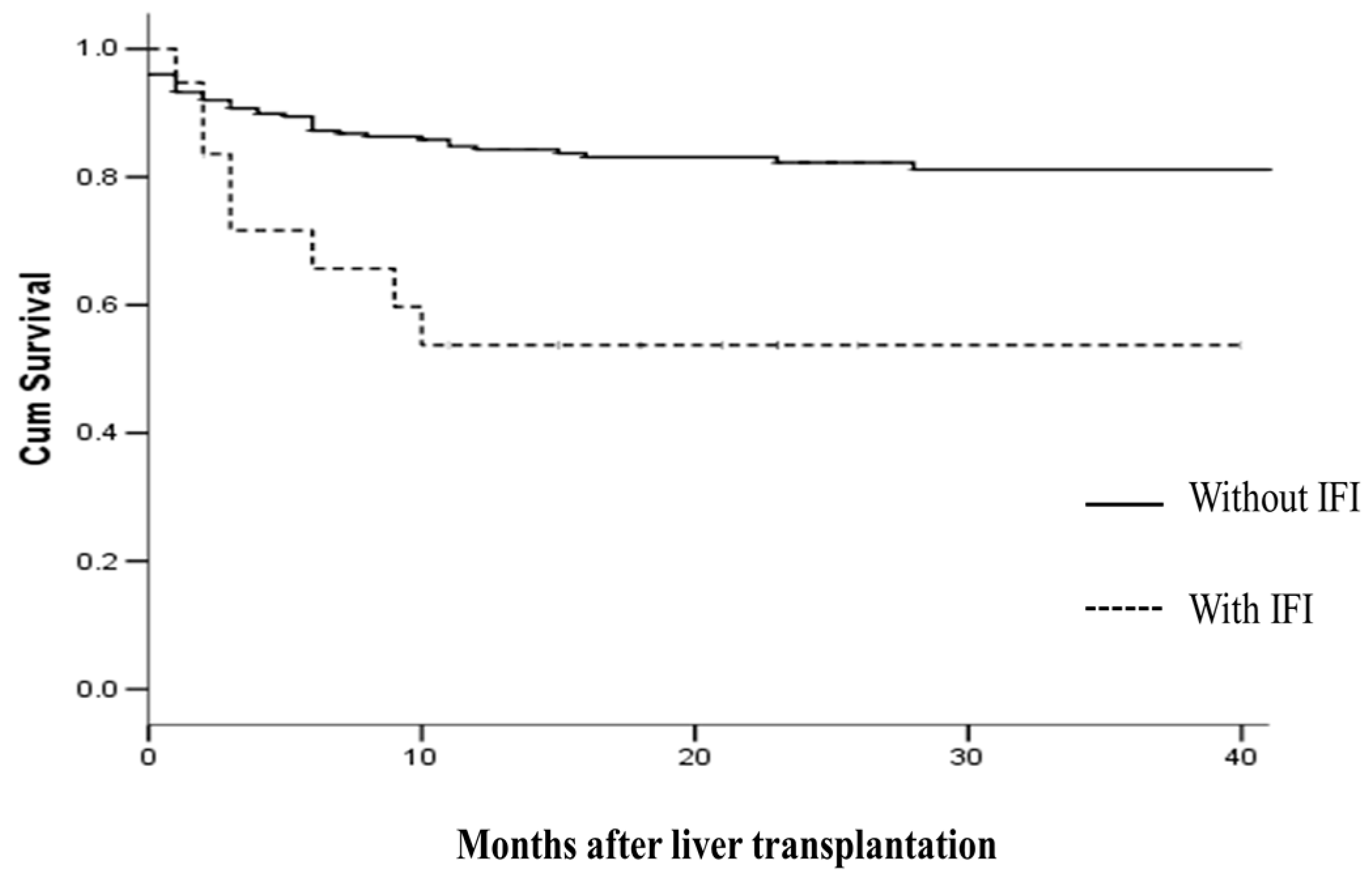
2.4. IFI and Survival
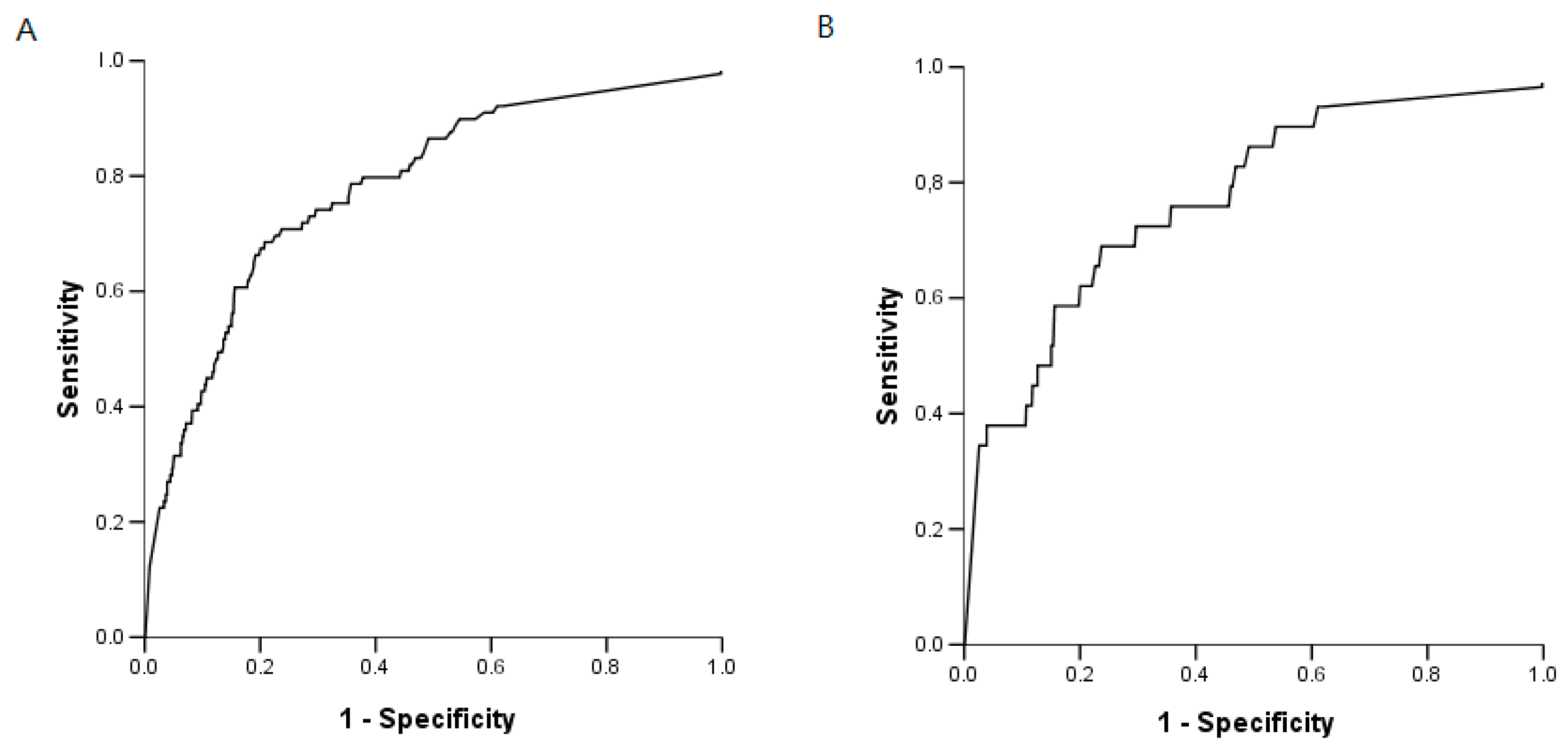
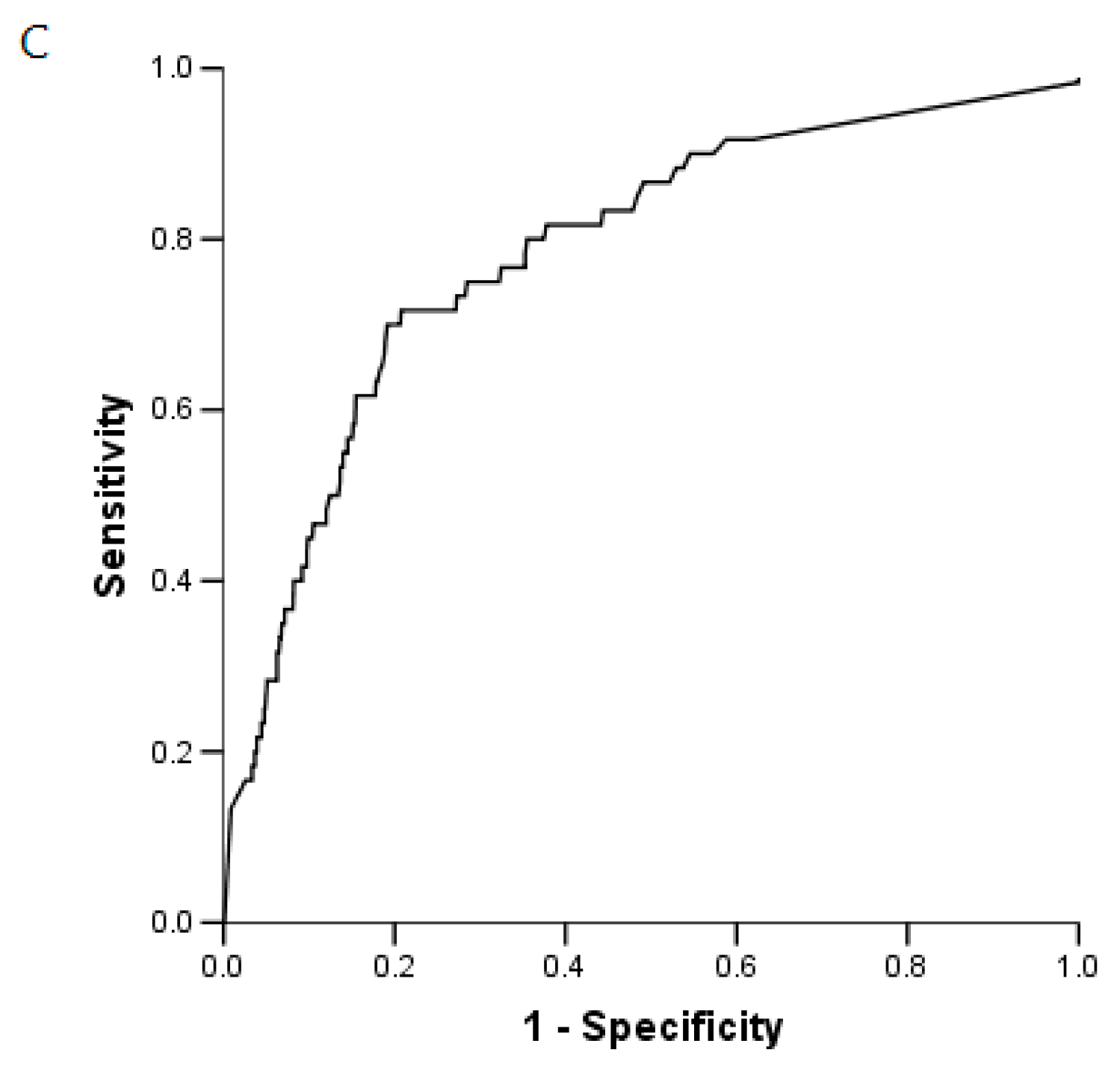
The median ICU and hospital stays after LT were 7.9 ± 9.8 and 33.8 ± 28.2 days, respectively. The median follow-up period was 20 months, and no patient was lost to follow-up. Notwithstanding the difficulty of assessing the primary cause of death in these recipients, which was sepsis/multi-organ failure in 73% of cases, no deaths appeared directly to be due to IFI. Patient survival rates at six months and one year after LT were 87% and 83.4%, and graft survival rates were 85% and 81.6%, respectively. Recipients with IFIs had significantly lower patient survival rates at six months and one year when compared to recipients with no IFI (89.3% vs. 68.9%; p = 0.007 at six months, and 85.3% vs. 57.9%; p = 0.002 at one year, respectively). Finally, Kaplan Meier curves were fitted for the two groups of patients according to the IFI experienced, and compared using the log-rank test (Figure 1).

2.5. BG Analysis

For the BG test, 1019 samples were obtained from the 236 recipients. Thus, the average number of samples obtained from each patient was 4.31 (range: 1 to 18), or about one per week. In 35 recipients without IFI risk factors and a short ICU stay, no sample was collected during their stay. None of these recipients developed any fungal infections. Using the cut-off of 80 pg/mL, the BG positivity was 48% (113/236 patients) at baseline, 35% (83/236 patients) at week 1 and 30% (56/189 patients) at week 2 (Table 4). In patients without IFI, 34% (318/924 samples) were over 80 pg/mL. Factors associated with BG positivity at the time of sampling were: renal replacement therapy (n = 56 samples from 22 patients), surgery (LT or repeat surgery <5 days) (n = 196 samples from 121 patients), the administration of piperacillin-tazobactam (n = 66 samples from 52 patients) and, in 25 positive samples, neither IFI nor factors associated with BG positivity was found. During the follow-up of patients without IFI, the BG concentration is significantly different between patients receiving echinocandin prophylaxis (n = 124 patients with 375 samples) and patients without echinocandin prophylaxis (n = 131 patients with 304 samples) (109 ± 116 vs. 89 ± 95 pg/mL, p < 0.001).

2.6. IFI and BG

When applying the cut-off value of 80 pg/mL, and using a single BG sample for the primary analysis of IFI, 95% (21/22) of the subjects with IFI had positive BG at the time of IFI diagnosis (between −7 and +7 days of onset of clinical symptoms of IFI) (Table 4). The mean BG concentration during the 28-day diagnostic window for recipients with proven or probable IFI was significantly higher than the mean value seen in those without IFI (244 ± 173 vs. 99 ± 106 pg/mL, respectively; p < 0.001).
Receiver operating characteristic (ROC) curves were used to evaluate the potential for BG to diagnose IFI (Figure 2). The area under the ROC curves was 0.78 (95% CI: 0.73–0.83). The sensitivity, specificity, positive predictive value (PPV) and negative predictive value (NPV) of BG for IFI, overall, were 75% (95% CI: 65–83), 65% (95% CI: 62–68), 17% (95% CI: 13–21), and 96% (95% CI: 94–97), respectively. If IC and IPA were considered separately, the same results in terms of AUROC (area under receiver operating characteristic) were observed: the AUROC value was 0.77 (5% CI: 0.67–0.86) for the diagnosis of IPA and 0.78 (95% CI: 0.72–0.84) for the diagnosis of IC (Figure 2).
In recipients with IFI, the median time elapsing between the first clinical signs of IFI and a BG value ≥ 80 pg/mL was −2 days (range: −7 to 14 days). The peak BG value was reached after a median of four days (range: −7 to 28 days) after the diagnosis of IFI. After antifungal treatment, the negativity of BG (below 80 pg/mL) was obtained in 66% (14/21 patients) ranged from six to 21 days after the BG peak (median 13 days). In addition, the BG decreases with the antifungal treatment in 90% of the subjects (19/21).

3. Discussion

In this report, we investigated the incidence of IFI in the era of antifungal prophylaxis and assessed the performance of BG in diagnosing IFI among liver transplant recipients. Out of a total of 271 recipients, 19 (7%) (who experienced twenty-one episodes) developed proven or probable IFI during their ICU stay.
The incidence of IFI in our patients receiving targeted prophylactic antifungal therapy was 7%, but this reached 12% among liver recipients with high risk of IFI. Our result was similar to the incidence reported elsewhere, with rates reaching 17.7% [1,4,15,16,17,18,19,20,21]. In the era of MELD and targeted prophylactic antifungal therapy, our study was able to offer three interesting pieces of information: (i) all patients who developed IFI within two months of LT had one or more risk factors and were receiving antifungal therapy after LT; (ii) the time to onset of IFI in the ICU after LT ranged from five days to 252 days. In eight cases, IFI occurred before the tenth post-operative day due to IC (n = 5, associated with surgical complications) or IPA (n = 3). These IA were probably present before LT but had not been diagnosed. This raises the question regarding the need to determine the presence of IA in end-stage liver patients with a high MELD score or organ failure, because this may reflect a greater severity of their liver disease [17]; and (iii) our findings confirm that IFI represent a significant burden in terms of mortality and morbidity, and highlight the importance of their early detection.
The BG assay has been shown to be useful for the diagnosis of IFI in haematological patients [22,23,24]. In LT, only a few studies have to date evaluated the utility of BG. During a preliminary study, we investigated the impact of the systematic monitoring of BG levels in liver transplant recipients. We observed that serum BG levels achieved good diagnostic accuracy for the diagnosis of IC [14]. The principal limitation of that preliminary study was the low incidence of IFI cases (six episodes of invasive candidiasis out of a total of 52 patients). In this new study, concerning a large patient sample and 21 episodes of IFI, our findings agree with our previous results and with those obtained by Akamatsu et al. [12,14]. In all three studies, the AUROC curves for the detection of IFI using BG ranged from 0.72 to 0.79. By contrast, Singh et al. found that the BG test was ineffective in predicting IFI, with an AUROC curve of 0.56 [13]. This was due to the numerous false positive tests observed in their study. Indeed, the baseline marker (after surgery) was positive at about 90% in the Singh study, but declined over time, with approximately 50% of the patients still having positive tests up to four weeks later [13]. A number of factors can cause the false positivity of this marker in the clinical setting: patients who are receiving renal replacement therapy, albumin or immunoglobulin infusions, β-lactam inhibitors, antibiotics, surgical gauze, etc. [25,26,27,28,29]. In our study, the baseline marker (just after LT) was positive in only half of recipients and declined over time. Moreover, in 92% of BG positivity, at least one factor associated with BG positivity was found (renal replacement therapy, after surgery, the administration of piperacillin-tazobactam). Finally, we observe that an echinocandin treatment can increase the BG value, by release of BG, but in order to reduce this high proportion of false positives, we have proposed confirming a positive result by testing rapidly (or quickly) a second sample or to increase the cut-off value rather than 80 pg/mL [14]. The most important finding of our study is that the IFI diagnosis may be excluded when the BG value is less than 80 pg/mL. In this case, as shown by Singh et al. [13], this test has a sensitivity close to 100% (BG values were higher than 80 pg/mL in 95% of IFI cases (20/21 episodes).
We are aware of the limitations of this study. Nearly half of the recipients in our study received antifungal prophylaxis because of their risk factors, but without evident signs of an IFI. They may have been affected by a latent infection that was treated with the antifungal therapy. The BG had to also be affected by echinocandin treatment, probably by the release of BG. In addition, the prophylaxis in high-risk patients may have directly affected the estimations of BG efficacy in this population. For these reasons, our results can only be extrapolated to patients who were being treated initially with echinocandin (90% of antifungal prophylaxis). Furthermore, our study was not designed to assess BG as a predictor of survival.

4. Patients and Methods

This study was carried out in liver transplant patients at Henri Mondor Hospital in the suburbs of Paris, France. Between January 2013 and May 2016, all patients admitted consecutively to our Intensive Care Unit (ICU) following LT were enrolled in the study. Demographic and clinical characteristics, investigations for mycological and bacterial infections, and antimicrobial therapies were all recorded prospectively. The patients were studied during their hospitalization in and after the ICU. Our Institutional Review Board approved the study (2012-12) and the database was declared to the French Data Protection Authority (Commission Nationale Informatique et Liberté) (No. 1699340).

4.1. Clinical and Biological Management

Following LT, the recipients were hospitalized in our liver ICU. All patients received similar post-operative intensive care with a standard triple immunosuppressive regimen that included corticosteroids, mycophenolate mofetil and FK506 (tacrolimus) or cyclosporin, with basiliximab (day 1 and day 4) in the event of a rising, or initially high, serum creatinine level. All patients received post-operative antibiotic therapy for 48 h (piperacillin) and trimethoprim-sulfamethoxazol (TMP/SMX) as prophylaxis against PjP in high-risk patients (fulminant hepatitis, re-transplantation). To prevent cytomegalovirus (CMV) disease, pre-emptive therapy was instituted. The patients were monitored for evidence of CMV replication, and antiviral therapy (valganciclovir or ganciclovir) was administered pre-emptively, if necessary, to prevent progression to symptomatic clinical disease. Graft and recipient outcomes were recorded prospectively for all transplanted patients. The clinical course of recipients was followed for a minimum of three months after LT.

4.2. Antifungal Prophylaxis

Identification of patients at increased risk of fungal infection (re-transplantation, renal failure (creatinine clearance <50 mL/min or requiring replacement therapy), fulminant hepatic failure, primary non-function, patients with thymoglobulin as immunosuppressive agents, complicated surgery or reoperation, recipient MELD scores before LT > 30, more than 40 pre-operatory transfusion blood products, bilio-digestive anastomosis and multifocal colonization (≥2 localisations) by Candida spp., and the yeast preservation fluid contamination) is the key of prevention [14,30]. Patients affected by any of these factors received caspofungin at a dose of 70 mg on the first day, and then 50 mg per day (or 70 mg per day if the recipient weighed >80 kg), or micafungin at a dose of 100 mg per day for 28 days. The choice between capsofungin and mcafungin was left to the discretion of the physician. The few patients who were only considered to be at risk of Candida infection, but not Aspergillus (patients with choledocho-jejunostomy or colonized at the time of liver transplantation by Candida spp.), received fluconazole 400 mg per day for three weeks in the absence of Candida glabrata, Candida krusei, or the prior use of azoles.

4.3. Colonisation

All patients were subject to weekly monitoring for Candida colonization and were screened for Candida infections in blood cultures and at other sterile sites, depending on their clinical signs. As part of this routine surveillance of Candida colonization, swabs were systematically taken from five superficial sites (mouth, nose, axillary surface, inguinal fold, and anus) on the day of admission to undergo LT, and then once a week thereafter until discharge from the ICU or death. Colonisation is defined as the presence of Candida spp. isolates in a swab from at least one surveillance site.

4.4. Galactomannan

Serum from all patients at risk of invasive pulmonary aspergillosis (IPA) was screened twice weekly for galactomannan (Platelia Aspergillus, Biorad, Marne la Coquette, France) during the patient's ICU stay. The threshold was 0.5, according to the revised European Organization for Research and Treatment of Cancer/Mycoses Study Group (EORTC/MSG) criteria [31]. If the patients had a broncho-alveolar lavage, a galactomannan assay was performed systematically at the same time as the culture.

4.5. (1,3)-β-d-Glucan (BG) Assay

In addition to clinical and laboratory evaluations to determine the presence of any infection, serum was collected weekly during the patient’s ICU (each monday) and according to the discretion of the physician during the hospital stay and tested for BG using the Fungitell kit (Fungitell®, Associates Cape Code Inc., Falmouth, MA, USA). These samples were stored at −20 °C until batch testing was performed. It was necessary for serum samples to be collected from patients with proven or probable IFI during the seven days before and 21 days after the diagnosis (thus providing a 28-day diagnostic window).

4.6. Definition

The diagnostic criteria used for IPA were those of the EORTC/MSG [31]. All patients were reviewed by a committee composed of four members (two intensive care clinicians and two mycologists), who classified aspergillosis as probable aspergillosis or colonization.
Invasive candidiasis (IC) included candidemia and deep-seated candidiasis, defined as the recovery of Candida species from blood and/or a sterile site, respectively. Candida infections can manifest themselves as peritonitis, candidemia, surgical anastomosis infection, urinary tract infection, or wound infection. IFI were, thus, classified according to the EORTC/MSG criteria [31].
Only patients with proven or probable IFI were evaluated as cases for the present study and the PjP were separated from the IFI.
Controls were defined as liver transplant recipients with no clinical or microbiological evidence of IC (patients only colonized by Candida were also considered as controls) and those free of invasive pulmonary aspergillosis.

4.7. Statistical Analysis

Continuous data were presented as medians and ranges. Fisher’s exact test (categorical variables) and the Mann-Whitney test (continuous variables) were used to compare demographic data based on the patient’s IC status. Kaplan-Meier curves were generated, and a log-rank test was utilized to compare patients with and without IFI. The following parameters for diagnostic performance, and their 95% confidence intervals, were calculated: sensitivity, specificity, positive predictive value (PPV), and negative predictive value (NPV). To identify the ability of BG to diagnose invasive candidiasis, receiver operating characteristic (ROC) curves were constructed. All statistical analyses were performed using SPSS (version 18; SPPS, Inc, IBM, Chicago, IL, USA). The areas under the curves (AUCs) reflected the discriminative ability of BG. A p value lower than 0.05 was considered to be statistically significant.

5. Conclusions

In a large sample of patients, the present study was able to confirm that the detection of BG in serum after LT may be a tool which can rule out IC in high-risk recipients based on its high NPV.

Author Contributions

Participated in research design: Eric Levesque, Françoise Botterel; participated in the writing of the paper: Eric Levesque, Françoise Botterel, Catherine Cordonnier-Jourdin, and Daniel Azoulay; participated in the performance of the research (acquisition data): Eric Levesque, Sarra El Anbassi, Fadi Rizk, Zaid Noorah, Nawel Aït-Ammar, Françoise Botterel, and Catherine Cordonnier-Jourdin; contributed new reagents or analytic tools (interpretation of data): Eric Levesque, Nawel Aït-Ammar, Jean-Claude Merle, Françoise Botterel, and Christine Bonnal; and participated in data analysis: Eric Levesque, and Daniel Azoulay.

Conflicts of Interest

The authors declare no conflict of interest.

Abbreviations

AUROCArea Under Receiver Operating Characteristic
BG(1,3)-β-d-glucan
CMVCytomegalovirus
EORTC/MSGEuropean Organization for the Research and Treatment of Cancer/Mycoses Study Group
ICInvasive Candidiasis
ICUIntensive Care Unit
IFIInvasive Fungal Infection
IPAInvasive Pulmonary Aspergillosis
LTLiver Transplantation
MELD ScoreModel for End-Stage Liver Disease
NPVNegative Predictive Value
PjPPneumocystis jirovecii pneumonia
PPVPositive Predictive Value
ROCReceiver Operating Characteristic

References

  1. Pappas, P.G.; Alexander, B.D.; Andes, D.R.; Hadley, S.; Kauffman, C.A.; Freifeld, A.; Kontoyiannis, D.P. Invasive fungal infections among organ transplant recipients: Results of the Transplant-Associated Infection Surveillance Network (TRANSNET). Clin. Infect. Dis. 2010, 50, 1101–1111. [Google Scholar] [CrossRef] [PubMed]
  2. Neofytos, D.; Fishman, J.A.; Horn, D.; Anaissie, E.; Chang, C.H.; Olyaei, A.; Marr, K.A. Epidemiology and outcome of invasive fungal infections in solid organ transplant recipients. Transpl. Infect. Dis. 2010, 12, 220–229. [Google Scholar] [CrossRef] [PubMed]
  3. Yang, C.H.; He, X.S.; Chen, J.; Ouyang, B.; Zhu, X.F.; Chen, M.Y.; Chen, X.X. Fungal infection in patients after liver transplantation in years 2003 to 2012. Ann. Transplant. Q. Pol. Transplant. Soc. 2012, 17, 59–63. [Google Scholar]
  4. Grossi, P.A. Clinical aspects of invasive candidiasis in solid organ transplant recipients. Drugs 2009, 69, 15–20. [Google Scholar] [CrossRef] [PubMed]
  5. Singh, N.; Avery, R.K.; Munoz, P.; Pruett, T.L.; Alexander, B.; Jacobs, R.; Husain, S. Trends in risk profiles for and mortality associated with invasive aspergillosis among liver transplant recipients. Clin. Infect. Dis. 2003, 36, 46–52. [Google Scholar] [CrossRef] [PubMed]
  6. Li, C.; Wen, T.F.; Mi, K.; Wang, C.; Yan, L.N.; Li, B. Analysis of infections in the first 3-month after living donor liver transplantation. World J. Gastroenterol. 2012, 18, 1975–1980. [Google Scholar] [CrossRef] [PubMed]
  7. Hou, T.Y.; Wang, S.H.; Liang, S.X.; Jiang, W.X.; Luo, D.D.; Huang, D.H. The screening performance of serum 1,3-β-d-glucan in patients with invasive fungal diseases: A meta-analysis of prospective cohort studies. PLoS ONE 2015, 10, e0131602. [Google Scholar] [CrossRef] [PubMed]
  8. He, S.; Hang, J.P.; Zhang, L.; Wang, F.; Zhang, D.C.; Gong, F.H. A systematic review and meta-analysis of diagnostic accuracy of serum 1,3-β-d-glucan for invasive fungal infection: Focus on cutoff levels. J. Microbiol. Immunol. Infect. 2015, 48, 351–361. [Google Scholar] [CrossRef] [PubMed]
  9. Onishi, A.; Sugiyama, D.; Kogata, Y.; Saegusa, J.; Sugimoto, T.; Kawano, S.; Kumagai, S. Diagnostic accuracy of serum 1,3-β-d-glucan for pneumocystis jiroveci pneumonia, invasive candidiasis, and invasive aspergillosis: Systematic review and meta-analysis. J. Clin. Microbiol. 2012, 50, 7–15. [Google Scholar] [CrossRef] [PubMed]
  10. Lamoth, F.; Cruciani, M.; Mengoli, C.; Castagnola, E.; Lortholary, O.; Richardson, M.; Marchetti, O. β-Glucan antigenemia assay for the diagnosis of invasive fungal infections in patients with hematological malignancies: A systematic review and meta-analysis of cohort studies from the Third European Conference on Infections in Leukemia (ECIL-3). Clin. Infect. Dis. 2012, 54, 633–643. [Google Scholar] [PubMed]
  11. Karageorgopoulos, D.E.; Voulouman, E.K.; Ntziora, F.; Michalopoulos, A.; Rafailidis, P.I.; Falagas, M.E. β-d-Glucan assay for the diagnosis of invasive fungal infections: A meta-analysis. Clin. Infect. Dis. 2011, 52, 750–770. [Google Scholar] [CrossRef] [PubMed]
  12. Akamatsu, N.; Sugawara, Y.; Kaneko, J.; Tamura, S.; Makuuchi, M. Preemptive treatment of fungal infection based on plasma (1→3) β-d-glucan levels after liver transplantation. Infection 2007, 35, 346–351. [Google Scholar] [CrossRef] [PubMed]
  13. Singh, N.; Winston, D.J.; Limaye, A.P.; Pelletier, S.; Safdar, N.; Morris, M.I.; Wheat, L.J. Performance characteristics of galactomannan and β-d-glucan in high-risk liver transplant recipients. Transplantation 2015, 99, 2543–2550. [Google Scholar] [CrossRef] [PubMed]
  14. Levesque, E.; El Anbassi, S.; Sitterle, E.; Foulet, F.; Merle, J.C.; Botterel, F. Contribution of (1,3)-β-d-glucan to diagnosis of invasive candidiasis after liver transplantation. J. Clin. Microbiol. 2015, 53, 771–776. [Google Scholar] [CrossRef] [PubMed]
  15. Hadley, S.; Huckabee, C.; Pappas, P.G.; Daly, J.; Rabkin, J.; Kauffman, C.A.; Karchmer, A.W. Outcomes of antifungal prophylaxis in high-risk liver transplant recipients. Transpl. Infect. Dis. 2009, 11, 40–48. [Google Scholar] [CrossRef] [PubMed]
  16. Collins, L.A.; Samore, M.H.; Roberts, M.S.; Luzzati, R.; Jenkins, R.L.; Lewis, W.D.; Karchmer, A.W. Risk factors for invasive fungal infections complicating orthotopic liver transplantation. J. Infect. Dis. 1994, 170, 644–652. [Google Scholar] [CrossRef] [PubMed]
  17. Saliba, F.; Delva, V.; Ichaï, P.; Kassis, N.; Botterel, F.; Mihaila, L.; Samuel, D. Fungal infections after liver transplantation: Outcomes and risk factors revisited in the MELD era. Clin. Transplant. 2013, 27, 454–461. [Google Scholar] [CrossRef] [PubMed]
  18. Silveira, F.P.; Husain, S. Fungal infections in solid organ transplantation. Med. Mycol. 2007, 45, 305–320. [Google Scholar] [CrossRef] [PubMed]
  19. Winston, D.J.; Limaye, A.P.; Pelletier, S.; Safdar, N.; Morris, M.I.; Meneses, K.; Singh, N. Randomized, double-blind trial of anidulafungin versus fluconazole for prophylaxis of invasive fungal infections in high-risk liver transplant recipients. Am. J. Transplant. 2014, 14, 2758–2764. [Google Scholar] [CrossRef] [PubMed]
  20. Saliba, F.; Pascher, A.; Cointault, O.; Laterre, P.F.; Cervera, C.; de Waele, J.J.; Phillips, S. Randomized trial of micafungin for the prevention of invasive fungal infection in high-risk liver transplant recipients. Clin. Infect. Dis. 2015, 60, 997–1006. [Google Scholar] [CrossRef] [PubMed]
  21. Fortún, J.; Muriel, A.; Martín-Dávila, P.; Montejo, M.; Len, O.; Torre-Cisneros, J.; Fresco, G. Caspofungin versus fluconazole as prophylaxis of invasive fungal infection in high-risk liver transplantation recipients: A propensity score analysis. Liver Transpl. 2016, 22, 427–435. [Google Scholar] [CrossRef] [PubMed]
  22. Ostrosky-Zeichner, L.; Alexander, B.D.; Kett, D.H.; Vazquez, J.; Pappas, P.G.; Saeki, F.; Finkelman, M.A. Multicenter clinical evaluation of the (1→3) β-d-glucan assay as an aid to diagnosis of fungal infections in humans. Clin. Infect. Dis. 2005, 41, 654–659. [Google Scholar] [CrossRef] [PubMed]
  23. Odabasi, Z.; Mattiuzzi, G.; Estey, E.; Kantarjian, H.; Saeki, F.; Ridge, R.J.; Ostrosky-Zeichner, L. β-d-Glucan as a diagnostic adjunct for invasive fungal infections: Validation, cutoff development, and performance in patients with acute myelogenous leukemia and myelodysplastic syndrome. Clin. Infect. Dis. 2004, 39, 199–205. [Google Scholar] [CrossRef] [PubMed]
  24. Racil, Z.; Kocmanova, I.; Lengerova, M.; Weinbergerova, B.; Buresova, L.; Toskova, M.; Mayer, J. Difficulties in using 1,3-β-d-glucan as the screening test for the early diagnosis of invasive fungal infections in patients with haematological malignancies—High frequency of false-positive results and their analysis. J. Med. Microbiol. 2010, 59, 1016–1022. [Google Scholar] [CrossRef] [PubMed]
  25. Kanda, H.; Kubo, K.; Hamasaki, K.; Kanda, Y.; Nakao, A.; Kitamura, T.; Mimura, T. Influence of various hemodialysis membranes on the plasma (1→3)-β-d-glucan level. Kidney Int. 2001, 60, 319–323. [Google Scholar] [CrossRef] [PubMed]
  26. Usami, M.; Ohata, A.; Horiuchi, T.; Nagasawa, K.; Wakabayashi, T.; Tanaka, S. Positive (1→3)-β-d-glucan in blood components and release of (1→3)-β-d-glucan from depth-type membrane filters for blood processing. Transfusion 2002, 42, 1189–1195. [Google Scholar] [CrossRef] [PubMed]
  27. Mennink-Kersten, M.A.; Warris, A.; Verweij, P.E. 1,3-β-d-Glucan in patients receiving intravenous amoxicillin-clavulanic acid. N. Engl. J. Med. 2006, 354, 2834–2835. [Google Scholar] [CrossRef] [PubMed]
  28. Nett, J.; Lincoln, L.; Marchillo, K.; Andes, D. β-1,3-Glucan as a test for central venous catheter biofilm infection. J. Infect. Dis. 2007, 195, 1705–1712. [Google Scholar] [CrossRef] [PubMed]
  29. Mohr, J.F.; Sims, C.; Paetznick, V.; Rodriguez, J.; Finkelman, M.A.; Rex, J.H.; Ostrosky-Zeichner, L. Prospective survey of (1→3)-β-d-glucan and its relationship to invasive candidiasis in the surgical intensive care unit setting. J. Clin. Microbiol. 2011, 49, 58–61. [Google Scholar] [CrossRef] [PubMed]
  30. Gavaldà, J.; Meije, Y.; Fortún, J.; Roilides, E.; Saliba, F.; Lortholary, O.; Cuenca-Estrella, M. Invasive fungal infections in SOT recipients. Clin. Microbiol. Infect. 2014, 20, 27–48. [Google Scholar] [CrossRef] [PubMed]
  31. De Pauw, B.; Walsh, T.J.; Donnelly, J.P.; Stevens, D.A.; Edwards, J.E.; Calandra, T.; Denning, D.W. Revised definitions of invasive fungal disease from the European Organization for Research and Treatment of Cancer/Invasive Fungal Infections Cooperative Group and the National Institute of Allergy and Infectious Diseases Mycoses Study Group (EORTC/MSG) Consensus Group. Clin. Infect. Dis. 2008, 46, 1813–1821. [Google Scholar] [PubMed]
Figure 1. Patient survival following liver transplantation as a function of invasive fungal infection status, p = 0.003.
Ijms 18 00862 g001
Figure 2. Receiver operating characteristic (ROC) curves used to evaluate the ability of BG to diagnose (A) an invasive fungal infection (IFI): AUROC (Area under ROC) value of 0.78 (95% confidence interval (CI): 0.73–0.83); (B) Invasive pulmonary aspergillosis (IPA): AUROC value 0.77 (95% CI: 0.67–0.86); and (C) invasive candidiasis (IC): AUROC value 0.78 (95% CI: 0.72–0.84).
Ijms 18 00862 g002aIjms 18 00862 g002b
Table 1. Characteristics of 271 liver transplants and risk factors for invasive fungal infection (IFI) at the time of transplant. Data are presented as the mean ± SD or n (%). HBV: viral hepatitis B; HCV: viral hepatitis C; MELD: model for end-stage liver disease; LT: liver transplantation.
VariableLiver Transplants n = 271
Age (years)52.7 ± 12.7
Gender M/F202/69
Indication for liver transplantation
  Hepatocellular carcinoma85 (31)
  Cirrhosis126 (46)
  Fulminant hepatic failure17 (7)
  Other43 (16)
Alcoholic liver disease130 (48)
Viral hepatitis (HCV and HBV)54 (20)
Other87 (32)
MELD score19.1 ± 11.4
  MELD < 20164 (60)
  MELD 20–3047 (18)
  MELD > 3060 (22)
Antifungal prophylaxis n (%)157 (58)
  Caspofungin, n (%)132 (84.1)
  Micafungin, n (%)6 (3.8)
  Fluconazole, n (%)18 (11.5)
  Ampho B, n (%)1 (0.6)
duration of treatment (days)24.3 ± 18.5
Risk factors for IFI
  MELD > 3060 (22)
  Fulminant hepatic failure17 (6.3)
  Re-transplantation20 (7.5)
Graft type
  Whole cadaveric liver transplant246 (91)
  Partial liver transplant (split)22 (8)
  Domino3 (1)
Multi-organ transplantation (kidney/liver)14 (5)
>40 transfusions of blood products8 (3)
Renal Replacement therapy44 (16)
Early re-intervention after LT51 (19)
Choledocojejunostomy anastomosis (Roux-en-Y)38 (14)
Table 2. Characteristics of patients with invasive candidiasis. IC: invasive candidiasis, LT: liver transplantation, BG: (1,3)-β-d-glucan assay, CI: colonization index.
Patient NumberDelay between LT and IC (days)Localisation ICCultureClassification of ICValues BG (pg/mL)CIProphylactic Treatment (Delay before IC)Curative TreatmentOutcome (after LT)
146Intra-abdominalC. glabrataProven2010.5Caspofungin (45)VoriconazoleDied 301 days
252CandidemiaC. parapsilosisProven5010.8Caspofungin (25)Fluconazole
265Intra-abdominalC. glabrataProven5010.8Caspofungin (10)VoriconazoleDied 81 days
326Intra-abdominal candidiasisC. glabrata, C. albicansProven3660.8-CaspofunginDied 91 days
47Intra-abdominalC. albicansProven3600.8Caspofungin (7)FluconazoleDied 47 days
58CandidemiaC. albicansProven5010.6Fluconazole (8)FluconazoleAlive 692 days
67Intra-abdominal/biliary abscessC. albicansProven5010.8Caspofungin (7)FluconazoleAlive 677 days
76CandidemiaC. albicansProven1700.6Caspofungin (6)FluconazoleAlive 438 days
822Intra-abdominalC. kruseiProven3200.2Caspofungin (22)CaspofunginAlive 317 days
962CandidemiaC. orthopsilosisProven1730.5-Liposomal Amphotericin BAlive 177 days
1010Intra-abdominalC. krusei, C. albicansProven2090.6Caspofungin (10)Liposomal Amphotericin BAlive 49 days
1112Intra-abdominal and candidemiaC. glabrataProven2460.6Caspofungin (10)CaspofunginDied 280 days
86abscess and biliary infectionC. glabrata, C. albicans, C. tropicalisProbable5010.5-Caspofungin
Table 3. Characteristics of patients with invasive pulmonary aspergillosis (IPA). LT: liver transplantation, BG: (1,3)-β-d-glucan assay; GM: galactomannan, BAL: bronchoalveolar lavage, VHB: viral hepatitis B; VHC: viral hepatitis C; NA: Not available.
Patient NumberTime to Onset after LT (days)Underlying DiseasesIPA ClassificationBAL Direct ExaminationCultureGM in Serum (Index)GM in BAL (Index)BG Values (pg/mL)Prophylactic Treatment (Delay)Curative TreatmentOutcome (after LT)
125Cryptogenetic cirrhosisProbableNAA. fumigatus+ (0.602)NA207Caspofungin (5)VoriconazoleAlive at 1197 days
1344Acute liver failure (autoimmune hepatitis)Probable-A. fumigatus− (0.125)+ (1.25)501Caspofungin (35)VoriconazoleAlive at 1197 days
1445HCC/VHB cirrhosisProbable-A. fumigatus+ (>6)+ (5.58)198Caspofungin (9)VoriconazoleDied 61 days
157Acute liver failure (toxic hepatitis)ProbableNAA. fumigatus+ (1.39)+ (1.85)219Caspofungin (7)VoriconazoleAlive at 599 days
165Acute liver failure Reactivation of VHB infectionProbable-A. fumigatus− (0.06)− (0.13)501Caspofungin (5)VoriconazoleAlive at 542 days
1732Re-LT Chronic graft rejectionProbable-A. fumigatus+ (2.361)NA42Caspofungin (31)VoriconazoleAlive at 41 days after LT
1825VHB/VHC cirrhosisProbable-A. fumigatus− (0.157)+ (1.6)170Caspofungin (19)VoriconazoleDied 26 days
255Acute liver failure (toxic hepatitis)Probable-A. fumigatus+ (5.30)+ (5.68)423Caspofungin (35)VoriconazoleDied 81 days
1922Acute liver failure Reactivation of VHB infectionProven Septic arthritis of the hip Scedosporium sp.+ (2.83)NA501Micafungin (22)Surgery Caspofungin + voriconazoleAlive at 599 days
Table 4. Kinetics of (1,3)-β-d-glucan (BG) in patients with IFI. Day 0 is defined as the beginning of clinical symptoms of IFI. IFI: Invasive fungal infection; IC: invasive candidiasis, IPA: invasive pulmonary aspergillosis, NA: not available.
IFIPatient NumberDay 7Day 0Day 7Day 14Day 21Day 28
IC1201147NANA4144
1501356393501501446
2234423501501501NA
3NA36366NA5077
4360464501NA31NA
5NA501269269191221
632434814501323192
71061531701109051
8232NA320311NANA
93115117376140NA
1031209148NANANA
111703158180NA246
11187501NANA293170
IPA121432075355NANA
13501NA174NANANA
14198169NANANA30
15NA21912850NANA
16501501501NA501501
17313342NANANA
1875170NANANANA
2423131NANANA99
Scedosporium sp.19423501501501NANA

