Next Issue
Volume 18, September
Previous Issue
Volume 18, July
 
 
ijms-logo

Journal Browser

Journal Browser

Int. J. Mol. Sci., Volume 18, Issue 8 (August 2017) – 223 articles

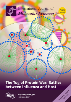
Cover Story (view full-size image): During infection, the influenza A virus must engage with the host cellular protein interaction network to hijack host molecular machinery in order to fulfill the viral life cycle and perturb host defense to evade immune surveillance. On the other hand, the interaction elicits various host responses to eliminate the virus. Thus, the protein interactions between influenza A virus and host determine the outcome of viral pathogenesis. View this paper
  • Issues are regarded as officially published after their release is announced to the table of contents alert mailing list.
  • You may sign up for e-mail alerts to receive table of contents of newly released issues.
  • PDF is the official format for papers published in both, html and pdf forms. To view the papers in pdf format, click on the "PDF Full-text" link, and use the free Adobe Reader to open them.
Order results
Result details
Section
Select all
Export citation of selected articles as:
2479 KiB  
Article
Circadian and Light Regulated Expression of CBFs and their Upstream Signalling Genes in Barley
by Krisztián Gierczik, Aliz Novák, Mohamed Ahres, András Székely, Alexandra Soltész, Ákos Boldizsár, Zsolt Gulyás, Balázs Kalapos, István Monostori, László Kozma-Bognár, Gábor Galiba and Attila Vágújfalvi
Int. J. Mol. Sci. 2017, 18(8), 1828; https://0-doi-org.brum.beds.ac.uk/10.3390/ijms18081828 - 22 Aug 2017
Cited by 22 | Viewed by 5377
Abstract
CBF (C-repeat binding factor) transcription factors show high expression levels in response to cold; moreover, they play a key regulatory role in cold acclimation processes. Recently, however, more and more information has led to the conclusion that, apart from cold, light—including its spectra—also [...] Read more.
CBF (C-repeat binding factor) transcription factors show high expression levels in response to cold; moreover, they play a key regulatory role in cold acclimation processes. Recently, however, more and more information has led to the conclusion that, apart from cold, light—including its spectra—also has a crucial role in regulating CBF expression. Earlier, studies established that the expression patterns of some of these regulatory genes follow circadian rhythms. To understand more of this complex acclimation process, we studied the expression patterns of the signal transducing pathways, including signal perception, the circadian clock and phospholipid signalling pathways, upstream of the CBF gene regulatory hub. To exclude the confounding effect of cold, experiments were carried out at 22 °C. Our results show that the expression of genes implicated in the phospholipid signalling pathway follow a circadian rhythm. We demonstrated that, from among the tested CBF genes expressed in Hordeum vulgare (Hv) under our conditions, only the members of the HvCBF4-phylogenetic subgroup showed a circadian pattern. We found that the HvCBF4-subgroup genes were expressed late in the afternoon or early in the night. We also determined the expression changes under supplemental far-red illumination and established that the transcript accumulation had appeared four hours earlier and more intensely in several cases. Based on our results, we propose a model to illustrate the effect of the circadian clock and the quality of the light on the elements of signalling pathways upstream of the HvCBFs, thus integrating the complex regulation of the early cellular responses, which finally lead to an elevated abiotic stress tolerance. Full article
(This article belongs to the Special Issue Abiotic Stress and Gene Networks in Plants 2017)
Show Figures

Graphical abstract

928 KiB  
Article
Systemic Chemokine Levels with “Gut-Specific” Vedolizumab in Patients with Inflammatory Bowel Disease—A Pilot Study
by Stephanie Zwicker, Ronaldo Lira-Junior, Charlotte Höög, Sven Almer and Elisabeth A. Boström
Int. J. Mol. Sci. 2017, 18(8), 1827; https://0-doi-org.brum.beds.ac.uk/10.3390/ijms18081827 - 22 Aug 2017
Cited by 7 | Viewed by 4929
Abstract
Vedolizumab, a gut-specific biological treatment for inflammatory bowel disease (IBD), is an antibody that binds to the α4β7 integrin and blocks T-cell migration into intestinal mucosa. We aimed to investigate chemokine levels in serum of IBD-patients treated with vedolizumab. In [...] Read more.
Vedolizumab, a gut-specific biological treatment for inflammatory bowel disease (IBD), is an antibody that binds to the α4β7 integrin and blocks T-cell migration into intestinal mucosa. We aimed to investigate chemokine levels in serum of IBD-patients treated with vedolizumab. In this pilot study, we included 11 IBD patients (8 Crohn’s disease, 3 ulcerative colitis) previously non-respondent to anti-tumor necrosis factor (TNF)-agents. Patients received vedolizumab at week 0, 2 and 6 and were evaluated for clinical efficacy at week 10. Clinical characteristics and routine laboratory parameters were obtained and patients were classified as responders or non-responders. Expression of 21 chemokines in serum was measured using Proximity Extension Assay and related to clinical outcome. At week 10, 6 out of 11 patients had clinically responded. Overall expression of CCL13 increased after treatment. In non-responders, expression of CCL13 and CXCL8 increased after treatment, and CCL20 and CXCL1 expressions were higher compared to responders. In responders, CCL28 decreased after treatment. C-reactive protein (CRP) correlated negatively with 6 chemokines before therapy, but not after therapy. Systemic CCL13 expression increases in IBD-patients after vedolizumab therapy and several chemokine levels differ between responders and non-responders. An increased CCL13-level when starting vedolizumab treatment, might indicate potential prognostic value of measuring chemokine levels when starting therapy with vedolizumab. This study provides new information on modulation of systemic chemokine levels after vedolizumab treatment. Full article
Show Figures

Figure 1

17895 KiB  
Article
MiR-30a-5p Inhibits Epithelial-to-Mesenchymal Transition and Upregulates Expression of Tight Junction Protein Claudin-5 in Human Upper Tract Urothelial Carcinoma Cells
by Yueh-Hua Chung, Sung-Chou Li, Ying-Hsien Kao, Hao-Lun Luo, Yuan-Tso Cheng, Pey-Ru Lin, Ming-Hong Tai and Po-Hui Chiang
Int. J. Mol. Sci. 2017, 18(8), 1826; https://0-doi-org.brum.beds.ac.uk/10.3390/ijms18081826 - 22 Aug 2017
Cited by 29 | Viewed by 5290
Abstract
The involvement of microRNAs (miRNAs) in cancer development and their potential as prognostic biomarkers are becoming increasingly known. However, the signature of miRNAs and their regulatory roles in tumorigenesis of upper tract urothelial carcinoma (UTUC) remain to be elucidated. This study aimed to [...] Read more.
The involvement of microRNAs (miRNAs) in cancer development and their potential as prognostic biomarkers are becoming increasingly known. However, the signature of miRNAs and their regulatory roles in tumorigenesis of upper tract urothelial carcinoma (UTUC) remain to be elucidated. This study aimed to profile the miRNA expression pattern in UTUC tumor tissues and identify candidate miRNAs with prognostic and/or therapeutic functions. Methods and Results: We collected 22 UTUC tissue and adjacent normal tissues samples from patients who underwent nephroureterectomy. The miRNAs signatures of three selected UTUC samples using next-generation sequencing showed that miR-30a-5p was significantly downregulated in UTUC tumors compared to adjacent normal tissues. The differentially-expressed miRNAs were specifically validated by quantitative real-time polymerase chain reaction. In addition, the miRNA expression signatures were analyzed with the transcriptome profile characterized by microarray. Further in vitro studies indicated that overexpression of miR-30a-5p significantly suppressed proliferation, migration, and epithelial-to-mesenchymal transition (EMT) in cultured BFTC-909 UTUC cells. As a potential target gene of miR-30a-5p in the tight junction pathway suggested by the pathway enrichment analysis, the reduced expression of tight junction protein claudin-5 in UTUC cells was demonstrated to be upregulated by miR-30a-5p genetic delivery. Conclusions: Taken together, our findings demonstrated that miR-30a-5p inhibits proliferation, metastasis, and EMT, and upregulates the expression of tight junction claudin-5 in UTUC cells. Thus, miR-30a-5p may provide a promising therapeutic strategy for UTUC treatment. Full article
(This article belongs to the Collection Regulation by Non-coding RNAs)
Show Figures

Graphical abstract

1361 KiB  
Review
Chemokines as a Conductor of Bone Marrow Microenvironment in Chronic Myeloid Leukemia
by Naofumi Mukaida, Yamato Tanabe and Tomohisa Baba
Int. J. Mol. Sci. 2017, 18(8), 1824; https://0-doi-org.brum.beds.ac.uk/10.3390/ijms18081824 - 22 Aug 2017
Cited by 27 | Viewed by 7962
Abstract
All blood lineage cells are generated from hematopoietic stem cells (HSCs), which reside in bone marrow after birth. HSCs self-renew, proliferate, and differentiate into mature progeny under the control of local microenvironments including hematopoietic niche, which can deliver regulatory signals in the form [...] Read more.
All blood lineage cells are generated from hematopoietic stem cells (HSCs), which reside in bone marrow after birth. HSCs self-renew, proliferate, and differentiate into mature progeny under the control of local microenvironments including hematopoietic niche, which can deliver regulatory signals in the form of bound or secreted molecules and from physical cues such as oxygen tension and shear stress. Among these mediators, accumulating evidence indicates the potential involvement of several chemokines, particularly CXCL12, in the interaction between HSCs and bone marrow microenvironments. Fusion between breakpoint cluster region (BCR) and Abelson murine leukemia viral oncogene homolog (ABL)-1 gene gives rise to BCR-ABL protein with a constitutive tyrosine kinase activity and transforms HSCs and/or hematopoietic progenitor cells (HPCs) into disease-propagating leukemia stem cells (LSCs) in chronic myeloid leukemia (CML). LSCs can self-renew, proliferate, and differentiate under the influence of the signals delivered by bone marrow microenvironments including niche, as HSCs can. Thus, the interaction with bone marrow microenvironments is indispensable for the initiation, maintenance, and progression of CML. Moreover, the crosstalk between LSCs and bone marrow microenvironments can contribute to some instances of therapeutic resistance. Furthermore, evidence is accumulating to indicate the important roles of bone marrow microenvironment-derived chemokines. Hence, we will herein discuss the roles of chemokines in CML with a focus on bone marrow microenvironments. Full article
(This article belongs to the Special Issue Tumor Microenvironment)
Show Figures

Graphical abstract

2763 KiB  
Article
Identification of Reference and Biomarker Proteins in Chlamydomonas reinhardtii Cultured under Different Stress Conditions
by Jianan Shi, Teng Huang, Shuaijie Chai, Yalu Guo, Jian Wei, Shijuan Dou, Liyun Li and Guozhen Liu
Int. J. Mol. Sci. 2017, 18(8), 1822; https://0-doi-org.brum.beds.ac.uk/10.3390/ijms18081822 - 22 Aug 2017
Cited by 17 | Viewed by 6049
Abstract
Reference proteins and biomarkers are important for the quantitative evaluation of protein abundance. Chlamydomonas reinhardtii was grown under five stress conditions (dark, cold, heat, salt, and glucose supplementation), and the OD750 and total protein contents were evaluated on days 0, 1, 2, [...] Read more.
Reference proteins and biomarkers are important for the quantitative evaluation of protein abundance. Chlamydomonas reinhardtii was grown under five stress conditions (dark, cold, heat, salt, and glucose supplementation), and the OD750 and total protein contents were evaluated on days 0, 1, 2, 4, and 6 of culture. Antibodies for 20 candidate proteins were generated, and the protein expression patterns were examined by western blotting. Reference protein(s) for each treatment were identified by calculating the Pearson’s correlation coefficient (PCC) between target protein abundance and total protein content. Histone H3, beta tubulin 1 (TUB-1), ribulose-1,5-bisphosphate carboxylase/oxygenase large subunit (RBCL), and mitochondrial F1F0 ATP synthase subunit 6 (ATPs-6) were the top reference proteins, because they were expressed stably under multiple stress conditions. The average relative-fold change (ARF) value of each protein was calculated to identify biomarkers. Heat shock protein 90B (HSP90B), flagellar associated protein (FAP127) and ATP synthase CF0 A subunit (ATPs-A) were suitable biomarkers for multiple treatments, while receptor of activated protein kinase C1 (RCK1), biotin carboxylase (BCR1), mitochondrial phosphate carrier protein (MPC1), and rubisco large subunit N-methyltransferase (RMT1) were suitable biomarkers for the dark, cold, heat, and glucose treatments, respectively. Full article
(This article belongs to the Special Issue Selected Papers from the 6th National Plant Protein Research Congress)
Show Figures

Figure 1

945 KiB  
Review
Principal Aspects Regarding the Maintenance of Mammalian Mitochondrial Genome Integrity
by Panagiotis V. S. Vasileiou, Iordanis Mourouzis and Constantinos Pantos
Int. J. Mol. Sci. 2017, 18(8), 1821; https://0-doi-org.brum.beds.ac.uk/10.3390/ijms18081821 - 22 Aug 2017
Cited by 20 | Viewed by 6094
Abstract
Mitochondria have emerged as key players regarding cellular homeostasis not only due to their contribution regarding energy production through oxidative phosphorylation, but also due to their involvement in signaling, ion regulation, and programmed cell death. Indeed, current knowledge supports the notion that mitochondrial [...] Read more.
Mitochondria have emerged as key players regarding cellular homeostasis not only due to their contribution regarding energy production through oxidative phosphorylation, but also due to their involvement in signaling, ion regulation, and programmed cell death. Indeed, current knowledge supports the notion that mitochondrial dysfunction is a hallmark in the pathogenesis of various diseases. Mitochondrial biogenesis and function require the coordinated action of two genomes: nuclear and mitochondrial. Unfortunately, both intrinsic and environmental genotoxic insults constantly threaten the integrity of nuclear as well as mitochondrial DNA. Despite the extensive research that has been made regarding nuclear genome instability, the importance of mitochondrial genome integrity has only recently begun to be elucidated. The specific architecture and repair mechanisms of mitochondrial DNA, as well as the dynamic behavior that mitochondria exert regarding fusion, fission, and autophagy participate in mitochondrial genome stability, and therefore, cell homeostasis. Full article
(This article belongs to the Special Issue Mechanisms Leading to Genomic Instability)
Show Figures

Graphical abstract

3959 KiB  
Article
GLUT10—Lacking in Arterial Tortuosity Syndrome—Is Localized to the Endoplasmic Reticulum of Human Fibroblasts
by Alessandra Gamberucci, Paola Marcolongo, Csilla E. Németh, Nicoletta Zoppi, András Szarka, Nicola Chiarelli, Tamás Hegedűs, Marco Ritelli, Giulia Carini, Andy Willaert, Bert L. Callewaert, Paul J. Coucke, Angiolo Benedetti, Éva Margittai, Rosella Fulceri, Gábor Bánhegyi and Marina Colombi
Int. J. Mol. Sci. 2017, 18(8), 1820; https://0-doi-org.brum.beds.ac.uk/10.3390/ijms18081820 - 22 Aug 2017
Cited by 16 | Viewed by 5195
Abstract
GLUT10 belongs to a family of transporters that catalyze the uptake of sugars/polyols by facilitated diffusion. Loss-of-function mutations in the SLC2A10 gene encoding GLUT10 are responsible for arterial tortuosity syndrome (ATS). Since subcellular distribution of the transporter is dubious, we aimed to clarify [...] Read more.
GLUT10 belongs to a family of transporters that catalyze the uptake of sugars/polyols by facilitated diffusion. Loss-of-function mutations in the SLC2A10 gene encoding GLUT10 are responsible for arterial tortuosity syndrome (ATS). Since subcellular distribution of the transporter is dubious, we aimed to clarify the localization of GLUT10. In silico GLUT10 localization prediction suggested its presence in the endoplasmic reticulum (ER). Immunoblotting showed the presence of GLUT10 protein in the microsomal, but not in mitochondrial fractions of human fibroblasts and liver tissue. An even cytosolic distribution with an intense perinuclear decoration of GLUT10 was demonstrated by immunofluorescence in human fibroblasts, whilst mitochondrial markers revealed a fully different decoration pattern. GLUT10 decoration was fully absent in fibroblasts from three ATS patients. Expression of exogenous, tagged GLUT10 in fibroblasts from an ATS patient revealed a strict co-localization with the ER marker protein disulfide isomerase (PDI). The results demonstrate that GLUT10 is present in the ER. Full article
(This article belongs to the Section Molecular Pathology, Diagnostics, and Therapeutics)
Show Figures

Graphical abstract

743 KiB  
Communication
Impact of Platelet-Rich Plasma on Viability and Proliferation in Wound Healing Processes after External Radiation
by Yvonne Reinders, Oliver Felthaus, Gero Brockhoff, Fabian Pohl, Norbert Ahrens, Lukas Prantl and Frank Haubner
Int. J. Mol. Sci. 2017, 18(8), 1819; https://0-doi-org.brum.beds.ac.uk/10.3390/ijms18081819 - 22 Aug 2017
Cited by 17 | Viewed by 5397
Abstract
Platelet-rich plasma is a current subject of studies on chronic wound healing therapy due to possible pro-angiogenic effects. Microvascular compromise represents the major component in radiogenic wound healing complications. The effects of platelet-rich plasma on irradiated cells of the cutaneous wound healing process [...] Read more.
Platelet-rich plasma is a current subject of studies on chronic wound healing therapy due to possible pro-angiogenic effects. Microvascular compromise represents the major component in radiogenic wound healing complications. The effects of platelet-rich plasma on irradiated cells of the cutaneous wound healing process are poorly understood so far. In this study, the interaction of endothelial cells and adipose-derived stem cells in conjunction with treatment with platelet-rich plasma is investigated in the context of radiation effects. Therefore, the expression of surface-marker CD90 and CD31 was determined. Moreover, cell proliferation and viability after external radiation was analyzed with and without treatment by platelet-rich plasma. Full article
(This article belongs to the Special Issue Molecular Research of Epidermal Stem Cells 2017)
Show Figures

Graphical abstract

912 KiB  
Case Report
Peritoneal Mesothelioma with Residential Asbestos Exposure. Report of a Case with Long Survival (Seventeen Years) Analyzed by Cgh-Array
by Gabriella Serio, Federica Pezzuto, Andrea Marzullo, Anna Scattone, Domenica Cavone, Alessandra Punzi, Francesco Fortarezza, Mattia Gentile, Antonia Lucia Buonadonna, Mattia Barbareschi and Luigi Vimercati
Int. J. Mol. Sci. 2017, 18(8), 1818; https://0-doi-org.brum.beds.ac.uk/10.3390/ijms18081818 - 22 Aug 2017
Cited by 26 | Viewed by 4424
Abstract
Malignant mesothelioma is a rare and aggressive tumor with limited therapeutic options. We report a case of a malignant peritoneal mesothelioma (MPM) epithelioid type, with environmental asbestos exposure, in a 36-year-old man, with a long survival (17 years). The patient received standard treatment [...] Read more.
Malignant mesothelioma is a rare and aggressive tumor with limited therapeutic options. We report a case of a malignant peritoneal mesothelioma (MPM) epithelioid type, with environmental asbestos exposure, in a 36-year-old man, with a long survival (17 years). The patient received standard treatment which included cytoreductive surgery (CRS) and hyperthermic intraperitoneal chemotherapy (HIPEC). Methods and Results: Molecular analysis with comparative genomic hybridization (CGH)-array was performed on paraffin-embedded tumoral samples. Multiple chromosomal imbalances were detected. The gains were prevalent. Losses at 1q21, 2q11.1→q13, 8p23.1, 9p12→p11, 9q21.33→q33.1, 9q12→q21.33, and 17p12→p11.2 are observed. Chromosome band 3p21 (BAP1), 9p21 (CDKN2A) and 22q12 (NF2) are not affected. Conclusions: the defects observed in this case are uncommon in malignant peritoneal mesothelioma. Some chromosomal aberrations that appear to be random here, might actually be relevant events explaining the response to therapy, the long survival and, finally, may be considered useful prognostic factors in peritoneal malignant mesothelioma (PMM). Full article
(This article belongs to the Special Issue Mesothelioma Heterogeneity: Potential Mechanisms)
Show Figures

Figure 1

338 KiB  
Review
Current Advances in Thyroid Cancer Management. Are We Ready for the Epidemic Rise of Diagnoses?
by Dagmara Rusinek, Ewa Chmielik, Jolanta Krajewska, Michal Jarzab, Malgorzata Oczko-Wojciechowska, Agnieszka Czarniecka and Barbara Jarzab
Int. J. Mol. Sci. 2017, 18(8), 1817; https://0-doi-org.brum.beds.ac.uk/10.3390/ijms18081817 - 22 Aug 2017
Cited by 33 | Viewed by 6614
Abstract
A rising incidence of thyroid cancers (TCs) mainly small tumors, observed during recent years, lead to many controversies regarding treatment strategies. TCs represent a distinct molecular background and clinical outcome. Although in most cases TCs are characterized by a good prognosis, there are [...] Read more.
A rising incidence of thyroid cancers (TCs) mainly small tumors, observed during recent years, lead to many controversies regarding treatment strategies. TCs represent a distinct molecular background and clinical outcome. Although in most cases TCs are characterized by a good prognosis, there are some aggressive forms, which do not respond to standard treatment. There are still some questions, which have to be resolved to avoid dangerous simplifications in the clinical management. In this article, we focused on the current advantages in preoperative molecular diagnostic tests and histopathological examination including noninvasive follicular thyroid neoplasm with papillary-like nuclear features (NIFTP). We discussed the controversies regarding the extent of thyroid surgery and adjuvant radioiodine therapy, as well as new treatment modalities for radioiodine-refractory differentiated thyroid cancer (RR-DTC). Considering medullary thyroid cancer (MTC), we analyzed a clinical management based on histopathology and RET (ret proto-oncogene) mutation genotype, disease follow-up with a special attention to serum calcitonin doubling time as an important prognostic marker, and targeted therapy applied in advanced MTC. In addition, we provided some data regarding anaplastic thyroid cancer (ATC), a highly lethal neoplasm, which lead to death in nearly 100% of patients due to the lack of effective treatment options. Full article
(This article belongs to the Special Issue Cell and Molecular Biology of Thyroid Disorders)
Show Figures

Graphical abstract

1292 KiB  
Article
Bioaccessibility, Intestinal Permeability and Plasma Stability of Isorhamnetin Glycosides from Opuntia ficus-indica (L.)
by Marilena Antunes-Ricardo, César Rodríguez-Rodríguez, Janet A. Gutiérrez-Uribe, Eduardo Cepeda-Cañedo and Sergio O. Serna-Saldívar
Int. J. Mol. Sci. 2017, 18(8), 1816; https://0-doi-org.brum.beds.ac.uk/10.3390/ijms18081816 - 22 Aug 2017
Cited by 40 | Viewed by 5276
Abstract
Isorhamnetin glycosides are representative compounds of Opuntia ficus-indica that possess different biological activities. There is slight information about the changes in bioaccessibility induced by the glycosylation pattern of flavonoids, particularly for isorhamnetin. In this study, the bioaccessibility and permeability of isorhamnetin glycosides extracted [...] Read more.
Isorhamnetin glycosides are representative compounds of Opuntia ficus-indica that possess different biological activities. There is slight information about the changes in bioaccessibility induced by the glycosylation pattern of flavonoids, particularly for isorhamnetin. In this study, the bioaccessibility and permeability of isorhamnetin glycosides extracted from O. ficus-indica were contrasted with an isorhamnetin standard. Also, the plasma stability of these isorhamnetin glycosides after intravenous administration in rats was evaluated. Recoveries of isorhamnetin after oral and gastric digestion were lower than that observed for its glycosides. After intestinal digestion, isorhamnetin glycosides recoveries were reduced to less than 81.0%. The apparent permeability coefficient from apical (AP) to basolateral (BL) direction (Papp(AP-BL)) of isorhamnetin was 2.6 to 4.6-fold higher than those obtained for its glycosides. Isorhamnetin diglycosides showed higher Papp(AP-BL) values than triglycosides. Sugar substituents affected the Papp(AP-BL) of the triglycosides. Isorhamnetin glycosides were better retained in the circulatory system than the aglycone. After intravenous dose of the isorhamnetin standard, the elimination half-life was 0.64 h but increased to 1.08 h when the O. ficus-indica extract was administered. These results suggest that isorhamnetin glycosides naturally found in O. ficus-indica could be a controlled delivery system to maintain a constant plasmatic concentration of this important flavonoid to exert its biological effects in vivo. Full article
(This article belongs to the Special Issue Bioactive Phenolics and Polyphenols 2018)
Show Figures

Graphical abstract

6958 KiB  
Article
Successive Onset of Molecular, Cellular and Tissue-Specific Responses in Midgut Gland of Littorina littorea Exposed to Sub-Lethal Cadmium Concentrations
by Denis Benito, Michael Niederwanger, Urtzi Izagirre, Reinhard Dallinger and Manu Soto
Int. J. Mol. Sci. 2017, 18(8), 1815; https://0-doi-org.brum.beds.ac.uk/10.3390/ijms18081815 - 22 Aug 2017
Cited by 23 | Viewed by 5236
Abstract
Cadmium (Cd) is one of the most harmful metals, being toxic to most animal species, including marine invertebrates. Among marine gastropods, the periwinkle (Littorina littorea) in particular can accumulate high amounts of Cd in its midgut gland. In this organ, the [...] Read more.
Cadmium (Cd) is one of the most harmful metals, being toxic to most animal species, including marine invertebrates. Among marine gastropods, the periwinkle (Littorina littorea) in particular can accumulate high amounts of Cd in its midgut gland. In this organ, the metal can elicit extensive cytological and tissue-specific alterations that may reach, depending on the intensity of Cd exposure, from reversible lesions to pathological cellular disruptions. At the same time, Littorina littorea expresses a Cd-specific metallothionein (MT) that, due to its molecular features, expectedly exerts a protective function against the adverse intracellular effects of this metal. The aim of the present study was, therefore, to assess the time course of MT induction in the periwinkle’s midgut gland on the one hand, and cellular and tissue-specific alterations in the digestive organ complex (midgut gland and digestive tract) on the other, upon exposure to sub-lethal Cd concentrations (0.25 and 1 mg Cd/L) over 21 days. Depending on the Cd concentrations applied, the beginning of alterations of the assessed parameters followed distinct concentration-dependent and time-dependent patterns, where the timeframe for the onset of the different response reactions became narrower at higher Cd concentrations compared to lower exposure concentrations. Full article
(This article belongs to the Special Issue Metal Metabolism in Animals II)
Show Figures

Graphical abstract

2809 KiB  
Article
The Herbal Bitter Drug Gentiana lutea Modulates Lipid Synthesis in Human Keratinocytes In Vitro and In Vivo
by Ute Wölfle, Birgit Haarhaus, Jasmin Seiwerth, Anja Cawelius, Kay Schwabe, Karl-Werner Quirin and Christoph M. Schempp
Int. J. Mol. Sci. 2017, 18(8), 1814; https://0-doi-org.brum.beds.ac.uk/10.3390/ijms18081814 - 22 Aug 2017
Cited by 30 | Viewed by 6816
Abstract
Gentiana lutea is a herbal bitter drug that is used to enhance gastrointestinal motility and secretion. Recently we have shown that amarogentin, a characteristic bitter compound of Gentiana lutea extract (GE), binds to the bitter taste receptors TAS2R1 and TAS2R38 in human keratinocytes, [...] Read more.
Gentiana lutea is a herbal bitter drug that is used to enhance gastrointestinal motility and secretion. Recently we have shown that amarogentin, a characteristic bitter compound of Gentiana lutea extract (GE), binds to the bitter taste receptors TAS2R1 and TAS2R38 in human keratinocytes, and stimulates the synthesis of epidermal barrier proteins. Here, we wondered if GE also modulates lipid synthesis in human keratinocytes. To address this issue, human primary keratinocytes were incubated for 6 days with GE. Nile Red labeling revealed that GE significantly increased lipid synthesis in keratinocytes. Similarly, gas chromatography with flame ionization detector indicated that GE increases the amount of triglycerides in keratinocytes. GE induced the expression of epidermal ceramide synthase 3, but not sphingomyelinase. Lipid synthesis, as well as ceramide synthase 3 expression, could be specifically blocked by inhibitors of the p38 MAPK and PPARγ signaling pathway. To assess if GE also modulates lipid synthesis in vivo, we performed a proof of concept half side comparison on the volar forearms of 33 volunteers. In comparison to placebo, GE significantly increased the lipid content of the treated skin areas, as measured with a sebumeter. Thus, GE enhances lipid synthesis in human keratinocytes that is essential for building an intact epidermal barrier. Therefore, GE might be used to improve skin disorders with an impaired epidermal barrier, e.g., very dry skin and atopic eczema. Full article
(This article belongs to the Special Issue Inflammatory Skin Conditions 2017)
Show Figures

Graphical abstract

1063 KiB  
Review
Towards a Better Understanding of GABAergic Remodeling in Alzheimer’s Disease
by Karan Govindpani, Beatriz Calvo-Flores Guzmán, Chitra Vinnakota, Henry J. Waldvogel, Richard L. Faull and Andrea Kwakowsky
Int. J. Mol. Sci. 2017, 18(8), 1813; https://0-doi-org.brum.beds.ac.uk/10.3390/ijms18081813 - 21 Aug 2017
Cited by 132 | Viewed by 11990
Abstract
γ-aminobutyric acid (GABA) is the primary inhibitory neurotransmitter in the vertebrate brain. In the past, there has been a major research drive focused on the dysfunction of the glutamatergic and cholinergic neurotransmitter systems in Alzheimer’s disease (AD). However, there is now growing evidence [...] Read more.
γ-aminobutyric acid (GABA) is the primary inhibitory neurotransmitter in the vertebrate brain. In the past, there has been a major research drive focused on the dysfunction of the glutamatergic and cholinergic neurotransmitter systems in Alzheimer’s disease (AD). However, there is now growing evidence in support of a GABAergic contribution to the pathogenesis of this neurodegenerative disease. Previous studies paint a complex, convoluted and often inconsistent picture of AD-associated GABAergic remodeling. Given the importance of the GABAergic system in neuronal function and homeostasis, in the maintenance of the excitatory/inhibitory balance, and in the processes of learning and memory, such changes in GABAergic function could be an important factor in both early and later stages of AD pathogenesis. Given the limited scope of currently available therapies in modifying the course of the disease, a better understanding of GABAergic remodeling in AD could open up innovative and novel therapeutic opportunities. Full article
(This article belongs to the Section Molecular Pathology, Diagnostics, and Therapeutics)
Show Figures

Figure 1

1706 KiB  
Review
Regulation of Mitochondrial Structure and Dynamics by the Cytoskeleton and Mechanical Factors
by Erzsébet Bartolák-Suki, Jasmin Imsirovic, Yuichiro Nishibori, Ramaswamy Krishnan and Béla Suki
Int. J. Mol. Sci. 2017, 18(8), 1812; https://0-doi-org.brum.beds.ac.uk/10.3390/ijms18081812 - 21 Aug 2017
Cited by 109 | Viewed by 14502
Abstract
Mitochondria supply cells with energy in the form of ATP, guide apoptosis, and contribute to calcium buffering and reactive oxygen species production. To support these diverse functions, mitochondria form an extensive network with smaller clusters that are able to move along microtubules aided [...] Read more.
Mitochondria supply cells with energy in the form of ATP, guide apoptosis, and contribute to calcium buffering and reactive oxygen species production. To support these diverse functions, mitochondria form an extensive network with smaller clusters that are able to move along microtubules aided by motor proteins. Mitochondria are also associated with the actin network, which is involved in cellular responses to various mechanical factors. In this review, we discuss mitochondrial structure and function in relation to the cytoskeleton and various mechanical factors influencing cell functions. We first summarize the morphological features of mitochondria with an emphasis on fission and fusion as well as how network properties govern function. We then review the relationship between the mitochondria and the cytoskeletal structures, including mechanical interactions. We also discuss how stretch and its dynamic pattern affect mitochondrial structure and function. Finally, we present preliminary data on how extracellular matrix stiffness influences mitochondrial morphology and ATP generation. We conclude by discussing the more general role that mitochondria may play in mechanobiology and how the mechanosensitivity of mitochondria may contribute to the development of several diseases and aging. Full article
(This article belongs to the Special Issue Mitochondria Crosstalks with other Organelles in Pathophysiology)
Show Figures

Graphical abstract

1673 KiB  
Article
NPFFR2 Activates the HPA Axis and Induces Anxiogenic Effects in Rodents
by Ya-Tin Lin, Yu-Lian Yu, Wei-Chen Hong, Ting-Shiuan Yeh, Ting-Chun Chen and Jin-Chung Chen
Int. J. Mol. Sci. 2017, 18(8), 1810; https://0-doi-org.brum.beds.ac.uk/10.3390/ijms18081810 - 21 Aug 2017
Cited by 20 | Viewed by 11261
Abstract
Neuropeptide FF (NPFF) belongs to the RFamide family and is known as a morphine-modulating peptide. NPFF regulates various hypothalamic functions through two receptors, NPFFR1 and NPFFR2. The hypothalamic-pituitary-adrenal (HPA) axis participates in physiological stress response by increasing circulating glucocorticoid levels and modulating emotional [...] Read more.
Neuropeptide FF (NPFF) belongs to the RFamide family and is known as a morphine-modulating peptide. NPFF regulates various hypothalamic functions through two receptors, NPFFR1 and NPFFR2. The hypothalamic-pituitary-adrenal (HPA) axis participates in physiological stress response by increasing circulating glucocorticoid levels and modulating emotional responses. Other RFamide peptides, including neuropeptide AF, neuropeptide SF and RFamide related peptide also target NPFFR1 or NPFFR2, and have been reported to activate the HPA axis and induce anxiety- or depression-like behaviors. However, little is known about the action of NPFF on HPA axis activity and anxiety-like behaviors, and the role of the individual receptors remains unclear. In this study, NPFFR2 agonists were used to examine the role of NPFFR2 in activating the HPA axis in rodents. Administration of NPFFR2 agonists, dNPA (intracerebroventricular, ICV) and AC-263093 (intraperitoneal, IP), time-dependently (in rats) and dose-dependently (in mice) increased serum corticosteroid levels and the effects were counteracted by the NPFF receptor antagonist, RF9 (ICV), as well as corticotropin-releasing factor (CRF) antagonist, α-helical CRF(9-41) (intravenous, IV). Treatment with NPFFR2 agonist (AC-263093, IP) increased c-Fos protein expression in the hypothalamic paraventricular nucleus and induced an anxiogenic effect, which was evaluated in mice using an elevated plus maze. These findings reveal, for the first time, that the direct action of hypothalamic NPFFR2 stimulates the HPA axis and triggers anxiety-like behaviors. Full article
Show Figures

Graphical abstract

2020 KiB  
Article
Protocatechuic Acid from Pear Inhibits Melanogenesis in Melanoma Cells
by Xuan T. Truong, Seo-Hee Park, Yu-Geon Lee, Hang Yeon Jeong, Jae-Hak Moon and Tae-Il Jeon
Int. J. Mol. Sci. 2017, 18(8), 1809; https://0-doi-org.brum.beds.ac.uk/10.3390/ijms18081809 - 21 Aug 2017
Cited by 33 | Viewed by 11496
Abstract
Despite the critical role of melanin in the protection of skin against UV radiation, excess production of melanin can lead to hyperpigmentation and skin cancer. Pear fruits are often used in traditional medicine for the treatment of melasma; therefore, we investigated the effects [...] Read more.
Despite the critical role of melanin in the protection of skin against UV radiation, excess production of melanin can lead to hyperpigmentation and skin cancer. Pear fruits are often used in traditional medicine for the treatment of melasma; therefore, we investigated the effects of pear extract (PE) and its component, protocatechuic acid (PCA), on melanogenesis in mouse melanoma cells. We found that PE and PCA significantly suppressed melanin content and cellular tyrosinase activity through a decrease in the expression of melanogenic enzymes and microphthalmia-associated transcription factor (Mitf) in α-melanocyte stimulating hormone-stimulated mouse melanoma cells. Moreover, PCA decreased cyclic adenosine monophosphate (cAMP) levels and cAMP-responsive element-binding protein phosphorylation, which downregulated Mitf promoter activation and subsequently mediated the inhibition of melanogenesis. These results suggested that pear may be an effective skin lightening agent that targets either a tyrosinase activity or a melanogenic pathway. Full article
(This article belongs to the Special Issue Melanins and Melanogenesis: From Nature to Applications)
Show Figures

Graphical abstract

6318 KiB  
Article
Muscle Conditional Medium Reduces Intramuscular Adipocyte Differentiation and Lipid Accumulation through Regulating Insulin Signaling
by Haiyin Han, Wei Wei, Weiwei Chu, Kaiqing Liu, Ye Tian, Zaohang Jiang and Jie Chen
Int. J. Mol. Sci. 2017, 18(8), 1799; https://0-doi-org.brum.beds.ac.uk/10.3390/ijms18081799 - 20 Aug 2017
Cited by 14 | Viewed by 5977
Abstract
Due to the paracrine effects of skeletal muscle, the lipid metabolism of porcine intramuscular (i.m.) preadipocytes was different from that of subcutaneous (s.c.) preadipocytes. To investigate the development of i.m. preadipocytes in vivo, the s.c. preadipocytes were cultured with muscle conditional cultured medium [...] Read more.
Due to the paracrine effects of skeletal muscle, the lipid metabolism of porcine intramuscular (i.m.) preadipocytes was different from that of subcutaneous (s.c.) preadipocytes. To investigate the development of i.m. preadipocytes in vivo, the s.c. preadipocytes were cultured with muscle conditional cultured medium (MCM) for approximating extracellular micro-environment of the i.m. preadipocytes. Insulin signaling plays a fundamental role in porcine adipocyte differentiation. The expression levels of insulin receptor (INSR) and insulin-like growth factor 1 receptor (IGF-1R) in i.m. Preadipocytes were higher than that in s.c. preadipocytes. The effects of MCM on adipocyte differentiation, lipid metabolism and insulin signaling transdution were verified. MCM induced the apoptosis of s.c. preadipocytes but not of s.c. adipocytes. Moreover, MCM inhibited adipocyte differentiation at pre-differentiation and early stages of differentiation, while the expression levels of INSR and IGF-1R were increased. Furthermore, MCM treatment increased adipocyte lipolysis and fatty acid oxidation through induction of genes involved in lipolysis, thermogenesis, and fatty acid oxidation in mitochondria. Consistent with the above, treatment of s.c. adipocytes with MCM upregulated mitochondrial biogenesis. Taken together, MCM can approximate the muscle micro-environment and reduce intramuscular adipocyte differentiation and lipid accumulation via regulating insulin signaling. Full article
(This article belongs to the Special Issue Adipose Stem Cells)
Show Figures

Figure 1

891 KiB  
Review
Crosstalk between DNA Damage and Inflammation in the Multiple Steps of Carcinogenesis
by Shosuke Kawanishi, Shiho Ohnishi, Ning Ma, Yusuke Hiraku and Mariko Murata
Int. J. Mol. Sci. 2017, 18(8), 1808; https://0-doi-org.brum.beds.ac.uk/10.3390/ijms18081808 - 19 Aug 2017
Cited by 174 | Viewed by 11022
Abstract
Inflammation can be induced by chronic infection, inflammatory diseases and physicochemical factors. Chronic inflammation is estimated to contribute to approximately 25% of human cancers. Under inflammatory conditions, inflammatory and epithelial cells release reactive oxygen (ROS) and nitrogen species (RNS), which are capable of [...] Read more.
Inflammation can be induced by chronic infection, inflammatory diseases and physicochemical factors. Chronic inflammation is estimated to contribute to approximately 25% of human cancers. Under inflammatory conditions, inflammatory and epithelial cells release reactive oxygen (ROS) and nitrogen species (RNS), which are capable of causing DNA damage, including the formation of 8-oxo-7,8-dihydro-2′-deoxyguanosine and 8-nitroguanine. We reported that 8-nitroguanine was clearly formed at the sites of cancer induced by infectious agents including Helicobacter pylori, inflammatory diseases including Barrett’s esophagus, and physicochemical factors including asbestos. DNA damage can lead to mutations and genomic instability if not properly repaired. Moreover, DNA damage response can also induce high mobility group box 1-generating inflammatory microenvironment, which is characterized by hypoxia. Hypoxia induces hypoxia-inducible factor and inducible nitric oxide synthase (iNOS), which increases the levels of intracellular RNS and ROS, resulting DNA damage in progression with poor prognosis. Furthermore, tumor-producing inflammation can induce nuclear factor-κB, resulting in iNOS-dependent DNA damage. Therefore, crosstalk between DNA damage and inflammation may play important roles in cancer development. A proposed mechanism for the crosstalk may explain why aspirin decreases the long-term risk of cancer mortality. Full article
(This article belongs to the Special Issue Mechanisms Leading to Genomic Instability)
Show Figures

Figure 1

3009 KiB  
Article
Signaling Cascade Involved in Rapid Stimulation of Cystic Fibrosis Transmembrane Conductance Regulator (CFTR) by Dexamethasone
by Miriam Bossmann, Benjamin W. Ackermann, Ulrich H. Thome and Mandy Laube
Int. J. Mol. Sci. 2017, 18(8), 1807; https://0-doi-org.brum.beds.ac.uk/10.3390/ijms18081807 - 19 Aug 2017
Cited by 6 | Viewed by 5815
Abstract
Impairment of mucociliary clearance with reduced airway fluid secretion leads to chronically inflamed airways. Cystic fibrosis transmembrane conductance regulator (CFTR) is crucially involved in airway fluid secretion and dexamethasone (dexa) has previously been shown to elevate CFTR activity in airway epithelial cells. However, [...] Read more.
Impairment of mucociliary clearance with reduced airway fluid secretion leads to chronically inflamed airways. Cystic fibrosis transmembrane conductance regulator (CFTR) is crucially involved in airway fluid secretion and dexamethasone (dexa) has previously been shown to elevate CFTR activity in airway epithelial cells. However, the pathway by which dexa increases CFTR activity is largely unknown. We aimed to determine whether the increase of CFTR activity by dexa is achieved by non-genomic signaling and hypothesized that the phosphoinositide 3-kinase (PI3K) pathway is involved in CFTR stimulation. Primary rat airway epithelial cells and human bronchial submucosal gland-derived Calu-3 cells were analyzed in Ussing chambers and kinase activation was determined by Western blots. Results demonstrated a critical involvement of PI3K and protein kinase B (AKT) signaling in the dexa-induced increase of CFTR activity, while serum and glucocorticoid dependent kinase 1 (SGK1) activity was not essential. We further demonstrated a reduced neural precursor cell expressed, developmentally downregulated 4-like (NEDD4L) ubiquitin E3 ligase activity induced by dexa, possibly responsible for the elevated CFTR activity. Finally, increases of CFTR activity by dexa were demonstrated within 30 min accompanied by rapid activation of AKT. In conclusion, dexa induces a rapid stimulation of CFTR activity which depends on PI3K/AKT signaling in airway epithelial cells. Glucocorticoids might thus represent, in addition to their immunomodulatory actions, a therapeutic strategy to rapidly increase airway fluid secretion. Full article
(This article belongs to the Special Issue Physiological and Pathological Roles of ABC Transporters)
Show Figures

Graphical abstract

2039 KiB  
Article
A Comparison of Lysosomal Enzymes Expression Levels in Peripheral Blood of Mild- and Severe-Alzheimer’s Disease and MCI Patients: Implications for Regenerative Medicine Approaches
by Francesco Morena, Chiara Argentati, Rosa Trotta, Lucia Crispoltoni, Anna Stabile, Alessandra Pistilli, Angela Di Baldassarre, Riccardo Calafiore, Pia Montanucci, Giuseppe Basta, Anna Pedrinolla, Nicola Smania, Massimo Venturelli, Federico Schena, Fabio Naro, Carla Emiliani, Mario Rende and Sabata Martino
Int. J. Mol. Sci. 2017, 18(8), 1806; https://0-doi-org.brum.beds.ac.uk/10.3390/ijms18081806 - 19 Aug 2017
Cited by 35 | Viewed by 5563
Abstract
The association of lysosomal dysfunction and neurodegeneration has been documented in several neurodegenerative diseases, including Alzheimer’s Disease (AD). Herein, we investigate the association of lysosomal enzymes with AD at different stages of progression of the disease (mild and severe) or with mild cognitive [...] Read more.
The association of lysosomal dysfunction and neurodegeneration has been documented in several neurodegenerative diseases, including Alzheimer’s Disease (AD). Herein, we investigate the association of lysosomal enzymes with AD at different stages of progression of the disease (mild and severe) or with mild cognitive impairment (MCI). We conducted a screening of two classes of lysosomal enzymes: glycohydrolases (β-Hexosaminidase, β-Galctosidase, β-Galactosylcerebrosidase, β-Glucuronidase) and proteases (Cathepsins S, D, B, L) in peripheral blood samples (blood plasma and PBMCs) from mild AD, severe AD, MCI and healthy control subjects. We confirmed the lysosomal dysfunction in severe AD patients and added new findings enhancing the association of abnormal levels of specific lysosomal enzymes with the mild AD or severe AD, and highlighting the difference of AD from MCI. Herein, we showed for the first time the specific alteration of β-Galctosidase (Gal), β-Galactosylcerebrosidase (GALC) in MCI patients. It is notable that in above peripheral biological samples the lysosomes are more sensitive to AD cellular metabolic alteration when compared to levels of Aβ-peptide or Tau proteins, similar in both AD groups analyzed. Collectively, our findings support the role of lysosomal enzymes as potential peripheral molecules that vary with the progression of AD, and make them useful for monitoring regenerative medicine approaches for AD. Full article
(This article belongs to the Special Issue Molecular Research on Neurodegenerative Diseases)
Show Figures

Graphical abstract

4050 KiB  
Article
Single-Construct Polycistronic Doxycycline-Inducible Vectors Improve Direct Cardiac Reprogramming and Can Be Used to Identify the Critical Timing of Transgene Expression
by Tomohiko C. Umei, Hiroyuki Yamakawa, Naoto Muraoka, Taketaro Sadahiro, Mari Isomi, Sho Haginiwa, Hidenori Kojima, Shota Kurotsu, Fumiya Tamura, Rina Osakabe, Hidenori Tani, Kaori Nara, Hiroyuki Miyoshi, Keiichi Fukuda and Masaki Ieda
Int. J. Mol. Sci. 2017, 18(8), 1805; https://0-doi-org.brum.beds.ac.uk/10.3390/ijms18081805 - 19 Aug 2017
Cited by 19 | Viewed by 7052
Abstract
Direct reprogramming is a promising approach in regenerative medicine. Overexpression of the cardiac transcription factors Gata4, Mef2c, and Tbx5 (GMT) or GMT plus Hand2 (GHMT) directly reprogram fibroblasts into cardiomyocyte-like cells (iCMs). However, the critical timing of transgene expression and the molecular mechanisms [...] Read more.
Direct reprogramming is a promising approach in regenerative medicine. Overexpression of the cardiac transcription factors Gata4, Mef2c, and Tbx5 (GMT) or GMT plus Hand2 (GHMT) directly reprogram fibroblasts into cardiomyocyte-like cells (iCMs). However, the critical timing of transgene expression and the molecular mechanisms for cardiac reprogramming remain unclear. The conventional doxycycline (Dox)-inducible temporal transgene expression systems require simultaneous transduction of two vectors (pLVX-rtTA/pLVX-cDNA) harboring the reverse tetracycline transactivator (rtTA) and the tetracycline response element (TRE)-controlled transgene, respectively, leading to inefficient cardiac reprogramming. Herein, we developed a single-construct-based polycistronic Dox-inducible vector (pDox-cDNA) expressing both the rtTA and TRE-controlled transgenes. Fluorescence activated cell sorting (FACS) analyses, quantitative RT-PCR, and immunostaining revealed that pDox-GMT increased cardiac reprogramming three-fold compared to the conventional pLVX-rtTA/pLVX-GMT. After four weeks, pDox-GMT-induced iCMs expressed multiple cardiac genes, produced sarcomeric structures, and beat spontaneously. Co-transduction of pDox-Hand2 with retroviral pMX-GMT increased cardiac reprogramming three-fold compared to pMX-GMT alone. Temporal Dox administration revealed that Hand2 transgene expression is critical during the first two weeks of cardiac reprogramming. Microarray analyses demonstrated that Hand2 represses cell cycle-promoting genes and enhances cardiac reprogramming. Thus, we have developed an efficient temporal transgene expression system, which could be invaluable in the study of cardiac reprogramming. Full article
(This article belongs to the Special Issue Stem Cell Research)
Show Figures

Graphical abstract

615 KiB  
Review
FANCD2 and DNA Damage
by Manoj Nepal, Raymond Che, Chi Ma, Jun Zhang and Peiwen Fei
Int. J. Mol. Sci. 2017, 18(8), 1804; https://0-doi-org.brum.beds.ac.uk/10.3390/ijms18081804 - 19 Aug 2017
Cited by 43 | Viewed by 9046
Abstract
Investigators have dedicated considerable effort to understanding the molecular basis underlying Fanconi Anemia (FA), a rare human genetic disease featuring an extremely high incidence of cancer and many congenital defects. Among those studies, FA group D2 protein (FANCD2) has emerged as the focal [...] Read more.
Investigators have dedicated considerable effort to understanding the molecular basis underlying Fanconi Anemia (FA), a rare human genetic disease featuring an extremely high incidence of cancer and many congenital defects. Among those studies, FA group D2 protein (FANCD2) has emerged as the focal point of FA signaling and plays crucial roles in multiple aspects of cellular life, especially in the cellular responses to DNA damage. Here, we discuss the recent and relevant studies to provide an updated review on the roles of FANCD2 in the DNA damage response. Full article
(This article belongs to the Special Issue DNA Injury and Repair Systems)
Show Figures

Figure 1

305 KiB  
Review
Platelet Aggregometry Testing: Molecular Mechanisms, Techniques and Clinical Implications
by Katalin Koltai, Gabor Kesmarky, Gergely Feher, Antal Tibold and Kalman Toth
Int. J. Mol. Sci. 2017, 18(8), 1803; https://0-doi-org.brum.beds.ac.uk/10.3390/ijms18081803 - 18 Aug 2017
Cited by 84 | Viewed by 10707
Abstract
Platelets play a fundamental role in normal hemostasis, while their inherited or acquired dysfunctions are involved in a variety of bleeding disorders or thrombotic events. Several laboratory methodologies or point-of-care testing methods are currently available for clinical and experimental settings. These methods describe [...] Read more.
Platelets play a fundamental role in normal hemostasis, while their inherited or acquired dysfunctions are involved in a variety of bleeding disorders or thrombotic events. Several laboratory methodologies or point-of-care testing methods are currently available for clinical and experimental settings. These methods describe different aspects of platelet function based on platelet aggregation, platelet adhesion, the viscoelastic properties during clot formation, the evaluation of thromboxane metabolism or certain flow cytometry techniques. Platelet aggregometry is applied in different clinical settings as monitoring response to antiplatelet therapies, the assessment of perioperative bleeding risk, the diagnosis of inherited bleeding disorders or in transfusion medicine. The rationale for platelet function-driven antiplatelet therapy was based on the result of several studies on patients undergoing percutaneous coronary intervention (PCI), where an association between high platelet reactivity despite P2Y12 inhibition and ischemic events as stent thrombosis or cardiovascular death was found. However, recent large scale randomized, controlled trials have consistently failed to demonstrate a benefit of personalised antiplatelet therapy based on platelet function testing. Full article
(This article belongs to the Special Issue Mechanisms of Platelet Thrombus Formation)
2284 KiB  
Article
EpCAM Expression in Lymph Node Metastases of Urothelial Cell Carcinoma of the Bladder: A Pilot Study
by Christa A. M. Van der Fels, Stefano Rosati and Igle J. De Jong
Int. J. Mol. Sci. 2017, 18(8), 1802; https://0-doi-org.brum.beds.ac.uk/10.3390/ijms18081802 - 18 Aug 2017
Cited by 10 | Viewed by 4923
Abstract
In this retrospective pilot study, the feasibility of the epithelial cell adhesion molecule (EpCAM) as an imaging target for lymph node (LN) metastatic disease of urothelial cell carcinoma (UCC) of the bladder was investigated. LN metastases and LNs without metastases of patients who [...] Read more.
In this retrospective pilot study, the feasibility of the epithelial cell adhesion molecule (EpCAM) as an imaging target for lymph node (LN) metastatic disease of urothelial cell carcinoma (UCC) of the bladder was investigated. LN metastases and LNs without metastases of patients who underwent pelvic lymph node dissection because of muscle invasive bladder cancer (MIBC) were used. Primary tumors of the same patients were used from cystectomy specimen, transurethral resections, and biopsies. A pathologist, blinded to clinical data, scored EpCAM immunoreactivity. This method determines a total immunostaining score, which is the product of a proportion score and an intensity score. EpCAM expression was observed in 19/20 (95%) LNs with UCC metastases and in 11/12 (92%) of the primary tumors. EpCAM expression was absent in 14/14 (100%) LNs without metastases. Median EpCAM expression (TIS) in LN metastases was 5 (IQR 2.0–8.0) and in the primary tumors 6 (IQR 2.3–11.0). Based on the absence of staining in LNs without metastases, EpCAM show high tumor distinctiveness. EpCAM seems to be a feasible imaging target in LN metastases of UCC of the bladder. Pre- and perioperative visualization of these metastases will improve disease staging and improve the complete resection of LN metastases in MIBC. Full article
(This article belongs to the Special Issue Molecular Research on Urology)
Show Figures

Figure 1

2620 KiB  
Article
Enhanced Susceptibility of Ogg1 Mutant Mice to Multiorgan Carcinogenesis
by Anna Kakehashi, Naomi Ishii, Takahiro Okuno, Masaki Fujioka, Min Gi and Hideki Wanibuchi
Int. J. Mol. Sci. 2017, 18(8), 1801; https://0-doi-org.brum.beds.ac.uk/10.3390/ijms18081801 - 18 Aug 2017
Cited by 15 | Viewed by 4365
Abstract
The role of deficiency of oxoguanine glycosylase 1 (Ogg1) Mmh homolog, a repair enzyme of the 8-hydroxy-2’-deoxyguanosine (8-OHdG) residue in DNA, was investigated using the multiorgan carcinogenesis bioassay in mice. A total of 80 male and female six-week-old mice of C57BL/6J [...] Read more.
The role of deficiency of oxoguanine glycosylase 1 (Ogg1) Mmh homolog, a repair enzyme of the 8-hydroxy-2’-deoxyguanosine (8-OHdG) residue in DNA, was investigated using the multiorgan carcinogenesis bioassay in mice. A total of 80 male and female six-week-old mice of C57BL/6J background carrying a mutant Mmh allele of the Mmh/Ogg1 gene (Ogg1/) and wild type (Ogg1+/+) mice were administered N-diethylnitrosamine (DEN), N-methyl-N-nitrosourea (MNU), N-butyl-N-(4-hydroxybutyl) nitrosamine (BBN), N-bis (2-hydroxypropyl) nitrosamine (DHPN) and 1,2-dimethylhydrazine dihydrochloride (DMH) (DMBDD) to induce carcinogenesis in multiple organs, and observed up to 34 weeks. Significant increase of lung adenocarcinomas incidence was observed in DMBDD-treated Ogg1/ male mice, but not in DMBDD-administered Ogg1+/+ animals. Furthermore, incidences of lung adenomas were significantly elevated in both Ogg1/ males and females as compared with respective Ogg1/ control and DMBDD-treated Ogg1+/+ groups. Incidence of total liver tumors (hepatocellular adenomas, hemangiomas and hemangiosarcomas) was significantly higher in the DMBDD-administered Ogg1/ males and females. In addition, in DMBDD-treated male Ogg1/ mice, incidences of colon adenomas and total colon tumors showed a trend and a significant increase, respectively, along with significant rise in incidence of simple hyperplasia of the urinary bladder, and a trend to increase for renal tubules hyperplasia in the kidney. Furthermore, incidence of squamous cell hyperplasia in the forestomach of DMBDD-treated Ogg1/ male mice was significantly higher than that of Ogg1+/+ males. Incidence of small intestine adenomas in DMBDD Ogg1/ groups showed a trend for increase, as compared to the wild type mice. The current results demonstrated increased susceptibility of Ogg1 mutant mice to the multiorgan carcinogenesis induced by DMBDD. The present bioassay could become a useful tool to examine the influence of various targets on mouse carcinogenesis. Full article
(This article belongs to the Special Issue Chemically-Induced DNA Damage, Mutagenesis, and Cancer)
Show Figures

Graphical abstract

1705 KiB  
Article
Determination of Sphingosine-1-Phosphate in Human Plasma Using Liquid Chromatography Coupled with Q-Tof Mass Spectrometry
by Emmanuel E. Egom, Ross Fitzgerald, Rebecca Canning, Rebabonye B. Pharithi, Colin Murphy and Vincent Maher
Int. J. Mol. Sci. 2017, 18(8), 1800; https://0-doi-org.brum.beds.ac.uk/10.3390/ijms18081800 - 18 Aug 2017
Cited by 9 | Viewed by 5699
Abstract
Evidence suggests that high-density lipoprotein (HDL) components distinct from cholesterol, such as sphingosine-1-phosphate (S1P), may account for the anti-atherothrombotic effects attributed to this lipoprotein. The current method for the determination of plasma levels of S1P as well as levels associated with HDL particles [...] Read more.
Evidence suggests that high-density lipoprotein (HDL) components distinct from cholesterol, such as sphingosine-1-phosphate (S1P), may account for the anti-atherothrombotic effects attributed to this lipoprotein. The current method for the determination of plasma levels of S1P as well as levels associated with HDL particles is still cumbersome an assay method to be worldwide practical. Recently, a simplified protocol based on liquid chromatography-tandem mass spectrometry (LC-MS/MS) for the sensitive and specific quantification of plasma levels of S1P with good accuracy has been reported. This work utilized a triple quadrupole (QqQ)-based LC-MS/MS system. Here we adapt that method for the determination of plasma levels of S1P using a quadrupole time of flight (Q-Tof) based LC-MS system. Calibration curves were linear in the range of 0.05 to 2 µM. The lower limit of quantification (LOQ) was 0.05 µM. The concentration of S1P in human plasma was determined to be 1 ± 0.09 µM (n = 6). The average accuracy over the stated range of the method was found to be 100 ± 5.9% with precision at the LOQ better than 10% when predicting the calibration standards. The concentration of plasma S1P in the prepared samples was stable for 24 h at room temperature. We have demonstrated the quantification of plasma S1P using Q-Tof based LC-MS with very good sensitivity, accuracy, and precision that can used for future studies in this field. Full article
(This article belongs to the Section Biochemistry)
Show Figures

Figure 1

3575 KiB  
Review
Aquaporin-4 Functionality and Virchow-Robin Space Water Dynamics: Physiological Model for Neurovascular Coupling and Glymphatic Flow
by Tsutomu Nakada, Ingrid L. Kwee, Hironaka Igarashi and Yuji Suzuki
Int. J. Mol. Sci. 2017, 18(8), 1798; https://0-doi-org.brum.beds.ac.uk/10.3390/ijms18081798 - 18 Aug 2017
Cited by 54 | Viewed by 13089
Abstract
The unique properties of brain capillary endothelium, critical in maintaining the blood-brain barrier (BBB) and restricting water permeability across the BBB, have important consequences on fluid hydrodynamics inside the BBB hereto inadequately recognized. Recent studies indicate that the mechanisms underlying brain water dynamics [...] Read more.
The unique properties of brain capillary endothelium, critical in maintaining the blood-brain barrier (BBB) and restricting water permeability across the BBB, have important consequences on fluid hydrodynamics inside the BBB hereto inadequately recognized. Recent studies indicate that the mechanisms underlying brain water dynamics are distinct from systemic tissue water dynamics. Hydrostatic pressure created by the systolic force of the heart, essential for interstitial circulation and lymphatic flow in systemic circulation, is effectively impeded from propagating into the interstitial fluid inside the BBB by the tightly sealed endothelium of brain capillaries. Instead, fluid dynamics inside the BBB is realized by aquaporin-4 (AQP-4), the water channel that connects astrocyte cytoplasm and extracellular (interstitial) fluid. Brain interstitial fluid dynamics, and therefore AQP-4, are now recognized as essential for two unique functions, namely, neurovascular coupling and glymphatic flow, the brain equivalent of systemic lymphatics. Full article
(This article belongs to the Special Issue Cerebral Blood Flow and Metabolism)
Show Figures

Graphical abstract

3786 KiB  
Article
Immune-Response Patterns and Next Generation Sequencing Diagnostics for the Detection of Mycoses in Patients with Septic Shock—Results of a Combined Clinical and Experimental Investigation
by Sebastian O. Decker, Annette Sigl, Christian Grumaz, Philip Stevens, Yevhen Vainshtein, Stefan Zimmermann, Markus A. Weigand, Stefan Hofer, Kai Sohn and Thorsten Brenner
Int. J. Mol. Sci. 2017, 18(8), 1796; https://0-doi-org.brum.beds.ac.uk/10.3390/ijms18081796 - 18 Aug 2017
Cited by 45 | Viewed by 9664
Abstract
Fungi are of increasing importance in sepsis. However, culture-based diagnostic procedures are associated with relevant weaknesses. Therefore, culture- and next-generation sequencing (NGS)-based fungal findings as well as corresponding plasma levels of β-d-glucan, interferon gamma (INF-γ), tumor necrosis factor alpha (TNF-α), interleukin [...] Read more.
Fungi are of increasing importance in sepsis. However, culture-based diagnostic procedures are associated with relevant weaknesses. Therefore, culture- and next-generation sequencing (NGS)-based fungal findings as well as corresponding plasma levels of β-d-glucan, interferon gamma (INF-γ), tumor necrosis factor alpha (TNF-α), interleukin (IL)-2, -4, -6, -10, -17A, and mid-regional proadrenomedullin (MR-proADM) were evaluated in 50 septic patients at six consecutive time points within 28 days after sepsis onset. Furthermore, immune-response patterns during infections with Candida spp. were studied in a reconstituted human epithelium model. In total, 22% (n = 11) of patients suffered from a fungal infection. An NGS-based diagnostic approach appeared to be suitable for the identification of fungal pathogens in patients suffering from fungemia as well as in patients with negative blood cultures. Moreover, MR-proADM and IL-17A in plasma proved suitable for the identification of patients with a fungal infection. Using RNA-seq., adrenomedullin (ADM) was shown to be a target gene which is upregulated early after an epithelial infection with Candida spp. In summary, an NGS-based diagnostic approach was able to close the diagnostic gap of routinely used culture-based diagnostic procedures, which can be further facilitated by plasmatic measurements of MR-proADM and IL-17A. In addition, ADM was identified as an early target gene in response to epithelial infections with Candida spp. Full article
(This article belongs to the Special Issue Sepsis)
Show Figures

Graphical abstract

1662 KiB  
Article
Rho-Kinase Blockade Attenuates Podocyte Apoptosis by Inhibiting the Notch Signaling Pathway in Diabetic Nephropathy
by Keiichiro Matoba, Daiji Kawanami, Yosuke Nagai, Yusuke Takeda, Tomoyo Akamine, Sho Ishizawa, Yasushi Kanazawa, Tamotsu Yokota and Kazunori Utsunomiya
Int. J. Mol. Sci. 2017, 18(8), 1795; https://0-doi-org.brum.beds.ac.uk/10.3390/ijms18081795 - 18 Aug 2017
Cited by 33 | Viewed by 5711
Abstract
Podocyte apoptosis is a key process in the onset of diabetic nephropathy. A significant body of evidence shows that the Notch signaling pathway plays a central role in this process. We found that Rho-kinase mediates transforming growth factor β (TGF-β)-induced Notch ligand Jag1 [...] Read more.
Podocyte apoptosis is a key process in the onset of diabetic nephropathy. A significant body of evidence shows that the Notch signaling pathway plays a central role in this process. We found that Rho-kinase mediates transforming growth factor β (TGF-β)-induced Notch ligand Jag1 expression. Importantly, TGF-β-mediated podocyte apoptosis was attenuated by Rho-kinase inhibition. Mechanistically, Rho-kinase regulated Jag1 induction via the extracellular signal-regulated kinase (ERK) 1/2 and c-Jun N-terminal kinase (JNK) but not Smad pathways. Consistently, the Rho-kinase inhibitor fasudil prevented albuminuria and the urinary excretion of nephrin in db/db mice and reduced the prevalence of podocyte apoptosis and Jag1 expression. Finally, the expression of Jag1 and apoptosis markers such as Bax and cyclin-dependent kinase inhibitor 1A (CDKN1A) was decreased in podocytes derived from db/db mice treated with fasudil. The present study provides evidence that Rho-kinase plays a key role in podocyte apoptosis. Rho-kinase is an attractive therapeutic target for diabetic nephropathy. Full article
(This article belongs to the Special Issue Advances in Chronic Kidney Disease 2017)
Show Figures

Graphical abstract

Previous Issue
Back to TopTop