Review

Chemical Modifications of Nucleic Acid Aptamers for Therapeutic Purposes

1
Institute of Precision Medicine and Innovative Drug Discovery, School of Chinese Medicine, Hong Kong Baptist University (HKBU), Hong Kong 999077, China
2
Institute for Advancing Translational Medicine in Bone & Joint Diseases, School of Chinese Medicine, Hong Kong Baptist University (HKBU), Hong Kong 999077, China
3
Faculty of Materials Science and Chemical Engineering, the State Key Laboratory Base of Novel Functional Materials and Preparation Science, Ningbo University, Ningbo 315211, Zhejiang, China
*
Authors to whom correspondence should be addressed.
These authors contributed equally to this work.
Int. J. Mol. Sci. 2017, 18(8), 1683; https://0-doi-org.brum.beds.ac.uk/10.3390/ijms18081683
Submission received: 10 July 2017 / Revised: 27 July 2017 / Accepted: 1 August 2017 / Published: 2 August 2017
(This article belongs to the Special Issue Aptamers)

Abstract

:
Nucleic acid aptamers have minimal immunogenicity, high chemical synthesis production, low cost and high chemical stability when compared with antibodies. However, the susceptibility to nuclease degradation, rapid excretion through renal filtration and insufficient binding affinity hindered their development as drug candidates for therapeutic applications. In this review, we will discuss methods to conquer these challenges and highlight recent developments of chemical modifications and technological advances that may enable early aptamers to be translated into clinical therapeutics.

Graphical Abstract

1. Introduction

In 1990, several groups isolated the first nucleic acid aptamers by “SELEX” (Systematic Evolution of Ligands by Exponential Enrichment) or “in vitro selection” (a demarcation resulting from whether the technique was learned from Tuerk and Gold [1] or Ellington and Szostak [2], respectively). Through 3D conformational complementarities, aptamers bind to a wide range of targets, including small metal ions and organic molecules, peptides, proteins, viruses, bacteria, whole cells and even targets within live animals [3]. Being similar to the binding of antibodies and antigens, the binding between aptamer and its target has comparable binding affinity and specificity, which makes aptamers a promising class of therapeutic alternatives to antibodies [4].
In addition, nucleic acid aptamers have minimal immunogenicity, high chemical synthesis production, low cost and high chemical stability, drawing extensive attention of researchers to the development of aptamer therapeutics [5].
However, the susceptibility to nuclease degradation and rapid excretion through renal filtration severely limit the practical usage of aptamers [6,7]. Many aptamers with potent activities have unacceptable short half-lives in vivo [8,9]. Besides, the binding affinity and specificity of unmodified nucleic acid aptamers are sometimes insufficient for successful implementation as therapeutic agent [10]. The generation of high quality aptamers from conventional SELEX is generally below 30% [11]. Therefore, many attempts of post-SELEX chemical modifications should be done in order to solve these challenges (Figure 1).
In this review, the standard synthetic method of solid phase phosphoramidite chemistry for nucleic acid aptamers preparation will be introduced firstly [18,19]. Then, the chemical modification strategies of aptamers for resisting nuclease degradation [12,20,21,22,23,24,25,26], improving target binding affinities [10,27,28,29,30,31] and resisting renal clearance [32,33,34,35,36] will be summarized, sequentially. Among the modifications, such as modifications on the terminals of nucleic acids, modifications on the phosphodiester linkage, modifications on the sugar ring and modifications on the bases, the 3′ end capping with inverted thymidine [6,12] and PEGylation [13] have been the common strategies in the chemical modifications of nucleic acid aptamers for development clinical therapeutics (e.g., pegaptanib [14,15,16,17], etc.). More excitingly, aptamers with improved binding affinities are being generated with modifications on the bases [29] or substitutions of two non-bridging phosphate oxygen atoms in nucleic acids by sulfur replacement [10] (see “SOMAmers” and “PS2 walk” below).

2. Chemical Synthesis of Nucleic Acid Aptamers

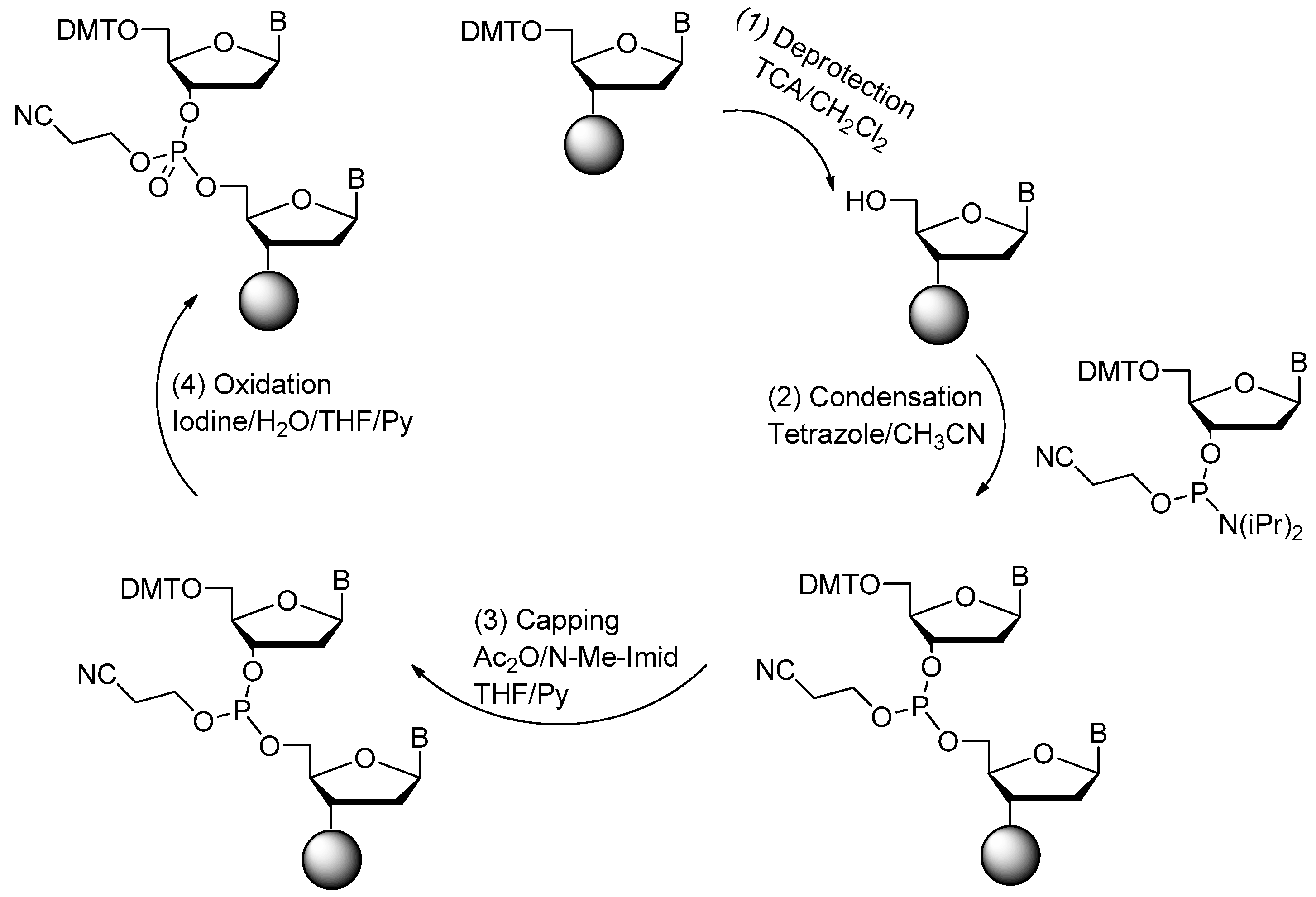
2.1. Synthesis of DNA Aptamers

DNA aptamers can be synthesized through the classic solid phase phosphoramidite four-step process on the automated DNA synthesizer [18].
The four-step method is shown in Figure 2. First, the 4,4′-Dimethoxytriphenylmethyl (DMT) group is removed from the deoxynucleoside (5′-end) which is linked to the control pore glass (CPG) columns. Large excess of acid solution (trichloroacetic acid (TCA)) could be used for the deprotection of DMT. In the second step of the cycle, an internucleotide bond called phosphite trimester is synthesized. Then, in the third step, the reaction product from Step 2 should be treated with capping agent to cap the unreacted free 5′-OH group. In the last step (Step 4), the new phosphite is oxidized to the corresponding phosphotriester by iodine. The cycle is repeated, once for each base, to produce the required oligonucleotide. Finally, the nucleic acid aptamers could be cleaved from the CPG by concentrated ammonium hydroxide. The protecting groups for phosphates and heterocyclic bases could be removed at the same time [18,37,38].
At present, the application of four-step method is very common. For the most part, progress in the solid phase nucleic acid synthesis field has not changed this fundamental approach. For R&D purposes, shortened aptamers with 20 to 50 nucleotides in length can be generated in individual labs using “lab scale” DNA or RNA synthesizers [39] (e.g., Expedite 8909, ABI394).

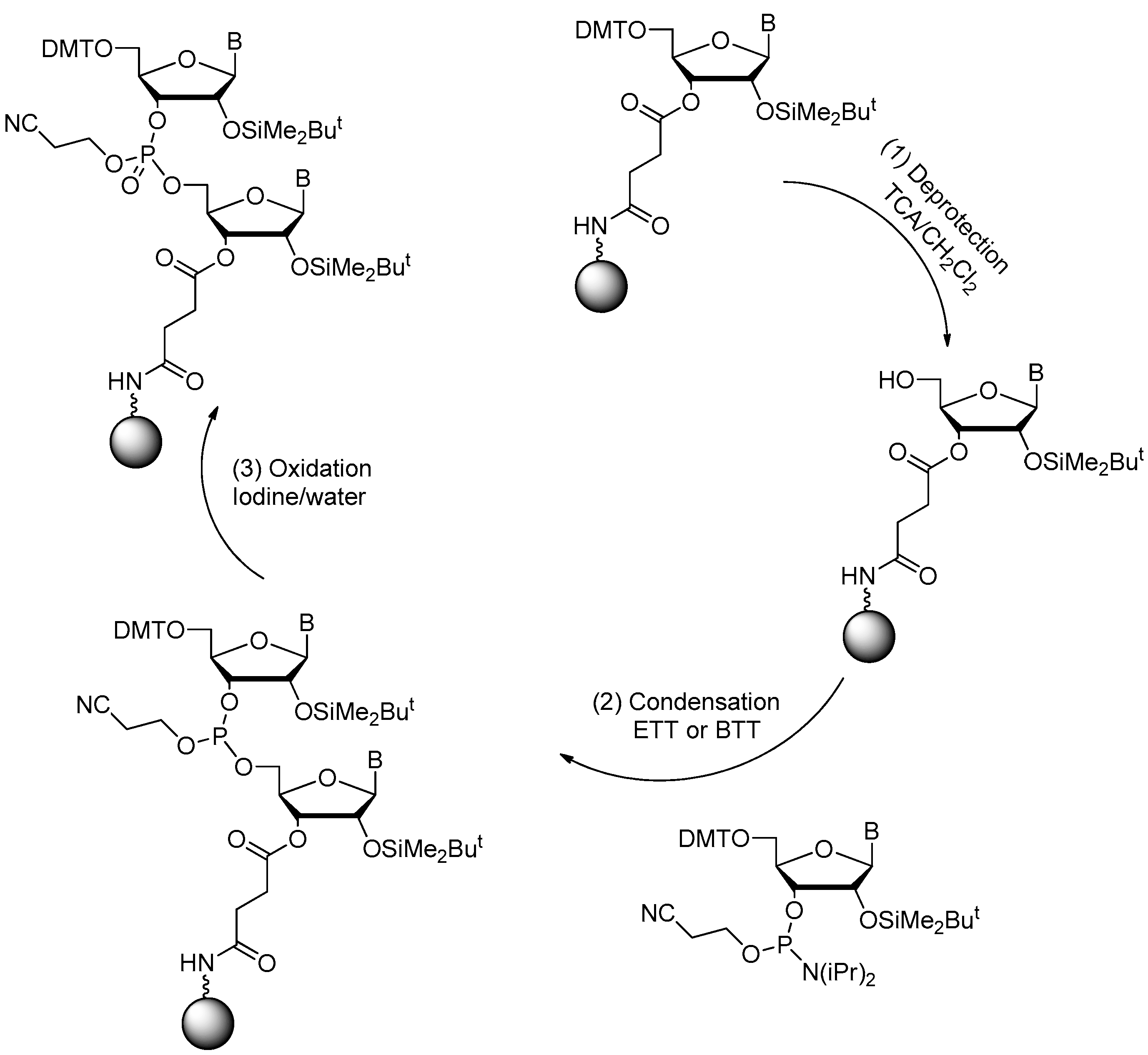
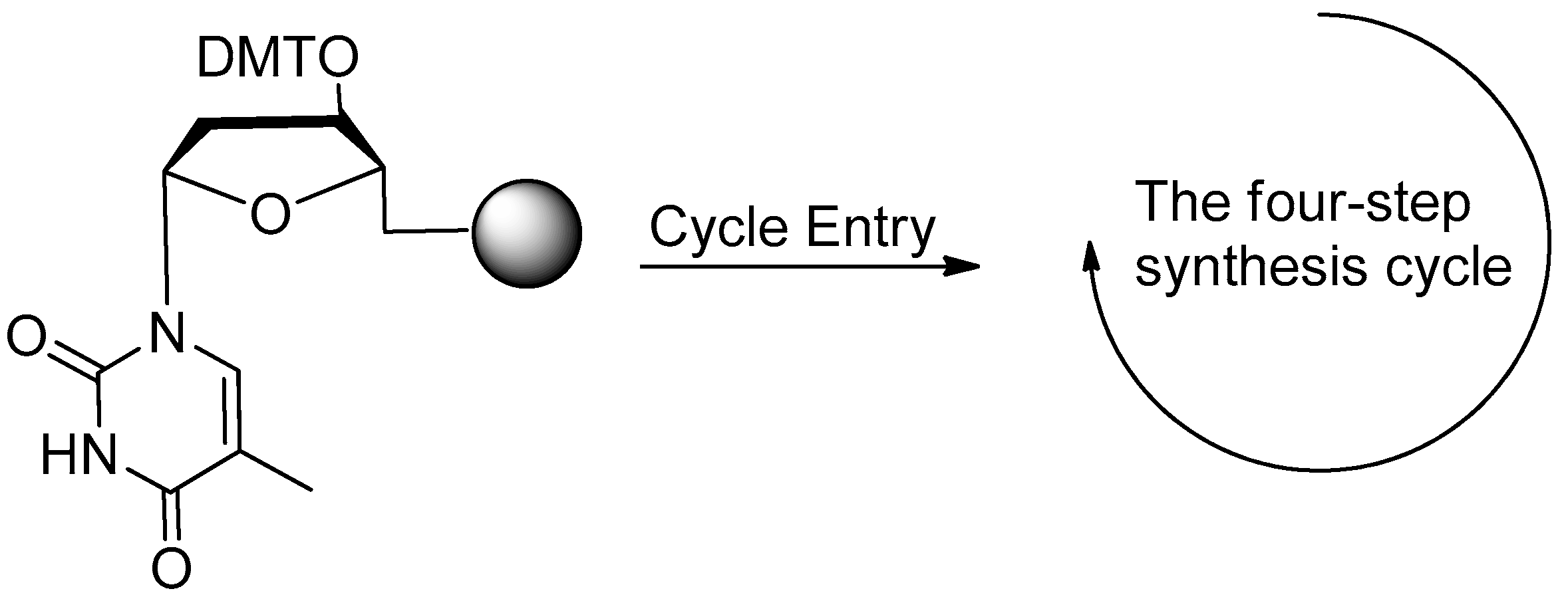
2.2. Synthesis of RNA Aptamers

Several of synthetic strategies for the solid-phase synthesis of RNA had been reported [19,40,41,42]. Among the combinations of different coupling/activation chemistries and protecting groups for the 2′-hydroxyl and exocyclic amine groups, the tert-butyldimethylsilyl protection of the ribose 2′-hydroxyl group combined with the standard protecting groups for the exocyclic amine groups (benzoyl for adenosine, acetyl for cytidine, and isobutyryl for guanosine) were most widely used [40,43]. Phosphoramidite monomers were usually activated with 4,5-dicyanoimidazole, 5-ethylthio-1H-tetrazole (ETT) or 5-benzylthio-1H-tetrazole (BTT) (Figure 3). The solid supports for RNA synthesis were polymeric supports or CPGs with different linkers and pore sizes. The final product could be cleaved from the CPG by concentrated ammonium hydroxide. The protection groups can also be removed at the same time [19,40,44,45,46].

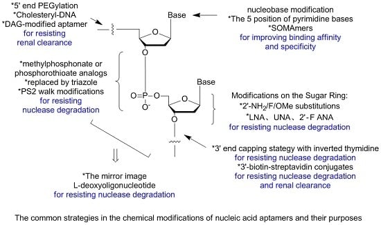
3. Modifications of Nucleic Acid Aptamers

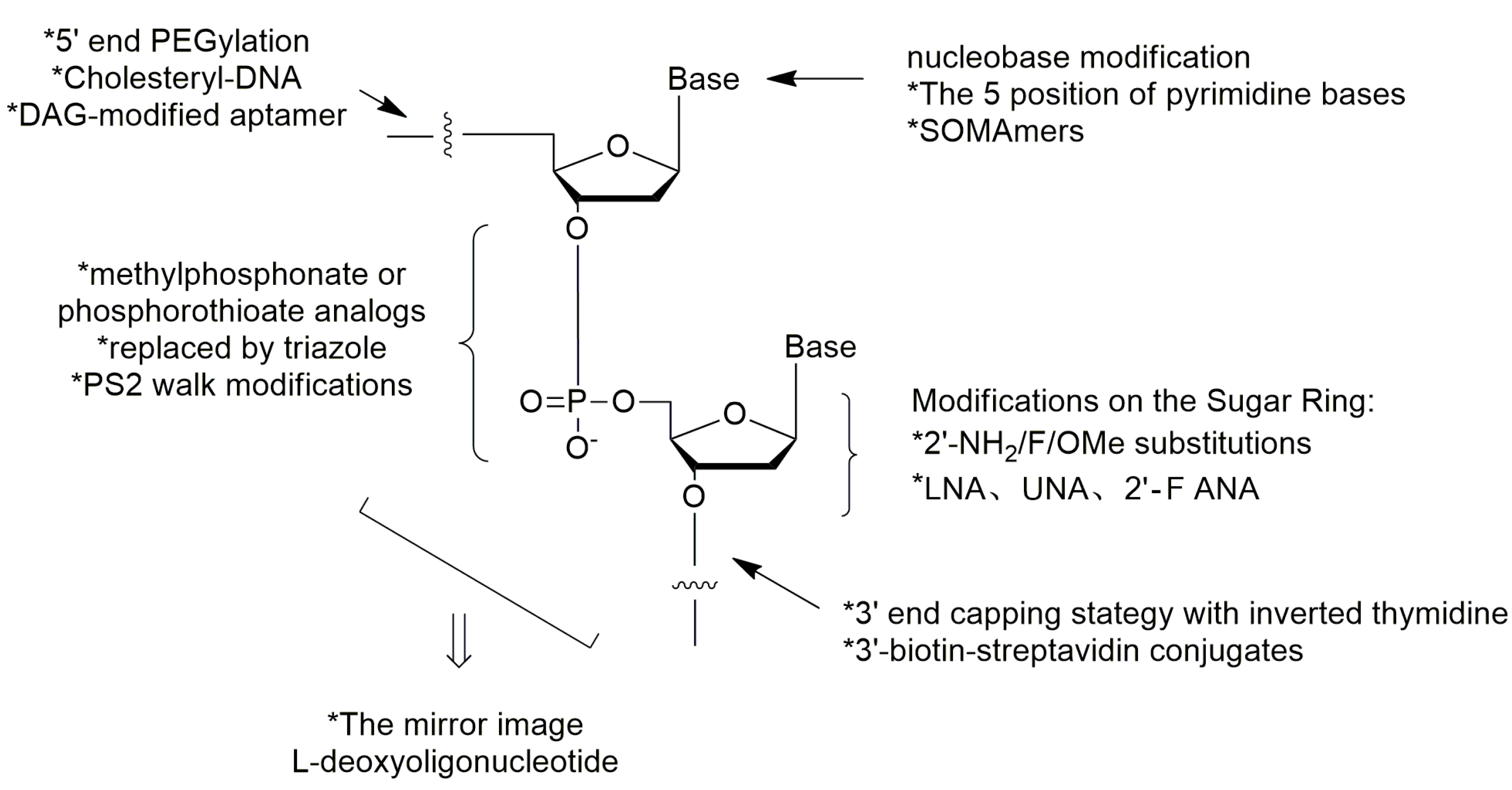
3.1. Aptamer Derivatives for Resisting Nuclease Degradation

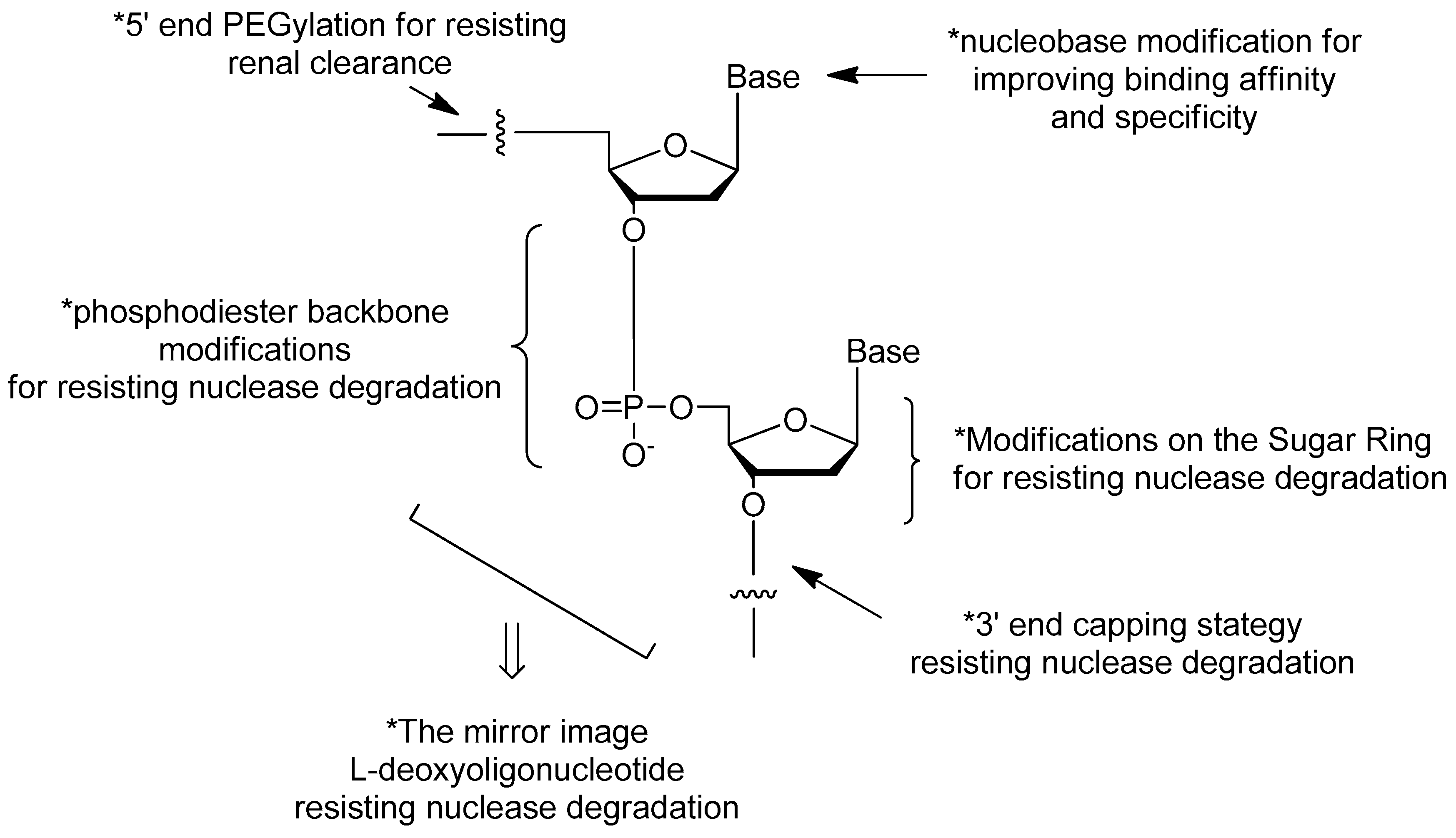
3.1.1. Terminal 3′–3′ and 5′–5′ Internucleotide Linkage

The 3′–3′ and 5′–5′ inversions were tested in 1991 by Seliger et al. [12]. The 3′-end capping with inverted thymidine has also been a common strategy among aptamers for diseases therapy in ongoing or completed clinical trials [15,47]. Research suggested that 3′-inverted dT modification could increase the stability and resistance of aptamers to 3′-exonuclease in human serum. Synthesis of 3′-inverted dT modified aptamers (Figure 4) needed modified CPG with the 5′-hydroxyl of the first nucleoside attached, followed by chain elongation in standard 3′→5′ fashion [12,20,21].

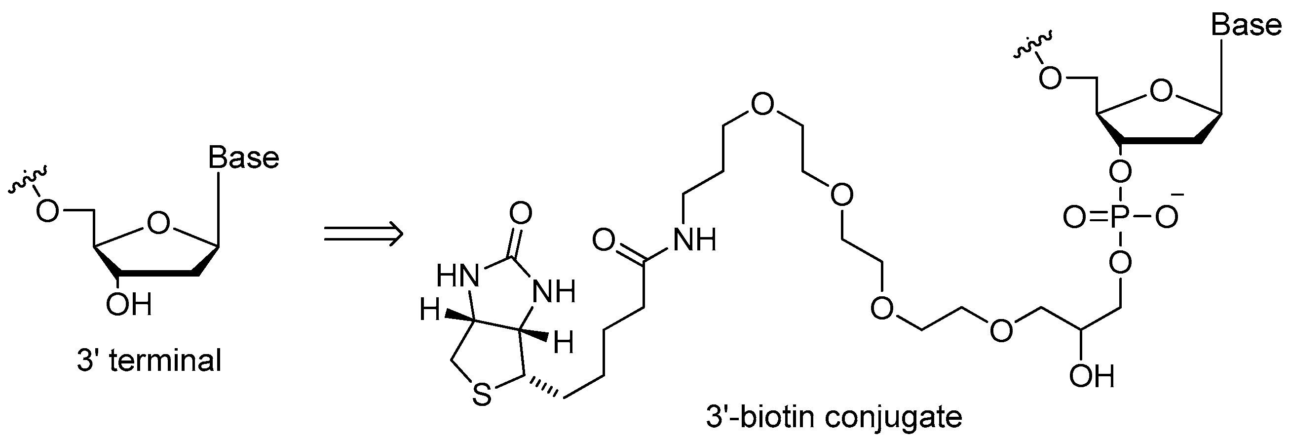
3.1.2. 3′-Biotin Conjugates

In some ways, 3′-biotin (Figure 5) could resist the activity of 3′-exonuclease, which was similar to 3′-inverted dT modification. Dougan et al. [36] investigated the 3′-biotin-streptavidin conjugates of the thrombin aptamer to find that the 3′-biotin rendered resistance to the 3′-exonuclease in the blood of mouse or rabbits. In addition, the 3′-biotin-streptavidin conjugates slowed down the clearance rate of aptamers in blood circulation system in vivo [36]. A similar 3′-biotin approach was also used to protect the DNA aptamer targeting the SARS coronavirus helicase for up to 31 and 16 h in 5% and 10% fetal bovine serum, whereas the original aptamer can only sustain half of that time [20].

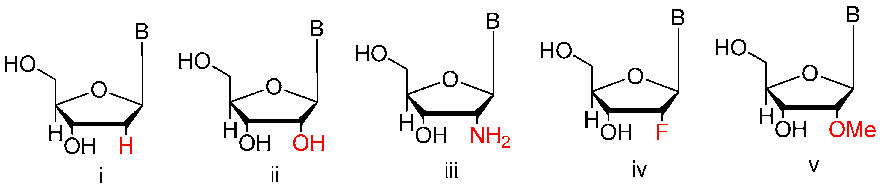
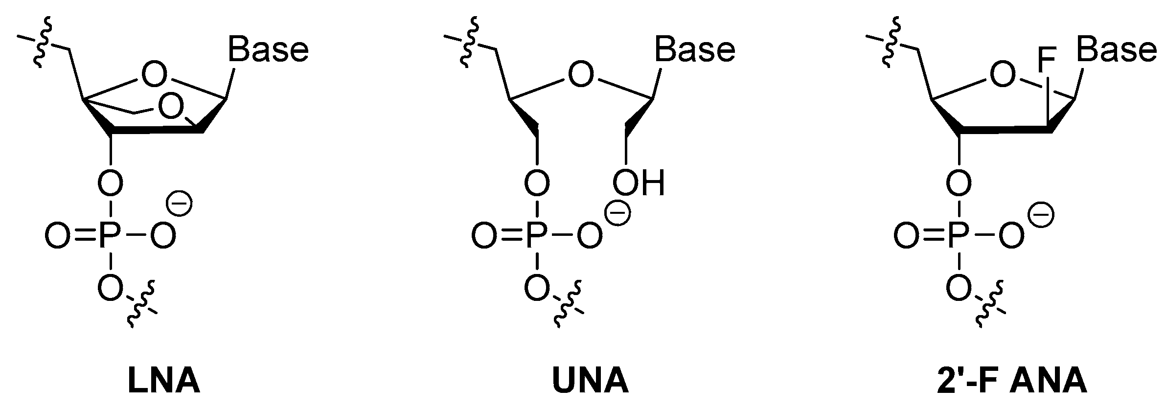
3.1.3. Modifications on the Sugar Ring

2′-Substitutions

Modifications to the sugars such as 2′-fluoro (2′-F) or 2′-amino (2′-NH2) ribose groups (Figure 6) on the pyrimidine residues have been available for incorporation into enzymatically derived nucleic acids for some years. Although both are effective at improving serum half-life, 2′-F modifications quickly garnered favor over 2′-NH2 due to the increased coupling efficiency during solid-phase synthesis, and elimination of extra deprotection steps during 2′-NH2 purification. The more bulky 2′-O-methyl (2′-OMe) modifications have been previously used as a post-selection modification due to their increased nuclease resistance and high duplex melting temperature which could be seen in the clinical examples [48,49].

LNA, UNA, 2′-F ANA

Locked nucleic acid (LNA) (Figure 7) is an analog of ribonucleotide with a methylene linkage between 2′-O and 4′-C of the sugar ring. This modification showed great resistance to nucleases and increased thermostability thus could be used to generate the most stable pairs [50,51]. Darfeuille et al. also found that the LNA/DNA chimera LNA5, a stable complex that against HIV-1 trans-activating response (TAR) RNA, was able to maintain the intact structure within 20 h in bovine serum [52]. Shi et al. developed a new LNA/DNA chimeric aptamer probe through proper LNA incorporation and 3′-3′-thymidine (3′-3′-T) capping. The serum stability of original aptamer was gradually enhanced while its specificity and affinity were perfectly maintained. Especially TD05.6 aptamer which had a 7-base pair-LNA substitution exhibited a ten-fold elevated stability in serum and a much slower clearance rate in mice [53].
Unlike LNA, a structurally rigid modification that increases the thermostability of a modified-oligonucleotide thus protects it from nucleases degradation in cells, unlocked nucleic acid (UNA) (Figure 7) in which a bond between C2′ and C3′ of the sugar ring was absent makes aptamers more flexible [54]. Due to its nature of flexibility, UNA could alleviate strain in tight loop structures. Pasternak et al. found that UNA modifications on the loop regions of a 15-mer thrombin targeted DNA aptamer increased its thermodynamic stability. However, modifications within the G-quartet structures were unfavorable for quadruplex formation [55]. They also demonstrated that UNA could be placed in many positions without affecting the thrombin-binding affinity and anticoagulant efficiency of the aptamer [55].
It has been found that modifications at the 2′-position of the sugar ring would bring about different effects on thermostability based on the molecularity of G-quadruplex. Peng et al. discovered that, in both anti-HIV phosphorothioate aptamer and thrombin-binding aptamer, substitution of guanines (G) that adopted anti-conformation with 2′-F-G could maintain the quadruplex conformation, while substituting guanines with syn-conformation was not favored [25]. More importantly, two 2′-F-modified thrombin-binding aptamers (PG13 and PG14) showed approximately four-fold increased binding affinity to thrombin and up to seven-fold higher nuclease resistance. As a result, the 2′-deoxy-2′-fluoro-d-arabinonucleic acid (2′-F ANA) (Figure 7) modification was very suitable for improving the biological and physicochemical properties of DNA G-quartets [25].

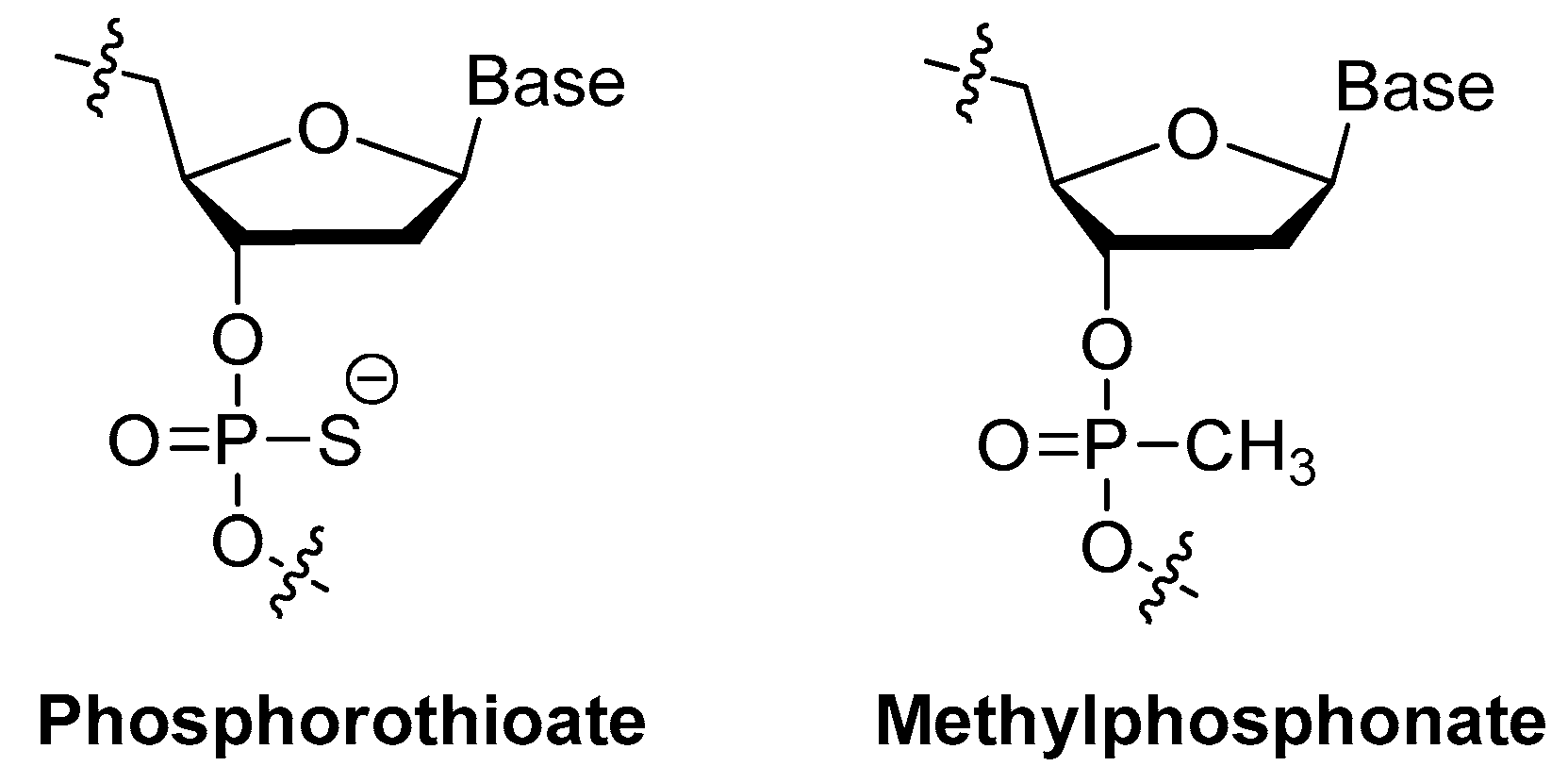
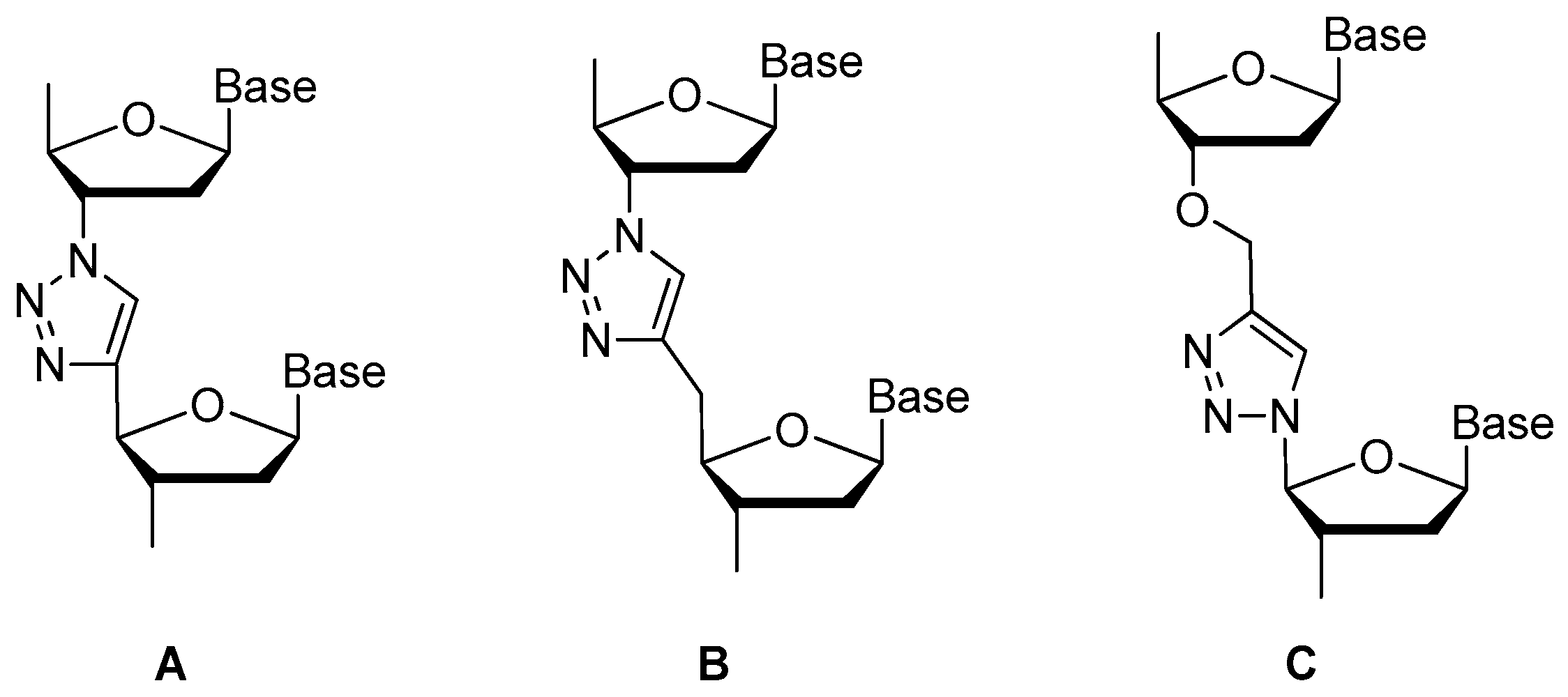
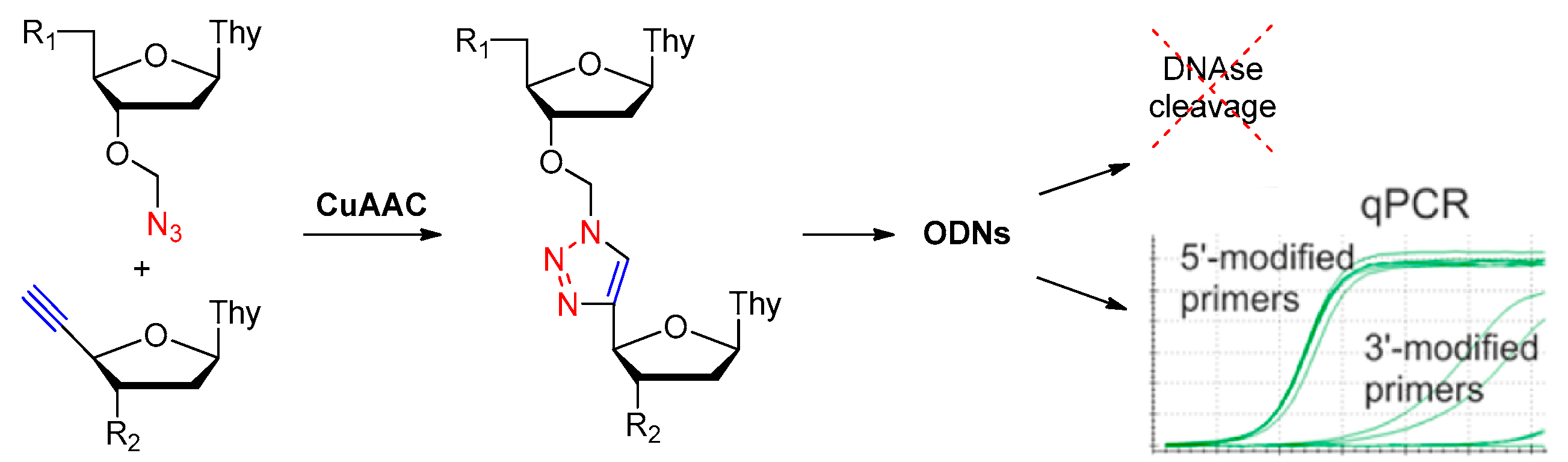
3.1.4. Modifications on the Phosphodiester Linkage

Methylphosphonate or Phosphorothioate

Replaced phosphodiester linkage of DNA with methylphosphonate or phosphorothioate analog is commonly used for aptamer modification. Thermodynamic studies revealed that loss of the negative charge of the phosphate backbone, as the methylphosphonate analog (Figure 8), destabilized the G-quadruplex structure [56]. The ionic radii of the oligonucleotide backbone atoms also have an impact in the stabilization of G-quadruplex structures. Sacca et al. found that substitution of the phosphate backbone atom O with S (phosphorothioate analog, Figure 8) might influence the thermal stability of the G-quadruplex structure in a molecularity-dependent manner [56].
The thermodynamic stability of the phosphodiester linkage of the thrombin-binding aptamer d(GGTTGGTGTGGTTGG) with thiophosphoryl substitutions at different internucleotide sites were studied [23,24]. Complete substitution by thiophosphorylated oligonucleotides was limited as their high toxicities, so partial substitutions with the maximum thermal stability were selected for evaluating their stabilities under conditions of nuclease RQ1 DNAse hydrolysis and their antithrombin activities in blood plasma [24]. Aptamer d(GGSTSTSGGTGTGGSTSTSGG) with thio-substitutions in both TT loops exhibited similar antithrombin efficiency to the unmodified aptamer but better resistance to the degradation of DNA nuclease in blood serum [23].
More recently, phosphorodiothioate linkages (PS2) were employed to stabilize phosphate backbone. The substitution of both non-bridging oxygen atoms with sulfur could give rise to a phosphorodithioate linkage, which, similar to natural DNA, is achiral at phosphorus. In addition, it was reported that PS2 substitutions dramatically improved target binding affinity by ~1000-fold (see PS2 walk below) [10].

Replaced by Triazole

Replacement of the oligonucleotide phosphodiester linkage with triazole linkages has shown great promise [57,58,59,60]. These triazole analogs can be obtained through automated phosphoramidite synthesis with modified dinucleoside blocks [61] or the click reaction between azide- and alkyne-bearing nucleosides [62,63]. Figure 9 shows three types of promising triazole internucleotide modifications [64].
Varizhuk et al. synthesized several new oligonucleotide analogs with triazole internucleotide linkages through the click reaction as shown in Figure 10. These analogs bore DNA hybridization affinities similar to those of original oligonucleotides and increased resistance to nuclease cleavage [64].
Later in 2013, Varizhuk et al. synthesized a series of triazole-modified DNA aptamers with structure similar to thrombin-inhibiting G-quadruplexes TBA15 (Thrombin-Binding Aptamer) and TBA31, then tested their secondary structure stabilities, binding affinities for thrombin and anticoagulant effects [65]. A modification in the central loop of the aptamer quadruplex resulted in an anticoagulant activity similar to that of TBA15. Although the modification failed to enhance thrombin binding affinity, it protected aptamers from nuclease hydrolysis thus increased their stabilities. The novel aptamers were potent thrombin inhibitors and could be an alternative to the known anticoagulant drugs [59].

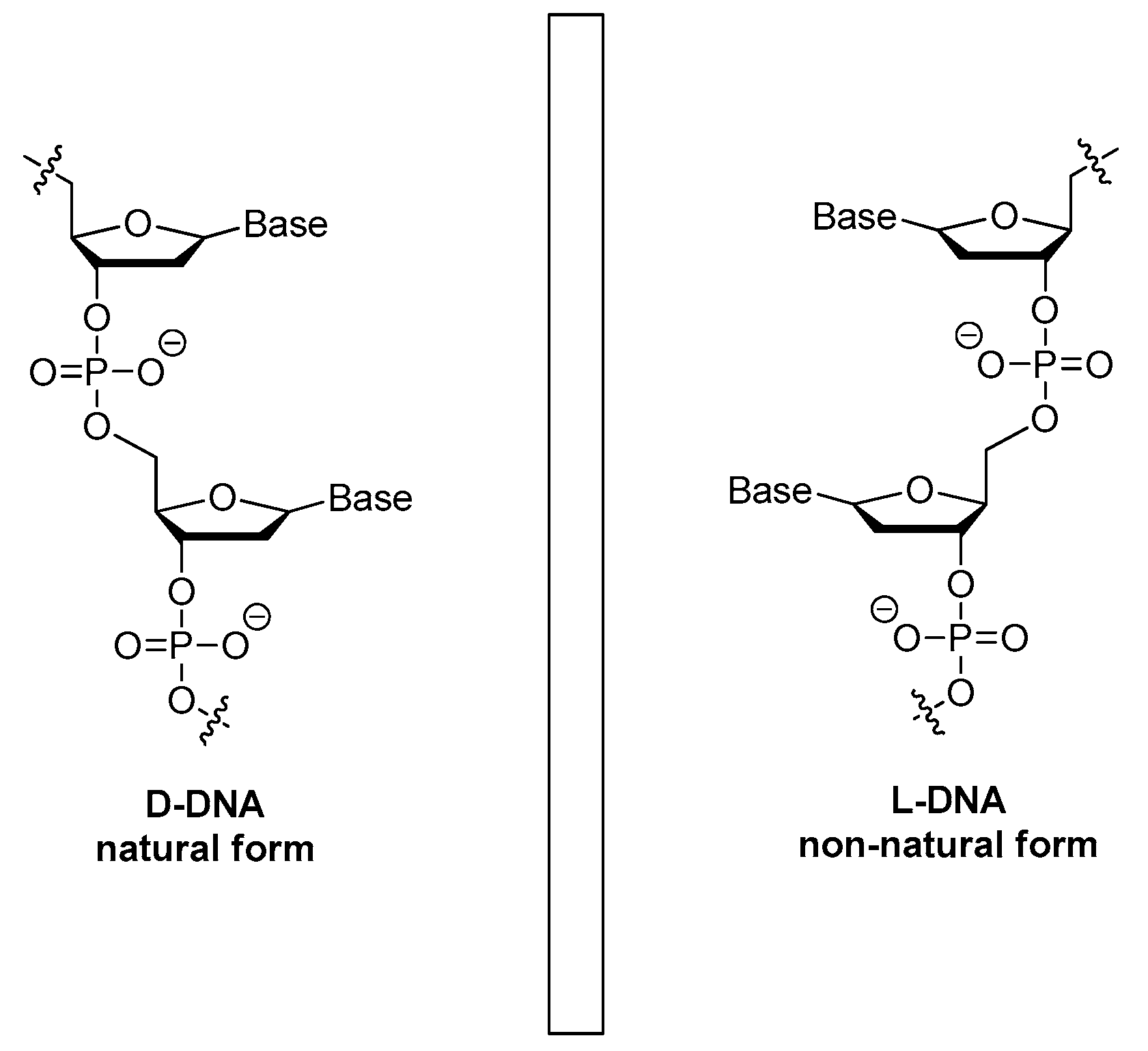
3.1.5. The Mirror Image l-DNA

Natural DNAs are all in d-form. A chiral transition could result in the mirror image l-DNA (Figure 11) that may display high resistance to the degradation of nucleases and retain the affinity to targets. Based on the sequences of d-form aptamers, the l-enantiomeric oligonucleotide aptamers (also called as Spiegelmers) were then chemically synthesized [66]. Based on the domain approach, Purschke et al. found a 65-mer Spiegelmer that bound to a stable 25-amino acids length domain of bacterial staphylococcal enterotoxin B [64]. The l-DNA Spiegelmer showed comparable binding affinity to the l-peptide domain and slightly reduced affinity to the whole bacterial staphylococcal enterotoxin B protein.
Through an in vitro-selection process, which was started from a random pool of oligonucleotides, a 67-mer Spiegelmer with a dissociation constant (Kd) of 20 nM for gonadotropin-releasing hormone (GnRH) was reported by Wlotzka et al. [67]. This Spiegelmer was an effective antagonist to GnRH in Chinese hamster and castrated rat models. Besides, the PEGylated Spiegelmer showed more pronounced inhibition activity and longer plasma half-life [67]. Towards the same target, other Spiegelmers with high specificity and affinity were identified through the usage of Spiegelmer technology by Leva et al. [68]. Firstly, aptamers that bind to d-GnRH with Kd of 50–100 nM were isolated, and then their enantiomers were synthesized. The resulting Spiegelmers had similar affinities to that of d-aptamers [68]. Many clinical evaluated aptamers such as NOX-A12, NOX-H94 and NOX-E36 are all l-aptamers [69,70].
A number of different strategies and chemical modifications are now available to enhance the stability of aptamers to nuclease (Table 1). Among these modifications, 2′-fluoro or 2′-O-methyl-substitutions and 3′ end capping with inverted thymidine have been the common strategies in the chemical modifications of nucleic acid aptamers for resisting nuclease degradation.

3.2. Aptamer Derivatives for Resisting Renal Clearance

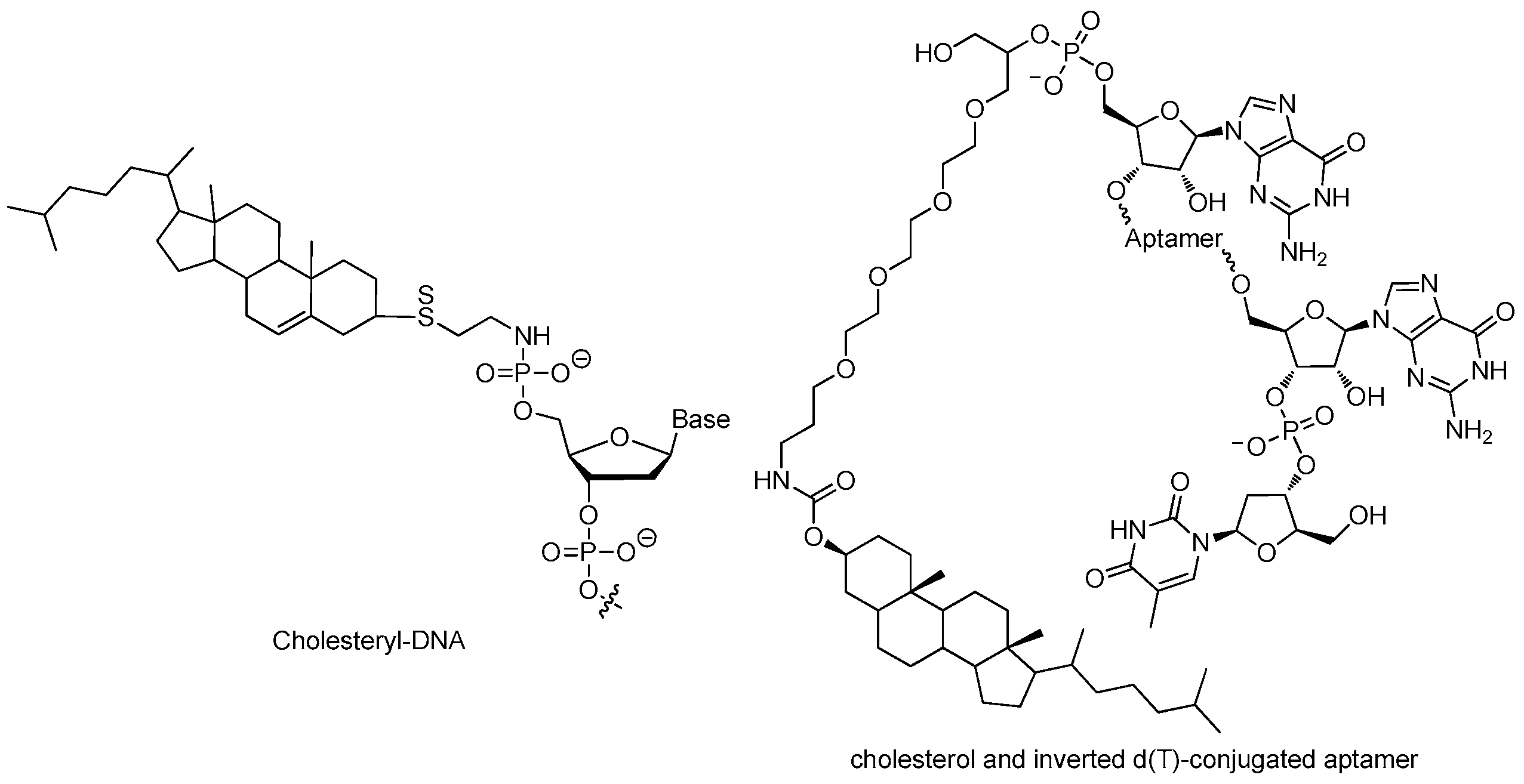
3.2.1. 5′-End with Cholesterol

Even with stabilizing backbone modification, small aptamers are subjected to rapid excretion through renal clearance mainly through glomerular filtration. Formulation with bulky moiety enlarges the size of aptamers, overcoming the renal filtration and extending circulation time, evidently [32,33].
Cholesterol can be derivatized to the 5′-end of an aptamer to form a cholesterol-oligonucleotide (cholODN) conjugate. Smidt et al. added cholesterol at the 5′-end of a 16-mer oligonucleotide (ODN) through a phosphate spacer (Figure 12), the half-time of the resulting cholODN (9–11 min) in plasma was considerably longer than the unmodified ODN (<1 min) [71]. The resulting cholODN can be further linked with low-density lipoprotein (LDL) to form cholODN-LDL complex that turned out to be stable against degradation by rat serum nucleases. The cholODN had a roughly 10-fold longer plasma half-life than the unmodified ODN [71].
Lee and coworkers modified a 29 nucleotide-long 2′-F pyrimidine modified RNA aptamer with cholesterol to form a cholesterol-conjugated aptamer (chol-aptamer) (Figure 12) which can be efficiently absorbed into the cell and inhibits Hepatitis C virus RNA replication [71]. The chol-aptamer had no toxicity in vitro or in vivo. It did not induce any notable alteration in the gene expression profile, including innate immune-related genes. Moreover, administration of the chol-aptamer was well tolerated in mice without any abnormalities observed. Noticeably, cholesterol conjugation showed longer half-life with approximately nine times lower of clearance rate in plasma. In other words, it extended the duration time that the aptamer stayed in plasma, thus enhanced the stability when the aptamer was exposed to body [32].

3.2.2. 5′-End with Dialkyl Lipids

Willis et al. reported the preparation and functional properties of a nuclease-resistant vascular endothelial growth factor (VEGF) aptamer which was attached to liposome bilayers through a lipid group. The resulting liposome-anchored aptamer maintained the high binding affinity to VEGF. Moreover, the residence time in plasma was considerably improved when compared with that of the original aptamer [72]. They used the solid phase phosphoramidite method to prepare a dialkylglycerol (DAG) modified VEGF aptamer in which two 18-carbon saturated unbranched hydrocarbon chains were attached via a tetraethylene glycol linker. The DAG phosphoramidite was synthesized in seven steps and then introduced to the 5′-end of the VEGF aptamer (Figure 13) [73]. Afterwards, the DAG-modified VEGF aptamer was incorporated into the bilayers of liposomes, which resulted in aptamers with improved inhibitory activity toward VEGF-induced endothelial cell proliferation in vitro and increased vascular permeability in vivo [73].

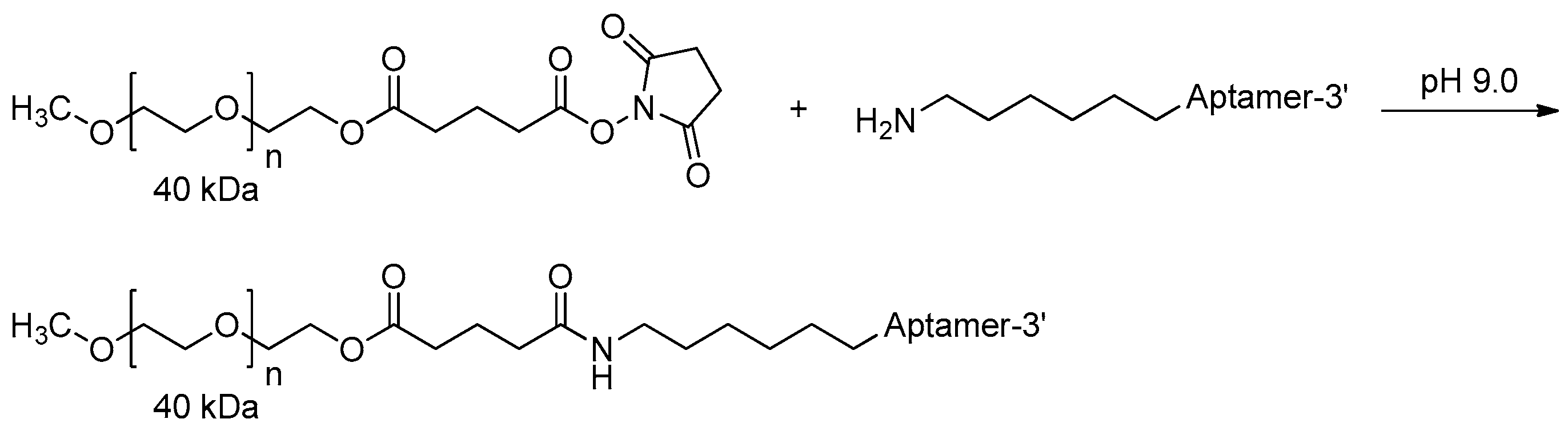
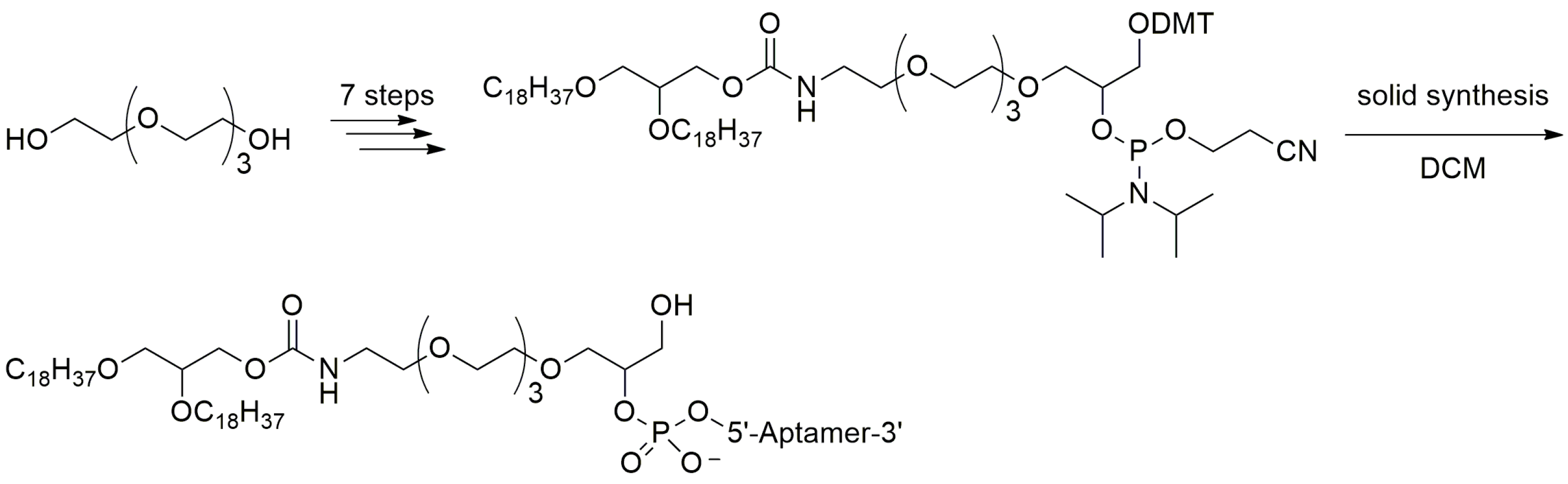
3.2.3. 5′-End PEGylation

In 2011, Hoffmann et al. described the PEGylation of amino-modified NOX-E36 oligonucleotide by using N-hydroxysuccinimide (NHS)-ester-activated polyethylene glycol (PEG), which was most widely used, especially for manufacturing large quantities of PEGylated oligonucleotides. Following synthesis and two-step deprotection, the resulting intermediate amino-modified oligonucleotide reacted with NHS-ester-activated PEG to form oligonucleotide-PEG conjugate (Figure 14). Other coupling methods such as activation by p-nitrophenyl carbonate or thiol-maleimide coupling could also be used [74]. The choice of coupling strategies should be made under consideration of the following factors: (1) compatibility with the oligonucleotide; (2) accessibility of the modified oligonucleotide; and (3) reactivity of the activated PEG, which should only react at the functionalization site of the oligonucleotide.
MP7 is one of the DNA aptamers that bind specifically to the murine extracellular domain of PD-1 (Programmed death protein 1) and block the PD-1:PD-L1 (Programmed death-ligand 1) interaction. However, the unmodified DNA aptamer exhibited very short in vivo half-time (<1 h) owing to the rapid renal filtration of such small molecule [75]. It has been reported that conjugation of aptamers with high molecular weight PEG could limit the rate of filtration and extended half-life up to 24–48 h [32,75]. Thus, MP7 was modified at its 5′-termini with a 40 kDa PEG (Figure 15). The PEGylated form of MP7 retained the ability to block PD-1 binding to PD-L1, and significantly suppressed the growth of PD-L1 positive colon carcinoma in vivo [76,77] (Table 2).

3.3. Aptamer Derivatives for Improving Binding Affinity and Target Selectivity

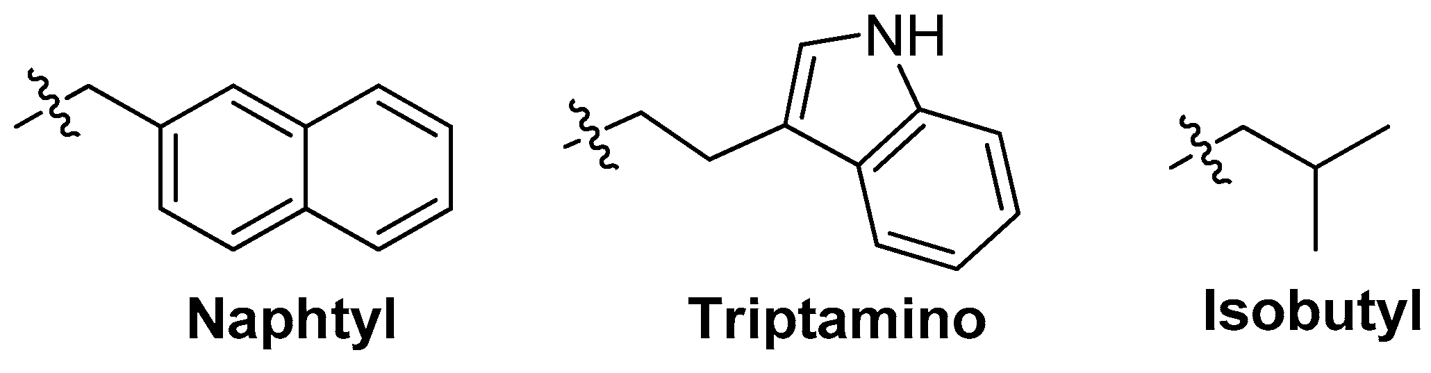
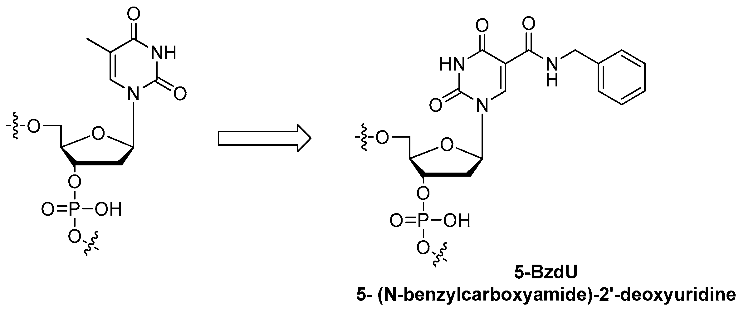
3.3.1. Modifications on the Bases; SOMAmers

Aptamers with improved binding affinities are being generated with modifications on the base. AS1411 aptamer is a 26-mer single strand DNA 5-d(GGTGGTGGTGGTTGTGGTGGTGGTGG)-3′ which binds to the nucleolin protein expressed on the surfaces of cancer cells [78,79,80]. Recent research has shown that 5-BzdU (5-(N-benzylcarboxyamide)-2-deoxyuridine) modification (Figure 16) of the AS1411 aptamer might selectively increase its targeting affinity to cancer cells while the normal healthy cells have no significant influence [26].
The benzyl could be replaced by the other functional groups such as naphtyl, triptamino, isobutyl and so on (Figure 17). These additional groups might increase the affinities of aptamers to their targets [81,82,83,84] (Table 3).
The base modifications have also made significant advancements to give aptamers protein-like functionality [11,87]. The SOMAmers (Slow Off-rate Modified Aptamers) not only display improved binding affinities and binding kinetics (in particular, slow off-rates) when compared to traditional aptamers, but also the inclusion of these modifications in their libraries significantly increased the selection “hit rate” [88]. The power of this kind of base modifications has been further demonstrated through the discovery of a 32 nucleotide SOMAmer, SL1025, which binds IL-6 with 200 pmol/L binding affinity and exhibits very little nuclease degradation over a 48-hour incubation in human serum [47,89].

3.3.2. Crystal Structure Based Modifications

Nucleic acid aptamers are much smaller than antibodies. In recent years, there are many studies on the crystallization and X-ray diffraction analysis of aptamers or the complex of aptamers and enzyme [90,91,92,93]. It is an effective method to develop modified aptamers with higher affinity and selectivity according to the crystal structures. Autotaxin (ATX) is a plasma lysophospholipase D which can hydrolyze lysophosphatidylcholine (LPC) and generate lysophosphatidic acid (LPA) [94,95]. DNA aptamer RB011 is an inhibitor against ATX. Nureki and coworkers had investigated the crystal structure of ATX in complex with RB011 [96]. The results showed that RB011 inhibited the activity of ATX by preventing its binding to LPC substrates. The hydrophobic pocket of ATX could be occupied by some inhibitors such as HA155 or 3BoA [97,98], but RB011 did not occlude the hydrophobic pocket. Thus, the researchers introduced some hydrophobic groups such as p-methyl and p-isopropyl into the backbone phosphate of RB011, resulting in RB012 and RB013, respectively. The activities of both RB012 and RB013 (IC50 values for LPC were 1.8 and 0.85 nM, respectively) were more potent than that of RB011 (4.4 nM) [96]. These results suggested that modifications aimed to occlude the hydrophobic pocket could significantly increase inhibitory activity. This is a successful modification based on the crystal structural information.

3.3.3. NMR Spectroscopy Guided Aptamer Optimization
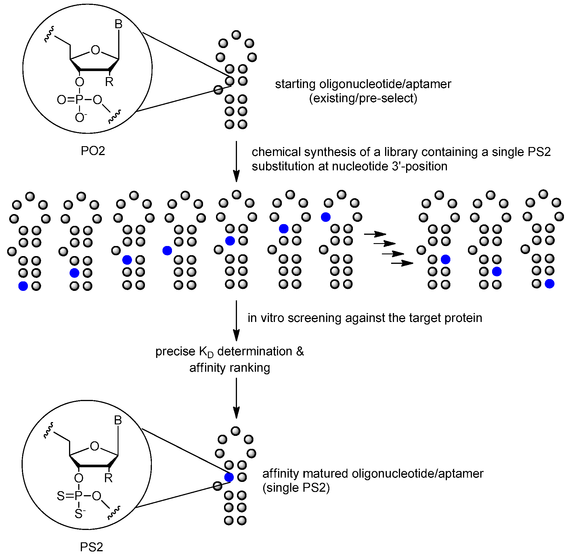
Nucleic acid aptamers are widely used for biotechnological or biomedical purpose. High resolution structure information of aptamer–ligand complexes could help reveal the fundamental aspects of nucleic acid folding and nucleic acid-small molecule interactions. Structure information of aptamers and aptamer–ligand complexes constitute the starting point for rational function directed chemical modifications. Duchardt-Ferner E et al. reported the NMR resonance assignment of an RNA aptamer binding to the fluorescent ligand tetramethylrhodamine (TMR) in complex with the ligand 5-carboxy-tetramethylrhodamine (5-TAMRA) as a starting point for a high-resolution structure determination using NMR spectroscopy in solution [99]. This and other reports indicated that NMR guided aptamer optimization could be an optional strategy for aptamer improving binding affinity [100,101,102].

3.3.4. PS2 Walk

The binding affinity and specificity of unmodified nucleic acid aptamers are sometimes insufficient for successful implementation as therapeutic agents, compared with monoclonal antibody. Post-SELEX optimization of one Bn-dU and one Nap-dU SOMAmer led to improvements in IL-6 binding (10-fold) and inhibition activity (greater than 20-fold), resulting in lead SOMAmers with sub-nanomolar affinity (Kd = 0.2 nM) and potency (IC50 = 0.2 nM) [92]. The PS2 (phosphorodithioate) walk strategy is another option [85,86] (Figure 18). It was reported that the application of the PS2 substitution on a single nucleotide of nucleic acid aptamers could significantly improve target binding affinity by ~1000-fold (from nanomolar to picomolar). An X-ray co-crystal structure of the α-thrombin-PS2-aptamer complex revealed a localized induced-fit folding of the PS2-containing aptamer which leads to increased target interaction [10].
It is worth noting that the effect of PSO substitution (see Section 3.1.4. above) cannot be predicted since the PSO backbone modification is chiral and the chemical synthesis of PSO using phosphoramidite methodology typically results in a mixture of diastereoisomers with a fairly limited influence on the affinity improvement. The promising PS2 derivatives are achiral, representing a class of closely related mimics of natural nucleic acids.

4. Conclusions

In this review, we introduced the general solid phase synthesis method of nucleic acid aptamers. In addition, a number of chemical modifications of both DNA and RNA aptamers are summarized here. Among all the modifications shown in the Figure 19, 5′-end PEGylation (for resisting renal clearance) and 3′-end capping strategy (for resisting nuclease degradation) with inverted thymidine are the most commonly used strategy in recent studies. These two methods have been used in the aptamers for disease therapy in ongoing or completed clinical trials [15,47].
The nucleobase and phosphodiester linkage modifications (for improving target binding affinity) can also optimize the properties of aptamers. Excitingly, the established technologies provide an opportunity to generate nucleic acid aptamers of substantially improved affinity with a SOMAmer strategy or a single PS2-moiety substitution and without negatively affecting specificity. These technologies also provide crucial insights that could significantly accelerate the development of nucleic acid aptamer-based therapeutics for clinical applications. With the development of post-SELEX modifications of nucleic acid aptamers, the inherent physicochemical characteristics (metabolic instability, insufficient binding affinity and rapid renal filtration) of nucleic acid aptamers have been improved constantly, which provide a strong impetus of developing nucleic acid aptamers for therapeutic purposes (Table 4).

Acknowledgments

We thank the other academic staff members in Aiping Lu and Ge Zhang’s group at Hong Kong Baptist University (Hong Kong, China). We also thank Hong Kong Baptist University (Hong Kong, China) and the State Key Laboratory of Bioorganic and Natural Products Chemistry (Shanghai, China) for providing critical comments and technical support. This study was supported by the Hong Kong General Research Fund (HKBU12102914 to Ge Zhang), the Faculty Research Grant of Hong Kong Baptist University (FRG2/12-13/027 to Ge Zhang) and Open Project of Shanghai Institute of Organic Chemistry (SKLBNPC17344 to Feng Jiang).

Author Contributions

Shuaijian Ni, Houzong Yao, Lili Wang and Jun Lu wrote the manuscript. Feng Jiang, Aiping Lu and Ge Zhang revised and approved the manuscript.

Conflicts of Interest

The authors declare no conflict of interest.

References

  1. Tuerk, C.; Gold, L. Systematic evolution of ligands by exponential enrichment: RNA ligands to bacteriophage T4 DNA polymerase. Science 1990, 249, 505–510. [Google Scholar] [CrossRef] [PubMed]
  2. Ellington, A.D.; Szostak, J.W. In vitro selection of RNA molecules that bind specific ligands. Nature 1990, 346, 818. [Google Scholar] [CrossRef] [PubMed]
  3. Zhou, J.; Rossi, J. Aptamers as targeted therapeutics: Current potential and challenges. Nat. Rev. Drug Discov. 2017, 16, 181–202. [Google Scholar] [CrossRef] [PubMed]
  4. Gelinas, A.D.; Davies, D.R.; Janjic, N. Embracing proteins: Structural themes in aptamer-protein complexes. Curr. Opin. Struct. Biol. 2016, 36, 122–132. [Google Scholar] [CrossRef] [PubMed]
  5. Ashrafuzzaman, M. Aptamers as both drugs and drug-carriers. BioMed Res. Int. 2014, 2014. [Google Scholar] [CrossRef] [PubMed]
  6. Dass, C.R.; Saravolac, E.G.; Li, Y.; Sun, L.Q. Cellular uptake, distribution, and stability of 10–23 deoxyribozymes. Antisense Nucleic Acid Drug Dev. 2002, 12, 289–299. [Google Scholar] [CrossRef] [PubMed]
  7. Morrissey, D.V.; Blanchard, K.; Shaw, L.; Jensen, K.; Lockridge, J.A.; Dickinson, B.; McSwiggen, J.A.; Vargeese, C.; Bowman, K.; Shaffer, C.S.; et al. Activity of stabilized short interfering RNA in a mouse model of hepatitis B virus replication. Hepatology 2005, 41, 1349–1356. [Google Scholar] [CrossRef] [PubMed]
  8. Griffin, L.C.; Tidmarsh, G.F.; Bock, L.C.; Toole, J.J.; Leung, L.L. In vivo anticoagulant properties of a novel nucleotide-based thrombin inhibitor and demonstration of regional anticoagulation in extracorporeal circuits. Blood 1993, 81, 3271–3276. [Google Scholar] [PubMed]
  9. Pagratis, N.C.; Bell, C.; Chang, Y.F.; Jennings, S.; Fitzwater, T.; Jellinek, D.; Dang, C. Potent 2′-amino-, and 2′-fluoro-2′-deoxyribonucleotide RNA inhibitors of keratinocyte growth factor. Nat. Biotechnol. 1997, 15, 68–73. [Google Scholar] [CrossRef] [PubMed]
  10. Abeydeera, N.D.; Egli, M.; Cox, N.; Mercier, K.; Conde, J.N.; Pallan, P.S.; Mizurini, D.M.; Sierant, M.; Hibti, F.E.; Hassell, T.; et al. Evoking picomolar binding in RNA by a single phosphorodithioate linkage. Nucleic Acids Res. 2016, 44, 8052–8064. [Google Scholar] [CrossRef] [PubMed]
  11. Gold, L.; Ayers, D.; Bertino, J.; Bock, C.; Bock, A.; Brody, E.N.; Carter, J.; Dalby, A.B.; Eaton, B.E.; Fitzwater, T.; et al. Aptamer-based multiplexed proteomic technology for biomarker discovery. PLoS ONE 2010, 5, e15004. [Google Scholar] [CrossRef] [PubMed]
  12. Ortigao, J.R.; Rosch, H.; Montenarh, M.; Frohlich, A.; Seliger, H. Oligonucleotide analogs with terminal 3′, 3′-and 5′, 5′-internucleotidic linkages as antisense inhibitors of viral replication. Antisense Res. Dev. 1991, 1, 380. [Google Scholar] [CrossRef]
  13. Ng, E.W.; Shima, D.T.; Calias, P.; Cunningham, E.T., Jr.; Guyer, D.R.; Adamis, A.P. Pegaptanib, a targeted anti-VEGF aptamer for ocular vascular disease. Nat. Rev. Drug Discov. 2006, 5, 123–132. [Google Scholar] [CrossRef] [PubMed]
  14. Doggrell, S.A. Pegaptanib: The first antiangiogenic agent approved for neovascular macular degeneration. Expert Opin. Pharmacother. 2005, 6, 1421–1423. [Google Scholar] [CrossRef] [PubMed]
  15. Fine, S.L.; Martin, D.F.; Kirkpatrick, P. Pegaptanib sodium. Nat. Rev. Drug Discov. 2005, 4, 187–188. [Google Scholar] [CrossRef] [PubMed]
  16. Kiire, C.A.; Morjaria, R.; Rudenko, A.; Fantato, A.; Smith, L.; Smith, A.; Chong, V. Intravitreal pegaptanib for the treatment of ischemic diabetic macular edema. Clin. Ophthalmol. 2015, 9, 2305–2311. [Google Scholar] [CrossRef] [PubMed]
  17. Vinores, S.A. Technology evaluation: Pegaptanib, Eyetech/Pfizer. Curr. Opin. Mol. Ther. 2003, 5, 673–679. [Google Scholar] [PubMed]
  18. Caruthers, M.H.; Barone, A.D.; Beaucage, S.L.; Dodds, D.R.; Fisher, E.F.; McBride, L.J.; Matteucci, M.; Stabinsky, Z.; Tang, J.Y. Chemical synthesis of deoxyoligonucleotides by the phosphoramidite method. Methods Enzymol. 1987, 154, 287–313. [Google Scholar] [PubMed]
  19. Sproat, B.S. RNA synthesis using 2′-O-(tert-butyldimethylsilyl) protection. Oligonucleotide Synth. 2005, 17–31. [Google Scholar]
  20. Shum, K.T.; Tanner, J.A. Differential inhibitory activities and stabilisation of DNA aptamers against the SARS coronavirus helicase. Chembiochem 2008, 9, 3037–3045. [Google Scholar] [CrossRef] [PubMed]
  21. Shaw, J.P.; Kent, K.; Bird, J.; Fishback, J.; Froehler, B. Modified deoxyoligonucleotides stable to exonuclease degradation in serum. Nucleic Acids Res. 1991, 19, 747–750. [Google Scholar] [CrossRef] [PubMed]
  22. De Smidt, P.C.; le Doan, T.; de Falco, S.; van Berkel, T.J. Association of antisense oligonucleotides with lipoproteins prolongs the plasma half-life and modifies the tissue distribution. Nucleic Acids Res. 1991, 19, 4695–4700. [Google Scholar] [CrossRef] [PubMed]
  23. Zaitseva, M.; Kaluzhny, D.; Shchyolkina, A.; Borisova, O.; Smirnov, I.; Pozmogova, G. Conformation and thermostability of oligonucleotide d(GGTTGGTGTGGTTGG) containing thiophosphoryl internucleotide bonds at different positions. Biophys. Chem. 2010, 146, 1–6. [Google Scholar] [CrossRef] [PubMed]
  24. Pozmogova, G.; Zaitseva, M.; Smirnov, I.; Shvachko, A.; Murina, M.; Sergeenko, V. Anticoagulant effects of thioanalogs of thrombin-binding DNA-aptamer and their stability in the plasma. Bull. Exp. Biol. Med. 2010, 150, 180–184. [Google Scholar] [CrossRef] [PubMed]
  25. Peng, C.G.; Damha, M.J. G-quadruplex induced stabilization by 2′-deoxy-2′-fluoro-d-arabinonucleic acids (2′F-ANA). Nucleic Acids Res. 2007, 35, 4977–4988. [Google Scholar] [CrossRef] [PubMed]
  26. Lee, K.Y.; Kang, H.; Ryu, S.H.; Lee, D.S.; Lee, J.H.; Kim, S. Bioimaging of nucleolin aptamer-containing 5-(N-benzylcarboxyamide)-2′-deoxyuridine more capable of specific binding to targets in cancer cells. J. Biomed. Biotechnol. 2010, 2010, 168306. [Google Scholar] [CrossRef] [PubMed]
  27. Pallan, P.S.; Yang, X.; Sierant, M.; Abeydeera, N.D.; Hassell, T.; Martinez, C.; Janicka, M.; Nawrot, B.; Egli, M. Crystal structure, stability and Ago2 affinity of phosphorodithioate-modified RNAs. Rsc. Adv. 2014, 4, 64901–64904. [Google Scholar] [CrossRef]
  28. Ashley, S.L.; Xia, M.; Murray, S.; O’Dwyer, D.N.; Grant, E.; White, E.S.; Flaherty, K.R.; Martinez, F.J.; Moore, B.B. Six-SOMAmer Index relating to immune, protease and angiogenic functions predicts progression in IPF. PLoS ONE 2016, 11, e0159878. [Google Scholar] [CrossRef] [PubMed]
  29. Eid, C.; Palko, J.W.; Katilius, E.; Santiago, J.G. Rapid slow off-rate modified aptamer (SOMAmer)-based detection of C-reactive protein using isotachophoresis and an ionic spacer. Anal. Chem. 2015, 87, 6736–6743. [Google Scholar] [CrossRef] [PubMed]
  30. Kraemer, S.; Vaught, J.D.; Bock, C.; Gold, L.; Katilius, E.; Keeney, T.R.; Kim, N.; Saccomano, N.A.; Wilcox, S.K.; Zichi, D.; et al. From SOMAmer-based biomarker discovery to diagnostic and clinical applications: A SOMAmer-based, streamlined multiplex proteomic assay. PLoS ONE 2011, 6, e26332. [Google Scholar] [CrossRef] [PubMed]
  31. Park, N.J.; Wang, X.; Diaz, A.; Goos-Root, D.M.; Bock, C.; Vaught, J.D.; Sun, W.; Strom, C.M. Measurement of cetuximab and panitumumab-unbound serum EGFR extracellular domain using an assay based on slow off-rate modified aptamer (SOMAmer) reagents. PLoS ONE 2013, 8, e71703. [Google Scholar] [CrossRef] [PubMed]
  32. Healy, J.M.; Lewis, S.D.; Kurz, M.; Boomer, R.M.; Thompson, K.M.; Wilson, C.; McCauley, T.G. Pharmacokinetics and biodistribution of novel aptamer compositions. Pharm. Res. 2004, 21, 2234–2246. [Google Scholar] [CrossRef] [PubMed]
  33. Watson, S.R.; Chang, Y.F.; O’Connell, D.; Weigand, L.; Ringquist, S.; Parma, D.H. Anti-l-selectin aptamers: Binding characteristics, pharmacokinetic parameters, and activity against an intravascular target in vivo. Antisense Nucleic Acid Drug Dev. 2000, 10, 63–75. [Google Scholar] [CrossRef] [PubMed]
  34. Gilbert, J.C.; DeFeo-Fraulini, T.; Hutabarat, R.M.; Horvath, C.J.; Merlino, P.G.; Marsh, H.N.; Healy, J.M.; Boufakhreddine, S.; Holohan, T.V.; Schaub, R.G. First-in-human evaluation of anti von Willebrand factor therapeutic aptamer ARC1779 in healthy volunteers. Circulation 2007, 116, 2678–2686. [Google Scholar] [CrossRef] [PubMed]
  35. Van Eijk, L.; Swinkels, D.; John, A.; Schwoebel, F.; Fliegert, F.; Summo, L.; Vauleon, S.; Laarakkers, J.; Riecke, K.; Pickkers, P. Randomized double-blind placebo-controlled PK/PD study on the effects of a single intravenous dose of the anti-hepcidin Spiegelmer NOX-H94 on serum iron during experimental human endotoxemia. Crit. Care 2013, 17, P352. [Google Scholar]
  36. Dougan, H.; Lyster, D.M.; Vo, C.V.; Stafford, A.; Weitz, J.I.; Hobbs, J.B. Extending the lifetime of anticoagulant oligodeoxynucleotide aptamers in blood. Nucl. Med. Biol. 2000, 27, 289–297. [Google Scholar] [CrossRef]
  37. Dellinger, D.J.; Betley, J.R.; Wyrzykiewicz, T.K.; Caruthers, M.H. Synthesis of DNA using a new two-step cycle. Oligonucleotide Synth. 2005, 288, 1–16. [Google Scholar]
  38. Beaucage, S.L.; Caruthers, M.H. Synthetic strategies and parameters involved in the synthesis of oligodeoxyribonucleotides according to the phosphoramidite method. Curr. Protoc. Nucleic Acid Chem. 2001. [Google Scholar] [CrossRef]
  39. Maier, K.E.; Levy, M. From selection hits to clinical leads: Progress in aptamer discovery. Mol. Ther. Methods Clin. Dev. 2016, 5, 16014. [Google Scholar] [CrossRef] [PubMed]
  40. Usman, N.; Ogilvie, K.; Jiang, M.; Cedergren, R. The automated chemical synthesis of long oligoribuncleotides using 2′-O-silylated ribonucleoside 3′-O-phosphoramidites on a controlled-pore glass support: Synthesis of a 43-nucleotide sequence similar to the 3′-half molecule of an Escherichia coli formylmethionine tRNA. J. Am. Chem. Soc. 1987, 109, 7845–7854. [Google Scholar]
  41. Shiba, Y.; Masuda, H.; Watanabe, N.; Ego, T.; Takagaki, K.; Ishiyama, K.; Ohgi, T.; Yano, J. Chemical synthesis of a very long oligoribonucleotide with 2-cyanoethoxymethyl (CEM) as the 2′-O-protecting group: Structural identification and biological activity of a synthetic 110mer precursor-microRNA candidate. Nucleic Acids Res. 2007, 35, 3287–3296. [Google Scholar] [CrossRef] [PubMed]
  42. Ohgi, T.; Masutomi, Y.; Ishiyama, K.; Kitagawa, H.; Shiba, Y.; Yano, J. A new RNA synthetic method with a 2′-O-(2-cyanoethoxymethyl) protecting group. Org. Lett. 2005, 7, 3477–3480. [Google Scholar] [CrossRef] [PubMed]
  43. Usman, N.; Pon, R.T.; Ogilvie, K.K. Preparation of ribonucleoside 3′-O-phosphoramidites and their application to the automated solid phase synthesis of oligonucleotides. Tetrahedron Lett. 1985, 26, 4567–4570. [Google Scholar] [CrossRef]
  44. Sinha, N.; Davis, P.; Usman, N.; Perez, J.; Hodge, R.; Kremsky, J.; Casale, R. Labile exocyclic amine protection of nucleosides in DNA, RNA and oligonucleotide analog synthesis facililating N-deacylation, minimizing depurination and chain degradation. Biochimie 1993, 75, 13–23. [Google Scholar] [CrossRef]
  45. Welz, R.; Müller, S. 5-(Benzylmercapto)-1H-tetrazole as activator for 2′-O-TBDMS phosphoramidite building blocks in RNA synthesis. Tetrahedron Lett. 2002, 43, 795–797. [Google Scholar] [CrossRef]
  46. Westman, E.; Stromberg, R. Removal of t-butyldimethylsilyl protection in RNA-synthesis. Triethylamine trihydrofluoride (TEA, 3HF) is a more reliable alternative to tetrabutylammonium fluoride (TBAF). Nucleic Acids Res. 1994, 22, 2430–2431. [Google Scholar] [CrossRef] [PubMed]
  47. Gupta, S.; Hirota, M.; Waugh, S.M.; Murakami, I.; Suzuki, T.; Muraguchi, M.; Shibamori, M.; Ishikawa, Y.; Jarvis, T.C.; Carter, J.D.; et al. Chemically modified DNA aptamers bind interleukin-6 with high affinity and inhibit signaling by blocking its interaction with interleukin-6 receptor. J. Biol. Chem. 2014, 289, 8706–8719. [Google Scholar] [CrossRef] [PubMed]
  48. Padilla, R.; Sousa, R. Efficient synthesis of nucleic acids heavily modified with non-canonical ribose 2′-groups using a mutantT7 RNA polymerase (RNAP). Nucleic Acids Res. 1999, 27, 1561–1563. [Google Scholar] [CrossRef] [PubMed]
  49. Ruckman, J.; Green, L.S.; Beeson, J.; Waugh, S.; Gillette, W.L.; Henninger, D.D.; Claesson-Welsh, L.; Janjic, N. 2′-Fluoropyrimidine RNA-based aptamers to the 165-amino acid form of vascular endothelial growth factor (VEGF165). Inhibition of receptor binding and VEGF-induced vascular permeability through interactions requiring the exon 7-encoded domain. J. Biol. Chem. 1998, 273, 20556–20567. [Google Scholar] [CrossRef] [PubMed]
  50. Obika, S.; Nanbu, D.; Hari, Y.; Morio, K.-I.; In, Y.; Ishida, T.; Imanishi, T. Synthesis of 2′-O,4′-C-methyleneuridine and -cytidine. Novel bicyclic nucleosides having a fixed C3, -endo sugar puckering. Tetrahedron Lett. 1997, 38, 8735–8738. [Google Scholar] [CrossRef]
  51. Koshkin, A.A.; Singh, S.K.; Nielsen, P.; Rajwanshi, V.K.; Kumar, R.; Meldgaard, M.; Olsen, C.E.; Wengel, J. LNA (Locked Nucleic Acids): Synthesis of the adenine, cytosine, guanine, 5-methylcytosine, thymine and uracil bicyclonucleoside monomers, oligomerisation, and unprecedented nucleic acid recognition. Tetrahedron 1998, 54, 3607–3630. [Google Scholar] [CrossRef]
  52. Darfeuille, F.; Hansen, J.B.; Orum, H.; Di Primo, C.; Toulme, J.J. LNA/DNA chimeric oligomers mimic RNA aptamers targeted to the TAR RNA element of HIV-1. Nucleic Acids Res. 2004, 32, 3101–3107. [Google Scholar] [CrossRef] [PubMed]
  53. Shi, H.; He, X.; Cui, W.; Wang, K.; Deng, K.; Li, D.; Xu, F. Locked nucleic acid/DNA chimeric aptamer probe for tumor diagnosis with improved serum stability and extended imaging window in vivo. Anal. Chim. Acta 2014, 812, 138–144. [Google Scholar] [CrossRef] [PubMed]
  54. Campbell, M.A.; Wengel, J. Locked vs. unlocked nucleic acids (LNA vs. UNA): Contrasting structures work towards common therapeutic goals. Chem. Soc. Rev. 2011, 40, 5680–5689. [Google Scholar] [CrossRef] [PubMed]
  55. Pasternak, A.; Hernandez, F.J.; Rasmussen, L.M.; Vester, B.; Wengel, J. Improved thrombin binding aptamer by incorporation of a single unlocked nucleic acid monomer. Nucleic Acids Res. 2011, 39, 1155–1164. [Google Scholar] [CrossRef] [PubMed]
  56. Sacca, B.; Lacroix, L.; Mergny, J.L. The effect of chemical modifications on the thermal stability of different G-quadruplex-forming oligonucleotides. Nucleic Acids Res. 2005, 33, 1182–1192. [Google Scholar] [CrossRef] [PubMed]
  57. El-Sagheer, A.H.; Brown, T. Click chemistry with DNA. Chem. Soc. Rev. 2010, 39, 1388–1405. [Google Scholar] [CrossRef] [PubMed]
  58. Mutisya, D.; Selvam, C.; Kennedy, S.D.; Rozners, E. Synthesis and properties of triazole-linked RNA. Bioorg. Med. Chem. Lett. 2011, 21, 3420–3422. [Google Scholar] [CrossRef] [PubMed]
  59. Sau, S.P.; Hrdlicka, P.J. C2′-pyrene-functionalized triazole-linked DNA: Universal DNA/RNA hybridization probes. J. Org. Chem. 2012, 77, 5–16. [Google Scholar] [CrossRef] [PubMed]
  60. El-Sagheer, A.H.; Brown, T. Click nucleic acid ligation: Applications in biology and nanotechnology. Acc. Chem. Res. 2012, 45, 1258–1267. [Google Scholar] [CrossRef] [PubMed]
  61. Chandrasekhar, S.; Srihari, P.; Nagesh, C.; Kiranmai, N.; Nagesh, N.; Idris, M.M. Synthesis of readily accessible triazole-linked dimer deoxynucleoside phosphoramidite for solid-phase oligonucleotide synthesis. Synthesis 2010, 2010, 3710–3714. [Google Scholar] [CrossRef]
  62. Nuzzi, A.; Massi, A.; Dondoni, A. Model studies toward the synthesis of thymidine oligonucleotides with triazole internucleosidic linkages via iterative Cu(I)-promoted azide–alkyne ligation chemistry. Mol. Inform. 2007, 26, 1191–1199. [Google Scholar]
  63. Lucas, R.; Zerrouki, R.; Granet, R.; Krausz, P.; Champavier, Y. A rapid efficient microwave-assisted synthesis of a 3′,5′-pentathymidine by copper (I)-catalyzed [3 + 2] cycloaddition. Tetrahedron 2008, 64, 5467–5471. [Google Scholar] [CrossRef]
  64. Varizhuk, A.M.; Kaluzhny, D.N.; Novikov, R.A.; Chizhov, A.O.; Smirnov, I.P.; Chuvilin, A.N.; Tatarinova, O.N.; Fisunov, G.Y.; Pozmogova, G.E.; Florentiev, V.L. Synthesis of triazole-linked oligonucleotides with high affinity to DNA complements and an analysis of their compatibility with biosystems. J. Org. Chem. 2013, 78, 5964–5969. [Google Scholar] [CrossRef] [PubMed]
  65. Varizhuk, A.M.; Tsvetkov, V.B.; Tatarinova, O.N.; Kaluzhny, D.N.; Florentiev, V.L.; Timofeev, E.N.; Shchyolkina, A.K.; Borisova, O.F.; Smirnov, I.P.; Grokhovsky, S.L.; et al. Synthesis, characterization and in vitro activity of thrombin-binding DNA aptamers with triazole internucleotide linkages. Eur. J. Med. Chem. 2013, 67, 90–97. [Google Scholar] [CrossRef] [PubMed]
  66. Hoellenriegel, J.; Zboralski, D.; Maasch, C.; Rosin, N.Y.; Wierda, W.G.; Keating, M.J.; Kruschinski, A.; Burger, J.A. The Spiegelmer NOX-A12, a novel CXCL12 inhibitor, interferes with chronic lymphocytic leukemia cell motility and causes chemosensitization. Blood 2014, 123, 1032–1039. [Google Scholar] [CrossRef] [PubMed]
  67. Wlotzka, B.; Leva, S.; Eschgfaller, B.; Burmeister, J.; Kleinjung, F.; Kaduk, C.; Muhn, P.; Hess-Stumpp, H.; Klussmann, S. In vivo properties of an anti-GnRH Spiegelmer: An example of an oligonucleotide-based therapeutic substance class. Proc. Natl. Acad. Sci. USA 2002, 99, 8898–8902. [Google Scholar] [CrossRef] [PubMed]
  68. Leva, S.; Lichte, A.; Burmeister, J.; Muhn, P.; Jahnke, B.; Fesser, D.; Erfurth, J.; Burgstaller, P.; Klussmann, S. GnRH binding RNA and DNA Spiegelmers: A novel approach toward GnRH antagonism. Chem. Biol. 2002, 9, 351–359. [Google Scholar] [CrossRef]
  69. Yu, Y.; Liang, C.; Lv, Q.; Li, D.; Xu, X.; Liu, B.; Lu, A.; Zhang, G. Molecular selection, modification and development of therapeutic oligonucleotide aptamers. Int. J. Mol. Sci. 2016, 17, 358. [Google Scholar] [CrossRef] [PubMed]
  70. Purschke, W.G.; Radtke, F.; Kleinjung, F.; Klussmann, S. A DNA Spiegelmer to staphylococcal enterotoxin B. Nucleic Acids Res. 2003, 31, 3027–3032. [Google Scholar] [CrossRef] [PubMed]
  71. Lee, C.H.; Lee, S.H.; Kim, J.H.; Noh, Y.H.; Noh, G.J.; Lee, S.W. Pharmacokinetics of a cholesterol-conjugated aptamer against the Hepatitis C Virus (HCV) NS5B Protein. Mol. Ther. Nucleic Acids 2015, 4, e254. [Google Scholar] [CrossRef] [PubMed]
  72. Willis, M.C.; Collins, B.D.; Zhang, T.; Green, L.S.; Sebesta, D.P.; Bell, C.; Kellogg, E.; Gill, S.C.; Magallanez, A.; Knauer, S.; et al. Liposome-anchored vascular endothelial growth factor aptamers. Bioconjug. Chem. 1998, 9, 573–582. [Google Scholar] [CrossRef] [PubMed]
  73. Green, L.S.; Jellinek, D.; Bell, C.; Beebe, L.A.; Feistner, B.D.; Gill, S.C.; Jucker, F.M.; Janjic, N. Nuclease-resistant nucleic acid ligands to vascular permeability factor/vascular endothelial growth factor. Chem. Biol. 1995, 2, 683–695. [Google Scholar] [CrossRef]
  74. Hoffmann, S.; Hoos, J.; Klussmann, S.; Vonhoff, S. RNA aptamers and spiegelmers: Synthesis, purification, and post-synthetic PEG conjugation. Curr. Protoc. Nucleic Acid Chem. 2011, 46, 1–30. [Google Scholar]
  75. Da Pieve, C.; Blackshaw, E.; Missailidis, S.; Perkins, A.C. PEGylation and biodistribution of an anti-MUC1 aptamer in MCF-7 tumor-bearing mice. Bioconjug. Chem. 2012, 23, 1377–1381. [Google Scholar] [CrossRef] [PubMed]
  76. Prodeus, A.; Abdul-Wahid, A.; Fischer, N.W.; Huang, E.H.; Cydzik, M.; Gariépy, J. Targeting the PD-1/PD-L1 immune evasion axis with DNA aptamers as a novel therapeutic strategy for the treatment of disseminated cancers. Mol. Ther. Nucleic Acids 2015, 4, e237. [Google Scholar] [CrossRef] [PubMed]
  77. Tan, L.; Neoh, K.G.; Kang, E.T.; Choe, W.S.; Su, X. PEGylated anti-MUC1 aptamer-doxorubicin complex for targeted drug delivery to MCF7 breast cancer cells. Macromol. Biosci. 2011, 11, 1331–1335. [Google Scholar] [CrossRef] [PubMed]
  78. Bates, P.J.; Laber, D.A.; Miller, D.M.; Thomas, S.D.; Trent, J.O. Discovery and development of the G-rich oligonucleotide AS1411 as a novel treatment for cancer. Exp. Mol. Pathol. 2009, 86, 151–164. [Google Scholar] [CrossRef] [PubMed]
  79. Reyes-Reyes, E.M.; Teng, Y.; Bates, P.J. A new paradigm for aptamer therapeutic AS1411 action: Uptake by macropinocytosis and its stimulation by a nucleolin-dependent mechanism. Cancer Res. 2010, 70, 8617–8629. [Google Scholar] [CrossRef] [PubMed]
  80. Trinh, T.L.; Zhu, G.; Xiao, X.; Puszyk, W.; Sefah, K.; Wu, Q.; Tan, W.; Liu, C. A synthetic aptamer-drug adduct for targeted liver cancer therapy. PLoS ONE 2015, 10, e0136673. [Google Scholar] [CrossRef] [PubMed]
  81. Kimoto, M.; Yamashige, R.; Matsunaga, K.I.; Yokoyama, S.; Hirao, I. Generation of high-affinity DNA aptamers using an expanded genetic alphabet. Nat. Biotechnol. 2013, 31, 453–457. [Google Scholar] [CrossRef] [PubMed]
  82. Sefah, K.; Yang, Z.; Bradley, K.M.; Hoshika, S.; Jimenez, E.; Zhang, L.; Zhu, G.; Shanker, S.; Yu, F.; Turek, D.; et al. In vitro selection with artificial expanded genetic information systems. Proc. Natl. Acad. Sci. USA 2014, 111, 1449–1454. [Google Scholar] [CrossRef] [PubMed]
  83. Yang, Z.; Durante, M.; Glushakova, L.G.; Sharma, N.; Leal, N.A.; Bradley, K.M.; Chen, F.; Benner, S.A. Conversion strategy using an expanded genetic alphabet to assay nucleic acids. Anal. Chem. 2013, 85, 4705–4712. [Google Scholar] [CrossRef] [PubMed]
  84. Chumakov, A.; Yuhina, E.; Frolova, E.; Kravchenko, J.; Chumakov, S. Expanding the application potential of DNA aptamers by their functionalization. Russ. J. Bioorg. Chem. 2016, 42, 1–13. [Google Scholar] [CrossRef]
  85. Tonkinson, J.L.; Guvakova, M.; Khaled, Z.; Lee, J.; Yakubov, L.; Marshall, W.S.; Caruthers, M.H.; Stein, C. Cellular pharmacology and protein binding of phosphoromonothioate and phosphorodithioate oligodeoxynucleotides: A comparative study. Antisense Res. Dev. 1994, 4, 269–278. [Google Scholar] [CrossRef] [PubMed]
  86. Zandarashvili, L.; Nguyen, D.; Anderson, K.M.; White, M.A.; Gorenstein, D.G.; Iwahara, J. Entropic enhancement of protein-DNA affinity by oxygen-to-sulfur substitution in DNA phosphate. Biophys. J. 2015, 109, 1026–1037. [Google Scholar] [CrossRef] [PubMed]
  87. Vaught, J.D.; Bock, C.; Carter, J.; Fitzwater, T.; Otis, M.; Schneider, D.; Rolando, J.; Waugh, S.; Wilcox, S.K.; Eaton, B.E. Expanding the chemistry of DNA for in vitro selection. J. Am. Chem. Soc. 2010, 132, 4141–4151. [Google Scholar] [CrossRef] [PubMed]
  88. Davies, D.R.; Gelinas, A.D.; Zhang, C.; Rohloff, J.C.; Carter, J.D.; O’Connell, D.; Waugh, S.M.; Wolk, S.K.; Mayfield, W.S.; Burgin, A.B.; et al. Unique motifs and hydrophobic interactions shape the binding of modified DNA ligands to protein targets. Proc. Natl. Acad. Sci. USA 2012, 109, 19971–19976. [Google Scholar] [CrossRef] [PubMed]
  89. Rohloff, J.C.; Gelinas, A.D.; Jarvis, T.C.; Ochsner, U.A.; Schneider, D.J.; Gold, L.; Janjic, N. Nucleic acid ligands with protein-like side chains: Modified aptamers and their use as diagnostic and therapeutic agents. Mol. Ther. Nucleic Acids 2014, 3, e201. [Google Scholar] [CrossRef] [PubMed]
  90. Forster, C.; Brauer, A.B.; Brode, S.; Schmidt, K.S.; Perbandt, M.; Meyer, A.; Rypniewski, W.; Betzel, C.; Kurreck, J.; Furste, J.P.; et al. Comparative crystallization and preliminary X-ray diffraction studies of locked nucleic acid and RNA stems of a tenascin C-binding aptamer. Acta Crystallogr. Sect. F Struct. Biol. Cryst. Commun. 2006, 62 Pt 7, 665–668. [Google Scholar] [CrossRef] [PubMed]
  91. Forster, C.; Oberthuer, D.; Gao, J.; Eichert, A.; Quast, F.G.; Betzel, C.; Nitsche, A.; Erdmann, V.A.; Furste, J.P. Crystallization and preliminary X-ray diffraction data of an LNA 7-mer duplex derived from a ricin aptamer. Acta Crystallogr. Sect. F Struct. Biol. Cryst. Commun. 2009, 65 Pt 9, 881–885. [Google Scholar] [CrossRef] [PubMed]
  92. Gelinas, A.D.; Davies, D.R.; Edwards, T.E.; Rohloff, J.C.; Carter, J.D.; Zhang, C.; Gupta, S.; Ishikawa, Y.; Hirota, M.; Nakaishi, Y.; et al. Crystal structure of interleukin-6 in complex with a modified nucleic acid ligand. J. Biol. Chem. 2014, 289, 8720–8734. [Google Scholar] [CrossRef] [PubMed]
  93. Hottin, A.; Marx, A. Structural insights into the processing of nucleobase-modified nucleotides by DNA polymerases. Acc. Chem. Res. 2016, 49, 418–427. [Google Scholar] [CrossRef] [PubMed]
  94. Umezu-Goto, M.; Kishi, Y.; Taira, A.; Hama, K.; Dohmae, N.; Takio, K.; Yamori, T.; Mills, G.B.; Inoue, K.; Aoki, J. Autotaxin has lysophospholipase d activity leading to tumor cell growth and motility by lysophosphatidic acid production. J. Cell Biol. 2002, 158, 227–233. [Google Scholar] [CrossRef] [PubMed]
  95. Tokumura, A.; Majima, E.; Kariya, Y.; Tominaga, K.; Kogure, K.; Yasuda, K.; Fukuzawa, K. Identification of human plasma lysophospholipase d, a lysophosphatidic acid-producing enzyme, as autotaxin, a multifunctional phosphodiesterase. J. Biol. Chem. 2002, 277, 39436–39442. [Google Scholar] [CrossRef] [PubMed]
  96. Kato, K.; Ikeda, H.; Miyakawa, S.; Futakawa, S.; Nonaka, Y.; Fujiwara, M.; Okudaira, S.; Kano, K.; Aoki, J.; Morita, J.; et al. Structural basis for specific inhibition of autotaxin by a DNA aptamer. Nat. Struct. Mol. Biol. 2016, 23, 395–401. [Google Scholar] [CrossRef] [PubMed]
  97. Hausmann, J.; Kamtekar, S.; Christodoulou, E.; Day, J.E.; Wu, T.; Fulkerson, Z.; Albers, H.M.; van Meeteren, L.A.; Houben, A.J.; van Zeijl, L.; et al. Structural basis of substrate discrimination and integrin binding by autotaxin. Nat. Struct. Mol. Biol. 2011, 18, 198–204. [Google Scholar] [CrossRef] [PubMed]
  98. Kawaguchi, M.; Okabe, T.; Okudaira, S.; Nishimasu, H.; Ishitani, R.; Kojima, H.; Nureki, O.; Aoki, J.; Nagano, T. Screening and X-ray crystal structure-based optimization of autotaxin (ENPP2) inhibitors, using a newly developed fluorescence probe. ACS Chem. Biol. 2013, 8, 1713–1721. [Google Scholar] [CrossRef] [PubMed]
  99. Duchardt-Ferner, E.; Juen, M.; Kreutz, C.; Wöhnert, J. NMR resonance assignments for the tetramethylrhodamine binding RNA aptamer 3 in complex with the ligand 5-carboxy-tetramethylrhodamine. Biomol. NMR Assign. 2017, 11, 29–34. [Google Scholar] [CrossRef] [PubMed]
  100. Amano, R.; Aoki, K.; Miyakawa, S.; Nakamura, Y.; Kozu, T.; Kawai, G.; Sakamoto, T. NMR monitoring of the SELEX process to confirm enrichment of structured RNA. Sci. Rep. 2017, 7, 283. [Google Scholar] [CrossRef] [PubMed]
  101. Minagawa, H.; Onodera, K.; Fujita, H.; Sakamoto, T.; Akitomi, J.; Kaneko, N.; Shiratori, I.; Kuwahara, M.; Horii, K.; Waga, I. Selection characterization and application of artificial DNA aptamer containing appended bases with sub-nanomolar affinity for a salivary biomarker. Sci. Rep. 2017, 7, 42716. [Google Scholar] [CrossRef] [PubMed]
  102. Thirunavukarasu, D.; Chen, T.; Liu, Z.; Hongdilokkul, N.; Romesberg, F.E. Selection of 2′-fluoro-modified aptamers with optimized properties. J. Am. Chem. Soc. 2017, 139, 2892–2895. [Google Scholar] [CrossRef] [PubMed]
Figure 1. The common strategies in the chemical modifications of nucleic acid aptamers and their purposes. Among the modifications, such as modifications on the terminals of nucleic acids, modifications on the phosphodiester linkage, modifications on the sugar ring and modifications on the bases, the 3′ end capping with inverted thymidine [6,12] and PEGylation [13] have been the common strategies in the chemical modifications of nucleic acid aptamers for development clinical therapeutics [14,15,16,17].
Ijms 18 01683 g001
Figure 2. Four-step phosphoramidite oligodeoxynucleotide synthesis cycle (adapted from [18]). The phosphoramidite method, pioneered by Marvin Caruthers in the early 1980s, and enhanced by the application of solid-phase technology and automation, is now firmly established as the method of choice. Phosphoramidite oligonucleotide synthesis proceeds in the 3′ to 5′ direction (opposite to the 5′ to 3′ direction of DNA biosynthesis in DNA replication). One nucleotide is added per synthesis cycle. The phosphoramidite DNA synthesis cycle consists of a series of steps outlined in the figure.
Ijms 18 01683 g002
Figure 3. Solid-phase RNA synthesis via the phosphoramidite method (adapted from [19]). In RNA synthesis, the 2′-hydroxy group is protected with TBDMS (t-butyldimethylsilyl) group, which can be removed by treatment with fluoride ion.
Ijms 18 01683 g003
Figure 4. Solid-phase synthesis of 3′-inverted dT modified aptamers. Synthesis of 3′-inverted dT modified aptamers needs modified CPG with the 5′-hydroxyl of the first nucleoside attached, followed by chain elongation in standard 3′→5′ fashion.
Ijms 18 01683 g004
Figure 5. Structure of the 3′-biotin conjugate. 3′-Biotin could inhibit the activity of 3′-exonuclease, which was similar to 3′-inverted dT modification. In addition, the 3′-biotin conjugates slowed down the clearance rate in blood circulation system in vivo.
Ijms 18 01683 g005
Figure 6. 2′-substitutions utilized to enhance the stability of aptamers in vivo (adapted from [39]). 2′-Substitutions can easily be incorporated into aptamers during chemical synthesis and include: (i) 2′-H; (ii) 2′-OH; (iii) 2′-NH2; (iv) 2′-F; and (v) 2′-OMe.
Ijms 18 01683 g006
Figure 7. Structures of Locked nucleic acid (LNA), unlocked nucleic acid (UNA) and 2′-deoxy-2′-fluoro-d-arabinonucleic acid (2′-F ANA). LNA is an analog of ribonucleotide with a methylene linkage between 2′-O and 4′-C of the sugar ring. UNA misses a bond between C2′ and C3′ of the sugar ring. 2′-F ANA adopts anti-conformation with 2′-F-G.
Ijms 18 01683 g007
Figure 8. Structures of methylphosphonate and phosphorothioate.
Ijms 18 01683 g008
Figure 9. Fragments of oligonucleotide analogs with different types of triazole internucleotide modifications (adapted from [64]). A, B, C represent three different types of triazole internucleotide modifications.
Ijms 18 01683 g009
Figure 10. Synthesis of the triazole internucleoside linked oligonucleotide analogs with increased resistance to DNAses and polymerases (adapted from [64]).
Ijms 18 01683 g010
Figure 11. Structures of l-deoxyoligonucleotide (l-DNA). Mirror image aptamers are composed of non-natural l-ribose nucleotides. The molecules are initially selected from natural d-ribose aptamer libraries against a non-natural target, for example a d-peptide. Once optimized as a d-aptamer, the mirror image l-aptamer (Spiegelmer) is synthesized chemically and intrinsically bound to the natural l-target, such as a naturally occurring protein.
Ijms 18 01683 g011
Figure 12. Structures of cholesterol-oligonucleotide conjugates (adapted from [71]). Cholesterol can be derivatized to the 5′-end of an aptamer to form a cholesterol-oligonucleotide (cholODN) conjugate. The half-time of the resulting cholODN in plasma was considerably longer than the control ODN.
Ijms 18 01683 g012
Figure 13. Synthesis of the dialkylglycerol (DAG) modified VEGF aptamer (adapted from [72]). Liposome-anchored aptamer maintained the high binding affinity to VEGF. Moreover, the plasma residence time was considerably improved when compared with that of the original aptamer.
Ijms 18 01683 g013
Figure 14. Addition of the aminolinker to 5′-end of the oligonucleotide and PEGylation of amino-modified oligonucleotide with 40 kDa Y-shaped PEG (n = ~450) (adapted from [74]). Amino-modified oligonucleotide could be reacted with NHS-ester-activated PEG to form oligonucleotide-PEG conjugate. Conjugation of aptamers with high molecular weight PEG could limit the rate of filtration and extended half-life up to 24–48 h.
Ijms 18 01683 g014
Figure 15. Reaction scheme of aptamer conjugating to a 40 kDa polyethylene glycol (PEG) at the 5′-termini (adapted from [76]).
Ijms 18 01683 g015
Figure 16. Structure of 5-BzdU (5-(N-benzylcarboxyamide)-2′-deoxyuridine).
Ijms 18 01683 g016
Figure 17. Structures of naphtyl, triptamino and isobutyl.
Ijms 18 01683 g017
Figure 18. Schematic of the PS2-walk library of sequence variants each containing a single PS2 modification. Modification hot spots along the phosphate backbone of the aptamer could be identified by phosphorodithioate (PS2) substitution on a single nucleotide of nucleic acid sequences.
Ijms 18 01683 g018
Figure 19. Summary of the chemical modifications of nucleic acid aptamers.
Ijms 18 01683 g019
Table 1. Chemical modifications of nucleic acid aptamers for resisting nuclease degradation.
Modification SitesStrategyApplications
ends of nucleic acid chainterminal 3′–3′or 5′–5′internucleotide linkage1, 3′-biotin conjugates;[12,15,36,47]
sugar ring of nucleoside2′-fluoro, 2′-O-methyl and 2′-amino-substitutions 1, locked nucleic acid (LNA), unlocked nucleic acid (UNA) and 2′-deoxy-2′-fluoro-d-arabinonucleic acid (2′-F ANA);[25,48,49,52,53,54,55]
phosphodiester linkagemethylphosphonate or phosphorothioate, replaced by triazole;[23,24,56,57,58,59]
mirror imagel-enantiomeric oligonucleotide aptamers (Spiegelmers)[66,67,68,69,70]
1 2′-fluoro or 2′-O-methyl-substitutions and 3′ end capping with inverted thymidine have been the common strategies in the chemical modifications of nucleic acid aptamers for resisting nuclease degradation.
Table 2. Aptamer derivatives for resisting renal clearance.
Modification SitesStrategyApplications
ends of nucleic acid chain5′-end with cholesterol;
5′-end with dialkyl lipids;
5′-end PEGylation 1
[32,33,71,72,73,74,75,76,77]
1 Terminal PEGylation has been the common strategy in the chemical modifications of nucleic acid aptamers for resisting renal clearance.
Table 3. Aptamer derivatives for improving binding affinity and specificity.
Modification SitesStrategyApplications
base of nucleoside5-(N-benzylcarboxyamide)-2′-deoxyuridine modification 1, Slow Off-rate Modified Aptamers (SOMAmers)[78,79,80,81,82,83,84]
phosphodiester linkagephosphorodithioate (PS2) substitution[10,85,86]
1 The benzyl could be replaced by the other functional groups such as naphtyl, triptamino, isobutyl and so on.
Table 4. Chemical modifications of nucleic acid aptamers for different purposes.
StrategyNuclease ResistanceImproving Binding Affinity and Target SelectivityResistance to Renal Clearance
3′-3′inversion/ 3′-T capping[12,20,21]
5′-5′inversion[12]
3′-biotin conjugates[20,36]
2′-fluoro, 2′-O-methyl and 2′-amino-substitutions1[39,48,49]
locked nucleic acid (LNA)[52,53]
unlocked nucleic acid (UNA)[54,55]
2′-deoxy-2′-fluoro-d-arabinonucleic acid (2′-F ANA)[25]
methylphosphonate[56]
phosphorothioate[23,24]
replaced by triazole[57,58,59,60]
l-enantiomeric oligonucleotide aptamers (Spiegelmers)[66,67,68,69,70]
5′-end with cholesterol [32,33,71]
5′-end with dialkyl lipids [72,73]
5′-end PEGylation [32,74,75,76,77]
5-(N-benzylcarboxyamide)-2-deoxyuridine modification1, Slow Off-rate Modified Aptamers (SOMAmers) [78,79,80,81,82,83,84]
phosphorodithioate (PS2) substitution [10,85,86]

