Adipose Stem Cell Translational Applications: From Bench-to-Bedside


Abstract
:1. Introduction
The Stem Cell Paradigm
2. Adipose Stem Cells
2.1. History of Adipose Stem Cells
2.2. Isolation Methods of Human Adipose Stem Cells
2.3. Property of Multipotency and Self-Renewal of Adipose Stem Cells
2.3.1. Mesenchymal Cell Lineages
2.3.2. Non-Mesenchymal Lineage
3. Adipose Stem Cells and Regenerative Medicine
3.1. Modulation of the Cell/Tissue Milieu Capability of ASCs
3.2. Adipose Stem Cell Transplantation
3.3. Gene/Cell Therapy Approaches with Adipose Stem Cells
3.4. Adipose Stem Cells and Tissue Engineering
3.4.1. Mesenchymal Tissues
3.4.2. Non-Mesenchymal Tissues
4. Conclusions
Author Contributions
Funding
Acknowledgments
Conflicts of Interest
References
- He, S.; Nakada, D.; Morrison, S.J. Mechanisms of stem cell self-renewal. Annu. Rev. Cell Dev. Biol. 2009, 25, 377–406. [Google Scholar] [CrossRef] [PubMed]
- Slack, J.M.W. What is a stem cell? Wiley Interdiscip. Rev. Dev. Biol. 2018, e323. [Google Scholar] [CrossRef]
- Weissman, I.L. Stem cells: Units of development, units of regeneration, and units in evolution. Cell 2000, 100, 157–168. [Google Scholar] [CrossRef]
- Martino, S.; D’Angelo, F.; Armentano, I.; Kenny, J.M.; Orlacchio, A. Stem cell-biomaterial interactions for regenerative medicine. Biotechnol. Adv. 2012, 30, 338–351. [Google Scholar] [CrossRef] [PubMed]
- Martino, S.; Morena, F.; Barola, C.; Bicchi, I.; Emiliani, C. Proteomics and Epigenetic Mechanisms in Stem Cells. Curr. Proteom. 2014, 11, 193–209. [Google Scholar] [CrossRef]
- Heitman, N.; Saxena, N.; Rendl, M. Advancing insights into stem cell niche complexities with next-generation technologies. Curr. Opin. Cell Biol. 2018, 55, 87–95. [Google Scholar] [CrossRef] [PubMed]
- Lin, H.; Sohn, J.; Shen, H.; Langhans, M.T.; Tuan, R.S. Bone marrow mesenchymal stem cells: Aging and tissue engineering applications to enhance bone healing. Biomaterials 2018. [Google Scholar] [CrossRef] [PubMed]
- Donnelly, H.; Salmeron-Sanchez, M.; Dalby, M.J. Designing stem cell niches for differentiation and self-renewal. J. R. Soc. Interface 2018, 15. [Google Scholar] [CrossRef] [PubMed]
- Williams, S.E.; Beronja, S.; Pasolli, H.A.; Fuchs, E. Asymmetric cell divisions promote Notch-dependent epidermal differentiation. Nature 2011, 470, 353–358. [Google Scholar] [CrossRef] [PubMed] [Green Version]
- Yamashita, Y.M. The centrosome and asymmetric cell division. Prion 2009, 3, 84–88. [Google Scholar] [CrossRef] [PubMed] [Green Version]
- Mukherjee, S.; Kong, J.; Brat, D.J. Cancer stem cell division: When the rules of asymmetry are broken. Stem Cells Dev. 2015, 24, 405–416. [Google Scholar] [CrossRef] [PubMed]
- Baumann, K. Stem cells: Dividing with symmetry. Nat. Rev. Mol. Cell Biol. 2010, 11, 752. [Google Scholar] [CrossRef] [PubMed]
- Carlesso, N.; Cardoso, A.A. Stem cell regulatory niches and their role in normal and malignant hematopoiesis. Curr. Opin. Hematol. 2010, 17, 281–286. [Google Scholar] [CrossRef] [PubMed]
- Stefano, B.D.; Ueda, M.; Sabri, S.; Brumbaugh, J.; Huebner, A.J.; Sahakyan, A.; Clement, K.; Clowers, K.J.; Erickson, A.R.; Shioda, K.; et al. Reduced MEK inhibition preserves genomic stability in naive human embryonic stem cells. Nat. Methods 2018, 15, 732–740. [Google Scholar] [CrossRef] [PubMed]
- Takahashi, S.; Kobayashi, S.; Hiratani, I. Epigenetic differences between naïve and primed pluripotent stem cells. Cell. Mol. Life Sci. 2018, 75, 1191–1203. [Google Scholar] [CrossRef] [PubMed]
- Ware, C.B. Concise Review: Lessons from Naïve Human Pluripotent Cells. Stem Cells 2017, 35, 35–41. [Google Scholar] [CrossRef] [PubMed]
- Morena, F.; Argentati, C.; Bazzucchi, M.; Emiliani, C.; Martino, S. Above the Epitranscriptome: RNA Modifications and Stem Cell Identity. Genes 2018, 9, 329. [Google Scholar] [CrossRef] [PubMed]
- Zimmerlin, L.; Park, T.S.; Zambidis, E.T. Capturing Human Naïve Pluripotency in the Embryo and in the Dish. Stem Cells Dev. 2017, 26, 1141–1161. [Google Scholar] [CrossRef] [PubMed]
- Broughton, K.M.; Sussman, M.A. Enhancement Strategies for Cardiac Regenerative Cell Therapy: Focus on Adult Stem Cells. Circ. Res. 2018, 123, 177–187. [Google Scholar] [CrossRef] [PubMed]
- Navarro Quiroz, E.; Navarro Quiroz, R.; Ahmad, M.; Gomez Escorcia, L.; Villarreal, J.L.; Fernandez Ponce, C.; Aroca Martinez, G. Cell Signaling in Neuronal Stem Cells. Cells 2018, 7, 75. [Google Scholar] [CrossRef] [PubMed]
- Ratajczak, M.Z. Circulating Stem Cells in Physiology and Pathology—Recent Studies Published in Stem Cell Reviews and Reports. Stem Cell Rev. 2018. [Google Scholar] [CrossRef] [PubMed]
- Kaneko, T.; Gu, B.; Sone, P.P.; Zaw, S.Y.M.; Murano, H.; Zaw, Z.C.T.; Okiji, T. Dental Pulp Tissue Engineering Using Mesenchymal Stem Cells: A Review with a Protocol. Stem Cell Rev. 2018. [Google Scholar] [CrossRef] [PubMed]
- Nguyen, P.K.; Rhee, J.-W.; Wu, J.C. Adult Stem Cell Therapy and Heart Failure, 2000 to 2016: A Systematic Review. JAMA Cardiol. 2016, 1, 831–841. [Google Scholar] [CrossRef] [PubMed]
- Dulak, J.; Szade, K.; Szade, A.; Nowak, W.; Józkowicz, A. Adult stem cells: Hopes and hypes of regenerative medicine. Acta Biochim. Pol. 2015, 62, 329–337. [Google Scholar] [CrossRef] [PubMed]
- Ermolaeva, M.; Neri, F.; Ori, A.; Rudolph, K.L. Cellular and epigenetic drivers of stem cell ageing. Nat. Rev. Mol. Cell Biol. 2018, 19, 594–610. [Google Scholar] [CrossRef] [PubMed]
- Laurenti, E.; Göttgens, B. From haematopoietic stem cells to complex differentiation landscapes. Nature 2018, 553, 418–426. [Google Scholar] [CrossRef] [PubMed]
- Gao, X.; Xu, C.; Asada, N.; Frenette, P.S. The hematopoietic stem cell niche: From embryo to adult. Development 2018, 145. [Google Scholar] [CrossRef] [PubMed]
- Quesenberry, P. Stem Cell Reviews and Reports: Cancer Stem Cells and Aging Section. Stem Cell Rev. 2017, 13, 6. [Google Scholar] [CrossRef] [PubMed]
- Batlle, E.; Clevers, H. Cancer stem cells revisited. Nat. Med. 2017, 23, 1124–1134. [Google Scholar] [CrossRef] [PubMed]
- Takahashi, K.; Yamanaka, S. Induction of pluripotent stem cells from mouse embryonic and adult fibroblast cultures by defined factors. Cell 2006, 126, 663–676. [Google Scholar] [CrossRef] [PubMed] [Green Version]
- Assou, S.; Bouckenheimer, J.; De Vos, J. Concise Review: Assessing the Genome Integrity of Human Induced Pluripotent Stem Cells: What Quality Control Metrics? Stem Cells 2018, 36, 814–821. [Google Scholar] [CrossRef] [PubMed] [Green Version]
- Nishizawa, M.; Chonabayashi, K.; Nomura, M.; Tanaka, A.; Nakamura, M.; Inagaki, A.; Nishikawa, M.; Takei, I.; Oishi, A.; Tanabe, K.; et al. Epigenetic Variation between Human Induced Pluripotent Stem Cell Lines Is an Indicator of Differentiation Capacity. Cell Stem Cell 2016, 19, 341–354. [Google Scholar] [CrossRef] [PubMed]
- Takahashi, K.; Yamanaka, S. Induced pluripotent stem cells in medicine and biology. Development 2013, 140, 2457–2461. [Google Scholar] [CrossRef] [PubMed] [Green Version]
- Takahashi, K.; Yamanaka, S. A decade of transcription factor-mediated reprogramming to pluripotency. Nat. Rev. Mol. Cell Biol. 2016, 17, 183–193. [Google Scholar] [CrossRef] [PubMed]
- Wang, Y.; Bi, Y.; Gao, S. Epigenetic regulation of somatic cell reprogramming. Curr. Opin. Genet. Dev. 2017, 46, 156–163. [Google Scholar] [CrossRef] [PubMed]
- Avior, Y.; Sagi, I.; Benvenisty, N. Pluripotent stem cells in disease modelling and drug discovery. Nat. Rev. Mol. Cell Biol. 2016, 17, 170–182. [Google Scholar] [CrossRef] [PubMed]
- Ortmann, D.; Vallier, L. Variability of human pluripotent stem cell lines. Curr. Opin. Genet. Dev. 2017, 46, 179–185. [Google Scholar] [CrossRef] [PubMed]
- Fraser, J.K.; Wulur, I.; Alfonso, Z.; Hedrick, M.H. Fat tissue: An underappreciated source of stem cells for biotechnology. Trends Biotechnol. 2006, 24, 150–154. [Google Scholar] [CrossRef] [PubMed]
- Meyer, J.; Salamon, A.; Mispagel, S.; Kamp, G.; Peters, K. Energy metabolic capacities of human adipose-derived mesenchymal stromal cells in vitro and their adaptations in osteogenic and adipogenic differentiation. Exp. Cell Res. 2018, 370, 632–642. [Google Scholar] [CrossRef] [PubMed]
- Bateman, M.E.; Strong, A.L.; Gimble, J.M.; Bunnell, B.A. Concise Review: Using Fat to Fight Disease: A Systematic Review of Nonhomologous Adipose-Derived Stromal/Stem Cell Therapies. Stem Cells 2018. [Google Scholar] [CrossRef] [PubMed]
- Nordberg, R.C.; Loboa, E.G. Our Fat Future: Translating Adipose Stem Cell Therapy. Stem Cells Transl. Med. 2015, 4, 974–979. [Google Scholar] [CrossRef] [PubMed] [Green Version]
- Aghajani, F.; Hooshmand, T.; Khanmohammadi, M.; Khanjani, S.; Edalatkhah, H.; Zarnani, A.-H.; Kazemnejad, S. Comparative Immunophenotypic Characteristics, Proliferative Features, and Osteogenic Differentiation of Stem Cells Isolated from Human Permanent and Deciduous Teeth with Bone Marrow. Mol. Biotechnol. 2016, 58, 415–427. [Google Scholar] [CrossRef] [PubMed]
- Sakaguchi, Y.; Sekiya, I.; Yagishita, K.; Ichinose, S.; Shinomiya, K.; Muneta, T. Suspended cells from trabecular bone by collagenase digestion become virtually identical to mesenchymal stem cells obtained from marrow aspirates. Blood 2004, 104, 2728–2735. [Google Scholar] [CrossRef] [PubMed] [Green Version]
- Wang, C.; Meng, H.; Wang, X.; Zhao, C.; Peng, J.; Wang, Y. Differentiation of Bone Marrow Mesenchymal Stem Cells in Osteoblasts and Adipocytes and its Role in Treatment of Osteoporosis. Med. Sci. Monit. 2016, 22, 226–233. [Google Scholar] [CrossRef] [PubMed] [Green Version]
- Zhu, H.; Guo, Z.-K.; Jiang, X.-X.; Li, H.; Wang, X.-Y.; Yao, H.-Y.; Zhang, Y.; Mao, N. A protocol for isolation and culture of mesenchymal stem cells from mouse compact bone. Nat. Protoc. 2010, 5, 550–560. [Google Scholar] [CrossRef] [PubMed]
- Wang, B.; Li, W.; Dean, D.; Mishra, M.K.; Wekesa, K.S. Enhanced hepatogenic differentiation of bone marrow derived mesenchymal stem cells on liver ECM hydrogel. J. Biomed. Mater. Res. A 2018, 106, 829–838. [Google Scholar] [CrossRef] [PubMed]
- Martino, S.; D’Angelo, F.; Armentano, I.; Tiribuzi, R.; Pennacchi, M.; Dottori, M.; Mattioli, S.; Caraffa, A.; Cerulli, G.G.; Kenny, J.M.; et al. Hydrogenated amorphous carbon nanopatterned film designs drive human bone marrow mesenchymal stem cell cytoskeleton architecture. Tissue Eng. Part A 2009, 15, 3139–3149. [Google Scholar] [CrossRef] [PubMed]
- Riezzo, I.; Pascale, N.; La Russa, R.; Liso, A.; Salerno, M.; Turillazzi, E. Donor Selection for Allogenic Hemopoietic Stem Cell Transplantation: Clinical and Ethical Considerations. Stem Cells Int. 2017, 2017, 5250790. [Google Scholar] [CrossRef] [PubMed]
- Ivanovs, A.; Rybtsov, S.; Ng, E.S.; Stanley, E.G.; Elefanty, A.G.; Medvinsky, A. Human haematopoietic stem cell development: From the embryo to the dish. Development 2017, 144, 2323–2337. [Google Scholar] [CrossRef] [PubMed]
- Eaves, C.J. Hematopoietic stem cells: Concepts, definitions, and the new reality. Blood 2015, 125, 2605–2613. [Google Scholar] [CrossRef] [PubMed]
- Watt, F.M.; Lo Celso, C.; Silva-Vargas, V. Epidermal stem cells: An update. Curr. Opin. Genet. Dev. 2006, 16, 518–524. [Google Scholar] [CrossRef] [PubMed]
- Fuchs, E. Epithelial Skin Biology: Three Decades of Developmental Biology, a Hundred Questions Answered and a Thousand New Ones to Address. Curr. Top. Dev. Biol. 2016, 116, 357–374. [Google Scholar] [CrossRef] [PubMed]
- Ge, Y.; Gomez, N.C.; Adam, R.C.; Nikolova, M.; Yang, H.; Verma, A.; Lu, C.P.-J.; Polak, L.; Yuan, S.; Elemento, O.; et al. Stem Cell Lineage Infidelity Drives Wound Repair and Cancer. Cell 2017, 169, 636–650. [Google Scholar] [CrossRef] [PubMed]
- Gage, F.H. Mammalian neural stem cells. Science 2000, 287, 1433–1438. [Google Scholar] [CrossRef] [PubMed]
- Temple, S.; Alvarez-Buylla, A. Stem cells in the adult mammalian central nervous system. Curr. Opin. Neurobiol. 1999, 9, 135–141. [Google Scholar] [CrossRef]
- Kumamaru, H.; Kadoya, K.; Adler, A.F.; Takashima, Y.; Graham, L.; Coppola, G.; Tuszynski, M.H. Generation and post-injury integration of human spinal cord neural stem cells. Nat. Methods 2018, 15, 723–731. [Google Scholar] [CrossRef] [PubMed]
- Yoder, M.C. Endothelial stem and progenitor cells (stem cells): (2017 Grover Conference Series). Pulm. Circ. 2017, 8. [Google Scholar] [CrossRef] [PubMed] [Green Version]
- Kim, S.; von Recum, H. Endothelial stem cells and precursors for tissue engineering: Cell source, differentiation, selection, and application. Tissue Eng. Part B Rev. 2008, 14, 133–147. [Google Scholar] [CrossRef] [PubMed]
- Azzopardi, J.I.; Blundell, R. Review: Umbilical Cord Stem Cells. Stem Cell Discov. 2018, 8, 1–11. [Google Scholar] [CrossRef]
- Yu, Y.-B.; Song, Y.; Chen, Y.; Zhang, F.; Qi, F.-Z. Differentiation of umbilical cord mesenchymal stem cells into hepatocytes in comparison with bone marrow mesenchymal stem cells. Mol. Med. Rep. 2018, 18, 2009–2016. [Google Scholar] [CrossRef] [PubMed]
- Montanucci, P.; Basta, G.; Pescara, T.; Pennoni, I.; Di Giovanni, F.; Calafiore, R. New simple and rapid method for purification of mesenchymal stem cells from the human umbilical cord Wharton jelly. Tissue Eng. Part A 2011, 17, 2651–2661. [Google Scholar] [CrossRef] [PubMed]
- Roelofs, A.J.; Zupan, J.; Riemen, A.H.K.; Kania, K.; Ansboro, S.; White, N.; Clark, S.M.; De Bari, C. Joint morphogenetic cells in the adult mammalian synovium. Nat. Commun. 2017, 8, 15040. [Google Scholar] [CrossRef] [PubMed] [Green Version]
- Kubosch, E.J.; Lang, G.; Furst, D.; Kubosch, D.; Izadpanah, K.; Rolauffs, B.; Sudkamp, N.P.; Schmal, H. The Potential for Synovium-derived Stem Cells in Cartilage Repair. Curr. Stem Cell Res. Ther. 2018, 13, 174–184. [Google Scholar] [CrossRef] [PubMed]
- Palumbo, P.; Lombardi, F.; Siragusa, G.; Cifone, M.G.; Cinque, B.; Giuliani, M. Methods of Isolation, Characterization and Expansion of Human Adipose-Derived Stem Cells (ASCs): An Overview. Int. J. Mol. Sci. 2018, 19, 1897. [Google Scholar] [CrossRef] [PubMed]
- Feisst, V.; Meidinger, S.; Locke, M.B. From bench to bedside: Use of human adipose-derived stem cells. Stem Cells Cloning 2015, 8, 149–162. [Google Scholar] [CrossRef] [PubMed]
- Yang, H.J.; Kim, K.-J.; Kim, M.K.; Lee, S.J.; Ryu, Y.H.; Seo, B.F.; Oh, D.-Y.; Ahn, S.-T.; Lee, H.Y.; Rhie, J.W. The stem cell potential and multipotency of human adipose tissue-derived stem cells vary by cell donor and are different from those of other types of stem cells. Cells Tissues Organs (Print) 2014, 199, 373–383. [Google Scholar] [CrossRef] [PubMed]
- El-Badawy, A.; Amer, M.; Abdelbaset, R.; Sherif, S.N.; Abo-Elela, M.; Ghallab, Y.H.; Abdelhamid, H.; Ismail, Y.; El-Badri, N. Adipose Stem Cells Display Higher Regenerative Capacities and More Adaptable Electro-Kinetic Properties Compared to Bone Marrow-Derived Mesenchymal Stromal Cells. Sci. Rep. 2016, 6, 37801. [Google Scholar] [CrossRef] [PubMed]
- Bajek, A.; Gurtowska, N.; Olkowska, J.; Kazmierski, L.; Maj, M.; Drewa, T. Adipose-Derived Stem Cells as a Tool in Cell-Based Therapies. Arch. Immunol. Ther. Exp. (Warsz.) 2016, 64, 443–454. [Google Scholar] [CrossRef] [PubMed] [Green Version]
- Rosen, E.D.; Spiegelman, B.M. What we talk about when we talk about fat. Cell 2014, 156, 20–44. [Google Scholar] [CrossRef] [PubMed]
- Van Harmelen, V.; Skurk, T.; Röhrig, K.; Lee, Y.-M.; Halbleib, M.; Aprath-Husmann, I.; Hauner, H. Effect of BMI and age on adipose tissue cellularity and differentiation capacity in women. Int. J. Obes. Relat. Metab. Disord. 2003, 27, 889–895. [Google Scholar] [CrossRef] [PubMed] [Green Version]
- Aust, L.; Devlin, B.; Foster, S.J.; Halvorsen, Y.D.C.; Hicok, K.; du Laney, T.; Sen, A.; Willingmyre, G.D.; Gimble, J.M. Yield of human adipose-derived adult stem cells from liposuction aspirates. Cytotherapy 2004, 6, 7–14. [Google Scholar] [CrossRef] [PubMed]
- Deshpande, R.S.; Grayson, W.L.; Spector, A.A. A Modeling Insight into Adipose-Derived Stem Cell Myogenesis. PLoS ONE 2015, 10, e0137918. [Google Scholar] [CrossRef] [PubMed]
- Morena, F.; Argentati, C.; Calzoni, E.; Cordellini, M.; Emiliani, C.; D’Angelo, F.; Martino, S. Ex-Vivo Tissues Engineering Modeling for Reconstructive Surgery Using Human Adult Adipose Stem Cells and Polymeric Nanostructured Matrix. Nanomaterials 2016, 6, 57. [Google Scholar] [CrossRef] [PubMed]
- Rodbell, M. Metabolism of isolated fat cells. II. The similar effects of phospholipase C (Clostridium perfringens alpha toxin) and of insulin on glucose and amino acid metabolism. J. Biol. Chem. 1966, 241, 130–139. [Google Scholar] [PubMed]
- Yoshimura, K.; Suga, H.; Eto, H. Adipose-derived stem/progenitor cells: Roles in adipose tissue remodeling and potential use for soft tissue augmentation. Regen. Med. 2009, 4, 265–273. [Google Scholar] [CrossRef] [PubMed]
- Van, R.L.; Roncari, D.A. Isolation of fat cell precursors from adult rat adipose tissue. Cell Tissue Res. 1977, 181, 197–203. [Google Scholar] [PubMed]
- Hauner, H.; Entenmann, G.; Wabitsch, M.; Gaillard, D.; Ailhaud, G.; Negrel, R.; Pfeiffer, E.F. Promoting effect of glucocorticoids on the differentiation of human adipocyte precursor cells cultured in a chemically defined medium. J. Clin. Investig. 1989, 84, 1663–1670. [Google Scholar] [CrossRef] [PubMed]
- Illouz, Y.G. Body contouring by lipolysis: A 5-year experience with over 3000 cases. Plast. Reconstr. Surg. 1983, 72, 591–597. [Google Scholar] [CrossRef] [PubMed]
- Zuk, P.A.; Zhu, M.; Mizuno, H.; Huang, J.; Futrell, J.W.; Katz, A.J.; Benhaim, P.; Lorenz, H.P.; Hedrick, M.H. Multilineage cells from human adipose tissue: Implications for cell-based therapies. Tissue Eng. 2001, 7, 211–228. [Google Scholar] [CrossRef] [PubMed]
- Zuk, P.A.; Zhu, M.; Ashjian, P.; De Ugarte, D.A.; Huang, J.I.; Mizuno, H.; Alfonso, Z.C.; Fraser, J.K.; Benhaim, P.; Hedrick, M.H. Human adipose tissue is a source of multipotent stem cells. Mol. Biol. Cell 2002, 13, 4279–4295. [Google Scholar] [CrossRef] [PubMed]
- Mizuno, H.; Zuk, P.A.; Zhu, M.; Lorenz, H.P.; Benhaim, P.; Hedrick, M.H. Myogenic differentiation by human processed lipoaspirate cells. Plast. Reconstr. Surg. 2002, 109, 199–209, discussion 210–211. [Google Scholar] [CrossRef] [PubMed]
- Safford, K.M.; Hicok, K.C.; Safford, S.D.; Halvorsen, Y.-D.C.; Wilkison, W.O.; Gimble, J.M.; Rice, H.E. Neurogenic differentiation of murine and human adipose-derived stromal cells. Biochem. Biophys. Res. Commun. 2002, 294, 371–379. [Google Scholar] [CrossRef]
- Gimble, J.M.; Guilak, F. Differentiation potential of adipose derived adult stem (ADAS) cells. Curr. Top. Dev. Biol. 2003, 58, 137–160. [Google Scholar] [PubMed]
- Miranville, A.; Heeschen, C.; Sengenès, C.; Curat, C.A.; Busse, R.; Bouloumié, A. Improvement of postnatal neovascularization by human adipose tissue-derived stem cells. Circulation 2004, 110, 349–355. [Google Scholar] [CrossRef] [PubMed]
- Katz, A.J.; Tholpady, A.; Tholpady, S.S.; Shang, H.; Ogle, R.C. Cell surface and transcriptional characterization of human adipose-derived adherent stromal (hADAS) cells. Stem Cells 2005, 23, 412–423. [Google Scholar] [CrossRef] [PubMed]
- Mitchell, J.B.; McIntosh, K.; Zvonic, S.; Garrett, S.; Floyd, Z.E.; Kloster, A.; Di Halvorsen, Y.; Storms, R.W.; Goh, B.; Kilroy, G.; et al. Immunophenotype of human adipose-derived cells: Temporal changes in stromal-associated and stem cell-associated markers. Stem Cells 2006, 24, 376–385. [Google Scholar] [CrossRef] [PubMed]
- Strem, B.M.; Hicok, K.C.; Zhu, M.; Wulur, I.; Alfonso, Z.; Schreiber, R.E.; Fraser, J.K.; Hedrick, M.H. Multipotential differentiation of adipose tissue-derived stem cells. Keio J. Med. 2005, 54, 132–141. [Google Scholar] [CrossRef] [PubMed]
- Sullivan, M.O.; Gordon-Evans, W.J.; Fredericks, L.P.; Kiefer, K.; Conzemius, M.G.; Griffon, D.J. Comparison of Mesenchymal Stem Cell Surface Markers from Bone Marrow Aspirates and Adipose Stromal Vascular Fraction Sites. Front. Vet. Sci. 2015, 2, 82. [Google Scholar] [CrossRef] [PubMed]
- Wagner, W.; Wein, F.; Seckinger, A.; Frankhauser, M.; Wirkner, U.; Krause, U.; Blake, J.; Schwager, C.; Eckstein, V.; Ansorge, W.; et al. Comparative characteristics of mesenchymal stem cells from human bone marrow, adipose tissue, and umbilical cord blood. Exp. Hematol. 2005, 33, 1402–1416. [Google Scholar] [CrossRef] [PubMed]
- González-Garza, M.T.; Cruz-Vega, D.E.; Cárdenas-Lopez, A.; de la Rosa, R.M.; Moreno-Cuevas, J.E. Comparing stemness gene expression between stem cell subpopulations from peripheral blood and adipose tissue. Am. J. Stem Cells 2018, 7, 38–47. [Google Scholar] [PubMed]
- Dubois, S.G.; Floyd, E.Z.; Zvonic, S.; Kilroy, G.; Wu, X.; Carling, S.; Halvorsen, Y.D.C.; Ravussin, E.; Gimble, J.M. Isolation of human adipose-derived stem cells from biopsies and liposuction specimens. Methods Mol. Biol. 2008, 449, 69–79. [Google Scholar] [CrossRef] [PubMed]
- Rada, T.; Reis, R.L.; Gomes, M.E. Novel method for the isolation of adipose stem cells (ASCs). J. Tissue Eng. Regen. Med. 2009, 3, 158–159. [Google Scholar] [CrossRef] [PubMed] [Green Version]
- Sterodimas, A.; de Faria, J.; Nicaretta, B.; Papadopoulos, O.; Papalambros, E.; Illouz, Y.G. Cell-assisted lipotransfer. Aesthet. Surg. J. 2010, 30, 78–81. [Google Scholar] [CrossRef] [PubMed]
- De Francesco, F.; Mannucci, S.; Conti, G.; Dai Prè, E.; Sbarbati, A.; Riccio, M. A Non-Enzymatic Method to Obtain a Fat Tissue Derivative Highly Enriched in Adipose Stem Cells (ASCs) from Human Lipoaspirates: Preliminary Results. Int. J. Mol. Sci. 2018, 19, 2061. [Google Scholar] [CrossRef] [PubMed]
- Mahmood, R.; Shaukat, M.; Choudhery, M.S. Biological properties of mesenchymal stem cells derived from adipose tissue, umbilical cord tissue and bone marrow. CTE 2018, 2, 78–90. [Google Scholar] [CrossRef]
- Mamidi, M.K.; Nathan, K.G.; Singh, G.; Thrichelvam, S.T.; Mohd Yusof, N.A.N.; Fakharuzi, N.A.; Zakaria, Z.; Bhonde, R.; Das, A.K.; Majumdar, A.S. Comparative cellular and molecular analyses of pooled bone marrow multipotent mesenchymal stromal cells during continuous passaging and after successive cryopreservation. J. Cell. Biochem. 2012, 113, 3153–3164. [Google Scholar] [CrossRef] [PubMed]
- Roato, I.; Alotto, D.; Belisario, D.C.; Casarin, S.; Fumagalli, M.; Cambieri, I.; Piana, R.; Stella, M.; Ferracini, R.; Castagnoli, C. Adipose Derived-Mesenchymal Stem Cells Viability and Differentiating Features for Orthopaedic Reparative Applications: Banking of Adipose Tissue. Stem Cells Int. 2016, 2016, 4968724. [Google Scholar] [CrossRef] [PubMed]
- Devitt, S.M.; Carter, C.M.; Dierov, R.; Weiss, S.; Gersch, R.P.; Percec, I. Successful isolation of viable adipose-derived stem cells from human adipose tissue subject to long-term cryopreservation: Positive implications for adult stem cell-based therapeutics in patients of advanced age. Stem Cells Int. 2015, 2015, 146421. [Google Scholar] [CrossRef] [PubMed]
- Harris, D.T. Banking of Adipose- and Cord Tissue-Derived Stem Cells: Technical and Regulatory Issues. Adv. Exp. Med. Biol. 2016, 951, 147–154. [Google Scholar] [CrossRef] [PubMed]
- Ohta, Y.; Takenaga, M.; Hamaguchi, A.; Ootaki, M.; Takeba, Y.; Kobayashi, T.; Watanabe, M.; Iiri, T.; Matsumoto, N. Isolation of Adipose-Derived Stem/Stromal Cells from Cryopreserved Fat Tissue and Transplantation into Rats with Spinal Cord Injury. Int. J. Mol. Sci. 2018, 19, 1963. [Google Scholar] [CrossRef] [PubMed]
- Zhao, X.; Liang, M.; Li, X.; Qiu, X.; Cui, L. Identification of key genes and pathways associated with osteogenic differentiation of adipose stem cells. J. Cell. Physiol. 2018, 233, 9777–9785. [Google Scholar] [CrossRef] [PubMed]
- Huang, J.I.; Zuk, P.A.; Jones, N.F.; Zhu, M.; Lorenz, H.P.; Hedrick, M.H.; Benhaim, P. Chondrogenic potential of multipotential cells from human adipose tissue. Plast. Reconstr. Surg. 2004, 113, 585–594. [Google Scholar] [CrossRef] [PubMed]
- Parsons, A.M.; Ciombor, D.M.; Liu, P.Y.; Darling, E.M. Regenerative Potential and Inflammation-Induced Secretion Profile of Human Adipose-Derived Stromal Vascular Cells Are Influenced by Donor Variability and Prior Breast Cancer Diagnosis. Stem Cell Rev. 2018, 14, 546–557. [Google Scholar] [CrossRef] [PubMed]
- Bacou, F.; Andalousi, R.B.E.; Daussin, P.-A.; Micallef, J.-P.; Levin, J.M.; Chammas, M.; Casteilla, L.; Reyne, Y.; Nouguès, J. Transplantation of Adipose Tissue-Derived Stromal Cells Increases Mass and Functional Capacity of Damaged Skeletal Muscle. Cell Transplant. 2004, 13, 103–111. [Google Scholar] [CrossRef] [PubMed] [Green Version]
- Lee, J.; Abdeen, A.A.; Tang, X.; Saif, T.A.; Kilian, K.A. Matrix directed adipogenesis and neurogenesis of mesenchymal stem cells derived from adipose tissue and bone marrow. Acta Biomater. 2016, 42, 46–55. [Google Scholar] [CrossRef] [PubMed] [Green Version]
- Zhang, M.; Jiang, M.H.; Kim, D.-W.; Ahn, W.; Chung, E.; Son, Y.; Chi, G. Comparative Analysis of the Cell Fates of Induced Schwann Cells from Subcutaneous Fat Tissue and Naïve Schwann Cells in the Sciatic Nerve Injury Model. Biomed. Res. Int. 2017, 2017, 1252851. [Google Scholar] [CrossRef] [PubMed]
- Lee, M.-S.; Wang, J.; Yuan, H.; Jiao, H.; Tsai, T.-L.; Squire, M.W.; Li, W.-J. Endothelin-1 differentially directs lineage specification of adipose- and bone marrow-derived mesenchymal stem cells. FASEB J. 2018. [Google Scholar] [CrossRef] [PubMed]
- Luo, M.-L.; Liu, X.-P.; Wang, F.; Liu, X.-X.; Liu, W.-F.; Wu, D.; Tao, H.; Wang, R.-L.; Zhao, Y.; Zhu, J.-W.; et al. Conditioned Medium from Human Umbilical Vein Endothelial Cells Promotes Proliferation, Migration, Invasion and Angiogenesis of Adipose Derived Stem Cells. Curr. Med. Sci. 2018, 38, 124–130. [Google Scholar] [CrossRef] [PubMed]
- Baer, P.C. Adipose-derived stem cells and their potential to differentiate into the epithelial lineage. Stem Cells Dev. 2011, 20, 1805–1816. [Google Scholar] [CrossRef] [PubMed]
- Kim, I.; Bang, S.I.; Lee, S.K.; Park, S.Y.; Kim, M.; Ha, H. Clinical implication of allogenic implantation of adipogenic differentiated adipose-derived stem cells. Stem Cells Transl. Med. 2014, 3, 1312–1321. [Google Scholar] [CrossRef] [PubMed]
- Grottkau, B.E.; Lin, Y. Osteogenesis of Adipose-Derived Stem Cells. Bone Res. 2013, 1, 133–145. [Google Scholar] [CrossRef] [PubMed] [Green Version]
- Stromps, J.-P.; Paul, N.E.; Rath, B.; Nourbakhsh, M.; Bernhagen, J.; Pallua, N. Chondrogenic differentiation of human adipose-derived stem cells: A new path in articular cartilage defect management? Biomed. Res. Int. 2014, 2014, 740926. [Google Scholar] [CrossRef] [PubMed]
- López-Ruiz, E.; Jiménez, G.; Kwiatkowski, W.; Montañez, E.; Arrebola, F.; Carrillo, E.; Choe, S.; Marchal, J.A.; Perán, M. Impact of TGF-β family-related growth factors on chondrogenic differentiation of adipose-derived stem cells isolated from lipoaspirates and infrapatellar fat pads of osteoarthritic patients. Eur. Cell Mater. 2018, 35, 209–224. [Google Scholar] [CrossRef] [PubMed]
- Sung, M.S.; Mun, J.-Y.; Kwon, O.; Kwon, K.-S.; Oh, D.-B. Efficient myogenic differentiation of human adipose-derived stem cells by the transduction of engineered MyoD protein. Biochem. Biophys. Res. Commun. 2013, 437, 156–161. [Google Scholar] [CrossRef] [PubMed]
- Planat-Bénard, V.; Menard, C.; André, M.; Puceat, M.; Perez, A.; Garcia-Verdugo, J.-M.; Pénicaud, L.; Casteilla, L. Spontaneous cardiomyocyte differentiation from adipose tissue stroma cells. Circ. Res. 2004, 94, 223–229. [Google Scholar] [CrossRef] [PubMed]
- Ma, T.; Sun, J.; Zhao, Z.; Lei, W.; Chen, Y.; Wang, X.; Yang, J.; Shen, Z. A brief review: Adipose-derived stem cells and their therapeutic potential in cardiovascular diseases. Stem Cell Res. Ther. 2017, 8, 124. [Google Scholar] [CrossRef] [PubMed]
- Nagata, H.; Ii, M.; Kohbayashi, E.; Hoshiga, M.; Hanafusa, T.; Asahi, M. Cardiac Adipose-Derived Stem Cells Exhibit High Differentiation Potential to Cardiovascular Cells in C57BL/6 Mice. Stem Cells Transl. Med. 2016, 5, 141–151. [Google Scholar] [CrossRef] [PubMed]
- Kato, H.; Minamizato, H.; Ohno, H.; Ohira, Y.; Izawa, T. Exercise ameliorates high-fat diet-induced impairment of differentiation of adipose-derived stem cells into neuron-like cells in rats. J. Cell. Physiol. 2018. [Google Scholar] [CrossRef] [PubMed]
- Kang, S.K.; Lee, D.H.; Bae, Y.C.; Kim, H.K.; Baik, S.Y.; Jung, J.S. Improvement of neurological deficits by intracerebral transplantation of human adipose tissue-derived stromal cells after cerebral ischemia in rats. Exp. Neurol. 2003, 183, 355–366. [Google Scholar] [CrossRef]
- Xu, J.; Gong, T.; Heng, B.C.; Zhang, C.F. A systematic review: Differentiation of stem cells into functional pericytes. FASEB J. 2017, 31, 1775–1786. [Google Scholar] [CrossRef] [PubMed]
- Bacakova, L.; Zarubova, J.; Travnickova, M.; Musilkova, J.; Pajorova, J.; Slepicka, P.; Kasalkova, N.S.; Svorcik, V.; Kolska, Z.; Motarjemi, H.; et al. Stem cells: Their source, potency and use in regenerative therapies with focus on adipose-derived stem cells—A review. Biotechnol. Adv. 2018, 36, 1111–1126. [Google Scholar] [CrossRef] [PubMed]
- Jopling, C.; Boue, S.; Izpisua Belmonte, J.C. Dedifferentiation, transdifferentiation and reprogramming: Three routes to regeneration. Nat. Rev. Mol. Cell Biol. 2011, 12, 79–89. [Google Scholar] [CrossRef] [PubMed]
- Kokai, L.E.; Marra, K.; Rubin, J.P. Adipose stem cells: Biology and clinical applications for tissue repair and regeneration. Transl. Res. 2014, 163, 399–408. [Google Scholar] [CrossRef] [PubMed]
- Baraniak, P.R.; McDevitt, T.C. Stem cell paracrine actions and tissue regeneration. Regen. Med. 2010, 5, 121–143. [Google Scholar] [CrossRef] [PubMed] [Green Version]
- Barba, M.; Di Taranto, G.; Lattanzi, W. Adipose-derived stem cell therapies for bone regeneration. Expert Opin. Biol. Ther. 2017, 17, 677–689. [Google Scholar] [CrossRef] [PubMed]
- De Miguel, M.P.; Fuentes-Julián, S.; Blázquez-Martínez, A.; Pascual, C.Y.; Aller, M.A.; Arias, J.; Arnalich-Montiel, F. Immunosuppressive properties of mesenchymal stem cells: Advances and applications. Curr. Mol. Med. 2012, 12, 574–591. [Google Scholar] [CrossRef] [PubMed]
- Sessa, M.; Lorioli, L.; Fumagalli, F.; Acquati, S.; Redaelli, D.; Baldoli, C.; Canale, S.; Lopez, I.D.; Morena, F.; Calabria, A.; et al. Lentiviral haemopoietic stem-cell gene therapy in early-onset metachromatic leukodystrophy: An ad-hoc analysis of a non-randomised, open-label, phase 1/2 trial. Lancet 2016, 388, 476–487. [Google Scholar] [CrossRef]
- Biffi, A.; Montini, E.; Lorioli, L.; Cesani, M.; Fumagalli, F.; Plati, T.; Baldoli, C.; Martino, S.; Calabria, A.; Canale, S.; et al. Lentiviral hematopoietic stem cell gene therapy benefits metachromatic leukodystrophy. Science 2013, 341, 1233158. [Google Scholar] [CrossRef] [PubMed]
- Calbi, V.; Fumagalli, F.; Consiglieri, G.; Penati, R.; Acquati, S.; Redaelli, D.; Attanasio, V.; Marcella, F.; Cicalese, M.P.; Migliavacca, M.; et al. Use of Defibrotide to help prevent post-transplant endothelial injury in a genetically predisposed infant with metachromatic leukodystrophy undergoing hematopoietic stem cell gene therapy. Bone Marrow Transplant. 2018, 53, 913–917. [Google Scholar] [CrossRef] [PubMed] [Green Version]
- Ricca, A.; Rufo, N.; Ungari, S.; Morena, F.; Martino, S.; Kulik, W.; Alberizzi, V.; Bolino, A.; Bianchi, F.; Del Carro, U.; et al. Combined gene/cell therapies provide long-term and pervasive rescue of multiple pathological symptoms in a murine model of globoid cell leukodystrophy. Hum. Mol. Genet. 2015, 24, 3372–3389. [Google Scholar] [CrossRef] [PubMed] [Green Version]
- Lorioli, L.; Cesani, M.; Regis, S.; Morena, F.; Grossi, S.; Fumagalli, F.; Acquati, S.; Redaelli, D.; Pini, A.; Sessa, M.; et al. Critical issues for the proper diagnosis of Metachromatic Leukodystrophy. Gene 2014, 537, 348–351. [Google Scholar] [CrossRef] [PubMed]
- Frati, G.; Luciani, M.; Meneghini, V.; De Cicco, S.; Ståhlman, M.; Blomqvist, M.; Grossi, S.; Filocamo, M.; Morena, F.; Menegon, A.; et al. Human iPSC-based models highlight defective glial and neuronal differentiation from neural progenitor cells in metachromatic leukodystrophy. Cell Death Dis. 2018, 9, 698. [Google Scholar] [CrossRef] [PubMed]
- Meneghini, V.; Frati, G.; Sala, D.; De Cicco, S.; Luciani, M.; Cavazzin, C.; Paulis, M.; Mentzen, W.; Morena, F.; Giannelli, S.; et al. Generation of Human Induced Pluripotent Stem Cell-Derived Bona Fide Neural Stem Cells for Ex Vivo Gene Therapy of Metachromatic Leukodystrophy. Stem Cells Transl. Med. 2017, 6, 352–368. [Google Scholar] [CrossRef] [PubMed]
- Morena, F.; di Girolamo, I.; Emiliani, C.; Gritti, A.; Biffi, A.; Martino, S. A new analytical bench assay for the determination of arylsulfatase a activity toward galactosyl-3-sulfate ceramide: Implication for metachromatic leukodystrophy diagnosis. Anal. Chem. 2014, 86, 473–481. [Google Scholar] [CrossRef] [PubMed]
- Lattanzi, A.; Salvagno, C.; Maderna, C.; Benedicenti, F.; Morena, F.; Kulik, W.; Naldini, L.; Montini, E.; Martino, S.; Gritti, A. Therapeutic benefit of lentiviral-mediated neonatal intracerebral gene therapy in a mouse model of globoid cell leukodystrophy. Hum. Mol. Genet. 2014, 23, 3250–3268. [Google Scholar] [CrossRef] [PubMed] [Green Version]
- Ungari, S.; Montepeloso, A.; Morena, F.; Cocchiarella, F.; Recchia, A.; Martino, S.; Gentner, B.; Naldini, L.; Biffi, A. Design of a regulated lentiviral vector for hematopoietic stem cell gene therapy of globoid cell leukodystrophy. Mol. Ther. Methods Clin. Dev. 2015, 2, 15038. [Google Scholar] [CrossRef] [PubMed]
- Martino, S.; Cavalieri, C.; Emiliani, C.; Dolcetta, D.; Cusella De Angelis, M.G.; Chigorno, V.; Severini, G.M.; Sandhoff, K.; Bordignon, C.; Sonnino, S.; et al. Restoration of the GM2 ganglioside metabolism in bone marrow-derived stromal cells from Tay-Sachs disease animal model. Neurochem. Res. 2002, 27, 793–800. [Google Scholar] [CrossRef] [PubMed]
- Martino, S.; Montesano, S.; di Girolamo, I.; Tiribuzi, R.; Di Gregorio, M.; Orlacchio, A.; Datti, A.; Calabresi, P.; Sarchielli, P.; Orlacchio, A. Expression of cathepsins S and D signals a distinctive biochemical trait in CD34+ hematopoietic stem cells of relapsing-remitting multiple sclerosis patients. Mult. Scler. 2013, 19, 1443–1453. [Google Scholar] [CrossRef] [PubMed]
- Martino, S.; di Girolamo, I.; Cavazzin, C.; Tiribuzi, R.; Galli, R.; Rivaroli, A.; Valsecchi, M.; Sandhoff, K.; Sonnino, S.; Vescovi, A.; et al. Neural precursor cell cultures from GM2 gangliosidosis animal models recapitulate the biochemical and molecular hallmarks of the brain pathology. J. Neurochem. 2009, 109, 135–147. [Google Scholar] [CrossRef] [PubMed] [Green Version]
- Mizuno, H.; Tobita, M.; Uysal, A.C. Concise review: Adipose-derived stem cells as a novel tool for future regenerative medicine. Stem Cells 2012, 30, 804–810. [Google Scholar] [CrossRef] [PubMed]
- Bunnell, B.A.; Flaat, M.; Gagliardi, C.; Patel, B.; Ripoll, C. Adipose-derived stem cells: Isolation, expansion and differentiation. Methods 2008, 45, 115–120. [Google Scholar] [CrossRef] [PubMed]
- Kolaparthy, L.K.; Sanivarapu, S.; Moogla, S.; Kutcham, R.S. Adipose Tissue—Adequate, Accessible Regenerative Material. Int. J. Stem Cells 2015, 8, 121–127. [Google Scholar] [CrossRef] [PubMed]
- Mirotsou, M.; Jayawardena, T.M.; Schmeckpeper, J.; Gnecchi, M.; Dzau, V.J. Paracrine mechanisms of stem cell reparative and regenerative actions in the heart. J. Mol. Cell. Cardiol. 2011, 50, 280–289. [Google Scholar] [CrossRef] [PubMed] [Green Version]
- Wu, T.; Liu, Y.; Wang, B.; Li, G. The roles of mesenchymal stem cells in tissue repair and disease modification. Curr. Stem Cell Res. Ther. 2014, 9, 424–431. [Google Scholar] [CrossRef] [PubMed]
- Huang, S.; Wu, Y.; Gao, D.; Fu, X. Paracrine action of mesenchymal stromal cells delivered by microspheres contributes to cutaneous wound healing and prevents scar formation in mice. Cytotherapy 2015, 17, 922–931. [Google Scholar] [CrossRef] [PubMed]
- Gnecchi, M.; Danieli, P.; Malpasso, G.; Ciuffreda, M.C. Paracrine Mechanisms of Mesenchymal Stem Cells in Tissue Repair. Methods Mol. Biol. 2016, 1416, 123–146. [Google Scholar] [CrossRef] [PubMed]
- Otsuki, Y.; Nakamura, Y.; Harada, S.; Yamamoto, Y.; Ogino, K.; Morikawa, K.; Ninomiya, H.; Miyagawa, S.; Sawa, Y.; Hisatome, I.; et al. Adipose stem cell sheets improved cardiac function in the rat myocardial infarction, but did not alter cardiac contractile responses to β-adrenergic stimulation. Biomed. Res. 2015, 36, 11–19. [Google Scholar] [CrossRef] [PubMed] [Green Version]
- Qiu, G.; Zheng, G.; Ge, M.; Huang, L.; Tong, H.; Chen, P.; Lai, D.; Hu, Y.; Cheng, B.; Shu, Q.; et al. Adipose-derived mesenchymal stem cells modulate CD14++CD16+ expression on monocytes from sepsis patients in vitro via prostaglandin E2. Stem Cell Rese. Ther. 2017, 8, 97. [Google Scholar] [CrossRef] [PubMed] [Green Version]
- Bahrami, B.; Hosseini, A.; Talei, A.-R.; Ghaderi, A.; Razmkhah, M. Adipose Derived Stem Cells Exert Immunomodulatory Effects on Natural Killer Cells in Breast Cancer. Cell J. 2017, 19, 137–145. [Google Scholar] [PubMed]
- Ren, G.; Zhang, L.; Zhao, X.; Xu, G.; Zhang, Y.; Roberts, A.I.; Zhao, R.C.; Shi, Y. Mesenchymal stem cell-mediated immunosuppression occurs via concerted action of chemokines and nitric oxide. Cell Stem Cell 2008, 2, 141–150. [Google Scholar] [CrossRef] [PubMed]
- Maria, A.T.J.; Maumus, M.; Le Quellec, A.; Jorgensen, C.; Noël, D.; Guilpain, P. Adipose-Derived Mesenchymal Stem Cells in Autoimmune Disorders: State of the Art and Perspectives for Systemic Sclerosis. Clin. Rev. Allergy Immunol. 2017, 52, 234–259. [Google Scholar] [CrossRef] [PubMed]
- Chen, C.; Tang, Q.; Zhang, Y.; Yu, M.; Jing, W.; Tian, W. Physioxia: A more effective approach for culturing human adipose-derived stem cells for cell transplantation. Stem Cell Res. Ther. 2018, 9, 148. [Google Scholar] [CrossRef] [PubMed]
- Yoshimura, K.; Sato, K.; Aoi, N.; Kurita, M.; Inoue, K.; Suga, H.; Eto, H.; Kato, H.; Hirohi, T.; Harii, K. Cell-assisted lipotransfer for facial lipoatrophy: Efficacy of clinical use of adipose-derived stem cells. Dermatol. Surg. 2008, 34, 1178–1185. [Google Scholar] [CrossRef] [PubMed]
- Bonomi, R.; Fabio Rapisarda, I.; Toussoun, G.; Kalra, L. Fat Transfer in Oncoplastic and Reconstructive Breast Surgery. In Breast Cancer Management for Surgeons: A European Multidisciplinary Textbook; Wyld, L., Markopoulos, C., Leidenius, M., Senkus-Konefka, E., Eds.; Springer International Publishing: Cham, Switzerland, 2018; pp. 245–253. ISBN 978-3-319-56673-3. [Google Scholar]
- De Girolamo, L.; Lucarelli, E.; Alessandri, G.; Avanzini, M.A.; Bernardo, M.E.; Biagi, E.; Brini, A.T.; D’Amico, G.; Fagioli, F.; Ferrero, I.; et al. Italian Mesenchymal Stem Cell Group, null Mesenchymal stem/stromal cells: A new “cells as drugs” paradigm. Efficacy and critical aspects in cell therapy. Curr. Pharm. Des. 2013, 19, 2459–2473. [Google Scholar] [CrossRef] [PubMed]
- García-Olmo, D.; García-Arranz, M.; Herreros, D.; Pascual, I.; Peiro, C.; Rodríguez-Montes, J.A. A phase I clinical trial of the treatment of Crohn’s fistula by adipose mesenchymal stem cell transplantation. Dis. Colon Rectum 2005, 48, 1416–1423. [Google Scholar] [CrossRef] [PubMed]
- Yañez, R.; Lamana, M.L.; García-Castro, J.; Colmenero, I.; Ramírez, M.; Bueren, J.A. Adipose Tissue-Derived Mesenchymal Stem Cells Have In Vivo Immunosuppressive Properties Applicable for the Control of the Graft-Versus-Host Disease. Stem Cells 2006, 24, 2582–2591. [Google Scholar] [CrossRef] [PubMed] [Green Version]
- Riordan, N.H.; Ichim, T.E.; Min, W.-P.; Wang, H.; Solano, F.; Lara, F.; Alfaro, M.; Rodriguez, J.P.; Harman, R.J.; Patel, A.N.; et al. Non-expanded adipose stromal vascular fraction cell therapy for multiple sclerosis. J. Transl. Med. 2009, 7, 29. [Google Scholar] [CrossRef] [PubMed] [Green Version]
- Bowles, A.C.; Strong, A.L.; Wise, R.M.; Thomas, R.C.; Gerstein, B.Y.; Dutreil, M.F.; Hunter, R.S.; Gimble, J.M.; Bunnell, B.A. Adipose Stromal Vascular Fraction-Mediated Improvements at Late-Stage Disease in a Murine Model of Multiple Sclerosis. Stem Cells 2017, 35, 532–544. [Google Scholar] [CrossRef] [PubMed]
- Bowles, A.C.; Wise, R.M.; Gerstein, B.Y.; Thomas, R.C.; Ogelman, R.; Febbo, I.; Bunnell, B.A. Immunomodulatory Effects of Adipose Stromal Vascular Fraction Cells Promote Alternative Activation Macrophages to Repair Tissue Damage. Stem Cells 2017, 35, 2198–2207. [Google Scholar] [CrossRef] [PubMed] [Green Version]
- Siliprandi, M.; Bandi, V.; Lisa, A.; Vinci, V.; Maione, L.; Klinger, F. Minimally Invasive Face Lifting and Lipofilling. Plast. Reconstr. Surg. Glob. Open 2018, 6, e1663. [Google Scholar] [CrossRef]
- Philips, B.J.; Marra, K.G.; Rubin, J.P. Healing of grafted adipose tissue: Current clinical applications of adipose-derived stem cells for breast and face reconstruction. Wound Repair Regen. 2014, 22 (Suppl. 1), 11–13. [Google Scholar] [CrossRef] [PubMed] [Green Version]
- Nelson, L.; Stewart, K.J. Experience in the treatment of HIV-associated lipodystrophy. J. Plast. Reconstr. Aesthet. Surg. 2008, 61, 366–371. [Google Scholar] [CrossRef] [PubMed]
- Sargunar, B.; Ebenezer, V.; Balakrishnan, R.; Priya, S. Parry Romberg Syndrome-A Review of Treatment Options. Biomed. Pharmacol. J. 2015, 7, 207–211. [Google Scholar] [CrossRef]
- Cihan, Y.B. Autologous grafts in radiotherapy received breast cancer patients. J. Radiol. Oncol. 2018, 2, 1–2. [Google Scholar] [Green Version]
- Yoshimura, K.; Sato, K.; Aoi, N.; Kurita, M.; Hirohi, T.; Harii, K. Cell-assisted lipotransfer for cosmetic breast augmentation: Supportive use of adipose-derived stem/stromal cells. Aesthet. Plast. Surg. 2008, 32, 48–55. [Google Scholar] [CrossRef] [PubMed]
- Zhu, M.; Zhou, Z.; Chen, Y.; Schreiber, R.; Ransom, J.T.; Fraser, J.K.; Hedrick, M.H.; Pinkernell, K.; Kuo, H.-C. Supplementation of fat grafts with adipose-derived regenerative cells improves long-term graft retention. Ann. Plast. Surg. 2010, 64, 222–228. [Google Scholar] [CrossRef] [PubMed]
- Sterodimas, A.; de Faria, J.; Nicaretta, B.; Boriani, F. Autologous fat transplantation versus adipose-derived stem cell-enriched lipografts: A study. Aesthet. Surg. J. 2011, 31, 682–693. [Google Scholar] [CrossRef] [PubMed]
- Tanikawa, D.Y.S.; Aguena, M.; Bueno, D.F.; Passos-Bueno, M.R.; Alonso, N. Fat grafts supplemented with adipose-derived stromal cells in the rehabilitation of patients with craniofacial microsomia. Plast. Reconstr. Surg. 2013, 132, 141–152. [Google Scholar] [CrossRef] [PubMed]
- Mailey, B.; Saba, S.; Baker, J.; Tokin, C.; Hickey, S.; Wong, R.; Wallace, A.M.; Cohen, S.R. A comparison of cell-enriched fat transfer to conventional fat grafting after aesthetic procedures using a patient satisfaction survey. Ann. Plast. Surg. 2013, 70, 410–415. [Google Scholar] [CrossRef] [PubMed]
- Mesimäki, K.; Lindroos, B.; Törnwall, J.; Mauno, J.; Lindqvist, C.; Kontio, R.; Miettinen, S.; Suuronen, R. Novel maxillary reconstruction with ectopic bone formation by GMP adipose stem cells. Int. J. Oral. Maxillofac. Surg. 2009, 38, 201–209. [Google Scholar] [CrossRef] [PubMed]
- Lendeckel, S.; Jödicke, A.; Christophis, P.; Heidinger, K.; Wolff, J.; Fraser, J.K.; Hedrick, M.H.; Berthold, L.; Howaldt, H.-P. Autologous stem cells (adipose) and fibrin glue used to treat widespread traumatic calvarial defects: Case report. J. Craniomaxillofac. Surg. 2004, 32, 370–373. [Google Scholar] [CrossRef] [PubMed]
- Effectiveness of Adipose Tissue Derived Mesenchymal Stem Cells as Osteogenic Component in Composite Grafts. Available online: https://clinicaltrials.gov/ct2/show/NCT01532076 (accessed on 10 September 2018).
- Naldini, L. Ex vivo gene transfer and correction for cell-based therapies. Nat. Rev. Genet. 2011, 12, 301–315. [Google Scholar] [CrossRef] [PubMed]
- Naldini, L.; Verma, I.M. Lentiviral vectors. Adv. Virus Res. 2000, 55, 599–609. [Google Scholar] [PubMed]
- Naldini, L. Gene therapy returns to centre stage. Nature 2015, 526, 351–360. [Google Scholar] [CrossRef] [PubMed]
- Kay, M.A.; Glorioso, J.C.; Naldini, L. Viral vectors for gene therapy: The art of turning infectious agents into vehicles of therapeutics. Nat. Med. 2001, 7, 33–40. [Google Scholar] [CrossRef] [PubMed]
- Alenzi, F.Q.; Lotfy, M.; Tamimi, W.G.; Wyse, R.K.H. Review: Stem cells and gene therapy. Lab. Hematol. 2010, 16, 53–73. [Google Scholar] [CrossRef] [PubMed]
- Studeny, M.; Marini, F.C.; Champlin, R.E.; Zompetta, C.; Fidler, I.J.; Andreeff, M. Bone marrow-derived mesenchymal stem cells as vehicles for interferon-beta delivery into tumors. Cancer Res. 2002, 62, 3603–3608. [Google Scholar] [PubMed]
- Altanerova, U.; Jakubechova, J.; Benejova, K.; Priscakova, P.; Pesta, M.; Pitule, P.; Topolcan, O.; Kausitz, J.; Zduriencikova, M.; Repiska, V.; et al. Prodrug suicide gene therapy for cancer targeted intracellular by mesenchymal stem cell exosomes. Int. J. Cancer 2018. [Google Scholar] [CrossRef]
- Li, B.; Zhang, Y.; Li, M.; Zhao, X.; Xie, H.; Guo, X.; Wang, F.; Xia, H.; Chen, B. Genetic correction of adipose tissue-derived mesenchymal stem cells mediated by TALEN targeting the GDF5 gene. Int. J. Mol. Med. 2018, 41, 2397–2405. [Google Scholar] [CrossRef] [PubMed]
- Aiuti, A.; Roncarolo, M.G.; Naldini, L. Gene therapy for ADA-SCID, the first marketing approval of an ex vivo gene therapy in Europe: Paving the road for the next generation of advanced therapy medicinal products. EMBO Mol. Med. 2017, 9, 737–740. [Google Scholar] [CrossRef] [PubMed]
- Cavazzana, M.; Antoniani, C.; Miccio, A. Gene Therapy for β-Hemoglobinopathies. Mol. Ther. 2017, 25, 1142–1154. [Google Scholar] [CrossRef] [PubMed] [Green Version]
- Morizono, K.; De Ugarte, D.A.; Zhu, M.; Zuk, P.; Elbarbary, A.; Ashjian, P.; Benhaim, P.; Chen, I.S.Y.; Hedrick, M.H. Multilineage cells from adipose tissue as gene delivery vehicles. Hum. Gene Ther. 2003, 14, 59–66. [Google Scholar] [CrossRef] [PubMed]
- Jing, H.X.; Duan, D.J.; Zhou, H.; Hu, Q.M.; Lei, T.C. Adipose-derived mesenchymal stem cell-facilitated TRAIL expression in melanoma treatment in vitro. Mol. Med. Rep. 2016, 14, 195–201. [Google Scholar] [CrossRef] [PubMed] [Green Version]
- Ok Ahn, J.; woo Lee, H.; won Seo, K.; keun Kang, S.; chan Ra, J.; Young Youn, H. young Anti-tumor effect of adipose tissue derived-mesenchymal stem cells expressing interferon-β and treatment with cisplatin in a xenograft mouse model for canine melanoma. PLoS ONE 2013, 8, e74897. [Google Scholar] [CrossRef]
- Akbar, M.A.; Lu, Y.; Elshikha, A.S.; Chen, M.-J.; Yuan, Y.; Whitley, E.M.; Holliday, L.S.; Chang, L.; Song, S. Transplantation of Adipose Tissue–Derived Mesenchymal Stem Cell (ATMSC) Expressing Alpha-1 Antitrypsin Reduces Bone Loss in Ovariectomized Osteoporosis Mice. Hum. Gene Ther. 2016, 28, 179–189. [Google Scholar] [CrossRef] [PubMed]
- Kucerova, L.; Altanerova, V.; Matuskova, M.; Tyciakova, S.; Altaner, C. Adipose tissue-derived human mesenchymal stem cells mediated prodrug cancer gene therapy. Cancer Res. 2007, 67, 6304–6313. [Google Scholar] [CrossRef] [PubMed]
- Altaner, C.; Altanerova, V.; Cihova, M.; Ondicova, K.; Rychly, B.; Baciak, L.; Mravec, B. Complete regression of glioblastoma by mesenchymal stem cells mediated prodrug gene therapy simulating clinical therapeutic scenario. Int. J. Cancer 2014, 134, 1458–1465. [Google Scholar] [CrossRef] [PubMed]
- Zhu, X.-Y.; Zhang, X.-Z.; Xu, L.; Zhong, X.-Y.; Ding, Q.; Chen, Y.-X. Transplantation of adipose-derived stem cells overexpressing hHGF into cardiac tissue. Biochem. Biophys. Res. Commun. 2009, 379, 1084–1090. [Google Scholar] [CrossRef] [PubMed]
- Rostami, M.; Haidari, K.; Shahbazi, M. Genetically Engineered Adipose Mesenchymal Stem Cells Using HIV-Based Lentiviral Vectors as Gene Therapy for Autoimmune Diseases. Cell. Reprogr. 2018. [Google Scholar] [CrossRef] [PubMed]
- D’Angelo, F.; Tiribuzi, R.; Armentano, I.; Kenny, J.M.; Martino, S.; Orlacchio, A. Mechanotransduction: Tuning stem cells fate. J. Funct. Biomater. 2011, 2, 67–87. [Google Scholar] [CrossRef] [PubMed]
- Murphy, W.L.; McDevitt, T.C.; Engler, A.J. Materials as stem cell regulators. Nat. Mater. 2014, 13, 547–557. [Google Scholar] [CrossRef] [PubMed] [Green Version]
- Ye, K.; Wang, X.; Cao, L.; Li, S.; Li, Z.; Yu, L.; Ding, J. Matrix Stiffness and Nanoscale Spatial Organization of Cell-Adhesive Ligands Direct Stem Cell Fate. Nano Lett. 2015, 15, 4720–4729. [Google Scholar] [CrossRef] [PubMed]
- Downing, T.L.; Soto, J.; Morez, C.; Houssin, T.; Fritz, A.; Yuan, F.; Chu, J.; Patel, S.; Schaffer, D.V.; Li, S. Biophysical regulation of epigenetic state and cell reprogramming. Nat. Mater. 2013, 12, 1154–1162. [Google Scholar] [CrossRef] [PubMed]
- Trappmann, B.; Gautrot, J.E.; Connelly, J.T.; Strange, D.G.T.; Li, Y.; Oyen, M.L.; Cohen Stuart, M.A.; Boehm, H.; Li, B.; Vogel, V.; et al. Extracellular-matrix tethering regulates stem-cell fate. Nat. Mater. 2012, 11, 642–649. [Google Scholar] [CrossRef] [PubMed]
- Morena, F.; Armentano, I.; Montanucci, P.; Argentati, C.; Fortunati, E.; Montesano, S.; Bicchi, I.; Pescara, T.; Pennoni, I.; Mattioli, S.; et al. Design of a nanocomposite substrate inducing adult stem cell assembly and progression toward an Epiblast-like or Primitive Endoderm-like phenotype via mechanotransduction. Biomaterials 2017, 144, 211–229. [Google Scholar] [CrossRef] [PubMed]
- D’Angelo, F.; Armentano, I.; Cacciotti, I.; Tiribuzi, R.; Quattrocelli, M.; Del Gaudio, C.; Fortunati, E.; Saino, E.; Caraffa, A.; Cerulli, G.G.; et al. Tuning multi/pluri-potent stem cell fate by electrospun poly(L-lactic acid)-calcium-deficient hydroxyapatite nanocomposite mats. Biomacromolecules 2012, 13, 1350–1360. [Google Scholar] [CrossRef] [PubMed]
- Williams, D.F. On the nature of biomaterials. Biomaterials 2009, 30, 5897–5909. [Google Scholar] [CrossRef] [PubMed]
- Albertsson, A.-C.; Varma, I.K. Aliphatic Polyesters: Synthesis, Properties and Applications. In Degradable Aliphatic Polyesters; Advances in Polymer Science; Springer: Berlin/Heidelberg, Germany, 2002; pp. 1–40. ISBN 978-3-540-45734-3. [Google Scholar]
- Zhang, E.; Yin, D.; Xu, L.; Yang, L.; Yang, K. Microstructure, mechanical and corrosion properties and biocompatibility of Mg–Zn–Mn alloys for biomedical application. Mater. Sci. Eng. C 2009, 29, 987–993. [Google Scholar] [CrossRef]
- D’Angelo, F.; Armentano, I.; Mattioli, S.; Crispoltoni, L.; Tiribuzi, R.; Cerulli, G.G.; Palmerini, C.A.; Kenny, J.M.; Martino, S.; Orlacchio, A. Micropatterned hydrogenated amorphous carbon guides mesenchymal stem cells towards neuronal differentiation. Eur. Cell Mater. 2010, 20, 231–244. [Google Scholar] [CrossRef] [PubMed]
- Lv, H.; Wang, H.; Zhang, Z.; Yang, W.; Liu, W.; Li, Y.; Li, L. Biomaterial stiffness determines stem cell fate. Life Sci. 2017, 178, 42–48. [Google Scholar] [CrossRef] [PubMed]
- Lin, J.; Zhou, W.; Han, S.; Bunpetch, V.; Zhao, K.; Liu, C.; Yin, Z.; Ouyang, H. Cell-material interactions in tendon tissue engineering. Acta Biomater. 2018, 70, 1–11. [Google Scholar] [CrossRef] [PubMed]
- Kenry; Lee, W.C.; Loh, K.P.; Lim, C.T. When stem cells meet graphene: Opportunities and challenges in regenerative medicine. Biomaterials 2018, 155, 236–250. [Google Scholar] [CrossRef] [PubMed]
- Yang, L.; Chueng, S.-T.D.; Li, Y.; Patel, M.; Rathnam, C.; Dey, G.; Wang, L.; Cai, L.; Lee, K.-B. A biodegradable hybrid inorganic nanoscaffold for advanced stem cell therapy. Nat. Commun. 2018, 9, 3147. [Google Scholar] [CrossRef] [PubMed]
- Zhou, J.; Li, J.; Du, X.; Xu, B. Supramolecular biofunctional materials. Biomaterials 2017, 129, 1–27. [Google Scholar] [CrossRef] [PubMed]
- Shin, J.-W.; Mooney, D.J. Improving Stem Cell Therapeutics with Mechanobiology. Cell Stem Cell 2016, 18, 16–19. [Google Scholar] [CrossRef] [PubMed]
- Armentano, I.; Dottori, M.; Fortunati, E.; Mattioli, S.; Kenny, J.M. Biodegradable polymer matrix nanocomposites for tissue engineering: A review. Polym. Degrad. Stab. 2010, 95, 2126–2146. [Google Scholar] [CrossRef]
- Li, J.; Mooney, D.J. Designing hydrogels for controlled drug delivery. Nat. Rev. Mater. 2016, 1, 16071. [Google Scholar] [CrossRef] [PubMed]
- Armentano, I.; Puglia, D.; Luzi, F.; Arciola, C.R.; Morena, F.; Martino, S.; Torre, L. Nanocomposites Based on Biodegradable Polymers. Materials 2018, 11, 795. [Google Scholar] [CrossRef] [PubMed]
- Tarpani, L.; Morena, F.; Gambucci, M.; Zampini, G.; Massaro, G.; Argentati, C.; Emiliani, C.; Martino, S.; Latterini, L. The Influence of Modified Silica Nanomaterials on Adult Stem Cell Culture. Nanomaterials 2016, 6, 104. [Google Scholar] [CrossRef] [PubMed]
- Armentano, I.; Gigli, M.; Morena, F.; Argentati, C.; Torre, L.; Martino, S.; Armentano, I.; Gigli, M.; Morena, F.; Argentati, C.; et al. Recent Advances in Nanocomposites Based on Aliphatic Polyesters: Design, Synthesis, and Applications in Regenerative Medicine. Appl. Sci. 2018, 8, 1452. [Google Scholar] [CrossRef]
- Silva, J.M.; Rodrigues, L.C.; Silva, S.S.; Reis, R.L.; Duarte, A.R.C. Engineered tubular structures based on chitosan for tissue engineering applications. J. Biomater. Appl. 2018, 32, 841–852. [Google Scholar] [CrossRef] [PubMed]
- Jaikumar, D.; Sajesh, K.M.; Soumya, S.; Nimal, T.R.; Chennazhi, K.P.; Nair, S.V.; Jayakumar, R. Injectable alginate-O-carboxymethyl chitosan/nano fibrin composite hydrogels for adipose tissue engineering. Int. J. Biol. Macromol. 2015, 74, 318–326. [Google Scholar] [CrossRef] [PubMed]
- Rubin, J.P.; Bennett, J.M.; Doctor, J.S.; Tebbets, B.M.; Marra, K.G. Collagenous microbeads as a scaffold for tissue engineering with adipose-derived stem cells. Plast. Reconstr. Surg. 2007, 120, 414–424. [Google Scholar] [CrossRef] [PubMed]
- Song, K.; Li, L.; Yan, X.; Zhang, W.; Zhang, Y.; Wang, Y.; Liu, T. Characterization of human adipose tissue-derived stem cells in vitro culture and in vivo differentiation in a temperature-sensitive chitosan/β- glycerophosphate/collagen hybrid hydrogel. Mater. Sci. Eng. C Mater. Biol. Appl. 2017, 70, 231–240. [Google Scholar] [CrossRef] [PubMed]
- Tapp, H.; Hanley, E.N.; Patt, J.C.; Gruber, H.E. Adipose-derived stem cells: Characterization and current application in orthopaedic tissue repair. Exp. Biol. Med. 2009, 234, 1–9. [Google Scholar] [CrossRef]
- Poon, C.J.; Pereira, E.; Cotta, M.V.; Sinha, S.; Palmer, J.A.; Woods, A.A.; Morrison, W.A.; Abberton, K.M. Preparation of an adipogenic hydrogel from subcutaneous adipose tissue. Acta Biomater. 2013, 9, 5609–5620. [Google Scholar] [CrossRef] [PubMed]
- Altman, A.M.; Abdul Khalek, F.J.; Seidensticker, M.; Pinilla, S.; Yan, Y.; Coleman, M.; Song, Y.-H.; Butler, C.E.; Alt, E.U. Human tissue-resident stem cells combined with hyaluronic acid gel provide fibrovascular-integrated soft-tissue augmentation in a murine photoaged skin model. Plast. Reconstr. Surg. 2010, 125, 63–73. [Google Scholar] [CrossRef] [PubMed]
- Södergård, A.; Stolt, M. Properties of lactic acid based polymers and their correlation with composition. Prog. Polym. Sci. 2002, 27, 1123–1163. [Google Scholar] [CrossRef]
- Jung, Y.K.; Kim, T.Y.; Park, S.J.; Lee, S.Y. Metabolic engineering of Escherichia coli for the production of polylactic acid and its copolymers. Biotechnol. Bioeng. 2010, 105, 161–171. [Google Scholar] [CrossRef] [PubMed]
- Garlotta, D. A Literature Review of Poly(Lactic Acid). J. Polym. Environ. 2001, 9, 63–84. [Google Scholar] [CrossRef]
- Armentano, I.; Bitinis, N.; Fortunati, E.; Mattioli, S.; Rescignano, N.; Verdejo, R.; Lopez-Manchado, M.A.; Kenny, J.M. Multifunctional nanostructured PLA materials for packaging and tissue engineering. Prog. Polym. Sci. 2013, 38, 1720–1747. [Google Scholar] [CrossRef]
- Santoro, M.; Shah, S.R.; Walker, J.L.; Mikos, A.G. Poly(Lactic Acid) Nanofibrous Scaffolds for Tissue Engineering. Adv. Drug Deliv. Rev. 2016, 107, 206–212. [Google Scholar] [CrossRef] [PubMed]
- Ho, C.M.B.; Mishra, A.; Lin, P.T.P.; Ng, S.H.; Yeong, W.Y.; Kim, Y.-J.; Yoon, Y.-J. 3D Printed Polycaprolactone Carbon Nanotube Composite Scaffolds for Cardiac Tissue Engineering. Macromol. Biosci. 2017, 17. [Google Scholar] [CrossRef] [PubMed]
- Bianco, A.; Bozzo, B.M.; Del Gaudio, C.; Cacciotti, I.; Armentano, I.; Dottori, M.; D’Angelo, F.; Martino, S.; Orlacchio, A.; Kenny, J.M. Poly(l-lactic acid)/calcium-deficient nanohydroxyapatite electrospun mats for bone marrow stem cell cultures. J. Bioact. Compat. Polym. 2011, 26, 225–241. [Google Scholar] [CrossRef]
- Ciapetti, G.; Granchi, D.; Devescovi, V.; Baglio, S.R.; Leonardi, E.; Martini, D.; Jurado, M.J.; Olalde, B.; Armentano, I.; Kenny, J.M.; et al. Enhancing osteoconduction of PLLA-based nanocomposite scaffolds for bone regeneration using different biomimetic signals to MSCs. Int. J. Mol. Sci. 2012, 13, 2439–2458. [Google Scholar] [CrossRef] [PubMed] [Green Version]
- Dozois, M.D.; Bahlmann, L.C.; Zilberman, Y.; Tang, X. (Shirley) Carbon nanomaterial-enhanced scaffolds for the creation of cardiac tissue constructs: A new frontier in cardiac tissue engineering. Carbon 2017, 120, 338–349. [Google Scholar] [CrossRef]
- Lizundia, E.; Sarasua, J.R.; D’Angelo, F.; Orlacchio, A.; Martino, S.; Kenny, J.M.; Armentano, I. Biocompatible poly(l-lactide)/MWCNT nanocomposites: Morphological characterization, electrical properties, and stem cell interaction. Macromol. Biosci. 2012, 12, 870–881. [Google Scholar] [CrossRef] [PubMed]
- Ege, D.; Kamali, A.R.; Boccaccini, A.R. Graphene Oxide/Polymer-Based Biomaterials. Adv. Eng. Mater. 2017, 19, 1700627. [Google Scholar] [CrossRef] [Green Version]
- Du, Y.; Ge, J.; Li, Y.; Ma, P.X.; Lei, B. Biomimetic elastomeric, conductive and biodegradable polycitrate-based nanocomposites for guiding myogenic differentiation and skeletal muscle regeneration. Biomaterials 2018, 157, 40–50. [Google Scholar] [CrossRef] [PubMed]
- Erdal, N.B.; Hakkarainen, M. Construction of Bioactive and Reinforced Bioresorbable Nanocomposites by Reduced Nano-Graphene Oxide Carbon Dots. Biomacromolecules 2018, 19, 1074–1081. [Google Scholar] [CrossRef] [PubMed]
- Shevach, M.; Fleischer, S.; Shapira, A.; Dvir, T. Gold nanoparticle-decellularized matrix hybrids for cardiac tissue engineering. Nano Lett. 2014, 14, 5792–5796. [Google Scholar] [CrossRef] [PubMed]
- Armentano, I.; Ciapetti, G.; Pennacchi, M.; Dottori, M.; Devescovi, V.; Granchi, D.; Baldini, N.; Olalde, B.; Jurado, M.J.; Alava, J.I.M.; et al. Role of PLLA plasma surface modification in the interaction with human marrow stromal cells. J. Appl. Polym. Sci. 2009, 114, 3602–3611. [Google Scholar] [CrossRef]
- Argentati, C.; Morena, F.; Montanucci, P.; Rallini, M.; Basta, G.; Calabrese, N.; Calafiore, R.; Cordellini, M.; Emiliani, C.; Armentano, I.; et al. Surface Hydrophilicity of Poly(l-lactide) Acid Polymer Film Changes the Human Adult Adipose Stem Cell Architecture. Polymers 2018, 10, 140. [Google Scholar] [CrossRef]
- Kim, S.-H.; Ahn, K.; Park, J.Y. Responses of human adipose-derived stem cells to interstitial level of extremely low shear flows regarding differentiation, morphology, and proliferation. Lab Chip 2017, 17, 2115–2124. [Google Scholar] [CrossRef] [PubMed]
- McNamara, L.E.; Burchmore, R.; Riehle, M.O.; Herzyk, P.; Biggs, M.J.P.; Wilkinson, C.D.W.; Curtis, A.S.G.; Dalby, M.J. The role of microtopography in cellular mechanotransduction. Biomaterials 2012, 33, 2835–2847. [Google Scholar] [CrossRef] [PubMed]
- Kim, J.H.; Kim, H.W.; Cha, K.J.; Han, J.; Jang, Y.J.; Kim, D.S.; Kim, J.-H. Nanotopography Promotes Pancreatic Differentiation of Human Embryonic Stem Cells and Induced Pluripotent Stem Cells. ACS Nano 2016, 10, 3342–3355. [Google Scholar] [CrossRef] [PubMed]
- Kim, J.S.; Choi, J.S.; Cho, Y.W. Cell-Free Hydrogel System Based on a Tissue-Specific Extracellular Matrix for In Situ Adipose Tissue Regeneration. ACS Appl. Mater. Interfaces 2017, 9, 8581–8588. [Google Scholar] [CrossRef] [PubMed]
- Gwon, K.; Kim, E.; Tae, G. Heparin-hyaluronic acid hydrogel in support of cellular activities of 3D encapsulated adipose derived stem cells. Acta Biomater. 2017, 49, 284–295. [Google Scholar] [CrossRef] [PubMed]
- Clevenger, T.N.; Luna, G.; Boctor, D.; Fisher, S.K.; Clegg, D.O. Cell-mediated remodeling of biomimetic encapsulating hydrogels triggered by adipogenic differentiation of adipose stem cells. J. Tissue Eng. 2016, 7. [Google Scholar] [CrossRef] [PubMed] [Green Version]
- Frydrych, M.; Román, S.; MacNeil, S.; Chen, B. Biomimetic poly(glycerol sebacate)/poly(l-lactic acid) blend scaffolds for adipose tissue engineering. Acta Biomater. 2015, 18, 40–49. [Google Scholar] [CrossRef] [PubMed]
- Francis, M.P.; Sachs, P.C.; Madurantakam, P.A.; Sell, S.A.; Elmore, L.W.; Bowlin, G.L.; Holt, S.E. Electrospinning adipose tissue-derived extracellular matrix for adipose stem cell culture. J. Biomed. Mater. Res. A 2012, 100, 1716–1724. [Google Scholar] [CrossRef] [PubMed]
- Liao, H.T.; Tsai, M.-J.; Brahmayya, M.; Chen, J.-P. Bone Regeneration Using Adipose-Derived Stem Cells in Injectable Thermo-Gelling Hydrogel Scaffold Containing Platelet-Rich Plasma and Biphasic Calcium Phosphate. Int. J. Mol. Sci. 2018, 19, 2537. [Google Scholar] [CrossRef] [PubMed]
- Kashef-Saberi, M.S.; Hayati Roodbari, N.; Parivar, K.; Vakilian, S.; Hanaee-Ahvaz, H. Enhanced Osteogenic Differentiation of Mesenchymal Stem Cells on Electrospun Polyethersulfone/Poly(Vinyl) Alcohol/Platelet Rich Plasma Nanofibrous Scaffolds. ASAIO J. 2018, 64, e115–e122. [Google Scholar] [CrossRef] [PubMed]
- Hu, S.; Chen, H.; Zhou, X.; Chen, G.; Hu, K.; Cheng, Y.; Wang, L.; Zhang, F. Thermally induced self-agglomeration 3D scaffolds with BMP-2-loaded core-shell fibers for enhanced osteogenic differentiation of rat adipose-derived stem cells. Int. J. Nanomed. 2018, 13, 4145–4155. [Google Scholar] [CrossRef] [PubMed]
- Wenz, A.; Tjoeng, I.; Schneider, I.; Kluger, P.J.; Borchers, K. Improved vasculogenesis and bone matrix formation through coculture of endothelial cells and stem cells in tissue-specific methacryloyl gelatin-based hydrogels. Biotechnol. Bioeng. 2018. [Google Scholar] [CrossRef] [PubMed]
- Sayin, E.; Rashid, R.H.; Rodríguez-Cabello, J.C.; Elsheikh, A.; Baran, E.T.; Hasirci, V. Human adipose derived stem cells are superior to human osteoblasts (HOB) in bone tissue engineering on a collagen-fibroin-ELR blend. Bioact. Mater. 2017, 2, 71–81. [Google Scholar] [CrossRef] [PubMed]
- Liu, J.; Zhou, P.; Long, Y.; Huang, C.; Chen, D. Repair of bone defects in rat radii with a composite of allogeneic adipose-derived stem cells and heterogeneous deproteinized bone. Stem Cell Res. Ther. 2018, 9, 79. [Google Scholar] [CrossRef] [PubMed] [Green Version]
- Wang, C.-C.; Wang, C.-H.; Chen, H.-C.; Cherng, J.-H.; Chang, S.-J.; Wang, Y.-W.; Chang, A.; Yeh, J.-Z.; Huang, Y.-H.; Liu, C.-C. Combination of resveratrol-containing collagen with adipose stem cells for craniofacial tissue-engineering applications. Int. Wound J. 2018, 15, 660–672. [Google Scholar] [CrossRef] [PubMed]
- Kang, E.-S.; Kim, D.-S.; Han, Y.; Son, H.; Chung, Y.-H.; Min, J.; Kim, T.-H. Three-Dimensional Graphene-RGD Peptide Nanoisland Composites That Enhance the Osteogenesis of Human Adipose-Derived Mesenchymal Stem Cells. Int. J. Mol. Sci. 2018, 19. [Google Scholar] [CrossRef] [PubMed]
- Li, D.-W.; He, J.; He, F.-L.; Liu, Y.-L.; Liu, Y.-Y.; Ye, Y.-J.; Deng, X.; Yin, D.-C. Silk fibroin/chitosan thin film promotes osteogenic and adipogenic differentiation of rat bone marrow-derived mesenchymal stem cells. J. Biomater. Appl. 2018, 32, 1164–1173. [Google Scholar] [CrossRef] [PubMed]
- Pandey, S.; Rathore, K.; Johnson, J.; Cekanova, M. Aligned nanofiber material supports cell growth and increases osteogenesis in canine adipose-derived mesenchymal stem cells in vitro. J. Biomed. Mater. Res. A 2018, 106, 1780–1788. [Google Scholar] [CrossRef] [PubMed]
- Maisani, M.; Ziane, S.; Ehret, C.; Levesque, L.; Siadous, R.; Le Meins, J.-F.; Chevallier, P.; Barthélémy, P.; De Oliveira, H.; Amédée, J.; et al. A new composite hydrogel combining the biological properties of collagen with the mechanical properties of a supramolecular scaffold for bone tissue engineering. J. Tissue Eng. Regen. Med. 2018, 12, e1489–e1500. [Google Scholar] [CrossRef] [PubMed]
- Kim, Y.J.; Park, S.G.; Shin, B.; Kim, J.; Kim, S.W.; Choo, O.-S.; Yin, X.Y.; Min, B.H.; Choung, Y.-H. Osteogenesis for postoperative temporal bone defects using human ear adipose-derived stromal cells and tissue engineering: An animal model study. J. Biomed. Mater. Res. A 2017, 105, 3493–3501. [Google Scholar] [CrossRef] [PubMed]
- Ardeshirylajimi, A.; Delgoshaie, M.; Mirzaei, S.; Khojasteh, A. Different Porosities of Chitosan Can Influence the Osteogenic Differentiation Potential of Stem Cells. J. Cell. Biochem. 2018, 119, 625–633. [Google Scholar] [CrossRef] [PubMed]
- Thesleff, T.; Lehtimäki, K.; Niskakangas, T.; Huovinen, S.; Mannerström, B.; Miettinen, S.; Seppänen-Kaijansinkko, R.; Öhman, J. Cranioplasty with Adipose-Derived Stem Cells, Beta-Tricalcium Phosphate Granules and Supporting Mesh: Six-Year Clinical Follow-Up Results. Stem Cells Transl. Med. 2017, 6, 1576–1582. [Google Scholar] [CrossRef] [PubMed] [Green Version]
- Zhang, J.; Neoh, K.G.; Kang, E.-T. Electrical stimulation of adipose-derived mesenchymal stem cells and endothelial cells co-cultured in a conductive scaffold for potential orthopaedic applications. J. Tissue Eng. Regen. Med. 2018, 12, 878–889. [Google Scholar] [CrossRef] [PubMed]
- Ko, E.; Lee, J.S.; Kim, H.; Yang, S.Y.; Yang, D.; Yang, K.; Lee, J.; Shin, J.; Yang, H.S.; Ryu, W.; et al. Electrospun Silk Fibroin Nanofibrous Scaffolds with Two-Stage Hydroxyapatite Functionalization for Enhancing the Osteogenic Differentiation of Human Adipose-Derived Mesenchymal Stem Cells. ACS Appl Mater. Interfaces 2018, 10, 7614–7625. [Google Scholar] [CrossRef] [PubMed]
- Bostancioglu, R.B.; Gurbuz, M.; Akyurekli, A.G.; Dogan, A.; Koparal, A.S.; Koparal, A.T. Adhesion profile and differentiation capacity of human adipose tissue derived mesenchymal stem cells grown on metal ion (Zn, Ag and Cu) doped hydroxyapatite nano-coated surfaces. Colloids Surf. B Biointerfaces 2017, 155, 415–428. [Google Scholar] [CrossRef] [PubMed]
- Wang, Q.F.; Huang, Y.; He, G.C.; Wang, H.S.; Chen, Z.H.; Cai, X.H.; Xie, Y.H.; Liu, Q. Osteoblast differentiation of rabbit adipose-derived stem cells by polyethylenimine-mediated BMP-2 gene transfection in vitro. Genet. Mol. Res. 2017, 16. [Google Scholar] [CrossRef] [PubMed]
- Osinga, R.; Di Maggio, N.; Todorov, A.; Allafi, N.; Barbero, A.; Laurent, F.; Schaefer, D.J.; Martin, I.; Scherberich, A. Generation of a Bone Organ by Human Adipose-Derived Stromal Cells Through Endochondral Ossification. Stem Cells Transl. Med. 2016, 5, 1090–1097. [Google Scholar] [CrossRef] [PubMed] [Green Version]
- Oliveira, M.B.; Custódio, C.A.; Gasperini, L.; Reis, R.L.; Mano, J.F. Autonomous osteogenic differentiation of hASCs encapsulated in methacrylated gellan-gum hydrogels. Acta Biomater. 2016, 41, 119–132. [Google Scholar] [CrossRef] [PubMed]
- Lee, J.B.; Kim, J.E.; Balikov, D.A.; Bae, M.S.; Heo, D.N.; Lee, D.; Rim, H.J.; Lee, D.-W.; Sung, H.-J.; Kwon, I.K. Poly(l-lactic acid)/Gelatin Fibrous Scaffold Loaded with Simvastatin/Beta-Cyclodextrin-Modified Hydroxyapatite Inclusion Complex for Bone Tissue Regeneration. Macromol. Biosci. 2016, 16, 1027–1038. [Google Scholar] [CrossRef] [PubMed]
- Kao, C.-T.; Lin, C.-C.; Chen, Y.-W.; Yeh, C.-H.; Fang, H.-Y.; Shie, M.-Y. Poly(dopamine) coating of 3D printed poly(lactic acid) scaffolds for bone tissue engineering. Mater. Sci. Eng. C 2015, 56, 165–173. [Google Scholar] [CrossRef] [PubMed]
- Feng, Z.-Q.; Yan, K.; Shi, C.; Xu, X.; Wang, T.; Li, R.; Dong, W.; Zheng, J. Neurogenic differentiation of adipose derived stem cells on graphene-based mat. Mater. Sci. Eng. C Mater. Biol. Appl. 2018, 90, 685–692. [Google Scholar] [CrossRef] [PubMed]
- Sun, X.; Zhu, Y.; Yin, H.-Y.; Guo, Z.-Y.; Xu, F.; Xiao, B.; Jiang, W.-L.; Guo, W.-M.; Meng, H.-Y.; Lu, S.-B.; et al. Differentiation of adipose-derived stem cells into Schwann cell-like cells through intermittent induction: Potential advantage of cellular transient memory function. Stem Cell Res. Ther. 2018, 9, 133. [Google Scholar] [CrossRef] [PubMed]
- Fesharaki, M.; Razavi, S.; Ghasemi-Mobarakeh, L.; Behjati, M.; Yarahmadian, R.; Kazemi, M.; Hejazi, H. Differentiation of Human Scalp Adipose-Derived Mesenchymal Stem Cells into Mature Neural Cells on Electrospun Nanofibrous Scaffolds for Nerve Tissue Engineering Applications. Cell J. 2018, 20, 168–176. [Google Scholar] [CrossRef] [PubMed]
- Jung, A.R.; Kim, R.Y.; Kim, H.W.; Shrestha, K.R.; Jeon, S.H.; Cha, K.J.; Park, Y.H.; Kim, D.S.; Lee, J.Y. Nanoengineered Polystyrene Surfaces with Nanopore Array Pattern Alters Cytoskeleton Organization and Enhances Induction of Neural Differentiation of Human Adipose-Derived Stem Cells. Tissue Eng. Part A 2015, 21, 2115–2124. [Google Scholar] [CrossRef] [PubMed]
- Han, I.H.; Sun, F.; Choi, Y.J.; Zou, F.; Nam, K.H.; Cho, W.H.; Choi, B.K.; Song, G.S.; Koh, K.; Lee, J. Cultures of Schwann-like cells differentiated from adipose-derived stem cells on PDMS/MWNT sheets as a scaffold for peripheral nerve regeneration. J. Biomed. Mater. Res. A 2015, 103, 3642–3648. [Google Scholar] [CrossRef] [PubMed]
- Arslan, Y.E.; Galata, Y.F.; Sezgin Arslan, T.; Derkus, B. Trans-differentiation of human adipose-derived mesenchymal stem cells into cardiomyocyte-like cells on decellularized bovine myocardial extracellular matrix-based films. J. Mater. Sci. Mater. Med. 2018, 29, 127. [Google Scholar] [CrossRef] [PubMed]
- Baghalishahi, M.; Efthekhar-Vaghefi, S.H.; Piryaei, A.; Nematolahi-Mahani, S.N.; Mollaei, H.R.; Sadeghi, Y. Cardiac extracellular matrix hydrogel together with or without inducer cocktail improves human adipose tissue-derived stem cells differentiation into cardiomyocyte-like cells. Biochem. Biophys. Res. Commun. 2018, 502, 215–225. [Google Scholar] [CrossRef] [PubMed]
- Yong, K.W.; Li, Y.; Liu, F.; Gao, B.; Lu, T.J.; Wan Abas, W.A.B.; Wan Safwani, W.K.Z.; Pingguan-Murphy, B.; Ma, Y.; Xu, F.; et al. Paracrine Effects of Adipose-Derived Stem Cells on Matrix Stiffness-Induced Cardiac Myofibroblast Differentiation via Angiotensin II Type 1 Receptor and Smad7. Sci. Rep. 2016, 6, 33067. [Google Scholar] [CrossRef] [PubMed] [Green Version]
- Kim, J.H.; Park, Y.; Jung, Y.; Kim, S.H.; Kim, S.-H. Combinatorial therapy with three-dimensionally cultured adipose-derived stromal cells and self-assembling peptides to enhance angiogenesis and preserve cardiac function in infarcted hearts. J. Tissue Eng. Regen. Med. 2017, 11, 2816–2827. [Google Scholar] [CrossRef] [PubMed]
- Zhou, Z.; Yan, H.; Liu, Y.; Xiao, D.; Li, W.; Wang, Q.; Zhao, Y.; Sun, K.; Zhang, M.; Lu, M. Adipose-derived stem-cell-implanted poly(ϵ-caprolactone)/chitosan scaffold improves bladder regeneration in a rat model. Regen. Med. 2018, 13, 331–342. [Google Scholar] [CrossRef] [PubMed]
- Kook, Y.-M.; Kim, H.; Kim, S.; Heo, C.Y.; Park, M.H.; Lee, K.; Koh, W.-G. Promotion of Vascular Morphogenesis of Endothelial Cells Co-Cultured with Human Adipose-Derived Mesenchymal Stem Cells Using Polycaprolactone/Gelatin Nanofibrous Scaffolds. Nanomaterials 2018, 8, 117. [Google Scholar] [CrossRef] [PubMed]
- Navarro-Requena, C.; Weaver, J.D.; Clark, A.Y.; Clift, D.A.; Pérez-Amodio, S.; Castaño, Ó.; Zhou, D.W.; García, A.J.; Engel, E. PEG hydrogel containing calcium-releasing particles and mesenchymal stromal cells promote vessel maturation. Acta Biomater. 2018, 67, 53–65. [Google Scholar] [CrossRef] [PubMed]
- Staubli, S.M.; Cerino, G.; Gonzalez De Torre, I.; Alonso, M.; Oertli, D.; Eckstein, F.; Glatz, K.; Rodríguez Cabello, J.C.; Marsano, A. Control of angiogenesis and host response by modulating the cell adhesion properties of an Elastin-Like Recombinamer-based hydrogel. Biomaterials 2017, 135, 30–41. [Google Scholar] [CrossRef] [PubMed] [Green Version]
- Caracciolo, P.C.; Rial-Hermida, M.I.; Montini-Ballarin, F.; Abraham, G.A.; Concheiro, A.; Alvarez-Lorenzo, C. Surface-modified bioresorbable electrospun scaffolds for improving hemocompatibility of vascular grafts. Mater. Sci. Eng. C 2017, 75, 1115–1127. [Google Scholar] [CrossRef] [PubMed]
- Linh, N.T.B.; Abueva, C.D.G.; Lee, B.-T. Enzymatic in situ formed hydrogel from gelatin-tyramine and chitosan-4-hydroxylphenyl acetamide for the co-delivery of human adipose-derived stem cells and platelet-derived growth factor towards vascularization. Biomed. Mater. 2017, 12, 015026. [Google Scholar] [CrossRef] [PubMed]
- Krawiec, J.T.; Liao, H.-T.; Kwan, L.L.; D’Amore, A.; Weinbaum, J.S.; Rubin, J.P.; Wagner, W.R.; Vorp, D.A. Evaluation of the stromal vascular fraction of adipose tissue as the basis for a stem cell-based tissue-engineered vascular graft. J. Vasc. Surg. 2017, 66, 883–890. [Google Scholar] [CrossRef] [PubMed]
- Zhou, R.; Zhu, L.; Fu, S.; Qian, Y.; Wang, D.; Wang, C. Small Diameter Blood Vessels Bioengineered From Human Adipose-derived Stem Cells. Sci. Rep. 2016, 6, 35422. [Google Scholar] [CrossRef] [PubMed] [Green Version]
- Gonçalves, A.I.; Rodrigues, M.T.; Gomes, M.E. Tissue-engineered magnetic cell sheet patches for advanced strategies in tendon regeneration. Acta Biomater. 2017, 63, 110–122. [Google Scholar] [CrossRef] [PubMed]
- Vuornos, K.; Björninen, M.; Talvitie, E.; Paakinaho, K.; Kellomäki, M.; Huhtala, H.; Miettinen, S.; Seppänen-Kaijansinkko, R.; Haimi, S. Human Adipose Stem Cells Differentiated on Braided Polylactide Scaffolds Is a Potential Approach for Tendon Tissue Engineering. Tissue Eng. Part A 2016, 22, 513–523. [Google Scholar] [CrossRef] [PubMed]
- Wu, S.-C.; Chen, C.-H.; Wang, J.-Y.; Lin, Y.-S.; Chang, J.-K.; Ho, M.-L. Hyaluronan size alters chondrogenesis of adipose-derived stem cells via the CD44/ERK/SOX-9 pathway. Acta Biomater. 2018, 66, 224–237. [Google Scholar] [CrossRef] [PubMed]
- Yin, H.; Wang, Y.; Sun, X.; Cui, G.; Sun, Z.; Chen, P.; Xu, Y.; Yuan, X.; Meng, H.; Xu, W.; et al. Functional tissue-engineered microtissue derived from cartilage extracellular matrix for articular cartilage regeneration. Acta Biomater. 2018, 77, 127–141. [Google Scholar] [CrossRef] [PubMed]
- Wang, C.-Z.; Eswaramoorthy, R.; Lin, T.-H.; Chen, C.-H.; Fu, Y.-C.; Wang, C.-K.; Wu, S.-C.; Wang, G.-J.; Chang, J.-K.; Ho, M.-L. Enhancement of chondrogenesis of adipose-derived stem cells in HA-PNIPAAm-CL hydrogel for cartilage regeneration in rabbits. Sci. Rep. 2018, 8, 10526. [Google Scholar] [CrossRef] [PubMed]
- Sasaki, H.; Rothrauff, B.B.; Alexander, P.G.; Lin, H.; Gottardi, R.; Fu, F.H.; Tuan, R.S. In Vitro Repair of Meniscal Radial Tear With Hydrogels Seeded With Adipose Stem Cells and TGF-β3. Am. J. Sports Med. 2018, 46, 2402–2413. [Google Scholar] [CrossRef] [PubMed]
- Leslie, S.K.; Cohen, D.J.; Hyzy, S.L.; Dosier, C.R.; Nicolini, A.; Sedlaczek, J.; Schwartz, Z.; Boyan, B.D. Microencapsulated rabbit adipose stem cells initiate tissue regeneration in a rabbit ear defect model. J. Tissue Eng. Regen. Med. 2018, 12, 1742–1753. [Google Scholar] [CrossRef] [PubMed]
- Bahrami, M.; Valiani, A.; Amirpour, N.; Ra Rani, M.Z.; Hashemibeni, B. Cartilage Tissue Engineering Via Icariin and Adipose-derived Stem Cells in Fibrin Scaffold. Adv. Biomed. Res. 2018, 7, 36. [Google Scholar] [CrossRef] [PubMed]
- Chen, C.-H.; Kuo, C.-Y.; Chen, J.-P. Effect of Cyclic Dynamic Compressive Loading on Chondrocytes and Adipose-Derived Stem Cells Co-Cultured in Highly Elastic Cryogel Scaffolds. Int. J. Mol. Sci. 2018, 19, 370. [Google Scholar] [CrossRef] [PubMed]
- Moradian Tehrani, R.; Mirzaei, H.; Verdi, J.; Sahebkar, A.; Noureddini, M.; Salehi, R.; Alani, B.; Kianmehr, M. Chondrogenic differentiation of human scalp adipose-derived stem cells in Polycaprolactone scaffold and using Freeze Thaw Freeze method. J. Cell. Physiol. 2018, 233, 6705–6713. [Google Scholar] [CrossRef] [PubMed]
- Moradi, L.; Vasei, M.; Dehghan, M.M.; Majidi, M.; Farzad Mohajeri, S.; Bonakdar, S. Regeneration of meniscus tissue using adipose mesenchymal stem cells-chondrocytes co-culture on a hybrid scaffold: In vivo study. Biomaterials 2017, 126, 18–30. [Google Scholar] [CrossRef] [PubMed]
- Amann, E.; Wolff, P.; Breel, E.; van Griensven, M.; Balmayor, E.R. Hyaluronic acid facilitates chondrogenesis and matrix deposition of human adipose derived mesenchymal stem cells and human chondrocytes co-cultures. Acta Biomater. 2017, 52, 130–144. [Google Scholar] [CrossRef] [PubMed]
- Prabhakar, A.; Lynch, A.P.; Ahearne, M. Self-Assembled Infrapatellar Fat-Pad Progenitor Cells on a Poly-ε-Caprolactone Film For Cartilage Regeneration. Artif. Organs 2016, 40, 376–384. [Google Scholar] [CrossRef] [PubMed]
- Gorecka, A.; Salemi, S.; Haralampieva, D.; Moalli, F.; Stroka, D.; Candinas, D.; Eberli, D.; Brügger, L. Autologous transplantation of adipose-derived stem cells improves functional recovery of skeletal muscle without direct participation in new myofiber formation. Stem Cell Res. Ther. 2018, 9, 195. [Google Scholar] [CrossRef] [PubMed]
- Björninen, M.; Gilmore, K.; Pelto, J.; Seppänen-Kaijansinkko, R.; Kellomäki, M.; Miettinen, S.; Wallace, G.; Grijpma, D.; Haimi, S. Electrically Stimulated Adipose Stem Cells on Polypyrrole-Coated Scaffolds for Smooth Muscle Tissue Engineering. Ann. Biomed. Eng. 2017, 45, 1015–1026. [Google Scholar] [CrossRef] [PubMed]
- Feng, C.; Hu, J.; Liu, C.; Liu, S.; Liao, G.; Song, L.; Zeng, X. Association of 17-β Estradiol with Adipose-Derived Stem Cells: New Strategy to Produce Functional Myogenic Differentiated Cells with a Nano-Scaffold for Tissue Engineering. PLoS ONE 2016, 11, e0164918. [Google Scholar] [CrossRef] [PubMed]
- Taghiabadi, E.; Beiki, B.; Aghdami, N.; Bajouri, A. Cultivation of Adipose-Derived Stromal Cells on Intact Amniotic Membrane-Based Scaffold for Skin Tissue Engineering. Methods Mol. Biol. 2018. [Google Scholar] [CrossRef]
- Young, S.; Flynn, L.E.; Amsden, B.G. Adipose-derived stem cells in a resilient in situ forming hydrogel modulate macrophage phenotype. Tissue Eng. Part A 2018. [Google Scholar] [CrossRef] [PubMed]
- Burmeister, D.M.; Stone, R.; Wrice, N.; Laborde, A.; Becerra, S.C.; Natesan, S.; Christy, R.J. Delivery of Allogeneic Adipose Stem Cells in Polyethylene Glycol-Fibrin Hydrogels as an Adjunct to Meshed Autografts After Sharp Debridement of Deep Partial Thickness Burns. Stem Cells Transl. Med. 2018, 7, 360–372. [Google Scholar] [CrossRef] [PubMed]
- Enderami, S.E.; Soleimani, M.; Mortazavi, Y.; Nadri, S.; Salimi, A. Generation of insulin-producing cells from human adipose-derived mesenchymal stem cells on PVA scaffold by optimized differentiation protocol. J. Cell. Physiol. 2018, 233, 4327–4337. [Google Scholar] [CrossRef] [PubMed]
- Huang, Q.; Zou, Y.; Arno, M.C.; Chen, S.; Wang, T.; Gao, J.; Dove, A.P.; Du, J. Hydrogel scaffolds for differentiation of adipose-derived stem cells. Chem. Soc. Rev. 2017, 46, 6255–6275. [Google Scholar] [CrossRef] [PubMed]
- Brett, E.; Chung, N.; Leavitt, W.T.; Momeni, A.; Longaker, M.T.; Wan, D.C. A Review of Cell-Based Strategies for Soft Tissue Reconstruction. Tissue Eng. Part B Rev. 2017, 23, 336–346. [Google Scholar] [CrossRef] [PubMed]
- Clevenger, T.N.; Hinman, C.R.; Ashley Rubin, R.K.; Smither, K.; Burke, D.J.; Hawker, C.J.; Messina, D.; Van Epps, D.; Clegg, D.O. Vitronectin-Based, Biomimetic Encapsulating Hydrogel Scaffolds Support Adipogenesis of Adipose Stem Cells. Tissue Eng. Part A 2016, 22, 597–609. [Google Scholar] [CrossRef] [PubMed] [Green Version]
- Guerrero, J.; Pigeot, S.; Müller, J.; Schaefer, D.J.; Martin, I.; Scherberich, A. Fractionated human adipose tissue as a native biomaterial for the generation of a bone organ by endochondral ossification. Acta Biomater. 2018, 77, 142–154. [Google Scholar] [CrossRef] [PubMed]
- Leite, Á.J.; Gonçalves, A.I.; Rodrigues, M.T.; Gomes, M.E.; Mano, J.F. Strontium-Doped Bioactive Glass Nanoparticles in Osteogenic Commitment. ACS Appl. Mater. Interfaces 2018, 10, 23311–23320. [Google Scholar] [CrossRef] [PubMed]
- Zhang, S.; Ma, B.; Liu, F.; Duan, J.; Wang, S.; Qiu, J.; Li, D.; Sang, Y.; Liu, C.; Liu, D.; et al. Polylactic Acid Nanopillar Array-Driven Osteogenic Differentiation of Human Adipose-Derived Stem Cells Determined by Pillar Diameter. Nano Lett. 2018, 18, 2243–2253. [Google Scholar] [CrossRef] [PubMed]
- Tseng, L.-F.; Wang, J.; Baker, R.M.; Wang, G.; Mather, P.T.; Henderson, J.H. Osteogenic Capacity of Human Adipose-Derived Stem Cells is Preserved Following Triggering of Shape Memory Scaffolds. Tissue Eng. Part A 2016, 22, 1026–1035. [Google Scholar] [CrossRef] [PubMed]
- Yang, G.; Rothrauff, B.B.; Lin, H.; Yu, S.; Tuan, R.S. Tendon-Derived Extracellular Matrix Enhances Transforming Growth Factor-β3-Induced Tenogenic Differentiation of Human Adipose-Derived Stem Cells. Tissue Eng. Part A 2017, 23, 166–176. [Google Scholar] [CrossRef] [PubMed] [Green Version]
- Gonçalves, A.I.; Rotherham, M.; Markides, H.; Rodrigues, M.T.; Reis, R.L.; Gomes, M.E.; El Haj, A.J. Triggering the activation of Activin A type II receptor in human adipose stem cells towards tenogenic commitment using mechanomagnetic stimulation. Nanomedicine 2018, 14, 1149–1159. [Google Scholar] [CrossRef] [PubMed]
- Sowa, Y.; Kishida, T.; Imura, T.; Numajiri, T.; Nishino, K.; Tabata, Y.; Mazda, O. Adipose-Derived Stem Cells Promote Peripheral Nerve Regeneration In Vivo without Differentiation into Schwann-Like Lineage. Plast. Reconstr. Surg. 2016, 137, 318e–330e. [Google Scholar] [CrossRef] [PubMed]
- Young, S.A.; Sherman, S.E.; Cooper, T.T.; Brown, C.; Anjum, F.; Hess, D.A.; Flynn, L.E.; Amsden, B.G. Mechanically resilient injectable scaffolds for intramuscular stem cell delivery and cytokine release. Biomaterials 2018, 159, 146–160. [Google Scholar] [CrossRef] [PubMed]
- Kesireddy, V. Evaluation of adipose-derived stem cells for tissue-engineered muscle repair construct-mediated repair of a murine model of volumetric muscle loss injury. Int. J. Nanomed. 2016, 11, 1461–1473. [Google Scholar] [CrossRef] [PubMed]
- Srijaya, T.C.; Ramasamy, T.S.; Kasim, N.H.A. Advancing stem cell therapy from bench to bedside: Lessons from drug therapies. J. Transl. Med. 2014, 12, 243. [Google Scholar] [CrossRef] [PubMed]


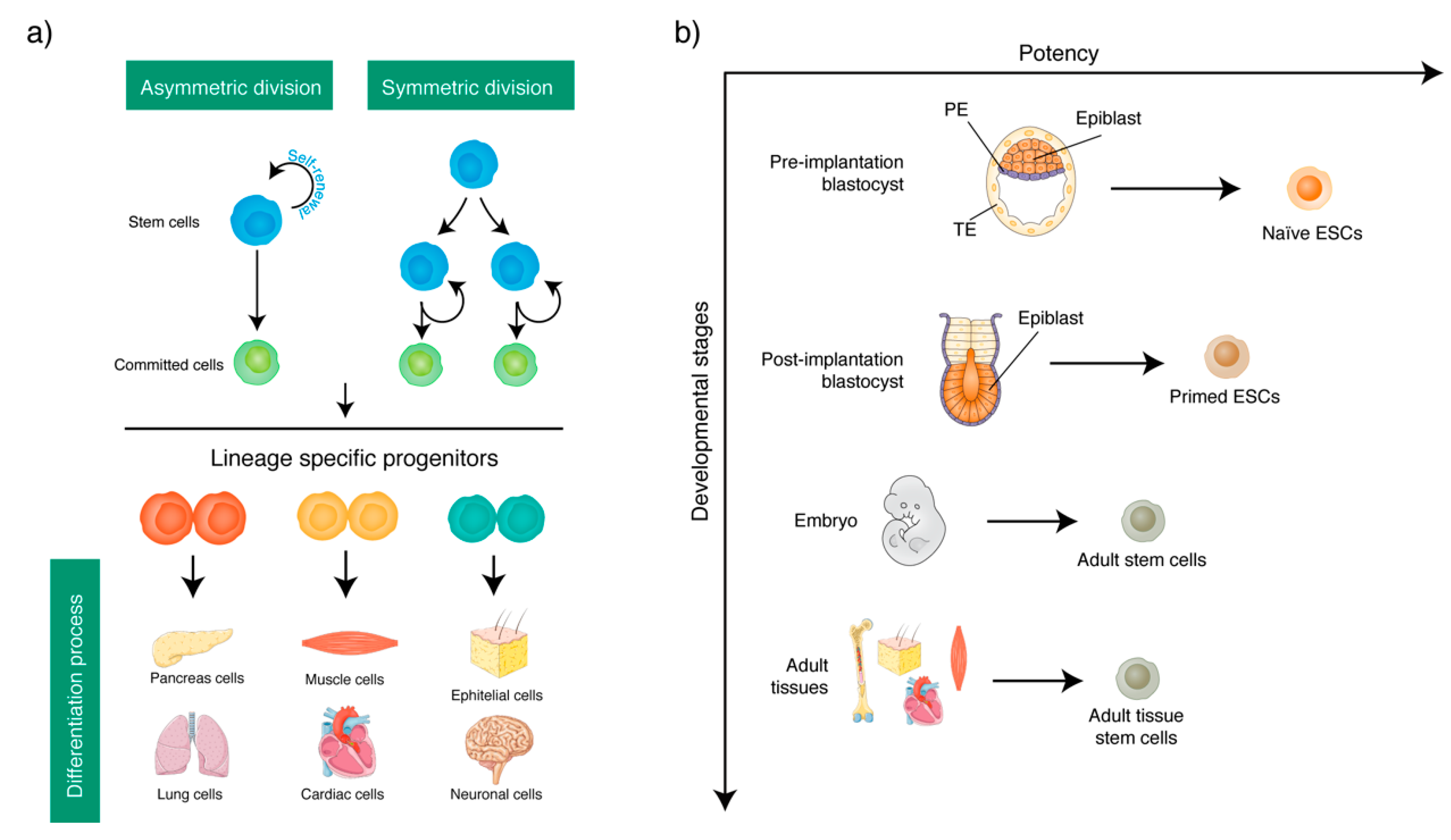
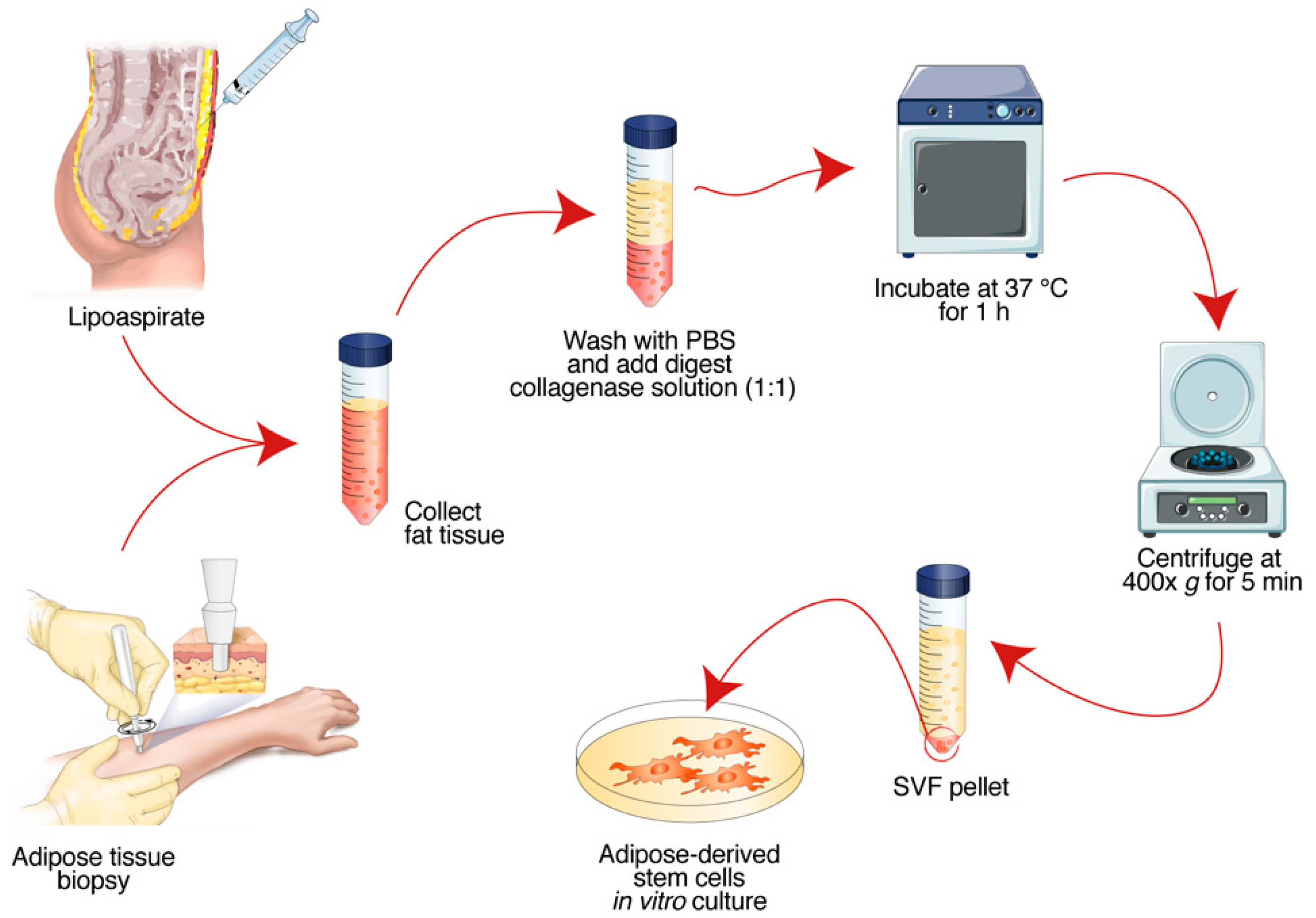
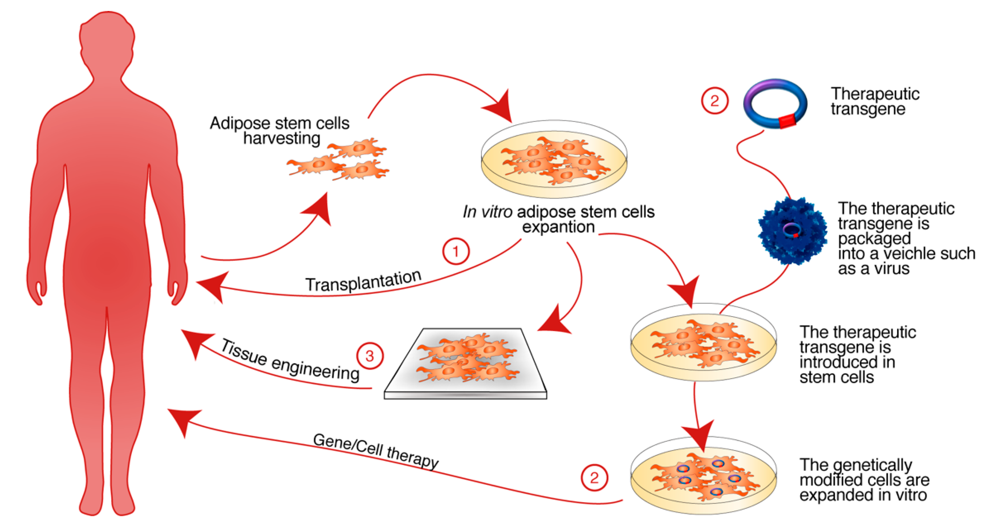
| Adult Stem Cell Types | Embryonic Origin | Living Tissue | Multipotency | References |
|---|---|---|---|---|
| Adipose Stem Cells | Mesoderm | Adipose tissue and lipoaspirate | The adipose stem cells have the ability to differentiate toward different cells of several tissues: fat, bone, cartilage, skeletal, smooth and cardiac muscle, endothelium, hematopoietic, and liver and neuronal. | [38,39,40,41] |
| Dental Stem Cells | Mesoderm | Dental pulp | Dental stem cells may differentiate toward mesenchymal (osteoblasts, adipocytes, chondrocytes and myocytes) and non-mesenchymal (neuronal and endothelial cells, hepatocytes and melanocytes) tissue cells. | [42] |
| Bone Marrow Mesenchymal Stem Cells | Mesoderm | Bone-marrow | These stem cells could be induced to differentiate to the lineages of the mesenchymal tissues, including bone, cartilage, fat, tendon, muscle and medullary stroma. Recently, the ability of mesenchymal bone marrow stem cells to differentiate into cardiac, neuronal lineages, and hepatocyte-like cells has also been demonstrated. | [43,44,45,46,47] |
| Hematopoietic Stem Cells | Mesoderm | Peripheral blood, bone marrow, and umbilical cord blood | Hemopoietic stem cells give rise to both the myeloid (monocytes, macrophages, neutrophils, basophils, eosinophils, erythrocytes, megakaryocytes platelets), and lymphoid (T cells, B cells, and natural killer) cells lineages of blood cells. | [48,49,50] |
| Epidermal Stem Cells | Ectoderm | Epidermis and hair follicles | Epidermal stem cells can differentiate toward cells of at least three specialized structures: epidermis, hair follicles, and sebaceous glands. | [51,52,53] |
| Neural Stem Cells | Ectoderm | Adult brain and spinal cord | Neural stem cells give rise to differentiated neurons, astrocytes, and oligodendrocytes. | [54,55,56] |
| Endothelial Stem Cells | Endoderm | Endothelial intima of blood vessels, peripheral blood, bone marrow, and umbilical cord blood | Endothelial stem cells are implicated in functional blood vessels and lymphatic vascular systems formation. | [57,58] |
| Umbilical Cord Mesenchymal Stem Cells | Endoderm | Umbilical cord blood, umbilical vein subendothelium, and Wharton’s jelly | These stem cells may be differentiated in osteoblasts, chondrocytes, adipocytes, skeletal muscle cells, endothelial cells, hepatocytes, cardiomyocytes-like cells, and neurons. | [59,60,61] |
| Joint/Synovium-Derived Mesenchymal Stem Cells | Mesoderm | adult skeletal tissues synovium and bone marrow | These stem cells may be differentiated in chondrocytes, adipocytes, osteoblasts. | [62,63] |
| NCT Number | Title | Status | Conditions | Interventions | Sponsor/Collaborators | Phases |
|---|---|---|---|---|---|---|
| NCT03608579 | Autologous Culture Expanded Adipose Derived MSCs for Treatment of Painful Hip OA | Recruiting | Osteoarthritis, Hip | Drug: Autologous Adipose Derived Mesenchymal Stromal Cells | Mayo Clinic | Phase I |
| NCT03570450 | Regenerative Stem Cell Therapy for Stroke in Europe | Recruiting | Stroke | Drug: Adipose derived Stem Cell|Drug: placebo | University Hospital, Grenoble|European Commission H2020 program | Phase I |
| NCT03308565 | Adipose Stem Cells for Traumatic Spinal Cord Injury | Recruiting | Spinal Cord Injuries|Paralysis | Biological: Autologous, Adipose derived Mesenchymal Stem Cells | Allan Dietz|Mayo Clinic | Phase I |
| NCT03279081 | Adult Allogeneic Expanded Adipose-derived Stem Cells (eASC) for the Treatment of Complex Perianal Fistula(s) in Patients with Crohn’s Disease | Recruiting | Crohn’s Disease | Drug: Cx601|Other: Placebo | TiGenix S.A.U.|Cellerix | Phase III |
| NCT03268603 | Intrathecal Autologous Adipose-derived Mesenchymal Stromal Cells for Amyotrophic Lateral Sclerosis (ALS) | Recruiting | ALS|Amyotrophic Lateral Sclerosis | Drug: Autologous Adipose-derived Mesenchymal Stromal Cells | Mayo Clinic|State of Minnesota Regenerative Medicine Minnesota | Phase II |
| NCT03171194 | Pilot Trial of Mesenchymal Stem Cells for Systemic Lupus Erythematosus | Active, not recruiting | System; Lupus Erythematosus | Drug: Low Dose Mesenchymal Stem Cells (MSCs) | Medical University of South Carolina | Phase I |
| NCT03092284 | Allogeneic Stem Cell Therapy in Heart Failure | Recruiting | Heart Failure | Biological: Cardiology Stem Cell Centre Adipose Stem Cell (CSCC_ASC)|Biological: Placebo | JKastrup|Rigshospitalet, Denmark | Phase II |
| NCT02952131 | Use of Autologous, Adult Adipose-Derived Stem/Stromal Cells in Inflammatory Bowel Disease | Recruiting | Inflammatory Bowel Diseases | Procedure: Lipoaspiration|Procedure: AD-cSVF|Procedure: Normal Saline IV | Healeon Medical Inc|Terry, Glenn C., M.D. | Phase I|Phase II |
| NCT02904824 | Injection Laryngoplasty Using Autologous Fat Enriched with Adipose Derived Regenerative Stem Cells (ADRC) | Completed | Vocal Cord Paralysis, Unilateral | Biological: adipose derived regenerative cells|Biological: centrifuged autologous fat | Hospital General Universitario Gregorio Marañon | Phase I|Phase II |
| NCT02808208 | Autologous Adipose Derived Mesenchymal Stem Cells (AMSC) in Reducing Hemodialysis Arteriovenous Fistula Failure | Recruiting | End Stage Renal Disease (ESRD)|Vascular Access Complication | Biological: Adipose Derived Mesenchymal Stem Cells (AMSC) | Mayo Clinic | Phase I |
| NCT02741362 | Safety and Efficacy of Adipose Derived Stem Cells in Refractory Rheumatoid Arthritis, Systemic Lupus Erythematosus or Sharp’s Syndrome | Terminated | Systemic Lupus Erythematosus|Rheumatoid Arthritis|Sharp’s Syndrome | Other: Intravenous injection of Stromal Vascular Fraction Cells (SVF) containing ADSCs|Other: Lipoaspiration | Arkansas Heart Hospital | Phase I |
| NCT02387723 | CSCC_ASC Therapy in Patients with Severe Heart Failure | Completed | Heart Failure | Biological: Allogeneic adipose derived stem cells (CSCC_ASC) | JKastrup|Rigshospitalet, Denmark | Phase I |
| NCT02287974 | Clinical Trial I/II Opened, Randomized and Controlled for the Study of the Use of Stem Cells Therapy in Insulinized Diabetic Patients Type 2 With Critical Ischemia in Lower Limbs (CLI): Study of the Needs of Insulin | Completed | Critical Limb Ischemia (CLI) | Drug: Stem cell infusion | Andalusian Initiative for Advanced Therapies—Fundación Pública Andaluza Progreso y Salud|Iniciativa Andaluza en Terapias Avanzadas | Phase I|Phase II |
| NCT02208713 | Intramuscular Transplantation of Muscle Derived Stem Cell and Adipose Derived Mesenchymal Stem Cells in Patients with Facioscapulohumeral Dystrophy (FSHD) | Recruiting | Dystrophy | Biological: Intramuscular injection | Royan Institute | Phase I |
| NCT02161744 | Safety, Tolerability and Preliminary Efficacy of Adipose Derive Stem Cells for Patients With COPD | Active, not recruiting | Chronic Obstructive Pulmonary Disease | Biological: ADSCs administration | Arkansas Heart Hospital | Phase I |
| NCT02068794 | MV-NIS Infected Mesenchymal Stem Cells in Treating Patients with Recurrent Ovarian Cancer | Recruiting | Malignant Ovarian Brenner Tumor | Ovarian Serous Adenocarcinoma|Ovarian Transitional Cell Carcinoma| |Undifferentiated Ovarian Carcinoma | Other: Laboratory Biomarker Analysis|Procedure: Mesenchymal Stem Cell Transplantation|Biological: Oncolytic Measles Virus Encoding Thyroidal Sodium Iodide Symporter | Mayo Clinic|National Cancer Institute (NCI) | Phase I|Phase II |
| NCT02035085 | 19F Hot Spot MRI of Human Adipose-derived Stem Cells for Breast Reconstruction | Recruiting | Breast Cancer | Drug: CS-1000 labeled SVF cells | Johns Hopkins University|Cosmeticsurg.net | Phase I |
| NCT01828723 | Safety Study of Antria Cell Preparation Process to Enhance Facial Fat Grafting with Adipose Derived Stem Cells | Completed | Lipoatrophy|Aging|Wrinkles | Biological: SVF | Antria | Phase I |
| NCT01678534 | Reparative Therapy in Acute Ischemic Stroke with Allogenic Mesenchymal Stem Cells from Adipose Tissue, Safety Assessment, a Randomised, Double Blind Placebo Controlled Single Center Pilot Clinical Trial | Completed | Ischemic Stroke | Drug: Allogenic mesenchymal stem cells from adipose tissue|Drug: Placebo | Instituto de Investigación Hospital Universitario La Paz | Phase II |
| NCT01649687 | Treatment of Cerebellar Ataxia with Mesenchymal Stem Cells | Completed | Cerebellar Ataxia | Biological: Allogeneic adult adipose-derived mesenchymal stem cells | National Yang Ming University | Phase I|Phase II |
| NCT01585857 | ADIPOA—Clinical Study | Completed | Osteoarthritis | Biological: Autologous adipose derived stem cells administrated for intra-articular use | University Hospital, Montpellier | Phase I |
| NCT01532076 | Effectiveness of Adipose Tissue Derived Mesenchymal Stem Cells as Osteogenic Component in Composite Grafts | Terminated | Osteoporotic Fractures | Procedure: Cellularized composite graft augmentation|Procedure: Acellular composite graft augmentation | University Hospital, Basel, Switzerland | Phase II |
| NCT01257776 | Human Adipose Derived Mesenchymal Stem Cells for Critical Limb Ischemia (CLI) in Diabetic Patients | Completed | Critical Limb Ischemia (CLI)|Diabetes | Drug: Autologous adipose derived mesenchymal stem cells | Andalusian Initiative for Advanced Therapies—Fundación Pública Andaluza Progreso y Salud|Iniciativa Andaluza en Terapias Avanzadas | Phase I|Phase II |
| NCT01222039 | Multicenter Clinical Trial for the Evaluation of Mesenchymal Stem Cells from Adipose Tissue in Patients with Chronic Graft Versus Host Disease | Completed | Graft Versus Host Disease|Chronic and Expanded Graft Versus Host Disease|Immune System Diseases | Other: Conventional treatment plus intravenous infusion of allogenic mesenchymal stem cells from adipose tissue | Andalusian Initiative for Advanced Therapies—Fundación Pública Andaluza Progreso y Salud|Iniciativa Andaluza en Terapias Avanzadas | Phase I|Phase II |
| NCT01157650 | Treatment of Fistulous Crohn’s Disease by Implant of Autologous Mesenchymal Stem Cells Derived from Adipose Tissue | Completed | Crohn Disease | Other: Autologous mesenchymal stem cells | Clinica Universidad de Navarra, Universidad de Navarra | Phase I|Phase II |
| NCT01056471 | Autologous Mesenchymal Stem Cells from Adipose Tissue in Patients with Secondary Progressive Multiple Sclerosis | Completed | Demyelinating Autoimmune Diseases, CNS|Autoimmune Diseases of the Nervous System | Other: Autologous mesenchymal stem cells from adipose tissue | Andalusian Initiative for Advanced Therapies—Fundación Pública Andaluza Progreso y Salud|Carlos III Health Institute | Phase I|Phase II |
| NCT00913289 | Liver Regeneration Therapy Using Autologous Adipose Tissue Derived Stromal Cells | Terminated | Liver Cirrhosis | Biological: adipose tissue derived stromal cells | Kanazawa University | Phase I |
| NCT00442806 | Randomized Clinical Trial of Adipose-Derived Stem Cells in the Treatment of Pts With ST-elevation Myocardial Infarction | Completed | Myocardial Infarction|Coronary Arteriosclerosis|Cardiovascular Disease|Coronary Disease | Drug: Injection of ADRC’s|Other: Injection of Placebo | Cytori Therapeutics | Phase I |
| Biomaterial | Description | References |
|---|---|---|
| Natural Polymers | Collagen, glycosaminoglycans, chitin, and chitosan have been used to repair a large number of defects on various organs such as nerves, skin, cartilage, and bones. | [214,215,216,217,218] |
| Hyaluronic acid, used also as a gel, is widely used in the regeneration of soft tissues. | [219,220] | |
| Synthetic Polymers | Such as: polyphenylene esters, polyanhydrides, and polyortoesters. This class of polymers includes polyglycolic (PGA), polylactic acid (PLA), and polycaprolactone (PCL) with different degradation times. | |
| Poly-l-lactide (PLLA) produced by the polymerization of l, l-lactide is a biodegradable and bioactive thermoplastic aliphatic polyester derived from renewable resources such as corn starch (in the United States and Canada), cassava, fried potatoes or starch (especially in Asia), or sugar cane (in the rest of the world). PLLA has been employed for a wide range of tissue engineering purposes, such as bone, cartilage, tendon, neural, and vascular regeneration. | [221,222,223,224,225] | |
| PCL is synthesized by ring-opening polymerization of the cyclic monomer ε-caprolactone. It has been used since the 70s as a long-term resorbable sutures and implants. | [209,226] | |
| Nanocomposite Biomaterials | By reinforcing the matrix with particles of nanometric dimensions, a clear improvement can be achieved in many physical and chemical properties of the scaffold, with a very low charge content, or even through the attribution of absent characteristics in the polymer as the thermal or electrical conductivity. | |
| NanoHydroxyapatite (NHAP). Hydroxyapatite is the main part of the bone. Can be natural or synthetic. It is mainly used in bone regeneration. | [198,227] | |
| Single walled carbon nanotubes (SWCNTs) have diameter typically around 1 nm and length until some micrometer and are tubes made of a single sheet of graphene. | [228,229] | |
| Multi-walled carbon nanotubes (MWCNTs) are made from more graphene sheets, with diameter in the range of 10–20 nm. | [197,230] | |
| Graphene, Graphene Oxide. Graphene is a single layer of sp2-bonded carbon atoms in a hexagonal lattice. It is one of the most popular nanomaterials due to its excellent physical, electrical, and thermal properties. It is the strongest material ever measured. It has a Young’s modulus of 1 TPa, fracture toughness of 130 GPa, thermal conductivity of ≈103 W·m−1·K−1, and electrical conductivity of ≈102 S·cm−1. Graphene oxide is the oxidized form of graphene with hydroxyls, epoxides, diols, ketones, and carboxyl functional groups. The presence of oxygen on the edges and basal planes of graphene oxide increases its hydrophilicity. | [231,232,233] | |
| Gold Nanoparticles (Au NPs). Nanoparticles of noble metals as gold have nanometer diameter and they show a very intense color, which is absent in the bulk material, due to the collective oscillation of the free conduction electrons. | [234] | |
| Surface Modifications | Plasma processing. Surface modification techniques are mainly applied in order to modify the first part of the scaffold that come into contact with the biological entities. | [235,236] |
| Application | Biomaterials | Biological Effect | References |
|---|---|---|---|
| Adipose Tissue | Semicircular microfluidic channel | Quantification of responses and changes of stem cells and tumor cells to cutting streams at the interstitial level. | [237] |
| Free hydrogel system based on a tissue-specific extracellular matrix | This application is a promising cell-free therapeutic approach for in situ adipose tissue regeneration. | [240] | |
| Hydrogel crosslinked by thiolated heparin and methacrylated hyaluronic acid | Induction and differentiation of ASCs towards the adipogenic lineage. | [241] | |
| Chitosan/β-glycerophosphate/collagen hybrid hydrogel | Generation of a larger number of adipocytes and vascularized adipose tissues. | [217] | |
| Biomimetic poly(ethylene)-glycol hydrogel with embedded peptides | It provides niches for stem cell differentiation and for soft tissue regeneration. | [242] | |
| Modified silica nanomaterials | Different functionalized silica nanoparticles surfaces generate different responses in ASC cultures. | [212] | |
| Large three-dimensional poly(glycerol sebacate)/poly(l-lactic acid) scaffolds | Adipose tissue engineering. | [243] | |
| Extracellular matrix from adipose tissue in electrospinning scaffold of polydioxanone | Adipose stem cell culture. | [244] | |
| Bone Tissue | Thermo-gelling hydrogel scaffold containing platelet rich plasma and biphasic calcium phosphate | New bone formation at the site of the calvular bone defect in rabbits. | [245] |
| Electrospun polyethersulfone/poly(vinyl) alcohol/platelet rich plasma nanofibrous scaffolds | Osteogenic differentiation for bone tissue engineering. | [246] | |
| 3-D scaffolds with BMP-2 loaded core-shell fibers | Bone tissue engineering. | [247] | |
| Methacryloyl gelatin-based hydrogels | Interplay between osteogenesis and angiogenesis in vitro in bone tissue engineering application. | [248] | |
| The collagen fibroin-ELR (elastin-like recombinamer) blend | Improvement of the mechanical tensile properties of engineered scaffolds to promote bone differentiation. | [249] | |
| The heterogeneous deproteinized bone | Repair segmental bone defects and have a good potential to be used as graft material. | [250] | |
| Collagen containing resveratrol scaffolds | Provide useful biological signals that then stimulate the regeneration of the craniofacial tissue. | [251] | |
| 3-D-graphene/arginine-glycine-aspartic acid peptide nano-island composite | Promote differentiation of ASCs to osteoblasts. | [252] | |
| Silk fibroin/chitosan thin film | Tissue engineering of bone, cartilage, adipose, and skin. | [253] | |
| The aligned-(NanoAligned™) and random-(NanoECM™) oriented PCL nanofiber-coated plates | PCL nanofiber is a suitable regenerative medicine application for canine patients in vivo. | [254] | |
| A composite hydrogel of collagen and supramolecular scaffold | Bone tissue engineering applications. | [255] | |
| PCL scaffolds and osteogenic differentiation medium | Anatomical and functional reconstruction of temporal bone defects following mastoidectomy. | [256] | |
| Different porosities of chitosan scaffolds | Osteogenic differentiation. | [257] | |
| Beta-tricalcium phosphate granules and supporting mesh | Cranial repair. | [258] | |
| Polypyrrole/chitosan scaffold with electrical stimulation | Bone defect therapy. | [259] | |
| Electrospun silk fibroin nanofibrous scaffolds with two-stage hydroxyapatite functionalization | Bone tissue engineering. | [260] | |
| The metal ion (Zn, Ag, and Cu) doped hydroxyapatite nano-coated surfaces | Osteogenic differentiation and cell adhesion capacity are higher on nanocoated surfaces that include Zn, Ag, and/or Cu metal ions. | [261] | |
| Polyethylenimine-mediated BMP-2 gene transfection in vitro | BMP-2 gene delivery and induction of osteogenic differentiation. | [262] | |
| Collagen sponges | Endochondral ossification. | [263] | |
| Methacrylated gellan-gum alone and combined with collagen type I hydrogels | Both hydrogel formulations induced ASCs towards osteogenic differentiation. | [264] | |
| Poly(l-lactic acid)/gelatin fibrous scaffold loaded with Simvastatin/β-Cyclodextrin-Modified Hydroxyapatite Inclusion Complex | Potential application in bone tissue engineering. | [265] | |
| Poly(dopamine) coating of 3-D printed poly(lactic acid) scaffolds | Promotes the osteogenic differentiation of ASCs. | [266] | |
| Peripheral Nerve/Neural Cells | Graphene oxide and reduced graphene oxide mats | Neural differentiation of the ASCs and improvement of nerve repair. | [267] |
| Cell transplantation of ASCs differentiated in to Schwann cell-like cells (SCLCs) in vitro | Repair sciatic nerve defects in rats and in general the peripheral nerve injury. | [268] | |
| Poly (ε-caprolactone) (PCL) and PCL/gelatin nanofibrous scaffolds coated with platelet-rich plasma (PRP) | Application in nerve tissue engineering. | [269] | |
| Polystyrene surface containing nanopore array-patterned substrate (NP) | NP lead to greater adhesion of ASCs on the substrate, growth of filopods, elongation of nuclei, and expression of specific neural markers compared to flat substrates. | [270] | |
| PDMS/MWNT sheets as a scaffold | Regeneration of peripheral nerves, similar to Schwann cells. | [271] | |
| Cardiomyocytes | Decellularized bovine myocardial extracellular matrix-based films (dMEbF) | dMEbF mimics native ECM, but also induces cardiomyocyte-like cells differentiation. | [272] |
| Cardiac extracellular matrix (cECM) hydrogel | Cardiac tissue engineering applications. | [273] | |
| Collagen coated polyacrylamide hydrogel substrates | Therapeutic use of ASCs in cardiac fibrosis therapy in future. | [274] | |
| 3-D cell masses and self-assembling peptides | Promising application for therapeutic angiogenesis to treat myocardial infarction. | [275] | |
| Endothelial | Poly(ε-caprolactone)/chitosan scaffold | Bladder tissue engineering. | [276] |
| Polycaprolactone/gelatin nanofibrous scaffolds | Promote the generation of robust and functional microvasal structures that could be valuable for regeneration of blood vessels. | [277] | |
| PEG hydrogel containing calcium-releasing particles | Vascular stabilization and revascularization of ischemic tissues. | [278] | |
| Elastin-like recombinamer-based hydrogel | Improve the successful integration of engineered substitutes in angiogenic and inflammation process. | [279] | |
| Surface-modified bioresorbable electrospun scaffolds from blends of poly(l-lactic acid) and segmented polyurethane | Vascular tissue engineering due to its biomimetic behaviors and its ability to avoid thrombus formation and provide antimicrobial characteristics. | [280] | |
| Hydrogel from gelatin-tyramine and chitosan-4-hydroxylphenyl acetamide | Vehicle for delivering signals to cells and growth factors by supporting the vascularization process in tissue engineering applications. | [281] | |
| Polyurethane urea (PEUU) | Engineered vascular grafts. | [282] | |
| Polycaprolactone (PCL)-gelatin mesh | Smooth muscle cells and endothelial cells for the bioengineering of small-diameter blood vessels. | [283] | |
| Tendon | Magnetic cell sheet | Tendon therapies. | [284] |
| Poly(l/d)lactide 96l/4d copolymer filament scaffolds in tenogenic medium compared to foamed poly(l-lactide-co-ε-caprolactone) 70L/30CL scaffolds | Tendon injury treatments. | [285] | |
| Cartilaginous | Hyaluronan (HA) | Regeneration of hASC-mediated cartilage in chondral defects and cartilage joint. | [286] |
| Cartilage extracellular matrix-derived particles | Alternative method for the culture of chondrocytes and stem cell differentiation; promising strategy for the construction of cartilage microtexes and repair of articular cartilage in vivo. | [287] | |
| Hyaluronic acid (HA)-modified thermoresponsive poly(N-isopropylacrylamide) hydrogels | Chondrogenesis and repair of the articular cartilage. | [288] | |
| 3-d-photocrosslinkable methacrylated gelatin hydrogels | The hydrogel pre-loaded with TGF-β3 allows improvement of healing of the radial meniscus in an in vitro meniscal repair model. | [289] | |
| Alginate microbeads | Regeneration of auricular cartilage. | [290] | |
| Herbal extracts icariin and transforming growth factor β3 (TGFβ3) were added in fibrin-cell constructions | Chondrogenesis and together with TGF3 it could decrease its hypertrophic effects. | [291] | |
| Gelatin/chondroitin-6-sulfate/hyaluronan/chitosan highly elastic cryogels | Cartilage engineering. | [292] | |
| Polycaprolatone and with freeze thaw freeze scaffolds | Transition mechanism from chondroblast to chondrocyte. | [293] | |
| Polyvinyl alcohol/chitosan scaffold | Regeneration of the tear meniscus lesion. | [294] | |
| Composite gel based on collagen/hyaluronic acid | Chondrogenic differentiation in a dose-dependent manner. | [295] | |
| Poly-ε-caprolactone films | Implantation at sites of defects as a possible treatment of cartilage defects. | [296] | |
| Other Tissues | Collagen hydrogel | Regeneration of muscle tissue. | [297] |
| Polypyrrole-coated polymer scaffolds and electrical stimulation | Vascular smooth muscle regeneration. | [298] | |
| Poly(l-lactide)/Poly(e-caprolactone) electrospinning nano-scaffold | Myogenic proliferation and differentiation of ASCs. | [299] | |
| Acellular human amniotic membrane (HAM) | Application of cell-based skin substitutes. | [300] | |
| β-tricalcium phosphate (betaTCP) | Reconstruction of cranial defects. | [258] | |
| Injectable hydrogels | Immunomodulatory effects, angiogenic stimulation angiogenic. | [301] | |
| Polyethylene glycol-fibrin hydrogels | Improve wound healing and minimize donor or scar sites. | [302] | |
| Autologous growth factors and nanofibrous scaffolds | Pancreatic tissue engineering applications and beta cell replacement therapies in type 1 diabetes mellitus. | [303] |
© 2018 by the authors. Licensee MDPI, Basel, Switzerland. This article is an open access article distributed under the terms and conditions of the Creative Commons Attribution (CC BY) license (http://creativecommons.org/licenses/by/4.0/).
Share and Cite
Argentati, C.; Morena, F.; Bazzucchi, M.; Armentano, I.; Emiliani, C.; Martino, S. Adipose Stem Cell Translational Applications: From Bench-to-Bedside. Int. J. Mol. Sci. 2018, 19, 3475. https://0-doi-org.brum.beds.ac.uk/10.3390/ijms19113475
Argentati C, Morena F, Bazzucchi M, Armentano I, Emiliani C, Martino S. Adipose Stem Cell Translational Applications: From Bench-to-Bedside. International Journal of Molecular Sciences. 2018; 19(11):3475. https://0-doi-org.brum.beds.ac.uk/10.3390/ijms19113475
Chicago/Turabian StyleArgentati, Chiara, Francesco Morena, Martina Bazzucchi, Ilaria Armentano, Carla Emiliani, and Sabata Martino. 2018. "Adipose Stem Cell Translational Applications: From Bench-to-Bedside" International Journal of Molecular Sciences 19, no. 11: 3475. https://0-doi-org.brum.beds.ac.uk/10.3390/ijms19113475