Article

Respiratory Bordetella bronchiseptica Carriage is Associated with Broad Phenotypic Alterations of Peripheral CD4+CD25+ T Cells and Differentially Affects Immune Responses to Secondary Non-Infectious and Infectious Stimuli in Mice

by
Andreas Jeron
1,2,†,
Julia D. Boehme
1,2,†,
Julia Volckmar
1,2,
Marcus Gereke
1,2,
Tetyana Yevsa
3,§,
Robert Geffers
4,
Carlos A. Guzmán
3,
Jens Schreiber
5,
Sabine Stegemann-Koniszewski
1,2,5,‡ and
Dunja Bruder
1,2,*,‡
1
Infection Immunology Group, Institute of Medical Microbiology, Infection Control and Prevention, Health Campus Immunology, Infectiology and Inflammation, Otto-von-Guericke University Magdeburg, 39120 Magdeburg, Germany
2
Immune Regulation Group, Helmholtz Centre for Infection Research, 38124 Braunschweig, Germany
3
Department of Vaccinology and Applied Microbiology, Helmholtz Centre for Infection Research, 38124 Braunschweig, Germany
4
Genome Analytics Group, Helmholtz Centre for Infection Research, 38124 Braunschweig, Germany
5
Experimental Pneumology, University Hospital for Pneumology, Health Campus Immunology, Infectiology and Inflammation, Otto-von-Guericke University Magdeburg, 39120 Magdeburg, Germany
*
Author to whom correspondence should be addressed.
These authors contributed equally.
§
Current address Department of Gastroenterology, Hepatology and Endocrinology, Hannover Medical School, Hannover, Germany
Int. J. Mol. Sci. 2018, 19(9), 2602; https://0-doi-org.brum.beds.ac.uk/10.3390/ijms19092602
Submission received: 30 May 2018 / Revised: 3 August 2018 / Accepted: 28 August 2018 / Published: 1 September 2018
(This article belongs to the Special Issue Host-Microbe Interaction 2018)

Abstract

:
The respiratory tract is constantly exposed to the environment and displays a favorable niche for colonizing microorganisms. However, the effects of respiratory bacterial carriage on the immune system and its implications for secondary responses remain largely unclear. We have employed respiratory carriage with Bordetella bronchiseptica as the underlying model to comprehensively address effects on subsequent immune responses. Carriage was associated with the stimulation of Bordetella-specific CD4+, CD8+, and CD4+CD25+Foxp3+ T cell responses, and broad transcriptional activation was observed in CD4+CD25+ T cells. Importantly, transfer of leukocytes from carriers to acutely B. bronchiseptica infected mice, resulted in a significantly increased bacterial burden in the recipient’s upper respiratory tract. In contrast, we found that respiratory B. bronchiseptica carriage resulted in a significant benefit for the host in systemic infection with Listeria monocytogenes. Adaptive responses to vaccination and influenza A virus infection, were unaffected by B. bronchiseptica carriage. These data showed that there were significant immune modulatory processes triggered by B. bronchiseptica carriage, that differentially affect subsequent immune responses. Therefore, our results demonstrated the complexity of immune regulation induced by respiratory bacterial carriage, which can be beneficial or detrimental to the host, depending on the pathogen and the considered compartment.

1. Introduction

The lung as the central site of gas exchange is, together with the conducting airways, constantly exposed to all kinds of environmental factors and thereby confronted with potential hazards, such as pollutants and pathogenic microorganisms. While it was long believed to represent a sterile compartment, research of the last years has delivered evidence that the lower respiratory tract displays a favorable niche for commensal microorganisms [1,2]. The lungs central role in oxygen supply, its delicate structure, its constant exposure to harmful as well as harmless microorganisms, and its potential to serve as an entry site for invading pathogens, altogether demand for an effective and at the same time tightly regulated immune system.
Immunity in the lung is conferred through the combined action of different types of sentinel cells, such as airway and alveolar epithelial cells, alveolar macrophages (AM), and dendritic cells (DC), which surveil the respiratory tract and in the case of pathogen encounter induce well-orchestrated innate and adaptive responses, involving multiple cell-types and mediators [3,4,5,6]. To protect from overshooting and harmful responses, sophisticated regulatory mechanisms are in place. These can create an immune regulatory environment and can suppress innate and adaptive immune cell function through several mechanisms, such as the production of anti-inflammatory mediators or direct cell-to-cell interactions [3,4,7,8]. However, next to protecting the lung from the detrimental effects of host immunity, these regulatory mechanisms can in turn favor the persistence of colonizing microorganisms and frequently display targets of microbial evasion strategies [9,10,11].
Given its function, as well as its anatomic exposition and resulting immunological features, the respiratory tract harbors an exceptional challenge regarding the regulation of immune responses towards multiple antigens simultaneously present. These can be polymicrobial interactions, such as co-infections with different respiratory pathogens, the co-occurrence of commensal and pathogenic bacteria, or interactions between respiratory infections and non-infectious stimuli, such as inhaled allergens [12,13,14]. At the same time, there is an increasing awareness of immune regulatory effects across compartments to remote sites, which to date are most appreciated for the gut microbiome [13,15,16]. Nevertheless, there is also evidence for inflammatory processes in the respiratory tract to influence systemic immunity [13,17,18,19]. While we are only beginning to understand these multi-faceted interactions, the underlying mechanisms to date remain mostly elusive.
Bordetella bronchiseptica is a gram-negative respiratory pathogen of several mammalian species, which is causing a number of veterinary respiratory syndromes in domestic as well as agriculturally important and food-producing animals, whilst humans are only rarely infected [20,21]. B. bronchiseptica is genetically closely related to the major human pathogen Bordetella pertussis, the cause of whooping cough. Both B. bronchiseptica and B. pertussis regulate the expression of several virulence factors, including toxins and a type III secretion system (T3SS), by a two-component system that controls phenotypic modulation [21,22]. Due to the lack of B. pertussis models in commonly used laboratory animal species and its close relatedness to B. bronchiseptica, the latter is frequently utilized as an animal model to explore mechanisms of B. pertussis pathogenesis (reviewed in [21]). Nevertheless, there are fundamental differences namely that B. bronchiseptica can persist in the respiratory tract of its hosts and is able to cause chronic infections [20,23], whereas in humans there is no evidence of prolonged B. pertussis carriage [24]. B. bronchiseptica interacts with the host immune system in multiple ways, affecting a variety of immune cells. Both intracellular survival [25,26], as well as clear cytotoxic effects on macrophages, have been described for B. bronchiseptica [27,28,29]. Importantly, B. bronchiseptica is able to modulate cytokine production by macrophages and DC through its T3SS, with clear consequences for subsequent T cell responses [30,31]. Furthermore, it has been described that B. bronchiseptica invades and survives in mouse DC in vitro [32], and that nasal persistence affects DC dynamics in the upper and lower respiratory tract [33]. In vivo, B. bronchiseptica induces a strong influx of neutrophils, monocytes/macrophages, and lymphocytes into the lungs [20]. Furthermore, the T3SS of B. bronchiseptica was shown to drive DC migration to the secondary lymphoid tissue, which was observed together with a strong induction of anti-inflammatory interleukin (IL) −10 and a down-regulation of the interferon (IFN)-γ response [34,35,36]. Based on these findings, the induction of an immune suppressive Treg response during B. bronchiseptica carriage, as a mechanism of immune evasion has been suggested, but to our knowledge this was not addressed in detail [35,36].
In our study, we aimed at assessing the regulatory potential of respiratory microbial carriage on secondary immune responses, both in the lung and the periphery. We took advantage of B. bronchiseptica’s natural potential to colonize the host [21] and chose it as a model for respiratory carriage. In this model, we have comprehensively elucidated its effects on local and peripheral immune responses to different secondary stimuli. For these secondary immunological challenges, we employed models of both respiratory and systemic infection with viral and bacterial pathogens, as well as vaccination with a model antigen. Furthermore, our study included the analysis of the peripheral CD4+ T cell pool on the functional and gene transcriptional level during acute B. bronchiseptica infection, as well as during subsequent carriage. We show for the first time that clear and broad changes in the peripheral T cell pool, including the induction of pathogen-specific Treg, are mediated by B. bronchiseptica carriage. Furthermore, our study reveals significant immune regulatory processes triggered by B. bronchiseptica carriage, which differentially affect secondary immune responses.

2. Results

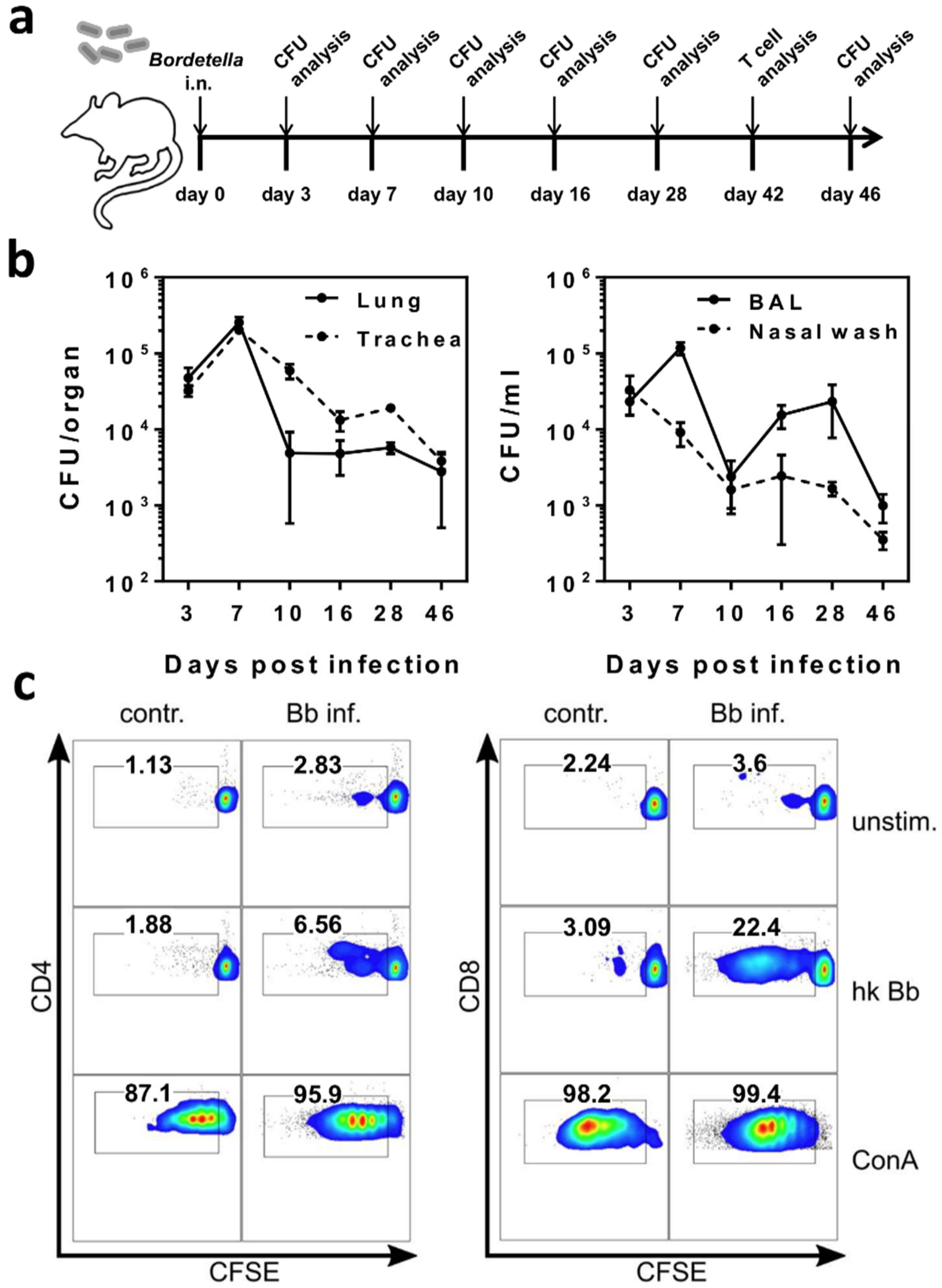
2.1. B. bronchiseptica Establishes Respiratory Tract Carriage Despite the Presence of Pathogen-Specific T Cells in Mice

We chose B. bronchiseptica as an experimental infection model to study the effects of chronic microbial carriage on immune regulatory mechanisms, and on subsequent immunological challenges in the lung and in the periphery. In our mouse model, B. bronchiseptica was still detectable in the lung, trachea, bronchoalveolar space, and nasal cavity, seven weeks following intranasal infection with 5 × 10⁵ CFU. In all compartments, peak bacterial loads were detected 7 days post infection, followed by respiratory carriage of B. bronchiseptica with reduced bacterial numbers from day 10 onwards (Figure 1a,b). B. bronchiseptica-specific lymphocyte proliferation was detectable in cells isolated from the cervical lymph nodes from day 16 post infection, and in splenocytes by day 46 post infection (data not shown). As our aim was to address the regulatory potential of respiratory microbial carriage, after acute infection and independent of the primary adaptive immune response, we chose late time points of 42 days or longer—post infection for our analyses. Importantly, due to the presence of Bordetella-specific CD4+, as well as CD8+ T cells in the periphery, a completed specific adaptive T cell response was confirmed during bacterial carriage (Figure 1c). Immune regulatory effects of B. bronchiseptica involving multiple cell types of the immune system have been described in References [25,26,27,28,29,30,31]. Therefore, we hypothesized that inefficient bacterial clearance from the respiratory tract was the potential consequence of Bordetella-induced alterations of host immunity, possibly associated with the induction of immunosuppressive cells. To experimentally address this hypothesis, we adoptively transferred B-cell depleted leukocytes from 6-week B. bronchiseptica infected mice into recipient mice, which had been infected with B. bronchiseptica one week before (Figure 2a). To assess potentially suppressive effects of the transferred cells on the clearance of B. bronchiseptica, we determined the bacterial load in the upper respiratory tract of the recipient mice three days after transfer. Control of B. bronchiseptica growth was significantly less efficient in the nasal cavity of mice that received leukocytes from carriers, as compared to those that received cells from uninfected donors (Figure 2b). In conclusion, these data showed that B. bronchiseptica was able to cause respiratory carriage, despite the presence of pathogen-specific T cells and that leukocytes transferred from B. bronchiseptica carriers interfered with pathogen clearance in acute infection.

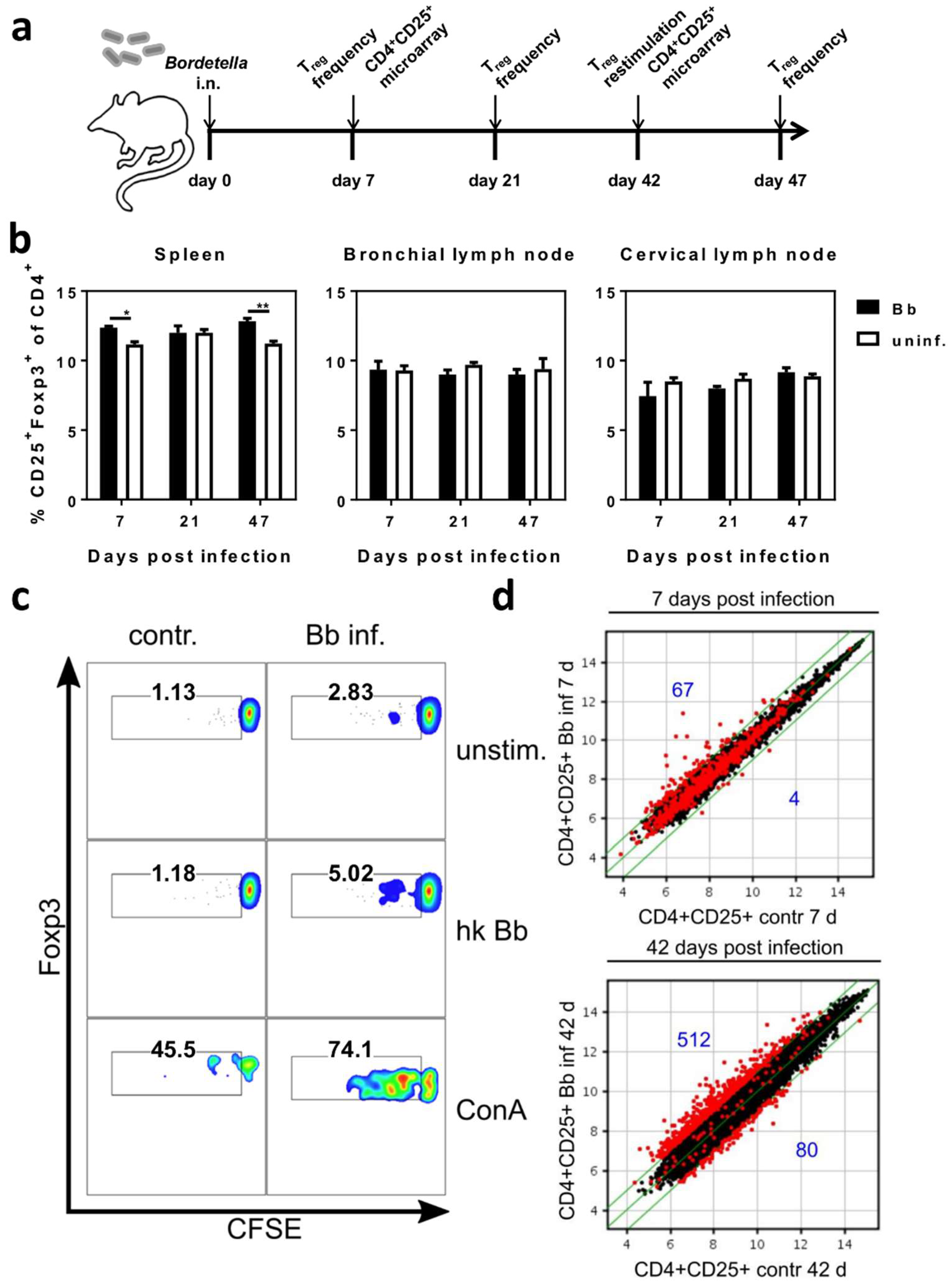
2.2. Respiratory B. bronchiseptica Carriage Is Associated with Broad Phenotypic Alterations of Peripheral CD4+CD25+ T Cells and the Induction of Pathogen-Specific Regulatory T Cells

The effect of the adoptive transfer of leukocytes from 6-week B. bronchiseptica infected mice to acutely infected mice, pointed towards the presence of immunosuppressive cells that impede the elimination of B. bronchiseptica from the respiratory tract. Furthermore, the induction of a Treg response by B. bronchiseptica has been implicated in past studies [35,36]. Therefore, we characterized the peripheral CD4+ T cell pool at different time points following B. bronchiseptica infection (Figure 3a). First, we investigated the frequency of CD4+CD25+Foxp3+ Treg in B. bronchiseptica infected mice, in comparison to control mice on days 7, 21, and 47, post infection (Figure 3b). Unexpectedly, Treg frequencies were not changed over the course of infection. Of note, this was consistently the case in the draining (bronchial and cervical) lymph nodes, as well as in the periphery (spleen). At the same time, comparing Treg frequencies between infected and control mice revealed a slight though significant reduction in the spleens, but not lymph nodes, of infected mice on day 7 and day 47. We furthermore analyzed the presence of B. bronchiseptica-specific CD4+CD25+Foxp3+ Treg in infected mice, six weeks following the infection. Indeed, and despite the reduced overall frequency of Treg in B. bronchiseptica carriers, in vitro stimulation of splenocytes isolated from B. bronchiseptica carriers with heat-killed bacteria revealed the expansion of Bordetella-specific CD4+CD25+Foxp3+ Treg within the polyclonal Treg pool (Figure 3c). These results clearly showed distinct Bordetella-induced alterations to peripheral Treg. Based on these findings, we aimed at further characterizing the peripheral CD4+CD25+ T cell pool, which also contained CD4+CD25+Foxp3+ Treg, during B. bronchiseptica carriage, in an unbiased approach. To this end, we performed transcriptional profiling of CD4+CD25+ T cells isolated from the spleens of B. bronchiseptica infected and control mice on day 7 post infection, when peak bacterial loads were detected (acute infection); and day 42 post infection, when B. bronchiseptica specific CD4+ T cells, as well as Treg, were systemically detectable (carriage) (Figure 3d). Of note, we chose the late time point of day 42 post infection to address changes in the CD4+CD25+ T cell compartment that were mediated by carriage of B. bronchiseptica, but were not linked to the T cell activation mounted during peak pathogenic load, and during the subsequent generation of the specific adaptive immune response. Strikingly, whilst changes were only subtle during acute infection, transcriptional profiling showed that respiratory B. bronchiseptica carriage induced dramatic alterations in the expression profile of the peripheral polyclonal CD4+CD25+ T cell pool. Our analyses revealed 512 genes to be up-regulated and 80 genes to be down-regulated at least 2-fold in splenic CD4+CD25+ T cells isolated on day 42 post B. bronchiseptica infection, when compared to uninfected controls. In contrast, only 67 transcripts were up- and 4 transcripts were down-regulated on day 7 post B. bronchiseptica infection. Of note, CD4+CD25 T cells that were analyzed in parallel only underwent minor changes at both time points (Supplementary Materials Figure S1a). Furthermore, the transcriptional changes observed in the CD4+CD25 T cell compartment were largely independent from those regulated in the CD4+CD25+ T cell pool (Figure S1b). Table 1 summarizes the top 30 genes most up- and down-regulated exclusively in CD4+CD25+ T cells, but not in CD4+CD25 T cells, isolated from B. bronchiseptica carriers, when compared to CD4+CD25+ T cells isolated from the respective uninfected controls. We detected transcriptional regulation of immunologically relevant proteins typically associated with the cell surface of T cells, such as CD38 (fold change 2.8) and Toll-like receptor (TLR) 4 (fold change 2.6). Additionally, IL-4 (fold change 2); the NFk-B inhibitor alpha (Nfkbia; fold change 2.1); and transcription factors such as the forkhead box P1 (Foxp1; fold change 2.8), interferon regulatory factor 2 (Irf2; fold change 4.7), and the RAR-related orphan receptor alpha (Rora; fold change 2.8) were up-regulated in CD4+CD25+ T cells during B. bronchiseptica carriage. Furthermore, the IL-2 receptor beta chain (Il2rb; fold change −2.2) and the transcription factor special AT-rich sequence binding protein 1 (Satb1; fold change −2.3) were down-regulated during carriage. To capture the overall significance of the broad transcriptional regulation in CD4+CD25+ T cells isolated from B. bronchiseptica carriers on a functional level, we performed gene ontology (GO)-term enrichment analysis on the list of transcripts regulated more than two-fold (Figure 4). Next to the immune-associated GO-terms “leukocyte differentiation” and “cytoplasmic pattern recognition receptor signaling pathway”, GO-terms of general cell biological processes, such as “transcription, DNA-templated” and “negative regulation of RNA metabolic processes” were significantly enriched in the transcripts regulated in CD4+CD25+ T cells isolated from B. bronchiseptica carriers. Nevertheless, the extensive regulation that exclusively occurred during B. bronchiseptica carriage, but not acute infection, suggested immune regulatory mechanisms rather than mere CD4+ T cell activation as a driving force. In line with this, typically Treg-associated transcripts such as neuropilin 1 (Nrp1; fold change 2.1) [37], were found to be transcriptionally up-regulated in the CD4+CD25+ T cell pool isolated from B. bronchiseptica carriers, as compared to uninfected controls. Importantly, comparing gene expression levels between CD4+CD25+ and CD4+CD25 T cells revealed a clear Treg signature for the CD4+CD25+ T cell pool, independent of B. bronchiseptica infection or carriage (Table 2). This Treg signature was marked by transcriptional up-regulation of established Treg-associated factors, such as Gpr83 [38,39], CTLA4 [40], CD103 (Itgae) [41], the Zinc-finger protein Eos (Ikzf4) [42] and Foxp3 [43], all of which were among the 30 most intensely up-regulated transcripts, when comparing CD4+CD25+ and CD4+CD25 T cells. Importantly, up-regulated expression of these Treg-associated factors also held true for CD4+CD25+ T cells isolated from B. bronchiseptica carriers.
Taken together, these analyses show that B. bronchiseptica carriage in the respiratory tract was associated with the expansion of Bordetella-specific CD4+CD25+Foxp3+ Treg, as well as with broad alterations in the transcriptional profile of the overall peripheral CD4+CD25+ T cell pool, which also pointed to the induction of Treg. In turn, these modulations to the peripheral Treg pool displayed one possible mechanism by which leukocyte transfer from B. bronchiseptica carriers to acutely infected mice, hindered bacterial clearance in the recipient’s upper respiratory tract. Importantly, immune modulation through B. bronchiseptica carriage could also affect the responses to unrelated immunological stimuli in the lung or the periphery.

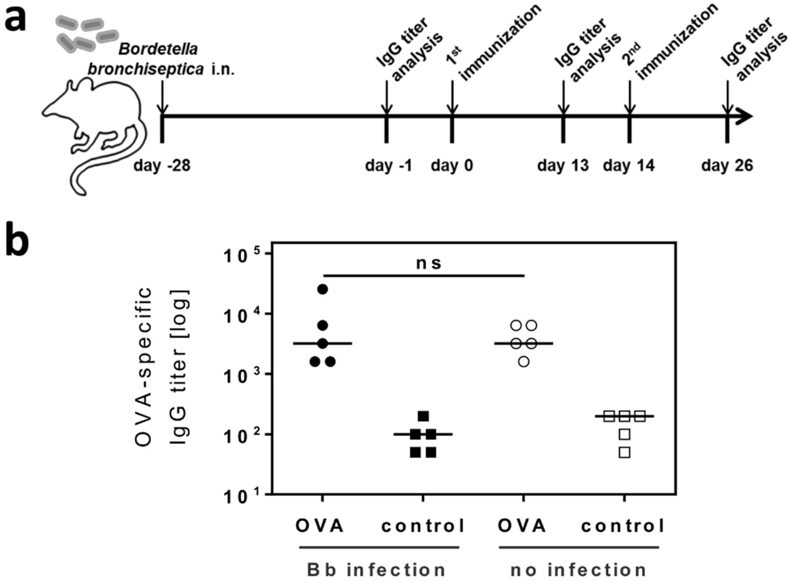
2.3. Respiratory B. bronchiseptica Carriage Does Not Alter the Humoral Immune Response to Vaccination

Possible influences of microbial carriage and microbiota on vaccination efficiency are currently being discussed [44,45,46,47]. Thus, we investigated whether immune modulation through respiratory B. bronchiseptica carriage would affect the immune response to vaccination with an unrelated antigen. To this end, B. bronchiseptica infected mice were first immunized subcutaneously with the model antigen ovalbumin (OVA) on day 28 post infection, followed by a second booster immunization with the same antigen 14 days later (Figure 5a). To quantify the antibody response mounted towards the model antigen, serum samples were collected from vaccinated B. bronchiseptica carriers, as well as control animals, which were analyzed for OVA-specific IgG titers (Figure 5b). These analyses revealed that B. bronchiseptica infected and uninfected animals responded equally well to vaccination. Therefore, respiratory carriage with B. bronchiseptica and the associated changes to the peripheral CD4+CD25+ T cell pool, did not alter humoral immunity to an unrelated antigen administered to the periphery.

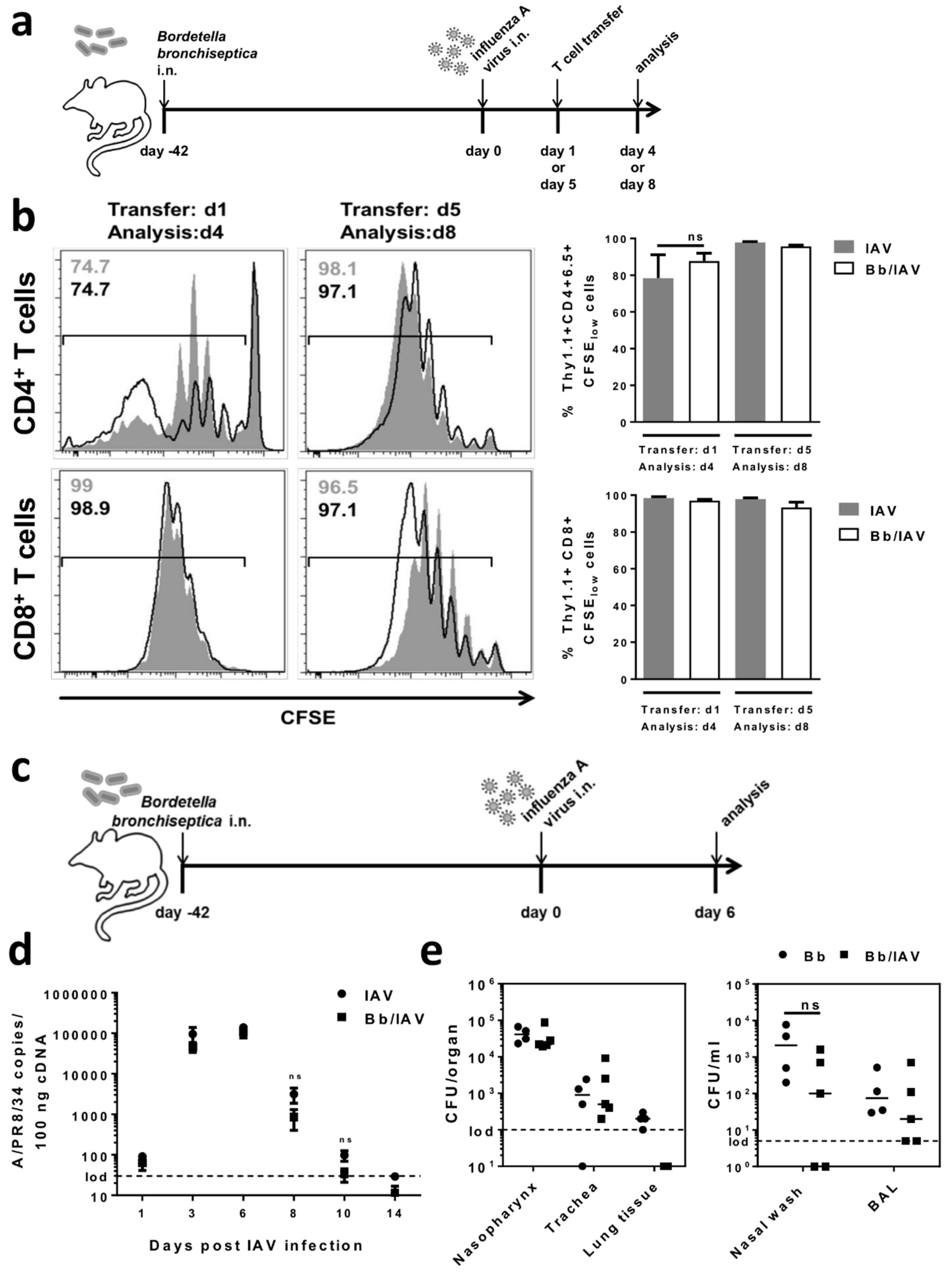
2.4. Respiratory B. bronchiseptica Carriage Does Not Affect the Local Antiviral Immune Response Following Secondary Infection with Influenza A Virus

In addition to assessing the effects of adoptively transferred immune cells of B. bronchiseptica carriers on bacterial clearance in an acute B. bronchiseptica challenge, we asked whether carriage would also impinge on cellular adaptive immunity to a secondary respiratory tract infection with an unrelated pathogen. To experimentally address this issue, we employed an antigen-specific adoptive T cell transfer system, in combination with a respiratory influenza A virus (IAV) infection (Figure 6a). Respiratory B. bronchiseptica carriers were infected with IAV. To enable the analysis of antigen-specific T cell proliferation in this model, the mice received adoptive transfer of either CD4+ or CD8+ T cells, specific for the IAV hemagglutinin (HA) on day 1 or day 5 following IAV infection. Both the IAV-specific CD4+ and CD8+ T cells that were recovered from the draining lymph nodes of IAV only infected recipient mice and of IAV infected B. bronchiseptica carriers, had proliferated equally well in response to viral antigen recognition, as shown in Figure 6b. Of note, the dissemination of proliferated HA-specific CD4+ and CD8+ T cells to the periphery was unchanged (Supplementary Materials Figure S2). These observations indicated that B. bronchiseptica carriage in the respiratory tract did not negatively affect antigen-presentation and the subsequent induction of specific T cell responses to a secondary viral pathogen in the lung. To assess possible effects of B. bronchiseptica carriage on the anti-IAV immune response that were independent of the unchanged T cell response, e.g., on innate antiviral defense, we analyzed viral clearance in B. bronchiseptica carriers infected with IAV (Figure 6c). The viral load of the lung was quantified at different time points over the course of the infection (Figure 6d). However, respiratory B. bronchiseptica carriage neither affected early viral replication nor clearance from the lung, suggesting equally efficient anti-viral mechanisms to be present in B. bronchiseptica carriers. Importantly, respiratory IAV infection triggers a full anti-viral immune response even at sublethal doses and causes substantial inflammation in the respiratory tract, involving a variety of different immune cell types, as well as inflammatory and anti-inflammatory mechanisms [48,49]. Therefore, effects of the IAV infection on B. bronchiseptica carriage and distribution within the respiratory tract were well conceivable. Thus, we quantified bacterial carriage in IAV infected B. bronchiseptica carriers at the peak of viral replication (day 6 post IAV infection), and there was a trend towards improved bacterial clearance in the lung and nose of IAV infected B. bronchiseptica carriers (Figure 6e). However, this did not reach statistical significance, demonstrating that secondary IAV infection, neither significantly boosted antibacterial immune mechanisms nor supported the outgrowth of B. bronchiseptica in the upper or lower respiratory tract of carriers. Taken together, these analyses showed that respiratory B. bronchiseptica carriage and the associated changes to the immune system do not affect immunity to secondary IAV infection in the lung.

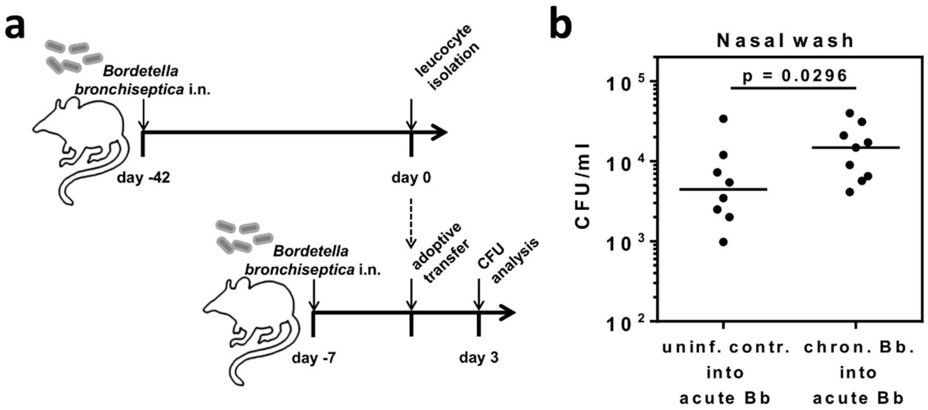
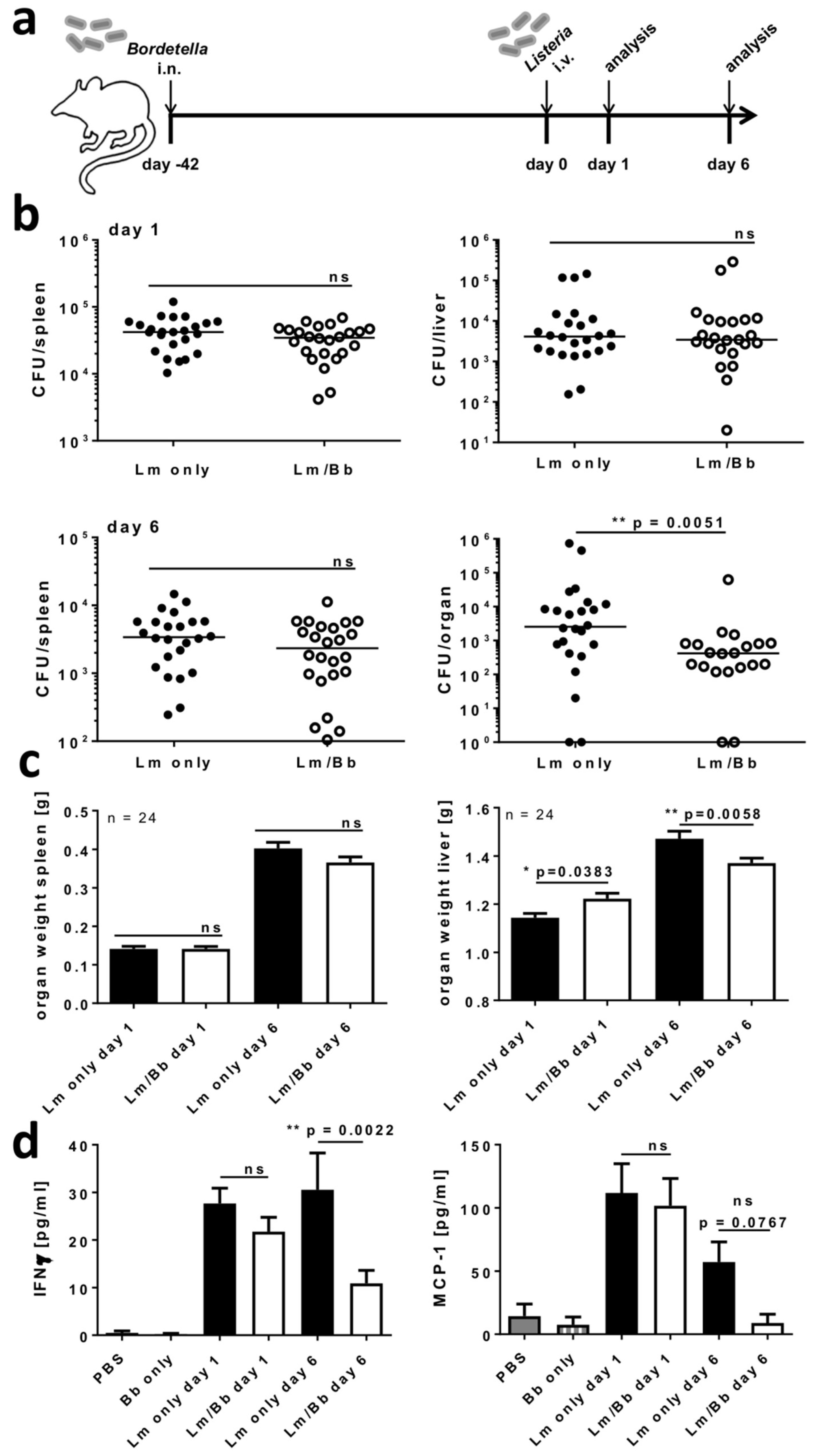
2.5. Respiratory B. bronchiseptica Carriage Alters Immunity to Systemic L. monocytogenes Infection

IAV, like B. bronchiseptica, is a respiratory pathogen, and thus, we further assessed the effect of B. bronchiseptica carriage on a secondary systemic infection by L. monocytogenes. To this end, mice were first intranasally infected with B. bronchiseptica, followed by intravenous infection with a sublethal dose of L. monocytogenes six weeks later (Figure 7a). We assessed the organ dissemination of L. monocytogenes by quantifying bacterial loads in the spleen and the liver, on days 1 and 6 post infection (Figure 7b). Strikingly, while L. monocytogenes loads were unchanged in the spleen at both time points and in the liver early post infection, we detected a significantly reduced L. monocytogenes burden in the livers of B. bronchiseptica carriers, compared to only L. monocytogenes infected mice six days after the secondary infection. These results suggested a significant benefit from respiratory B. bronchiseptica carriage, for the clearance of L. monocytogenes in the liver. Next to the L. monocytogenes bacterial burden, we assessed the organ weight as a measure for inflammation and tissue damage [50] (Figure 7c). While no differences were detected for the spleens, the liver weight was significantly reduced in L. monocytogenes infected B. bronchiseptica carriers on day 6 following secondary infection, compared to only L. monocytogenes infected mice. This observation was well in line with the significantly reduced bacterial burden in the liver at this time point. Systemic L. monocytogenes infection is associated with a substantial inflammatory response [51]. Thus, in a next step, we assessed the pro-inflammatory response and evaluated if it correlated with the effects of B. bronchiseptica carriage on L. monocytogenes loads in the liver. To this end, we assessed the levels of interferon (IFN)-γ and monocyte chemotactic protein 1 (MCP-1) in the serum of L. monocytogenes infected mice (Figure 7d). We detected a strong induction of both mediators in the serum of L. monocytogenes infected naïve mice, as well as B. bronchiseptica carriers, on day 1 following L. monocytogenes infection. Strikingly, both IFN-γ and MCP-1 were strongly reduced in L. monocytogenes infected B. bronchiseptica carriers, compared to only L. monocytogenes infected mice, by day 6 following infection. These results were well in line with the reduced pathogen burden in the liver, detected in L. monocytogenes infected B. bronchiseptica carriers at this time point. Ultimately, these data indicated that chronic respiratory B. bronchiseptica carriage, significantly affects peripheral immunity towards an unrelated secondary bacterial pathogen.

3. Discussion

Immune reactions are versatile, well-coordinated processes involving a number of different players. Next to immune activation, tight regulatory mechanisms are in place to protect the host from immune mediated pathology. Such regulation can, however, also result in pathogen immune evasion and promotion of persistence. Currently, increasing attention is being directed to the interactions between responses to different simultaneous or sequential immunologically relevant stimuli in single hosts. Furthermore, next to the gut and skin, the lower respiratory tract is now being acknowledged as a niche for colonization by harmless and potentially harmful microorganisms [1,2]. The questions arising are, what regulatory mechanisms are potentially induced through respiratory pathogen carriage and how these affect the response to secondary immunological triggers. Our study comprehensively addresses these questions by in vivo utilizing B. bronchiseptica as a mouse model, for carriage with a potentially pathogenic microorganism. Of note, while B. bronchiseptica infection has frequently served as a mouse model for studies on the closely related major human pathogen B. pertussis in the past [21], our study employed B. bronchiseptica primarily as a robust model for respiratory microbial carriage in the mouse.
We clearly show immune modulatory effects resulting from respiratory B. bronchiseptica carriage. This was demonstrated by the adoptive transfer of leukocytes isolated from carriers to acutely B. bronchiseptica infected mice, which resulted in significantly increased bacterial numbers in the recipient’s upper respiratory tract. The induction of immunosuppressive cells such as Treg during B. bronchiseptica carriage, represents one possible mechanism underlying this observation. Interestingly, Treg induction has been described for B. pertussis infected mice and has been implicated and discussed, however, it has not been addressed experimentally for B. bronchiseptica infection [35,36,52]. We now show the induction of Bordetella-specific Treg following respiratory infection. Furthermore, our analyses of the peripheral CD4+CD25+ T cell pool revealed exceptionally broad transcriptional changes, induced by respiratory B. bronchiseptica carriage. These broad changes together with the detected imbalance in favor of up-regulated over down-regulated transcripts revealed transcriptional activation in CD4+CD25+ T cells of chronic B. bronchiseptica carriers that was probably associated with some gain or change in function rather than mere functional suppression. However, the most intensely up-regulated transcripts in CD4+CD25+ T cells during chronic carriage (listed in Table 1) do not clearly associate with any apparent annotated function in the defense against bacterial pathogens or the suppression of immune responses. Instead, GO-term analysis of all the genes differentially expressed in CD4+CD25+ T cells isolated from B. bronchiseptica carriers revealed that mainly transcription- and RNA-metabolism-associated genes are regulated. Nevertheless, up-regulated transcripts did include genes known to be associated to immune responses mounted towards B. bronchiseptica or B. pertussis. TLR4, which was up-regulated 2.6-fold in CD4+CD25+ T cells from B. bronchiseptica carriers, compared to uninfected controls, has been described to play a role in very early infection with B. bronchiseptica [53] and to affect transmission [54]. Nevertheless, there are also implications for its up-regulation by CD4+CD25+ T cells, as distinct TLR4-mediated responses of T cells have been reported [55]. Il-4 (2.0-fold up-regulated in CD4+CD25+ T cells from B. bronchiseptica carriers, compared to uninfected controls), has been suggested to display an important regulator of the recruitment of inflammatory cells in the context of B. pertussis challenge, following vaccination [56]. Importantly, the detected transcriptional changes in the CD4+CD25+ T cell pool of B. bronchiseptica carriers were predominantly detected six, but not one week, following infection, and thereby pointed to carriage-associated mechanisms that were most likely of a regulatory manner. Indeed, this conclusion is supported by the finding that several Treg-associated transcripts were differentially expressed in CD4+CD25+ T cells during respiratory carriage, compared to CD4+CD25+ T cells isolated from controls. Here, Nrp1 stands out, in as much as it is a murine Treg-marker not expressed in activated conventional T cells [37]. Nrp1 promotes long-lasting interactions between Treg and immature DC, and functions as receptor for immune suppressive TGF-ß [57]. Moreover, up-regulation of Nfkbia, an inhibitor of NFκB, suggests reduced responsiveness to T cell stimulation, which typically involves activation of the NFκB pathway [58]. Importantly, CD4+CD25+ T cells generally, and also during B. bronchiseptica carriage, revealed a clear transcriptional Treg signature when compared to the CD4+CD25 T cell pool isolated at the same time. Here, well established Treg-specific factors, such as Gpr83 [38,39], CTLA4 [40], CD103 [41], the Zinc-finger protein Eos (Ikzf4) [42] and Foxp3 [43] were among the 30 most intensely up-regulated transcripts, pointing to the strong presence of Treg in the CD4+CD25+ T cell pool, also during respiratory B. bronchiseptica carriage. Functionally, it has been shown that CD4+CD25+Foxp3+ Treg not only act on adaptive lymphocytes, but also affect innate immune cells. Here, Treg have the potential to induce the alternative activation of monocytes/macrophages [59]. In line with this, persistent colonization with B. bronchiseptica has been suggested to rely on the differential modulation of both macrophage and DC function, in turn leading to an altered adaptive immune response [31]. Therefore, the Treg signature detected in CD4+CD25+ T cells isolated from B. bronchiseptica carriers as compared to the respective CD4+CD25 T cell population, together with the detection of B. bronchiseptica-specific Treg during carriage, suggest Treg-associated mechanisms of B. bronchiseptica carriage. Additionally, it makes a transfer of suppressive Treg together with the adoptively transferred leukocytes from B. bronchiseptica carriers likely and is a possible mechanism underlying the increased bacterial load in acutely infected recipients. Generally, the regulatory mechanisms that act in the respiratory tract, next to Treg, also include but are not restricted to alternatively activated (M2) macrophages and myeloid derived suppressor cells (MDSC) [3,4,7,8]. Therefore, at this point, it remains unclear whether the effects we observed for the adoptive transfer of leukocytes from B. bronchiseptica carriers into acutely infected hosts were linked to the accumulation of Treg specific for B. bronchiseptica; or to overall changes in the CD4+CD25+ T cell pool; or to independent mechanisms, such as the transfer of likewise induced MDSC. Future studies will have to address these details and will have to clarify the mechanisms interfering with bacterial clearance in the upper respiratory tract, following the adoptive transfer of leukocytes isolated from B. bronchiseptica carriers. Nevertheless, our results clearly show the regulatory potential of respiratory B. bronchiseptica carriage.
With the clear immune modulatory effects observed, we hypothesized that B. bronchiseptica carriage had the potential to also affect responses to unrelated immunological stimuli, such as vaccination and infection. Of note, possible influences of microbial carriage on vaccination efficiencies are currently being discussed [44,45,46,47,60]. However, we found that B. bronchiseptica carriers mounted a full humoral response to subcutaneous vaccination with a model antigen. This response was unchanged when compared to naïve control mice, suggesting that under these experimental conditions, respiratory colonization with a potentially pathogenic microorganism neither displayed beneficial nor adverse effects on vaccination efficacy. In contrast to vaccination, infections are typically accompanied by pathogen replication and at least some extent of host cell damage, which together lead to stronger inflammation and immune stimulation, despite the use of adjuvants in vaccination. For IAV, strong anti-viral responses and collateral immune pathology are hallmark features of the infection, and in turn there is a particular need for regulation to protect the host from immune mediated damage [48,61]. Therefore, we hypothesized that adaptive anti-IAV responses might be exceptionally “susceptible” to the regulatory mechanisms induced by B. bronchiseptica carriage. Of note, B. bronchiseptica pulsed macrophages have previously been shown to inhibit antigen-specific CD4+ T cell proliferation in vitro [31]. However, we detected unchanged IAV-specific T cell proliferation and dissemination in B. bronchiseptica carriers, compared to naïve mice following IAV infection. Therefore, presumably antigen-presentation following the viral infection was fully functional, despite the effects of B. bronchiseptica on macrophages and DC, which have previously been described in References [30,33,36]. Importantly, viral clearance from the respiratory tract was equally efficient in B. bronchiseptica carriers, pointing at likewise intact innate responses. This observation was somewhat in contrast to the findings of a human study, describing asymptomatic neonatal airway colonization with different respiratory pathogens to increase the risk of pneumonia and bronchiolitis in early life [62]. Moreover, pre-infection with B. bronchiseptica has been shown to favor colonization with Haemophilus parasuis, and simultaneous acute infection with B. bronchiseptica and IAV has been shown to lead to increased bacterial titers in swine [63,64]. In our model, we focused on respiratory carriage with B. bronchiseptica, which neither affected clearance of the viral respiratory pathogen nor the underlying respiratory bacterial carriage. Such effects most likely do not represent general mechanisms, but depend on the specific microorganisms, the phase of the infection, as well as on the age and underlying conditions of the host. For example, neonates, as were studied in Reference [58], harbor a still developing and thereby unique immune system [65], where microbial carriage most likely has different effects than later in life. In addition, distinct microbiological features of the airway microbiome have been associated with specific respiratory diseases [66], underlining the complexity of respiratory immune regulation with respect to microbial carriage.
Despite the fact that respiratory IAV infection went unchanged, we explored whether respiratory carriage with B. bronchiseptica affected responses against a systemic bacterial infection. Of note, only recently asymptomatic colonization with Moraxella catharralis, Haemophilus influenzae, and/or Streptococcus pneumoniae in the hypopharynx, was shown to be associated with low-grade systemic inflammation in new-born children [18]. Surprisingly, we found B. bronchiseptica carriage to display a significant benefit for the host, regarding the control of systemic L. monocytogenes infection. Next to decreased bacterial numbers in the liver, we further detected reduced levels of MCP-1 and IFN-γ in the serum of L. monocytogenes infected B. bronchiseptica carriers. In line with this, the B. bronchiseptica T3SS has been shown to inhibit IFN-γ production via IL-10 to promote persistent carriage [35]. B. bronchiseptica mediated inhibition of IFN-γ production, could in turn possibly have led to the reduced bacterial burden in the liver, as a consequence of reduced inflammation and immune pathology. Such a mechanistic link is strongly supported by the observation that excessive IFN-γ production following L. monocytogenes infection, exacerbates pathology and impairs anti-bacterial defense mechanisms [67]. Ultimately, our study clearly shows the potential of respiratory carriage with a pathogenic microorganism, to significantly alter systemic responses to a secondary bacterial infection.
Taken together, our results show clear and significant effects of respiratory B. bronchiseptica carriage on the immune system. Considering the results of previous studies, these immune modulatory processes are most likely aimed at supporting microbial persistence in the host [34,35,36]. We have now for the first time comprehensively addressed their significance, for secondary immune responses in vivo, employing B. bronchiseptica as a model for respiratory pathogen carriage. We did not observe general, but highly differential effects, depending on the considered compartment and the nature of the secondary stimulus. Interestingly, the effects we described can be beneficial for the pathogen on the one hand, as observed in the adoptive transfer experiments, or beneficial for the host, as demonstrated by the improved containment of L. monocytogenes in the livers of B. bronchiseptica carriers. Therefore, our results demonstrated the complexity of the versatile local and systemic immune regulation, induced by respiratory carriage with a pathogenic microorganism. Furthermore, our study provided a valuable basis for future investigations, which will address the decisive factors and elucidate specific mechanisms of such interactions, that will be essential for prophylactic and therapeutic considerations.

4. Materials and Methods

4.1. Mice

BALB/c mice were obtained from Harlan (Borchen, Germany). Clone 4 (CL4) mice expressing a major histocompatibility complex (MHC)-I–restricted HA-specific T cell receptor, and TCR-HA mice expressing a MHC-II-restricted T cell receptor, have been described previously in References [68,69]. Transgenic T cell receptor bearing mice were used on the Thy1.1 congenic background to allow for tracking of HA-specific T cells, after adoptive transfer into BALB/c recipient mice of the Thy1.2 congenic background. Transgenic mice were bred under specific pathogen-free conditions, at the Helmholtz Centre for Infection Research (Braunschweig, Germany). Animal experiments were performed according to national and institutional guidelines, under the project identification codes 11/0425, 10/0108, and 10/0256 (Nds. Landesamt für Verbraucherschutz und Lebensmittelsicherheit).

4.2. Bacterial and Viral Pathogens

B. bronchispetica wild type strain BB7865 [70] was obtained from the Culture Collection of the University of Gothenburg, Sweden. Bacteria were prepared as described in Reference [20]. Briefly, B. bronchiseptica was grown for 24 h, on Bordet-Gengou agar plates (supplemented with 10% sheep blood and 100 µg/mL streptomycin). Colonies were suspended in PBS, the OD600nm was determined, and bacterial inocula were prepared by dilution. The dose/mouse was retrospectively validated by plating serial dilutions of the inoculum.
Madin-Darby canine kidney (MDCK) cell-derived IAV PR8/A/34 (H1N1), was obtained as described in Reference [71] and diluted in PBS to the appropriate concentration before infection.
L. monocytogenes strain EGD was grown in Brain Heart Infusion (BHI). For mouse infections, bacteria were collected in mid-log phase, centrifuged, and resuspended in PBS to the desired concentration. The dose/mouse was retrospectively validated by plating serial dilutions of the inoculum on BHI agar plates.

4.3. Bacterial and Viral Infections

For intranasal B. bronchiseptica infection, mice were lightly anaesthetized by isoflurane inhalation, and 20 µL (10 µL per nostril) of the bacterial inoculum (~ 0.5–1 × 10⁶ CFU) were administered. For intranasal IAV infection, mice were infected with 100.9 TCID50 of the virus in 25 µL PBS under ketamine/xylazine anesthesia. Holding the animals upright, the viral or bacterial inoculum was delivered to the nostrils to be taken up by the mouse upon breathing.
For systemic L. monocytogenes infection, 2 × 10³ CFU/mouse in sterile PBS were intravenously injected into the tail vein.

4.4. Quantification of Pathogen Loads Following Infection

For the quantification of B. bronchiseptica CFU, bronchoalveolar lavage (BAL) was obtained by flushing the lungs with 1 mL PBS through the trachea. Nasal wash fluid was obtained by flushing the nasopharynx with 200 µL PBS. Lungs and tracheas were aseptically removed and homogenized separately in PBS (Polytron® PT 1300D, Kinematica, Lucerne, Switzerland). Serial dilutions of airway fluids and tissue homogenates were plated onto LB agar plates (supplemented with streptomycin), incubated overnight, and CFU counts were calculated.
For the quantification of the lung viral load following IAV infection, lungs were homogenized in TriFastFL reagent (PeqLab now VWR Life Science Competence Center, Erlangen, Germany). RNA was extracted by addition of 1-Br-3-Cl-Propane (Merck, Darmstadt, Germany), purified by precipitation and treated with DNase (Ambion, Thermo Fisher Scientific, Waltham, MA, USA), prior to reverse transcription to cDNA (M-MLV Reverse Transcriptase; Invitrogen, Thermo Fisher Scientific, Waltham, MA, USA). Quantitative real-time PCR of 100 ng cDNA samples, was conducted using the LightCycler® 480 SYBR Green I Master Kit (Roche, Basel, Switzerland) and primers specific for the IAV nucleoprotein (NP). Quantitative real-time PCR of samples containing known numbers of a plasmid carrying the NP sequence (pVI-PmH5-PR8-NP; provided by G. Sutter, Munich, Germany) was performed to obtain a CT-value/NP copy number standard, which was used to quantify NP copy numbers in the samples. NP primers were GAGGGGTGAGAATGGACGAAAAAC (5′-NP) and CAGGCAGGCAGGCAGGACTT (3′-NP) and were used in a final concentration of 500 nmol/L. Quantitative real-time PCR was performed using a LightCycler® instrument.
For the quantification of L. monocytogenes bacterial counts, spleens and livers (without gall bladder) were harvested from infected mice, weighed, and homogenized (Polytron® PT 1300D, Kinematica, Lucerne, Switzerland) in PBS containing 0.2% IGEPAL CA-630. Organ homogenates were plated on BHI agar plates, and CFU were counted after 24 h of incubation.

4.5. Restimulation of Splenocytes with Heat-Killed B. bronchiseptica

B. bronchiseptica was cultured on Bordet-Gengou agar plates for 24 h at 37 °C, and resuspended and diluted to the desired concentration in sterile PBS. Bacteria were heat-inactivated for 30 min at 55–56 °C. Splenocytes were isolated from groups of three mice per group, were pooled, and re-stimulated in vitro, with or without heat killed bacteria in a B. bronchiseptica-to-cell ratio of 10:1, for 4 days (2 × 10⁶ cells/well). As a positive control for proliferation, lymphocytes were stimulated with Concavalin A (5 µg/mL; Sigma-Aldrich, Schnelldorf, Germany).

4.6. Flow Cytometry

For lymphocyte isolation, the spleens, cervical lymph nodes and bronchial lymph nodes were rinsed with PBS and meshed through a 100 µm cell strainer followed by centrifugation and the lysis of red blood cells through osmotic shock. Antibody staining of up to 106 cells was performed in 96-well plates. Cells were washed in FACS (fluorescence activated cell sorting) buffer (PBS, 2% [v/v] fetal bovine serum, 2 mM EDTA) and incubated with the indicated antibodies (fluorochrome-conjugated) diluted in FACS buffer for 10 min at 4 °C in the dark. Antibody concentrations were separately titrated for each antibody, fluorophore and lot. Following surface staining, cells were washed and immediately analyzed or alternatively fixed in 100 µL/sample 1% (w/v) paraformaldehyde in PBS for 20 min at room temperature. For the intracellular staining of Foxp3 following surface staining, the Foxp3 staining buffer set (eBioscience, Thermo Fisher Scientific, Waltham, MA, USA) was used according to the manufacturer’s recommendations. Acquisition of flow cytometry samples was performed on a FACSCanto (BD, Franklin Lakes, NJ, USA), and data were analyzed with the FlowJo software (Tree Star, Becton, Dickinson & Company, Franklin Lakes, NJ, USA). A monoclonal antibody specific to mouse CD90.1 (OX-7) was obtained from BioLegend (San Diego, CA, USA). Antibodies specific to mouse CD4 (RM4-5), CD8 (53-6.7) and Foxp3 (FJK-16s) were obtained from eBioscience (Thermo Fisher Scientific, Waltham, MA, USA). Antibodies specific for mouse CD25 (PC61 and 7D4) were obtained from BD Pharmingen (Franklin Lakes, NJ, USA). In addition, HA-specific CD4+ T cells were identified using α-6.5 (α-TCR-HA; Clone: 14.3d) antibody, purified from hybridoma supernatant by affinity chromatography.

4.7. Adoptive Transfer of Leukocytes Isolated from B. bronchiseptica Carriers

For leukocyte isolation; spleens, cervical lymph nodes, and bronchial lymph nodes were rinsed with PBS and meshed through a 100 µm cell strainer, followed by centrifugation and lysis of red blood cells through osmotic shock. For adoptive transfer, leukocytes were labelled with a biotinylated CD45R/B22 antibody (RA3-6B2), and B-cell depleted using anti-biotin Micro-Beads (Miltenyi Biotec, Bergisch Gladbach, Germany) and an AutoMACS device, according to the manufacturer’s instructions. Recipient mice received 8 × 10⁶ cells in PBS intravenously.

4.8. Microarray Analyses of Sorted CD4+CD25+ T Cells

Gene expression arrays were performed in the microarray facility of the Helmholtz Centre for Infection Research Braunschweig. Splenocytes from both infected and control mice were isolated and pooled (n = 6 per group) on day 7 or 42, post infection. CD4+CD25+ cells were flow cytometrically sorted on a BD FACSAria II instrument. Total RNA of sorted CD4+CD25+ cells was isolated using the Rneasy Mini Kit (Quiagen, Hilden, Germany), including the enzymatic digestion of DNA (Rnase-free Dnase Set, Quiagen), according to the manufacturer’s recommendations. RNA was concentrated by ethanol precipitation following elution, and RNA integrity was tested using an Agilent 2100 Bioanalyzer (Agilent Technologies, Santa Clara, CA, USA). Equal amounts of RNA were amplified using a T7dT23 primer (Metabion, Steinkirchen, Germany) and SuperScript II reverse transcriptase (Invitrogen). Second-strand cDNA synthesis was performed using DNA-polymerase I (Escherichia coli DNA ligase; Invitrogen). Double-stranded cDNA was amplified using the Promega P1300 RiboMax Kit, for T7 amplification (Promega, Madison, WI, USA). Resulting cRNA was amplified for first-strand synthesis of cDNA using random hexamer primers (Pharmacia, GE Healthcare, Chicago, IL, USA). Degradation of cRNA was performed through RNase H treatment, and second-strand synthesis was performed as described above, using T7dT23 primers. A second in vitro transcription was performed using the GeneChip expression 3’-Amplification Reagent Kit (Affymetrix, Santa Clara, CA, USA). Recommendations of the Affymetrix protocol, were followed for hybridization with the GeneChip Mouse Genome 430 2.0 Array (Affymetrix, Santa Clara, CA, USA), staining and scanning. Microarray data were analyzed using the GeneSpring GX 10.0 software (Agilent Technologies, Santa Clara, CA, USA), applying the RMA algorithm, including a quantile normalization. Gene ontology enrichment analyses were performed using the ClueGO Cytoscape plug-in software, as outlined in Reference [72]. Microarray data were deposited in the Gene Expression Omnibus (GEO), under the reference ID GSE116913.

4.9. Immunization with the Model Antigen OVA

Mice were subcutaneously (s.c.) immunized on days 0 and 14; either with 10 µg EndoGrade OVA (>98% purity) (Hyglos, Bernried, Germany), in addition to 50 µg Poly (I:C) (Invivogen, San Diego, CA, USA) and 50 µg CpG (Eurofins MWG Operon, Ebersberg, Germany) in a total volume of 50 µL PBS per animal, or with the adjuvants alone as control.

4.10. Detection of OVA-Specific Serum IgG

For the preparation of serum samples, 75 µL of blood were collected from the retro-orbital sinus, and samples were incubated for 45 min at 37 °C, followed by 45 min incubation at 4 °C, and subsequent centrifugation (10 min at 420× g). Sera were assayed for the presence of OVA-specific IgG by enzyme-linked immunosorbent assay (ELISA), using 96-well Nunc-Immuno MaxiSorp plates (Nunc, Thermo Fisher Scientific, Waltham, MA, USA) coated with 2 µg/mL OVA protein, in 0.05 M carbonate buffer (pH 9.6). After overnight coating at 4 °C, the plates were washed with PBS supplemented with 0.1% Tween 20, and blocked with 3% bovine serum albumin (BSA) in PBS for 1 h at 37 °C. Serial 2-fold dilutions of sera in 3% BSA/PBS were added and the plates were incubated for 2 h at 37 °C. After washing, antibody binding was detected using biotin-conjugated goat anti-mouse IgG (Sigma-Aldrich, Schnelldorf, Germany) (1 h, 37 °C) and Streptavidin-HRPO (BD Biosciences, Franklin Lakes, NJ, USA) (30 min, 37 °C). ABTS (2,2′-azino-bis (3-ethylbenzothioazoline-6-sulfonic acid) diammonium salt) in 0.1 M citrate-phosphate buffer (pH 4.35) containing 0.03% H2O2 was added to each well, and the absorbance at 405 nm was recorded after 30 min of incubation. Endpoint titers were expressed as the reciprocal value of the last serum dilution, which yielded an absorbance two times above the values of negative controls.

4.11. Adoptive Transfer of IAV-Specific CD4+ and CD8+ T Cells

CD4+ and CD8+ T cells were isolated from the spleen and cervical lymph nodes of TCR-HA or CL4 mice, and purified by negative selection, using the CD4+ and CD8+ T cell isolation kit and an AutoMACS instrument (Miltenyi Biotec, Bergisch Gladbach, Germany), respectively. Purified T cells were labeled with 2.5 µM carboxyfluorescein diacetate succinimidyl ester (CFSE; Invitrogen, Thermo Fisher Scientific, Waltham, MA, USA) and i.v. injected into the tail veins (1.5–1.8 × 10⁶ HA-specific CD4+ T cells, 5–7 × 10⁶ HA-specific CD8+ T cells) of recipient mice. Mice were sacrificed 3 days after T cell transfer and T cell proliferation in the bronchial lymph nodes (BLN), and spleen was assessed by flow cytometry.

4.12. Quantification of IFN-γ and MCP-1 in Serum

Serum was prepared as described above. Cytokine levels of IFN-γ and MCP-1 in serum samples were quantified using a BD™ Cytometric Bead Array (CBA) from BD Biosciences (Franklin Lakes, NJ, USA), following the manufacturer’s instructions.

4.13. Statistical Analyses

Groups were compared by the indicated statistical test, using the Graph Pad Prism software (Graph Pad Software, La Jolla, CA, USA). p values < 0.05 were considered to indicate statistical significance.

Supplementary Materials

Author Contributions

Conceptualization, A.J.; J.D.B.; J.V.; M.G.; T.Y.; C.A.G.; S.S.-K. and D.B.; Methodology, A.J.; J.D.B.; J.V.; M.G.; T.Y.; R.G. and S.S.-K.; Software, R.G.; Formal Analysis, A.J.; J.D.B.; J.V.; M.G.; T.Y. and S.S.-K.; Investigation, A.J.; J.D.B.; J.V.; M.G.; T.Y.; R.G.; C.A.G.; S.S.-K. and D.B.; Resources, R.G.; C.A.G.; J.S. and D.B.; Data Curation, A.J.; J.D.B.; J.V.; M.G.; T.Y.; R.G.; C.A.G.; S.S.-K. and D.B.; Writing—Original Draft Preparation, S.S.-K. and D.B.; Writing—Review & Editing, A.J.; J.D.B.; J.V.; M.G.; T.Y.; C.A.G.; S.S.-K. and D.B.; Visualization, A.J.; J.D.B.; J.V.; M.G. and S.S.-K; Supervision, R.G.; C.A.G.; D.B.; Funding Acquisition, D.B.

Funding

This research was funded by the International Research Training Group 1273 of the German Research Foundation (Deutsche Forschungsgemeinschaft, DFG) and by the Helmholtz Association (Helmholtz-Gemeinschaft Deutscher Forschungszentren, HGF), grant number W2/W3-029 (to D.B.).

Acknowledgments

We thank S. Prettin, A. Höhne, P. Hagendorff and C. Wylegalla for expert technical assistance and L. Gröbe for assistance in cell sorting.

Conflicts of Interest

The authors declare no conflict of interest.

Abbreviations

MDPIMultidisciplinary Digital Publishing Institute
DOAJDirectory of open access journals

References

  1. Dickson, R.P.; Erb-Downward, J.R.; Martinez, F.J.; Huffnagle, G.B. The microbiome and the respiratory tract. Annu. Rev. Physiol. 2016, 78, 481–504. [Google Scholar] [CrossRef] [PubMed]
  2. Wu, B.G.; Segal, L.N. Lung microbiota and its impact on the mucosal immune phenotype. Microbiol. Spectr. 2017, 5. [Google Scholar] [CrossRef] [PubMed]
  3. Chen, K.; Kolls, J.K. T cell-mediated host immune defenses in the lung. Annu. Rev. Immunol. 2013, 31, 605–633. [Google Scholar] [CrossRef] [PubMed]
  4. Duan, W.; Croft, M. Control of regulatory T cells and airway tolerance by lung macrophages and dendritic cells. Ann. Am. Thorac. Soc. 2014, 11 (Suppl. 5), S306–S313. [Google Scholar] [CrossRef] [PubMed]
  5. Hasenberg, M.; Stegemann-Koniszewski, S.; Gunzer, M. Cellular immune reactions in the lung. Immunol. Rev. 2013, 251, 189–214. [Google Scholar] [CrossRef] [PubMed]
  6. Weitnauer, M.; Mijosek, V.; Dalpke, A.H. Control of local immunity by airway epithelial cells. Mucosal Immunol. 2016, 9, 287–298. [Google Scholar] [CrossRef] [PubMed]
  7. Garbi, N.; Lambrecht, B.N. Location, function, and ontogeny of pulmonary macrophages during the steady state. Pflugers Arch. 2017, 469, 561–572. [Google Scholar] [CrossRef] [PubMed]
  8. Kolahian, S.; Oz, H.H.; Zhou, B.; Griessinger, C.M.; Rieber, N.; Hartl, D. The emerging role of myeloid-derived suppressor cells in lung diseases. Eur. Respir. J. 2016, 47, 967–977. [Google Scholar] [CrossRef] [PubMed] [Green Version]
  9. Garib, F.Y.; Rizopulu, A.P. T-regulatory cells as part of strategy of immune evasion by pathogens. Biochemistry (Mosc.) 2015, 80, 957–971. [Google Scholar] [CrossRef] [PubMed]
  10. Cyktor, J.C.; Turner, J. Interleukin-10 and immunity against prokaryotic and eukaryotic intracellular pathogens. Infect. Immun. 2011, 79, 2964–2973. [Google Scholar] [CrossRef] [PubMed]
  11. Levast, B.; Li, Z.; Madrenas, J. The role of IL-10 in microbiome-associated immune modulation and disease tolerance. Cytokine 2015, 75, 291–301. [Google Scholar] [CrossRef] [PubMed]
  12. Bisgaard, H.; Hermansen, M.N.; Buchvald, F.; Loland, L.; Halkjaer, L.B.; Bonnelykke, K.; Brasholt, M.; Heltberg, A.; Vissing, N.H.; Thorsen, S.V.; et al. Childhood asthma after bacterial colonization of the airway in neonates. N. Engl. J. Med. 2007, 357, 1487–1495. [Google Scholar] [CrossRef] [PubMed]
  13. Taylor, S.L.; Wesselingh, S.; Rogers, G.B. Host-microbiome interactions in acute and chronic respiratory infections. Cell. Microbiol. 2016, 18, 652–662. [Google Scholar] [CrossRef] [PubMed] [Green Version]
  14. Von Linstow, M.L.; Schonning, K.; Hoegh, A.M.; Sevelsted, A.; Vissing, N.H.; Bisgaard, H. Neonatal airway colonization is associated with troublesome lung symptoms in infants. Am. J. Respir. Crit. Care Med. 2013, 188, 1041–1042. [Google Scholar] [CrossRef] [PubMed]
  15. McAleer, J.P.; Kolls, J.K. Contributions of the intestinal microbiome in lung immunity. Eur. J. Immunol. 2018, 48, 39–49. [Google Scholar] [CrossRef] [PubMed]
  16. Samuelson, D.R.; Welsh, D.A.; Shellito, J.E. Regulation of lung immunity and host defense by the intestinal microbiota. Front. Microbiol. 2015, 6, 1085. [Google Scholar] [CrossRef] [PubMed]
  17. Liu, Y.Y.; Chiang, C.H.; Chuang, C.H.; Liu, S.L.; Jheng, Y.H.; Ryu, J.H. Spillover of cytokines and reactive oxygen species in ventilator-induced lung injury associated with inflammation and apoptosis in distal organs. Respir. Care 2014, 59, 1422–1432. [Google Scholar] [CrossRef] [PubMed]
  18. Rahman Fink, N.; Chawes, B.L.; Thorsen, J.; Stokholm, J.; Krogfelt, K.; Schjorring, S.; Kragh, M.; Bonnelykke, K.; Brix, S.; et al. Neonates colonized with pathogenic bacteria in the airways have a low-grade systemic inflammation. Allergy 2018, 6, 1085. [Google Scholar] [CrossRef] [PubMed]
  19. Teichert, T.; Vossoughi, M.; Vierkotter, A.; Sugiri, D.; Schikowski, T.; Hoffmann, B.; Schulte, T.; Roden, M.; Raulf-Heimsoth, M.; Luckhaus, C.; et al. Investigating the spill-over hypothesis: Analysis of the association between local inflammatory markers in sputum and systemic inflammatory mediators in plasma. Environ. Res. 2014, 134, 24–32. [Google Scholar] [CrossRef] [PubMed]
  20. Yevsa, T.; Ebensen, T.; Fuchs, B.; Zygmunt, B.; Libanova, R.; Gross, R.; Schulze, K.; Guzman, C.A. Development and characterization of attenuated metabolic mutants of Bordetella bronchiseptica for applications in vaccinology. Environ. Microbiol. 2013, 15, 64–76. [Google Scholar] [CrossRef] [PubMed]
  21. Melvin, J.A.; Scheller, E.V.; Miller, J.F.; Cotter, P.A. Bordetella pertussis pathogenesis: Current and future challenges. Nat. Rev. Microbiol. 2014, 12, 274–288. [Google Scholar] [CrossRef] [PubMed]
  22. Cotter, P.A.; Jones, A.M. Phosphorelay control of virulence gene expression in Bordetella. Trends Microbiol. 2003, 11, 367–373. [Google Scholar] [CrossRef]
  23. Trainor, E.A.; Nicholson, T.L.; Merkel, T.J. Bordetella pertussis transmission. Pathog. Dis. 2015, 73, ftv068. [Google Scholar] [CrossRef] [PubMed]
  24. Hewlett, E.L.; Burns, D.L.; Cotter, P.A.; Harvill, E.T.; Merkel, T.J.; Quinn, C.P.; Stibitz, E.S. Pertussis pathogenesis—What we know and what we don’t know. J. Infect. Dis. 2014, 209, 982–985. [Google Scholar] [CrossRef] [PubMed]
  25. Banemann, A.; Gross, R. Phase variation affects long-term survival of Bordetella bronchiseptica in professional phagocytes. Infect. Immun. 1997, 65, 3469–3473. [Google Scholar] [PubMed]
  26. Forde, C.B.; Parton, R.; Coote, J.G. Bioluminescence as a reporter of intracellular survival of Bordetella bronchiseptica in murine phagocytes. Infect. Immun. 1998, 66, 3198–3207. [Google Scholar] [PubMed]
  27. Buboltz, A.M.; Nicholson, T.L.; Weyrich, L.S.; Harvill, E.T. Role of the type III secretion system in a hypervirulent lineage of Bordetella bronchiseptica. Infect. Immun. 2009, 77, 3969–3977. [Google Scholar] [CrossRef] [PubMed]
  28. Mann, P.; Goebel, E.; Barbarich, J.; Pilione, M.; Kennett, M.; Harvill, E. Use of a genetically defined double mutant strain of Bordetella bronchiseptica lacking adenylate cyclase and type III secretion as a live vaccine. Infect. Immun. 2007, 75, 3665–3772. [Google Scholar] [CrossRef] [PubMed]
  29. Weyrich, L.S.; Rolin, O.Y.; Muse, S.J.; Park, J.; Spidale, N.; Kennett, M.J.; Hester, S.E.; Chen, C.; Dudley, E.G.; Harvill, E.T. A type VI secretion system encoding locus is required for Bordetella bronchiseptica immunomodulation and persistence in vivo. PLoS ONE 2012, 7, e45892. [Google Scholar] [CrossRef] [PubMed]
  30. Reissinger, A.; Skinner, J.A.; Yuk, M.H. Downregulation of mitogen-activated protein kinases by the Bordetella bronchiseptica Type III secretion system leads to attenuated nonclassical macrophage activation. Infect. Immun. 2005, 73, 308–316. [Google Scholar] [CrossRef] [PubMed]
  31. Siciliano, N.A.; Skinner, J.A.; Yuk, M.H. Bordetella bronchiseptica modulates macrophage phenotype leading to the inhibition of CD4+ T cell proliferation and the initiation of a Th17 immune response. J. Immunol. 2006, 177, 7131–7138. [Google Scholar] [CrossRef] [PubMed]
  32. Guzman, C.A.; Rohde, M.; Bock, M.; Timmis, K.N. Invasion and intracellular survival of Bordetella bronchiseptica in mouse dendritic cells. Infect. Immun. 1994, 62, 5528–5537. [Google Scholar] [PubMed]
  33. Gueirard, P.; Ave, P.; Balazuc, A.M.; Thiberge, S.; Huerre, M.; Milon, G.; Guiso, N. Bordetella bronchiseptica persists in the nasal cavities of mice and triggers early delivery of dendritic cells in the lymph nodes draining the lower and upper respiratory Tract. Infect. Immun. 2003, 71, 4137–4143. [Google Scholar] [CrossRef] [PubMed]
  34. Nagamatsu, K.; Kuwae, A.; Konaka, T.; Nagai, S.; Yoshida, S.; Eguchi, M.; Watanabe, M.; Mimuro, H.; Koyasu, S.; Abe, A. Bordetella evades the host immune system by inducing IL-10 through a type III effector, BopN. J. Exp. Med. 2009, 206, 3073–3088. [Google Scholar] [CrossRef] [PubMed]
  35. Pilione, M.R.; Harvill, E.T. The Bordetella bronchiseptica type III secretion system inhibits gamma interferon production that is required for efficient antibody-mediated bacterial clearance. Infect. Immun. 2006, 74, 1043–1049. [Google Scholar] [CrossRef] [PubMed]
  36. Skinner, J.A.; Pilione, M.R.; Shen, H.; Harvill, E.T.; Yuk, M.H. Bordetella type III secretion modulates dendritic cell migration resulting in immunosuppression and bacterial persistence. J. Immunol. 2005, 175, 4647–4652. [Google Scholar] [CrossRef] [PubMed]
  37. Bruder, D.; Probst-Kepper, M.; Westendorf, A.M.; Geffers, R.; Beissert, S.; Loser, K.; von Boehmer, H.; Buer, J.; Hansen, W. Neuropilin-1: A surface marker of regulatory T cells. Eur. J. Immunol. 2004, 34, 623–630. [Google Scholar] [CrossRef] [PubMed]
  38. Hansen, W.; Loser, K.; Westendorf, A.M.; Bruder, D.; Pfoertner, S.; Siewert, C.; Huehn, J.; Beissert, S.; Buer, J. G protein-coupled receptor 83 overexpression in naive CD4+CD25 T cells leads to the induction of Foxp3+ regulatory T cells in vivo. J. Immunol. 2006, 177, 209–215. [Google Scholar] [CrossRef] [PubMed]
  39. Sugimoto, N.; Oida, T.; Hirota, K.; Nakamura, K.; Nomura, T.; Uchiyama, T.; Sakaguchi, S. Foxp3-dependent and -independent molecules specific for CD25+CD4+ natural regulatory T cells revealed by DNA microarray analysis. Int. Immunol. 2006, 18, 1197–1209. [Google Scholar] [CrossRef] [PubMed] [Green Version]
  40. Wing, K.; Onishi, Y.; Prieto-Martin, P.; Yamaguchi, T.; Miyara, M.; Fehervari, Z.; Nomura, T.; Sakaguchi, S. CTLA-4 control over Foxp3+ regulatory T cell function. Science 2008, 322, 271–275. [Google Scholar] [CrossRef] [PubMed]
  41. Lehmann, J.; Huehn, J.; de la Rosa, M.; Maszyna, F.; Kretschmer, U.; Krenn, V.; Brunner, M.; Scheffold, A.; Hamann, A. Expression of the integrin alpha Ebeta 7 identifies unique subsets of CD25+ as well as CD25 regulatory T cells. Proc. Natl. Acad. Sci. USA 2002, 99, 13031–13036. [Google Scholar] [CrossRef] [PubMed]
  42. Pan, F.; Yu, H.; Dang, E.V.; Barbi, J.; Pan, X.; Grosso, J.F.; Jinasena, D.; Sharma, S.M.; McCadden, E.M.; Getnet, D.; et al. Eos mediates Foxp3-dependent gene silencing in CD4+ regulatory T cells. Science 2009, 325, 1142–1146. [Google Scholar] [CrossRef] [PubMed]
  43. Hori, S.; Nomura, T.; Sakaguchi, S. Control of regulatory T cell development by the transcription factor Foxp3. Science 2003, 299, 1057–1061. [Google Scholar] [CrossRef] [PubMed]
  44. Kim, Y.G. Microbiota influences vaccine and mucosal adjuvant efficacy. Immune Netw. 2017, 17, 20–24. [Google Scholar] [CrossRef] [PubMed]
  45. Lynn, D.J.; Pulendran, B. The potential of the microbiota to influence vaccine responses. J. Leukoc. Biol. 2018, 103, 225–231. [Google Scholar] [CrossRef] [PubMed]
  46. Nguyen, Q.N.; Himes, J.E.; Martinez, D.R.; Permar, S.R. The impact of the gut microbiota on humoral immunity to pathogens and vaccination in early infancy. PLoS Pathog. 2016, 12, e1005997. [Google Scholar] [CrossRef] [PubMed]
  47. Zimmermann, P.; Curtis, N. The influence of probiotics on vaccine responses—A systematic review. Vaccine 2018, 36, 207–213. [Google Scholar] [CrossRef] [PubMed]
  48. Braciale, T.J.; Sun, J.; Kim, T.S. Regulating the adaptive immune response to respiratory virus infection. Nat. Rev. Immunol. 2012, 12, 295–305. [Google Scholar] [CrossRef] [PubMed] [Green Version]
  49. McGill, J.; Heusel, J.W.; Legge, K.L. Innate immune control and regulation of influenza virus infections. J. Leukoc. Biol. 2009, 86, 803–812. [Google Scholar] [CrossRef] [PubMed] [Green Version]
  50. Wilder, M.S.; Sword, C.P. Mechanisms of pathogenesis in Listeria monocytogenes infection. II. Characterization of listeriosis in the CD-1 mouse and survey of biochemical lesions. J. Bacteriol. 1967, 93, 531–537. [Google Scholar] [PubMed]
  51. Calame, D.G.; Mueller-Ortiz, S.L.; Wetsel, R.A. Innate and adaptive immunologic functions of complement in the host response to Listeria monocytogenes infection. Immunobiology 2016, 221, 1407–1417. [Google Scholar] [CrossRef] [PubMed]
  52. McGuirk, P.; McCann, C.; Mills, K.H.G. Pathogen-specific T regulatory 1 cells induced in the respiratory tract by a bacterial molecule that stimulates interleukin 10 production by dendritic Cells. J. Exp. Med. 2002, 195, 221–231. [Google Scholar] [CrossRef] [PubMed]
  53. Moreno, G.; Errea, A.; Van Maele, L.; Roberts, R.; Léger, H.; Sirard, J.C.; Benecke, A.; Rumbo, M.; Hozbor, D. Toll-like receptor 4 orchestrates neutrophil recruitment into airways during the first hours of Bordetella pertussis infection. Microbes Infect. 2013, 15, 708–718. [Google Scholar] [CrossRef] [PubMed]
  54. Rolin, O.; Smallridge, W.; Henry, M.; Goodfield, L.; Place, D.; Harvill, E.T. Toll-like receptor 4 limits transmission of Bordetella bronchiseptica. PLoS ONE 2014, 9, e85229. [Google Scholar] [CrossRef] [PubMed]
  55. Zanin-Zhorov, A.; Tal-Lapidot, G.; Cahalon, L.; Cohen-Sfady, M.; Pevsner-Fischer, M.; Lider, O.; Cohen, I.R. Cutting edge: T cells respond to lipopolysaccharide innately via TLR4 signaling. J. Immunol. 2007, 179, 41–44. [Google Scholar] [CrossRef] [PubMed]
  56. McGuirk, P.; Mills, K.H. A regulatory role for interleukin 4 in differential inflammatory responses in the lung following infection of mice primed with Th1- or Th2-inducing pertussis vaccines. Infect. Immun. 2000, 68, 1383–1390. [Google Scholar] [CrossRef] [PubMed]
  57. Glinka, Y.; Prud’homme, G.J. Neuropilin-1 is a receptor for transforming growth factor beta-1, activates its latent form, and promotes regulatory T cell activity. J. Leukoc. Biol. 2008, 84, 302–310. [Google Scholar] [CrossRef] [PubMed]
  58. Gerondakis, S.; Siebenlist, U. Roles of the NF-kappaB pathway in lymphocyte development and function. Cold Spring Harb. Perspect. Biol. 2010, 2, a000182. [Google Scholar] [CrossRef] [PubMed]
  59. Tiemessen, M.M.; Jagger, A.L.; Evans, H.G.; van Herwijnen, M.J.; John, S.; Taams, L.S. CD4+CD25+Foxp3+ regulatory T cells induce alternative activation of human monocytes/macrophages. Proc. Natl. Acad. Sci. USA 2007, 104, 19446–19451. [Google Scholar] [CrossRef] [PubMed]
  60. Przemska-Kosicka, A.; Childs, C.E.; Maidens, C.; Dong, H.; Todd, S.; Gosney, M.A.; Tuohy, K.M.; Yaqoob, P. Age-related changes in the natural killer cell response to seasonal influenza vaccination are not influenced by a synbiotic: A randomised controlled trial. Front. Immunol. 2018, 9, 591. [Google Scholar] [CrossRef] [PubMed]
  61. Betts, R.J.; Prabhu, N.; Ho, A.W.; Lew, F.C.; Hutchinson, P.E.; Rotzschke, O.; Macary, P.A.; Kemeny, D.M. Influenza A virus infection results in a robust, antigen-responsive, and widely disseminated Foxp3+ regulatory T cell response. J. Virol. 2012, 86, 2817–2825. [Google Scholar] [CrossRef] [PubMed]
  62. Vissing, N.H.; Chawes, B.L.; Bisgaard, H. Increased risk of pneumonia and bronchiolitis after bacterial colonization of the airways as neonates. Am. J. Respir. Crit. Care Med. 2013, 188, 1246–1252. [Google Scholar] [CrossRef] [PubMed]
  63. Brockmeier, S.L. Prior infection with Bordetella bronchiseptica increases nasal colonization by Haemophilus parasuis in swine. Vet. Microbiol. 2004, 99, 75–78. [Google Scholar] [CrossRef] [PubMed]
  64. Loving, C.L.; Brockmeier, S.L.; Vincent, A.L.; Palmer, M.V.; Sacco, R.E.; Nicholson, T.L. Influenza virus coinfection with Bordetella bronchiseptica enhances bacterial colonization and host responses exacerbating pulmonary lesions. Microb. Pathog. 2010, 49, 237–245. [Google Scholar] [CrossRef] [PubMed]
  65. Basha, S.; Surendran, N.; Pichichero, M. Immune responses in neonates. Expert Rev. Clin. Immunol. 2014, 10, 1171–1184. [Google Scholar] [CrossRef] [PubMed] [Green Version]
  66. Lynch, S.V. The lung microbiome and airway disease. Ann. Am. Thorac. Soc. 2016, 13 (Suppl. 5), S462–S465. [Google Scholar] [CrossRef] [PubMed]
  67. Viegas, N.; Andzinski, L.; Wu, C.F.; Komoll, R.M.; Gekara, N.; Dittmar, K.E.; Weiss, S.; Jablonska, J. IFN-gamma production by CD27(+) NK cells exacerbates Listeria monocytogenes infection in mice by inhibiting granulocyte mobilization. Eur. J. Immunol. 2013, 43, 2626–2637. [Google Scholar] [CrossRef] [PubMed]
  68. Kirberg, J.; Baron, A.; Jakob, S.; Rolink, A.; Karjalainen, K.; von Boehmer, H. Thymic selection of CD8+ single positive cells with a class II major histocompatibility complex-restricted receptor. J. Exp. Med. 1994, 180, 25–34. [Google Scholar] [CrossRef] [PubMed] [Green Version]
  69. Morgan, D.J.; Liblau, R.; Scott, B.; Fleck, S.; McDevitt, H.O.; Sarvetnick, N.; Lo, D.; Sherman, L.A. CD8(+) T cell-mediated spontaneous diabetes in neonatal mice. J. Immunol. 1996, 157, 978–983. [Google Scholar] [PubMed]
  70. Arico, B.; Rappuoli, R. Bordetella parapertussis and Bordetella bronchiseptica contain transcriptionally silent pertussis toxin genes. J. Bacteriol. 1987, 169, 2847–2853. [Google Scholar] [CrossRef] [PubMed]
  71. Stegemann, S.; Dahlberg, S.; Kroger, A.; Gereke, M.; Bruder, D.; Henriques-Normark, B.; Gunzer, M. Increased susceptibility for superinfection with Streptococcus pneumoniae during influenza virus infection is not caused by TLR7-mediated lymphopenia. PLoS ONE 2009, 4, e4840. [Google Scholar] [CrossRef] [PubMed]
  72. Bindea, G.; Mlecnik, B.; Hackl, H.; Charoentong, P.; Tosolini, M.; Kirilovsky, A.; Fridman, W.H.; Pages, F.; Trajanoski, Z.; Galon, J. ClueGO: A Cytoscape plug-in to decipher functionally grouped gene ontology and pathway annotation networks. Bioinformatics 2009, 25, 1091–1093. [Google Scholar] [CrossRef] [PubMed]
Figure 1. B. bronchiseptica establishes respiratory tract carriage, despite the presence of pathogen-specific T cells in mice. Wild type BALB/c mice were intranasally infected with 5 × 10⁵ CFU B. bronchiseptica (Bb). (a) Experimental setup. (b) Mice were sacrificed at the indicated time points, and the CFU load was determined in homogenized lung and tracheal tissue, as well as in bronchoalveolar lavage (BAL) and nasal wash. Data represent the mean ± SEM of three mice per time point. (c) On day 42 post infection, splenocytes were isolated from three B. bronchiseptica (Bb) infected mice per group, pooled, carboxyfluorescein diacetate succinimidyl ester (CFSE) labelled, and re-stimulated in vitro, with or without heat-killed bacteria at a Bb-to-cell ratio of 10:1 for 4 days (2 × 10⁶ splenocytes/well). Concanavalin A was added as a positive control. Cells were analyzed by flow cytometry, for CFSE loss in the CD4+ (left) and CD8+ (right) T cell compartment. Data are representative for one out of two independent experiments.
Ijms 19 02602 g001
Figure 2. Transfer of leukocytes isolated from B. bronchiseptica carriers interferes with anti-bacterial control in acutely infected recipients. Donor BALB/c mice were infected with B. bronchiseptica and six weeks later leukocytes were isolated from spleen, bronchial, and cervical lymph nodes, followed by B220-marker-depletion. Leukocytes were injected intravenously into recipients likewise infected with B. bronchiseptica seven days before transfer. (a) Experimental setup. (b) On day 10 post infection (day 3 post transfer) recipient mice were sacrificed and the CFU burden in the nasal wash was determined. Data and the median/group are shown for individual mice and were compiled from two independent experiments. Groups were compared by unpaired, one-sided Mann-Whitney test.
Ijms 19 02602 g002
Figure 3. Respiratory B. bronchiseptica carriage is associated with broad phenotypic alterations of peripheral CD4+CD25+ T cells and induction of specific regulatory T cells. BALB/c mice were intranasally infected with 5 × 10⁵ CFU B. bronchiseptica (Bb) or treated with PBS. (a) Experimental setup. (b) Mice were sacrificed at the indicated time points and lymphocytes were isolated from the spleen, bronchial, and cervical lymph nodes, and analyzed for the frequency of CD25+Foxp3+ Treg within the CD4+ T cell population by flow cytometry. Data represent the mean ± SEM of n = 4–5 mice per group. Groups were compared by two-way ANOVA with multiple comparisons, followed by Bonferroni’s post-test. * p = 0.012; ** p = 0.0017. (c) On day 42 post infection, splenocytes were isolated from groups of three mice per group, were pooled, carboxyfluorescein diacetate succinimidyl ester (CFSE) labelled, and re-stimulated in vitro, with or without heat-killed bacteria in a Bb-to-cell ratio of 10:1 for 4 days (2 × 10⁶ cells/well). Concanavalin A was added as a positive control. Cells were analyzed by flow cytometry, for CFSE loss of the CD4+CD25+Foxp3+ Treg population. Data are representative for one out of two independent experiments with similar results. (d) Splenocytes from both infected and control mice were isolated and pooled (n = 6 per group) on day 7 or 42 post infection. CD4+CD25+ cells were flow cytometrically sorted for RNA preparation. Samples were analyzed on whole transcriptome microarrays. Data represent scatter plots of normalized log2 transformed signal intensities. For both the time points day 7 and day 42 post infection, the fold changes of transcripts were calculated for CD4+CD25+ T cells from infected mice vs. CD4+CD25+ T cells from uninfected controls. Red dots represent regulated genes with a fold change greater than ±2 on either of the analyzed time points. The numbers of up- and down-regulated genes are indicated in blue. Green lines indicate the fold change threshold.
Ijms 19 02602 g003
Figure 4. Gene ontology enrichment analysis of the genes regulated in CD4+CD25+ T cells isolated from B. bronchiseptica carriers. Transcripts that were regulated more than two-fold in CD4+CD25+ T cells on day 42 post-infection as compared to uninfected controls were analyzed for significant gene ontology (GO)-term enrichment (GO: biological process), with a right-sided hypergeometric test and Bonferroni step-down p-value correction (p < 0.1). GO-term levels were restricted from level 8 to 16 and at least five genes per GO-term were required. The analysis resulted in 13 significantly enriched GO-term groups (color coded). Enriched GO-terms were grouped and connected if they were >50% similar regarding their gene symbol content. The length of the displayed connecting edges and the resulting node distances between individual GO-terms (individual colored circles) within the 13 GO-term groups represent the gene content similarities (kappa-score) of the GO-terms shown. The circle size represents the enrichment p-value (see p-value legend). Each GO-term group is named according to the GO-term in the group that yielded the lowest enrichment p-value of all group members. The numbers of experimentally (microarray)-defined genes in the respective GO-term group is stated. Supplementary table S1 lists the displayed GO-term groups together with the fold-change regulation of the genes included in the respective group (top 10 up-and down-regulated).
Ijms 19 02602 g004
Figure 5. Respiratory B. bronchiseptica carriage does not alter the humoral immune response to vaccination. BALB/c mice were intranasally infected with 1 × 10⁶ CFU B. bronchiseptica or left uninfected for four weeks (day −28) before subcutaneous immunization with 10 µg OVA + Poly (I:C)/CpG, or with the adjuvants alone on days 0 and 14. OVA-specific serum IgG titers were determined for all animals at the indicated time points (days −1, 13 and 26). (a) Experimental setup. (b) OVA-specific total IgG titer of individual mice on day 26. Results are expressed as endpoint titers of individual mice and show the median/group. Groups were statistically analyzed using the one-way ANOVA, followed by Bonferroni´s post-test. ns = not significant (p > 0.05).
Ijms 19 02602 g005
Figure 6. Respiratory B. bronchiseptica carriage does not affect the antiviral immune response towards secondary infection with influenza A virus. BALB/c mice were intranasally infected with 1 × 10⁶ CFU B. bronchiseptica (Bb) or left uninfected, followed by intranasal infection of all mice with 0.04 LD50 IAV PR8 on day 42. (a) Experimental setup. (b) Naive CD4+Thy1.1+ or CD8+Thy1.1+ T lymphocytes isolated from spleens and cervical lymph nodes of TCR-HA mice (CD4+ T cells) or CL4 mice (CD8+ T cells) were labelled with carboxyfluorescein diacetate succinimidyl ester (CFSE). Cells were adoptively transferred into infected mice on day 1 or day 5 post IAV infection. On day 3, following adoptive transfer, lymphocytes were recovered from the bronchial lymph nodes and proliferation was determined by the analysis of CFSE dilution. Histogram plots show overlays of adoptively transferred cells in the two experimental groups: black lines for the IAV-only infected groups and grey shades for the IAV infected B. bronchiseptica carriers. The respective percentage of HA-specific T cells that underwent proliferation (CFSElow) is indicated. Data were compiled from two independent experiments (n = 4–6 mice/group). Bar graphs indicate the mean ± SEM/group. Groups were compared using the two-tailed Mann-Whitney test. (c) Experimental setup. (d) At the indicated time points, RNA was extracted from the lung tissue and IAV genome copies were quantified by qRT-PCR. Data are shown as mean ± SEM of n = 4–5 mice per group and time point. The dashed line indicates the limit of detection. (e) Mice were intranasally inoculated with 1.5 × 10⁶ CFU of B. bronchiseptica (Bb) and with IAV or PBS, according to the experimental setup shown in (c). On day 6, following IAV infection, B. bronchiseptica CFU were determined in homogenized lung and tracheal tissue, the nasopharynx, as well as in bronchoalveolar lavage (BAL) and nasal wash. Data show individual mice and the median/group. Dashed lines indicate the limit of detection. Groups were compared by unpaired, two-tailed Mann-Whitney test. ns = not significant (p > 0.05).
Ijms 19 02602 g006
Figure 7. Respiratory B. bronchiseptica carriage alters immunity to systemic L. monocytogenes infection. BALB/c mice were intranasally infected with 1 × 10⁶ CFU of B. bronchiseptica (Bb) or left uninfected, followed by secondary systemic L. monocytogenes (Lm) infection. (a) Experimental setup. On day 1 or day 6 post Lm infection the bacterial burden in the spleen and the liver (b), the organ weight (c), and the levels of IFN-γ and MCP-1 in the serum (d) of these mice were assessed. In (b) values for single mice and the group median, and in (c)/(d) group means ± SEM of 24 mice/group and 9–16 mice/group, respectively, are shown. Data are compiled from three independent experiments, and groups were compared by unpaired, two-tailed Mann-Whitney test in (b) and one-way ANOVA, followed by Bonferroni’s post-test in (c) and (d). ns = not significant (p > 0.05).
Ijms 19 02602 g007
Table 1. Top-regulated genes in CD4+CD25+ T cells isolated from B. bronchiseptica carriers.
Probe Set IDGene SymbolDay 7Day 42
1438082_atTmem2061.17.8
1437641_atRprd2−1.06.7
1433575_atSox41.66.4
1437668_atCcrl11.15.4
1436981_a_atYwhaz−1.05.3
1452247_atFxr1−1.55.3
1421144_atRpgrip11.54.9
1418265_s_atIrf21.24.7
1431037_a_atElavl11.24.5
1438908_atMap3k121.14.4
1436567_a_atNdufa71.34.4
1419913_atStrap−1.14.3
1419918_atTmed71.24.3
1418199_atHemgn1.44.2
1426358_atTaok11.34.2
1448011_atVps13c−1.04.1
1419924_atFnip11.14.0
1422307_atSppl3−1.14.0
1420175_atTax1bp1−1.13.9
1422185_a_atCyb5r3−1.13.9
1454169_a_atEpsti11.03.9
1438216_atRreb1−1.13.8
1431645_a_atGdi2−1.23.8
1429969_at4833403J16Rik1.03.8
1419866_s_atAtxn21.03.8
1420535_a_atNub1−1.03.7
1437801_atMorf4l1−1.03.7
1438009_atHist1h2ad1.63.6
1435749_atGda1.93.6
1439005_x_atYwhaz−1.13.6
Table 1. BALB/c mice were intranasally infected with 5 × 10⁵ CFU B. bronchiseptica or treated with PBS. Mice were sacrificed 7 or 42 days post infection. Splenocytes from both infected and control mice were isolated and pooled (n = 6 per group), and CD4+CD25+ T cells were flow cytometrically sorted, followed by RNA preparation. Samples were analyzed on whole transcriptome microarrays. Fold changes of differential gene regulation were calculated for each time point, comparing CD4+CD25+ T cells from infected mice vs. CD4+CD25+ T cells from the respective uninfected controls. Same was performed for CD4+CD25 T cells. Genes for which a fold change regulation of more than 2-fold (up or down) was detected, either 7 days or 42 days post infection, in CD4+CD25+ but not CD4+CD25 T cells, were ranked according to their fold change transcriptional regulation on Day 42. The table represents the top 30 up- and down-regulated genes of this ranked list.
Table 2. Comparison of gene expression between CD4+CD25+ and CD4+CD25 T cells reveals Treg signature.
Probe Set IDGene SymbolCD25+ vs. CD25
contr.Bb inf.
Day 7Day 42Day 7Day 42
1420692_atIl2ra53.814.729.725.1
1420765_a_atFoxp339.911.932.016.2
1439569_atGpr8315.311.310.714.6
1429159_atItih516.57.08.313.9
1418507_s_atSocs212.54.813.013.6
1447541_s_atItgae27.810.720.713.1
1434141_atGucy1a35.44.58.613.1
1457342_atIkzf410.59.98.513.0
1456956_atIkzf222.613.711.712.8
1419334_atCtla412.89.99.512.1
1448943_atNrp112.65.96.311.4
1429918_atArhgap209.28.89.010.9
1449216_atItgae16.18.415.010.5
1437542_atIkzf215.28.410.710.4
1423626_atDst9.07.45.99.4
1418084_atNrp111.85.68.28.8
1425145_atIl1rl112.04.111.08.5
1448944_atNrp17.64.85.37.8
1426208_x_atPlagl113.68.19.57.7
1438274_atIkzf48.07.56.37.7
1428329_a_atIft803.94.43.57.6
1428074_atTmem1585.03.24.37.1
1449109_atSocs27.94.28.46.8
1423415_atGpr838.311.68.06.8
1417601_atRgs12.74.93.06.4
1420788_atKlrg113.25.310.06.4
1419219_atCyp4f1812.210.38.66.2
1457198_atNrp17.76.76.15.9
1460469_atTnfrsf910.95.06.95.8
1418057_atTiam18.34.15.55.5
Table 2. BALB/c mice were intranasally infected with 5 × 10⁵ CFU B. bronchiseptica (Bb) or treated with PBS. Mice were sacrificed 7 or 42 days post infection. Splenocytes from both infected and control mice were isolated and pooled (n = 6 per group), and CD4+CD25+ as well as CD4+CD25 T cells were flow cytometrically sorted, followed by RNA preparation. Samples were analyzed on whole transcriptome microarrays. Fold changes of differential gene regulation were calculated for each time point, comparing CD4+CD25+ T cells vs. CD4+CD25 T cells from all four conditions. Genes with a fold change of more than 2-fold in all four conditions were ranked in descending order, according to their fold change in the CD4+CD25+ vs. CD4+CD25 comparison on Day 42 post infection. The table shows the top 30 up-regulated Probe Set IDs of this list.

