Review of the Effect of Natural Compounds and Extracts on Neurodegeneration in Animal Models of Diabetes Mellitus


Abstract
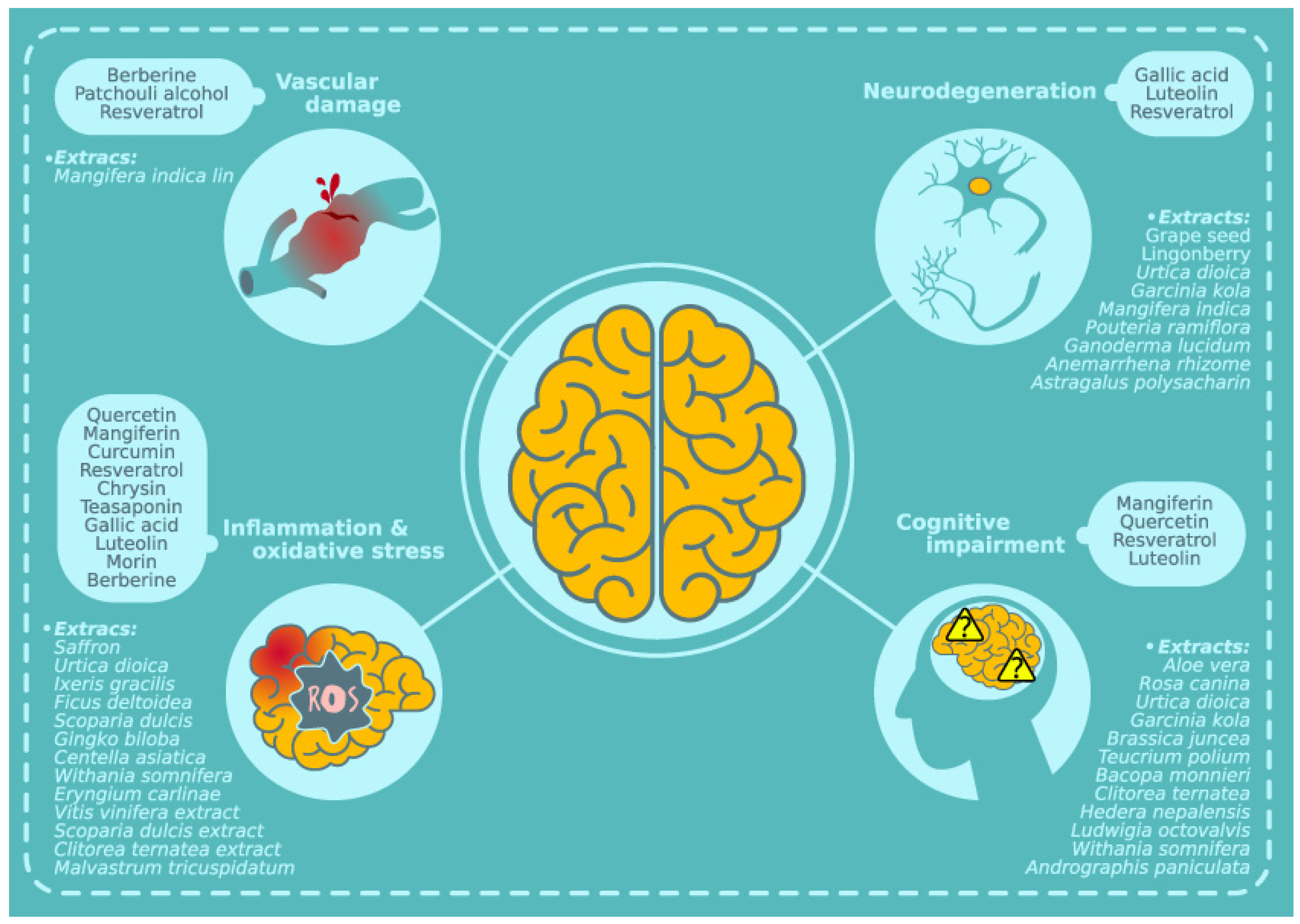
:1. Type 2 Diabetes Mellitus: Central Complications
2. Natural Compounds and Central Complications in DM
2.1. Natural Compounds and DM-Related Vascular Injury
2.1.1. Vascular Damage and DM
2.1.2. Natural Compounds and Extracts in Vascular Damage Associated with DM
2.2. Natural Compounds and Neuroinflammation Associated with DM
2.2.1. Brain Neuroinflammation and DM
2.2.2. Effect of Natural Compounds on DM-Related Inflammation
2.3. Natural Compounds and Brain Neurodegeneration in DM
2.3.1. Neurodegeneration in Diabetic Brain
2.3.2. Effect of Natural Compounds and Extracts on Brain Neurodegeneration Associated with DM
2.4. Natural Compounds and Cognitive Impairment in DM
2.4.1. Cognitive Dysfunction Associated with Diabetes
2.4.2. Effect of Natural Compounds and Extracts on Cognitive Impairment Associated with DM
Author Contributions
Funding
Conflicts of Interest
Abbreviations
| AGEs | Advanced glycation end products |
| DM | Diabetes mellitus |
| SOD | Superoxide dismutase |
| STZ | Streptozotocin |
| TNF-α | Tumor necrosis factor α |
| T1D | Type 1 diabetes |
| T2D | Type 2 diabetes |
| WHO | World Health Organization |
References
- Cornier, M.A.; Dabelea, D.; Hernandez, T.L.; Lindstrom, R.; Steig, A.J.; Stob, N.R.; Van Pelt, R.E.; Wang, H.; Eckel, R.H. The metabolic syndrome. Endocr. Rev. 2008, 29, 777–822. [Google Scholar] [CrossRef] [PubMed]
- Craft, S. The role of metabolic disorders in Alzheimer disease and vascular dementia: two roads converged. Arch Neurol. 2009, 66, 300–305. [Google Scholar] [CrossRef]
- Forouhi, N.G.; Wareham, N.J. Epidemiology of diabetes. Medicine 2014, 42, 698–702. [Google Scholar] [CrossRef] [PubMed] [Green Version]
- World Health Organization. Diabetes. Available online: https://www.who.int/diabetes/en/ (accessed on 30 April 2019).
- Skyler, J.S.; Bakris, G.L.; Bonifacio, E.; Darsow, T.; Eckel, R.H.; Groop, L.; Groop, P.H.; Handelsman, Y.; Insel, R.A.; Mathieu, C.; et al. Differentiation of Diabetes by Pathophysiology, Natural History, and Prognosis. Diabetes 2017, 66, 241–255. [Google Scholar] [CrossRef]
- Craig, M.E.; Jefferies, C.; Dabelea, D.; Balde, N.; Seth, A.; Donaghue, K.C. ISPAD Clinical Practice Consensus Guidelines Definition, epidemiology, and classification of diabetes in children and adolescents. Pediatr. Diabetes 2014, 15, 4–17. [Google Scholar] [CrossRef]
- Lascar, N.; Brown, J.; Pattison, H.; Barnett, A.H.; Bailey, C.J.; Bellary, S. Type 2 diabetes in adolescents and young adults. Lancet Diabetes Endocrinol. 2018, 6, 69–80. [Google Scholar] [CrossRef]
- Martin-Timon, I.; Sevillano-Collantes, C.; Segura-Galindo, A.; Del Canizo-Gomez, F.J. Type 2 diabetes and cardiovascular disease: Have all risk factors the same strength? World J. Diabetes 2014, 5, 444–470. [Google Scholar] [CrossRef]
- Rosenson, R.S.; Fioretto, P.; Dodson, P.M. Does microvascular disease predict macrovascular events in type 2 diabetes? Atherosclerosis 2011, 218, 13–18. [Google Scholar] [CrossRef]
- Craft, S. Alzheimer disease: Insulin resistance and AD--extending the translational path. Nat. Rev. Neurol. 2012, 8, 360–362. [Google Scholar] [CrossRef]
- Strachan, M.W.; Reynolds, R.M.; Frier, B.M.; Mitchell, R.J.; Price, J.F. The role of metabolic derangements and glucocorticoid excess in the aetiology of cognitive impairment in type 2 diabetes. Implications for future therapeutic strategies. Diabetes Obesity Metab. 2009, 11, 407–414. [Google Scholar] [CrossRef] [PubMed]
- Hamed, S.A. Brain injury with diabetes mellitus: evidence, mechanisms and treatment implications. Expert Rev. Clin. Pharmacol. 2017, 10, 409–428. [Google Scholar]
- Kodl, C.T.; Franc, D.T.; Rao, J.P.; Anderson, F.S.; Thomas, W.; Mueller, B.A.; Lim, K.O.; Seaquist, E.R. Diffusion tensor imaging identifies deficits in white matter microstructure in subjects with type 1 diabetes that correlate with reduced neurocognitive function. Diabetes 2008, 57, 3083–3089. [Google Scholar] [CrossRef]
- Ryan, C.M.; Geckle, M.O.; Orchard, T.J. Cognitive efficiency declines over time in adults with Type 1 diabetes: effects of micro- and macrovascular complications. Diabetologia 2003, 46, 940–948. [Google Scholar] [CrossRef] [Green Version]
- Moran, C.; Beare, R.; Phan, T.G.; Bruce, D.G.; Callisaya, M.L.; Srikanth, V.; Alzheimer’s Disease Neuroimaging Initiative (ADNI). Type 2 diabetes mellitus and biomarkers of neurodegeneration. Neurology 2015, 85, 1123–1130. [Google Scholar] [CrossRef] [Green Version]
- Fishel, M.A.; Watson, G.S.; Montine, T.J.; Wang, Q.; Green, P.S.; Kulstad, J.J.; Cook, D.G.; Peskind, E.R.; Baker, L.D.; Goldgaber, D.; et al. Hyperinsulinemia provokes synchronous increases in central inflammation and beta-amyloid in normal adults. Arch. Neurol. 2005, 62, 1539–1544. [Google Scholar] [CrossRef]
- Wang, T.; Fu, F.; Han, B.; Zhang, L.; Zhang, X. Danshensu ameliorates the cognitive decline in streptozotocin-induced diabetic mice by attenuating advanced glycation end product-mediated neuroinflammation. J. Neuroimmunol. 2012, 245, 79–86. [Google Scholar] [CrossRef]
- Infante-Garcia, C.; Jose Ramos-Rodriguez, J.; Marin-Zambrana, Y.; Teresa Fernandez-Ponce, M.; Casas, L.; Mantell, C.; Garcia-Alloza, M. Mango leaf extract improves central pathology and cognitive impairment in a type 2 diabetes mouse model. Brain Pathol. 2017, 27, 499–507. [Google Scholar] [CrossRef]
- Moran, C.; Beare, R.; Wang, W.; Callisaya, M.; Srikanth, V.; Alzheimer’s Disease Neuroimaging Initiative (ADNI). Type 2 diabetes mellitus, brain atrophy, and cognitive decline. Neurology 2019, 92, e823–e830. [Google Scholar] [CrossRef]
- Luchsinger, J.A.; Reitz, C.; Honig, L.S.; Tang, M.X.; Shea, S.; Mayeux, R. Aggregation of vascular risk factors and risk of incident Alzheimer disease. Neurology 2005, 65, 545–551. [Google Scholar] [CrossRef] [Green Version]
- Luchsinger, J.A.; Tang, M.X.; Shea, S.; Mayeux, R. Hyperinsulinemia and risk of Alzheimer disease. Neurology 2004, 63, 1187–1192. [Google Scholar] [CrossRef]
- Matsuzaki, T.; Sasaki, K.; Tanizaki, Y.; Hata, J.; Fujimi, K.; Matsui, Y.; Sekita, A.; Suzuki, S.O.; Kanba, S.; Kiyohara, Y.; et al. Insulin resistance is associated with the pathology of Alzheimer disease: the Hisayama study. Neurology 2010, 75, 764–770. [Google Scholar] [CrossRef]
- Schrijvers, E.M.; Witteman, J.C.; Sijbrands, E.J.; Hofman, A.; Koudstaal, P.J.; Breteler, M.M. Insulin metabolism and the risk of Alzheimer disease: the Rotterdam Study. Neurology 2010, 75, 1982–1987. [Google Scholar] [CrossRef]
- Strachan, M.W.; Reynolds, R.M.; Frier, B.M.; Mitchell, R.J.; Price, J.F. The relationship between type 2 diabetes and dementia. Br. Med. Bull. 2008, 88, 131–146. [Google Scholar] [CrossRef]
- Newman, D.J.; Cragg, G.M. Natural Products as Sources of New Drugs from 1981 to 2014. J. Nat. Prod. 2016, 79, 629–661. [Google Scholar] [CrossRef] [Green Version]
- Flores-Jimenez, N.G.; Rojas-Lemus, M.; Fortoul, T.I.; Zepeda-Rodriguez, A.; Lopez-Camacho, P.Y.; Anacleto-Santos, J.; Malagon-Gutierrez, F.; Basurto-Islas, G.; Rivera-Fernandez, N. Histopathological alterations in mice under sub-acute treatment with Hintonia latiflora methanolic stem bark extract. Histol. Histopathol. 2018, 33, 1299–1309. [Google Scholar]
- Spagnuolo, C.; Napolitano, M.; Tedesco, I.; Moccia, S.; Milito, A.; Russo, G.L. Neuroprotective Role of Natural Polyphenols. Curr. Top. Med. Chem. 2016, 16, 1943–1950. [Google Scholar] [CrossRef]
- Cheynier, V.; Comte, G.; Davies, K.M.; Lattanzio, V.; Martens, S. Plant phenolics: recent advances on their biosynthesis, genetics, and ecophysiology. Plant Physiol. Biochem. 2013, 72, 1–20. [Google Scholar] [CrossRef]
- Sevastre-Berghian, A.C.; Toma, V.A.; Sevastre, B.; Hanganu, D.; Vlase, L.; Benedec, D.; Oniga, I.; Baldea, I.; Olteanu, D.; Moldovan, R.; et al. Characterization and biological effects of Hypericum extracts on experimentally-induced - anxiety, oxidative stress and inflammation in rats. J. Physiol. Pharmacol. 2018, 6, 9. [Google Scholar]
- Spagnuolo, C.; Moccia, S.; Russo, G.L. Anti-inflammatory effects of flavonoids in neurodegenerative disorders. Eur. J. Med. Chem. 2018, 153, 105–115. [Google Scholar] [CrossRef]
- Lima, M.C.; Paiva de Sousa, C.; Fernandez-Prada, C.; Harel, J.; Dubreuil, J.D.; de Souza, E.L. A review of the current evidence of fruit phenolic compounds as potential antimicrobials against pathogenic bacteria. Microb. Pathog. 2019, 130, 259–270. [Google Scholar] [CrossRef]
- Christman, L.M.; Dean, L.L.; Allen, J.C.; Godinez, S.F.; Toomer, O.T. Peanut skin phenolic extract attenuates hyperglycemic responses in vivo and in vitro. PloS ONE 2019, 14, e0214591. [Google Scholar] [CrossRef]
- Pohl, F.; Kong Thoo Lin, P. The Potential Use of Plant Natural Products and Plant Extracts with Antioxidant Properties for the Prevention/Treatment of Neurodegenerative Diseases: In Vitro, In Vivo and Clinical Trials. Molecules 2018, 23, 3283. [Google Scholar] [CrossRef]
- Infante-Garcia, C.; Ramos-Rodriguez, J.J.; Delgado-Olmos, I.; Gamero-Carrasco, C.; Fernandez-Ponce, M.T.; Casas, L.; Mantell, C.; Garcia-Alloza, M. Long-Term Mangiferin Extract Treatment Improves Central Pathology and Cognitive Deficits in APP/PS1 Mice. Mol. Neurobiol. 2017, 54, 4696–4704. [Google Scholar] [CrossRef]
- Figueira, I.; Menezes, R.; Macedo, D.; Costa, I.; Dos Santos, C.N. Polyphenols Beyond Barriers: A Glimpse into the Brain. Curr. Neuropharmacol. 2017, 15, 562–594. [Google Scholar] [CrossRef] [Green Version]
- Tsao, R. Chemistry and biochemistry of dietary polyphenols. Nutrients 2010, 2, 1231–1246. [Google Scholar] [CrossRef]
- Garcia-Alloza, M.; Dodwell, S.A.; Meyer-Luehmann, M.; Hyman, B.T.; Bacskai, B.J. Plaque-derived oxidative stress mediates distorted neurite trajectories in the Alzheimer mouse model. J. Neuropathol. Exp. Neurol. 2006, 65, 1082–1089. [Google Scholar] [CrossRef]
- Liu, Y.W.; Liu, X.L.; Kong, L.; Zhang, M.Y.; Chen, Y.J.; Zhu, X.; Hao, Y.C. Neuroprotection of quercetin on central neurons against chronic high glucose through enhancement of Nrf2/ARE/glyoxalase-1 pathway mediated by phosphorylation regulation. Biomed. Pharmacother. 2019, 109, 2145–2154. [Google Scholar] [CrossRef]
- Fu, Q.Y.; Li, Q.S.; Lin, X.M.; Qiao, R.Y.; Yang, R.; Li, X.M.; Dong, Z.B.; Xiang, L.P.; Zheng, X.Q.; Lu, J.L.; et al. Antidiabetic Effects of Tea. Molecules 2017, 22, 849. [Google Scholar] [CrossRef]
- Dominguez Avila, J.A.; Rodrigo Garcia, J.; Gonzalez Aguilar, G.A.; de la Rosa, L.A. The Antidiabetic Mechanisms of Polyphenols Related to Increased Glucagon-Like Peptide-1 (GLP1) and Insulin Signaling. Molecules 2017, 22, 903. [Google Scholar] [CrossRef]
- Serna-Thome, G.; Castro-Eguiluz, D.; Fuchs-Tarlovsky, V.; Sanchez-Lopez, M.; Delgado-Olivares, L.; Coronel-Martinez, J.; Molina-Trinidad, E.M.; de la Torre, M.; Cetina-Perez, L. Use of Functional Foods and Oral Supplements as Adjuvants in Cancer Treatment. Rev. Inves. Clin. 2018, 70, 136–146. [Google Scholar] [CrossRef] [Green Version]
- Biessels, G.J.; Staekenborg, S.; Brunner, E.; Brayne, C.; Scheltens, P. Risk of dementia in diabetes mellitus: A systematic review. Lancet Neurol. 2006, 5, 64–74. [Google Scholar] [CrossRef]
- Crane, P.K.; Walker, R.; Hubbard, R.A.; Li, G.; Nathan, D.M.; Zheng, H.; Haneuse, S.; Craft, S.; Montine, T.J.; Kahn, S.E.; et al. Glucose levels and risk of dementia. N. Engl. J. Med. 2013, 369, 540–548. [Google Scholar] [CrossRef] [PubMed]
- Infante-Garcia, C.; Ramos-Rodriguez, J.J.; Hierro-Bujalance, C.; Ortegon, E.; Pickett, E.; Jackson, R.; Hernandez-Pacho, F.; Spires-Jones, T.; Garcia-Alloza, M. Antidiabetic Polypill Improves Central Pathology and Cognitive Impairment in a Mixed Model of Alzheimer’s Disease and Type 2 Diabetes. Mol. Neurobiol. 2018, 55, 6130–6144. [Google Scholar] [CrossRef] [PubMed]
- Munhoz, A.C.M.; Frode, T.S. Isolated Compounds from Natural Products with Potential Antidiabetic Activity - A Systematic Review. Curr. Diabetes Rev. 2018, 14, 36–106. [Google Scholar] [CrossRef]
- Chen, T.Y.; Ferruzzi, M.G.; Wu, Q.L.; Simon, J.E.; Talcott, S.T.; Wang, J.; Ho, L.; Todd, G.; Cooper, B.; Pasinetti, G.M.; et al. Influence of diabetes on plasma pharmacokinetics and brain bioavailability of grape polyphenols and their phase II metabolites in the Zucker diabetic fatty rat. Mol. Nutr. Food Res. 2017, 61, 1700111. [Google Scholar] [CrossRef]
- Domingueti, C.P.; Dusse, L.M.; Carvalho, M.; de Sousa, L.P.; Gomes, K.B.; Fernandes, A.P. Diabetes mellitus: The linkage between oxidative stress, inflammation, hypercoagulability and vascular complications. J. Diabetes Complicat. 2016, 30, 738–745. [Google Scholar] [CrossRef]
- Goldberg, R.B. Cytokine and cytokine-like inflammation markers, endothelial dysfunction, and imbalanced coagulation in development of diabetes and its complications. J. Clin. Endocrinol. Metab. 2009, 94, 3171–3182. [Google Scholar] [CrossRef]
- Wautier, J.L.; Guillausseau, P.J. Diabetes, advanced glycation endproducts and vascular disease. Vasc. Med. 1998, 3, 131–137. [Google Scholar] [CrossRef] [Green Version]
- Reddy, G.K. AGE-related cross-linking of collagen is associated with aortic wall matrix stiffness in the pathogenesis of drug-induced diabetes in rats. Microvasc. Res. 2004, 68, 132–142. [Google Scholar] [CrossRef]
- Idris, I.; Thomson, G.A.; Sharma, J.C. Diabetes mellitus and stroke. Int. J. Clin. Pract. 2006, 60, 48–56. [Google Scholar] [CrossRef]
- Callahan, A.; Amarenco, P.; Goldstein, L.B.; Sillesen, H.; Messig, M.; Samsa, G.P.; Altafullah, I.; Ledbetter, L.Y.; MacLeod, M.J.; Scott, R.; et al. Risk of stroke and cardiovascular events after ischemic stroke or transient ischemic attack in patients with type 2 diabetes or metabolic syndrome: secondary analysis of the Stroke Prevention by Aggressive Reduction in Cholesterol Levels (SPARCL) trial. Arch. Neurol. 2011, 68, 1245–1251. [Google Scholar] [CrossRef] [PubMed]
- Mogi, M.; Horiuchi, M. Neurovascular coupling in cognitive impairment associated with diabetes mellitus. Circ. J. 2011, 75, 1042–1048. [Google Scholar] [CrossRef] [PubMed]
- Hayden, M.R.; Grant, D.G.; Aroor, A.R.; DeMarco, V.G. Empagliflozin Ameliorates Type 2 Diabetes-Induced Ultrastructural Remodeling of the Neurovascular Unit and Neuroglia in the Female db/db Mouse. Brain Sci. 2019, 9, 57. [Google Scholar] [CrossRef]
- Infante-Garcia, C.; Ramos-Rodriguez, J.J.; Galindo-Gonzalez, L.; Garcia-Alloza, M. Long-term central pathology and cognitive impairment are exacerbated in a mixed model of Alzheimer’s disease and type 2 diabetes. Psychoneuroendocrinology 2016, 65, 15–25. [Google Scholar] [CrossRef] [PubMed]
- Ramos-Rodriguez, J.J.; Jimenez-Palomares, M.; Murillo-Carretero, M.I.; Infante-Garcia, C.; Berrocoso, E.; Hernandez-Pacho, F.; Lechuga-Sancho, A.M.; Cozar-Castellano, I.; Garcia-Alloza, M. Central vascular disease and exacerbated pathology in a mixed model of type 2 diabetes and Alzheimer’s disease. Psychoneuroendocrinology 2015, 62, 69–79. [Google Scholar] [CrossRef] [PubMed]
- Zhang, L.; Chopp, M.; Zhang, Y.; Xiong, Y.; Li, C.; Sadry, N.; Rhaleb, I.; Lu, M.; Zhang, Z.G. Diabetes Mellitus Impairs Cognitive Function in Middle-Aged Rats and Neurological Recovery in Middle-Aged Rats After Stroke. Stroke 2016, 47, 2112–2118. [Google Scholar] [CrossRef] [Green Version]
- Pasquier, F.; Boulogne, A.; Leys, D.; Fontaine, P. Diabetes mellitus and dementia. Diabetes Metab. 2006, 32, 403–414. [Google Scholar] [CrossRef]
- Wang, S.; Cao, C.; Chen, Z.; Bankaitis, V.; Tzima, E.; Sheibani, N.; Burridge, K. Pericytes regulate vascular basement membrane remodeling and govern neutrophil extravasation during inflammation. PloS ONE 2012, 7, e45499. [Google Scholar] [CrossRef]
- Bogush, M.; Heldt, N.A.; Persidsky, Y. Blood Brain Barrier Injury in Diabetes: Unrecognized Effects on Brain and Cognition. J. Neuroimmune Pharmacol. 2017, 12, 593–601. [Google Scholar] [CrossRef]
- Manasson, J.; Tien, T.; Moore, C.; Kumar, N.M.; Roy, S. High glucose-induced downregulation of connexin 30.2 promotes retinal vascular lesions: implications for diabetic retinopathy. Investig. Ophthalmol. Vis. Sci. 2013, 54, 2361–2366. [Google Scholar] [CrossRef]
- Sajja, R.K.; Prasad, S.; Cucullo, L. Impact of altered glycaemia on blood-brain barrier endothelium: an in vitro study using the hCMEC/D3 cell line. Fluids Barriers CNS. 2014, 11, 8. [Google Scholar] [CrossRef]
- Li, B.; Li, Y.; Liu, K.; Wang, X.; Qi, J.; Wang, B.; Wang, Y. High glucose decreases claudins-5 and -11 in cardiac microvascular endothelial cells: Antagonistic effects of tongxinluo. Endocr. Res. 2017, 42, 15–21. [Google Scholar] [CrossRef]
- Maile, L.A.; Gollahon, K.; Wai, C.; Dunbar, P.; Busby, W.; Clemmons, D. Blocking alphaVbeta3 integrin ligand occupancy inhibits the progression of albuminuria in diabetic rats. J. Diabetes Res. 2014, 2014, 421827. [Google Scholar] [CrossRef]
- Park, S.W.; Yun, J.H.; Kim, J.H.; Kim, K.W.; Cho, C.H.; Kim, J.H. Angiopoietin 2 induces pericyte apoptosis via alpha3beta1 integrin signaling in diabetic retinopathy. Diabetes 2014, 63, 3057–3068. [Google Scholar] [CrossRef]
- Lee, Y.J.; Jung, S.H.; Kim, S.H.; Kim, M.S.; Lee, S.; Hwang, J.; Kim, S.Y.; Kim, Y.M.; Ha, K.S. Essential Role of Transglutaminase 2 in Vascular Endothelial Growth Factor-Induced Vascular Leakage in the Retina of Diabetic Mice. Diabetes 2016, 65, 2414–2428. [Google Scholar] [CrossRef]
- Abu El-Asrar, A.M.; Mohammad, G.; Nawaz, M.I.; Abdelsaid, M.; Siddiquei, M.M.; Alam, K.; Van den Eynde, K.; De Hertogh, G.; Opdenakker, G.; Al-Shabrawey, M.; et al. The Chemokine Platelet Factor-4 Variant (PF-4var)/CXCL4L1 Inhibits Diabetes-Induced Blood-Retinal Barrier Breakdown. Investig. Ophthalmol. Vis. Sci. 2015, 56, 1956–1964. [Google Scholar] [CrossRef]
- Price, T.O.; Eranki, V.; Banks, W.A.; Ercal, N.; Shah, G.N. Topiramate treatment protects blood-brain barrier pericytes from hyperglycemia-induced oxidative damage in diabetic mice. Endocrinology 2012, 153, 362–372. [Google Scholar] [CrossRef]
- Takechi, R.; Lam, V.; Brook, E.; Giles, C.; Fimognari, N.; Mooranian, A.; Al-Salami, H.; Coulson, S.H.; Nesbit, M.; Mamo, J.C.L. Blood-Brain Barrier Dysfunction Precedes Cognitive Decline and Neurodegeneration in Diabetic Insulin Resistant Mouse Model: An Implication for Causal Link. Front. Aging Neurosci. 2017, 9, 399. [Google Scholar] [CrossRef]
- Ramos-Rodriguez, J.J.; Ortiz, O.; Jimenez-Palomares, M.; Kay, K.R.; Berrocoso, E.; Murillo-Carretero, M.I.; Perdomo, G.; Spires-Jones, T.; Cozar-Castellano, I.; Lechuga-Sancho, A.M.; et al. Differential central pathology and cognitive impairment in pre-diabetic and diabetic mice. Psychoneuroendocrinology 2013, 38, 2462–2475. [Google Scholar] [CrossRef]
- Jin, Y.; Khadka, D.B.; Cho, W.J. Pharmacological effects of berberine and its derivatives: A patent update. Expert Opin. Ther. Pat. 2016, 26, 229–243. [Google Scholar] [CrossRef]
- Yin, J.; Xing, H.; Ye, J. Efficacy of berberine in patients with type 2 diabetes mellitus. Metabolism 2008, 57, 712–717. [Google Scholar] [CrossRef] [Green Version]
- Yin, S.; Bai, W.; Li, P.; Jian, X.; Shan, T.; Tang, Z.; Jing, X.; Ping, S.; Li, Q.; Miao, Z.; et al. Berberine suppresses the ectopic expression of miR-133a in endothelial cells to improve vascular dementia in diabetic rats. Clin. Exp. Hypertens. 2018, 1–9. [Google Scholar] [CrossRef]
- Wei, L.L.; Chen, Y.; Yu, Q.Y.; Wang, Y.; Liu, G. Patchouli alcohol protects against ischemia/reperfusion-induced brain injury via inhibiting neuroinflammation in normal and obese mice. Brain Res. 2018, 1682, 61–70. [Google Scholar] [CrossRef]
- Patel, S.S.; Udayabanu, M. Effect of natural products on diabetes associated neurological disorders. Rev. Neurosci. 2017, 28, 271–293. [Google Scholar] [CrossRef]
- Wong, R.H.; Raederstorff, D.; Howe, P.R. Acute Resveratrol Consumption Improves Neurovascular Coupling Capacity in Adults with Type 2 Diabetes Mellitus. Nutrients 2016, 8, 425. [Google Scholar] [CrossRef]
- Jing, Y.H.; Chen, K.H.; Kuo, P.C.; Pao, C.C.; Chen, J.K. Neurodegeneration in streptozotocin-induced diabetic rats is attenuated by treatment with resveratrol. Neuroendocrinology 2013, 98, 116–127. [Google Scholar] [CrossRef]
- Newcombe, E.A.; Camats-Perna, J.; Silva, M.L.; Valmas, N.; Huat, T.J.; Medeiros, R. Inflammation: The link between comorbidities, genetics, and Alzheimer’s disease. J. Neuroinflammation 2018, 15, 276. [Google Scholar] [CrossRef]
- Hotamisligil, G.S. Inflammation and metabolic disorders. Nature 2006, 444, 860–867. [Google Scholar] [CrossRef]
- Hotamisligil, G.S.; Shargill, N.S.; Spiegelman, B.M. Adipose expression of tumor necrosis factor-alpha: Direct role in obesity-linked insulin resistance. Science 1993, 259, 87–91. [Google Scholar] [CrossRef]
- Wellen, K.E.; Hotamisligil, G.S. Inflammation, stress, and diabetes. J. Clin. Investig. 2005, 115, 1111–1119. [Google Scholar] [CrossRef] [Green Version]
- Colombo, E.; Farina, C. Astrocytes: Key Regulators of Neuroinflammation. Trends Immunol. 2016, 37, 608–620. [Google Scholar] [CrossRef]
- Jeong, H.K.; Ji, K.; Min, K.; Joe, E.H. Brain inflammation and microglia: Facts and misconceptions. Exp. Neurobiol. 2013, 22, 59–67. [Google Scholar] [CrossRef] [PubMed]
- Ransohoff, R.M.; Engelhardt, B. The anatomical and cellular basis of immune surveillance in the central nervous system. Nat. Rev. Immunol. 2012, 12, 623–635. [Google Scholar] [CrossRef]
- Ferreira, R.; Bernardino, L. Dual role of microglia in health and disease: pushing the balance toward repair. Front Cell Neurosci. 2015, 9, 51. [Google Scholar] [CrossRef]
- Chen, Z.; Trapp, B.D. Microglia and neuroprotection. J. Neurochem. 2016, 136, 10–17. [Google Scholar] [CrossRef]
- Ramos-Rodriguez, J.J.; Ortiz-Barajas, O.; Gamero-Carrasco, C.; de la Rosa, P.R.; Infante-Garcia, C.; Zopeque-Garcia, N.; Lechuga-Sancho, A.M.; Garcia-Alloza, M. Prediabetes-induced vascular alterations exacerbate central pathology in APPswe/PS1dE9 mice. Psychoneuroendocrinology 2014, 48, 123–135. [Google Scholar] [CrossRef]
- Ramos-Rodriguez, J.J.; Infante-Garcia, C.; Galindo-Gonzalez, L.; Garcia-Molina, Y.; Lechuga-Sancho, A.; Garcia-Alloza, M. Increased Spontaneous Central Bleeding and Cognition Impairment in APP/PS1 Mice with Poorly Controlled Diabetes Mellitus. Mol. Neurobiol. 2016, 53, 2685–2697. [Google Scholar] [CrossRef]
- Ramos-Rodriguez, J.J.; Sanchez-Sotano, D.; Doblas-Marquez, A.; Infante-Garcia, C.; Lubian-Lopez, S.; Garcia-Alloza, M. Intranasal insulin reverts central pathology and cognitive impairment in diabetic mother offspring. Mol. Neurodegener. 2017, 12, 57. [Google Scholar] [CrossRef]
- Van Dyken, P.; Lacoste, B. Impact of Metabolic Syndrome on Neuroinflammation and the Blood-Brain Barrier. Front. Neurosci. 2018, 12, 930. [Google Scholar] [CrossRef]
- Hwang, I.K.; Choi, J.H.; Nam, S.M.; Park, O.K.; Yoo, D.Y.; Kim, W.; Yi, S.S.; Won, M.H.; Seong, J.K.; Yoon, Y.S. Activation of microglia and induction of pro-inflammatory cytokines in the hippocampus of type 2 diabetic rats. Neurol. Res. 2014, 36, 824–832. [Google Scholar] [CrossRef]
- Ibrahim, A.S.; El-Shishtawy, M.M.; Pena, A., Jr.; Liou, G.I. Genistein attenuates retinal inflammation associated with diabetes by targeting of microglial activation. Mol. Vis. 2010, 16, 2033–2042. [Google Scholar]
- Lauridsen, J.K.; Olesen, R.H.; Vendelbo, J.; Hyde, T.M.; Kleinman, J.E.; Bibby, B.M.; Brock, B.; Rungby, J.; Larsen, A. High BMI levels associate with reduced mRNA expression of IL10 and increased mRNA expression of iNOS (NOS2) in human frontal cortex. Transl. Psychiatry 2017, 7, e1044. [Google Scholar] [CrossRef]
- Chen, W.; Jia, Z.; Pan, M.-H.; Babu, P.V.A. Natural Products for the Prevention of Oxidative Stress-Related Diseases: Mechanisms and Strategies. Oxidative Med. Cell. Longev. 2016, 2016, 1–2. [Google Scholar] [CrossRef]
- Jia, Z.; Babu, P.V.A.; Chen, W.; Sun, X. Natural Products Targeting on Oxidative Stress and Inflammation: Mechanisms, Therapies, and Safety Assessment. Oxidative Med. Cell. Longev. 2018, 2018, 1–3. [Google Scholar] [CrossRef] [Green Version]
- Maitra, I.; Marcocci, L.; Droy-Lefaix, M.T.; Packer, L. Peroxyl radical scavenging activity of Ginkgo biloba extract EGb 761. Biochem. Pharmacol. 1995, 49, 1649–1655. [Google Scholar] [CrossRef]
- Frandsen, J.R.; Narayanasamy, P. Neuroprotection through flavonoid: Enhancement of the glyoxalase pathway. Redox Biol. 2018, 14, 465–473. [Google Scholar] [CrossRef]
- Mahesh, T.; Menon, V.P. Quercetin allievates oxidative stress in streptozotocin-induced diabetic rats. Phytother. Res. 2004, 18, 123–127. [Google Scholar] [CrossRef]
- Bournival, J.; Francoeur, M.A.; Renaud, J.; Martinoli, M.G. Quercetin and sesamin protect neuronal PC12 cells from high-glucose-induced oxidation, nitrosative stress, and apoptosis. Rejuvenation Res. 2012, 15, 322–333. [Google Scholar] [CrossRef]
- Marquez, L.; Garcia-Bueno, B.; Madrigal, J.L.; Leza, J.C. Mangiferin decreases inflammation and oxidative damage in rat brain after stress. Eur. J. Nutr. 2012, 51, 729–739. [Google Scholar] [CrossRef]
- Liu, Y.W.; Cheng, Y.Q.; Liu, X.L.; Hao, Y.C.; Li, Y.; Zhu, X.; Zhang, F.; Yin, X.X. Mangiferin Upregulates Glyoxalase 1 Through Activation of Nrf2/ARE Signaling in Central Neurons Cultured with High Glucose. Mol. Neurobiol. 2017, 54, 4060–4070. [Google Scholar] [CrossRef]
- Faheem, N.M.; El Askary, A. Neuroprotective role of curcumin on the hippocampus against the structural and serological alterations of streptozotocin-induced diabetes in Sprague Dawely rats. Iran. J. Basic Med. Sci. 2017, 20, 690–699. [Google Scholar]
- Daugherty, D.J.; Marquez, A.; Calcutt, N.A.; Schubert, D. A novel curcumin derivative for the treatment of diabetic neuropathy. Neuropharmacology 2018, 129, 26–35. [Google Scholar] [CrossRef]
- Sadi, G.; Konat, D. Resveratrol regulates oxidative biomarkers and antioxidant enzymes in the brain of streptozotocin-induced diabetic rats. Pharm. Biol. 2016, 54, 1156–1163. [Google Scholar] [CrossRef]
- Nurdiana, S.; Goh, Y.M.; Hafandi, A.; Dom, S.M.; Nur Syimal’ain, A.; Noor Syaffinaz, N.M.; Ebrahimi, M. Improvement of spatial learning and memory, cortical gyrification patterns and brain oxidative stress markers in diabetic rats treated with Ficus deltoidea leaf extract and vitexin. J. Tradit. Complement. Med. 2018, 8, 190–202. [Google Scholar] [CrossRef]
- Samarghandian, S.; Azimi-Nezhad, M.; Samini, F. Ameliorative Effect of Saffron Aqueous Extract on Hyperglycemia, Hyperlipidemia, and Oxidative Stress on Diabetic Encephalopathy in Streptozotocin Induced Experimental Diabetes Mellitus. BioMed Int. 2014, 2014, 1–12. [Google Scholar] [CrossRef] [Green Version]
- Pari, L.; Latha, M. Protective role of Scoparia dulcis plant extract on brain antioxidant status and lipidperoxidation in STZ diabetic male Wistar rats. BMC Complement Altern Med. 2004, 4, 16. [Google Scholar] [CrossRef]
- Li, R.; Zang, A.; Zhang, L.; Zhang, H.; Zhao, L.; Qi, Z.; Wang, H. Chrysin ameliorates diabetes-associated cognitive deficits in Wistar rats. Neurol. Sci. 2014, 35, 1527–1532. [Google Scholar] [CrossRef]
- Marcocci, L.; Packer, L.; Droy-Lefaix, M.T.; Sekaki, A.; Gardes-Albert, M. Antioxidant action of Ginkgo biloba extract EGb. Methods Enzymol. 1994, 234, 462–475. [Google Scholar]
- Choi, H.R.; Choi, J.S.; Han, Y.N.; Bae, S.J.; Chung, H.Y. Peroxynitrite scavenging activity of herb extracts. Phytother. Res. 2002, 16, 364–367. [Google Scholar] [CrossRef]
- Yu, Y.; Wu, Y.; Szabo, A.; Wu, Z.; Wang, H.; Li, D.; Huang, X.F. Teasaponin reduces inflammation and central leptin resistance in diet-induced obese male mice. Endocrinology 2013, 154, 3130–3140. [Google Scholar] [CrossRef]
- Talpate, K.A.; Bhosale, U.A.; Zambare, M.R.; Somani, R.S. Neuroprotective and nootropic activity of Clitorea ternatea Linn.(Fabaceae) leaves on diabetes induced cognitive decline in experimental animals. J. Pharm. Bioallied Sci. 2014, 6, 48–55. [Google Scholar]
- Yonguc, G.N.; Dodurga, Y.; Adiguzel, E.; Gundogdu, G.; Kucukatay, V.; Ozbal, S.; Yilmaz, I.; Cankurt, U.; Yilmaz, Y.; Akdogan, I. Grape seed extract has superior beneficial effects than vitamin E on oxidative stress and apoptosis in the hippocampus of streptozotocin induced diabetic rats. Gene 2015, 555, 119–126. [Google Scholar] [CrossRef]
- Lu, M.; Xu, L.; Li, B.; Zhang, W.; Zhang, C.; Feng, H.; Cui, X.; Gao, H. Protective effects of grape seed proanthocyanidin extracts on cerebral cortex of streptozotocin-induced diabetic rats through modulating AGEs/RAGE/NF-kappaB pathway. J. Nutr. Sci. Vitaminol. 2010, 56, 87–97. [Google Scholar] [CrossRef]
- Patel, S.S.; Gupta, S.; Udayabanu, M. Urtica dioica modulates hippocampal insulin signaling and recognition memory deficit in streptozotocin induced diabetic mice. Metab. Brain Dis. 2016, 31, 601–611. [Google Scholar] [CrossRef]
- Patel, S.S.; Parashar, A.; Udayabanu, M. Urtica dioica leaves modulates muscarinic cholinergic system in the hippocampus of streptozotocin-induced diabetic mice. Metab. Brain Dis. 2015, 30, 803–811. [Google Scholar] [CrossRef]
- Jahanshahi, M.; Golalipour, M.J.; Afshar, M. The effect of Urtica dioica extract on the number of astrocytes in the dentate gyrus of diabetic rats. Folia Morphol. 2009, 68, 93–97. [Google Scholar]
- Abdel-Moneim, A.; Yousef, A.I.; Abd El-Twab, S.M.; Abdel Reheim, E.S.; Ashour, M.B. Gallic acid and p-coumaric acid attenuate type 2 diabetes-induced neurodegeneration in rats. Metab. Brain Dis. 2017, 32, 1279–1286. [Google Scholar] [CrossRef]
- Liu, Y.; Fu, X.; Lan, N.; Li, S.; Zhang, J.; Wang, S.; Li, C.; Shang, Y.; Huang, T.; Zhang, L. Luteolin protects against high fat diet-induced cognitive deficits in obesity mice. Behav. Brain Res. 2014, 267, 178–188. [Google Scholar] [CrossRef]
- Liu, Y.; Tian, X.; Gou, L.; Sun, L.; Ling, X.; Yin, X. Luteolin attenuates diabetes-associated cognitive decline in rats. Brain Res. Bull. 2013, 94, 23–29. [Google Scholar] [CrossRef]
- Kaur, T.; Kaur, G. Withania somnifera as a potential candidate to ameliorate high fat diet-induced anxiety and neuroinflammation. J. Neuroinflammation 2017, 14, 201. [Google Scholar] [CrossRef] [Green Version]
- Pena-Montes, D.J.; Huerta-Cervantes, M.; Rios-Silva, M.; Trujillo, X.; Huerta, M.; Noriega-Cisneros, R.; Salgado-Garciglia, R.; Saavedra-Molina, A. Protective Effect of the Hexanic Extract of Eryngium carlinae Inflorescences In Vitro, in Yeast, and in Streptozotocin-Induced Diabetic Male Rats. Antioxidants 2019, 8, 73. [Google Scholar] [CrossRef]
- Ola, M.S.; Aleisa, A.M.; Al-Rejaie, S.S.; Abuohashish, H.M.; Parmar, M.Y.; Alhomida, A.S.; Ahmed, M.M. Flavonoid, morin inhibits oxidative stress, inflammation and enhances neurotrophic support in the brain of streptozotocin-induced diabetic rats. Neurol. Sci. 2014, 35, 1003–1008. [Google Scholar] [CrossRef]
- Bachewal, P.; Gundu, C.; Yerra, V.G.; Kalvala, A.K.; Areti, A.; Kumar, A. Morin exerts neuroprotection via attenuation of ROS induced oxidative damage and neuroinflammation in experimental diabetic neuropathy. BioFactors 2018, 44, 109–122. [Google Scholar] [CrossRef]
- Ola, M.S.; Ahmed, M.M.; Ahmad, R.; Abuohashish, H.M.; Al-Rejaie, S.S.; Alhomida, A.S. Neuroprotective Effects of Rutin in Streptozotocin-Induced Diabetic Rat Retina. J. Mol. Neurosci. 2015, 56, 440–448. [Google Scholar] [CrossRef]
- Moghaddam, H.K.; Baluchnejadmojarad, T.; Roghani, M.; Khaksari, M.; Norouzi, P.; Ahooie, M.; Mahboobi, F. Berberine ameliorate oxidative stress and astrogliosis in the hippocampus of STZ-induced diabetic rats. Mol. Neurobiol. 2014, 49, 820–826. [Google Scholar] [CrossRef]
- Masola, B.; Oguntibeju, O.O.; Oyenihi, A.B. Centella asiatica ameliorates diabetes-induced stress in rat tissues via influences on antioxidants and inflammatory cytokines. Biomed. Pharmacother. 2018, 101, 447–457. [Google Scholar] [CrossRef]
- Syiem, D.; Warjri, P. Antidiabetic, antioxidant, and TNF-alpha lowering properties of extract of the traditionally used plant Ixeris gracilis in alloxan-induced diabetic mice. Pharmaceutical Biol. 2015, 53, 494–502. [Google Scholar] [CrossRef]
- Solanki, I.; Parihar, P.; Shetty, R.; Parihar, M.S. Synaptosomal and mitochondrial oxidative damage followed by behavioral impairments in streptozotocin induced diabetes mellitus: restoration by Malvastrum tricuspidatum. Cell Mol. Biol. 2017, 63, 94–101. [Google Scholar] [CrossRef]
- van Harten, B.; de Leeuw, F.E.; Weinstein, H.C.; Scheltens, P.; Biessels, G.J. Brain imaging in patients with diabetes: a systematic review. Diabetes Care 2006, 29, 2539–2548. [Google Scholar] [CrossRef]
- Marseglia, A.; Fratiglioni, L.; Kalpouzos, G.; Wang, R.; Backman, L.; Xu, W. Prediabetes and diabetes accelerate cognitive decline and predict microvascular lesions: A population-based cohort study. Alzheimer’s Dement. 2019, 15, 25–33. [Google Scholar] [CrossRef]
- Ramos-Rodriguez, J.J.; Spires-Jones, T.; Pooler, A.M.; Lechuga-Sancho, A.M.; Bacskai, B.J.; Garcia-Alloza, M. Progressive Neuronal Pathology and Synaptic Loss Induced by Prediabetes and Type 2 Diabetes in a Mouse Model of Alzheimer’s Disease. Mol. Neurobiol. 2017, 54, 3428–3438. [Google Scholar] [CrossRef]
- Moran, C.; Tapp, R.J.; Hughes, A.D.; Magnussen, C.G.; Blizzard, L.; Phan, T.G.; Beare, R.; Witt, N.; Venn, A.; Munch, G.; et al. The Association of Type 2 Diabetes Mellitus with Cerebral Gray Matter Volume Is Independent of Retinal Vascular Architecture and Retinopathy. J. Diabetes Res. 2016, 2016, 6328953. [Google Scholar] [CrossRef] [PubMed]
- Bednarik, P.; Moheet, A.A.; Grohn, H.; Kumar, A.F.; Eberly, L.E.; Seaquist, E.R.; Mangia, S. Type 1 Diabetes and Impaired Awareness of Hypoglycemia Are Associated with Reduced Brain Gray Matter Volumes. Front. Neurosci. 2017, 11, 529. [Google Scholar] [CrossRef] [PubMed]
- McCrimmon, R.J.; Ryan, C.M.; Frier, B.M. Diabetes and cognitive dysfunction. Lancet 2012, 379, 2291–2299. [Google Scholar] [CrossRef]
- Kumar, A.; Haroon, E.; Darwin, C.; Pham, D.; Ajilore, O.; Rodriguez, G.; Mintz, J. Gray matter prefrontal changes in type 2 diabetes detected using MRI. J. Magn. Reson. Imaging: Jmri. 2008, 27, 14–19. [Google Scholar] [CrossRef]
- de Bresser, J.; Tiehuis, A.M.; van den Berg, E.; Reijmer, Y.D.; Jongen, C.; Kappelle, L.J.; Mali, W.P.; Viergever, M.A.; Biessels, G.J.; Utrecht Diabetic Encephalopathy Study Group. Progression of cerebral atrophy and white matter hyperintensities in patients with type 2 diabetes. Diabetes Care 2010, 33, 1309–1314. [Google Scholar] [CrossRef]
- Convit, A.; Wolf, O.T.; Tarshish, C.; de Leon, M.J. Reduced glucose tolerance is associated with poor memory performance and hippocampal atrophy among normal elderly. Proc. Natl. Acad. Sci. USA 2003, 100, 2019–2022. [Google Scholar] [CrossRef] [Green Version]
- Muriach, M.; Flores-Bellver, M.; Romero, F.J.; Barcia, J.M. Diabetes and the brain: oxidative stress, inflammation, and autophagy. Oxidative Med. Cell. Longev. 2014, 2014, 102158. [Google Scholar] [CrossRef]
- Zhao, W.Q.; Alkon, D.L. Role of insulin and insulin receptor in learning and memory. Mol. Cell. Endocrinol. 2001, 177, 125–134. [Google Scholar] [CrossRef]
- Beckman, K.B.; Ames, B.N. The free radical theory of aging matures. Physiol. Rev. 1998, 78, 547–581. [Google Scholar] [CrossRef]
- Merad-Boudia, M.; Nicole, A.; Santiard-Baron, D.; Saille, C.; Ceballos-Picot, I. Mitochondrial impairment as an early event in the process of apoptosis induced by glutathione depletion in neuronal cells: relevance to Parkinson’s disease. Biochem. Pharmacol. 1998, 56, 645–655. [Google Scholar] [CrossRef]
- Bharadwaj, P.; Wijesekara, N.; Liyanapathirana, M.; Newsholme, P.; Ittner, L.; Fraser, P.; Verdile, G. The Link between Type 2 Diabetes and Neurodegeneration: Roles for Amyloid-beta, Amylin, and Tau Proteins. J. Alzheimer’s Dis. 2017, 59, 421–432. [Google Scholar] [CrossRef]
- Kimura, Y.; Ito, H.; Ohnishi, R.; Hatano, T. Inhibitory effects of polyphenols on human cytochrome P450 3A4 and 2C9 activity. Food Chem. Toxicol. 2010, 48, 429–435. [Google Scholar] [CrossRef] [PubMed]
- Espargaro, A.; Ginex, T.; Vadell, M.D.; Busquets, M.A.; Estelrich, J.; Munoz-Torrero, D.; Luque, F.J.; Sabate, R. Combined in Vitro Cell-Based/in Silico Screening of Naturally Occurring Flavonoids and Phenolic Compounds as Potential Anti-Alzheimer Drugs. J. Nat. Products. 2017, 80, 278–289. [Google Scholar] [CrossRef]
- Cittadini, M.C.; Repossi, G.; Albrecht, C.; Di Paola Naranjo, R.; Miranda, A.R.; de Pascual-Teresa, S.; Soria, E.A. Effects of bioavailable phenolic compounds from Ilex paraguariensis on the brain of mice with lung adenocarcinoma. Phytother. Res. 2019, 33, 1142–1149. [Google Scholar] [CrossRef] [PubMed]
- Chan, E.W.L.; Yeo, E.T.Y.; Wong, K.W.L.; See, M.L.; Wong, K.Y.; Gan, S.Y. Piper sarmentosum Roxb. Root Extracts Confer Neuroprotection by Attenuating Beta Amyloid-Induced Pro-Inflammatory Cytokines Released from Microglial Cells. Curr. Alzheimer Res. 2019, 16, 251–260. [Google Scholar] [CrossRef] [PubMed]
- Kean, R.J.; Lamport, D.J.; Dodd, G.F.; Freeman, J.E.; Williams, C.M.; Ellis, J.A.; Butler, L.T.; Spencer, J.P. Chronic consumption of flavanone-rich orange juice is associated with cognitive benefits: an 8-wk, randomized, double-blind, placebo-controlled trial in healthy older adults. Am. J. Clin. Nutrition. 2015, 101, 506–514. [Google Scholar] [CrossRef] [PubMed] [Green Version]
- Vauzour, D. Dietary polyphenols as modulators of brain functions: biological actions and molecular mechanisms underpinning their beneficial effects. Oxidative Med. Cell. Longev. 2012, 2012, 914273. [Google Scholar] [CrossRef] [PubMed]
- Tian, X.; Liu, Y.; Ren, G.; Yin, L.; Liang, X.; Geng, T.; Dang, H.; An, R. Resveratrol limits diabetes-associated cognitive decline in rats by preventing oxidative stress and inflammation and modulating hippocampal structural synaptic plasticity. Brain Res. 2016, 1650, 1–9. [Google Scholar] [CrossRef]
- Tian, Z.; Wang, J.; Xu, M.; Wang, Y.; Zhang, M.; Zhou, Y. Resveratrol Improves Cognitive Impairment by Regulating Apoptosis and Synaptic Plasticity in Streptozotocin-Induced Diabetic Rats. Cell. Physiol. Biochem. 2016, 40, 1670–1677. [Google Scholar] [CrossRef]
- Zhang, G.; Fang, H.; Li, Y.; Xu, J.; Zhang, D.; Sun, Y.; Zhou, L.; Zhang, H. Neuroprotective Effect of Astragalus Polysacharin on Streptozotocin (STZ)-Induced Diabetic Rats. Med. Sci. Monit. 2019, 25, 135–141. [Google Scholar] [CrossRef]
- Matysek, M.; Mozel, S.; Szalak, R.; Zacharko-Siembida, A.; Obszanska, K.; Arciszewski, M.B. Effect of feeding with bilberry fruit on the expression pattern of alphaCaMKII in hippocampal neurons in normal and diabetic rats. Polish J. Vet. Sci. 2017, 20, 313–319. [Google Scholar] [CrossRef]
- da Costa, A.V.; Calabria, L.K.; Furtado, F.B.; de Gouveia, N.M.; Oliveira, R.J.; de Oliveira, V.N.; Beletti, M.E.; Espindola, F.S. Neuroprotective effects of Pouteria ramiflora (Mart.) Radlk (Sapotaceae) extract on the brains of rats with streptozotocin-induced diabetes. Metab. Brain Dis. 2013, 28, 411–419. [Google Scholar] [CrossRef]
- Seke Etet, P.F.; Farahna, M.; Satti, G.M.H.; Bushara, Y.M.; El-Tahir, A.; Hamza, M.A.; Osman, S.Y.; Dibia, A.C.; Vecchio, L. Garcinia kola seeds may prevent cognitive and motor dysfunctions in a type 1 diabetes mellitus rat model partly by mitigating neuroinflammation. J. Complement Integr. Med. 2017, 14. [Google Scholar] [CrossRef]
- Patel, S.S.; Ray, R.S.; Sharma, A.; Mehta, V.; Katyal, A.; Udayabanu, M. Antidepressant and anxiolytic like effects of Urtica dioica leaves in streptozotocin induced diabetic mice. Metab. Brain 2018, 33, 1281–1292. [Google Scholar] [CrossRef]
- Fazeli, S.A.; Gharravi, A.M.; Ghafari, S.; Jahanshahi, M.; Golalipour, M.J. The granule cell density of the dentate gyrus following administration of Urtica dioica extract to young diabetic rats. Folia Morphol. 2008, 67, 196–204. [Google Scholar]
- Molahosseini, A.; Taghavi, M.M.; Taghipour, Z.; Shabanizadeh, A.; Fatehi, F.; Kazemi Arababadi, M.; Eftekhar Vaghefe, S.H. The effect of the ginger on the apoptosis of hippochampal cells according to the expression of BAX and Cyclin D1 genes and histological characteristics of brain in streptozotocin male diabetic rats. Cell. Mol. Biol. 2016, 62, 1–5. [Google Scholar]
- Shin, M.S.; Kim, S.K.; Kim, Y.S.; Kim, S.E.; Ko, I.G.; Kim, C.J.; Kim, Y.M.; Kim, B.K.; Kim, T.S. Aqueous extract of Anemarrhena rhizome increases cell proliferation and neuropeptide Y expression in the hippocampal dentate gyrus on streptozotocin-induced diabetic rats. Fitoterapia 2008, 79, 323–327. [Google Scholar] [CrossRef]
- Reichert, K.P.; Schetinger, M.R.C.; Gutierres, J.M.; Pelinson, L.P.; Stefanello, N.; Dalenogare, D.P.; Baldissarelli, J.; Lopes, T.F.; Morsch, V.M. Lingonberry Extract Provides Neuroprotection by Regulating the Purinergic System and Reducing Oxidative Stress in Diabetic Rats. Mol. Nutr. Food Res. 2018, 62, e1800050. [Google Scholar] [CrossRef]
- Xuan, M.; Okazaki, M.; Iwata, N.; Asano, S.; Kamiuchi, S.; Matsuzaki, H.; Sakamoto, T.; Miyano, Y.; Iizuka, H.; Hibino, Y. Chronic Treatment with a Water-Soluble Extract from the Culture Medium of Ganoderma lucidum Mycelia Prevents Apoptosis and Necroptosis in Hypoxia/Ischemia-Induced Injury of Type 2 Diabetic Mouse Brain. Evid Based Complement Alternat. Med. 2015, 2015, 865986. [Google Scholar] [CrossRef]
- Koekkoek, P.S.; Kappelle, L.J.; van den Berg, E.; Rutten, G.E.; Biessels, G.J. Cognitive function in patients with diabetes mellitus: guidance for daily care. Lancet Neurol. 2015, 14, 329–340. [Google Scholar] [CrossRef]
- Gudala, K.; Bansal, D.; Schifano, F.; Bhansali, A. Diabetes mellitus and risk of dementia: A meta-analysis of prospective observational studies. J. Diabetes Investig. 2013, 4, 640–650. [Google Scholar] [CrossRef] [Green Version]
- Zhang, J.; Chen, C.; Hua, S.; Liao, H.; Wang, M.; Xiong, Y.; Cao, F. An updated meta-analysis of cohort studies: Diabetes and risk of Alzheimer’s disease. Diabetes Res. Clin. Pract. 2017, 124, 41–47. [Google Scholar] [CrossRef]
- Gaudieri, P.A.; Chen, R.; Greer, T.F.; Holmes, C.S. Cognitive function in children with type 1 diabetes: A meta-analysis. Diabetes Care 2008, 31, 1892–1897. [Google Scholar] [CrossRef]
- Hughes, T.M.; Ryan, C.M.; Aizenstein, H.J.; Nunley, K.; Gianaros, P.J.; Miller, R.; Costacou, T.; Strotmeyer, E.S.; Orchard, T.J.; Rosano, C. Frontal gray matter atrophy in middle aged adults with type 1 diabetes is independent of cardiovascular risk factors and diabetes complications. J. Diabetes Its Complicat. 2013, 27, 558–564. [Google Scholar] [CrossRef] [Green Version]
- Ferguson, S.C.; Blane, A.; Wardlaw, J.; Frier, B.M.; Perros, P.; McCrimmon, R.J.; Deary, I.J. Influence of an early-onset age of type 1 diabetes on cerebral structure and cognitive function. Diabetes Care 2005, 28, 1431–1437. [Google Scholar] [CrossRef]
- Biessels, G.J.; Despa, F. Cognitive decline and dementia in diabetes mellitus: mechanisms and clinical implications. Nat. Rev. Endocrinol. 2018, 14, 591–604. [Google Scholar] [CrossRef]
- Feinkohl, I.; Price, J.F.; Strachan, M.W.; Frier, B.M. The impact of diabetes on cognitive decline: Potential vascular, metabolic, and psychosocial risk factors. Alzheimer’s Res. Ther. 2015, 7, 46. [Google Scholar] [CrossRef]
- Geijselaers, S.L.C.; Sep, S.J.S.; Stehouwer, C.D.A.; Biessels, G.J. Glucose regulation, cognition, and brain MRI in type 2 diabetes: a systematic review. Lancet Diabetes Endocrinol. 2015, 3, 75–89. [Google Scholar] [CrossRef]
- Jeon, B.T.; Heo, R.W.; Jeong, E.A.; Yi, C.O.; Lee, J.Y.; Kim, K.E.; Kim, H.; Roh, G.S. Effects of caloric restriction on O-GlcNAcylation, Ca(2+) signaling, and learning impairment in the hippocampus of ob/ob mice. Neurobiol. Aging 2016, 44, 127–137. [Google Scholar] [CrossRef]
- Liu, Y.W.; Zhu, X.; Yang, Q.Q.; Lu, Q.; Wang, J.Y.; Li, H.P.; Wei, Y.Q.; Yin, J.L.; Yin, X.X. Suppression of methylglyoxal hyperactivity by mangiferin can prevent diabetes-associated cognitive decline in rats. Psychopharmacology 2013, 228, 585–594. [Google Scholar] [CrossRef]
- Bhutada, P.; Mundhada, Y.; Bansod, K.; Bhutada, C.; Tawari, S.; Dixit, P.; Mundhada, D. Ameliorative effect of quercetin on memory dysfunction in streptozotocin-induced diabetic rats. Neurobiol. Learn. Mem. 2010, 94, 293–302. [Google Scholar] [CrossRef]
- Maciel, R.M.; Carvalho, F.B.; Olabiyi, A.A.; Schmatz, R.; Gutierres, J.M.; Stefanello, N.; Zanini, D.; Rosa, M.M.; Andrade, C.M.; Rubin, M.A.; et al. Neuroprotective effects of quercetin on memory and anxiogenic-like behavior in diabetic rats: Role of ectonucleotidases and acetylcholinesterase activities. Biomed. Pharmacother. 2016, 84, 559–568. [Google Scholar] [CrossRef]
- Thakur, A.K.; Rai, G.; Chatterjee, S.S.; Kumar, V. Beneficial effects of an Andrographis paniculata extract and andrographolide on cognitive functions in streptozotocin-induced diabetic rats. Pharm. Biol. 2016, 54, 1528–1538. [Google Scholar] [CrossRef]
- Hashmi, W.J.; Ismail, H.; Mehmood, F.; Mirza, B. Neuroprotective, antidiabetic and antioxidant effect of Hedera nepalensis and lupeol against STZ + AlCl3 induced rats model. Daru 2018, 26, 179–190. [Google Scholar] [CrossRef]
- Sanna, R.S.; Muthangi, S.; Devi, S.A. Grape seed proanthocyanidin extract and insulin prevents cognitive decline in type 1 diabetic rat by impacting Bcl-2 and Bax in the prefrontal cortex. Metab. Brain Dis. 2019, 34, 103–117. [Google Scholar] [CrossRef]
- Imam-Fulani, A.O.; Sanusi, K.O.; Owoyele, B.V. Effects of acetone extract of Cola nitida on brain sodium-potassium adenosine triphosphatase activity and spatial memory in healthy and streptozotocin-induced diabetic female Wistar rats. J. Basic Clin. Physiol. Pharmacol. 2018, 29, 411–416. [Google Scholar] [CrossRef]
- Thakur, A.K.; Chatterjee, S.S.; Kumar, V. Beneficial effects of Brassica juncea on cognitive functions in rats. Pharm. Biol. 2013, 51, 1304–1310. [Google Scholar] [CrossRef]
- Mousavi, S.M.; Niazmand, S.; Hosseini, M.; Hassanzadeh, Z.; Sadeghnia, H.R.; Vafaee, F.; Keshavarzi, Z. Beneficial Effects of Teucrium polium and Metformin on Diabetes-Induced Memory Impairments and Brain Tissue Oxidative Damage in Rats. Int. J. Alzheimers Dis. 2015, 2015, 493729. [Google Scholar]
- Farajpour, R.; Sadigh-Eteghad, S.; Ahmadian, N.; Farzipour, M.; Mahmoudi, J.; Majdi, A. Chronic Administration of Rosa canina Hydro-Alcoholic Extract Attenuates Depressive-Like Behavior and Recognition Memory Impairment in Diabetic Mice: A Possible Role of Oxidative Stress. Med Princ Pract. 2017, 26, 245–250. [Google Scholar] [CrossRef]
- Lin, W.S.; Lo, J.H.; Yang, J.H.; Wang, H.W.; Fan, S.Z.; Yen, J.H.; Wang, P.Y. Ludwigia octovalvis extract improves glycemic control and memory performance in diabetic mice. J. Ethnopharmacol. 2017, 207, 211–219. [Google Scholar] [CrossRef] [Green Version]
- Liu, Z.H.; Chen, H.G.; Wu, P.F.; Yao, Q.; Cheng, H.K.; Yu, W.; Liu, C. Flos Puerariae Extract Ameliorates Cognitive Impairment in Streptozotocin-Induced Diabetic Mice. Evid Based Complement Alternat Med. 2015, 2015, 873243. [Google Scholar] [CrossRef]
- Parihar, M.S.; Chaudhary, M.; Shetty, R.; Hemnani, T. Susceptibility of hippocampus and cerebral cortex to oxidative damage in streptozotocin treated mice: prevention by extracts of Withania somnifera and Aloe vera. J. Clin. Neurosci. 2004, 11, 397–402. [Google Scholar] [CrossRef]
- Pandey, S.P.; Singh, H.K.; Prasad, S. Alterations in Hippocampal Oxidative Stress, Expression of AMPA Receptor GluR2 Subunit and Associated Spatial Memory Loss by Bacopa monnieri Extract (CDRI-08) in Streptozotocin-Induced Diabetes Mellitus Type 2 Mice. PLoS ONE 2015, 10, e0131862. [Google Scholar] [CrossRef]
- Patel, S.S.; Udayabanu, M. Urtica dioica extract attenuates depressive like behavior and associative memory dysfunction in dexamethasone induced diabetic mice. Metab. Brain Dis. 2014, 29, 121–130. [Google Scholar] [CrossRef]


| Natural Compound | Action | Plant Source | References |
|---|---|---|---|
| Berberine | Regulation of glucose and lipid metabolism. Reduction of diabetes induced ectopic expression of miR-133a involved in endothelial dysfunction associated with DM. Inhibition of acetylcholine-induced vasorelaxation in the middle cerebral artery → better blood supply to the brain in STZ-treated rats. Reduction of oxidative stress and astrogliosis in the hippocampus from diabetic rats. | Coptis chinensis French and others. | [72,73,126] |
| Patchouli alcohol | Reduction of ischemia/reperfusion damage after middle cerebral artery occlusion in ob/ob mice by limiting infarct volume, protecting blood brain barrier function and decreasing inflammatory markers. | Pogostemonisherba | [74] |
| Resveratrol | Improvement of neurovascular coupling capacity in T2D patients. Reduction of blood brain barrier permeability and VEGF expression in the hippocampus of diabetic rats. Restriction of astrocytic activation as well as TNF-α, IL-6 transcripts the hippocampus of diabetic rats. Normalization of malonedialdehyde and oxidezed glutathione levels in diabetic rats and strengthening of the action of antioxidants enzymes SOD and catalase. Improvement of neuronal injury by attenuation of oxidative stress and neuroinflammation, and by reducing synaptic loss and increasing synaptic plasticity markers SYN and GAP-43, as well as by inhibiting hippocampal apoptosis through the Bcl-2, Bax and caspase-3 signaling pathways in STZ-induced diabetic rats. Protection against learning and memory alterations in diabetic rats. | Polygonum cuspidatum, Paeonia lactiflora and Vitis amurensis, among others | [75,76,77,104,150,151] |
| Quercetin | Enhancement glyoxalase pathway activity, inhibition of AGEs formation and reduction of oxidative stress. Increase of SOD and catalase activities, restoring blood levels of vitamin C and E and ameliorating diabetes-induced oxidative stress. Protection of neuronal PC12 cells against high-glucose-induced oxidative stress, inflammation and apoptosis. Improvement in learning and spatial memory in the Morris water maze. | Rosa canina, Opuntia ficusindica and Allium cepa | [38,75,97,98,99,173,174] |
| Mangiferin | Improvement of the function of glyoxalase-1 through activationNrf2/ARE pathway in neurons exposed to chronic high glucose. Protections against learning and memory impairments in diabetic rats, treated with STZ. | Mangifera indica Lin, Rhizoma Anemarrhenae and Rhizoma Belamcandae among others | [100,101,172] |
| Curcumin | Neuroprotective effects in diabetic rats reducing blood glucose, oxidative stress markers and astrocyte activation in hippocampus. Protection against structural alterations of the hippocampus associated with diabetes. | Curcuma longa | [75,102] |
| J147 curcumin derivative | Increase of curcumin bioavailability and blood brain barrier permeability. Reduction of inflammation by decreasing TNF-α pathway activation and several other markers of neuroinflammation in mice treated with STZ. Upregulation of nervous system development functions in diabetic mice including functions related with neuron growth, proliferation, axon growth and long-term potentiation. | Curcumin derivate | [103] |
| Chrysin | Amelioration of oxidative stress by reducing catalase levels, SOD, and glutathione in the cerebral cortex and hippocampus from diabetic rats. Improvement in spatial memory and learning abilities in Morris water maze test. | Oroxylum indicum, Passiflora caerulea, Passiflora incarnata, Teloxys graveolens and Artocarpus heterophyllus | [108] |
| Teasaponin | Reduction of proinflammatory citokines and inflammatory signaling in the hypothalamus from mice on high fat diet. | Camellia sinensis | [111] |
| Gallic acid | Inhibition of hippocampal neurodegeneration via its potent antioxidant and anti-inflammatory effects in diabetic rats as well as its anti-apoptotic properties. | Phaleria macrocarpa, Peltiphyllum peltatum, and Pistacia lentiscus | [118] |
| Luteolin | Neuroinflammation amelioration by reducingplasma and brain cytokines levels in a prediabetic mice. Antioxidant and anti-inflammatory effects in diabetic mice. Neuroprotection by increasing the levels of brain-derived neurotrophic factor, the action of synapsin I and postsynaptic density protein 95 in the cortex and hippocampus from mice on high fat diet. Protection against cognitive impairment in early metabolic alterations, such as prediabetic mice on a high fat diet, improvements in the Morris water maze and the step-through task. | Salvia officinalis, Artemisa annua, and others | [119,120] |
| Morin | Inhibition of oxidative stress and inflammation in the brain of STZ-induced diabetic rats. Neuroprotection via attenuation of ROS induced oxidative damage and neuroinflammation in experimental diabetic neuropathy. | Maclura pomifera and Maclura tinctoria | [123,124] |
| Rutin | Antioxidant properties in the diabetic rat retina. | Urtica dioica and others | [125] |
| Natural Extract | Action | References |
|---|---|---|
| Mangifera indica Lin. extract | Reduction of spontaneous central bleeding db/db mice Restriction of microglia activation and associated inflammation in db/db mice after long-term treatment. Limitation of brain atrophy and reduction of tau hyperphosphorylation in db/db mice. Protections against learning and memory impairments in db/db mice in the Morris water maze and new object discrimination tests. | [18] |
| Ficus deltoidea leaf extract | Increased SOD and glutathione peroxidase values and reduction of thiobarbituric acid reactive substances. | [105] |
| Scoparia dulcis extract | Increase of plasma SOD, catalase or glutahione peroxidase or glutathione-S-transferase activities and reduction of gluthatione in the brain from STZ diabetic male rats. Reduction of thiobarbituric acid reactive substances and hydroperoxides formation in the brain from diabetic rats | [107] |
| Gingko biloba extract EGb 761 | Scavenging reactive nitrogen and oxygen species, as well as peroxyl radicals. | [35,96,109] |
| Green tea extracts | Scavenging reactive nitrogen and oxygen species, as well as peroxyl radicals. | [35,110] |
| Clitorea ternatea leaf extract | Protection against oxidative stress increasing SOD, total nitric oxide, catalase and glutathione levels in the brain of diabetic rats. Improvement of spatial working memory, spatial reference memory, and spatial working-reference memory in the Y maze, the Morris water maze and radial arm maze in diabetic rats. | [112] |
| Grape seed extract | Beneficial effects on oxidative stress in the hippocampus of STZ-induced diabetes rats. Reduction in expression of inflammatory TNF-α, and NF-κB genes and modulation of AGEs/RAGE/NF-kappaB inflammatory pathway in the brain. Reduction of caspases 3 and 9 expression in the hippocampus, ameliorating apoptosis in diabetic rats. Improvement of cognitive impairment in diabetic rat models. | [113,114,177] |
| Urtica dioica leaves extract | Antioxidant and anti-inflamatory activities in hippocampus from STZ-induced diabetes in mice. Reduction in the number of astrocytes in the hippocampus from diabetic rats. Protection against memory deficits in different diabetic mouse models. Neuroprotective activities by iNOS downregulation, while it upregulates BDNF, TrKB, cyclin D1, Bcl2, autophagy5 and autophagy7 mRNA expression and reduces TNF-α expression in the hippocampus. Reduction of neuronal damage and DNA fragmentation. Limitation of granule cell loss of the dentate gyrus from young diabetic rats. | [115,116,117,156,157,186] |
| Withania somnifera leaf powder | Reduction of gliosis and microgliosis as well as expression of inflammation markers such as PPARγ, iNOS, MCP-1, TNF-α, IL-1β, and IL-6. Improvement of cognitive impairment STZ-treated mice, by reducing oxidative stress. | [121,184] |
| Extract of Eryngium carlinae inflorescences | Reduction of glucose levels by reducing lipid peroxidation, protein carbonylation and reactive oxigen species production, while increasing catalase activity in the brain of diabetic rats. | [122] |
| Centella asiatica extract | Protection of diabetes tissues from stress via antioxidant and anti-inflammatory mechanisms by brain reduced levels of malondialdehyde, TNF-α, IFN-γ, IL-4 or IL10. | [127] |
| Ixeris gracilis extract | Antidiabetic, antioxidant, and TNF- α lowering properties in alloxan-induced diabetic mice. | [128] |
| Malvastrum tricuspidatum extract | Restoration oxidative damage of mitochondrial status in STZ-induced diabetes. | [129] |
| Astragalus Polysacharin extract | Upregulation of phosphorylation levels of N-methyl-D-aspartate receptor, calcium/calmodulin-dependent protein kinase II and cAMP response element-binding protein, as well as reduction of the number of dead cells in the CA1 region of the hippocampus from STZ-treated diabetic rats. | [152] |
| Pouteria ramiflora extract | Hippocampal neuroprotection by restoring myosin-Va expression and the nuclear diameters of pyramidal neurons of the CA3 and the polymorphic cells of the hilus in STZ-treated rats. | [154] |
| Garcinia kola seeds | Reduced neuronal loss in regions involved in cognitive and motor functions, including the motor cortex, the medial septal nucleus a cerebellar Purkinje /granular cell layers in a T1D rat model. Improvement of cognitive abilities in diabetic rat models | [155] |
| Anemarrhena rhizome aqueous extract | Maintenance of hippocampus integrity by increasing cell proliferation and neurpeptide Y expression in the dentate gyrus from diabetic rats. | [159] |
| Lingonberry extract | Neuroprotective activity in diabetic rats by reducing oxidative stress and by restoring the density of purinergic receptors in the cortex. | [160] |
| Ganoderma lucidum mycelia extract | Increased resistance to apoptosis and necroptosis in T2D mice with cerebral ischemic injury. | [161] |
| Andrographis paniculata extract | Improvement of cognitive function in STZ-treated rats by reducing oxidative stress and acetylcholinesterase activity. | [175] |
| Hedera nepalensis extract | Improvement of cognitive abilities in STZ-aluminium trichloride rat model. | [176] |
| Kola nut extract | Protection against cognitive dysfunction in diabetic rat models. | [178] |
| Brassica juncea extract | Positive effects on learning and memory in diabetic rats. | [179] |
| hydroalcoholic extract of Teucrium polium | Limitation of cognitive impairment in the passive avoidance test and reduction of oxidative stress markers in diabetic rats. | [180] |
| Rosa canina hydro-alcoholic extract | Amelioration of cognitive impairment in mouse models after treatment. | [181] |
| Ludwigia octovalvis extract | Improvement of glycemic control and memory performance in mice fed with high fat diet. Protection against cognitive impairment in diabetic mice. | [182] |
| Flos Puerariae extract | Improvement of cognitive impairment after STZ administration, by reducing oxidative stress and restoring cholinergic activity (enhancing cholinacetyltransferase and alleviating acetylcholinesterase activities) in the cortex. | [183] |
| Aloe vera extract | Protection against cognitive impairment after STZ administration in mice, by reducing oxidative stress. | [184] |
| Bacopa monnieri extracts (CDRI-08) | Enhancement of spatial memory in T1D and T2D mice and reduction of oxidative stress. | [185] |
© 2019 by the authors. Licensee MDPI, Basel, Switzerland. This article is an open access article distributed under the terms and conditions of the Creative Commons Attribution (CC BY) license (http://creativecommons.org/licenses/by/4.0/).
Share and Cite
Infante-Garcia, C.; Garcia-Alloza, M. Review of the Effect of Natural Compounds and Extracts on Neurodegeneration in Animal Models of Diabetes Mellitus. Int. J. Mol. Sci. 2019, 20, 2533. https://0-doi-org.brum.beds.ac.uk/10.3390/ijms20102533
Infante-Garcia C, Garcia-Alloza M. Review of the Effect of Natural Compounds and Extracts on Neurodegeneration in Animal Models of Diabetes Mellitus. International Journal of Molecular Sciences. 2019; 20(10):2533. https://0-doi-org.brum.beds.ac.uk/10.3390/ijms20102533
Chicago/Turabian StyleInfante-Garcia, Carmen, and Monica Garcia-Alloza. 2019. "Review of the Effect of Natural Compounds and Extracts on Neurodegeneration in Animal Models of Diabetes Mellitus" International Journal of Molecular Sciences 20, no. 10: 2533. https://0-doi-org.brum.beds.ac.uk/10.3390/ijms20102533