Effect of Photodynamic Therapy on Microorganisms Responsible for Dental Caries: A Systematic Review and Meta-Analysis

 , , and
Abstract
:1. Introduction
2. Results
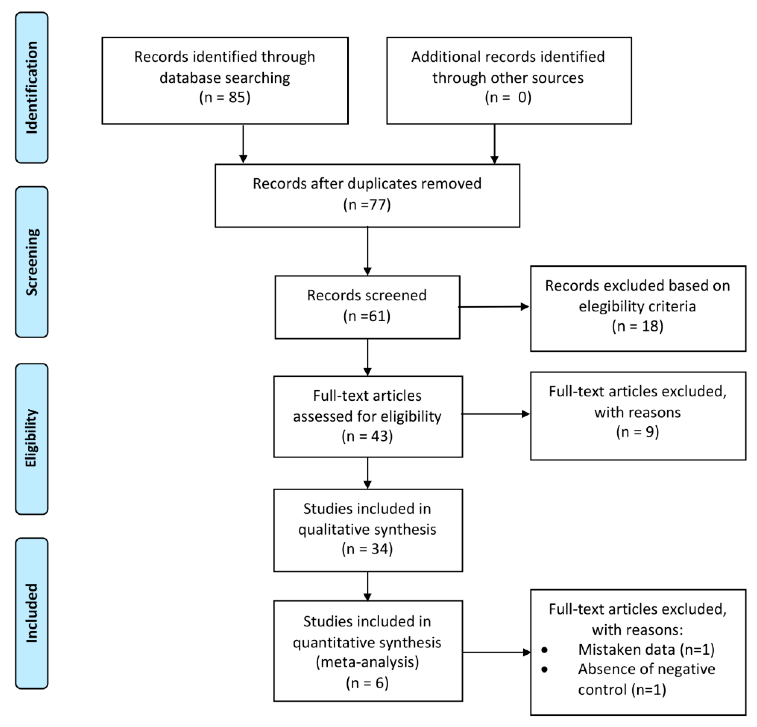
2.1. Search Results
2.2. Synthesis of Results
2.3. Level of Evidence
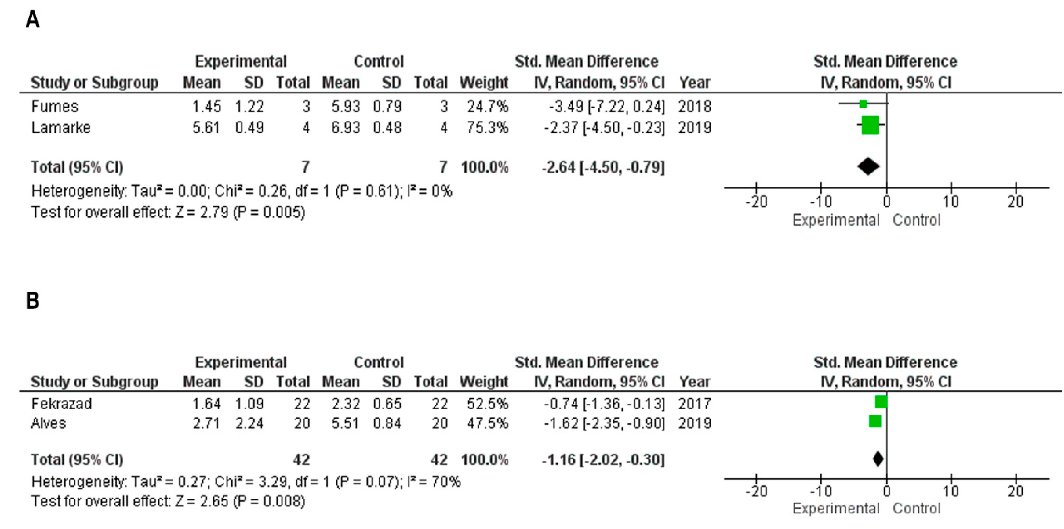
2.4. Meta-Analysis
3. Discussion
4. Materials and Methods
4.1. Eligibility Criteria
4.2. Search Strategy
4.3. Data Extraction and Analysis
4.4. Statistical Analysis
5. Conclusions
Author Contributions
Funding
Conflicts of Interest
Abbreviations
| PDT | Photodynamic Therapy |
| PS | Photosensitizer |
| ROS | Reactive Oxygen Species |
| ACS | Antioxidant Carrier Sensitizers |
| LoE | Level of Evidence |
| CFU | Colony Forming Units |
| MB | Methylene Blue |
References
- Sanz, M.; Beighton, D.; Curtis, M.A.; Cury, J.A.; Dige, I.; Dommisch, H.; Ellwood, R.; Giacaman, R.A.; Giacaman, R.A.; Herrera, D.; et al. Role of microbial biofilms in the maintenance of oral health and in the development of dental caries and periodontal diseases. Consensus report of group 1 of the Joint EFP/ORCA workshop on the boundaries between caries and periodontal disease. J. Clin. Periodontol. 2017, 44 (Suppl. 18), S5–S11. [Google Scholar] [CrossRef] [PubMed]
- Simón-Soro, A.; Mira, A. Solving the etiology of dental caries. Trends Microbiol. 2015, 23, 76–82. [Google Scholar] [CrossRef] [PubMed]
- Mathur, V.P.; Dhillon, J.K. Dental Caries: A Disease Which Needs Attention. Indian J. Pediatrics 2018, 85, 202–206. [Google Scholar] [CrossRef] [PubMed]
- Chen, F.; Wang, D. Novel technologies for the prevention and treatment of dental caries: A patent survey. Expert Opin. Ther. Pat. 2010, 20, 681–694. [Google Scholar] [CrossRef] [PubMed]
- Oshikawa, T.T. Antimicrobial resistance and aging: Beginning of the end of the antibiotic era? J. Am. Geriatr. Soc. 2002, 50, S226–S229. [Google Scholar] [CrossRef] [PubMed]
- Dobson, J.; Wilson, M. Sensitization of oral bacteria in biofilms to killing by light from a low-power laser. Arch. Oral. Biol. 1992, 37, 883–887. [Google Scholar] [CrossRef]
- Cieplik, F.; Wolfgang, B.; Hellwig, E.; Al-Ahmad, A.; Hiller, K.; Maisch, T.; Lamprini, K. Antimicrobial photodynamic therapy as an adjunct for treatment of deep carious lesions—A systematic review. Photodiagnosis Photodyn. Ther. 2017, 18, 54–62. [Google Scholar] [CrossRef]
- Kharkwal, G.B.; Sharma, S.K.; Huang, Y.Y.; Dai, T.; Hamblin, M.R. Photodynamic Therapy for Infections: Clinical Applications. Lasers Surg. Med. 2011, 43, 755–767. [Google Scholar] [CrossRef]
- Allison, R.R.; Moghissi, K. Photodynamic therapy (PDT): PDT mechanisms. Clin. Endosc. 2013, 46, 24–29. [Google Scholar] [CrossRef]
- Hamblim, M.R. Antimicrobial photodynamic inactivation: A bright new technique to kill resistant microbes. Curr. Opin. Microbiol. 2016, 33, 67–73. [Google Scholar] [CrossRef]
- Scherer, K.M.; Bisby, R.H.; Botchway, S.W.; Parker, A.W. New Approaches to Photodynamic Therapy from Types I, II and III to Type IV Using One or More Photons. Anticancer Agents Med Chem. 2017, 17, 171–189. [Google Scholar] [CrossRef] [PubMed]
- Zanin, I.C.; Lobo, M.M.; Rodrigues, L.K.; Pimenta, L.A.; Höfling, J.F.; Gonçalves, R.B. Photosensitization of in vitro biofilms by toluidine blue O combined with a light-emitting diode. Eur. J. Oral. Sci. 2006, 114, 64–69. [Google Scholar] [CrossRef] [PubMed]
- Müller, P.; Guggenheim, B.; Schmidlin, P.R. Efficacy of gasiform ozone and photodynamic therapy on a multispecies oral biofilm in vitro. Eur. J. Oral. Sci. 2007, 115, 77–80. [Google Scholar] [CrossRef] [PubMed]
- Luthi, M.; Gyenge, E.B.; Engstrom, M.; Bredell, M.; Gratz, K.; Walt, H.; Gmur, R.; Maake, A. Hypericin- and mTHPC-mediated photodynamic therapy for the treatment of cariogenic bactéria. Med. Laser Apl. 2009, 24, 227–236. [Google Scholar] [CrossRef]
- Mang, T.S.; Tayal, D.P.; Baier, R. Photodynamic therapy as na alternative treatment for disinfection of bacteria in oral biofilms. Lasers Surg. Med. 2012, 44, 588–596. [Google Scholar] [CrossRef] [PubMed]
- Rolim, J.P.; de-Melo, M.A.; Guedes, S.F.; Albuquerque-Filho, F.B.; de Souza, J.R.; Nogueira, N.A.; Zanin, I.C.; Rodrigues, L.K. The antimicrobial activity of photodynamic therapy against Streptococcus mutans using different photosensitizers. J. Photochem. Photobiol. B 2012, 106, 40–46. [Google Scholar] [CrossRef] [PubMed]
- Fekrazad, R.; Khoei, F.; Hakimiha, N.; Bahador, A. Photoelimination of Streptococcus mutans with two methods of photodynamic and photothermal therapy. Photodiagnosis Photodyn. Ther. 2013, 10, 626–631. [Google Scholar] [CrossRef]
- Spinei, A.; Spinei, I. The antimicrobial activity of photodynamic therapy against Streptococci species in dental biofilm using different photosensitizers: An in vitro study. In Proceedings of the E-Health Bioengineering Conference (EHB), Iasi, Romania, 21–23 November 2013. [Google Scholar]
- Araújo, N.C.; Fontana, C.R.; Bagnato, V.S.; Gerbi, M.E. Photodynamic antimicrobial therapy of curcumin in biofilms and carious dentine. Lasers Med. Sci. 2014, 29, 629–635. [Google Scholar] [CrossRef]
- Manoil, D.; Filieri, A.; Gameiro, C.; Lange, N.; Schrenzel, J.; Wataha, J.C.; Bouillaguet, S. Flow cytometric assessment of Streptococcus mutans viability after exposure to blue light-activated curcumin. Photodiagnosis Photodyn. Ther. 2014, 11, 372–379. [Google Scholar] [CrossRef]
- Diniz, I.M.; Horta, I.D.; Azevedo, C.S.; Elmadjian, T.R.; Matos, A.B.; Simionato, M.R.; Marques, M.M. Antimicrobial photodynamic therapy: A promise candidate for caries lesions treatment. Photodiagnosis Photodyn. Ther. 2015, 12, 511–518. [Google Scholar] [CrossRef]
- Melo, M.A.; Rolim, J.P.; Passos, V.F.; Lima, R.A.; Zanin, I.C.; Codes, B.M.; Rocha, S.S.; Rodrigues, L.K. Photodynamic antimicrobial chemotherapy and ultraconservative caries removal linked for management of deep caries lesions. Photodiagnosis Photodyn. Ther. 2015, 12, 581–586. [Google Scholar] [CrossRef] [PubMed] [Green Version]
- Soria-Lozano, P.; Gilaberte, Y.; Paz-Cristobal, M.P.; Pérez-Artiaga, L.; Lampaya-Pérez, V.; Aporta, J.; Pérez-Laguna, V.; García-Luque, I.; Revillo, M.J.; Rezusta, A. In Vitro effect photodynamic therapy with differents photosensitizers on cariogenic microorganisms. BMC Microbiol. 2015, 26, 15–187. [Google Scholar] [CrossRef] [PubMed]
- Leal, C.R.L.; Alvarenga, L.H.; Oliveira-Silva, T.; Kato, I.T.; Godoy, M.B.; Bussadori, S.K.; Ribeiro, M.S.; Prates, R.A. Antimicrobial photodynamic therapy on Streptococcus mutans is altered by glucose in the presence of methylene blue and red LED. Photodiagnosis Photodyn. Ther. 2017, 19, 1–4. [Google Scholar] [CrossRef] [PubMed]
- Fekrazad, R.; Seraj, B.; Chiniforush, N.; Rokouei, M.; Mousavi, N.; Ghadimi, S. Effect of antimicrobial photodynamic therapy on the counts of salivary Streptococcus mutans in children with severe early childhood caries. Photodiagnosis Photodyn. Ther. 2017, 18, 319–322. [Google Scholar] [CrossRef] [PubMed]
- Lee, H.J.; Kang, S.M.; Jeong, S.H.; Chung, K.H.; Kim, B.I. Antibacterial photodynamic therapy with curcumin and Curcuma xanthorrhiza extract against Streptococcus mutans. Photodiagnosis Photodyn. Ther. 2017, 20, 116–119. [Google Scholar] [CrossRef] [PubMed]
- Beytollahi, L.; Pourhajibagher, M.; Chiniforush, N.; Ghorbanzadeh, R.; Raoofian, R.; Pourakbari, B.; Bahador, A. The efficacy of photodynamic and photothermal therapy on biofilm formation of Streptococcus mutans: An in vitro study. Photodiagnosis Photodyn. Ther. 2017, 17, 56–60. [Google Scholar] [CrossRef]
- Nemezio, M.A.; de Souza Farias, S.S.; Borsatto, M.C.; Aires, C.P.; Corona, S.A.M. Effect of methylene blue-induced photodynamic therapy on a Streptococcus mutans biofilm model. Photodiagnosis Photodyn. Ther. 2017, 20, 234–237. [Google Scholar] [CrossRef]
- Pérez-Laguna, V.; Pérez-Artiaga, L.; Lampaya-Pérez, V.; López, S.C.; García-Luque, I.; Revillo, M.J.; Nonell, S.; Gilaberte, Y.; Rezusta, A. Comparative effect of photodynamic therapy on separated or mixed cultures of Streptococcus mutans and Streptococcus sanguinis. Photodiagnosis Photodyn. Ther. 2017, 19, 98–102. [Google Scholar] [CrossRef]
- Azizi, A.; Mousavian, S.; Taheri, S.; Lawaf, S.; Gonoudi, E.; Rahimi, A. Comparison of the antimicrobial efficacy of photodynamic therapy with two mediators against Lactobacillus acidophilus In Vitro. Photodiagnosis Photodyn. Ther. 2018, 21, 357–362. [Google Scholar] [CrossRef]
- Darmani, H.; Tawalbeh, K.H.; Al-Hiyasat, A.S.; Al-Akhras, M.A. Comparison of the Photosensitivity of Biofilms of Different Genera of Cariogenic Bacteria in Tooth Slices. Pol. J. Microbiol. 2018, 67, 455–462. [Google Scholar] [CrossRef] [Green Version]
- Esteban Florez, F.L.; Mendonça de Oliveira, M.R.; de Oliveira Júnior, O.B.; Hiers, R.D.; Khajotia, S.S.; Pretel, H. Bioluminescence Analysis of Antibacterial Photodynamic Therapy Using Methylene Blue Mediated by Low-Intensity Level Laser Against Cariogenic Biofilms. Photomed. Laser Surg. 2018, 36, 258–265. [Google Scholar] [CrossRef] [PubMed]
- Fumes, A.C.; Romualdo, P.C.; Monteiro, R.M.; Watanabe, E.; Corona, S.A.M.; Borsatto, M.C. Influence of pre-irradiation time employed in antimicrobial photodynamic therapy with diode laser. Lasers Med. Sci. 2018, 33, 67–73. [Google Scholar] [CrossRef] [PubMed]
- Garcia, T.M.; Pereira, A.H.C.; Figueiredo-Godoi, L.M.A.; Jorge, A.O.C.; Strixino, J.F.; Junqueira, J.C. Photodynamic therapy mediated by chlorin-type photosensitizers against Streptococcus mutans biofilms. Photodiagnosis Photodyn. Ther. 2018, 24, 256–261. [Google Scholar] [CrossRef]
- Gholibegloo, E.; Karbasi, A.; Pourhajibagher, M.; Chiniforush, N.; Ramazani, A.; Akbari, T.; Bahador, A.; Khoobi, M. Carnosine-graphene oxide conjugates decorated with hydroxyapatite as promising nanocarrier for ICG loading with enhanced antibacterial effects in photodynamic therapy against Streptococcus mutans. J. Photochem. Photobiol. B 2018, 181, 14–22. [Google Scholar] [CrossRef] [PubMed]
- Gómez, C.; Abellán, R.; Palma, J.C. Efficacy of photodynamic therapy vs ultrasonic scaler for preventing gingival inflammation and white spot lesions during orthodontic treatment. Photodiagnosis Photodyn. Ther. 2018, 24, 377–383. [Google Scholar] [CrossRef] [PubMed]
- Cusicanqui Méndez, D.A.; Gutierres, E.; José Dionisio, E.; Afonso, R.B.M.; Cardoso, O.R.; Andrade, M.M.M.A.; Cruvinel, T. Curcumin-mediated antimicrobial photodynamic therapy reduces the viability and vitality of infected dentin caries microcosms. Photodiagnosis Photodyn. Ther. 2018, 24, 102–108. [Google Scholar] [CrossRef] [PubMed]
- de Oliveira, F.S.; Cruvinel, T.; Cusicanqui, M.D.A.; Dionísio, E.J.; Rios, D.; Machado, M.A.A.M. The in vitro effect of Antimicrobial Photodynamic Therapy on dental microcosm biofilms from partially erupted permanent molars: A pilot study. Photodiagnosis Photodyn. Ther. 2018, 21, 163–167. [Google Scholar] [CrossRef]
- Tokubo, L.M.; Rosalen, P.L.; Sardi, J.D.C.O.; Freires, I.A.; Fujimaki, M.; Umeda, J.E.; Barbosa, P.M.; Tecchio, G.O.; Hioka, N.; de Freitas, C.F.; et al. Antimicrobial effect of photodynamic therapy using erythrosine/methylene blue combination on Streptococcus mutans biofilm. Photodiagnosis Photodyn. Ther. 2018, 23, 94–98. [Google Scholar] [CrossRef]
- Trigo-Gutierrez, J.K.; Sanitá, P.V.; Tedesco, A.C.; Pavarina, A.C.; Mima, E.G.O. Effect of Chloroaluminium phthalocyanine in cationic nanoemulsion on photoinactivation of multispecies biofilm. Photodiagnosis Photodyn. Ther. 2018, 24, 212–219. [Google Scholar] [CrossRef]
- Alexandrino, F.J.R.; Bezerra, E.M.; Da Costa, R.F.; Cavalcante, L.R.L.; Sales, F.A.M.; Francisco, T.S.; Rodrigues, L.K.A.; de Brito, D.A.; Ricardo, N.M.P.S.; Costa, S.N.; et al. Rose Bengal incorporated to α-cyclodextrin microparticles for photodynamic therapy against the cariogenic microorganism Streptococcus mutans. Photodiagnosis Photodyn. Ther. 2019, 25, 111–118. [Google Scholar] [CrossRef]
- Lara Alves, L.V.G.; Curylofo-Zotti, F.A.; Borsatto, M.C.; de Souza Salvador, S.L.; Valério, R.A.; Souza-Gabriel, A.E.; Corona, S.A.M. Influence of antimicrobial photodynamic therapy in carious lesion. Randomized split-mouth clinical trial in primary molars. Photodiagnosis Photodyn. Ther. 2019, 26, 124–130. [Google Scholar] [CrossRef] [PubMed]
- Esper, M.A.L.R.; Junqueira, J.C.; Uchoa, A.F.; Bresciani, E.; Rastelli, A.N.S.; Navarro, R.S.; Gonçalves, S.E.P. Photodynamic inactivation of planktonic cultures and Streptococcus mutans biofilms for prevention of white spot lesions during orthodontic treatment: An in vitro investigation. Am. J. Orthod. Dentofac. Orthop. 2019, 155, 243–253. [Google Scholar] [CrossRef] [PubMed]
- Lamarque, G.C.C.; Méndez, D.A.C.; Gutierrez, E.; Dionisio, E.J.; Machado, M.A.A.M.; Oliveira, T.M.; Rios, D.; Cruvinel, T. Could chlorhexidine be na adequate positive control for antimicrobial photodynamic therapy in-In Vitro studies? Photodiagnosis Photodyn. Ther. 2019, 25, 58–62. [Google Scholar] [CrossRef] [PubMed]
- Pourhajibagher, M.; Salehi Vaziri, A.; Takzaree, N.; Ghorbanzadeh, R. Physico-mechanical and antimicrobial properties of an orthodontic adhesive containing cationic curcumin doped zinc oxide nanoparticles subjected to photodynamic therapy. Photodiagnosis Photodyn Ther. 2019, 25, 239–246. [Google Scholar] [CrossRef] [PubMed]
- Kwiatkowski, S.; Knap, B.; Przystupski, D.; Saczko, J.; Kędzierska, E.; Knap-Czop, K.; Kotlińska, J.; Michel, O.; Kotowski, K.; Kulbacka, J. Photodynamic therapy—mechanisms, photosensitizers and combinations. Biomed. Pharm. 2018, 106, 1098–1107. [Google Scholar] [CrossRef] [PubMed]
- Durieux, N.; Vandenput, S.; Pasleau, F. OCEBM levels of evidence system. Rev. Med. Liege. 2013, 68, 644–649. [Google Scholar]
- Valkenburg, C.; Slot, D.E.; Bakker, E.W.; Van der Weijden, F.A. Does dentifrice use help to remove plaque? A systematic review. J. Clin. Periodontol. 2016, 43, 1050–1058. [Google Scholar] [CrossRef]
- Marsh, P.D. Contemporary perspective on plaque control. Br. Dent. J. 2012, 212, 601–606. [Google Scholar] [CrossRef] [PubMed] [Green Version]
- Dias, A.P.; Paschoal, M.A.B.; Diniz, R.S.; Lage, L.M.; Gonçalves, L.M. Antimicrobial action of chlorhexidine digluconate in self-ligating and conventional metal brackets infected with Streptococcus mutans biofilm. Clin. Cosmet. Investig. Dent. 2018, 19, 69–74. [Google Scholar] [CrossRef] [PubMed]
- McCoy, L.C.; Wehler, C.J.; Rich, S.E.; Garcia, R.I.; Miller, D.R.; Jones, J.A. Adverse events associated with chlorhexidine use Results from the Department of Veterans Affairs Dental Diabetes Study. J. Am. Dent. Assoc. 2008, 139, 178–183. [Google Scholar] [CrossRef] [PubMed]
- Williams, J.A.; Pearson, G.J.; Colles, M.J.; Wilson, M. The photo-activated antibacterial action of toluidine blue O in a collagen matrix and in carious dentine. Caries. Res. 2004, 38, 530–536. [Google Scholar] [CrossRef] [PubMed]
- Paschoal, M.A.; Tonon, C.C.; Spolidório, D.M.; Bagnato, V.S.; Giusti, J.S.; Santos-Pinto, L. Photodynamic potential of curcumin and blue LED against Streptococcus mutans in a planktonic culture. Photodiagnosis Photodyn. Ther. 2013, 10, 313–319. [Google Scholar] [CrossRef] [PubMed]
- Araújo, P.V.; Correia-Silva, J.F.; Gomez, R.S.; Massara, M.L.; Cortes, M.E.; Poletto, L.T. Antimicrobial effect of photodynamic therapy in carious lesions in vivo, using culture and real-time PCR methods. Photodiagnosis Photodyn. Ther. 2015, 12, 401–407. [Google Scholar] [CrossRef] [PubMed]
- Romão, I.Q.; Cavalcante, S.I.A.; Leite, H.L.A.; Gonçalves, L.M.; Branco-de-Almeida, L.S.; Paschoal, M.A.B. Effect of Combining Erythrosine with a High-Power Dental Curing Light Appliance on the Viability of a Planktonic Culture of Streptococcus mutans. Photomed Laser Surg. 2018, 36, 676–679. [Google Scholar] [CrossRef] [PubMed]
- Prażmo, E.J.; Kwaśny, M.; Łapiński, M.; Mielczarek, A. Photodynamic Therapy as a Promising Method Used in the Treatment of Oral Diseases. Adv. Clin. Exp. Med. 2016, 25, 799–807. [Google Scholar] [CrossRef] [PubMed]
- Sperandio, F.F.; Huang, Y.Y.; Hamblin, M.R. Antimicrobial photodynamic therapy to kill Gram-negative bacteria. Recent. Pat. Antiinfect. Drug Discov. 2013, 8, 108–120. [Google Scholar] [CrossRef] [PubMed]
- Larsen, T.; Fiehn, N.E. Dental biofilm infections—An update. APMIS 2017, 125, 376–384. [Google Scholar] [CrossRef]
- Pogue, B.W.; Elliott, J.T.; Kanick, S.C.; Davis, S.C.; Samkoe, K.S.; Maytin, E.V.; Pereira, S.P.; Hasan, T. Revisiting photodynamic therapy dosimetry: Reductionist & surrogate approaches to facilitate clinical success. Phys. Med. E Biol. 2016, 61, 57–89. [Google Scholar]
- Gomes, E.R.; Cruz, T.; Lopes, C.F.; Carvalho, A.P.; Duarte, C.B. Photosensitization of lymphoblastoid cells with phthalocyanines at different saturating incubation times. Cell Biol. Toxicol. 1999, 15, 249–260. [Google Scholar] [CrossRef] [Green Version]
- Tardivo, J.P.; Del Giglio, A.; de Oliveira, C.S.; Gabrielli, D.S.; Junqueira, H.C.; Tada, D.B.; Severino, D.; de Fátima Turchiello, R.; Baptista, M.S. Methylene blue in photodynamic therapy: From basic mechanisms to clinical applications. Photodiagnosis Photodyn. Ther. 2005, 2, 175–191. [Google Scholar] [CrossRef]
- Moreira, L.M.; Lyon, J.P.; Romani, A.P.; Severino, D.; Rodrigues, M.R.; de Oliveira, H.P. Phenotiazinium dyes as photosensitizers (PS) in photodynamic therapy (PDT): Spectroscopic properties and photochemical mechanisms. Intech Open Access Publ. 2012. [Google Scholar] [CrossRef]
- Fontana, C.R.; Abernethy, A.D.; Som, S.; Ruggiero, K.; Doucette, S.; Marcantonio, R.C.; Boussios, C.I.; Kent, R.; Goodson, J.M.; Tanner, A.C.R.; et al. The Antibacterial Effect of Photodynamic Therapy in Dental Plaque-Derived Biofilms. J. Periodontal Res. 2009, 44, 751–759. [Google Scholar] [CrossRef] [PubMed]
- De Annunzio, S.R.; De Freitas, L.M.; Blanco, A.L.; Da Costa, M.M.; Carmona-Vargas, C.C.; De Oliveira, K.T.; Fontana, C.R. Susceptibility Of Enterococcus Faecalis And Propionibacterium Acnes To Antimicrobial Photodynamic Therapy. J. Photochem. Photobiol. B Biol. 2018, 178, 545–550. [Google Scholar] [CrossRef] [PubMed]
- Mirouze, N.; Ferret, C.; Cornilleau, C.; Carballido-López, R. Antibiotic sensitivity reveals that wall teichoic acids mediate DNA binding during competence in Bacillus subtilis. Nat. Commun. 2018, 9, 5072. [Google Scholar] [CrossRef] [PubMed]
- Chabrier-Rosello, Y.; Foster, T.H.; Perez-Nazario, N.; Mitra, S.; Haidaris, C.G. Sensitivity of Candida albicans germ tubes and biofilms to photofrinmediated phototoxicity. Antimicrob. Agents Chemother. 2005, 49, 4288–4295. [Google Scholar] [CrossRef] [PubMed]
- Andrade, M.C.; Ribeiro, A.P.; Dovigo, L.N.; Brunetti, I.L.; Giampaolo, E.T.; Bagnato, V.S.; Pavarina, A.C. Effect of different pre-irradiation times on curcumin-mediated photodynamic therapy against planktonic cultures and biofilms of Candida spp. Arch. Oral. Biol. 2013, 58, 200–210. [Google Scholar] [CrossRef]
- Pankey, G.A.; Sabath, L.D. Clinical relevance of bacteriostatic versus bactericidal mechanisms of action in the treatment of Gram-positive bacterial infections. Clin. Infect. Dis. 2004, 38, 864–870. [Google Scholar] [CrossRef] [PubMed]
- Meyer, B. Approuches to prevention, removal and killing of biofilms. Int. Biodeterorat. Biodegrad. 2003, 51, 249–253. [Google Scholar] [CrossRef]
- Schwendicke, F.; Korte, F.; Dörfer, C.E.; Kneist, S.; Fawzy El-Sayed, K.; Paris, S. Inhibition of Streptococcus mutans Growth and Biofilm Formation by Probiotics in vitro. Caries Res. 2017, 51, 87–95. [Google Scholar] [CrossRef] [PubMed]
- Koo, H.; Xiao, J.; Klein, M.I. Extracellular polysaccharides matrix—Na often forgotten virulence fator in oral biofilm reserch. Int. J. Oral. Sci. 2009, 1, 229–234. [Google Scholar] [CrossRef] [PubMed]
- Zhao, K.; Tseng, B.S.; Beckerman, B.; Jin, F.; Gibiansky, M.L.; Harrison, J.J.; Luijten, E.; Parsek, M.R.; Wong, G.C.L. PSL trails guide exploration and microcolony formation in Pseudomonas aeruginosa biofilms. Nature 2013, 497, 388–391. [Google Scholar] [CrossRef] [PubMed]
- Koo, H.; Falsetta, M.L.; Klein, M.I. The exopolysaccharide matrix: A virulence determinant of cariogenic biofilm. J. Dent. Res. 2013, 92, 1065–1073. [Google Scholar] [CrossRef] [PubMed]
- Dewhirst, F.E.; Chen, T.; Izard, J.; Paster, B.J.; Tanner, A.C.; Yu, W.H.; Lakshmanan, A.; Wade, W.G. The human oral microbiome. J. Bacteriol. 2010, 192, 5002–5017. [Google Scholar] [CrossRef] [PubMed]
- Huang, R.; Li, M.; Gregory, R.L. Bacterial interactions in dental biofilm. Virulence 2011, 2, 435–444. [Google Scholar] [CrossRef] [PubMed]
- Exterkate, R.A.; Crielaard, W.; ten Cate, J.M. Different response to amine fluoride by Streptococcus mutans and polymicrobial biofilms in a novel high-throughput active attachment model. Caries Res. 2010, 44, 372–379. [Google Scholar] [CrossRef] [PubMed]
- Moher, D.; Shamseer, L.; Clarke, M.; Ghersi, D.; Liberati, A.; Petticrew, M.; Shekelle, P.; Stewart, L.A.; PRISMA-P Group. Preferred reporting items for systematic review and meta-analysis protocols (PRISMA-P) 2015 statement. Syst. Rev. 2015, 4, 1. [Google Scholar] [CrossRef] [PubMed]
- Moher, D.; Liberati, A.; Tetzlaff, J.; Altman, D.G. PRISMA Group, Preferred reporting items for systematic reviews and meta-analyses: The PRISMA statement. J. Clin. Epidemiol. 2009, 62, 1006–1012. [Google Scholar] [CrossRef] [PubMed]


| Study | Year | Study Design | Level of Evidence * | Sample Size | Irradiation Time ** | Photosensitizer | Biofilm Inhibition | Wave-Length | Microorganism | Control Group | Biofilm Reduction (Log CFU/mL) | |
|---|---|---|---|---|---|---|---|---|---|---|---|---|
| #1 | Zanin et al. [12] | 2006 | In vitro | III | 3 | 5 min | Toluidine blue | N/A | 660 nm | Streptococcus mutans | Negative | <3 |
| #2 | Muller et al. [13] | 2007 | In vitro | III | 9 | 1 min | Methylene blue | N/A | 665 nm | Multispecies biofilm | Negative and chlorexidine digluconate 2% | <1 |
| #3 | Lutti Martin et al. [14] | 2009 | In vitro | III | N/A | 1 min, 5 min, 15 min and 30 min | Fosfolipos and Hypericina | N/A | 400 nm–505 nm | Streptococcus mutans and Streptococcus subrinus | Negative | 3 (S. subrinus) and <3 (S. mutans) |
| #4 | Mang et al. [15] | 2012 | In vitro | III | N/A | 5 min | Porfimer sodium | N/A | 630 nm | Streptococcus mutans | Negative | N/A |
| #5 | Rolim et al. [16] | 2012 | In vitro | III | 10 | 5 min | Methylene blue, Toluidine blue, Ortho and Malachite green | N/A | N/A | Streptococcus mutans | Negative | 3 |
| #6 | Fekrazad et al. [17] | 2013 | In vitro | III | N/A | 5 min | Toluidine blue, Radachlorine and Indocyanine green | N/A | 660 mm and 810 nm | Streptococcus mutans | Negative | <3 |
| #7 | Spinei et al. [18] | 2013 | In vitro | III | N/A | N/A | Antocianine extract and methylene blue | N/A | 625 nm–635 nm | Streptococcus mutans, mitis, gordoni and sobrinus | Negative | 4.1 |
| #8 | Araujo et al. [19] | 2014 | In vitro | III | N/A | 5 min | Curcumin | N/A | 420 nm | Streptococcus mutans and Lactobacillus acidophillus | Negative | <1 |
| #9 | Manoil et al. [20] | 2014 | In vitro | III | 12 | 5 min and 10 min | Curcumin | N/A | 360 nm–550 nm | Streptococcus mutans | Negative | 2 |
| #10 | Diniz et al. [21] | 2015 | In vitro | III | 12 | 5 min | Methylene blue | N/A | 660 nm | Streptococcus mutans | Negative | 1.01 |
| #11 | Melo et al. [22] | 2015 | RCT | I | 45 | 5 min | Toluidine blue | N/A | 660 nm | Multispecies biofilm | Negative | <3 |
| #12 | Soria-Lozano et al. [23] | 2015 | In vitro | III | N/A | 1 min/ 1 h/3 h | Methylene blue, Rose Bengal, and Curcumin | N/A | N/A | Streptococcus mutans, Streptococcus sanguinis and Candida albicans | Negative | 6.0 (Streptococcus spp), 5.0 (C.albicans) |
| #13 | Cintia Lima et al. [24] | 2017 | In vitro | III | N/A | 10 min | Methylene blue | N/A | 660 nm | Streptococcus mutans | Negative | >3 |
| #14 | Fekrazad et al. [25] | 2017 | RCT | I | 22 | 1 min | Toluidine blue | N/A | 630 nm | Streptococcus mutans | Negative | 0.68 |
| #15 | Hyung-Jung et al. [26] | 2017 | In vitro | III | N/A | N/A | Curcumin and Curcuma xanthorrhiza extract | N/A | 405 nm | Streptococcus mutans | Negative | >3 |
| #16 | Leili Beytollahi [27] | 2017 | In vitro | III | N/A | 5 min | Methylene blue and Green Indocyanine | Yes | 635 nm | Streptococcus mutans | Negative | <3 |
| #17 | Nemezio et al. [28] | 2017 | In vitro | III | 4 | 5 min | Methylene blue | N/A | 660 nm | Streptococcus mutans | NaCL solution 0.9% and chlorhexidine digluconate 0.12% | 1 |
| #18 | Péres-Laguna et al. [29] | 2017 | In vitro | III | N/A | N/A | Methylene blue and Rose Bengal | N/A | N/A | Streptococcus mutansand sanguinis | Negative | 6 |
| #19 | Azizi et al. [30] | 2018 | In vitro | III | 6 | 5 min | Indocyanine green and Methylene blue | N/A | 660 nm and 808 nm | Lactobacillus acidophillus | Chlorexidine digluconate 0.2%, NaOCL2.5% and Penicilin 6.3.3 | N/A |
| #20 | Darmani et al. [31] | 2018 | In vitro | III | N/A | 5 min | Toluidine Blue | N/A | 670 nm | Streptococcus mutan, Streptococcus salivar, Lactobacillus casei and Actinomyces viscosus | Negative | <1 |
| #21 | Esteban Florez et al. [32] | 2018 | In vitro | III | 15 | 5 min | Methylene blue | N/A | 660 nm | Streptococcus mutans | Negative and chlorexidine digluconate 2% | 1,3 |
| #22 | Fumes et al. [33] | 2018 | In vitro | III | 3 | 1 min, 2 min, and 5 min | Methylene blue | N/A | N/A | Streptococcus mutans and Candida albicans | Negative and chlorexidine digluconate 0.12% | <3 |
| #23 | Garcia et al. [34] | 2018 | In vitro | III | 10 | N/A | Fotoencitine and Photoditazine | N/A | 660 nm | Streptococcus mutans | Negative and Methylene Blue | Complete eradication (Fotoencitine) and 6 (Photoditazine) |
| #24 | Gholibegloo et al. [35] | 2018 | In vitro | III | 3 | 5 min | Indocyanine green | Yes | N/A | Streptococcus mutans | Negative | <1 |
| #25 | Gomez et al. [36] | 2018 | RCT | I | 10 | 3 min | Methylene blue | N/A | 670 nm | Aggregatibacter actinomycetemcomitans, Porphyromonas gingivalis, Prevotella intermedia and Tannerella forsythia | US technique | N/A |
| #26 | Míndez et al. [37] | 2018 | In vitro | III | 9 | 2 min | Curcumin | N/A | 455 nm | Streptococcus mutans | Negative | <3 |
| #27 | Oliveira et al. [38] | 2018 | In vitro | III | 6 | 2 min | Methylene Blue | N/A | 630 nm | Multispecies biofilm from saliva | Negative | <3 |
| #28 | Tokubo et al. [39] | 2018 | In vitro | III | 3 | 5 min | Erythrosine and Methylene blue | N/A | N/A | Streptococcus mutans | Negative and chlorexidine digluconate 0.12% | 4.3 |
| #29 | Trigo-Gutierrez et al. [40] | 2018 | In vitro | III | N/A | 30 min | Cloroaluminium phthalocyanine nanoemulsion | N/A | N/A | Candida albicans, Candida glabrata and Streptococcus mutans | Negative | <3 |
| #30 | Alexandrino et al. [41] | 2019 | In vitro | III | N/A | N/A | Rose Bengal and Rose Bengal encapsulated with cyclodextrin | Yes | 520 nm | Streptococcus mutans | NaCL solution 0.9% and chlorhexidine digluconate 0.12% | Complete eradication |
| #31 | Alves et al. [42] | 2019 | RCT | I | 20 | 5 min | Methylene blue | N/A | 660 nm | Streptococcus mutans | Negative | 2.8 |
| #32 | Esper et al. [43] | 2019 | In vitro | III | 10 | 5 min | Hematoporfirine | N/A | 420 nm and 480 nm | Streptococcus mutans | Negative | <1 (biofilm) and 3.8 and 6.78 (planktonic) |
| #33 | Lamarke et al. [44] | 2019 | In vitro | III | 4 | 2 min | Curcumin | N/A | 420 nm | Multispecies biofilm | Negative and chlorexidine digluconate 0.12% | 1.32 |
| #34 | Pourbajibagher et al. [45] | 2019 | In vitro | III | 10 | 5 min | Cationic doped zinc oxide nanoparticle adhesive | Yes | 435 nm | Streptococcus mutans | Negative | 1.96 |
© 2019 by the authors. Licensee MDPI, Basel, Switzerland. This article is an open access article distributed under the terms and conditions of the Creative Commons Attribution (CC BY) license (http://creativecommons.org/licenses/by/4.0/).
Share and Cite
de Oliveira, A.B.; Ferrisse, T.M.; Marques, R.S.; de Annunzio, S.R.; Brighenti, F.L.; Fontana, C.R. Effect of Photodynamic Therapy on Microorganisms Responsible for Dental Caries: A Systematic Review and Meta-Analysis. Int. J. Mol. Sci. 2019, 20, 3585. https://0-doi-org.brum.beds.ac.uk/10.3390/ijms20143585
de Oliveira AB, Ferrisse TM, Marques RS, de Annunzio SR, Brighenti FL, Fontana CR. Effect of Photodynamic Therapy on Microorganisms Responsible for Dental Caries: A Systematic Review and Meta-Analysis. International Journal of Molecular Sciences. 2019; 20(14):3585. https://0-doi-org.brum.beds.ac.uk/10.3390/ijms20143585
Chicago/Turabian Stylede Oliveira, Analú Barros, Túlio Morandin Ferrisse, Raquel Souza Marques, Sarah Raquel de Annunzio, Fernanda Lourenção Brighenti, and Carla Raquel Fontana. 2019. "Effect of Photodynamic Therapy on Microorganisms Responsible for Dental Caries: A Systematic Review and Meta-Analysis" International Journal of Molecular Sciences 20, no. 14: 3585. https://0-doi-org.brum.beds.ac.uk/10.3390/ijms20143585