Article

Tobacco Cutworm (Spodoptera Litura) Larvae Silenced in the NADPH-Cytochrome P450 Reductase Gene Show Increased Susceptibility to Phoxim

1
Guangdong Province Key Laboratory of Eco-Circular Agriculture, South China Agricultural University, Guangzhou 510642, China
2
Key Laboratory of Agro-Environment in the Tropics, Ministry of Agriculture, South China Agricultural University, Guangzhou 510642, China
3
State Key Laboratory of Biocontrol and Guangdong Key Laboratory of Plant Resources, School of Life Sciences, Sun Yat-sen University, East Campus, Guangzhou 510006, China
4
New Rural Development Research Institute of South China Agricultural University, Guangzhou 510642, China
*
Authors to whom correspondence should be addressed.
Int. J. Mol. Sci. 2019, 20(15), 3839; https://0-doi-org.brum.beds.ac.uk/10.3390/ijms20153839
Submission received: 21 June 2019 / Revised: 28 July 2019 / Accepted: 5 August 2019 / Published: 6 August 2019

Abstract

:
Nicotinamide adenine dinucleotide phosphate (NADPH)-cytochrome P450 reductases (CPRs) function as redox partners of cytochrome P450 monooxygenases (P450s). CPRs and P450s in insects have been found to participate in insecticide resistance. However, the CPR of the moth Spodoptera litura has not been well characterized yet. Based on previously obtained transcriptome information, a full-length CPR cDNA of S. litura (SlCPR) was PCR-cloned. The deduced amino acid sequence contains domains and residues predicted to be essential for CPR function. Phylogenetic analysis with insect CPR amino acid sequences showed that SlCPR is closely related to CPRs of Lepidoptera. Quantitative reverse transcriptase PCR (RT-qPCR) was used to determine expression levels of SlCPR in different developmental stages and tissues of S. litura. SlCPR expression was strongest at the sixth-instar larvae stage and fifth-instar larvae showed highest expression in the midgut. Expression of SlCPR in the midgut and fat body was strongly upregulated when fifth-instar larvae were exposed to phoxim at LC15 (4 μg/mL) and LC50 (20 μg/mL) doses. RNA interference (RNAi) mediated silencing of SlCPR increased larval mortality by 34.6% (LC15 dose) and 53.5% (LC50 dose). Our results provide key information on the SlCPR gene and indicate that SlCPR expression levels in S. litura larvae influence their susceptibility to phoxim and possibly other insecticides.

1. Introduction

The tobacco cutworm, Spodoptera litura (F.) (Lepidoptera, Noctuidae) is a serious polyphagous insect pest. The moth has a broad host range that includes economically important crops such as tomato, cotton, and groundnut [1]. Over recent years, many field populations of S. litura acquired resistance to various insecticides, particularly in Pakistan, China, and India. The task of controlling insecticide-resistant S. litura populations is becoming exceedingly challenging [1,2,3]. Phoxim has become one of the most widely used organophosphate insecticides for the control of S. litura [1,2,3].
Cytochrome P450 monooxygenases (CYPs or P450s) belong to a superfamily of heme-containing enzymes that catalyze the monooxygenation of xenobiotics and endogenous compounds [4,5]. Insect P450s can metabolize and detoxify insecticides and thus play an important role in evolution of insecticide resistance [4,5]. Various studies suggested that expression of specific P450s is induced when insects are exposed to insecticides. For example, our previous work on S. litura P450s suggested that insecticide-induced CYP9A40 [6] and CYP321B1 [7] play an important role in insecticide detoxification. Similarly, transcript levels of CYP49A1, CYP6AB4, CYP9A19, and CYP9A22 in the fat body of Bombyx mori were expressed at high levels after 24, 48, and 72 h of phoxim treatment, suggesting that P450 genes expressed in the fat body are associated with detoxification of phoxim [8].
Ncotinamide adenine dinucleotide phosphate (NADPH)-cytochrome P450 reductases (CPRs), important redox partners of P450s, play a crucial role in providing electrons from nicotinamide adenine dinucleotide phosphate (NADPH) to P450s via two flavin cofactors [9,10]. CPRs belong to the electron transfer flavoprotein family whose members contain conserved binding domains to NADP, the flavin mononucleotide (FMN) cofactor, and the flavin adenine dinucleotide (FAD) cofactor [11,12]. CPR genes have been identified in various insect species such as S. litura [13], Spodoptera littoralis [14], Cimex lectularius [15], Helicoverpa armigera [16], Nilaparvata lugens [17], Bactrocera dorsalis [18], Spodoptera exigua [19], Laodelphax striatellus [12], Cnaphalocrocis medinalis [20], and Locusta migratoria [21]. Most of them such as the CPRs from S. exigua [19], B. dorsalis [18], Aphis (Toxoptera) citricidus (Kirkaldy) [22], L. striatellus [12], Plutella xylostella [23], and L. migratoria [21] have been found to be associated with metabolism and resistance to insecticides. Due to their possible role in insecticide detoxification, insect CPRs may represent possible molecular targets for new insecticides [5,12,16,21].
Little information is available on the function of the CPR gene in S. litura and whether silencing of this gene by RNA interference (RNAi) affects larval susceptibility to insecticides. RNAi is a powerful and widely used tool to down-regulate expression of specific genes in insects [24]. Previous studies in our laboratory showed that microinjection of double-stranded RNA (dsRNA) into S. litura larvae can effectively silence specific target genes [6,25].
In the present study, we cloned a full-length cDNA encoding CPR of S. litura (SlCPR). We used quantitative reverse transcriptase PCR (RT-qPCR) to analyze the SlCPR expression pattern at the insect’s different developmental stages and in various tissues prepared from fifth-instar larvae. To investigate whether SlCPR transcript levels affect the insect’s susceptibility to insecticides, SlCPR-silenced larvae were exposed to phoxim. The results showed increased mortality of SlCPR-silenced larvae as compared to control larvae.

2. Results

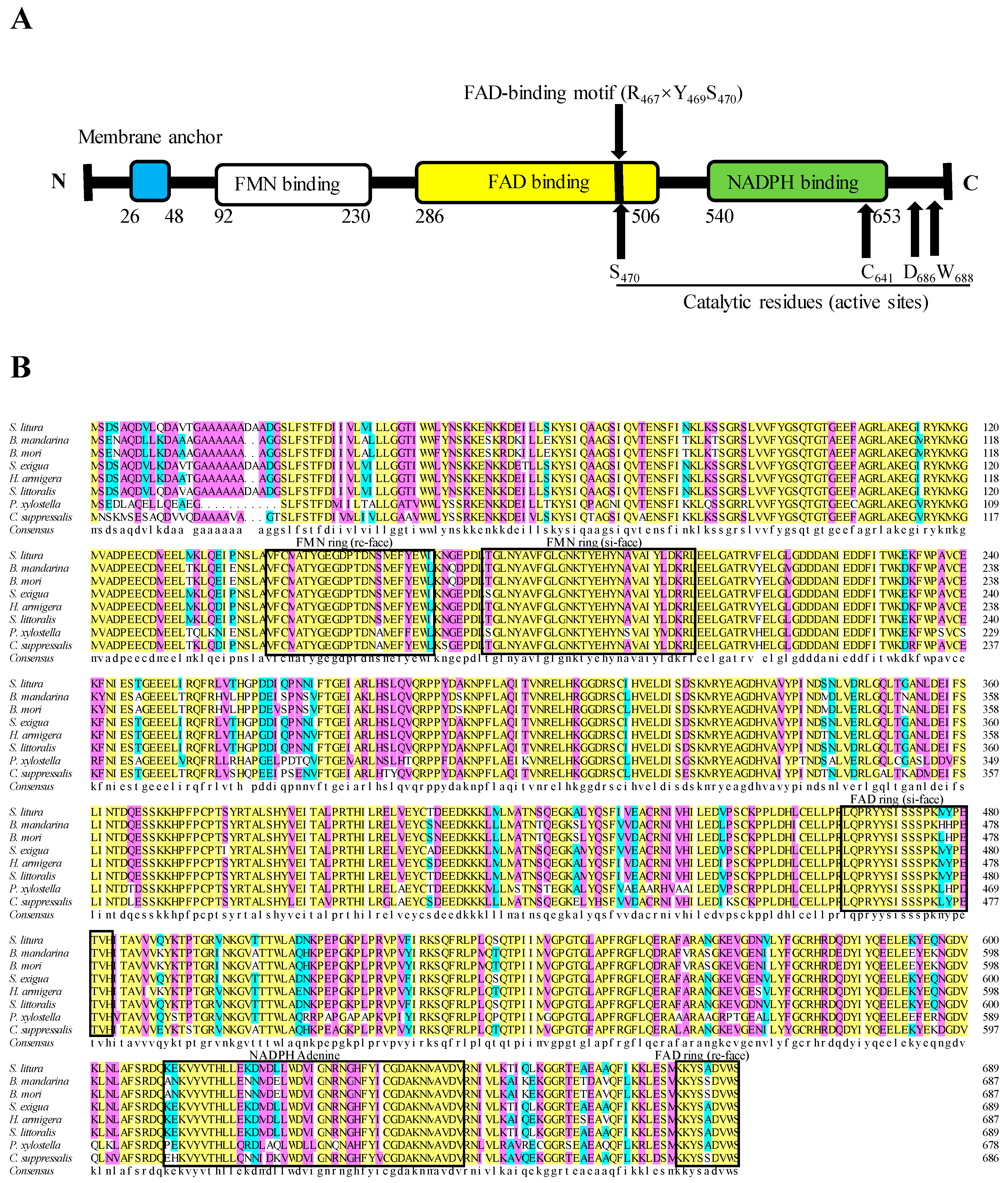
2.1. Cloning and Sequence Analysis of SlCPR

In a previous study, a whole transcriptome analysis was performed for the midgut of S. litura fourth-instar larvae [25]. Based on these data, we identified and cloned a full-length SlCPR cDNA (GenBank Acc. MH638288). The SlCPR cDNA sequence contains a 237-bp 5′-untranslated region (5′-UTR), a 2070-bp open reading frame, and a 1783-bp 3′-UTR with a poly-A nucleotide sequence. The predicted protein contains 689 amino acids (77.72 kDa) and possesses a theoretical pI of 5.32. No signal peptide was identified at the N-terminus of the protein. However, a hydrophobic transmembrane region consisting of 22 amino acids was predicted (Figure 1). The three amino acid residues R467, Y469, and S470 constitute a putative FAD binding motif which is ubiquitous in the FAD binding domain of CPR proteins [26]. Similar to rat and other CPRs, conserved catalytic residues are present in the SlCPR protein (S470, C641, D686, and W688) (Figure 1A). These active site residues have been demonstrated to be essential for CPR activity [17,27]. According to Cheng et al. (2017) [13] and nucleotide sequences deposited at the DDBJ/ENA/GenBank databases, S. litura possesses a single copy of the CPR gene. The alignment results of the deduced amino acid sequences of SlCPR and some other known CPRs showed that SlCPR shared 99.6%, 98.4%, and 95.6% amino acid identity with the CPR sequences of S. littoralis, S. exigua, and H. armigera, respectively. The results also demonstrated that SlCPR is a new member of the CPR family (Figure 1B).

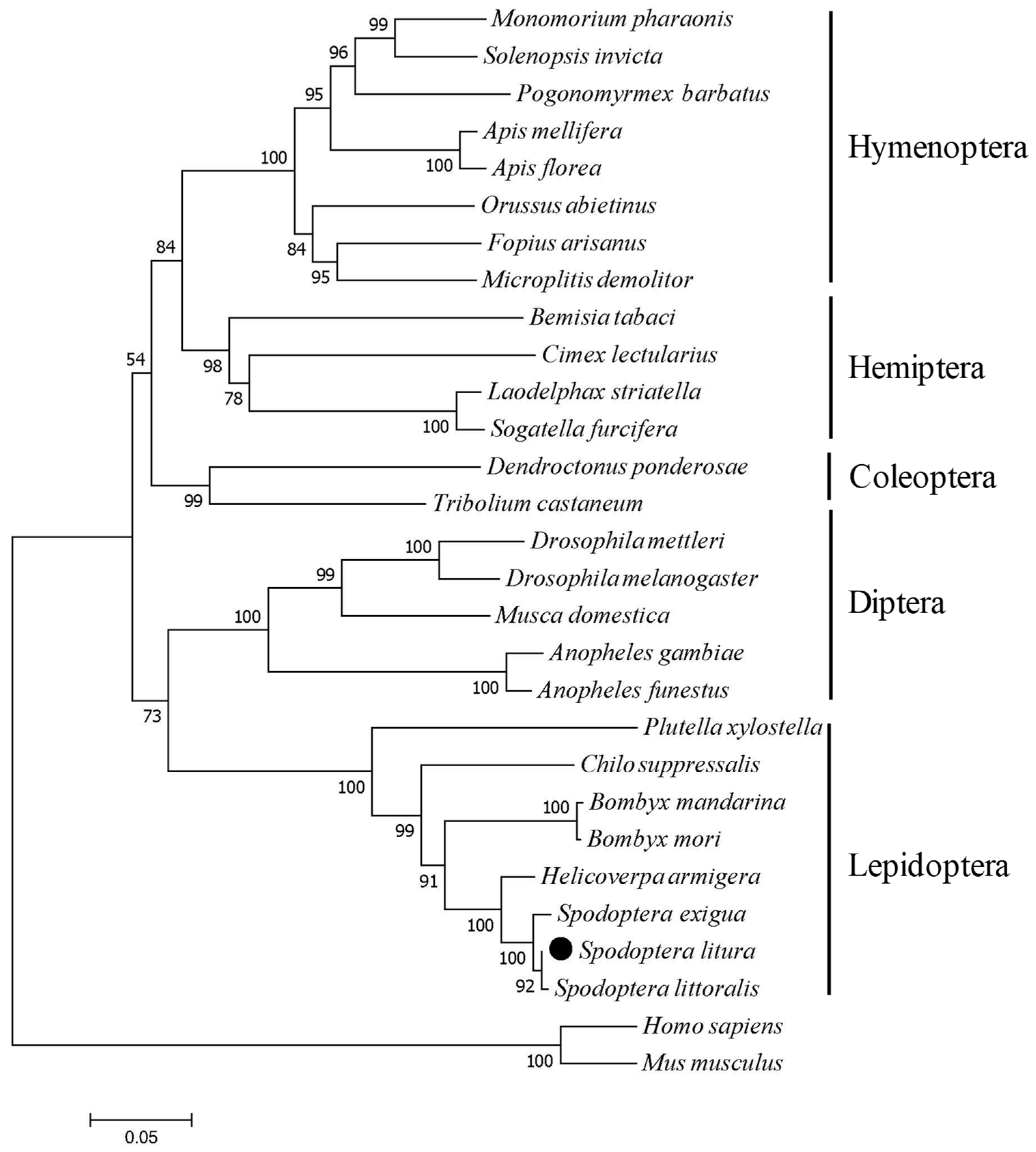
2.2. Phylogenetic Relation Between SlCPR and Other CPRs

Based on the deduced amino acid sequence of SlCPR and 28 other CPRs, phylogenetic analysis was performed using MEGA 7.0 software and the neighbor joining method. The constructed tree showed that CPRs from insects of the same order were grouped together. As expected, SlCPR was most related to CPRs of other Lepidoptera insects, including P. xylostella, Chilo suppressalis, Bombyx mandarina, Bombyx mori, H. armigera, S. exigua, and S. littoralis (Figure 2, Table 1).

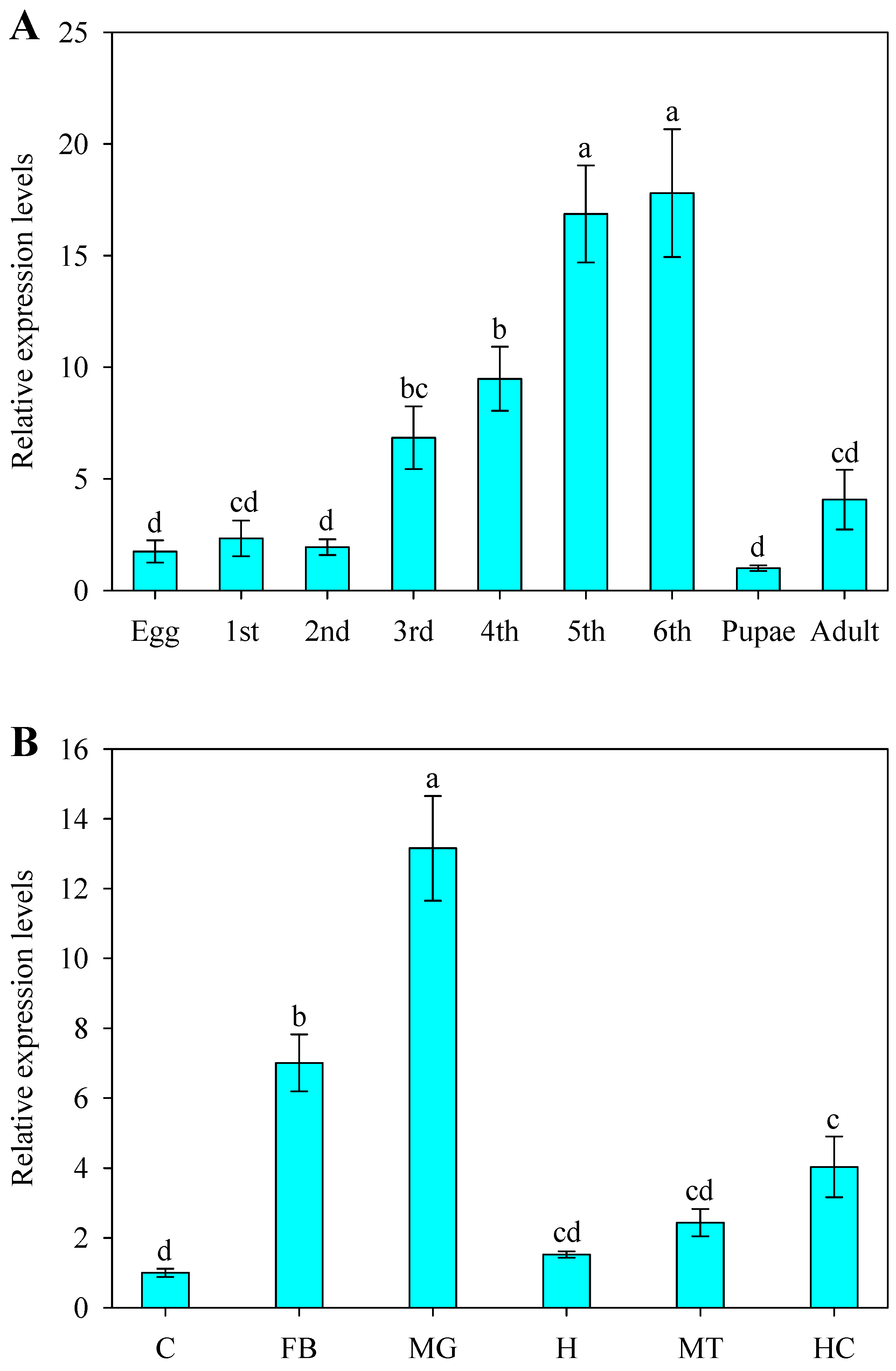
2.3. Developmental and Spatial Expression Patterns of SlCPR

We used RT-qPCR to examine the SlCPR expression pattern at different developmental stages of S. litura, namely eggs, first- to sixth-instar larvae, pupae, and adults. Highest expression levels were found in sixth-instar larvae (17.8-fold higher than in pupae), followed by fifth-instar larvae (16.9-fold higher than in pupae), and fifth-instar larvae (9.5-fold higher than in pupae) (Figure 3A). Tissue-specific expression of SlCPR was further analyzed for the cuticle, fat body, midgut, head, Malpighian tubule, and hemocytes of fifth-instar larvae (Figure 3B). Strongest expression levels of SlCPR were observed in the midgut (13.2-fold higher than in the cuticle) and fat body (7.0-fold higher than in the cuticle).

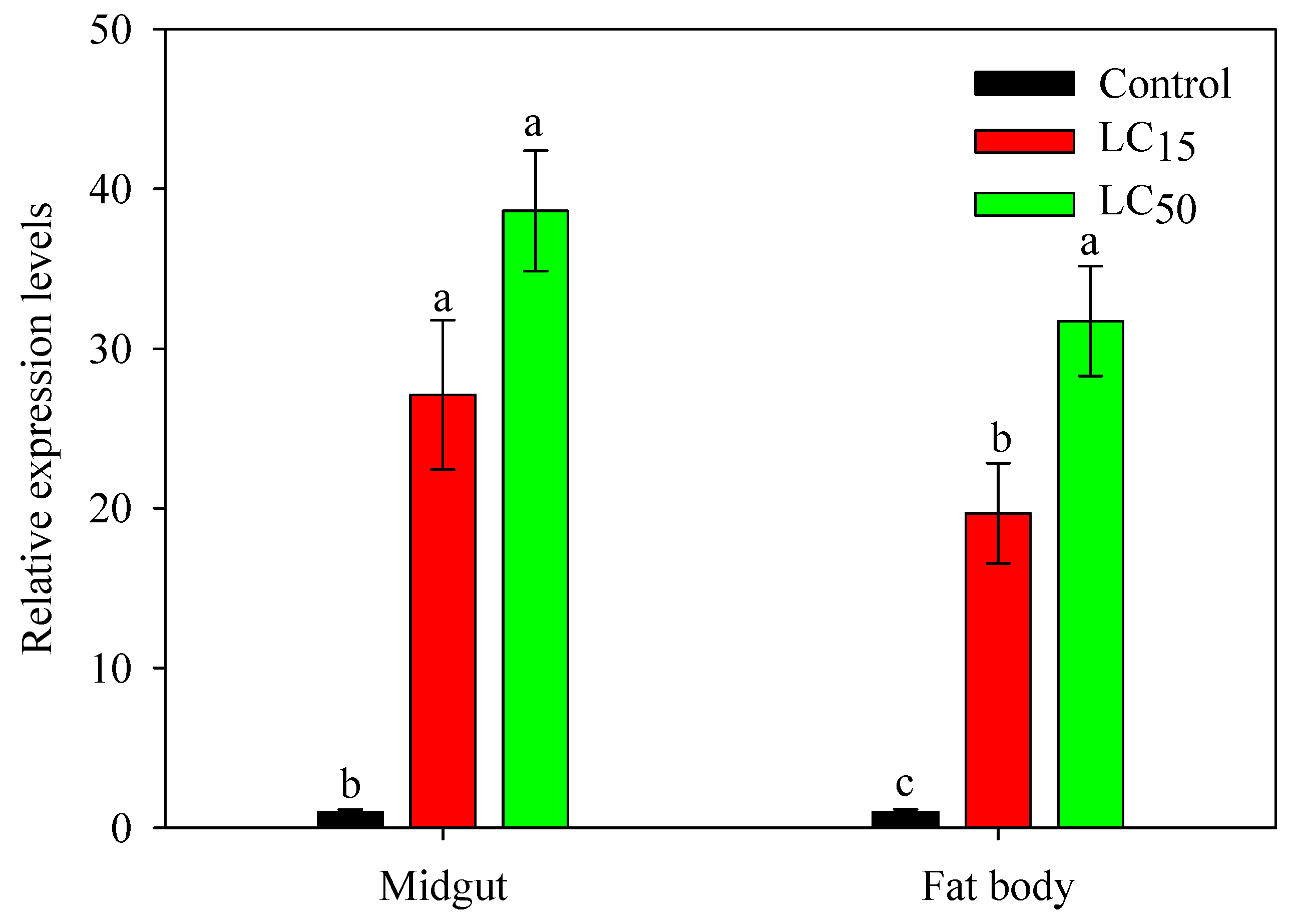
2.4. Expression Response of SlCPR in Larvae Exposed to Phoxim

Phoxim was selected to examine insecticide effects on SlCPR expression in the midgut and fat body of fifth-instar larvae (Figure 4). Compared to control treatments, larvae exposed to phoxim at LC15 (4 μg/mL) and LC50 (20 μg/mL) doses showed significantly increased SlCPR expression levels in the midgut. At LC50, phoxim caused 38.6-fold increased expression of SlCPR. Similarly, exposure to phoxim significantly induced SlCPR expression levels in the fat body (19.6-fold increase at LC15 and 31.7-fold increase at LC50) (Figure 4).

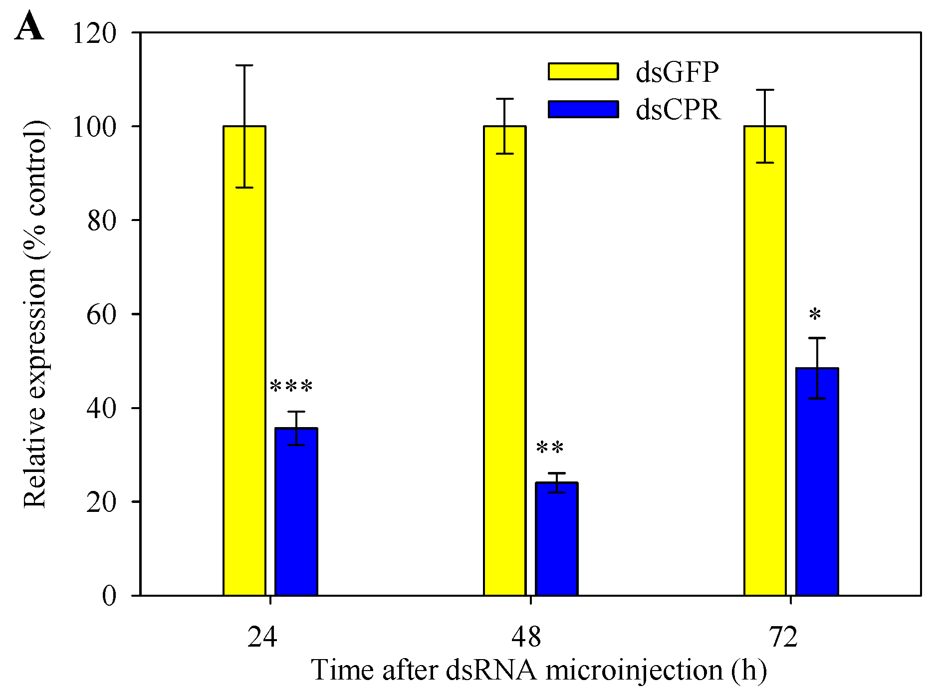
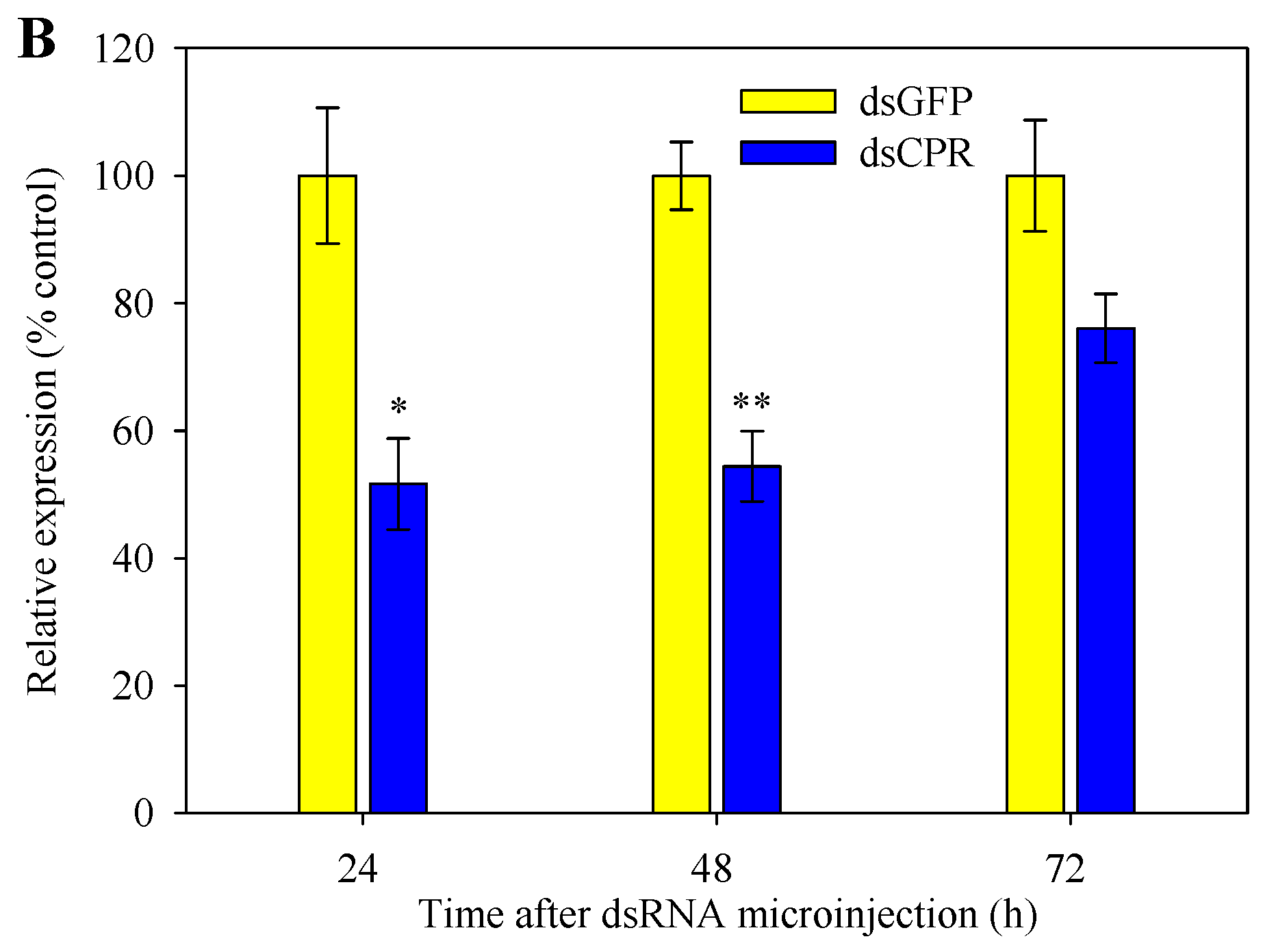
2.5. Silencing of SlCPR by RNAi

RNAi-mediated silencing of SlCPR by dsCPR microinjection was performed with fifth-instar larvae. To determine the efficiency of silencing, expression levels of the SlCPR-silenced larvae were determined by RT-qPCR. Compared to control larvae that were microinjected with dsGFP, SlCPR expression in the midgut of SlCPR-silenced larvae significantly decreased by 64.3%, 76.0%, and 51.5% when analyzed at 24, 48, and 72 h after dsCPR microinjection (Figure 5A). Likewise, expression levels of SlCPR-silenced larvae in the fat body decreased after dsCPR microinjection (by 48.4% at 24 h; by 45.6% at 48 h) (Figure 5B). These results indicated that RNAi suppressed the expression of SlCPR in S. litura larvae and that the silencing effect was retained for at least 48 h.

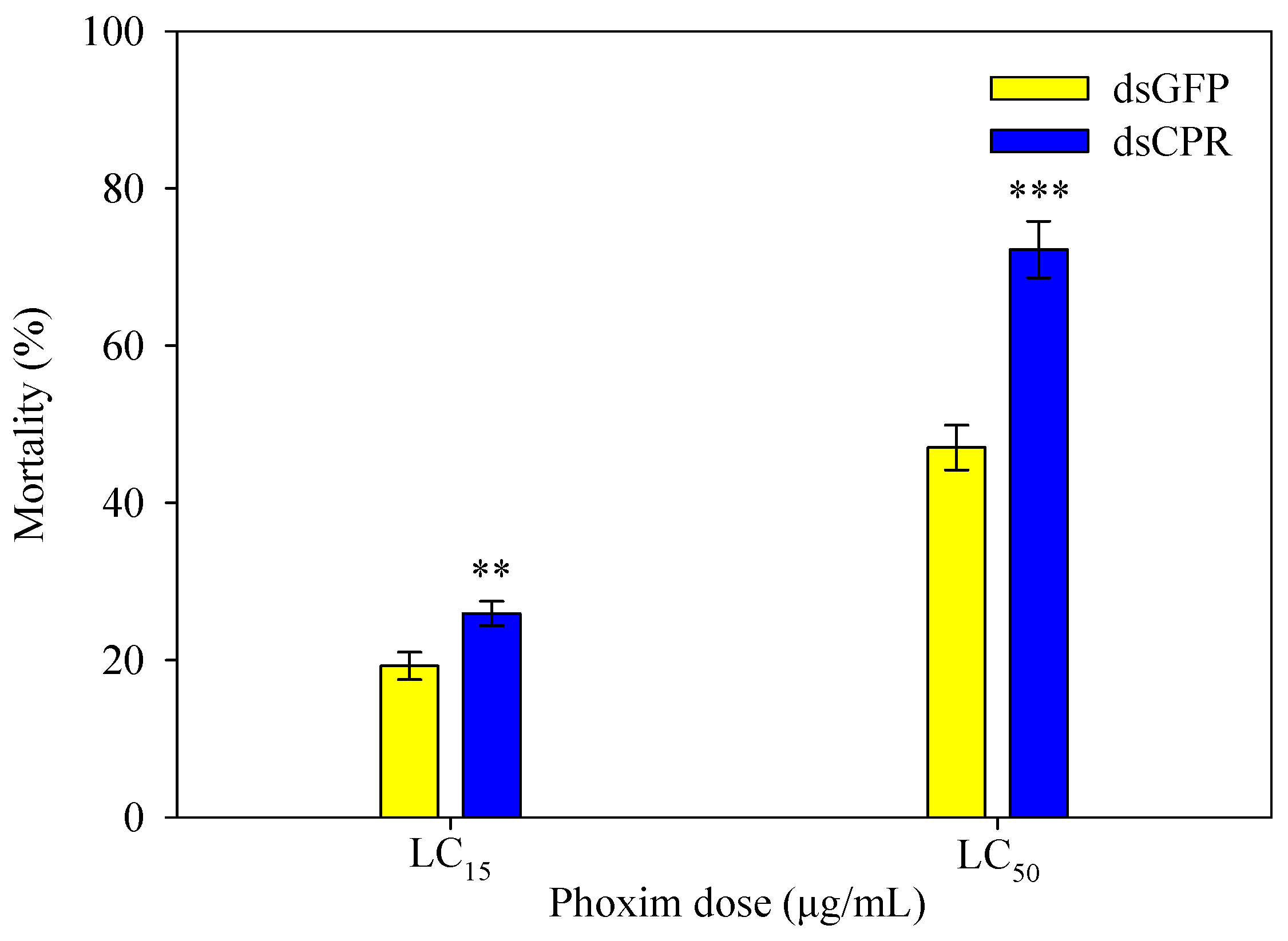
2.6. SlCPR-Silenced Larvae Show Increased Susceptibility to Phoxim

Mortality rates of fifth-instar larvae that were first microinjected with dsCPR (or dsGFP) and then exposed to phoxim are shown in Figure 6. When larvae were injected with dsGFP, mortality was 19.3% at the LC15 dose and 47.0% at the LC50 dose, respectively. However, compared to these control larvae, phoxim-induced mortality of SlCPR-silenced larvae was considerably increased (by 34.6% at the LC15 dose; by 53.5% at the LC50 dose) (Figure 6). These results indicate that SlCPR-silenced larvae exhibit an increased susceptibility to phoxim.

3. Discussion

Insect CPRs in phylogenetic trees are clearly segregated into clusters that correspond to different insect orders [16,21]. In the present study, we cloned and characterized the SlCPR gene of S. litura. The amino acid sequence of SlCPR shares high similarity with known CPRs. Our phylogenetic analysis of CPRs indicated that the SlCPR was more closely related to the CPR of S. littoralis than to the CPR of S. exigua. Likewise, previous phylogenetic analysis indicated that the P450 protein CYP321A7 of S. litura is most similar to CYP321A12 of S. littoralis [25], suggesting a close genetic relationship between S. litura and S. littoralis detoxification genes. Sequence comparisons also indicated that SlCPR most probably contains a hydrophobic N-terminal transmembrane domain, suggesting that SlCPR is a membrane anchored protein. In general, location of CPRs at the endoplasmic reticulum membrane is considered as essential for CPR function [15,17,19]. In this way, co-localized partner P450s are provided with electrons [17,28]. Similar hydrophobic transmembrane regions have been predicted for CPRs in related species such as S. exigua [19], C. lectularius [15], N. lugens [17], C. suppressalis [29], H. armigera [16], and L. migratoria [21]. Multiple sequence alignment further indicated that the hydrophilic C-terminal domain of SlCPR likely possesses FMN-, FAD-, and NADP-binding domains that are conserved among CPRs of insects [17,26]. Furthermore, putative catalytic residues (S470, C641, D686, and W688), known to be indispensable for rat and human CPR [27,30] were identified in the SlCPR sequence. Taking these sequence properties together, they indicate that SlCPR is likely an enzymatically functional CPR.
We further used RT-qPCR to investigate the expression profile of SlCPR in S. litura. The results showed that the expression levels of SlCPR varied among different development stages and tissues. Expression of SlCPR was strongest in the fifth- and sixth-instar larvae and highest expression levels were determined for the midgut and fat body of fifth-instar larvae. These differences likely reflect different levels of CPR activity. The expression pattern of SlCPR was found to be similar to that of CPRs in other insects such as N. lugens (NlCPR) [17], H. armigera (HaCPR) [31], and L. striatellus (LsCPR) [12]. CPRs likely possess conserved functions in insects [22]. The observed expression profile of SlCPR suggests that the protein is associated with different co-expressed P450s required for detoxification of plant allelochemicals and/or insecticides.
Previous studies have shown that CPRs of insects (together with partner P450s) may play an important role in detoxification of plant allelochemicals and insecticides [5,16,19]. An upregulation of CPR expression in insects may increase their resistance to insecticides [17,20,32]. For example, expression levels of the P. xylostella CPR gene in fourth-instar larvae were 13.2-fold higher in a β-cypermathrin resistant strain than in a susceptible strain [23]. Likewise, as compared to an insecticide-susceptible strain, CPR expression levels in apterous adult Rhopalosiphum padi were higher in an isoprocarb-resistant strain and imidacloprid-resistant strain (by 3.74- and 3.52-fold, respectively) [32]. In the present study, S. litura larvae exposed to phoxim showed significantly increased SlCPR transcript levels in the midgut and fat body. These findings suggested that SlCPR could be involved in insecticide detoxification and prompted us to further examine SlCPR-silenced larvae for their susceptibility to phoxim. In fact, previous reports on various insects showed that microinjection or feeding of dsRNA can result in successful silencing of CPR genes and this may influence the insect’s susceptibility to insecticides [12,15,21]. When exposed to β-cypermethrin, the mortality rate of the of NlCPR-silenced third-instar nymphs of N. lugens was 59.5% whereas control nymphs (microinjected with dsGFP) showed only 26.2%. Imidacloprid showed similar effects in NlCPR-silenced nymphs [17]. Furthermore, increased susceptibility to carbaryl was observed for third-instar nymphs of L. migratoria silenced in LmCPR [21]. Moreover, in A. citricidus, silencing of AcCPR caused significantly increased mortality when the adult aphids were exposed to abamectin [22]. In the present study, we successfully silenced the expression of SlCPR in S. litura fifth-instar larvae. Expression levels in the midgut and fat body were significantly reduced after dsCPR microinjection. When exposed to phoxim at LC15 and LC50 doses, SlCPR silencing significantly increased the mortality of S. litura as compared to the control group microinjected with dsGFP. Hence, reduced SlCPR expression levels enhanced the susceptibility of S. litura larvae to phoxim. These results suggest that SlCPR, in combination with partner P450s, is implicated in detoxification of phoxim.
In conclusion, we provide in this study key information on the SlCPR gene and our data indicate that SlCPR expression levels in S. litura larvae influence their susceptibility to phoxim. Further studies are needed to identify the redox partners of SlCPR and to study their role in resistance of phoxim and other insecticides.

4. Material and Methods

4.1. Insects

The phoxim susceptible population of S. litura used in this study was originally obtained from the Insectarium of the Institute of Entomology, Sun Yat-sen University (Guangzhou, China, May 11 2017). S. litura larvae were fed on an artificial diet [33] and maintained in an insectary (without exposure to any insecticides for more than two years) at 25 ± 2 °C and 70% ± 5% relative humidity under a 16:8 h light:dark regime at South China Agricultural University (Guangzhou, China).

4.2. RNA Extraction and cDNA Synthesis

RNA was extracted from eggs (20 eggs per RNA extraction), first- to sixth-instar larvae at day 2 (three larvae per RNA extraction), pupae at day 2 (three pupae per RNA extraction) and adult at day 1 (three adults per RNA extraction) for analyses of the SlCPR expression pattern at different development stages of S. litura. RNA was extracted from various tissues (cuticle, fat body, midgut, head, Malpighian tubule, and hemocytes) of fifth-instar larvae for analysis of the SlCPR expression pattern in different tissue types. Hemocytes were obtained with microcapillaries according to previously described procedures [34,35]. The material was centrifuged (10,000× g, 4 °C, 10 min) to remove debris. To obtain fat body tissue, the midgut was opened with tweezers and the content was carefully removed. The white-yellow fat body was then scraped from the midgut with tweezers and transferred into an Eppendorf tube containing phosphate-buffered saline (PBS). The sample was then centrifuged (2000 rpm, 4 °C, 3 min) to remove PBS. Finally, the fat body was washed twice with PBS.
Tissues from three individuals were pooled to obtain one RNA sample. Three independent biological replicates were performed for all samples. The RNA extraction procedure was performed with the RNAiso Plus kit (TaKaRa, Dalian, China) following the manual instructions. Isolated RNA (1 μg) was reverse transcribed using the ThermoScript™ RT-PCR System kit (Thermo Fisher Scientific, Carlsbad, CA, USA) following the manufacturer’s instruction.

4.3. Cloning of SlCPR

Based on obtained S. litura transcriptome data [25], primers (SlCPR-full-F: 5′-ATGTCAGACAGCGCACAGGACGTTC-3′; SlCPR-full-R: 5′-ACTCCAAACGTCAGCAGAATATTTC-3′)) were designed to amplify the complete SlCPR gene. cDNA derived from RNA isolated from S. litura fourth-instar larvae served as template. The PCR product was purified (Qiagen PCR Purification Kit, Qiagen, Netherlands) and cloned into the pMD18-T vector (Takara, Dalian, China). Finally, the plasmid was transformed into Escherichia coli DH5α competent cells (Invitrogen, Carlsbad, CA, USA) following the supplier’s guidelines and sequenced. The full-length SlCPR sequence can be found in the GenBank database under the accession number MH638288.

4.4. Bioinformatic Analyses

The predicted molecular weight and isoelectric point of SlCPR were calculated using corresponding programs available at the ExPASy Proteomics Server (http://cn.expasy.org/tools/pi_tool.html). Signal peptide and subcellular localization predictions were made with the SignalP 3.0 (http://www.cbs.dtu.dk/services/SignalP/) and the WoLF PSORT (http://wolfpsort.org/) programs. Multiple sequence alignment of CPR amino acid sequences was performed with DNAMAN software package (Version 6.0, Lynnon Biosoft, Vaudreuil, Quebec, Canada) [36]. MEGA 7.0 software (MEGA, PA, USA) [37] was employed to construct a corresponding phylogenetic tree using the neighbor-joining method with 1000 bootstrap replicates.

4.5. SlCPR Expression Analysis

Relative expression levels of SlCPR were quantified by RT-qPCR, using obtained cDNA and SlCPR specific primers (SlCPR-qF: 5′-TTACATAAGGGTGGAGATAGG-3′; SlCPR-qR: 5′-TGGTCAGTGTTGATGAGAGAG-3′). The PCR product (185 bp in length) corresponded to the nucleotide position 913 to 1097 of the SlCPR coding region. Two reference genes, β-actin (GenBank Acc. No. DQ494753) and Elongation factor-1 (EF1) (GenBank Acc. No. DQ192234) were used for normalizing the target gene expression. We confirm that β-actin and EF1 were the relatively stable genes for various target genes. The primers of the β-actin (β-actinF: 5′-TGAGACCTTCAACTCCCCCG-3′; β-actinR: 5′-GCGACCAGCCAAGTCCAGAC-3′) and EF1 (EF1F: 5′-CTCCTACATCAAGAAGATC-3′; EF1R: 5′-CTTGAGGATACCAGTTTC-3′) have been used before [25,38]. Each RT-qPCR was performed in a 20-μL reaction volume that contained 10 ng of cDNA template, 10 μL SYBR Green I Master Mix (Roche Diagnostics Corp., Indianapolis, IN, USA) and 0.2 μM of each primer. Reactions were performed with a MJ Research OpticonTM 2 instrument (Bio-Rad, Inc., Hercules, CA, USA) using the following parameters: (i) One cycle at 95 °C for 30 s and (ii) 40 cycles at 95 °C for 10 s and 60 °C for 25 s. The relative expression levels of SlCPR were calculated by the 2−∆∆Ct method [39] and normalized to the two reference genes (β-actin and EF1). All RT-qPCR experiments were performed with three independent biological replicates.

4.6. Analysis of SlCPR Expression in Larvae Exposed to Phoxim

Phoxim (99.0%, Shanghai Jiang Lai Biotechnology Co., Ltd., Shanghai, China) was diluted in acetone (99.5%, Guangzhou Chemical Reagent Factory, China) to obtain a 100 μg/mL stock solution. Then, the stock solution was diluted with sterilized water to prepare different concentrations for the tests. Concentrations of phoxim causing 15% and 50% lethality of fifth-instar larvae (LC15: 4 μg/mL; LC50: 20 μg/mL) were used in this study. The LC15 and LC50 values were obtained from a trial experiment with different phoxim doses. The mortality values were 6.7%, 11.1%, 45.6%, 73.3%, 81.1%, and 96.7% at 1, 4, 16, 64, 256, and 1024 μg/mL phoxim, respectively (Figure S1). The LC15 and LC50 values were determined using probit analysis (POLO-PC software). To test toxicity of phoxim on S. litura, fifth-instar (day 1) larvae were used in a standard leaf disc bioassay method [1]. Leaves (7 cm in diameter) of Chinese cabbage (Brassica campestris L. ssp. pekinensis) were immersed in the prepared phoxim solution (LC15 or LC50 dosages) for 10 s and allowed to air-dry for 1.5 h. Control leaves were immersed in sterilized water. A total of 30 fifth-instar larvae were placed on each treated leaf (three larvae per leaf) which were placed in a sterile glass Petri dish (9 cm in diameter). After 24 h incubation in the insectary, the midgut or fat bodies from three of surviving larvae were pooled as one sample for RNA exaction, respectively. Three independent replicates were used for each treatment (three biological replicates). SlCPR expression analysis by RT-qPCR was conducted as described above.

4.7. Silencing of SlCPR by RNAi

DNA for in vitro transcription reactions was amplified by PCRs using cDNAs of SlCPR and GFP (green fluorescent protein; accession number ACY56286) as a control. The PCRs were performed with the following primers: (i) CPR-RNAi-F (5′-ATGGTTGCTGATCCCGAAGAA-3′) and T7CPR-RNAi-F (5′-aatacgactcactatagggATGGTTGCTGATCCCGAAGAA-3′), (ii) CPR-RNAi-R (5′-AGGCCAAACACGGCATAATTT-3′) and T7CPR-RNAi-R (5′-aatacgactcactataggg AGGCCAAACACGGCATAATTT-3′), (iii) T7GFPdsRNAF (5′-AATACGACTCACTATAGGGAAGGGCGAGGAGCTGTTCACCG-3′) and GFPdsRNAR (5′CAGCAGGACCATGTGATCGCGC-3′), and (iv) GFPdsRNAF (5′-AAGGGCGAGGAGCTGTTCACCG-3′) and T7GFPdsRNAR (5′-AATACGACTCACTATAGGGCAGCAGGACCATGTGATCGCGC-3′) [25]. The SlCPR PCR product corresponded to the nucleotide position 361 to 551 of the SlCPR coding region. The PCR products were then purified with a PCR purification kit (Qiagen, Venlo, The Netherlands) and used as templates to synthesize double-stranded RNA (dsRNA) with the T7 RiboMAX™ Express RNAi System (Promega, Madison, WI, USA). The dsRNA was adjusted with DEPC-treated (RNase-free) water to a final concentration of 1.5 μg·μL−1 and kept at –80 °C for further use. Subsequently, 2 μL (3.0 μg) of dsRNA were injected into the side of the thorax of fifth-instar (day 2) larvae of S. litura using a manual microinjector (model No. MS05, Chengdu Centome Company Ltd., Chengdu, China). Thirty fifth-instar larvae microinjected with dsCPR or dsGFP were incubated in the insectary for 24, 48, and 72 h, respectively. RNA was then isolated from the midgut and fat bodies, respectively. Tissue from three larvae were used for each RNA extraction. SlCPR expression levels in the midgut and fat bodies were determined by RT-qPCR. Three independent replicates were conducted for all treatments.

4.8. Bioassays with Phoxim after RNAi

To explore a possible role of SlCPR in the insect’s susceptibility to phoxim, dsCPR or dsGFP was microinjected into 30 fifth-instar (day 1) larvae of S. litura, respectively. Leaves of Chinese cabbage were immersed in phoxim solution (LC15 or LC50 doses) and then air-dried. After dsRNA delivery, S. litura were placed on each prepared leaf and incubated in the insectary at the same condition as described above. Mortality rates of S. litura were recorded after 48 h. All tests were performed in three independent replicates.

4.9. Data Analysis

Data were expressed as means ± standard error (SE). Statistical analysis was carried out with the SPSS 13.0 Software Package (SPSS Inc., Chicago, IL, USA). One-way ANOVA followed by the Duncan’s multiple range test was employed to analyze differences among different development stages and tissues. The Student’s t-test was used to analyze data from SlCPR-silenced larvae and toxicity tests with phoxim. Statistical differences were considered as significant at p < 0.05.

Supplementary Materials

Author Contributions

Data curation, H.-Y.J., Y.-P.J., S.-W.L., Z.-H.M. and R.-L.W.; Formal analysis, H.-Y.J., C.S., Y.-P.J., S.-W.L., Z.-H.M., Y.-J.S., J.-E.Z. and R.-L.W.; Funding acquisition, J.-E.Z. and R.-L.W.; Methodology, H.-Y.J., Y.-P.J., S.-W.L. and R.-L.W.; Software, H.-Y.J., Z.-H.M., Y.-J.S.; Writing—original draft, H.-Y.J., C.S., J.-E.Z. and R.-L.W.; Writing—review and editing, C.S., J.-E.Z. and R.-L.W.

Funding

This research was financially supported by the Natural Science Foundation of Guangdong Province, China (Grant 2017A030313188[R.-L.W.]), the National Natural Science Foundation of China (Grant 31470576[R.-L.W.]) and the Science and Technology Planning Project of Guangdong Province (Grant 2019B030301007[J.-E.Z.]).

Conflicts of Interest

The authors declare no conflict of interest.

References

  1. Ahmad, M.; Sayyed, A.H.; Saleem, M.A.; Ahmad, M. Evidence for field evolved resistance to newer insecticides in Spodoptera litura (Lepidoptera: Noctuidae) from Pakistan. Crop Prot. 2008, 27, 1367–1372. [Google Scholar] [CrossRef]
  2. Huang, S.; Han, Z. Mechanisms for multiple resistances in field populations of common cutworm, Spodoptera litura (Fabricius) in China. Pestic. Biochem. Physiol. 2007, 87, 14–22. [Google Scholar] [CrossRef]
  3. Ahmad, M.; Ahmad, M.; Saleem, M.A.; Aslam, M.; Sayyed, A.H. Resistance to selected organochlorin, organophosphate, carbamate and pyrethroid, in Spodoptera litura (Lepidoptera: Noctuidae) from Pakistan. J. Econ. Entomol. 2008, 101, 1667–1675. [Google Scholar]
  4. Feyereisen, R. Arthropod CYPomes illustrate the tempo and mode in P450 evolution. Biochim. Biophys. Acta Proteins Proteomics 2011, 1814, 19–28. [Google Scholar] [CrossRef] [PubMed]
  5. Feyereisen, R. Insect P450 inhibitors and insecticides: Challenges and opportunities. Pest Manag. Sci. 2015, 71, 793–800. [Google Scholar] [CrossRef]
  6. Wang, R.L.; Staehelin, C.; Xia, Q.Q.; Su, Y.J.; Zeng, R.S. Identification and characterization of CYP9A40 from the tobacco cutworm moth (Spodoptera litura), a cytochrome P450 gene induced by plant allelochemicals and insecticides. Int. J. Mol. Sci. 2015, 16, 22606–22620. [Google Scholar] [CrossRef]
  7. Wang, R.L.; Zhu-Salzman, K.; Baerson, S.R.; Xin, X.W.; Li, J.; Su, Y.J.; Zeng, R.S. Identification of a novel cytochrome P450 CYP321B1 gene from tobacco cutworm (Spodoptera litura) and RNA interference to evaluate its role in commonly used insecticides. Insect Sci. 2017, 24, 235–247. [Google Scholar] [CrossRef]
  8. Li, F.; Ni, M.; Zhang, H.; Wang, B.; Xu, K.; Tian, J.; Hu, J.; Shen, W.; Li, B. Expression profile analysis of silkworm P450 family genes after phoxim induction. Pestic. Biochem. Physiol. 2015, 122, 103–109. [Google Scholar] [CrossRef]
  9. Porter, T.D.; Kasper, C.B. NADPH-cytochrome P-450 oxidoreductase: Flavin mononucleotide and flavin adenine dinucleotide domains evolved from different flavoproteins. Biochem. 1986, 25, 1682–1687. [Google Scholar] [CrossRef]
  10. Waskell, L.; Kim, J.J.P. Electron transfer partners of cytochrome P450. In Cytochrome P450: Structure, Mechanism, and Biochemistry, 4th ed.; Springer: Cham, Germany, 2015; pp. 33–68. [Google Scholar]
  11. Vermilion, J.L.; Ballou, D.P.; Massey, V.; Coon, M.J. Separate roles for FMN and FAD in catalysis by liver microsomal NADPH-cytochrome P-450 reductase. J. Biol. Chem. 1981, 256, 266–277. [Google Scholar]
  12. Zhang, Y.; Wang, Y.; Wang, L.; Yao, J.; Guo, H.; Fang, J. Knockdown of NADPH-cytochrome P450 reductase results in reduced resistance to buprofezin in the small brown planthopper, Laodelphax striatellus (fallén). Pestic. Biochem. Physiol. 2016, 127, 21–27. [Google Scholar] [CrossRef]
  13. Cheng, T.; Wu, J.; Wu, Y.; Chilukuri, R.V.; Huang, L.; Yamamoto, K.; Feng, L.; Li, W.; Chen, Z.; Guo, H.; et al. Genomic adaptation to polyphagy and insecticides in a major East Asian noctuid pest. Nat. Ecol. Evol. 2017, 1, 1747–1756. [Google Scholar] [CrossRef] [PubMed]
  14. Pottier, M.A.; Bozzolan, F.; Chertemps, T.; Jacquin-Joly, E.; Lalouette, L.; Siaussat, D.; Maïbèche-Coisne, M. Cytochrome P450s and cytochrome P450 reductase in the olfactory organ of the cotton leafworm Spodoptera littoralis. Insect Mol. Biol. 2012, 21, 568–580. [Google Scholar] [CrossRef] [PubMed]
  15. Zhu, F.; Sams, S.; Moural, T.; Haynes, K.F.; Potter, M.F.; Palli, S.R. RNA interference of NADPH-cytochrome P450 reductase results in reduced insecticide resistance in the bed bug, Cimex lectularius. PLoS ONE 2012, 7, e31037. [Google Scholar] [CrossRef] [PubMed]
  16. Zhao, C.; Tang, T.; Feng, X.; Qiu, L. Cloning and characterisation of NADPH-dependent cytochrome P450 reductase gene in the cotton bollworm, Helicoverpa armigera. Pest Manag. Sci. 2014, 70, 130–139. [Google Scholar] [CrossRef] [PubMed]
  17. Liu, S.; Liang, Q.M.; Zhou, W.W.; Jiang, Y.D.; Zhu, Q.Z.; Yu, H.; Zhang, C.X.; Gurr, G.M.; Zhu, Z.R. RNA interference of NADPH-cytochrome P450 reductase of the rice brown planthopper, Nilaparvata lugens, increases susceptibility to insecticides. Pest Manag. Sci. 2015, 71, 32–39. [Google Scholar] [CrossRef]
  18. Huang, Y.; Lu, X.P.; Wang, L.L.; Wei, D.; Feng, Z.J.; Zhang, Q.; Xiao, L.F.; Dou, W.; Wang, J.J. Functional characterization of NADPH-cytochrome P450 reductase from Bactrocera dorsalis: Possible involvement in susceptibility to malathion. Sci. Rep. 2015, 5, 18394. [Google Scholar] [CrossRef]
  19. Zhao, C.; Feng, X.; Tang, T.; Qiu, L.; Swale, D. Isolation and expression analysis of CYP9A11 and cytochrome P450 reductase gene in the beet armyworm (Lepidoptera: Noctuidae). J. Insect Sci. 2015, 15, 122. [Google Scholar] [CrossRef]
  20. Zhang, Y.X.; Li, S.G.; Rao, X.J.; Liu, S. Molecular characterization of a NADPH–cytochrome P450 reductase gene from the rice leaffolder, Cnaphalocrocis medinalis (Lepidoptera: Pyralidae). Appl. Entomol. Zool. 2018, 53, 19–27. [Google Scholar] [CrossRef]
  21. Zhang, X.; Wang, J.; Liu, J.; Li, Y.; Liu, X.; Wu, H.; Ma, E.; Zhang, J. Knockdown of NADPH-cytochrome P450 reductase increases the susceptibility to carbaryl in the migratory locust, Locusta migratoria. Chemosphere 2017, 188, 517–524. [Google Scholar] [CrossRef]
  22. Jing, T.X.; Tan, Y.; Ding, B.Y.; Dou, W.; Wei, D.D.; Wang, J.J. NADPH-cytochrome P450 reductase mediates the resistance of Aphis (Toxoptera) citricidus (Kirkaldy) to abamectin. Front. Physiol. 2018, 9, 986. [Google Scholar] [CrossRef] [PubMed]
  23. Chen, X.; Zhang, Y. Identification and characterization of NADPH-dependent cytochrome P450 reductase gene and cytochrome b5 gene from Plutella xylostella: Possible involvement in resistance to beta-cypermethrin. Gene 2015, 558, 208–214. [Google Scholar] [CrossRef] [PubMed]
  24. Terenius, O.; Papanicolaou, A.; Garbutt, J.S.; Eleftherianos, I.; Huvenne, H.; Kanginakudru, S.; Albrechtsen, M.; An, C.; Aymeric, J.L.; Barthel, A.; et al. RNA interference in Lepidoptera: An overview of successful and unsuccessful studies and implications for experimental design. J. Insect Physiol. 2011, 57, 231–245. [Google Scholar] [CrossRef] [PubMed] [Green Version]
  25. Wang, R.L.; He, Y.N.; Staehelin, C.; Liu, S.W.; Su, Y.J.; Zhang, J.E. Identification of two cytochrome monooxygenase P450 genes, CYP321A7 and CYP321A9, from the tobacco cutworm moth (Spodoptera litura) and their expression in response to plant allelochemicals. Int. J. Mol. Sci. 2017, 18, 2278. [Google Scholar] [CrossRef] [PubMed]
  26. Ingelman, M.; Bianchi, V.; Eklund, H. The three-dimensional structure of flavodoxin reductase from Escherichia coli at 1.7 Å resolution. J. Mol. Biol. 1997, 268, 147–157. [Google Scholar] [CrossRef]
  27. Hubbard, P.A.; Shen, A.L.; Paschke, R.; Kasper, C.B.; Kim, J.J.P. NADPH-cytochrome P450 oxidoreductase structural basis for hydride and electron transfer. J. Biol. Chem. 2001, 276, 29163–29170. [Google Scholar] [CrossRef] [PubMed]
  28. Lamb, D.C.; Warrilow, A.G.S.; Venkateswarlu, K.; Kelly, D.E.; Kelly, S.L. Activities and kinetic mechanisms of native and soluble NADPH-cytochrome P450 reductase. Biochem. Biophys. Res. Commun. 2001, 286, 48–54. [Google Scholar] [CrossRef] [PubMed]
  29. Liu, S.; Liang, Q.M.; Huang, Y.J.; Yuan, X.; Zhou, W.W.; Qiao, F.; Cheng, J.; Gurr, G.M.; Zhu, Z.R. Cloning, functional characterization, and expression profiles of NADPH-cytochrome P450 reductase gene from the Asiatic rice striped stem borer, Chilo suppressalis (Lepidoptera: Pyralidae). Comp. Biochem. Physiol. B Biochem. Mol. Biol. 2013, 166, 225–231. [Google Scholar] [CrossRef]
  30. Flück, C.E.; Mullis, P.E.; Pandey, A.V. Modeling of human P450 oxidoreductase structure by in silico mutagenesis and MD simulation. Mol. Cell. Endocrinol. 2009, 313, 17–22. [Google Scholar] [CrossRef]
  31. Liu, D.; Zhou, X.; Li, M.; Zhu, S.; Qiu, X. Characterization of NADPH-cytochrome P450 reductase gene from the cotton bollworm, Helicoverpa armigera. Gene 2014, 545, 262–270. [Google Scholar] [CrossRef]
  32. Wang, K.; Peng, X.; Zuo, Y.; Li, Y.; Chen, M. Molecular cloning, expression pattern and polymorphisms of NADPH-cytochrome P450 reductase in the bird cherry-oat aphid Rhopalosiphum padi (L.). PLoS ONE 2016, 11, e0154633. [Google Scholar] [CrossRef] [PubMed]
  33. Chen, Q.J.; Li, G.H.; Pang, Y. A simple artificial diet for mass rearing of some noctuid species. Entomol. Knowl. 2000, 37, 8–10. [Google Scholar]
  34. Miller, J.S.; Stanley, D.W. Lipopolysaccharide evokes microaggregation reactions in hemocytes isolated from tobacco hornworms, Manduca sexta. Comp. Biochem. Physiol. Part A: Mol. Integr. Physiol. 2004, 137, 285–295. [Google Scholar] [CrossRef] [PubMed]
  35. Zhou, J.; Zhang, G.; Zhou, Q. Molecular characterization of cytochrome P450 CYP6B47 cDNAs and 5′-flanking sequence from Spodoptera litura (Lepidoptera: Noctuidae): Its response to lead stress. J. Insect Physiol. 2012, 58, 726–736. [Google Scholar] [CrossRef] [PubMed]
  36. Chenna, R.; Sugawara, H.; Koike, T.; Lopez, R.; Gibson, T.J.; Higgins, D.G.; Thompson, J.D. Multiple sequence alignment with the Clustal series of programs. Nucleic Acids Res. 2003, 31, 3497–3500. [Google Scholar] [CrossRef] [PubMed] [Green Version]
  37. Kumar, S.; Stecher, G.; Tamura, K. MEGA7: Molecular evolutionary genetics analysis version 7.0 for bigger datasets. Mol. Biol. Evol. 2016, 33, 1870–1874. [Google Scholar] [CrossRef] [PubMed]
  38. Lu, Y.; Yuan, M.; Gao, X.; Kang, T.; Zhan, S.; Wan, H.; Li, J. Identification and validation of reference genes for gene expression analysis using quantitative PCR in Spodoptera litura (Lepidoptera: Noctuidae). PLoS ONE. 2013, 8, e68059. [Google Scholar] [CrossRef] [PubMed]
  39. Livak, K.J.; Schmittgen, T.D. Analysis of relative gene expression data using real-time quantitative PCR and the 2−ΔΔCT method. Methods 2001, 25, 402–408. [Google Scholar] [CrossRef] [PubMed]
Figure 1. Sequence analysis of the SlCPR protein (A). The protein contains predicted flavin mononucleotide (FMN)-, flavin adenine dinucleotide (FAD)-, and NADP-binding domains. The proteins also conserved residues such as the FAD-binding motif (R467, Y469, and S470) and the predicted catalytic residues (S470, C641, D686, and W688). (B) Comparison of the deduced amino acid sequence of SlCPR with other NADPH-cytochrome P450 reductases (CPRs). Accession numbers of indicated CPR amino acid sequences are shown in Table 1.
Ijms 20 03839 g001
Figure 2. Phylogenic analysis of SlCPR and related insect CPRs. Multiple sequence alignment of full-length amino acid sequences of 29 CPR proteins was performed with DNAMAN 6.0 software. The phylogenetic tree was constructed using MEGA7.0 with the neighbor-joining (NJ) method and 1000 bootstrap replicates. Numbers shown at the tree forks indicate frequency of occurrence among all bootstrap iterations performed. The scale bar indicates 0.05 amino acid substitutions per site. CPRs from human and mouse were used as an outgroup. SlCPR is marked by a black solid circle. Accession numbers of indicated CPR amino acid sequences are shown in Table 1.
Ijms 20 03839 g002
Figure 3. Relative expression levels of SlCPR at different development stages (A) and in various tissues (B). Whole body of S. litura larvae were used for the different development stages, while fifth-instar larvae were used for various tissues. Expression levels of SlCPR were determined by quantitative reverse transcriptase PCR (RT-qPCR), and β-actin and EF1 were selected as reference genes. Each RT-qPCR reaction for each sample was performed in three biological replicates and three technical replicates. Data shown are means ± SE. Different letters (a,b,c,d) above bars indicate significant differences (p < 0.05) according to Duncan’s multiple range test. Abbreviations: 1st to 6th—first- to sixth-instar larvae; C—cuticle; FB—fat body; MG—midgut; H—head; MT—Malpighian tubule; HC—hemocytes.
Ijms 20 03839 g003
Figure 4. Effects of phoxim on SlCPR expression in the midgut and fat body of fifth-instar larvae. Larvae were exposed to phoxim at LC15 (4 μg/mL) and LC50 (20 μg/mL) doses for 24 h. SlCPR expression levels were normalized to β-actin and EF1 expression and presented as the means ± SE with three independent biological replicates and three technical replicates. Different letters (a,b,c) above bars indicate significant differences (p < 0.05) according to Duncan’s multiple range test.
Ijms 20 03839 g004
Figure 5. RNA interference (RNAi)-mediated silencing of SlCPR in fifth-instar larvae. Larvae were microinjected with dsCPR or dsGFP (control). RNA was isolated at indicated time points after microinjection. Expression levels of SlCPR in the midgut (A) and fat body (B) were then determined by RT-qPCR. The expression levels of SlCPR were normalized using β-actin and EF1 as reference genes. Each RT-qPCR reaction for each sample was performed in three technical replicates and three biological replicates. Data indicate means ± SE. Asterisks indicate significantly reduced expression levels in SlCPR-silenced larvae as compared to the control group (Student’s t-test, * p < 0.05,** p < 0.01, *** p < 0.001).
Ijms 20 03839 g005aIjms 20 03839 g005b
Figure 6. Effect of SlCPR silencing on the susceptibility to phoxim. Fourth-instar larvae were microinjected with dsCPR or dsGFP (control). Thirty fifth-instar larvae were then exposed to phoxim at LC15 (4 μg/mL) or LC50 (20 μg/mL) doses for 48 h. All tests were performed in triplicate. Data shown are mortality rates (means ± SE). Asterisks indicate significant differences between SlCPR-silenced larvae as compared to the control group (Student’s t-test, ** p < 0.01, *** p < 0.001).
Ijms 20 03839 g006
Table 1. Percent amino acid identities between SlCPR and other CPRs.
OrderSpeciesAccession NumberIdentity (%)
HymenopteraMonomorium pharaonisXP_01254136462.8
Solenopsis invictaXP_01115706362.6
Pogonomyrmex barbatusXP_01164315261.5
Apis melliferaXP_00111994962.1
Apis floreaNP_00135166962.6
Orussus abietinusXP_01227516262.6
Fopius arisanusXP_01130634763.4
Microplitis demolitorXP_00854868462.3
HemipteraBemisia tabaciAGT1570161.9
Cimex lectulariusAFD5050762.7
Laodelphax striatellaAID5542263.8
Sogatella furciferaAHM9300964.4
ColeopteraDendroctonus ponderosaeAFI4500264.9
Tribolium castaneumXP_97117467.6
DipteraDrosophila mettleriAAB4896462.8
Drosophila melanogasterNP_47715866.6
Musca domesticaAAA2929568.4
Anopheles gambiaeAAO2476566.6
Anopheles funestusEF15257867.5
LepidopteraPlutella xylostellaNP_00129246979.4
Chilo suppressalisAGM2056585.4
Bombyx mandarinaABJ9770987.0
Bombyx moriNP_00110483487.0
Helicoverpa armigeraADK2506095.6
Spodoptera exiguaADX9574698.4
Spodoptera littoralisAFP2058499.6
Spodoptera lituraMH638288100
RodentiaMus musculusNM_00889853.5
PrimatesHomo sapiensNP_00093256.2

