Electrospun Chitosan/Poly (Vinyl Alcohol)/Graphene Oxide Nanofibrous Membrane with Ciprofloxacin Antibiotic Drug for Potential Wound Dressing Application


Abstract
:
1. Introduction
2. Results and Discussion
2.1. Morphology of Drug-Loaded Nanofibrous Membranes
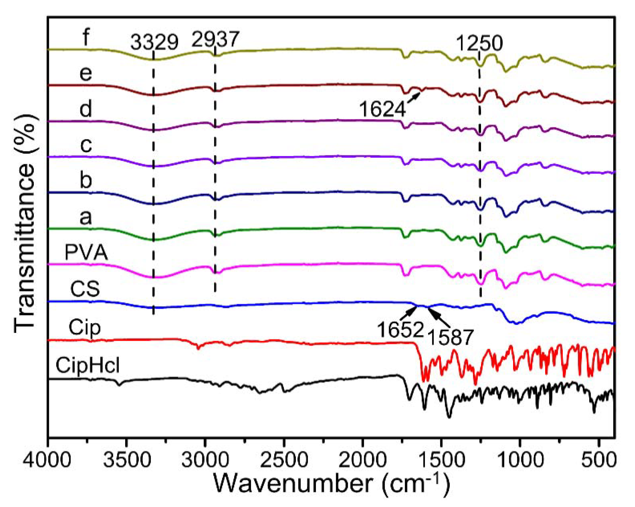
2.2. FTIR
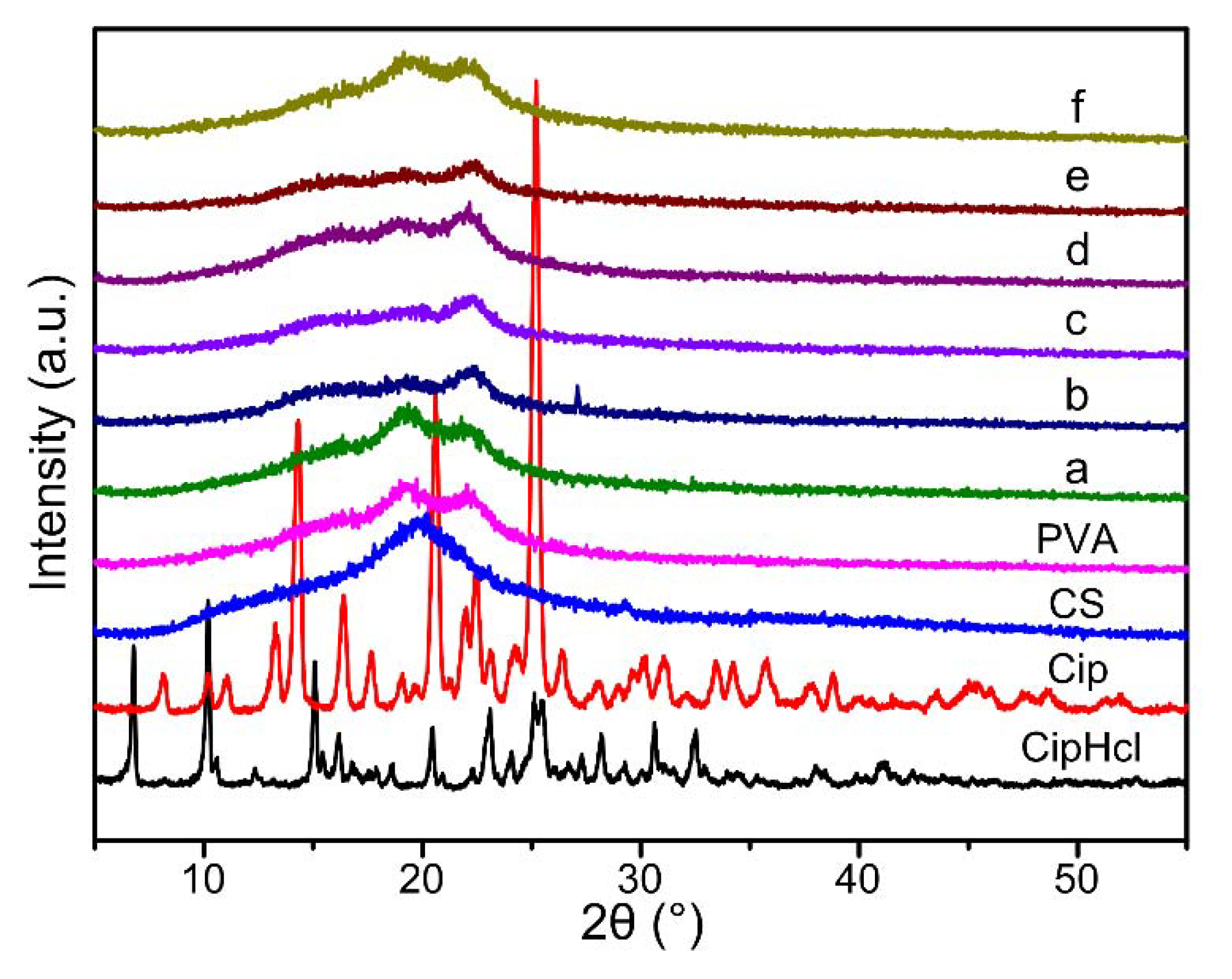
2.3. XRD
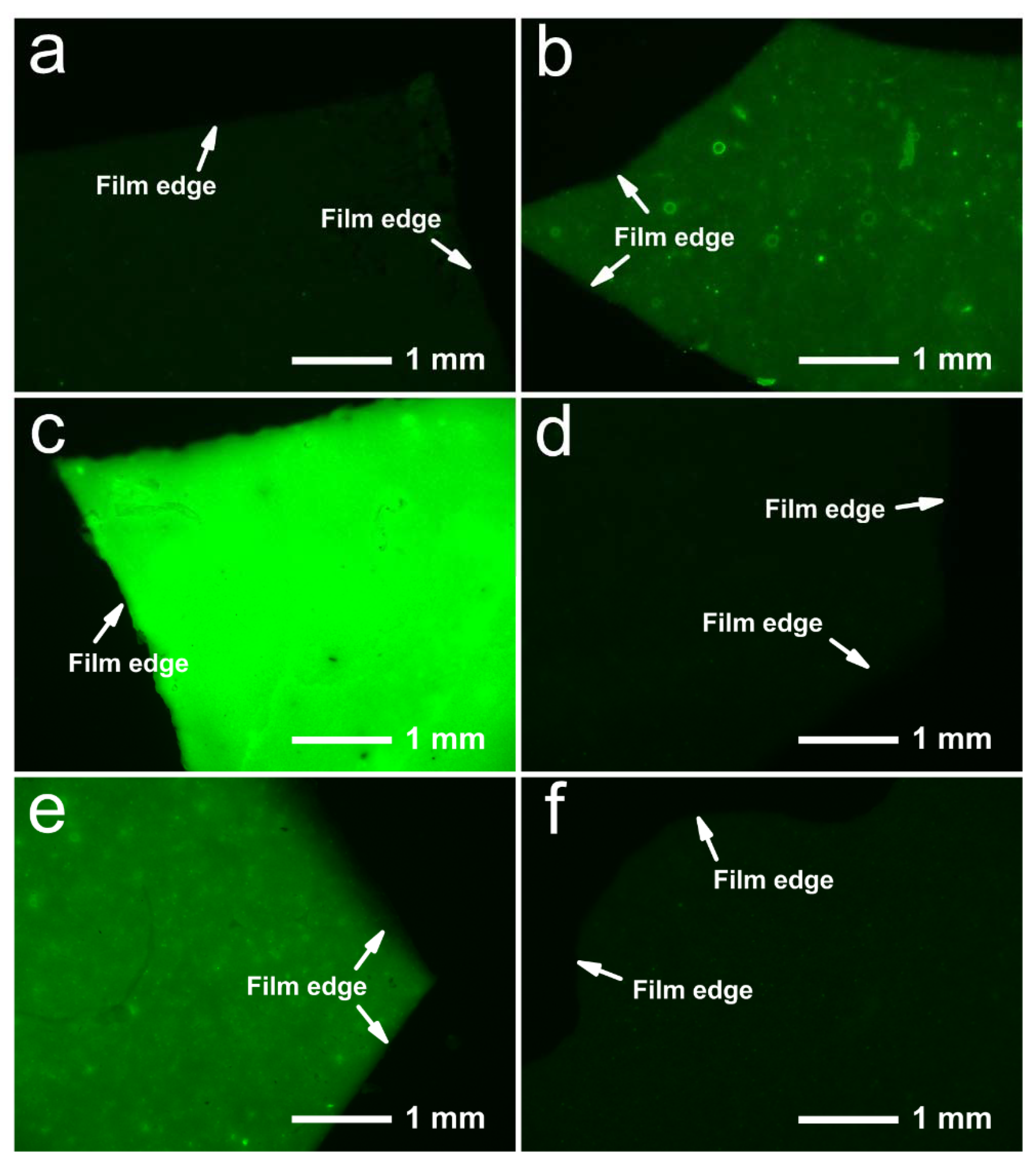
2.4. Fluorescence Effect
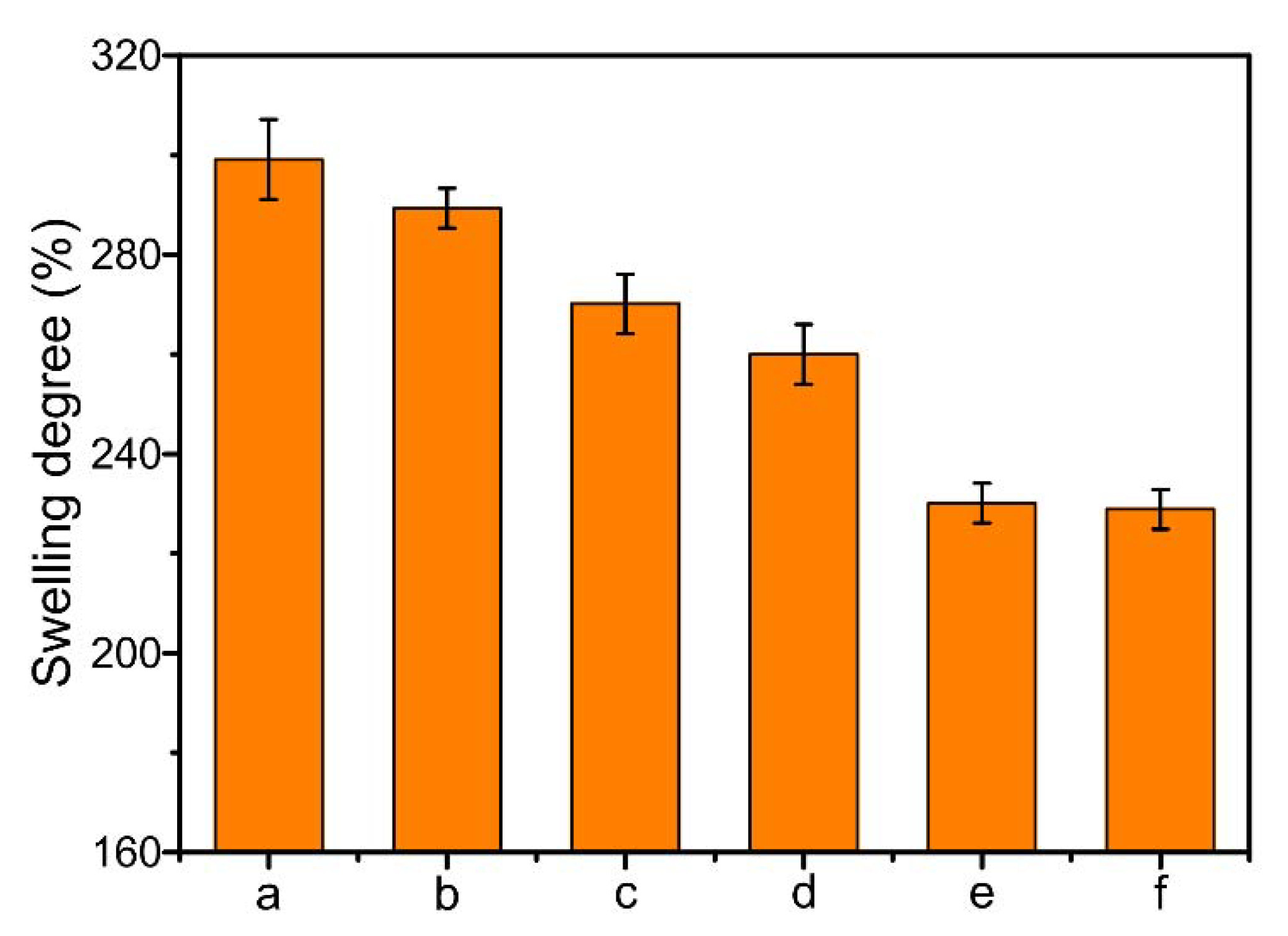
2.5. Swelling
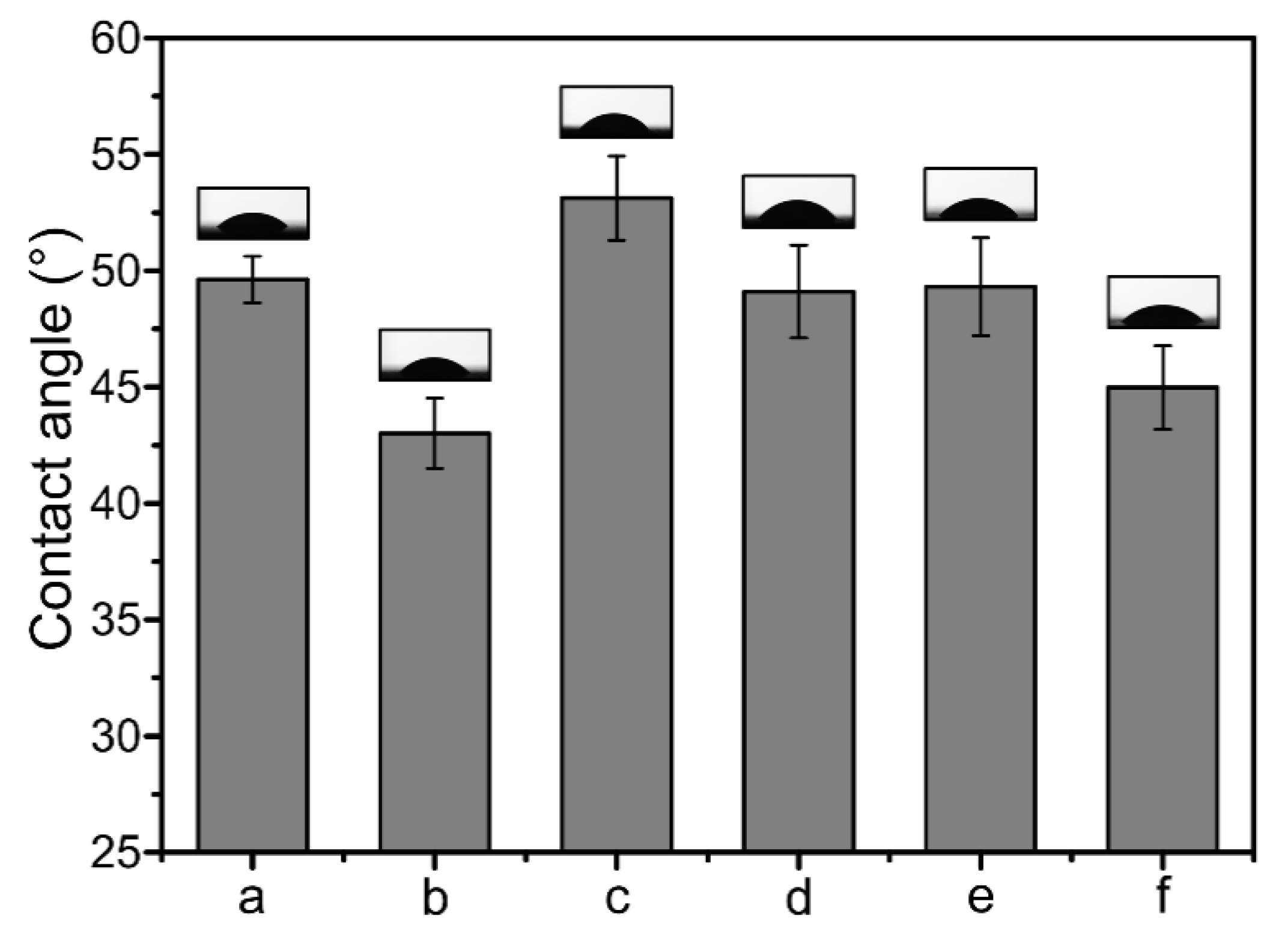
2.6. Contact Angle
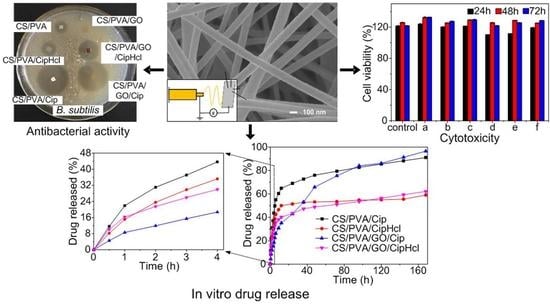
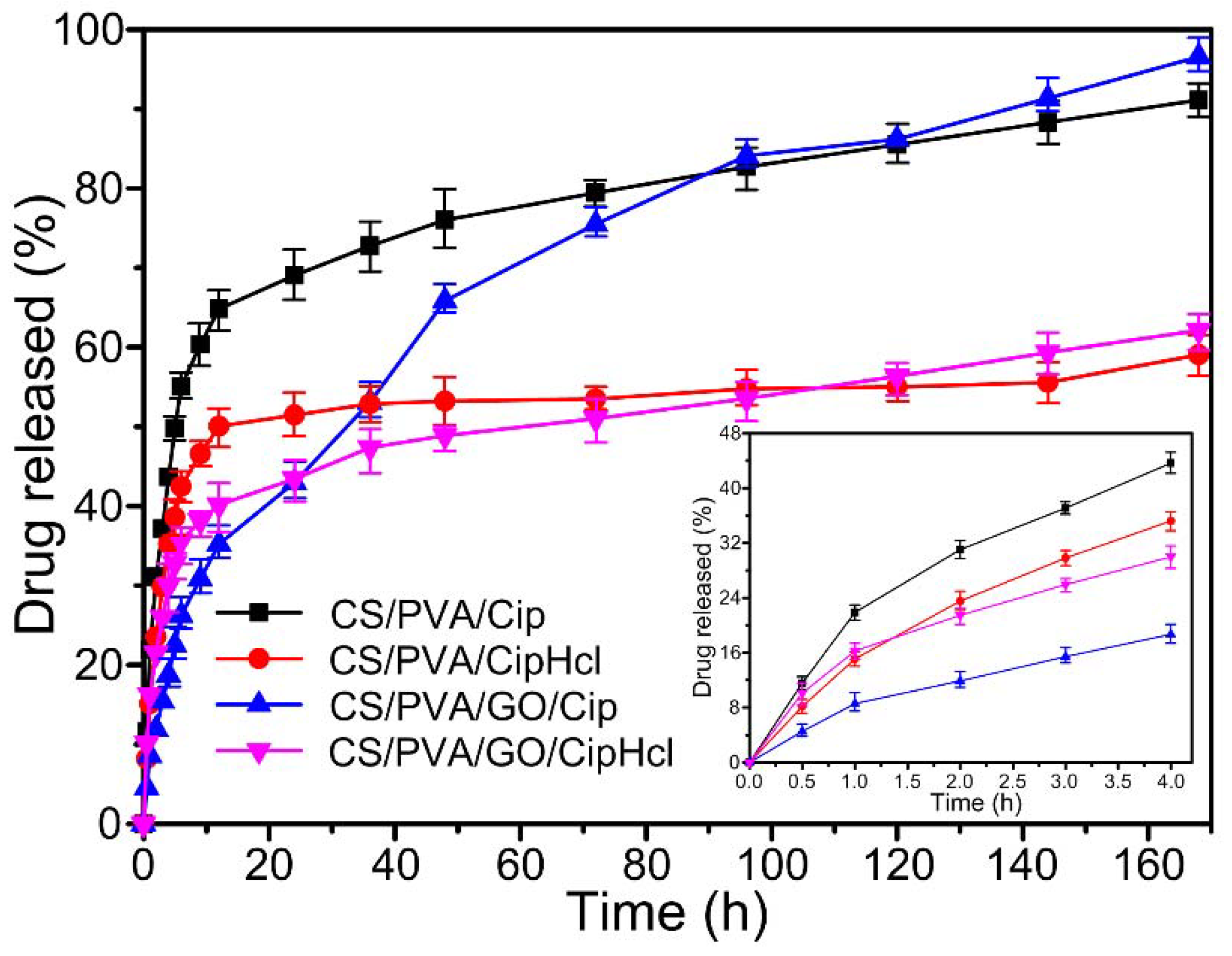
2.7. In Vitro Drug Release
2.8. Antibacterial Activity
2.9. Cytotoxicity
3. Experimental
3.1. Materials
3.2. Preparation of GO Dispersion
3.3. Preparation of Electrospun Precursor Solutions
3.4. Preparation of Drug-Loaded Nanofibrous Membranes
3.5. Characterization
3.6. Swelling Measurement
3.7. Contact Angle Measurement
3.8. In Vitro Drug Release Assay
3.9. Antibacterial Activity Assay
3.10. Cytotoxicity Assay
4. Conclusions
Author Contributions
Funding
Conflicts of Interest
References
- Jannesari, M.; Varshosaz, J.; Morshed, M.; Zamani, M. Composite poly(vinyl alcohol)/poly(vinyl acetate) electrospun nanofibrous mats as a novel wound dressing matrix for controlled release of drugs. Int. J. Nanomed. 2011, 6, 993–1003. [Google Scholar] [Green Version]
- Wu, J.M.; Su, C.; Jiang, L.; Ye, S.; Liu, X.F.; Shao, W. Green and Facile Preparation of Chitosan Sponges as Potential Wound Dressings. ACS Sustain. Chem. Eng. 2018, 6, 9145–9152. [Google Scholar] [CrossRef]
- Yang, S.B.; Han, X.G.; Jia, Y.H.; Zhang, H.B.; Tang, T.T. Hydroxypropyltrimethyl ammonium chloride chitosan functionalized-PLGA electrospun fibrous membranes as antibacterial wound dressing: In vitro and in vivo evaluation. Polymers 2017, 9, 697. [Google Scholar] [CrossRef] [PubMed]
- Maggi, L.; Friuli, V.; Chiesa, E.; Pisani, S.; Sakaj, M.; Celestini, P.; Bruni, G. Improvement of the Firocoxib Dissolution Performance Using Electrospun Fibers Obtained from Different Polymer/Surfactant Associations. Int. J. Mol. Sci. 2019, 20, 3084. [Google Scholar] [CrossRef] [PubMed]
- Sandri, G.; Miele, D.; Faccendini, A.; Bonferoni, M.C.; Rossi, S.; Grisoli, P.; Taglietti, A.; Ruggeri, M.; Bruni, G.; Vigani, B.; et al. Chitosan/Glycosaminoglycan Scaffolds: The Role of Silver Nanoparticles to Control Microbial Infections in Wound Healing. Polymers 2019, 11, 1207. [Google Scholar] [CrossRef] [PubMed]
- Liu, M.H.; Duan, X.P.; Li, Y.M.; Yang, D.P.; Long, Y.Z. Electrospun nanofibers for wound healing. Mater. Sci. Eng. 2017, 76, 1413–1423. [Google Scholar] [CrossRef] [PubMed]
- Lalani, R.; Liu, L.Y. Electrospun zwitterionic poly (sulfobetaine methacrylate) for nonadherent, superabsorbent, and antimicrobial wound dressing applications. Biomacromolecules 2012, 13, 1853–1863. [Google Scholar] [CrossRef] [PubMed]
- Kostopoulos, V.; Kotrotsos, A.; Fouriki, K. Graphene Nanoplatelet- and Hydroxyapatite-Doped Supramolecular Electrospun Fibers as Potential Materials for Tissue Engineering and Cell Culture. Int. J. Mol. Sci. 2019, 20, 1674. [Google Scholar] [CrossRef] [PubMed]
- Li, H.Y.; Williams, G.R.; Wu, J.Z.; Lv, Y.; Sun, X.Z.; Wu, H.L.; Zhu, L.M. Thermosensitive nanofibers loaded with ciprofloxacin as antibacterial wound dressing materials. Int. J. Pharmaceut. 2017, 517, 135–147. [Google Scholar] [CrossRef]
- Liu, Y.; Zhao, L.; Li, M.T.; Guo, L.J. TiO2/CdSe core-shell nanofiber film for photoelectrochemical hydrogen generation. Nanoscale 2014, 6, 7397–7404. [Google Scholar] [CrossRef] [PubMed]
- Liang, D.H.; Zhong, L.; Hao, Y.; Gao, J.T.; Chen, R. A Novel Asymmetric Wettable AgNPs/Chitosan Wound Dressing: In Vitro and In Vivo Evaluation. ACS Appl. Mater. Interfaces 2016, 8, 3958–3968. [Google Scholar] [CrossRef] [PubMed]
- Wang, H.; Xu, Y.D.; Zhou, X. Docetaxel-Loaded Chitosan Microspheres as a Lung Targeted Drug Delivery System: In Vitro and in Vivo Evaluation. Int. J. Mol. Sci. 2014, 15, 3519–3532. [Google Scholar] [CrossRef] [PubMed] [Green Version]
- Zhang, D.W.; Yang, S.; Zhang, K.K.; Zhou, G.L.; Jiang, Z.X.; Gu, J.Y. Shape memory effect of chitosan/glycerol composite film in mixed water/ethanol solution. J. Appl. Polym. Sci. 2019, 136, 47037. [Google Scholar] [CrossRef]
- Xie, J.W.; MacEwan, M.R.; Schwartz, A.G.; Xia, Y.N. Electrospun nanofibers for neural tissue engineering. Nanoscale 2010, 2, 35–44. [Google Scholar] [CrossRef] [PubMed]
- Conzatti, G.; Faucon, D.; Castel, M.; Ayadi, F.; Cavalie, S.; Tourrette, A. Alginate/chitosan polyelectrolyte complexes: A comparative study of the influence of the drying step on physicochemical properties. Carbohyd. Polym. 2017, 172, 142–151. [Google Scholar] [CrossRef] [PubMed] [Green Version]
- Mydhili, V.; Manivannan, S. Effect of microstructure on the dielectric properties of poly (vinyl alcohol)-poly (3, 4-ethylenedioxythiophene) doped with poly (styrenesulfonate) composite films. J. Appl. Polym. Sci. 2017, 134, 45079. [Google Scholar] [CrossRef]
- Dhandayuthapani, B.; Mallampati, R.; Sriramulu, D.; Dsouza, R.F.; Valiyaveettil, S. PVA/Gluten Hybrid Nanofibers for Removal of Nanoparticles from Water. ACS Sustain. Chem. Eng. 2014, 2, 1014–1021. [Google Scholar] [CrossRef]
- Ghasemi, H.H.; Rezvani, Z.; Nazm, B.M.; Shirinzadeh, H.; Seifalian, A.M.; Joghataei, M.T.; Razaghpour, M.; Alibakhshi, A.; Yazdanpanah, A.; Salimi, M.; et al. Chitosan-Intercalated Montmorillonite/Poly(vinyl alcohol) Nanofibers as a Platform to Guide Neuronlike Differentiation of Human Dental Pulp Stem Cells. ACS Appl. Mater. Interfaces 2017, 9, 11392–11404. [Google Scholar] [CrossRef]
- Habiba, U.; Afifi, A.M.; Salleh, A.; Ang, B.C. Chitosan/(polyvinyl alcohol)/zeolite electrospun composite nanofibrous membrane for adsorption of Cr6+, Fe3+ and Ni2+. J. Hazard. Mater. 2017, 322, 182–194. [Google Scholar] [CrossRef]
- Tavakoli, J.; Tang, Y.H. Honey/PVA hybrid wound dressings with controlled release of antibiotics: Structural, physico-mechanical and in-vitro biomedical studies. Mater. Sci. Eng. 2017, 77, 318–325. [Google Scholar] [CrossRef]
- Yang, S.; Lei, P.; Shan, Y.J.; Zhang, D.W. Preparation and characterization of antibacterial electrospun chitosan/poly (vinyl alcohol)/graphene oxide composite nanofibrous membrane. Appl. Surf. Sci. 2018, 435, 832–840. [Google Scholar] [CrossRef]
- Habiba, U.; Siddique, T.A.; Talebian, S.; Lee, J.; Salleh, A.; Ang, B.C.; Afifi, A.M. Effect of deacetylation on property of electrospun chitosan/PVA nanofibrous membrane and removal of methyl orange, Fe (III) and Cr (VI) ions. Carbohyd. Polym. 2017, 177, 32–39. [Google Scholar] [CrossRef] [PubMed]
- Cao, L.; Zhang, F.; Wang, Q.G.; Wu, X.F. Fabrication of chitosan/graphene oxide polymer nanofiber and its biocompatibility for cartilage tissue engineering. Mater. Sci. Eng. 2017, 79, 697–701. [Google Scholar] [CrossRef] [PubMed]
- Alavarse, A.C.; Silva, F.W.D.; Colque, J.T.; da Silva, V.M.; Prieto, T.; Venancio, E.C.; Bonvent, J.J. Electrospun Antimicrobial Wound Dressings: Novel Strategies to Fight Against Wound Infections. Mater. Sci. Eng. 2017, 77, 271–281. [Google Scholar] [CrossRef] [PubMed]
- Liu, S.J.; Bastola, A.K.; Li, L. A 3D Printable and Mechanically Robust Hydrogel Based on Alginate and Graphene Oxide. ACS Appl. Mater. Interfaces 2017, 9, 41473–41481. [Google Scholar] [CrossRef] [PubMed]
- Zhang, D.W.; Yang, S.; Chen, Y.Q.; Liu, S.Y.; Zhao, H.T.; Gu, J.Y. 60Co γ-ray Irradiation Crosslinking of Chitosan/Graphene Oxide Composite Film: Swelling, Thermal Stability, Mechanical, and Antibacterial Properties. Polymers 2018, 10, 294. [Google Scholar] [CrossRef]
- Xie, X.Y.; Hu, K.W.; Fang, D.D.; Shang, L.H.; Tran, S.D.; Cerruti, M. Graphene and hydroxyapatite self-assemble into homogeneous, free standing nanocomposite hydrogels for bone tissue engineering. Nanoscale 2015, 7, 7992–8002. [Google Scholar] [CrossRef]
- Shao, W.L.; He, J.X.; Wang, Q.; Cui, S.Z.; Ding, B. Biomineralized Poly(l-lactic-co-glycolic acid)/Graphene Oxide/Tussah Silk Fibroin Nanofiber Scaffolds with Multiple Orthogonal Layers Enhance Osteoblastic Differentiation of Mesenchymal Stem Cells. ACS Biomater. Sci. Eng. 2016, 3, 1370–1380. [Google Scholar] [CrossRef]
- Nguyen, H.N.; Rodrigues, D.F. Chronic toxicity of graphene and graphene oxide in sequencing batch bioreactors: A comparative investigation. J. Hazard. Mater. 2018, 343, 200–207. [Google Scholar] [CrossRef]
- Zhang, L.; Zhou, Q.; Song, W.; Wu, K.M.; Zhang, Y.M.; Zhao, Y.M. Dual-functionalized graphene oxide based siRNA delivery system for implant surface biomodification with enhanced osteogenesis. ACS Appl. Mater. Interfaces 2017, 9, 34722–34735. [Google Scholar] [CrossRef]
- Li, S.; Bai, Y.; Su, J.; Ma, W.; Jia, R.L. Graphene oxide/fluorhydroxyapatite composites with enhanced chemical stability, mechanical and biological properties for dental applications. Int. J. Appl. Ceram. Tec. 2017, 14, 1088–1100. [Google Scholar]
- Sun, H.J.; Gao, N.; Dong, K.; Ren, J.S.; Qu, X.G. Graphene quantum dots-band-aids used for wound disinfection. ACS Nano 2014, 8, 6202–6210. [Google Scholar] [CrossRef] [PubMed]
- Wu, X.; Xing, Y.Q.; Pierce, D.; Zhao, J.J.X. One-pot synthesis of reduced graphene oxide/metal (oxide) composites. ACS Appl. Mater. Interfaces 2017, 10, 1161–1171. [Google Scholar] [CrossRef] [PubMed]
- Duverger, E.; Picaud, F.; Stauffer, L.; Sonnet, P. Simulations of graphene nanoflake as a nanovector to improve ZnPc phototherapy toxicity; from vacuum to cell membrane. ACS Appl. Mater. Interfaces 2017, 9, 37554. [Google Scholar] [CrossRef] [PubMed]
- Saeednia, L.; Yao, L.; Berndt, M.; Cluff, K.; Asmatulu, R. Structural and biological properties of thermosensitive chitosan-graphene hybrid hydrogels for sustained drug delivery applications. J. Biomed. Mater. Res. A 2017, 105, 2381–2390. [Google Scholar] [CrossRef] [PubMed]
- Paipitak, K.; Pornpra, T.; Mongkontalang, P.; Techitdheera, W.; Pecharapa, W. Characterization of PVA-Chitosan Nanofibers Prepared by Electrospinning. Procedia Eng. 2011, 8, 101–105. [Google Scholar] [CrossRef] [Green Version]
- Yang, S.; Liu, Y.X.; Jiang, Z.X.; Gu, J.Y.; Zhang, D.W. Thermal and mechanical performance of electrospun chitosan/poly(vinyl alcohol) nanofibers with graphene oxide. Adv. Compos. Hybrid. Mater. 2018, 1, 722–730. [Google Scholar] [CrossRef]
- Ozcan, S.; Vempati, S.; Cirpan, A.; Uyar, T. Associative behaviour and role of functional groups on the fluorescence of graphene oxide. Phys. Chem. Chem. Phys. 2018, 20, 7559–7569. [Google Scholar] [CrossRef]
- Hwang, D.W.; Choi, Y.; Kim, D.; Park, H.Y.; Kim, K.W.; Kim, M.Y.; Park, C.K.; Lee, D.S. Graphene oxide-quenching-based fluorescence in situ hybridization (G-FISH) to detect RNA in tissue: Simple and fast tissue RNA diagnostics. Nanomed-Nanotechnol. 2019, 16, 162–172. [Google Scholar] [CrossRef]
- Marcano, D.C.; Kosynkin, D.V.; Berlin, J.M.; Sinitskii, A.; Sun, Z.; Slesarev, A.; Alemany, L.B.; Lu, W.; Tour, J.M. Improved synthesis of graphene oxide. ACS Nano 2010, 4, 4806–4814. [Google Scholar] [CrossRef]


© 2019 by the authors. Licensee MDPI, Basel, Switzerland. This article is an open access article distributed under the terms and conditions of the Creative Commons Attribution (CC BY) license (http://creativecommons.org/licenses/by/4.0/).
Share and Cite
Yang, S.; Zhang, X.; Zhang, D. Electrospun Chitosan/Poly (Vinyl Alcohol)/Graphene Oxide Nanofibrous Membrane with Ciprofloxacin Antibiotic Drug for Potential Wound Dressing Application. Int. J. Mol. Sci. 2019, 20, 4395. https://0-doi-org.brum.beds.ac.uk/10.3390/ijms20184395
Yang S, Zhang X, Zhang D. Electrospun Chitosan/Poly (Vinyl Alcohol)/Graphene Oxide Nanofibrous Membrane with Ciprofloxacin Antibiotic Drug for Potential Wound Dressing Application. International Journal of Molecular Sciences. 2019; 20(18):4395. https://0-doi-org.brum.beds.ac.uk/10.3390/ijms20184395
Chicago/Turabian StyleYang, Shuai, Xiaohong Zhang, and Dawei Zhang. 2019. "Electrospun Chitosan/Poly (Vinyl Alcohol)/Graphene Oxide Nanofibrous Membrane with Ciprofloxacin Antibiotic Drug for Potential Wound Dressing Application" International Journal of Molecular Sciences 20, no. 18: 4395. https://0-doi-org.brum.beds.ac.uk/10.3390/ijms20184395