Review

46,XX DSD due to Androgen Excess in Monogenic Disorders of Steroidogenesis: Genetic, Biochemical, and Clinical Features

1
Pediatric Unit, Department of Medical and Surgical Sciences, S.Orsola-Malpighi University Hospital, 40138 Bologna, Italy
2
Genetic Unit, Department of Medical and Surgical Sciences, S.Orsola-Malpighi University Hospital, 40138 Bologna, Italy
*
Author to whom correspondence should be addressed.
Int. J. Mol. Sci. 2019, 20(18), 4605; https://0-doi-org.brum.beds.ac.uk/10.3390/ijms20184605
Submission received: 3 September 2019 / Revised: 12 September 2019 / Accepted: 13 September 2019 / Published: 17 September 2019
(This article belongs to the Special Issue Molecular Aspects of Sex Development in Mammals: New Insight)

Abstract

:
The term ‘differences of sex development’ (DSD) refers to a group of congenital conditions that are associated with atypical development of chromosomal, gonadal, or anatomical sex. Disorders of steroidogenesis comprise autosomal recessive conditions that affect adrenal and gonadal enzymes and are responsible for some conditions of 46,XX DSD where hyperandrogenism interferes with chromosomal and gonadal sex development. Congenital adrenal hyperplasias (CAHs) are disorders of steroidogenesis that mainly involve the adrenals (21-hydroxylase and 11-hydroxylase deficiencies) and sometimes the gonads (3-beta-hydroxysteroidodehydrogenase and P450-oxidoreductase); in contrast, aromatase deficiency mainly involves the steroidogenetic activity of the gonads. This review describes the main genetic, biochemical, and clinical features that apply to the abovementioned conditions. The activities of the steroidogenetic enzymes are modulated by post-translational modifications and cofactors, particularly electron-donating redox partners. The incidences of the rare forms of CAH vary with ethnicity and geography. The elucidation of the precise roles of these enzymes and cofactors has been significantly facilitated by the identification of the genetic bases of rare disorders of steroidogenesis. Understanding steroidogenesis is important to our comprehension of differences in sexual development and other processes that are related to human reproduction and fertility, particularly those that involve androgen excess as consequence of their impairment.

Graphical Abstract

1. Introduction

The term ‘differences of sex development’ (DSD) refers to a group of congenital conditions that are associated with atypical development of chromosomal, gonadal, or anatomical sex [1]. The most common condition that presents a normal 46,XX karyotype and DSD is congenital adrenal hyperplasia (CAH) due to severe or complete 21-hydroxylase deficiency (21-OHD; an overall incidence in the Caucasian population of ~1:30,000 female newborns) [2]. Although rarer, other monogenic defects in steroidogenesis may cause androgen excess and determine similar phenotypic conditions that are related to adrenal and/or gonadal dysfunction (Table 1) [3]. The main genetic, biochemical, and clinical/therapeutic features that apply to these rare conditions will be described in this review and are summarized in Table 2.

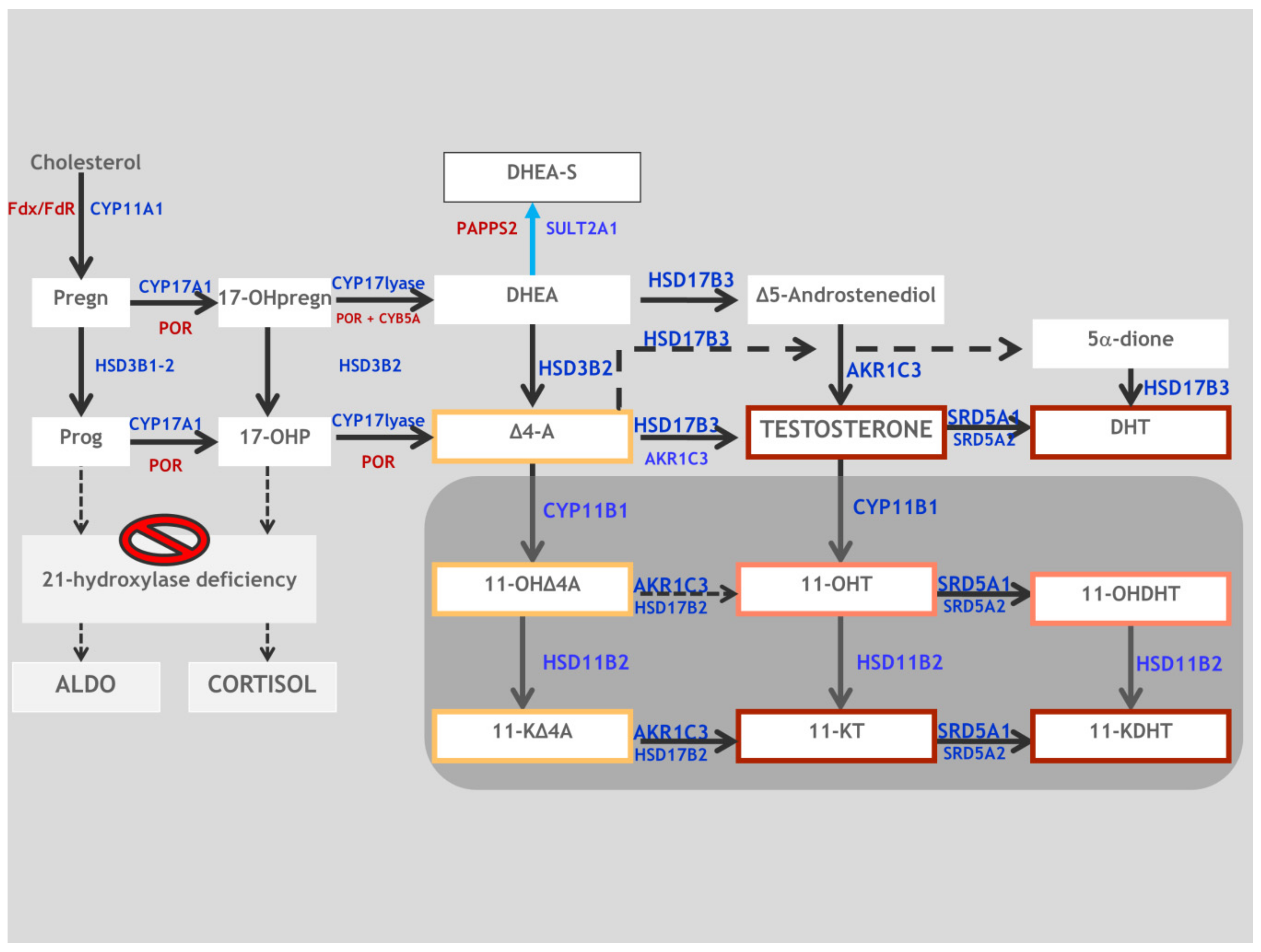
2. Synopsis of Steroidogenesis

Steroidogenesis is the process by which cholesterol is transformed into biologically active steroid hormones. Adrenals, gonads, placenta, and other steroidogenic tissues are responsible for a number of common processes with some cell-type-specific variations in each gland [5]. Figure 1 shows both the classical and the backdoor steroidogenic enzymatic steps that apply to adrenal and gonadal steroidogenesis. After the Steroidogenic Acute Regulatory (StAR)-protein-mediated transport of cholesterol from the outer to the inner mitochondrial membrane, the first and rate-limiting step in steroidogenesis is its conversion to pregnenolone (Preg) by a single enzyme, P450scc (CYP11A1) (Figure 1). Subsequently, many other enzymes and cofactors mediate the qualitative production of the type of steroid that is to be produced. Steroidogenic enzymes belong to two groups: cytochrome P450 enzymes and hydroxysteroid dehydrogenases. A cytochrome P450 may be either Type 1 (in mitochondria) or Type 2 (in endoplasmic reticulum), and a hydroxysteroid dehydrogenase may fit in either the aldo-keto reductase family or the short-chain dehydrogenase/reductase family. The activities of these enzymes are modulated by post-translational modifications and cofactors, particularly electron-donating redox partners. The elucidation of the precise roles of these enzymes and cofactors has been significantly facilitated by the identification of the genetic bases of rare disorders of steroidogenesis [5]. Some enzymes that are not primarily involved in steroidogenesis may also catalyze extraglandular steroidogenesis, modulating the phenotype that is expected to result from some mutations [6]. Understanding steroidogenesis is important to our comprehension of disorders of sexual differentiation and other processes that are related to human reproduction and fertility, particularly those that involve androgen excess as a consequence of their impairment.

3. 3β-Hydroxysteroid Dehydrogenase Type 2 Deficiency (3β-HSD2D)

3β-HSD2D is a very rare form of CAH that affects less than 5% of all CAH patients with an estimated prevalence of 1/1,000,000 at birth [5,7]. In steroidogenic tissues, it is expressed in both the adrenal glands and the gonads [8].

3.1. Genetics

There are two types of human 3β-HSD, which are encoded by two similar (more than 90% sequence identity) genes located on chromosome 1q13.1: HSD3B1 (encoding 3β-HSD1) is expressed in the placenta and multiple peripheral tissues; and HSD3B2 (encoding 3β-HSD2) is expressed in the adrenals and gonads. These two enzymes share and catalyze the same reactions, although the Michaelis–Menten constant (Km) for 3β-HSD2 is about 10-fold higher than that of 3β-HSD1 [6]. Overall, 43 female patients with or without virilization have been described so far, accounting for 26 reported mutations (Figure 2 and Supplementary Table S1) [9,10,11,12,13,14,15,16,17,18,19,20,21,22,23,24,25,26,27,28,29,30,31,32,33,34,35,36,37]. While severe loss-of-function mutations predict the neonatal salt-wasting (SW) phenotype, for missense mutations the correlation is good only with regard to mineralocorticoid (MC) deficiency.
All reported cases of 3β-HSD deficiency are in the HSD3B2 [5]; mutations in 3βHSD1 presumably would prevent placental biosynthesis of progesterone, resulting in a spontaneous first-trimester abortion [38].

3.2. Biochemistry

In the normal adrenals, the isoenzyme 3β-HSD2 plays a crucial role in all three steroidogenic pathways (aldosterone (Aldo), cortisol (F), and sex hormones production) (Figure 1). It catalyzes two sequential reactions (3β-hydroxysteroid dehydrogenation and Δ5–Δ4-isomerization) of four adrenal Δ5-steroids (pregnenolone (Preg), 17-OHpregnenolone (17-OHpreg), dehydroepiandrosterone (DHEA), and androstenediol (Adiol)), obtaining progesterone (P), 17-α-hydroxyprogesterone (17-OHP), androstenedione (A), and testosterone (T), respectively. The mitochondrial matrix appears to be the preferred location [39,40] for the conversion of Preg to P and the mineralocorticoid production in adrenal steroidogenic cells (Table 1). Interestingly, the Km of 3β-HSD2 (5.5 μM) is much higher than that of P450c17 (~0.8 μM), which would favor production of 17-OHpreg and downstream cortisol and sex steroids [6,41]. Similarly, in normal ovarian tissue, cholesterol is converted to Preg, 17-OHpreg, and DHEA. Moreover, 17β-hydroxysteroid dehydrogenase type 3 (HSD17B3)/17β-hydroxysteroid dehydrogenase type 5 (AKR1C3) converts androstendione or androstendiol into testosterone and then, via CYP19A1, into estrogens (Figure 1). Very high levels of Preg, 17-OHpreg, and DHEA are the biochemical markers of 3β-HSD Type II deficiency; however, a high concentration of serum 17-OHP is also paradoxically common in this condition. In fact, some of the secreted 17-OHPreg is then converted to 17-OHP by 3β-HSD1 in peripheral tissues. Plasma levels of A and other Δ4 steroids are also frequently elevated via the same mechanism. However, this elevated serum A may not be effectively taken up by the adrenals and peripheral tissues for the subsequent conversion to T because the circulating concentrations are much less than the Km of AKR1C3, which could explain the lack of virilization in most affected females [42]. Similarly, elevated 17-OHP is not effectively converted to cortisol in the adrenals as the Km of P450c21 is low [43]. Urinary steroid profiling, although less used, is similarly accurate and a less-invasive means by which to establish the diagnosis [44].
The principal diagnostic test in 3β-HSD deficiency is intravenous (i.v.) administration of synthetic ACTH with measurement of 17-OHpreg, cortisol, Δ4-17-OHP, DHEA, and androstenedione [7]. Steroidal responses to ACTH cannot reliably identify heterozygous carriers of 3β-HSD deficiency [45].
In general, levels of 17-OHpreg >150 nmol/L, either basal or post ACTH stimulation, is the most reliable single biochemical criterion of true 3β-HSD deficiency [41,46].

3.3. Clinical Features and Sex of Rearing

The phenotype of 3β-HSD2D varies, according to the severity of the genetic lesion, from the severe SW form in neonates to mild menstrual disorders in older females [10,11,46,47]. Classically, affected patients present with salt-wasting adrenal crisis, high-renin hypotension, and hypoglycemia, similar to 21-OHD. Affected 46,XX infants may have clitoromegaly and mild virilization because some fetal adrenal DHEA is converted into androstenedione in the periphery via 3β-HSD1, whose expression and function persist in the liver and elsewhere, causing androgen excess [5]. The females, although exhibiting fusion of the labia majora and enlargement of the clitoris, did not have the usual displacement of the urethral orifice as in the urogenital sinus of the other CAH types [47]. With the latter presentation, the diagnosis of 3B-HSD2D is generally done in the first weeks of life. However, this occurrence is not the most common phenotypic manifestation in the neonatal age, as demonstrated by the absence of ambiguous genitalia at birth in most of the cases reported in the literature (Table S1) [42]. Since 3β-HSD2D is a life-threatening condition that could easily be missed in female newborns in the absence of clinical signs of virilization, it is possible that female newborns with some of these severe forms may die undiagnosed due to neonatal adrenal crisis [11]. The low degree of genital atypia of this defect rarely determines sex misassignment (Table S1) and the sex of rearing is female. The low level of fetal hyperandrogenism should not significantly influence gender identity and behavior, although no long-term outcome data are available on this topic for 3β-HSD2D [7].
In the case of the non-SW phenotype, the diagnosis can occur at any pre- or post-pubertal age with symptoms of premature pubarche, hirsutism, and menstrual irregularities, including oligomenorrhea and primary amenorrhea [13]. Since patients with 3β-hydroxysteroid dehydrogenase deficiency may show elevated levels of 17-OHP, it is possible that newborns with false-positive results at neonatal screening for 21-OHD are actually affected by 3β-HSD2D, as confirmed by previous case reports [15,17,48,49].
Non-classical 3β-HSD2D had formerly been hypothesized for young females with premature adrenarche, hirsutism, virilism, or oligomenorrhea based on controversial hormonal criteria showing exaggerated Δ5 steroid production after an ACTH test and elevated 17-OHP/cortisol ratios. However, the HSD3B1 and HSD3B2 genes in these patients are normal [8,12,13] and the basis of this condition requires further study [50].
Since HSD3B2 is also necessary for the biosynthesis of sex hormones in the gonads, females may present menstrual irregularities and infertility. There are few long-term follow-up data, which, however, show, in most but not all [13] patients with good therapeutic compliance, a spontaneous process of progressive feminization with menstruation [11,19]. There are no reports of ovarian adrenal rest tumors in 3β-HSD deficiency. Although we know from the literature that HDS3B2D may be associated with obesity, short stature, infertility, or osteoporosis, the rarity of the condition precludes us from establishing its rate of mortality and the cardiovascular health, mental health, and adrenal tumor risk [7].

3.4. Therapy and Fertility Prognosis

During the developmental age, the treatment of choice is oral hydrocortisone at slightly higher doses (12–18 mg/m2/day) than in patients with 21-OHD; this is probably necessary due to the greater difficulty in suppressing androgens that are generated by the continuous peripheral transformation of DHEA/DHEA-S into testosterone and dihydrotestosterone (DHT) [7]. Long-acting glucocorticoids (prednisone, prednisolone, dexamethasone) might be used in adulthood [11,51], although they maintain the same contraindications that are given for 21-OHD in the most recent guidelines [52]. Administration of fludrocortisone at an average dose of 0.1 mg/day is sufficient to control mineralocorticoid deficiency. In the case of adrenal crisis with loss of salts, rapid rehydration, correction of hypoglycemia, and parenteral administration (IV or IM) of hydrocortisone are necessary. Sex hormone replacement therapy may be necessary in patients with a delay or an absence of pubertal development [13]. It is likely that fertility is reduced, although exact data on this topic are not available [7]. Given the low degree of genital atypia, any cosmetic surgery should be postponed and discussed at the proper time with the person with DSD in the context of a multidisciplinary and experienced DSD team. In the rare cases of severe genital ambiguity, the concerns that will be reported in Section 4.3 on 21-OHD apply.

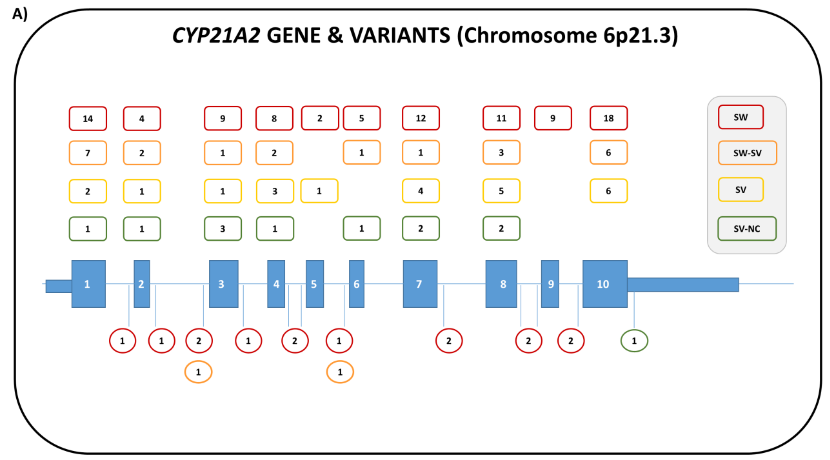
4. 21-Hydroxylase Deficiency (21-OHD)

The most common form of CAH is 21-OHD, which is responsible for about 90% of cases. According to the degree of impairment of 21-hydroxylase activity, three clinical phenotypes are commonly distinguished:
(1)
salt-wasting (<1% enzyme activity);
(2)
simple virilizing (1–2%); and
(3)
non-classical (20–50%).
The classical form (salt wasting and simple virilizing) has a variable incidence, according to ethnicity and location [53,54], and it ranges between 1:20,000 and 1:30,000 female live births [55], with a general heterozygous carriers frequency of 1 in 60 in most populations. The non-classical form generally has a higher incidence, which is estimated to be 1:1000 cases in the Caucasian population (a frequency of heterozygous carriers of 1:10 ) and even greater in other specific ethnic groups (Hebrew Ashkenazi 1:27, Hispanic 1:53, Italians 1:300) [56].

4.1. Genetics

The 21-OHD is caused by mutations in the CYP21A2 gene, which consists of 10 exons and is 3.4 kb long [57,58]. The gene is located on chromosome 6p21.3 within the Class III region of the major histocompatibility complex (MHC) in a complex locus that includes, in addition to the active CYP21A2 gene, an inactive pseudogene (CYP21P) that is about 98% homologous [59,60].
Both genes are arranged in a “tandem repeat” with the C4B and C4A genes within a repeating module called RCCX. This module consists of one part of or the whole RP gene (serine/threonine kinase), an entire C4 gene, an entire CYP21 gene, and either one part of or the whole TNX gene (tenascin-X).
Due to the high homology between the genes and the localization in the MHC region with a high frequency of recombination events, the locus rearrangements, such as gene deletions, duplications, or microconversions from CYP21P to CYP21A2, are very frequent and account for about 95% of the disease-causing mutations in 21-OHD [2], where about 20% are gene deletions. In addition to the recurrent mutations, another 300 pseudogene-independent rare mutations have been reported to date (Figure 3A) in both sexes [2,59]. Complex alleles, carrying more than one mutation, have also been described. For a correct diagnosis, it is therefore mandatory to analyze the entire CYP21A2 gene and the allelic segregation of the pathologic variants, and, if necessary, to determine the number of copies of the CYP1A2 gene. Given the complexity of the locus, in addition, it must be taken into account that, although not all of the rearrangements of the RCCX modules are pathological, they can still significantly interfere with a correct molecular diagnosis. It is, therefore, often necessary to use alternative/additional investigation techniques in order to identify these rearrangements, such as Multiplex Ligation-Dependent Probe Amplification (MLPA) [59,60].
Large deletions and a splicing mutation (c.293-13; A/C > G) are the most common molecular variations associated with the salt-wasting phenotype; loss-of-function mutations that cause the SW phenotype affect heme binding, substrate binding, and membrane anchoring or alter enzyme stability [61,62,63]. A non-conservative amino substitution in exon 4 (p.I73N; 1–2% enzyme activity) is frequently found in patients affected by the simple virilizing form. This phenotype is the result of mutations that affect the transmembrane region or conserved hydrophobic patches and reduce 21-OH activity by 2%. Interference in oxidoreductase interactions, salt-bridge and hydrogen bonding networks, and non-conserved hydrophobic clusters are responsible for the non-classical phenotype, where the enzyme activity is 20–50%. The point mutation in exon 7 (p.V282L) is frequently found in these patients [61,62,63].
Overall, the phenotype is extremely variable [63,64] since the majority (>65%) of CAH patients are compound heterozygotes with one or more mutations in both CYP21A2 alleles [64,65,66,67,68,69]. In these cases, the clinical phenotype depends on the less severely affected allele [53] (Figure 3B). Variants are reported according to the National Center for Biotechnology Information (NCBI) Reference Sequence NM_000500.9 [59].
In some CAH patients, a particular type of recombination occurs, caused by an unequal crossing-over event between TNXA and TNXB, that not only eliminates the interposed CYP21A2 gene, but produces a non-functional chimeric gene TNXA/TNXB. Therefore, in this situation, not only is the entire CYP21A2 gene missing, but a non-functioning TNX gene is formed that causes a “contiguous gene syndrome” characterized by CAH and Ehlers–Danlos Syndrome (EDS). This clinically distinct variant of CAH has recently been defined as “CAH-X”. The clinical findings observed in CAH-X patients include joint hypermobility, chronic joint pain, multiple joint dislocations, and structural cardiac valve abnormalities [70]. While homozygous mutations of TNXB cause a severe, autosomal recessive form of EDS [71], heterozygosity for TNXB mutations causing haploinsufficiency of TNX may be associated with the mild “hypermobility type” of EDS [72]. The CAH-X chimeras cause EDS in an autosomal-dominant manner regardless of CAH status, although patients with CAH usually have more severe EDS manifestations than do carriers without CAH [73,74]. CAH-X variants were initially found in 7–8.5% of patients with CAH [73,74], but this figure has recently been reported to be around 15.6% [75].
Three types of TNXA/TNXB chimeric gene have been reported. The first, which was described in 1997 [76] and later termed CAH-X CH-1, has TNXB exons 35–44 replaced with TNXA. Subsequently, novel TNXA/TNXB chimeras, named CAH-X CH-2 and CAH-X CH-3, were identified [77]: CAH-X CH-2 has TNXB exons 40–44 replaced with TNXA, and CAH-X CH-3 has TNXB exons 41–44 substituted by TNXA and has been reported in only one patient, with clinical significance still under investigation [70]. CAH-X CH-2 causes a more severe phenotype than CAH-X CH-1 that is characterized by a greater involvement of skin and joints [74].
The use of the commercial MLPA kit (P-050 MRC-Holland) to define CYP21A2 locus rearrangements is particularly helpful for the identification of a significant portion of CAH-X patients, in particular those that carry the CH-1 chimeric gene. Thus, MLPA may help the clinician provide a diagnosis in early infancy or perform a deep evaluation of clinical signs in adults. An experimental method for detecting both CH-1 and CH-2 has also been recently described [75].

4.2. Biochemistry

Both mineralocorticoid (MC) synthesis and glucocorticoid (GC) synthesis depend on CYP21A2 enzyme activity. In the adrenal zona glomerulosa, HSD3B2 converts Preg into P. Then, CYP21A2 converts P to deoxycorticosterone (DOC), which is converted by CYP11B2, which has 11-hydroxylase, 18-hydroxylase, and 18-oxidase activities, into aldosterone (Aldo) [5]. In the adrenal zona fasciculata, Preg is 17-hydroxylated to 17-OHPreg by CYP17A1, which also converts P to 17-OHP. 17-OHPreg is converted into 17-OHP by HSD3B2; CYP21A2 converts 17-OHP into 11-DF, and CYP11B1 finally transforms 11-DF into cortisol [5]. Therefore, in severe 21-OHD, GC and MC would not be sufficiently produced and the increased levels of ACTH, via negative cortisol feedback, would continue to stimulate the adrenal cortex. The excess 17-OHP, which represents the biochemical hallmark of the disease, is converted by the available accessible pathways into potent androgens, such as testosterone (T) and 5α-dihydrotestosterone (DHT). The mechanism by which hyperandrogenism occurs relies on the ready conversion by CYP17A1 of accumulated 17OH-Preg to DHEA (via the Δ5 pathway). Subsequently, DHEA is converted to A by HSD3B2. A very small amount of 17-OHP is converted to A (Δ4 pathway); therefore, this reaction does not represent a significant pathway of androgen synthesis. Both DHEA and A are considered to be weak androgens and a substrate for active androgen synthesis to T by 17β-HSD and, subsequently, DHT by 5alpha reductase type 2 (SRD5A2) in genital skin [78].
An alternative pathway to androgens—‘the backdoor pathway’ (Figure 1)—is responsible for the conversion of 17-OHP to DHT and bypasses the usual intermediate steroids DHEA, A, and T [79]. In this backdoor pathway, 17-OHP is converted to 5α-pregnane-3α, 17α-diol-20-one (pdiol), which is an excellent substrate for the 17-OHP, and 20 lyase activity of CYP17A1 to produce androsterone. After 17β-reduction to 5αAdiol, circulating 5αAdiol is 3a-oxidized to produce DHT, although it is not completely confirmed whether the final conversion of 5alpha-Diol to DHT occurs in genital skin or elsewhere or both [80]. After 17β-reduction to 5αAdiol, circulating 5αAdiol is 3a-oxidized to produce DHT in target tissues such as genital skin and prostate. This alternative pathway to androgens is active in fetal life in healthy individuals and plays a fundamental role in male sex development [79,80]. Androgen production by the backdoor pathway may explain why newborn girls with 21- and 11-hydroxlase deficiencies can be severely virilized, whereas those with 3β-HSD2 deficiency, whose adrenals cannot make 17-OHP, are minimally virilized [13].
In 2012, Kamrath et al. [81] showed that this pathway plays an important role in human hyperandrogenic disorders such as congenital adrenal hyperplasia caused by steroid 21-hydroxylase deficiency. They found significantly elevated ratios of pdiol to the Δ4 and Δ5 pathway metabolites as well as a higher androsterone to etiocholanolone ratio in untreated patients with 21-OHD. The authors found that the elevated pdiol level persisted after one year of age, whereas the androsterone to etiocholanolone ratio was highest only in the neonatal period and in the first year of life. Therefore, they concluded that the backdoor pathway in patients with 21-OHD is primarily activated during infancy.
The interaction between the fetal and the permanent adrenal plays a crucial role in the virilization of genitalia of 21-OHD 46,XX fetuses through both the front-door and the backdoor androgen pathway. In these fetuses, due to the activity of HSD3B2 that is present in the permanent adrenal, DHEA is converted to A [82], which is subsequently converted to T by AKR1C3 (HSD17B5) that is present in the fetal adrenal (the front-door pathway) [83]. At the same time, the high 17-OH progesterone levels activate, primarily in the fetal adrenal, the backdoor pathway to increase the expression of 5αAdiol, which, finally, is 3a-oxidized to produce DHT [79,81].
The utilization of liquid chromatography-tandem mass spectrometry (LC-MS/MS) has allowed us, in recent years, to extensively and more deeply evaluate androgen biosynthesis and the important role in hyperandrogenic disorders of another class of active adrenal androgens: the 11-oxygenated C19 steroids. These androgens derive from the 11-hydroxylation by CYP11B1 of A and T to 11OH-androstenedione (11-OHA4) and 11-hydroxytestosterone (11-OHT), respectively. Subsequently, they are converted by HSD11B2 to 11-ketoandrostenedione (11-KA4) and 11-ketotestosterone (11-KT), respectively. The 11-KT is a potent agonist of the human androgen receptor NR3C4, with an affinity that is comparable to that of testosterone [84,85], and its role as the dominant bioactive androgen in normal and premature adrenarche has recently been reported [86].
In 2016, Turcu et al. demonstrated that adrenal-derived 11-oxygenated 19-carbon steroids are the dominant androgens in classical 21-OHD (Figure 4). In particular, they showed that patients with 21-OHD have higher levels of 11-OHT, 11-KT, 11-OHA4, and 11-KA4 compared to healthy controls, and that 11-KT and 11-OHT only are potent activators of the human androgen receptor [87]. In the last few years, researchers have highlighted the importance and the potential clinical role of 11-oxygenated 19-carbon steroids as more specific markers of adrenal androgen excess compared with DHEA, A, and T, which, at present, are commonly utilized to monitor therapy control in 21-OHD patients [88,89].
Due to the reduced cortisol concentration in the adrenal medulla, CAH patients have impaired activity of medullary phenylethanolamine N-methyltransferase, which catalyzes the conversion of norepinephrine to epinephrine [90]; reduced production of epinephrine may increase the risk of hypoglycemia in these patients during adrenal crisis [90]. The 17-OHP levels directly correlate to the severity of disease, and most classical 21-OHD patients show basal 17-OHP levels above 10,000 ng/dL; basal levels above 200 ng/dL are found in most NC CAH patients, and stimulated 17-OHP levels >1000 ng/dL are useful for confirming the diagnosis [52].

4.2.1. Neonatal Screening

CAH due to 21-OHD can be detected in newborn screening (NS) programs by measuring the amount of 17-OHP, which is now performed in many countries to prevent SW crises during the neonatal period, particularly in males that do not show genital ambiguity, to prevent male sex assignment in affected females, and to reduce long-term morbidities. First-tier screening measures 17-OHP by various techniques, including radioimmunoassays (RIAs), enzyme-linked immunosorbent assays (ELISA), and time-resolved fluoroimmunoassays (DELFIA®), on dried blood spots, and mildly lower levels have been reported in female patients [91]. Diagnosis of 21-OHD can be made when 17-OHP levels are above the cut-off levels that have been elaborated specifically by each screening Centre. Since false-positive results are possible and frequent in premature and severely ill newborns, weight- and gestational-age-adjusted cutoffs for 17-OHP are usually utilized [92,93]. Second-tier strategies that use LC-MS/MS steroid profiling on the same spot sample have recently been developed to improve the positive predictive value of the test [94,95].

4.2.2. Prenatal Diagnosis and Treatment of 21-OHD

The prenatal diagnosis of affected CAH fetuses is possible at 10–12 weeks of gestation by chorionic villous sampling or at 15–16 weeks of gestation by amniocentesis. When, for the first time, cell-free fetal DNA in maternal plasma was detected in 1997, a noninvasive approach to prenatal diagnosis was conceivable [96]. In 2014, Lo et al. developed a noninvasive method for early prenatal diagnosis of fetuses at risk for CAH. With this method, it is possible to determine the inheritance of CYP21A2 pathogenic variants by noninvasive prenatal testing (NIPT) involving cell-free DNA from maternal blood. The test is performed with a correlative method that utilizes massively parallel sequencing (MPS) of cell-free DNA plasma drawn from an expectant mother. By the same complex technology, it is possible to identify a Y-sequence in maternal plasma via a traditional polymerase chain reaction using an SRY probe. Diagnosis can be made in a few hours and as early as at 5 weeks of gestation [97]. This approach is promising but currently is expensive and feasible only in highly specialized centers. Furthermore, in some countries, prenatal sex determination has been made illegal in order to prevent female feticide [98]. Another approach is preimplantation genetic testing (PGT), which allows for the selection of only embryos that are not affected by CAH for transfer [99].
The prenatal treatment of potentially affected CAH patients aims to prevent virilization of external genitalia of 21-OHD female fetuses by suppressing fetal ACTH and adrenal hyperandrogenism [2]. The treatment protocol involves the administration of a glucocorticoid that is not catabolized by the placenta (dexamethasone) to the mother as soon as pregnancy is suspected, because virilization of genitalia starts from the sixth week of gestation; treatment will be stopped if genetic testing reveals a male or unaffected female fetus [100,101]. In recent years, some important arguments have been raised against prenatal dexamethasone treatment. On the one hand, it reduces or prevents virilization in >85% of treated females [102]; on the other, it can produce negative effects on treated fetuses and their mothers. In line with these findings, in 2012, prenatal dexamethasone treatment in at-risk women was discontinued in Sweden [103]. Subsequent studies have shown that verbal working memory could be affected in children, particularly in girls, without CAH who were treated during the first trimester of fetal life [104,105]. Women with CAH who were treated during the entire gestational period were found to have worse cognitive performances than women with CAH who were not treated prenatally [106]. Furthermore, prenatal dexamethasone treatment in the first three months of life may affect fetal epigenetic programming [107]. Therefore, as stated by the Endocrine Society’s CAH Practice Guidelines [52], the prenatal diagnosis of fetuses who are potentially affected with CAH and their consequent prenatal treatment as formerly carried out in many countries remain controversial. All medical societies that have taken a position on prenatal treatment of 21-OHD with dexamethasone regard it as an experimental therapy that must only be done under research protocols approved by appropriate human experimentation and ethics boards.

4.3. Clinical Features and Sex of Rearing

The clinical picture is often complex and in some cases life threatening. In very severe and very mild cases of 21-OHD, genotype is predictive of phenotype [65]. The intermediate forms are not highly correlated with genotype, suggesting that other factors (environmental or genetic) may contribute to the clinical expression of the disease [2]. The cardinal feature that differentiates patients with classical CAH and patients with non-classical CAH is the genital ambiguity in female newborns with classical CAH. The classical form of 21-OHD is the phenotypic expression of an extremely reduced residual enzymatic activity (REA) of CYP21A2 that is less than 5% of the norm. In salt-wasting CAH, REA is 1–2%. In 46,XX newborns, exposure to an excess of intrauterine androgen causes different degrees of virilization and genital ambiguity as defined by the Prader Scale. Patients with 46,XX CAH have a normal vagina, cervix, and uterus, normal ovaries, and commonly a urogenital sinus and an enlarged clitoris. Secondary to impaired mineralocorticoid production and sodium loss, potassium retention occurs usually within the first 2–4 weeks of life. This metabolic impairment, which is associated with reduced production of epinephrine, can cause hypotension, cardiovascular collapse, and death if left untreated. The diagnosis of CAH due to 21-OHD should be rapidly ruled out in all 46,XX newborns with different degrees of virilization of the genitalia at birth without palpable gonads [52].
In its so-called simple virilizing forms, REA is greater than 2% and less than 5% due to missense mutations, typically p.I173N. Only cortisol deficiency and hyperandrogenism characterize the clinical features of these patients, because production of aldosterone is only moderately impaired [108]. However, for this reason, they could show increased plasma renin activity in the case of a moderate salt restriction. Those 46,XX newborns who are affected by the simple virilizing form show different degrees of virilization of genitalia at birth; 46,XY children affected by the simple virilizing form, which generally goes unrecognized at birth in the absence of a screening program, may be affected by premature pubarche/axillarche or precocious pseudo-puberty with increased growth velocity and rapid maturation of bone age.
The non-classical or late-onset forms show a 21-OH residual activity of >5% and the clinical picture is characterized by hyperandrogenism that is generally recognized in childhood with precocious pubarche/axillarche and advanced bone age. However, in some cases, some degree of external genitalia virilization (clitoral hypertrophy in females) has been described [109].
Regardless of the degree of genital virilization, 46,XX 21-OHD patients are usually raised as females. Since 46,XX 21 OHD patients have a female’s internal anatomy and fertility may be possible, it is still considered inappropriate to classify them as DSD [110]. New guidelines on CAH suggest that clinicians inform and psychologically support parents to help them to cope with the diagnosis and make decisions regarding surgical management of their children [52]. In patients with severe masculinization who are assigned to the female gender, it is generally recommended that surgical correction of internal genitalia be performed early [52] in order to bring the normal vagina to the perineum, repair the fistula between the vagina and the common urogenital channel, and locate the urethra in its normal position [111,112,113,114]. It is important to explain to the patient’s family that, in surgical procedures, the function of genitalia takes priority over their external appearance. Surgical management of clitoridomegaly, even performed by new nerve-sparing procedures, may not be fully satisfactory in terms of sensation, and some groups of CAH patients advocate for postponing this kind of cosmetic surgery until later in life, when the patients can make their own informed decisions. Overall, when these patients are assigned the female sex before 2 years of age and correctly managed by endocrinologists, surgeons, and psychologists who are expert in DSD management, they will generally achieve the female gender identity (88.7%), heterosexual behavior, and a positive psychosocial outcome [115,116]. However, the development of gender identity can only be predicted and not defined during infancy, and some CAH Groups have reported that up to 25% of individuals with CAH do not identify themselves as female [117]. Regarding sexual activities, more than 50% of cases complain of uncomfortable intercourse due to vaginal stenosis (27%) and impairment in the sensitivity of the clitoris. Low pregnancy rates (22.4%) are reported as well [116].

4.4. Therapy and Fertility

The goal of CAH treatment is to prevent adrenal crisis and virilization and allow for normal growth and development during childhood to the greatest possible extent.
The administration of glucocorticoids, besides being a substitute for cortisol ‘s own functions, also prevents the progression of virilization in its classical forms from inhibiting androgen hyperproduction by reducing ACTH secretion. However, to achieve this result, maintenance dosages are required that exceed the physiological secretion level of cortisol. Of the various types of cortisones and dosage schemes that have been proposed, the most advisable ones during the growth period consist in three administrations of oral hydrocortisone at doses of 10–15 mg/m2/day. Children who are affected by the classical form of 21-OHD also require mineralocorticoid. Newborns and infants require sodium chloride replacement due to MC resistance and immature renal tubules with a reduced sodium reabsorption capacity. Oral fluorohydrocortisone treatment is generally divided into two administrations: from 0.05 to 0.3 mg/day in the neonatal period and from 0.05 to 0.1 mg/day in infancy. Sodium chloride is divided into several administrations in the neonatal period and infancy (1–2 g/day is equal to 17–34 mEq/day).
In patients without an evident electrolyte imbalance, but with supraphysiologic levels of plasma renin activity (PRA), the administration of fluorohydrocortisone is recommended; this group of patients may also include subjects with the “non-classical” form characterized by the Pro30Leu mutation. Monitoring of renin activity, electrolyte levels, and blood pressure can help to evaluate the adequacy of mineralocorticoid replacement therapy.
However, attempts to completely normalize 17-OHP levels frequently result in overtreatment. Patients are at risk of overtreatment and developing Cushingoid side effects, and several evaluations (every 3–4 months in infancy and childhood) of clinical and biochemical features, including 17-OHP, androstenedione, and testosterone, with height velocity and annual bone age assessments after 2 years of age are required to reduce the potential for treatment failure or overtreatment and avoid possible detrimental effects on growth and metabolic health [52].
The reproductive outcome in women with classical CAH has been extensively studied, although conclusive data are lacking on this issue [118]. Fertility is reduced, particularly in patients with the SW form, as a result of several factors, such as hormonal, surgical, psychological, and sexual factors. Menstrual irregularities and anovulation affect from 30% to 75% of women with CAH. Chronically high androgen levels and progesterone overproduction are probably responsible for disturbing the reproductive axis in CAH females. Optimized and individualized glucocorticoid and mineralocorticoid regimens in these patients are needed to allow for spontaneous conception with a successful outcome. Reproductive techniques are now available for subfertile women despite effective adrenal androgen suppression.
The consequences of genital surgery are another factor that is implicated in the reduced fertility of CAH women. Surgery can include clitoroplasty and vaginoplasty, which are often performed in infancy and early childhood. Urinary incontinence, vaginal stenosis and inadequate introitus, poor cosmetics, anorgasmia, and painful intercourse have been reported. In the past few decades, techniques for feminizing surgery have evolved significantly (preserved innervation and clitoral sensation, improved vaginoplasty techniques). In a cohort study of 138 CAH women, Arlt et al. [119] reported that 46% of the participants stated that they were unhappy about their sexual life. In a more recent review of 151 patients with genitoplasty, assessments of cosmetic results showed that the majority of patients (between 60% and 94%) reported good or excellent outcomes [120].
Recently, a large population-based epidemiological study on psychosocial outcomes in CAH patients was conducted in Sweden. A total of 588 CAH patients, including 253 women, were compared to the general population. CAH women, particularly women with the SW form, were less often married, had fewer partnerships, and were less likely to have biological children than controls [121].
In conclusion, higher fertility and fecundity in CAH women will be largely dependent on surgical advances in genital reconstruction, earlier treatment, optimized therapy, the availability of psychological support, and better organization of the transition from pediatric to adult specialist care.

Experimental Therapy

To reduce the required doses of glucocorticoids and mineralocorticoids, several experimental treatments have been proposed in conjunction with classical therapies, such as the antiandrogen flutamide and the aromatase inhibitor testolactone [122,123]. In recent years, an extended-release formulation of hydrocortisone, which may help with treatment compliance and reduce the need for more potent glucocorticoids, has been made available [124]. However, Phase 2 and Phase 3 studies are ongoing to evaluate the efficacy and safety of this treatment in 21-OHD adult patients. To help treat younger patients and avoid an improper compounding effect of hydrocortisone from tablets, a very low dose of hydrocortisone in granules with high palatability has been recently released onto the market [125].

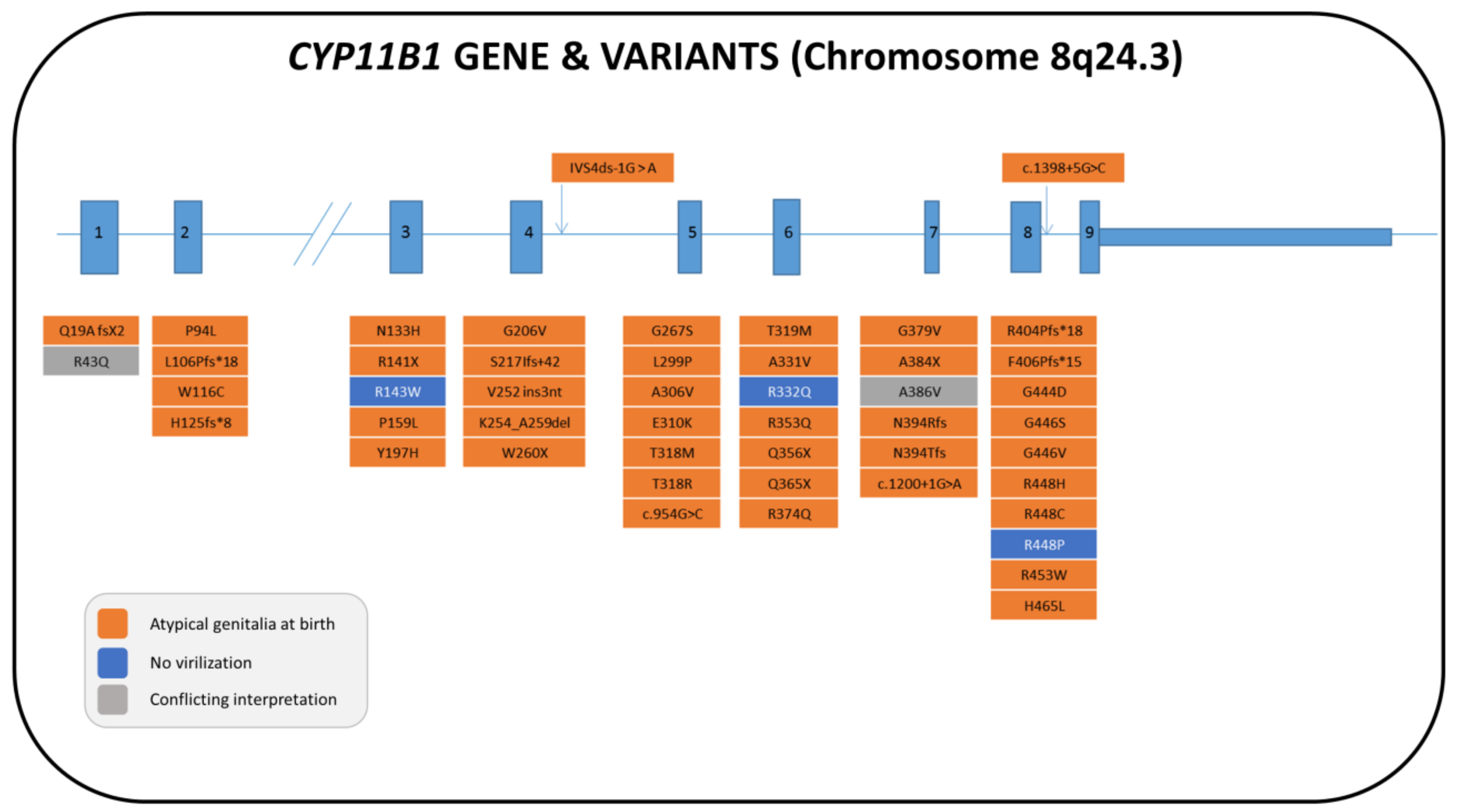
5. 11β-Hydroxylase Deficiency (11-OHD)

11-OHD, which is caused by mutations in the CYP11B1 gene, is the second most common cause of CAH with 46,XX DSD in the world, and accounts for about 5% of CAH patients with a European ancestry [126] and for about 15% of CAH patients in the Muslim and Jewish Middle Eastern populations [127]. 17-OHD and StAR deficiency (lipoid CAH) are the second most common form of CAH in China/Brazil and in Japan/Korea, respectively [6]; neither of these deficiencies, however, is causative for 46,XX DSD.

5.1. Genetics

The CYP11B1 gene is located on the long arm of chromosome 8 (8q21–q22), and it is in very close proximity to the highly homologous CYP11B2 gene, which encodes for aldosterone synthase. About 130 mutations of the CYP11B1 gene have been described and are localized all over the gene, with a minor presence in exons 1 and 9 [128,129,130,131,132,133,134,135,136,137,138,139,140,141,142,143,144,145,146,147,148,149,150]. Figure 5 summarizes the majority of allelic variants that have been described so far in the literature for females (62 patients) together with their influence on genital phenotype (see also Table S2).
A recent review [147] reported that the R448H mutation and eight other mutations (Q356X, G379V, T318M, c.53_54 insT, R454C, R448P, and R148X) are the most frequently found and account for approximately 40% of all cases. CYP11B1 mutations show significant ethnic specificity, and an updated description of their characteristics can be found in the studies of Khattab et al. [146] and Wang et al. [147]. Recombination events between the two homologous genes have been reported where the CYP11B1 gene is under the control of the CYP11B2 promoter such that it responds to angiotensin II and not to ACTH, resulting clinically in classical 11-OHD [151,152].

5.2. Biochemistry

In the normal adrenals, 11β-Hydroxylase is expressed in the zona fasciculata and converts 11-deoxycortisol to cortisol in response to ACTH (Figure 1). Aldosterone synthase is expressed at low levels in the zona glomerulosa and catalyzes the 11-hydroxylation, 18-hydroxylation, and 18-methyl oxidase activity needed to convert DOC to Aldo in response to angiotensin II and potassium. Both 11β-hydroxylase and aldosterone synthase can convert DOC into corticosterone (B). 11β-hydroxylase also has weak 18-hydroxylase activity; however, only aldosterone synthase can synthesize Aldo from 18OH-corticosterone [6]. 11-OHD disrupts the synthesis of cortisol with normal production of Aldo. ACTH excess due to a cortisol deficit causes overproduction of androgens and DOC, which causes virilization in females and hypertension, respectively. 11-deoxycortisol, which is hyper-responsive to ACTH administration, is the key steroid used in diagnosis [6]. Elevated serum B, DOC, and 17-OHP are also present, and it is possible for a wrong diagnosis of 21-OHD to occur with newborn screening for an accumulation of 17-OHP. Useful for diagnosis are urinary metabolites, such as tetrahydrocortisone, tetrahydro-11-deoxycorticosterone, and tetrahydro-11-deoxycortisol [3].

5.3. Clinical Features and Sex of Rearing

The classical form of 11-OHD has an estimated frequency of 1:200,000 live births [142] and is characterized by androgen excess and hypertension. Androgen excess causes ambiguous genitalia at birth in affected females (46,XX DSD) [3] and precocious pseudo-puberty with rapid somatic growth and bone age acceleration. Hypertension might not be apparent during the neonatal period due to mineralocorticoid resistance, and some patients can present with salt loss during the neonatal period [153]. Usually, sex assignment in these patients is female in accordance with the advice given for 21-OHD (Section 4.3 and Table 2).
The non-classical form of 11-OHD is similar to the non-classical form of 21-OHD with signs of androgen excess, including mild virilization, hirsutism, precocious pseudo-puberty, menstrual irregularities, and polycystic ovary syndrome in affected females. In this mild form, arterial hypertension does not occur often [154,155].

5.4. Therapy and Fertility Prognosis

Treatment of 11-OHD is glucocorticoid replacement at doses similar to those used in 21-OHD (orally administrated hydrocortisone at doses of 10–25 mg/m2/day) to ameliorate hypothalamic–pituitary–adrenal axis feedback to control androgen excess and hypertension; however, mineralocorticoid replacement is not needed [6]. Sometimes, treatment with mineralocorticoid receptor antagonists may be necessary [129]. As for 21-OHD, genital surgery may be necessary, but the timing of the surgery remains controversial. New guidelines for CAH suggest that clinicians inform parents about surgical options, including delaying surgery and/or observation until the child is older [52]. The existing studies underline that a significant number of individuals with CAH and the female gender identity evaluate their early surgery as a positive. However the development of gender identity can only be predicted and not defined during infancy; 25% of individuals with CAH do not identify themselves as female [117]. If the diagnosis of 11-OHD is late, it is possible that patients with a high Prader score will be reared as boys. There are some case reports in which these patients continued to be boys after diagnosis, and so the uterus and ovaries were removed [156].
Data on fertility are scarce, and only one successful pregnancy has been reported in the literature [157]. Caution should be taken when spironolactone is used to manage high blood pressure due to its teratogenic potential.

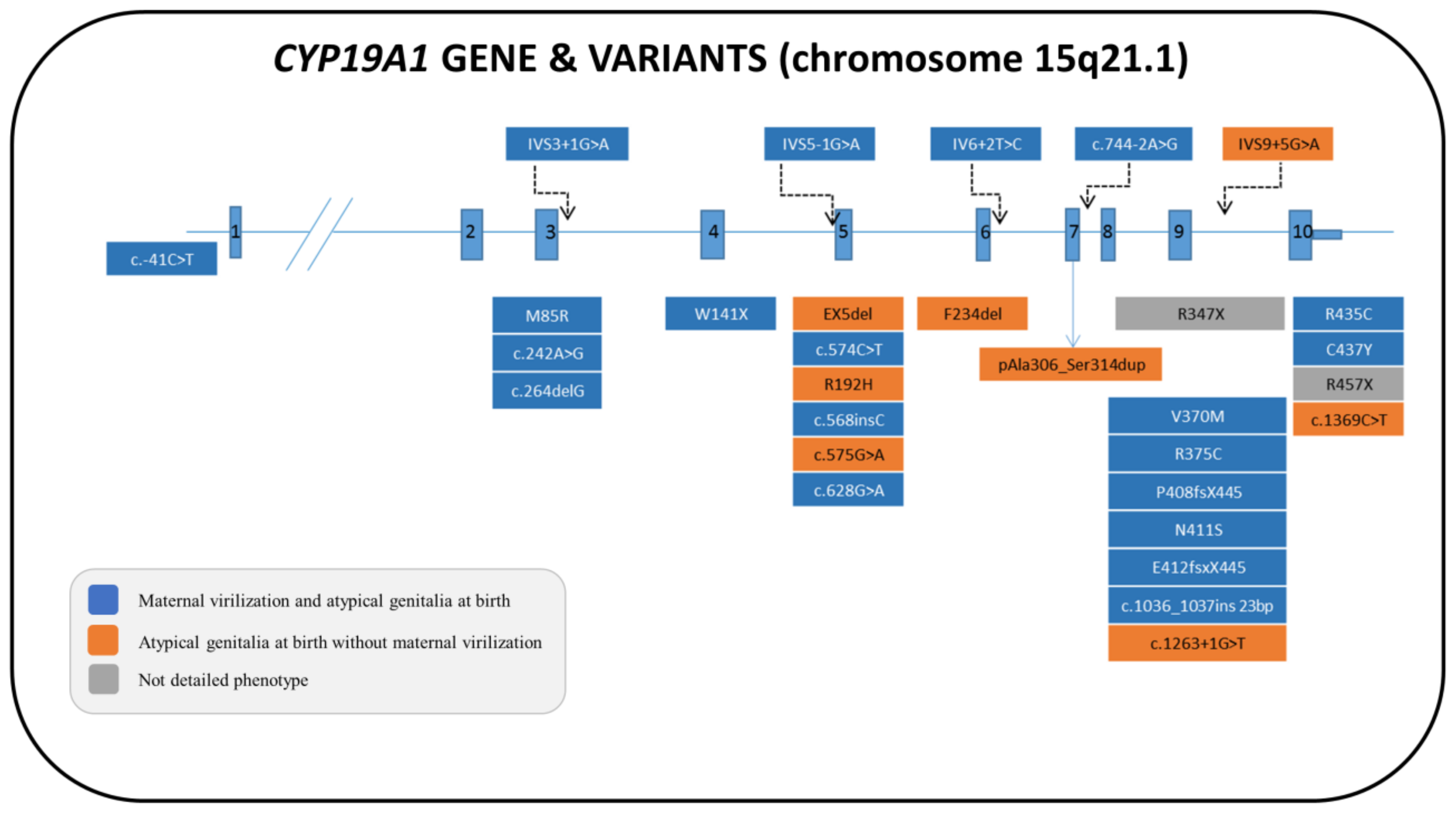
6. Aromatase Deficiency (AroD)

AroD is a rare genetic condition that is caused by mutations in the CYP19A1 gene, which is located on chromosome 15q21.1. Aromatase enzyme converts androgens into estrogens in many tissues. AroD causes atypical genitalia in 46,XX fetuses and maternal virilization during pregnancy due to an increased concentration of androgens. Ovaries are usually large and polycystic in girls with AroD [158].

6.1. Genetics

Aromatase deficiency is an autosomal recessive disorder that was first described by Shozu et al. [159]. To date, 30 46,XX cases with various ethnic origins have been described, and more than 29 distinct pathogenic variations have been identified in the CYP19A1 gene, including missense, nonsense, small deletions and insertions, splice-site mutations, and one large intragenic deletion [158,159,160,161,162,163,164,165,166,167,168,169,170,171,172,173,174,175,176,177,178,179,180]. Most of these mutations have been found to be located in exon 5 and exon 9 [173] (Figure 6 and Table S3).

6.2. Biochemistry

Aromatase catalyzes the three precursors androstenedione, testosterone, and 16-α-hydroxy dehydroepiandrosterone sulfate (after conversion to 16-α-hydroxyandrostenedione) into estrone, estradiol, and estriol, respectively [158,181]. Fetuses that lack aromatase activity are not able to convert the DHEA-S that is produced by the fetal adrenal gland into estrogens in the placenta; DHEA-S is, therefore, converted to testosterone, resulting in the virilization of both the fetus and the mother.
Both basal and GnRH-stimulated FSH levels have been shown to be higher in girls with AroD during the first two years of life compared to normal subjects (50–75 and 200–255 mIU/mL, respectively). However, the estradiol and estrone levels tend to be remarkably low during this same period [161,167]. Moreover, basal LH is often within normal limits or slightly elevated during infancy (5–10 mIU/mL). A previous study showed that, in a girl with AroD, the FSH and LH levels persistently increased and multicystic ovaries developed between the ages of three and four years [161]. However, Belgorosky et al. and Lin et al. [167,169] reported that the basal FSH and LH levels in a girl with AroD were found to be increased during mini-puberty and to show a dramatic decrease between two and five months of age. In the Unal et al. report [179], gonadotropin (FSH, LH) levels were found to be elevated since birth. The persistence, in some described cases, of high levels of gonadotropins, in particular of FSH, through infancy and childhood could suggest that a prolonged effect of an androgen or a lack of estrogen might result in an irreversible influence on the GnRH pulse generator. Moreover, an amplification of FSH signaling might occur in the presence of high intraovarian androgen production, and this mechanism could be involved in the development of ovarian follicular cysts [158].

6.3. Clinical Features and Sex of Rearing

The clinical characteristics of patients with aromatase deficiency vary depending on gender, age, and enzymatic activity [158]. AroD leads to an increase in intrauterine androgen concentration, which results in varying degrees of postnatal virilization in the external genitalia in girls. During infancy and childhood, there are usually no symptoms of aromatase deficiency, although some girl patients may present with abdominal symptoms of ovarian cysts because of mild changes in the hypothalamic–pituitary–gonadal axis due to a lack of feedback regulation [182]. AroD may lead to a number of clinical conditions in adolescent girls, such as delayed puberty, hypergonadotropic hypogonadism, multicystic ovaries, and primary amenorrhea in accordance with estrogen deficiency. Signs of virilization, such as acne, hirsutism, and cliteromegaly, in accordance with androgen excess, may also be present [158,181]. Estrogen deficiency, on the other hand, causes delayed epiphyseal closure, eunuchoid body habitus, osteopenia, and osteoporosis that develop in both genders [165].
In most fetuses with AroD, early-onset (12 weeks) or late-onset (up to 30 weeks) maternal virilization may be noted [161]. The non-aromatized fetoplacental and maternal androgen precursors are converted to testosterone in the placenta and also in peripheral maternal tissues, resulting in maternal virilization. After giving birth, the signs of virilization disappear gradually and the androgen levels return to normal [158].
There are few data in the literature on the possible effects of foetal hyperandrogenism in the programming of the brain for sexual identity and behavior. Although no detailed psychosexual studies have been reported, female psychosexual orientation has been observed and the female sex of rearing has been realized in most 46,XX subjects. Only one female patient, described by Lin et al. [169], with the homozygous F234del mutation, was raised as a male; this was probably due to the severe masculinization of the external genitalia at birth (Prader stage IV). This patient reported male gender identity, role, and orientation but experienced significant breast development (Tanner stage IV) at puberty that required a mastectomy, a salpingo-oophorectomy, and a hysterectomy. Tritiated androstenedione assays showed that this F234del mutant had between 16% and 19% wild-type activity, consistent with the significant uterine growth and breast development seen in this case. However, the same authors were not able to establish whether, in choosing the sex of rearing, the objective effect of prenatal androgen exposure coupled with estrogen insufficiency or social and cultural influences were predominant.

6.4. Therapy and Fertility Prognosis

To date, information on the effect of estrogen administration to prevent the consequences of estrogen insufficiency is limited and ambiguous. There is no consensus on the appropriate dosage of estrogen replacement therapy and on the usefulness of starting low-dose estrogen treatment from infancy and childhood in affected patients. Therapeutic follow-up data are available in 10 reported female patients who had been treated with estrogen at different ages in prepuberty and puberty [176]. In most cases, the treatment was performed in premenarcheal girls aged from 9.6 to 14 years to induce puberty. Generally, increasing doses of oral-conjugated estrogen have been used according to clinical signs of estrogen response, and progestogens were added to induce menarche. This therapeutic model, however, proved to be inadequate in about half of the cases to improve bone maturation delay, suppress elevated gonadotropin levels, regress ovarian cysts, and induce menarche. In the Janner et al. study [182], a girl with complete AroD started low-dose estrogen therapy at the age of 3.5 years and continued the treatment until the age of 15 years with increasing doses. The authors suggested that a low dose of estrogen (E2) (50–100 mg/day), even if insufficient to suppress gonadotropin feedback, is required for normal longitudinal growth and bone age maturation during early childhood, while higher E2 doses (1.4–2 mg/day) are needed more for successful negative feedback on the pituitary–ovarian axis, ovarian cyst regression, breast development, and endometrial reflex than for the normalization of growth and the appearance of menarche in late prepuberty and puberty.
Information about the course of the disease in adulthood and the long-term consequence for fertility remain unknown. However, the clinical, hormonal, and therapeutic problems that are observed in most patients, along with the surgical consequences of genital reconstruction, might affect reproductive capacity in these women [158].

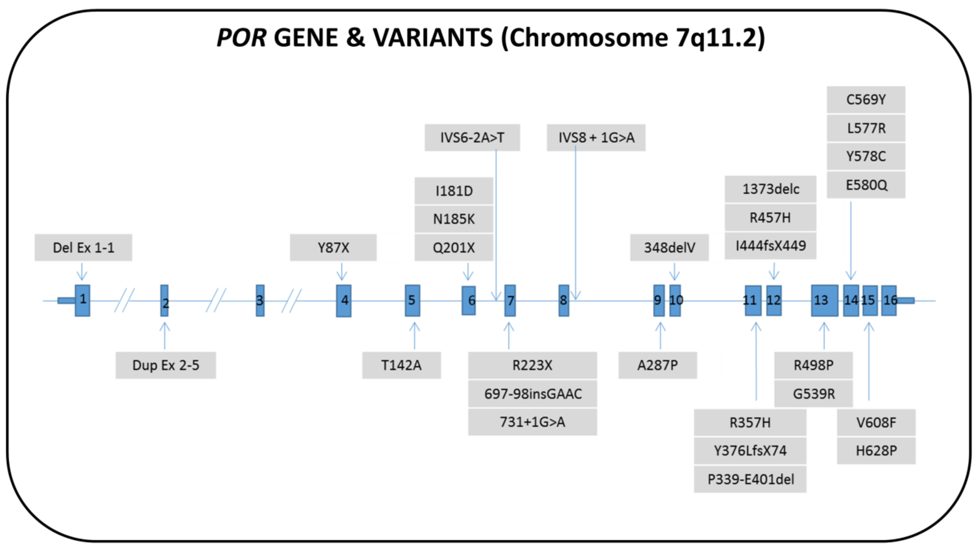
7. P450 Oxidoreductase Deficiency (PORD)

In humans, PORD causes an unusual and rare form of CAH, whose exact incidence is unknown [3]. Its presentation is characterized by DSD in both sexes and is often associated with skeletal defects [183,184].

7.1. Genetics

The genetic cause of this new form of CAH was discovered in 2004 [183,185] and explained the pathogenesis of a condition that, so far [186], has been referred to as “apparent associated 17α- and 21-hydroxylase deficiency”. The POR gene is located on the long arm of chromosome 7 (7q11.2). About 80 female patients affected by PORD with more than 30 different pathogenic variants have been reported to date (Figure 7 and Table S4) [183,184,185,187,188,189,190,191,192,193,194,195,196,197,198,199,200,201,202,203,204,205,206,207].
The phenotypic result in genetic females characteristically depends on the causative POR pathogenic variant [208]. In fact, depending on the type of mutation and its location within the POR gene, it may reduce the CYP17A1, CYP21A2, and CYP19A1 activities differently. Therefore, POR variants must be studied separately for each potential P450 target enzyme [204]. Interestingly, the POR mutation p.A287P, which characterizes European patients [183,188], alters CYP17A1 activity but not CYP21A2 activity or CYP19A1 activity as opposed to the POR mutation p.R457H, which is frequent in Japanese patients [184,189] and instead predominantly reduces CYP19A1 activity [209]. The latter process allows for an increase in androgens in the maternal circulation and virilizes the mother. Therefore, pregnant women with fetuses carrying the POR p.R457H mutation frequently show virilization during pregnancy [183,184,185,189]. Furthermore, the deviation of steroidogenesis towards the “backdoor pathway” (Figure 1) contributes to the virilization of mothers and affected female fetuses [189]. The ethnic variability and the high polymorphism of the POR gene was confirmed by the high recurrence of p.A503V sequence variation in mildly affected patients of Chinese origin, but not in those of African origin, as also occurs for incidences of other SNPs [6]. As for the “skeletal” phenotype, the greater the severity of the involved mutations, greater the severity of the “skeletal” phenotype [188,210]. Severe phenotypes can lead to stillbirth or early neonatal deaths [211]. Prenatal testing is possible through molecular genetic techniques once the POR pathogenic variant has been established for a known family [188].

7.2. Biochemistry

POR is a protein that has flavin adenine dinucleotide (FAD) and flavin mononucleotide (FMN) portions; the FAD transfers two electrons received from NADPH to the FMN, which, in turn, transfers them to all microsomal (type 2) CYP enzymes [212,213], including the steroidogenic enzymes CYP21A2, CYP17A1, and CYP19A1.
Measurements of serum and urinary steroid profiles have shown that POR deficiency is characterized by partially deficient CYP17A1 activity, with or without associated deficiencies of CYP21A2 and CYP19A1 enzymes [183,184,185,188]. POR-deficient patients typically have a normal mineralocorticoid function, low–normal cortisol levels that respond weakly to an ACTH stimulation test, high concentrations of 17-OHP that respond unpredictably to ACTH, and low levels of C19 precursors of sex steroids (Table S4). Anomalous elevations in some of the steroids (and their metabolites) from the "backdoor pathway" may support a diagnosis of POR deficiency [190]. The low estriol values seen in women carrying a fetus with some POR mutations may highlight a CYP19A1 deficiency and hyperandrogenism during pregnancy. Hyperandrogenism is explained by the production of 5α-reduced androgens via the alternative pathway in the fetus that cannot be aromatized by the placenta and is transferred to the maternal circulation [183,184,185,189].
Urinary steroid profiling by gas chromatography-mass spectrometry has disclosed the characteristic trait of a combined impairment of CYP21A2 activity and CYP17A1 activity [44,188] and a typical accumulation of pregnenolone metabolites (pregnanediol, PD) [44].
Prenatal testing is possible using maternal urinary steroid profiling from 12 weeks onwards [211]. Neonatal screening for 21-OHD may occasionally detect 17-OHP values above the cut-off level in patients with PORD [183,193].
Although POR is required by the main hepatic drug-metabolizing enzymes and several in vitro studies of these enzymes have shown a major impairment by POR mutations [208,213,214], effects in PORD patients are rare [215].

7.3. Clinical Features and Sex of Rearing

46,XX babies are born virilized without postnatal progression of virilization. This result depends on the existence of the alternative “backdoor pathway” (secondary to CYP21A2 deficiency), which leads to DHT production and is active only during the prenatal period [185,216]. After birth, the pathway’s activity declines; thus, a sex steroid deficiency appears in both sexes. During puberty, females can present with delayed development of sexual characteristics, often showing significant hypergonadotropic hypogonadism and presenting large ovarian cysts with a tendency to torsion [187,192]. In addition, pregnant mothers carrying a fetus affected by PORD may manifest virilization during pregnancy (see Section 7.2), which characteristically resolves postpartum [183,185,187,210]. Although hyperandrogenism declines after birth, the genital phenotype can range from Prader stage I to V. The majority of reported cases show Prader stages below IV and a female sex assignment. In a few cases, patients that scored Prader IV/V were assigned the male sex and subsequently shifted to the female sex after multidisciplinary and parental counselling (Table S4). No long-term outcome is available at present to determine the influence of prenatal androgenic imprinting on the gender identity of these cases.
Newborns with PORD often have an associated skeletal defect that has been described as part of the Antley–Bixler Syndrome (ABS) phenotype. Craniofacial anomalies involve brachycephaly, proptosis, choanal stenosis, and craniosynostosis with midface hypoplasia, which most commonly involves the coronal and lambdoid sutures and can be complicated by the hydrocephalus in severe forms [210]. Other frequent skeletal abnormalities may involve large joint synostosis, such as radio-humeral synostosis, congenital bowing of the femurs, hand and foot malformations such as long palms, camptodactyly, and arachnodactyly, and rocker bottom feet. A scoring system for classifying the severity of the Antley–Bixler syndrome phenotype has been proposed based on the number and severity of abnormal skeletal features [188].
ABS is a skeletal condition that can occur in two forms: the first is transmitted as an autosomal recessive trait [183,187,216], is associated with atypical genitalia in both sexes due to defects in steroidogenesis, and is caused by mutations in the POR gene; the second is transmitted as an autosomal dominant trait, does not involve steroidogenesis, and is caused by gain-of-function mutations in the fibroblast growth factor receptor 2 (FGFR2) gene [184,187].
PORD impairs CYP enzymes involved in sterol synthesis, causing retinoic acid accumulation at embryonic locations that usually form skeletal joints and sutures, leading to their premature fusion and the ABS skeletal phenotype [210,217]. In addition, PORD affects a number of key CYP enzymes involved in hepatic function (CYP1A2, CYP2C9, CYP2C19, CYP2D6, and CYP3A4) that metabolize a large number of the xenobiotics and drugs that are used in clinical practice [218].

7.4. Therapy and Fertility Prognosis

Treatment of PORD requires a multidisciplinary team of endocrinologists, geneticists, and orthopedic and cranio-facial surgeons and family support. The majority of affected patients (about 90%) will require low-dose glucocorticoid replacement therapy; 50% of them will need permanent, and the other 50% at least some, stress-related hydrocortisone replacement therapy; this should be determined individually through the assessment of cortisol response to ACTH, regardless of presence or absence of DSD [214]. Females may require estrogen replacement during puberty, preferably as an estrogen patch to avoid hepatic first-pass metabolism. This is important from the therapeutic point of view; in fact, since CYP3A4 metabolizes estrogens and glucocorticoids (GCs), it is likely that individuals with POR mutations affecting this enzyme activity may have a reduced hepatic clearance of these hormones and, consequently, higher than expected circulating levels [215].
Atypical genitalia may require surgical intervention, which should be discussed with the patient and the patient’s parents in the context of an experienced multidisciplinary team. Also, for this condition, information about the clinical course of the disease in adulthood and the long-term consequence for fertility remain unknown and the clinical, hormonal, and therapeutic problems that are observed in most patients, along with the surgical consequences of genital reconstruction, might affect reproductive capacity in these women.
Skeletal abnormalities may require physical and specialized therapy or surgical intervention.

8. Conclusions

46,XX DSD secondary to androgen excess represents a group of congenital conditions with atypical sex development. In these cases, an early diagnosis is mandatory not only for an appropriate sex assignment, but also to start the correct substitutive treatment to avoid life-threatening adrenal crisis. In most of these monogenic conditions, hormonal phenotypes correlate quite well with genotypes. Molecular analysis certainly is fundamental to a confirmation of the diagnostic suspicion and should firstly rely on an appropriate biochemical investigation. Patients with 46,XX DSD should be referred to a specialized Center where appropriate (i.e., tandem mass steroid profiles) treatment can be performed and skilled pediatric endocrinologists and geneticists lead a multidisciplinary team that can definitively manage these children from diagnosis to adulthood.

Supplementary Materials

Author Contributions

Conceptualization, A.B., L.B., and F.B.; methodology, A.B., L.B., and S.M.; software, A.B. and L.B.; resources, A.C.; data curation, A.B. and S.M.; writing—original draft preparation, A.B. and L.B. (3β-HSD2D and PORD), F.B., S.M., and G.T. (21-OHD), R.O., S.M., and B.V. (11-OHD), and A.C. and V.D.N. (AroD); writing—review and editing, A.B. and S.M.; visualization, A.B., L.B., and S.M.; supervision, A.B.

Funding

This review received no direct external funding.

Acknowledgments

We wish to thank the “arfSAG” ONLUS Association (Regional Association of Families with Congenital Adrenal Hyperplasia) for its support of our research on CAH.

Conflicts of Interest

The authors declare no conflicts of interest.

Graphic Abstract Illustration Credits

From Servier Medical Art (https://smart.servier.com/), reproduced under Creative Commons License attribution 3.0 Unported License.

References

  1. Hughes, I.A.; Houk, C.; Ahmed, S.F.; Lee, P.A. LWPES Consensus Group; ESPE Consensus Group. Consensus statement on management of intersex disorders. Arch. Dis. Child. 2006, 91, 554–563. [Google Scholar] [CrossRef] [PubMed]
  2. White, P.C.; Speiser, P.W. Congenital adrenal hyperplasia due to 21-hydroxylase deficiency. Endocr. Rev. 2000, 21, 245–291. [Google Scholar] [CrossRef] [PubMed]
  3. Baranowski, E.S.; Arlt, W.; Idkowiak, J. Monogenic Disorders of Adrenal Steroidogenesis. Horm. Res. Paediatr. 2018, 89, 292–310. [Google Scholar] [CrossRef] [PubMed]
  4. Ferreira, C.R.; van Karnebeek, C.D.M.; Vockley, J.; Blau, N. A proposed nosology of inborn errors of metabolism. Genet. Med. Off. J. Am. Coll. Med. Genet. 2019, 21, 102–106. [Google Scholar] [CrossRef] [PubMed]
  5. Miller, W.L.; Auchus, R.J. The molecular biology, biochemistry, and physiology of human steroidogenesis and its disorders. Endocr. Rev. 2011, 32, 81–151. [Google Scholar] [CrossRef]
  6. Miller, W.L. Mechanisms in endocrinology: Rare defects in adrenal steroidogenesis. Eur. J. Endocrinol. 2018, 179, R125–R141. [Google Scholar] [CrossRef] [PubMed]
  7. Al Alawi, A.M.; Nordenström, A.; Falhammar, H. Clinical perspectives in congenital adrenal hyperplasia due to 3β-hydroxysteroid dehydrogenase type 2 deficiency. Endocrine 2019, 63, 407–421. [Google Scholar] [CrossRef] [PubMed]
  8. Rhéaume, E.; Simard, J.; Morel, Y.; Mebarki, F.; Zachmann, M.; Forest, M.G.; New, M.I.; Labrie, F. Congenital adrenal hyperplasia due to point mutations in the type II 3 beta-hydroxysteroid dehydrogenase gene. Nat. Genet. 1992, 1, 239–245. [Google Scholar] [CrossRef]
  9. Schneider, G.; Genel, M.; Bongiovanni, A.M.; Goldman, A.S.; Rosenfield, R.L. Persistent testicular delta5-isomerase-3beta-hydroxysteroid dehydrogenase (delta5-3beta-HSD) deficiency in the delta5-3beta-HSD form of congenital adrenal hyperplasia. J. Clin. Investig. 1975, 55, 681–690. [Google Scholar] [CrossRef]
  10. Barnes, R.B.; Ehrmann, D.A.; Brigell, D.F.; Rosenfield, R.L. Ovarian steroidogenic responses to gonadotropin-releasing hormone agonist testing with nafarelin in hirsute women with adrenal responses to adrenocorticotropin suggestive of 3 beta-hydroxy-delta 5-steroid dehydrogenase deficiency. J. Clin. Endocrinol. Metab. 1993, 76, 450–455. [Google Scholar]
  11. Alos, N.; Moisan, A.M.; Ward, L.; Desrochers, M.; Legault, L.; Leboeuf, G.; Van Vliet, G.; Simard, J. A novel A10E homozygous mutation in the HSD3B2 gene causing severe salt-wasting 3beta-hydroxysteroid dehydrogenase deficiency in 46,XX and 46,XY French-Canadians: evaluation of gonadal function after puberty. J. Clin. Endocrinol. Metab. 2000, 85, 1968–1974. [Google Scholar] [PubMed]
  12. Rhéaume, E.; Sanchez, R.; Simard, J.; Chang, Y.T.; Wang, J.; Pang, S.; Labrie, F. Molecular basis of congenital adrenal hyperplasia in two siblings with classical nonsalt-losing 3 beta-hydroxysteroid dehydrogenase deficiency. J. Clin. Endocrinol. Metab. 1994, 79, 1012–1018. [Google Scholar] [PubMed]
  13. Moisan, A.M.; Ricketts, M.L.; Tardy, V.; Desrochers, M.; Mébarki, F.; Chaussain, J.L.; Cabrol, S.; Raux-Demay, M.C.; Forest, M.G.; Sippell, W.G.; et al. New insight into the molecular basis of 3beta-hydroxysteroid dehydrogenase deficiency: identification of eight mutations in the HSD3B2 gene eleven patients from seven new families and comparison of the functional properties of twenty-five mutant enzymes. J. Clin. Endocrinol. Metab. 1999, 84, 4410–4425. [Google Scholar] [PubMed]
  14. Huang, Y.; Zheng, J.; Xie, T.; Xiao, Q.; Lu, S.; Li, X.; Cheng, J.; Chen, L.; Liu, L. A novel homozygous mutation p.E25X in the HSD3B2 gene causing salt wasting 3β-hydroxysteroid dehydrogenases deficiency in a Chinese pubertal girl: a delayed diagnosis until recurrent ovary cysts. Zhonghua Er Ke Za Zhi Chin. J. Pediatr. 2014, 52, 948–951. [Google Scholar]
  15. Benkert, A.R.; Young, M.; Robinson, D.; Hendrickson, C.; Lee, P.A.; Strauss, K.A. Severe Salt-Losing 3β-Hydroxysteroid Dehydrogenase Deficiency: Treatment and Outcomes of HSD3B2 c.35G>A Homozygotes. J. Clin. Endocrinol. Metab. 2015, 100, E1105–E1115. [Google Scholar] [CrossRef] [PubMed]
  16. Mendonça, B.B.; Russell, A.J.; Vasconcelos-Leite, M.; Arnhold, I.J.; Bloise, W.; Wajchenberg, B.L.; Nicolau, W.; Sutcliffe, R.G.; Wallace, A.M. Mutation in 3 beta-hydroxysteroid dehydrogenase type II associated with pseudohermaphroditism in males and premature pubarche or cryptic expression in females. J. Mol. Endocrinol. 1994, 12, 119–122. [Google Scholar] [CrossRef] [PubMed]
  17. Nordenström, A.; Forest, M.G.; Wedell, A. A case of 3beta-hydroxysteroid dehydrogenase type II (HSD3B2) deficiency picked up by neonatal screening for 21-hydroxylase deficiency: difficulties and delay in etiologic diagnosis. Horm. Res. 2007, 68, 204–208. [Google Scholar]
  18. Pang, S.; Levine, L.S.; Stoner, E.; Opitz, J.M.; Pollack, M.S.; Dupont, B.; New, M.I. Nonsalt-losing congenital adrenal hyperplasia due to 3 beta-hydroxysteroid dehydrogenase deficiency with normal glomerulosa function. J. Clin. Endocrinol. Metab. 1983, 56, 808–818. [Google Scholar] [CrossRef]
  19. Chang, Y.T.; Kulin, H.E.; Garibaldi, L.; Suriano, M.J.; Bracki, K.; Pang, S. Hypothalamic-pituitary-gonadal axis function in pubertal male and female siblings with glucocorticoid-treated nonsalt-wasting 3 beta-hydroxysteroid dehydrogenase deficiency congenital adrenal hyperplasia. J. Clin. Endocrinol. Metab. 1993, 77, 1251–1257. [Google Scholar]
  20. Probst-Scheidegger, U.; Udhane, S.S.; l’Allemand, D.; Flück, C.E.; Camats, N. Non-Virilizing Congenital Adrenal Hyperplasia in a Female Patient with a Novel HSD3B2 Mutation. Sex. Dev. Genet. Mol. Biol. Evol. Endocrinol. Embryol. Pathol. Sex Determ. Differ. 2016, 10, 200–204. [Google Scholar] [CrossRef]
  21. Pang, S.; Wang, W.; Rich, B.; David, R.; Chang, Y.T.; Carbunaru, G.; Myers, S.E.; Howie, A.F.; Smillie, K.J.; Mason, J.I. A novel nonstop mutation in the stop codon and a novel missense mutation in the type II 3beta-hydroxysteroid dehydrogenase (3beta-HSD) gene causing, respectively, nonclassic and classic 3beta-HSD deficiency congenital adrenal hyperplasia. J. Clin. Endocrinol. Metab. 2002, 87, 2556–2563. [Google Scholar] [PubMed]
  22. Russell, A.J.; Wallace, A.M.; Forest, M.G.; Donaldson, M.D.; Edwards, C.R.; Sutcliffe, R.G. Mutation in the human gene for 3 beta-hydroxysteroid dehydrogenase type II leading to male pseudohermaphroditism without salt loss. J. Mol. Endocrinol. 1994, 12, 225–237. [Google Scholar] [CrossRef] [PubMed]
  23. Johannsen, T.H.; Mallet, D.; Dige-Petersen, H.; Müller, J.; Main, K.M.; Morel, Y.; Forest, M.G. Delayed diagnosis of congenital adrenal hyperplasia with salt wasting due to type II 3beta-hydroxysteroid dehydrogenase deficiency. J. Clin. Endocrinol. Metab. 2005, 90, 2076–2080. [Google Scholar] [CrossRef] [PubMed]
  24. Takasawa, K.; Ono, M.; Hijikata, A.; Matsubara, Y.; Katsumata, N.; Takagi, M.; Morio, T.; Ohara, O.; Kashimada, K.; Mizutani, S. Two novel HSD3B2 missense mutations with diverse residual enzymatic activities for Δ5-steroids. Clin. Endocrinol. 2014, 80, 782–789. [Google Scholar] [CrossRef] [PubMed]
  25. Katsumata, N.; Tanae, A.; Yasunaga, T.; Horikawa, R.; Tanaka, T.; Hibi, I. A novel missense mutation in the type II 3 beta-hydroxysteroid dehydrogenase gene in a family with classical salt-wasting congenital adrenal hyperplasia due to 3 beta-hydroxysteroid dehydrogenase deficiency. Hum. Mol. Genet. 1995, 4, 745–746. [Google Scholar] [CrossRef] [PubMed]
  26. Baquedano, M.S.; Ciaccio, M.; Marino, R.; Perez Garrido, N.; Ramirez, P.; Maceiras, M.; Turjanski, A.; Defelipe, L.A.; Rivarola, M.A.; Belgorosky, A. A novel missense mutation in the HSD3B2 gene, underlying nonsalt-wasting congenital adrenal hyperplasia. new insight into the structure-function relationships of 3β-hydroxysteroid dehidrogenase type II. J. Clin. Endocrinol. Metab. 2015, 100, E191–E196. [Google Scholar] [CrossRef] [PubMed]
  27. Rosenfield, R.L.; Rich, B.H.; Wolfsdorf, J.I.; Cassorla, F.; Parks, J.S.; Bongiovanni, A.M.; Wu, C.H.; Shackleton, C.H. Pubertal presentation of congenital delta 5-3 beta-hydroxysteroid dehydrogenase deficiency. J. Clin. Endocrinol. Metab. 1980, 51, 345–353. [Google Scholar] [CrossRef]
  28. Sanchez, R.; Rhéaume, E.; Laflamme, N.; Rosenfield, R.L.; Labrie, F.; Simard, J. Detection and functional characterization of the novel missense mutation Y254D in type II 3 beta-hydroxysteroid dehydrogenase (3 beta HSD) gene of a female patient with nonsalt-losing 3 beta HSD deficiency. J. Clin. Endocrinol. Metab. 1994, 78, 561–567. [Google Scholar]
  29. Barnes, R.B.; Rosenfield, R.L.; Burstein, S.; Ehrmann, D.A. Pituitary-ovarian responses to nafarelin testing in the polycystic ovary syndrome. N. Engl. J. Med. 1989, 320, 559–565. [Google Scholar] [CrossRef]
  30. Paula, F.J.; Dick-de-Paula, I.; Pontes, A.; Schmitt, F.C.; Mendonça, B.B.; Foss, M.C. Hyperandrogenism due to 3 beta-hydroxysteroid dehydrogenase deficiency with accessory adrenocortical tissue: a hormonal and metabolic evaluation. Braz. J. Med. Biol. Res. Rev. Bras. Pesqui. Medicas E Biol. 1994, 27, 1149–1158. [Google Scholar]
  31. Marui, S.; Russell, A.J.; Paula, F.J.; Dick-de-Paula, I.; Marcondes, J.A.; Mendonca, B.B. Genotyping of the type II 3beta-hydroxysteroid dehydrogenase gene (HSD3B2) in women with hirsutism and elevated ACTH-stimulated delta(5)-steroids. Fertil. Steril. 2000, 74, 553–557. [Google Scholar] [CrossRef]
  32. Marui, S.; Castro, M.; Latronico, A.C.; Elias, L.L.; Arnhold, I.J.; Moreira, A.C.; Mendonca, B.B. Mutations in the type II 3beta-hydroxysteroid dehydrogenase (HSD3B2) gene can cause premature pubarche in girls. Clin. Endocrinol. 2000, 52, 67–75. [Google Scholar] [CrossRef]
  33. Tajima, T.; Fujieda, K.; Nakae, J.; Shinohara, N.; Yoshimoto, M.; Baba, T.; Kinoshita, E.; Igarashi, Y.; Oomura, T. Molecular analysis of type II 3 beta-hydroxysteroid dehydrogenase gene in Japanese patients with classical 3 beta-hydroxysteroid dehydrogenase deficiency. Hum. Mol. Genet. 1995, 4, 969–971. [Google Scholar] [CrossRef]
  34. Zhang, L.; Sakkal-Alkaddour, H.; Chang, Y.T.; Yang, X.; Pang, S. A new compound heterozygous frameshift mutation in the type II 3 beta-hydroxysteroid dehydrogenase (3 beta-HSD) gene causes salt-wasting 3 beta-HSD deficiency congenital adrenal hyperplasia. J. Clin. Endocrinol. Metab. 1996, 81, 291–295. [Google Scholar]
  35. Scaramuzzo, R.T.; Menabò, S.; Baldazzi, L.; Moscuzza, F.; Saba, A.; Balsamo, A.; Boldrini, A.; Ghirri, P. Two Moroccan Sisters Presenting with a Severe Salt-Wasting Form of Congenital Adrenal Hyperplasia but Normal Female Genitalia. Sex. Dev. Genet. Mol. Biol. Evol. Endocrinol. Embryol. Pathol. Sex Determ. Differ. 2017, 11, 82–85. [Google Scholar] [CrossRef]
  36. Jeandron, D.D.; Sahakitrungruang, T. A novel homozygous Q334X mutation in the HSD3B2 gene causing classic 3β-hydroxysteroid dehydrogenase deficiency: an unexpected diagnosis after a positive newborn screen for 21-hydroxylase deficiency. Horm. Res. Paediatr. 2012, 77, 334–338. [Google Scholar] [CrossRef]
  37. Alswailem, M.M.; Alzahrani, O.S.; Alhomaidah, D.S.; Alasmari, R.; Qasem, E.; Murugan, A.K.; Alsagheir, A.; Brema, I.; Abbas, B.B.; Almehthel, M.; et al. Mutational analysis of rare subtypes of congenital adrenal hyperplasia in a highly inbred population. Mol. Cell. Endocrinol. 2018, 461, 105–111. [Google Scholar] [CrossRef]
  38. Flück, C.E.; Pandey, A.V. Steroidogenesis of the testis—New genes and pathways. Ann. Endocrinol. 2014, 75, 40–47. [Google Scholar] [CrossRef]
  39. Cherradi, N.; Rossier, M.F.; Vallotton, M.B.; Timberg, R.; Friedberg, I.; Orly, J.; Wang, X.J.; Stocco, D.M.; Capponi, A.M. Submitochondrial distribution of three key steroidogenic proteins (steroidogenic acute regulatory protein and cytochrome p450scc and 3beta-hydroxysteroid dehydrogenase isomerase enzymes) upon stimulation by intracellular calcium in adrenal glomerulosa cells. J. Biol. Chem. 1997, 272, 7899–7907. [Google Scholar]
  40. Rajapaksha, M.; Kaur, J.; Prasad, M.; Pawlak, K.J.; Marshall, B.; Perry, E.W.; Whittal, R.M.; Bose, H.S. An Outer Mitochondrial Translocase, Tom22, Is Crucial for Inner Mitochondrial Steroidogenic Regulation in Adrenal and Gonadal Tissues. Mol. Cell. Biol. 2016, 36, 1032–1047. [Google Scholar] [CrossRef] [Green Version]
  41. Mermejo, L.M.; Elias, L.L.K.; Marui, S.; Moreira, A.C.; Mendonca, B.B.; de Castro, M. Refining hormonal diagnosis of type II 3beta-hydroxysteroid dehydrogenase deficiency in patients with premature pubarche and hirsutism based on HSD3B2 genotyping. J. Clin. Endocrinol. Metab. 2005, 90, 1287–1293. [Google Scholar] [CrossRef]
  42. Simard, J.; Ricketts, M.-L.; Gingras, S.; Soucy, P.; Feltus, F.A.; Melner, M.H. Molecular biology of the 3beta-hydroxysteroid dehydrogenase/delta5-delta4 isomerase gene family. Endocr. Rev. 2005, 26, 525–582. [Google Scholar] [CrossRef]
  43. Auchus, R.J.; Lee, T.C.; Miller, W.L. Cytochrome b5 augments the 17,20-lyase activity of human P450c17 without direct electron transfer. J. Biol. Chem. 1998, 273, 3158–3165. [Google Scholar] [CrossRef]
  44. Krone, N.; Hughes, B.A.; Lavery, G.G.; Stewart, P.M.; Arlt, W.; Shackleton, C.H.L. Gas chromatography/mass spectrometry (GC/MS) remains a pre-eminent discovery tool in clinical steroid investigations even in the era of fast liquid chromatography tandem mass spectrometry (LC/MS/MS). J. Steroid Biochem. Mol. Biol. 2010, 121, 496–504. [Google Scholar] [CrossRef]
  45. Pang, S.; Carbunaru, G.; Haider, A.; Copeland, K.C.; Chang, Y.T.; Lutfallah, C.; Mason, J.I. Carriers for type II 3beta-hydroxysteroid dehydrogenase (HSD3B2) deficiency can only be identified by HSD3B2 genotype study and not by hormone test. Clin. Endocrinol. 2003, 58, 323–331. [Google Scholar] [CrossRef]
  46. Lutfallah, C.; Wang, W.; Mason, J.I.; Chang, Y.T.; Haider, A.; Rich, B.; Castro-Magana, M.; Copeland, K.C.; David, R.; Pang, S. Newly proposed hormonal criteria via genotypic proof for type II 3beta-hydroxysteroid dehydrogenase deficiency. J. Clin. Endocrinol. Metab. 2002, 87, 2611–2622. [Google Scholar]
  47. Bongiovanni, A.M. The adrenogenital syndrome with deficiency of 3 beta-hydroxysteroid dehydrogenase. J. Clin. Investig. 1962, 41, 2086–2092. [Google Scholar] [CrossRef]
  48. Levy-Shraga, Y.; Pinhas-Hamiel, O. High 17-hydroxyprogesterone level in newborn screening test for congenital adrenal hyperplasia. BMJ Case Rep. 2016, 2016, bcr2015213939. [Google Scholar] [CrossRef]
  49. de Araújo, V.G.B.; de Oliveira, R.S.; Gameleira, K.P.D.; Cruz, C.B.; Lofrano-Porto, A. 3β-hydroxysteroid dehydrogenase type II deficiency on newborn screening test. Arq. Bras. Endocrinol. Metabol. 2014, 58, 650–655. [Google Scholar] [CrossRef]
  50. Miller, W.L. Steroidogenesis: Unanswered Questions. Trends Endocrinol. Metab. TEM 2017, 28, 771–793. [Google Scholar] [CrossRef]
  51. Rushworth, R.L.; Torpy, D.J.; Stratakis, C.A.; Falhammar, H. Adrenal Crises in Children: Perspectives and Research Directions. Horm. Res. Paediatr. 2018, 89, 341–351. [Google Scholar] [CrossRef] [PubMed]
  52. Speiser, P.W.; Arlt, W.; Auchus, R.J.; Baskin, L.S.; Conway, G.S.; Merke, D.P.; Meyer-Bahlburg, H.F.L.; Miller, W.L.; Murad, M.H.; Oberfield, S.E.; et al. Congenital Adrenal Hyperplasia Due to Steroid 21-Hydroxylase Deficiency: An Endocrine Society Clinical Practice Guideline. J. Clin. Endocrinol. Metab. 2018, 103, 4043–4088. [Google Scholar] [CrossRef] [PubMed]
  53. Krone, N.; Arlt, W. Genetics of congenital adrenal hyperplasia. Best Pract. Res. Clin. Endocrinol. Metab. 2009, 23, 181–192. [Google Scholar] [CrossRef] [PubMed] [Green Version]
  54. Nimkarn, S.; Gangishetti, P.K.; Yau, M.; New, M.I. 21-Hydroxylase-Deficient Congenital Adrenal Hyperplasia. In GeneReviews®; Adam, M.P., Ardinger, H.H., Pagon, R.A., Wallace, S.E., Bean, L.J., Stephens, K., Amemiya, A., Eds.; University of Washington: Seattle, WA, USA, 1993. Available online: https://0-www-ncbi-nlm-nih-gov.brum.beds.ac.uk/books/NBK1171/ (accessed on 4 February 2016).
  55. Merke, D.P.; Bornstein, S.R. Congenital adrenal hyperplasia. Lancet Lond. Engl. 2005, 365, 2125–2136. [Google Scholar] [CrossRef]
  56. Speiser, P.W.; Dupont, B.; Rubinstein, P.; Piazza, A.; Kastelan, A.; New, M.I. High frequency of nonclassical steroid 21-hydroxylase deficiency. Am. J. Hum. Genet. 1985, 37, 650–667. [Google Scholar] [CrossRef] [PubMed]
  57. Chung, B.C.; Matteson, K.J.; Miller, W.L. Structure of a bovine gene for P-450c21 (steroid 21-hydroxylase) defines a novel cytochrome P-450 gene family. Proc. Natl. Acad. Sci. USA 1986, 83, 4243–4247. [Google Scholar] [CrossRef] [PubMed]
  58. Higashi, Y.; Yoshioka, H.; Yamane, M.; Gotoh, O.; Fujii-Kuriyama, Y. Complete nucleotide sequence of two steroid 21-hydroxylase genes tandemly arranged in human chromosome: a pseudogene and a genuine gene. Proc. Natl. Acad. Sci. USA 1986, 83, 2841–2845. [Google Scholar] [CrossRef]
  59. Simonetti, L.; Bruque, C.D.; Fernández, C.S.; Benavides-Mori, B.; Delea, M.; Kolomenski, J.E.; Espeche, L.D.; Buzzalino, N.D.; Nadra, A.D.; Dain, L. CYP21A2 mutation update: Comprehensive analysis of databases and published genetic variants. Hum. Mutat. 2018, 39, 5–22. [Google Scholar] [CrossRef]
  60. Balsamo, A.; Baldazzi, L.; Menabò, S.; Cicognani, A. Impact of molecular genetics on congenital adrenal hyperplasia management. Sex. Dev. Genet. Mol. Biol. Evol. Endocrinol. Embryol. Pathol. Sex Determ. Differ. 2010, 4, 233–248. [Google Scholar] [CrossRef]
  61. New, M.I.; Abraham, M.; Gonzalez, B.; Dumic, M.; Razzaghy-Azar, M.; Chitayat, D.; Sun, L.; Zaidi, M.; Wilson, R.C.; Yuen, T. Genotype-phenotype correlation in 1507 families with congenital adrenal hyperplasia owing to 21-hydroxylase deficiency. Proc. Natl. Acad. Sci. USA 2013, 110, 2611–2616. [Google Scholar] [CrossRef]
  62. Tusie-Luna, M.T.; Traktman, P.; White, P.C. Determination of functional effects of mutations in the steroid 21-hydroxylase gene (CYP21) using recombinant vaccinia virus. J. Biol. Chem. 1990, 265, 20916–20922. [Google Scholar]
  63. Balsamo, A.; Cacciari, E.; Baldazzi, L.; Tartaglia, L.; Cassio, A.; Mantovani, V.; Piazzi, S.; Cicognani, A.; Pirazzoli, P.; Mainetti, B.; et al. CYP21 analysis and phenotype/genotype relationship in the screened population of the Italian Emilia-Romagna region. Clin. Endocrinol. 2000, 53, 117–125. [Google Scholar] [CrossRef]
  64. Riedl, S.; Röhl, F.-W.; Bonfig, W.; Brämswig, J.; Richter-Unruh, A.; Fricke-Otto, S.; Bettendorf, M.; Riepe, F.; Kriegshäuser, G.; Schönau, E.; et al. Genotype/phenotype correlations in 538 congenital adrenal hyperplasia patients from Germany and Austria: discordances in milder genotypes and in screened versus prescreening patients. Endocr. Connect. 2019, 8, 86–94. [Google Scholar] [CrossRef] [Green Version]
  65. Speiser, P.W.; Dupont, J.; Zhu, D.; Serrat, J.; Buegeleisen, M.; Tusie-Luna, M.T.; Lesser, M.; New, M.I.; White, P.C. Disease expression and molecular genotype in congenital adrenal hyperplasia due to 21-hydroxylase deficiency. J. Clin. Investig. 1992, 90, 584–595. [Google Scholar] [CrossRef]
  66. Krone, N.; Braun, A.; Roscher, A.A.; Knorr, D.; Schwarz, H.P. Predicting phenotype in steroid 21-hydroxylase deficiency? Comprehensive genotyping in 155 unrelated, well defined patients from southern Germany. J. Clin. Endocrinol. Metab. 2000, 85, 1059–1065. [Google Scholar] [CrossRef]
  67. Stikkelbroeck, N.M.M.L.; Hoefsloot, L.H.; de Wijs, I.J.; Otten, B.J.; Hermus, A.R.M.M.; Sistermans, E.A. CYP21 gene mutation analysis in 198 patients with 21-hydroxylase deficiency in The Netherlands: six novel mutations and a specific cluster of four mutations. J. Clin. Endocrinol. Metab. 2003, 88, 3852–3859. [Google Scholar] [CrossRef]
  68. Wedell, A.; Thilén, A.; Ritzén, E.M.; Stengler, B.; Luthman, H. Mutational spectrum of the steroid 21-hydroxylase gene in Sweden: implications for genetic diagnosis and association with disease manifestation. J. Clin. Endocrinol. Metab. 1994, 78, 1145–1152. [Google Scholar] [CrossRef]
  69. Jääskeläinen, J.; Levo, A.; Voutilainen, R.; Partanen, J. Population-wide evaluation of disease manifestation in relation to molecular genotype in steroid 21-hydroxylase (CYP21) deficiency: good correlation in a well defined population. J. Clin. Endocrinol. Metab. 1997, 82, 3293–3297. [Google Scholar] [CrossRef]
  70. Miller, W.L.; Merke, D.P. Tenascin-X, Congenital Adrenal Hyperplasia, and the CAH-X Syndrome. Horm. Res. Paediatr. 2018, 89, 352–361. [Google Scholar] [CrossRef]
  71. Schalkwijk, J.; Zweers, M.C.; Steijlen, P.M.; Dean, W.B.; Taylor, G.; van Vlijmen, I.M.; van Haren, B.; Miller, W.L.; Bristow, J. A recessive form of the Ehlers-Danlos syndrome caused by tenascin-X deficiency. N. Engl. J. Med. 2001, 345, 1167–1175. [Google Scholar] [CrossRef]
  72. Zweers, M.C.; Bristow, J.; Steijlen, P.M.; Dean, W.B.; Hamel, B.C.; Otero, M.; Kucharekova, M.; Boezeman, J.B.; Schalkwijk, J. Haploinsufficiency of TNXB Is Associated with Hypermobility Type of Ehlers-Danlos Syndrome. Am. J. Hum. Genet. 2003, 73, 214–217. [Google Scholar] [CrossRef] [Green Version]
  73. Merke, D.P.; Chen, W.; Morissette, R.; Xu, Z.; Van Ryzin, C.; Sachdev, V.; Hannoush, H.; Shanbhag, S.M.; Acevedo, A.T.; Nishitani, M.; et al. Tenascin-X Haploinsufficiency Associated with Ehlers-Danlos Syndrome in Patients with Congenital Adrenal Hyperplasia. J. Clin. Endocrinol. Metab. 2013, 98, E379–E387. [Google Scholar] [CrossRef] [Green Version]
  74. Morissette, R.; Chen, W.; Perritt, A.F.; Dreiling, J.L.; Arai, A.E.; Sachdev, V.; Hannoush, H.; Mallappa, A.; Xu, Z.; McDonnell, N.B.; et al. Broadening the Spectrum of Ehlers Danlos Syndrome in Patients With Congenital Adrenal Hyperplasia. J. Clin. Endocrinol. Metab. 2015, 100, E1143–E1152. [Google Scholar] [CrossRef]
  75. Lao, Q.; Brookner, B.; Merke, D.P. High-Throughput Screening for CYP21A1P-TNXA/TNXB Chimeric Genes Responsible for Ehlers-Danlos Syndrome in Patients with Congenital Adrenal Hyperplasia. J. Mol. Diagn. JMD 2019, 21, 924–931. [Google Scholar] [CrossRef]
  76. Burch, G.H.; Gong, Y.; Liu, W.; Dettman, R.W.; Curry, C.J.; Smith, L.; Miller, W.L.; Bristow, J. Tenascin-X deficiency is associated with Ehlers-Danlos syndrome. Nat. Genet. 1997, 17, 104–108. [Google Scholar] [CrossRef]
  77. Chen, W.; Perritt, A.F.; Morissette, R.; Dreiling, J.L.; Bohn, M.-F.; Mallappa, A.; Xu, Z.; Quezado, M.; Merke, D.P. Ehlers-Danlos Syndrome Caused by Biallelic TNXB Variants in Patients with Congenital Adrenal Hyperplasia. Hum. Mutat. 2016, 37, 893–897. [Google Scholar] [CrossRef]
  78. Swart, A.C.; Schloms, L.; Storbeck, K.-H.; Bloem, L.M.; du Toit, T.; Quanson, J.L.; Rainey, W.E.; Swart, P. 11β-hydroxyandrostenedione, the product of androstenedione metabolism in the adrenal, is metabolized in LNCaP cells by 5α-reductase yielding 11β-hydroxy-5α-androstanedione. J. Steroid Biochem. Mol. Biol. 2013, 138, 132–142. [Google Scholar] [CrossRef]
  79. O’Shaughnessy, P.J.; Antignac, J.P.; Le Bizec, B.; Morvan, M.-L.; Svechnikov, K.; Söder, O.; Savchuk, I.; Monteiro, A.; Soffientini, U.; Johnston, Z.C.; et al. Alternative (backdoor) androgen production and masculinization in the human fetus. PLoS Biol. 2019, 17. [Google Scholar] [CrossRef]
  80. Flück, C.E.; Meyer-Böni, M.; Pandey, A.V.; Kempná, P.; Miller, W.L.; Schoenle, E.J.; Biason-Lauber, A. Why boys will be boys: two pathways of fetal testicular androgen biosynthesis are needed for male sexual differentiation. Am. J. Hum. Genet. 2011, 89, 201–218. [Google Scholar] [CrossRef]
  81. Kamrath, C.; Hochberg, Z.; Hartmann, M.F.; Remer, T.; Wudy, S.A. Increased activation of the alternative “backdoor” pathway in patients with 21-hydroxylase deficiency: evidence from urinary steroid hormone analysis. J. Clin. Endocrinol. Metab. 2012, 97, E367–E375. [Google Scholar] [CrossRef]
  82. Lin-Su, K.; Nimkarn, S.; New, M.I. Congenital adrenal hyperplasia in adolescents: diagnosis and management. Ann. N. Y. Acad. Sci. 2008, 1135, 95–98. [Google Scholar] [CrossRef]
  83. Goto, M.; Piper Hanley, K.; Marcos, J.; Wood, P.J.; Wright, S.; Postle, A.D.; Cameron, I.T.; Mason, J.I.; Wilson, D.I.; Hanley, N.A. In humans, early cortisol biosynthesis provides a mechanism to safeguard female sexual development. J. Clin. Investig. 2006, 116, 953–960. [Google Scholar] [CrossRef] [Green Version]
  84. Rege, J.; Nakamura, Y.; Satoh, F.; Morimoto, R.; Kennedy, M.R.; Layman, L.C.; Honma, S.; Sasano, H.; Rainey, W.E. Liquid chromatography-tandem mass spectrometry analysis of human adrenal vein 19-carbon steroids before and after ACTH stimulation. J. Clin. Endocrinol. Metab. 2013, 98, 1182–1188. [Google Scholar] [CrossRef]
  85. Imamichi, Y.; Yuhki, K.-I.; Orisaka, M.; Kitano, T.; Mukai, K.; Ushikubi, F.; Taniguchi, T.; Umezawa, A.; Miyamoto, K.; Yazawa, T. 11-Ketotestosterone Is a Major Androgen Produced in Human Gonads. J. Clin. Endocrinol. Metab. 2016, 101, 3582–3591. [Google Scholar] [CrossRef]
  86. Rege, J.; Turcu, A.F.; Kasa-Vubu, J.Z.; Lerario, A.M.; Auchus, G.C.; Auchus, R.J.; Smith, J.M.; White, P.C.; Rainey, W.E. 11-Ketotestosterone Is the Dominant Circulating Bioactive Androgen During Normal and Premature Adrenarche. J. Clin. Endocrinol. Metab. 2018, 103, 4589–4598. [Google Scholar] [CrossRef]
  87. Turcu, A.F.; Nanba, A.T.; Chomic, R.; Upadhyay, S.K.; Giordano, T.J.; Shields, J.J.; Merke, D.P.; Rainey, W.E.; Auchus, R.J. Adrenal-derived 11-oxygenated 19-carbon steroids are the dominant androgens in classic 21-hydroxylase deficiency. Eur. J. Endocrinol. 2016, 174, 601–609. [Google Scholar] [CrossRef] [Green Version]
  88. Kamrath, C.; Wettstaedt, L.; Boettcher, C.; Hartmann, M.F.; Wudy, S.A. Androgen excess is due to elevated 11-oxygenated androgens in treated children with congenital adrenal hyperplasia. J. Steroid Biochem. Mol. Biol. 2018, 178, 221–228. [Google Scholar] [CrossRef]
  89. Bacila, I.; Adaway, J.; Hawley, J.; Mahdi, S.; Krone, R.; Patel, L.; Alvi, S.; Randell, T.; Gevers, E.; Dattani, M.; et al. Measurement of salivary adrenal-specific androgens as biomarkers of therapy control in 21-hydroxylase deficiency. J. Clin. Endocrinol. Metab. 2019. [Google Scholar] [CrossRef]
  90. Merke, D.P.; Chrousos, G.P.; Eisenhofer, G.; Weise, M.; Keil, M.F.; Rogol, A.D.; Van Wyk, J.J.; Bornstein, S.R. Adrenomedullary dysplasia and hypofunction in patients with classic 21-hydroxylase deficiency. N. Engl. J. Med. 2000, 343, 1362–1368. [Google Scholar] [CrossRef]
  91. Varness, T.S.; Allen, D.B.; Hoffman, G.L. Newborn screening for congenital adrenal hyperplasia has reduced sensitivity in girls. J. Pediatr. 2005, 147, 493–498. [Google Scholar] [CrossRef]
  92. Steigert, M.; Schoenle, E.J.; Biason-Lauber, A.; Torresani, T. High reliability of neonatal screening for congenital adrenal hyperplasia in Switzerland. J. Clin. Endocrinol. Metab. 2002, 87, 4106–4110. [Google Scholar] [CrossRef]
  93. Balsamo, A.; Cacciari, E.; Piazzi, S.; Cassio, A.; Bozza, D.; Pirazzoli, P.; Zappulla, F. Congenital adrenal hyperplasia: neonatal mass screening compared with clinical diagnosis only in the Emilia-Romagna region of Italy, 1980–1995. Pediatrics 1996, 98, 362–367. [Google Scholar]
  94. Minutti, C.Z.; Lacey, J.M.; Magera, M.J.; Hahn, S.H.; McCann, M.; Schulze, A.; Cheillan, D.; Dorche, C.; Chace, D.H.; Lymp, J.F.; et al. Steroid profiling by tandem mass spectrometry improves the positive predictive value of newborn screening for congenital adrenal hyperplasia. J. Clin. Endocrinol. Metab. 2004, 89, 3687–3693. [Google Scholar] [CrossRef]
  95. Janzen, N.; Peter, M.; Sander, S.; Steuerwald, U.; Terhardt, M.; Holtkamp, U.; Sander, J. Newborn screening for congenital adrenal hyperplasia: additional steroid profile using liquid chromatography-tandem mass spectrometry. J. Clin. Endocrinol. Metab. 2007, 92, 2581–2589. [Google Scholar] [CrossRef]
  96. Lo, Y.M.; Corbetta, N.; Chamberlain, P.F.; Rai, V.; Sargent, I.L.; Redman, C.W.; Wainscoat, J.S. Presence of fetal DNA in maternal plasma and serum. Lancet Lond. Engl. 1997, 350, 485–487. [Google Scholar] [CrossRef]
  97. New, M.I.; Tong, Y.K.; Yuen, T.; Jiang, P.; Pina, C.; Chan, K.C.A.; Khattab, A.; Liao, G.J.W.; Yau, M.; Kim, S.-M.; et al. Noninvasive prenatal diagnosis of congenital adrenal hyperplasia using cell-free fetal DNA in maternal plasma. J. Clin. Endocrinol. Metab. 2014, 99, E1022–E1030. [Google Scholar] [CrossRef]
  98. Eunice, M.; Ammini, A.C. Prenatal treatment of mothers with fetuses at risk for congenital adrenal hyperplasia: How relevant is it to Indian context? Indian J. Endocrinol. Metab. 2013, 17, 373–375. [Google Scholar]
  99. Simpson, J.L.; Rechitsky, S. Prenatal genetic testing and treatment for congenital adrenal hyperplasia. Fertil. Steril. 2019, 111, 21–23. [Google Scholar] [CrossRef] [Green Version]
  100. Witchel, S.F.; Miller, W.L. Prenatal treatment of congenital adrenal hyperplasia-not standard of care. J. Genet. Couns. 2012, 21, 615–624. [Google Scholar] [CrossRef]
  101. New, M.I.; Carlson, A.; Obeid, J.; Marshall, I.; Cabrera, M.S.; Goseco, A.; Lin-Su, K.; Putnam, A.S.; Wei, J.Q.; Wilson, R.C. Prenatal diagnosis for congenital adrenal hyperplasia in 532 pregnancies. J. Clin. Endocrinol. Metab. 2001, 86, 5651–5657. [Google Scholar] [CrossRef]
  102. Lajic, S.; Nordenström, A.; Ritzén, E.M.; Wedell, A. Prenatal treatment of congenital adrenal hyperplasia. Eur. J. Endocrinol. 2004, 151 (Suppl. 3), U63–U69. [Google Scholar] [CrossRef]
  103. Hirvikoski, T.; Nordenström, A.; Wedell, A.; Ritzén, M.; Lajic, S. Prenatal Dexamethasone Treatment of Children at Risk for Congenital Adrenal Hyperplasia: The Swedish Experience and Standpoint. J. Clin. Endocrinol. Metab. 2012, 97, 1881–1883. [Google Scholar] [CrossRef] [Green Version]
  104. Wallensteen, L.; Zimmermann, M.; Thomsen Sandberg, M.; Gezelius, A.; Nordenström, A.; Hirvikoski, T.; Lajic, S. Sex-Dimorphic Effects of Prenatal Treatment With Dexamethasone. J. Clin. Endocrinol. Metab. 2016, 101, 3838–3846. [Google Scholar] [CrossRef] [Green Version]
  105. Hirvikoski, T.; Nordenström, A.; Lindholm, T.; Lindblad, F.; Ritzén, E.M.; Wedell, A.; Lajic, S. Cognitive functions in children at risk for congenital adrenal hyperplasia treated prenatally with dexamethasone. J. Clin. Endocrinol. Metab. 2007, 92, 542–548. [Google Scholar] [CrossRef]
  106. Karlsson, L.; Gezelius, A.; Nordenström, A.; Hirvikoski, T.; Lajic, S. Cognitive impairment in adolescents and adults with congenital adrenal hyperplasia. Clin. Endocrinol. 2017, 87, 651–659. [Google Scholar] [CrossRef]
  107. Karlsson, L.; Barbaro, M.; Ewing, E.; Gomez-Cabrero, D.; Lajic, S. Epigenetic Alterations Associated With Early Prenatal Dexamethasone Treatment. J. Endocrine Soc. 2019, 3, 250–263. [Google Scholar] [CrossRef]
  108. Zhang, L.H.; Rodriguez, H.; Ohno, S.; Miller, W.L. Serine phosphorylation of human P450c17 increases 17,20-lyase activity: implications for adrenarche and the polycystic ovary syndrome. Proc. Natl. Acad. Sci. USA 1995, 92, 10619–10623. [Google Scholar] [CrossRef]
  109. El-Maouche, D.; Arlt, W.; Merke, D.P. Congenital adrenal hyperplasia. Lancet Lond. Engl. 2017, 390, 2194–2210. [Google Scholar] [CrossRef]
  110. González, R.; Ludwikowski, B.M. Should CAH in Females Be Classified as DSD? Front. Pediatr. 2016, 4, 48. [Google Scholar] [CrossRef] [Green Version]
  111. Salle, J.L.P.; Lorenzo, A.J.; Jesus, L.E.; Leslie, B.; AlSaid, A.; Macedo, F.N.; Jayanthi, V.R.; de Castro, R. Surgical treatment of high urogenital sinuses using the anterior sagittal transrectal approach: a useful strategy to optimize exposure and outcomes. J. Urol. 2012, 187, 1024–1031. [Google Scholar] [CrossRef]
  112. Ludwikowski, B.M.; González, R. The Surgical Correction of Urogenital Sinus in Patients with DSD: 15 Years after Description of Total Urogenital Mobilization in Children. Front. Pediatr. 2013, 1, 41. [Google Scholar] [CrossRef] [Green Version]
  113. Merke, D.P.; Poppas, D.P. Management of adolescents with congenital adrenal hyperplasia. Lancet Diabetes Endocrinol. 2013, 1, 341–352. [Google Scholar] [CrossRef] [Green Version]
  114. Yankovic, F.; Cherian, A.; Steven, L.; Mathur, A.; Cuckow, P. Current practice in feminizing surgery for congenital adrenal hyperplasia; a specialist survey. J. Pediatr. Urol. 2013, 9, 1103–1107. [Google Scholar] [CrossRef]
  115. Suorsa, K.I.; Mullins, A.J.; Tackett, A.P.; Reyes, K.J.S.; Austin, P.; Baskin, L.; Bernabé, K.; Cheng, E.; Fried, A.; Frimberger, D.; et al. Characterizing Early Psychosocial Functioning of Parents of Children with Moderate to Severe Genital Ambiguity due to Disorders of Sex Development. J. Urol. 2015, 194, 1737–1742. [Google Scholar] [CrossRef] [Green Version]
  116. Almasri, J.; Zaiem, F.; Rodriguez-Gutierrez, R.; Tamhane, S.U.; Iqbal, A.M.; Prokop, L.J.; Speiser, P.W.; Baskin, L.S.; Bancos, I.; Murad, M.H. Genital Reconstructive Surgery in Females With Congenital Adrenal Hyperplasia: A Systematic Review and Meta-Analysis. J. Clin. Endocrinol. Metab. 2018, 103, 4089–4096. [Google Scholar] [CrossRef] [Green Version]
  117. Krege, S.; Eckoldt, F.; Richter-Unruh, A.; Köhler, B.; Leuschner, I.; Mentzel, H.-J.; Moss, A.; Schweizer, K.; Stein, R.; Werner-Rosen, K.; et al. Variations of sex development: The first German interdisciplinary consensus paper. J. Pediatr. Urol. 2019, 15, 114–123. [Google Scholar] [CrossRef]
  118. Bachelot, A.; Grouthier, V.; Courtillot, C.; Dulon, J.; Touraine, P. Management of endocrine disease: Congenital adrenal hyperplasia due to 21-hydroxylase deficiency: update on the management of adult patients and prenatal treatment. Eur. J. Endocrinol. 2017, 176, R167–R181. [Google Scholar] [CrossRef]
  119. Arlt, W.; Willis, D.S.; Wild, S.H.; Krone, N.; Doherty, E.J.; Hahner, S.; Han, T.S.; Carroll, P.V.; Conway, G.S.; Rees, D.A.; et al. Health status of adults with congenital adrenal hyperplasia: a cohort study of 203 patients. J. Clin. Endocrinol. Metab. 2010, 95, 5110–5121. [Google Scholar] [CrossRef]
  120. Wang, L.C.; Poppas, D.P. Surgical outcomes and complications of reconstructive surgery in the female congenital adrenal hyperplasia patient: What every endocrinologist should know. J. Steroid Biochem. Mol. Biol. 2017, 165, 137–144. [Google Scholar] [CrossRef]
  121. Strandqvist, A.; Falhammar, H.; Lichtenstein, P.; Hirschberg, A.L.; Wedell, A.; Norrby, C.; Nordenskjöld, A.; Frisén, L.; Nordenström, A. Suboptimal psychosocial outcomes in patients with congenital adrenal hyperplasia: epidemiological studies in a nonbiased national cohort in Sweden. J. Clin. Endocrinol. Metab. 2014, 99, 1425–1432. [Google Scholar] [CrossRef]
  122. Laue, L.; Merke, D.P.; Jones, J.V.; Barnes, K.M.; Hill, S.; Cutler, G.B. A preliminary study of flutamide, testolactone, and reduced hydrocortisone dose in the treatment of congenital adrenal hyperplasia. J. Clin. Endocrinol. Metab. 1996, 81, 3535–3539. [Google Scholar]
  123. Merke, D.P.; Keil, M.F.; Jones, J.V.; Fields, J.; Hill, S.; Cutler, G.B. Flutamide, testolactone, and reduced hydrocortisone dose maintain normal growth velocity and bone maturation despite elevated androgen levels in children with congenital adrenal hyperplasia. J. Clin. Endocrinol. Metab. 2000, 85, 1114–1120. [Google Scholar] [CrossRef]
  124. Verma, S.; Vanryzin, C.; Sinaii, N.; Kim, M.S.; Nieman, L.K.; Ravindran, S.; Calis, K.A.; Arlt, W.; Ross, R.J.; Merke, D.P. A pharmacokinetic and pharmacodynamic study of delayed- and extended-release hydrocortisone (Chronocort) vs. conventional hydrocortisone (Cortef) in the treatment of congenital adrenal hyperplasia. Clin. Endocrinol. 2010, 72, 441–447. [Google Scholar] [CrossRef]
  125. Porter, J.; Withe, M.; Ross, R.J. Immediate-release granule formulation of hydrocortisone, Alkindi®, for treatment of paediatric adrenal insufficiency (Infacort development programme). Expert Rev. Endocrinol. Metab. 2018, 13, 119–124. [Google Scholar] [CrossRef]
  126. Chabraoui, L.; Abid, F.; Menassa, R.; Gaouzi, A.; El Hessni, A.; Morel, Y. Three novel CYP11B1 mutations in congenital adrenal hyperplasia due to steroid 11Beta-hydroxylase deficiency in a moroccan population. Horm. Res. Paediatr. 2010, 74, 182–189. [Google Scholar] [CrossRef]
  127. Zachmann, M.; Tassinari, D.; Prader, A. Clinical and biochemical variability of congenital adrenal hyperplasia due to 11 beta-hydroxylase deficiency. A study of 25 patients. J. Clin. Endocrinol. Metab. 1983, 56, 222–229. [Google Scholar] [CrossRef]
  128. Curnow, K.M.; Slutsker, L.; Vitek, J.; Cole, T.; Speiser, P.W.; New, M.I.; White, P.C.; Pascoe, L. Mutations in the CYP11B1 gene causing congenital adrenal hyperplasia and hypertension cluster in exons 6, 7, and 8. Proc. Natl. Acad. Sci. USA 1993, 90, 4552–4556. [Google Scholar] [CrossRef]
  129. Geley, S.; Kapelari, K.; Jöhrer, K.; Peter, M.; Glatzl, J.; Vierhapper, H.; Schwarz, S.; Helmberg, A.; Sippell, W.G.; White, P.C.; et al. CYP11B1 mutations causing congenital adrenal hyperplasia due to 11 beta-hydroxylase deficiency. J. Clin. Endocrinol. Metab. 1996, 81, 2896–2901. [Google Scholar]
  130. Merke, D.P.; Tajima, T.; Chhabra, A.; Barnes, K.; Mancilla, E.; Baron, J.; Cutler, G.B. Novel CYP11B1 mutations in congenital adrenal hyperplasia due to steroid 11 beta-hydroxylase deficiency. J. Clin. Endocrinol. Metab. 1998, 83, 270–273. [Google Scholar]
  131. Solyom, J.; Racz, K.; Peter, F.; Homoki, J.; Sippell, W.G.; Peter, M. Clinical, Hormonal and Molecular Genetic Characterization of Hungarian Patients with 11β-Hydroxylase Deficiency. Int. J. Disabil. Hum. Dev. 2011, 2, 37–44. [Google Scholar] [CrossRef]
  132. Motaghedi, R.; Betensky, B.P.; Slowinska, B.; Cerame, B.; Cabrer, M.; New, M.I.; Wilson, R.C. Update on the prenatal diagnosis and treatment of congenital adrenal hyperplasia due to 11beta-hydroxylase deficiency. J. Pediatr. Endocrinol. Metab. JPEM 2005, 18, 133–142. [Google Scholar] [CrossRef]
  133. Krone, N.; Riepe, F.G.; Götze, D.; Korsch, E.; Rister, M.; Commentz, J.; Partsch, C.-J.; Grötzinger, J.; Peter, M.; Sippell, W.G. Congenital adrenal hyperplasia due to 11-hydroxylase deficiency: functional characterization of two novel point mutations and a three-base pair deletion in the CYP11B1 gene. J. Clin. Endocrinol. Metab. 2005, 90, 3724–3730. [Google Scholar] [CrossRef]
  134. Nimkarn, S.; New, M.I. Steroid 11beta- hydroxylase deficiency congenital adrenal hyperplasia. Trends Endocrinol. Metab. TEM 2008, 19, 96–99. [Google Scholar] [CrossRef]
  135. Soardi, F.C.; Penachioni, J.Y.; Justo, G.Z.; Bachega, T.A.S.S.; Inácio, M.; Mendonça, B.B.; de Castro, M.; de Mello, M.P. Novel mutations in CYP11B1 gene leading to 11 beta-hydroxylase deficiency in Brazilian patients. J. Clin. Endocrinol. Metab. 2009, 94, 3481–3485. [Google Scholar] [CrossRef]
  136. Parajes, S.; Loidi, L.; Reisch, N.; Dhir, V.; Rose, I.T.; Hampel, R.; Quinkler, M.; Conway, G.S.; Castro-Feijóo, L.; Araujo-Vilar, D.; et al. Functional consequences of seven novel mutations in the CYP11B1 gene: Four mutations associated with nonclassic and three mutations causing classic 11{beta}-hydroxylase deficiency. J. Clin. Endocrinol. Metab. 2010, 95, 779–788. [Google Scholar] [CrossRef]
  137. Dumic, K.; Wilson, R.; Thanasawat, P.; Grubic, Z.; Kusec, V.; Stingl, K.; New, M.I. Steroid 11-beta hydroxylase deficiency caused by compound heterozygosity for a novel mutation in intron 7 (IVS 7 DS+4A to G) in one CYP11B1 allele and R448H in exon 8 in the other. Eur. J. Pediatr. 2010, 169, 891–894. [Google Scholar] [CrossRef]
  138. Ben Charfeddine, I.; Riepe, F.G.; Kahloul, N.; Kulle, A.E.; Adala, L.; Mamaï, O.; Amara, A.; Mili, A.; Amri, F.; Saad, A.; et al. Two novel CYP11B1 mutations in congenital adrenal hyperplasia due to steroid 11β hydroxylase deficiency in a Tunisian family. Gen. Comp. Endocrinol. 2012, 175, 514–518. [Google Scholar] [CrossRef]
  139. Zhang, M.; Liu, Y.; Sun, S.; Zhang, H.; Wang, W.; Ning, G.; Li, X. A prevalent and three novel mutations in CYP11B1 gene identified in Chinese patients with 11-beta hydroxylase deficiency. J. Steroid Biochem. Mol. Biol. 2013, 133, 25–29. [Google Scholar] [CrossRef]
  140. Bin-Abbas, B.; Al-Humaida, D.; Al-Sagheir, A.; Qasem, E.; Almohanna, M.; Alzahrani, A.S. Divergent gender identity in three siblings with 46XX karyotype and severely virilizing congenital adrenal hyperplasia caused by a novel CYP11B1 mutation. Endocr. Pract. Off. J. Am. Coll. Endocrinol. Am. Assoc. Clin. Endocrinol. 2014, 20, e191–e197. [Google Scholar]
  141. Dumic, K.; Yuen, T.; Grubic, Z.; Kusec, V.; Barisic, I.; New, M.I. Two Novel CYP11B1 Gene Mutations in Patients from Two Croatian Families with 11β-Hydroxylase Deficiency. Int. J. Endocrinol. 2014, 2014. [Google Scholar] [CrossRef]
  142. Menabò, S.; Polat, S.; Baldazzi, L.; Kulle, A.E.; Holterhus, P.-M.; Grötzinger, J.; Fanelli, F.; Balsamo, A.; Riepe, F.G. Congenital adrenal hyperplasia due to 11-beta-hydroxylase deficiency: functional consequences of four CYP11B1 mutations. Eur. J. Hum. Genet. EJHG 2014, 22, 610–616. [Google Scholar] [CrossRef]
  143. Charnwichai, P.; Yeetong, P.; Suphapeetiporn, K.; Supornsilchai, V.; Sahakitrungruang, T.; Shotelersuk, V. Splicing analysis of CYP11B1 mutation in a family affected with 11β-hydroxylase deficiency: case report. BMC Endocr. Disord. 2016, 16, 37. [Google Scholar] [CrossRef]
  144. Matallana-Rhoades, A.M.; Corredor-Castro, J.D.; Bonilla-Escobar, F.J.; Mecias-Cruz, B.V.; Mejia de Beldjena, L. Congenital adrenal hyperplasia due to 11-beta-hydroxylase deficiency: description of a new mutation, R384X. Colomb. Méd. 2016, 47, 172–175. [Google Scholar]
  145. Kandemir, N.; Yilmaz, D.Y.; Gonc, E.N.; Ozon, A.; Alikasifoglu, A.; Dursun, A.; Ozgul, R.K. Novel and prevalent CYP11B1 gene mutations in Turkish patients with 11-β hydroxylase deficiency. J. Steroid Biochem. Mol. Biol. 2017, 165, 57–63. [Google Scholar] [CrossRef]
  146. Khattab, A.; Haider, S.; Kumar, A.; Dhawan, S.; Alam, D.; Romero, R.; Burns, J.; Li, D.; Estatico, J.; Rahi, S.; et al. Clinical, genetic, and structural basis of congenital adrenal hyperplasia due to 11β-hydroxylase deficiency. Proc. Natl. Acad. Sci. USA 2017, 114, E1933–E1940. [Google Scholar] [CrossRef]
  147. Wang, D.; Wang, J.; Tong, T.; Yang, Q. Non-classical 11β-hydroxylase deficiency caused by compound heterozygous mutations: a case study and literature review. J. Ovarian Res. 2018, 11, 82. [Google Scholar] [CrossRef]
  148. Baş, F.; Toksoy, G.; Ergun-Longmire, B.; Uyguner, Z.O.; Abalı, Z.Y.; Poyrazoğlu, Ş.; Karaman, V.; Avcı, Ş.; Altunoğlu, U.; Bundak, R.; et al. Prevalence, clinical characteristics and long-term outcomes of classical 11 β-hydroxylase deficiency (11BOHD) in Turkish population and novel mutations in CYP11B1 gene. J. Steroid Biochem. Mol. Biol. 2018, 181, 88–97. [Google Scholar] [CrossRef]
  149. Breil, T.; Yakovenko, V.; Inta, I.; Choukair, D.; Klose, D.; Mittnacht, J.; Schulze, E.; Alrajab, A.; Grulich-Henn, J.; Bettendorf, M. Typical characteristics of children with congenital adrenal hyperplasia due to 11β-hydroxylase deficiency: a single-centre experience and review of the literature. J. Pediatr. Endocrinol. Metab. JPEM 2019, 32, 259–267. [Google Scholar] [CrossRef]
  150. Valadares, L.P.; Pfeilsticker, A.C.V.; de Brito Sousa, S.M.; Cardoso, S.C.; de Moraes, O.L.; Gonçalves de Castro, L.C.; de Oliveira, R.S.; Lofrano-Porto, A. Insights on the phenotypic heterogenity of 11β-hydroxylase deficiency: clinical and genetic studies in two novel families. Endocrine 2018, 62, 326–332. [Google Scholar] [CrossRef]
  151. Hampf, M.; Dao, N.T.; Hoan, N.T.; Bernhardt, R. Unequal crossing-over between aldosterone synthase and 11beta-hydroxylase genes causes congenital adrenal hyperplasia. J. Clin. Endocrinol. Metab. 2001, 86, 4445–4452. [Google Scholar]
  152. Menabò, S.; Boccassini, S.; Gambineri, A.; Balsamo, A.; Pasquali, R.; Prontera, O.; Mazzanti, L.; Baldazzi, L. Improving the diagnosis of 11β-hydroxylase deficiency using home-made MLPA probes: identification of a novel chimeric CYP11B2/CYP11B1 gene in a Sicilian patient. J. Endocrinol. Investig. 2016, 39, 291–295. [Google Scholar] [CrossRef] [PubMed]
  153. Holcombe, J.H.; Keenan, B.S.; Nichols, B.L.; Kirkland, R.T.; Clayton, G.W. Neonatal salt loss in the hypertensive form of congenital adrenal hyperplasia. Pediatrics 1980, 65, 777–781. [Google Scholar] [PubMed]
  154. Joehrer, K.; Geley, S.; Strasser-Wozak, E.M.; Azziz, R.; Wollmann, H.A.; Schmitt, K.; Kofler, R.; White, P.C. CYP11B1 mutations causing non-classic adrenal hyperplasia due to 11 beta-hydroxylase deficiency. Hum. Mol. Genet. 1997, 6, 1829–1834. [Google Scholar] [CrossRef] [PubMed]
  155. Peters, C.J.; Nugent, T.; Perry, L.A.; Davies, K.; Morel, Y.; Drake, W.M.; Savage, M.O.; Johnston, L.B. Cosegregation of a novel homozygous CYP11B1 mutation with the phenotype of non-classical congenital adrenal hyperplasia in a consanguineous family. Horm. Res. 2007, 67, 189–193. [Google Scholar] [CrossRef] [PubMed]
  156. Razzaghy-Azar, M.; Karimi, S.; Shirazi, E. Gender Identity in Patients with Congenital Adrenal Hyperplasia. Int. J. Endocrinol. Metab. 2017, 15, e12537. [Google Scholar] [CrossRef] [PubMed]
  157. Simm, P.J.; Zacharin, M.R. Successful pregnancy in a patient with severe 11-beta-hydroxylase deficiency and novel mutations in CYP11B1 gene. Horm. Res. 2007, 68, 294–297. [Google Scholar] [CrossRef] [PubMed]
  158. Belgorosky, A.; Guercio, G.; Pepe, C.; Saraco, N.; Rivarola, M.A. Genetic and clinical spectrum of aromatase deficiency in infancy, childhood and adolescence. Horm. Res. 2009, 72, 321–330. [Google Scholar] [CrossRef] [PubMed]
  159. Shozu, M.; Akasofu, K.; Harada, T.; Kubota, Y. A new cause of female pseudohermaphroditism: placental aromatase deficiency. J. Clin. Endocrinol. Metab. 1991, 72, 560–566. [Google Scholar] [CrossRef]
  160. Harada, N.; Ogawa, H.; Shozu, M.; Yamada, K. Genetic studies to characterize the origin of the mutation in placental aromatase deficiency. Am. J. Hum. Genet. 1992, 51, 666–672. [Google Scholar] [PubMed]
  161. Conte, F.A.; Grumbach, M.M.; Ito, Y.; Fisher, C.R.; Simpson, E.R. A syndrome of female pseudohermaphrodism, hypergonadotropic hypogonadism, and multicystic ovaries associated with missense mutations in the gene encoding aromatase (P450arom). J. Clin. Endocrinol. Metab. 1994, 78, 1287–1292. [Google Scholar] [PubMed]
  162. Ito, Y.; Fisher, C.R.; Conte, F.A.; Grumbach, M.M.; Simpson, E.R. Molecular basis of aromatase deficiency in an adult female with sexual infantilism and polycystic ovaries. Proc. Natl. Acad. Sci. USA 1993, 90, 11673–11677. [Google Scholar] [CrossRef] [PubMed]
  163. Morishima, A.; Grumbach, M.M.; Simpson, E.R.; Fisher, C.; Qin, K. Aromatase deficiency in male and female siblings caused by a novel mutation and the physiological role of estrogens. J. Clin. Endocrinol. Metab. 1995, 80, 3689–3698. [Google Scholar] [PubMed]
  164. Portrat-Doyen, S.; Forest, M.G.; Nicolino, M.; Morel, Y.; Chatelain, P.C. Female pseudohermaphroditism (FPH) resulting from aromatase (P450arom) deficiency associated with a novel mutation (R457X) in the CYP19 gene. Hormone Res. 1996, 46/S2, 4, Oral Presentation n. 14. [Google Scholar]
  165. Mullis, P.E.; Yoshimura, N.; Kuhlmann, B.; Lippuner, K.; Jaeger, P.; Harada, H. Aromatase deficiency in a female who is compound heterozygote for two new point mutations in the P450arom gene: impact of estrogens on hypergonadotropic hypogonadism, multicystic ovaries, and bone densitometry in childhood. J. Clin. Endocrinol. Metab. 1997, 82, 1739–1745. [Google Scholar] [CrossRef] [PubMed]
  166. Ludwig, M.; Beck, A.; Wickert, L.; Bolkenius, U.; Tittel, B.; Hinkel, K.; Bidlingmaier, F. Female pseudohermaphroditism associated with a novel homozygous G-to-A (V370-to-M) substitution in the P-450 aromatase gene. J. Pediatr. Endocrinol. Metab. JPEM 1998, 11, 657–664. [Google Scholar] [CrossRef] [PubMed]
  167. Belgorosky, A.; Pepe, C.; Marino, R.; Guercio, G.; Saraco, N.; Vaiani, E.; Rivarola, M.A. Hypothalamic-pituitary-ovarian axis during infancy, early and late prepuberty in an aromatase-deficient girl who is a compound heterocygote for two new point mutations of the CYP19 gene. J. Clin. Endocrinol. Metab. 2003, 88, 5127–5131. [Google Scholar] [CrossRef] [PubMed]
  168. Pepe, C.M.; Saraco, N.I.; Baquedano, M.S.; Guercio, G.; Vaiani, E.; Marino, R.; Pandey, A.V.; Flück, C.E.; Rivarola, M.A.; Belgorosky, A. The cytochrome P450 aromatase lacking exon 5 is associated with a phenotype of nonclassic aromatase deficiency and is also present in normal human steroidogenic tissues. Clin. Endocrinol. 2007, 67, 698–705. [Google Scholar] [CrossRef]
  169. Lin, L.; Ercan, O.; Raza, J.; Burren, C.P.; Creighton, S.M.; Auchus, R.J.; Dattani, M.T.; Achermann, J.C. Variable phenotypes associated with aromatase (CYP19) insufficiency in humans. J. Clin. Endocrinol. Metab. 2007, 92, 982–990. [Google Scholar] [CrossRef]
  170. Richter-Unruh, A.; Schmidt, M.; Verhoef-Post, M.; Themmen, A. Aromatase deficiency: delayed puberty in a German girl is due to 2 new mutations of the CYP19 gene. In Proceedings of the ENDO 2008: The Endocrine Society 90th Annual Meeting, San Francisco, CA, USA, 15–18 June 2008; p. Abstr P2-590. [Google Scholar]
  171. Hauri-Hohl, A.; Meyer-Böni, M.; Lang-Muritano, M.; Hauri-Hohl, M.; Schoenle, E.J.; Biason-Lauber, A. Aromatase deficiency owing to a functional variant in the placenta promoter and a novel missense mutation in the CYP19A1 gene. Clin. Endocrinol. 2011, 75, 39–43. [Google Scholar] [CrossRef]
  172. Bouchoucha, N.; Samara-Boustani, D.; Pandey, A.V.; Bony-Trifunovic, H.; Hofer, G.; Aigrain, Y.; Polak, M.; Flück, C.E. Characterization of a novel CYP19A1 (aromatase) R192H mutation causing virilization of a 46,XX newborn, undervirilization of the 46,XY brother, but no virilization of the mother during pregnancies. Mol. Cell. Endocrinol. 2014, 390, 8–17. [Google Scholar] [CrossRef]
  173. Gagliardi, L.; Scott, H.S.; Feng, J.; Torpy, D.J. A case of Aromatase deficiency due to a novel CYP19A1 mutation. BMC Endocr. Disord. 2014, 14, 16. [Google Scholar] [CrossRef] [PubMed]
  174. Marino, R.; Perez Garrido, N.; Costanzo, M.; Guercio, G.; Juanes, M.; Rocco, C.; Ramirez, P.; Warman, D.M.; Ciaccio, M.; Pena, G.; et al. Five new cases of 46,XX aromatase deficiency: clinical follow-up from birth to puberty, a novel mutation, and a founder effect. J. Clin. Endocrinol. Metab. 2015, 100, E301–E307. [Google Scholar] [CrossRef] [PubMed]
  175. Saraco, N.; Nesi-Franca, S.; Sainz, R.; Marino, R.; Marques-Pereira, R.; La Pastina, J.; Perez Garrido, N.; Sandrini, R.; Rivarola, M.A.; de Lacerda, L.; et al. An Intron 9 CYP19 Gene Variant (IVS9+5G>A), Present in an Aromatase-Deficient Girl, Affects Normal Splicing and Is Also Present in Normal Human Steroidogenic Tissues. Horm. Res. Paediatr. 2015, 84, 275–282. [Google Scholar] [CrossRef] [PubMed]
  176. Zhu, W.-J.; Cheng, T.; Zhu, H.; Han, B.; Fan, M.-X.; Gu, T.; Zhao, S.-X.; Liu, Y.; Cheng, K.-X.; Song, H.-D.; et al. Aromatase deficiency: a novel compound heterozygous mutation identified in a Chinese girl with severe phenotype and obvious maternal virilization. Mol. Cell. Endocrinol. 2016, 433, 66–74. [Google Scholar] [CrossRef] [PubMed]
  177. Akçurin, S.; Türkkahraman, D.; Kim, W.-Y.; Durmaz, E.; Shin, J.-G.; Lee, S.-J. A Novel Null Mutation in P450 Aromatase Gene (CYP19A1) Associated with Development of Hypoplastic Ovaries in Humans. J. Clin. Res. Pediatr. Endocrinol. 2016, 8, 205–210. [Google Scholar] [CrossRef]
  178. Mazen, I.; McElreavey, K.; Elaidy, A.; Kamel, A.K.; Abdel-Hamid, M.S. Aromatase Deficiency due to a Homozygous CYP19A1 Mutation in a 46,XX Egyptian Patient with Ambiguous Genitalia. Sex. Dev. Genet. Mol. Biol. Evol. Endocrinol. Embryol. Pathol. Sex Determ. Differ. 2017, 11, 275–279. [Google Scholar] [CrossRef]
  179. Unal, E.; Yıldırım, R.; Taş, F.F.; Demir, V.; Onay, H.; Haspolat, Y.K. Aromatase Deficiency due to a Novel Mutation in CYP19A1 Gene. J. Clin. Res. Pediatr. Endocrinol. 2018, 10, 377–381. [Google Scholar]
  180. Ludwikowski, B.; Heger, S.; Datz, N.; Richter-Unruh, A.; González, R. Aromatase deficiency: Rare cause of virilization. Eur. J. Pediatr. Surg. 2013, 23, 418–422. [Google Scholar] [CrossRef]
  181. Verma, N.; Jain, V.; Birla, S.; Jain, R.; Sharma, A. Growth and hormonal profile from birth to adolescence of a girl with aromatase deficiency. J. Pediatr. Endocrinol. Metab. JPEM 2012, 25, 1185–1190. [Google Scholar] [CrossRef]
  182. Janner, M.; Flück, C.E.; Mullis, P.E. Impact of estrogen replacement throughout childhood on growth, pituitary-gonadal axis and bone in a 46,XX patient with CYP19A1 deficiency. Horm. Res. Paediatr. 2012, 78, 261–268. [Google Scholar] [CrossRef]
  183. Flück, C.E.; Tajima, T.; Pandey, A.V.; Arlt, W.; Okuhara, K.; Verge, C.F.; Jabs, E.W.; Mendonça, B.B.; Fujieda, K.; Miller, W.L. Mutant P450 oxidoreductase causes disordered steroidogenesis with and without Antley-Bixler syndrome. Nat. Genet. 2004, 36, 228–230. [Google Scholar] [CrossRef] [PubMed] [Green Version]
  184. Huang, N.; Pandey, A.V.; Agrawal, V.; Reardon, W.; Lapunzina, P.D.; Mowat, D.; Jabs, E.W.; Van Vliet, G.; Sack, J.; Flück, C.E.; et al. Diversity and function of mutations in p450 oxidoreductase in patients with Antley-Bixler syndrome and disordered steroidogenesis. Am. J. Hum. Genet. 2005, 76, 729–749. [Google Scholar] [CrossRef] [PubMed]
  185. Arlt, W.; Walker, E.A.; Draper, N.; Ivison, H.E.; Ride, J.P.; Hammer, F.; Chalder, S.M.; Borucka-Mankiewicz, M.; Hauffa, B.P.; Malunowicz, E.M.; et al. Congenital adrenal hyperplasia caused by mutant P450 oxidoreductase and human androgen synthesis: analytical study. Lancet Lond. Engl. 2004, 363, 2128–2135. [Google Scholar] [CrossRef]
  186. Peterson, R.E.; Imperato-McGinley, J.; Gautier, T.; Shackleton, C. Male pseudohermaphroditism due to multiple defects in steroid-biosynthetic microsomal mixed-function oxidases. A new variant of congenital adrenal hyperplasia. N. Engl. J. Med. 1985, 313, 1182–1191. [Google Scholar] [CrossRef] [PubMed]
  187. Idkowiak, J.; O’Riordan, S.; Reisch, N.; Malunowicz, E.M.; Collins, F.; Kerstens, M.N.; Köhler, B.; Graul-Neumann, L.M.; Szarras-Czapnik, M.; Dattani, M.; et al. Pubertal presentation in seven patients with congenital adrenal hyperplasia due to P450 oxidoreductase deficiency. J. Clin. Endocrinol. Metab. 2011, 96, E453–E462. [Google Scholar] [CrossRef]
  188. Krone, N.; Reisch, N.; Idkowiak, J.; Dhir, V.; Ivison, H.E.; Hughes, B.A.; Rose, I.T.; O’Neil, D.M.; Vijzelaar, R.; Smith, M.J.; et al. Genotype-phenotype analysis in congenital adrenal hyperplasia due to P450 oxidoreductase deficiency. J. Clin. Endocrinol. Metab. 2012, 97, E257–E267. [Google Scholar] [CrossRef] [PubMed]
  189. Fukami, M.; Horikawa, R.; Nagai, T.; Tanaka, T.; Naiki, Y.; Sato, N.; Okuyama, T.; Nakai, H.; Soneda, S.; Tachibana, K.; et al. Cytochrome P450 oxidoreductase gene mutations and Antley-Bixler syndrome with abnormal genitalia and/or impaired steroidogenesis: molecular and clinical studies in 10 patients. J. Clin. Endocrinol. Metab. 2005, 90, 414–426. [Google Scholar] [CrossRef]
  190. Homma, K.; Hasegawa, T.; Nagai, T.; Adachi, M.; Horikawa, R.; Fujiwara, I.; Tajima, T.; Takeda, R.; Fukami, M.; Ogata, T. Urine steroid hormone profile analysis in cytochrome P450 oxidoreductase deficiency: implication for the backdoor pathway to dihydrotestosterone. J. Clin. Endocrinol. Metab. 2006, 91, 2643–2649. [Google Scholar] [CrossRef]
  191. Scott, R.R.; Gomes, L.G.; Huang, N.; Van Vliet, G.; Miller, W.L. Apparent manifesting heterozygosity in P450 oxidoreductase deficiency and its effect on coexisting 21-hydroxylase deficiency. J. Clin. Endocrinol. Metab. 2007, 92, 2318–2322. [Google Scholar] [CrossRef]
  192. Fukami, M.; Nishimura, G.; Homma, K.; Nagai, T.; Hanaki, K.; Uematsu, A.; Ishii, T.; Numakura, C.; Sawada, H.; Nakacho, M.; et al. Cytochrome P450 oxidoreductase deficiency: identification and characterization of biallelic mutations and genotype-phenotype correlations in 35 Japanese patients. J. Clin. Endocrinol. Metab. 2009, 94, 1723–1731. [Google Scholar] [CrossRef]
  193. Sahakitrungruang, T.; Huang, N.; Tee, M.K.; Agrawal, V.; Russell, W.E.; Crock, P.; Murphy, N.; Migeon, C.J.; Miller, W.L. Clinical, genetic, and enzymatic characterization of P450 oxidoreductase deficiency in four patients. J. Clin. Endocrinol. Metab. 2009, 94, 4992–5000. [Google Scholar] [CrossRef] [PubMed]
  194. Flück, C.E.; Mallet, D.; Hofer, G.; Samara-Boustani, D.; Leger, J.; Polak, M.; Morel, Y.; Pandey, A.V. Deletion of P399_E401 in NADPH cytochrome P450 oxidoreductase results in partial mixed oxidase deficiency. Biochem. Biophys. Res. Commun. 2011, 412, 572–577. [Google Scholar] [CrossRef] [PubMed]
  195. Adachi, M.; Tachibana, K.; Asakura, Y.; Yamamoto, T.; Hanaki, K.; Oka, A. Compound heterozygous mutations of cytochrome P450 oxidoreductase gene (POR) in two patients with Antley-Bixler syndrome. Am. J. Med. Genet. A. 2004, 128, 333–339. [Google Scholar] [CrossRef] [PubMed]
  196. Roth, C.; Hinney, B.; Peter, M.; Steinberger, D.; Lakomek, M. Features of Antley-Bixler syndrome in an infant born to a mother with pregnancy luteoma. Eur. J. Pediatr. 2000, 159, 189–192. [Google Scholar] [CrossRef] [PubMed]
  197. Cragun, D.L.; Trumpy, S.K.; Shackleton, C.H.L.; Kelley, R.I.; Leslie, N.D.; Mulrooney, N.P.; Hopkin, R.J. Undetectable maternal serum uE3 and postnatal abnormal sterol and steroid metabolism in Antley-Bixler syndrome. Am. J. Med. Genet. A. 2004, 129, 1–7. [Google Scholar] [CrossRef]
  198. Shackleton, C.; Marcos, J.; Malunowicz, E.M.; Szarras-Czapnik, M.; Jira, P.; Taylor, N.F.; Murphy, N.; Crushell, E.; Gottschalk, M.; Hauffa, B.; et al. Biochemical diagnosis of Antley-Bixler syndrome by steroid analysis. Am. J. Med. Genet. A. 2004, 128, 223–231. [Google Scholar] [CrossRef] [PubMed]
  199. Williamson, L.; Arlt, W.; Shackleton, C.; Kelley, R.I.; Braddock, S.R. Linking Antley-Bixler syndrome and congenital adrenal hyperplasia: a novel case of P450 oxidoreductase deficiency. Am. J. Med. Genet. A. 2006, 140A, 1797–1803. [Google Scholar] [CrossRef]
  200. Ko, J.M.; Cheon, C.-K.; Kim, G.-H.; Yoo, H.-W. A case of Antley-Bixler syndrome caused by compound heterozygous mutations of the cytochrome P450 oxidoreductase gene. Eur. J. Pediatr. 2009, 168, 877–880. [Google Scholar] [CrossRef]
  201. But, W.M.; Lo, I.F.M.; Shek, C.C.; Tse, W.Y.; Lam, S.T.S. Ambiguous genitalia, impaired steroidogenesis, and Antley-Bixler syndrome in a patient with P450 oxidoreductase deficiency. Hong Kong Med. J. Xianggang Yi Xue Za Zhi 2010, 16, 59–62. [Google Scholar]
  202. Guaragna-Filho, G.; Castro, C.C.T.D.S.; De Carvalho, R.R.; Coeli, F.B.; Ferraz, L.F.C.; Petroli, R.J.; De Mello, M.P.; Sewaybricker, L.E.; Lemos-Marini, S.H.V.; D’Souza-Li, L.F.R.; et al. 46,XX DSD and Antley-Bixler syndrome due to novel mutations in the cytochrome P450 oxidoreductase gene. Arq. Bras. Endocrinol. Metabol. 2012, 56, 578–585. [Google Scholar] [CrossRef] [Green Version]
  203. Oldani, E.; Garel, C.; Bucourt, M.; Carbillon, L. Prenatal Diagnosis of Antley-Bixler Syndrome and POR Deficiency. Am. J. Case Rep. 2015, 16, 882–885. [Google Scholar] [PubMed]
  204. Burkhard, F.Z.; Parween, S.; Udhane, S.S.; Flück, C.E.; Pandey, A.V. P450 Oxidoreductase deficiency: Analysis of mutations and polymorphisms. J. Steroid Biochem. Mol. Biol. 2017, 165, 38–50. [Google Scholar] [CrossRef] [Green Version]
  205. Oh, J.; Song, J.S.; Park, J.E.; Jang, S.Y.; Ki, C.S.; Kim, D.K. A Case of Antley-Bixler Syndrome With a Novel Likely Pathogenic Variant (c.529G>C) in the POR Gene. Ann. Lab. Med. 2017, 37, 559–562. [Google Scholar] [CrossRef] [PubMed]
  206. Puiu, M.; Pienar, A.; Emandi, A.C.; Arghirescu, S.; Popa, C.; Micle, I. A case of antley bixler syndrome: diagnosis and outcome. Acta Endocrinol. 2012, 8, 479–484. [Google Scholar] [CrossRef]
  207. Fukami, M.; Hasegawa, T.; Horikawa, R.; Ohashi, T.; Nishimura, G.; Homma, K.; Ogata, T. Cytochrome P450 oxidoreductase deficiency in three patients initially regarded as having 21-hydroxylase deficiency and/or aromatase deficiency: diagnostic value of urine steroid hormone analysis. Pediatr. Res. 2006, 59, 276–280. [Google Scholar] [CrossRef] [PubMed]
  208. Miller, W.L.; Agrawal, V.; Sandee, D.; Tee, M.K.; Huang, N.; Choi, J.H.; Morrissey, K.; Giacomini, K.M. Consequences of POR mutations and polymorphisms. Mol. Cell. Endocrinol. 2011, 336, 174–179. [Google Scholar] [CrossRef] [PubMed]
  209. Pandey, A.V.; Kempná, P.; Hofer, G.; Mullis, P.E.; Flück, C.E. Modulation of human CYP19A1 activity by mutant NADPH P450 oxidoreductase. Mol. Endocrinol. Baltim. Md 2007, 21, 2579–2595. [Google Scholar] [CrossRef] [PubMed]
  210. Idkowiak, J.; Cragun, D.; Hopkin, R.J.; Arlt, W. Cytochrome P450 Oxidoreductase Deficiency. In GeneReviews®; Adam, M.P., Ardinger, H.H., Pagon, R.A., Wallace, S.E., Bean, L.J., Stephens, K., Amemiya, A., Eds.; University of Washington: Seattle, WA, USA, 1993. Available online: https://0-www-ncbi-nlm-nih-gov.brum.beds.ac.uk/books/NBK1419/ (accessed on 3 August 2017).
  211. Reisch, N.; Idkowiak, J.; Hughes, B.A.; Ivison, H.E.; Abdul-Rahman, O.A.; Hendon, L.G.; Olney, A.H.; Nielsen, S.; Harrison, R.; Blair, E.M.; et al. Prenatal diagnosis of congenital adrenal hyperplasia caused by P450 oxidoreductase deficiency. J. Clin. Endocrinol. Metab. 2013, 98, E528–E536. [Google Scholar] [CrossRef]
  212. Miller, W.L. Minireview: regulation of steroidogenesis by electron transfer. Endocrinology 2005, 146, 2544–2550. [Google Scholar] [CrossRef]
  213. Pandey, A.V.; Flück, C.E. NADPH P450 oxidoreductase: structure, function, and pathology of diseases. Pharmacol. Ther. 2013, 138, 229–254. [Google Scholar] [CrossRef]
  214. Hu, L.; Zhuo, W.; He, Y.-J.; Zhou, H.-H.; Fan, L. Pharmacogenetics of P450 oxidoreductase: Implications in drug metabolism and therapy. Pharmacogenet. Genomics 2012, 22, 812–819. [Google Scholar] [CrossRef] [PubMed]
  215. Tomalik-Scharte, D.; Maiter, D.; Kirchheiner, J.; Ivison, H.E.; Fuhr, U.; Arlt, W. Impaired hepatic drug and steroid metabolism in congenital adrenal hyperplasia due to P450 oxidoreductase deficiency. Eur. J. Endocrinol. 2010, 163, 919–924. [Google Scholar] [CrossRef] [PubMed] [Green Version]
  216. Scott, R.R.; Miller, W.L. Genetic and clinical features of p450 oxidoreductase deficiency. Horm. Res. 2008, 69, 266–275. [Google Scholar] [CrossRef] [PubMed]
  217. Laue, K.; Pogoda, H.-M.; Daniel, P.B.; van Haeringen, A.; Alanay, Y.; von Ameln, S.; Rachwalski, M.; Morgan, T.; Gray, M.J.; Breuning, M.H.; et al. Craniosynostosis and multiple skeletal anomalies in humans and zebrafish result from a defect in the localized degradation of retinoic acid. Am. J. Hum. Genet. 2011, 89, 595–606. [Google Scholar] [CrossRef] [PubMed]
  218. Nebert, D.W.; Russell, D.W. Clinical importance of the cytochromes P450. Lancet Lond. Engl. 2002, 360, 1155–1162. [Google Scholar] [CrossRef]
Figure 1. Adrenal and gonadal steroidogenesis (Classic and Backdoor pathways). StAR, steroidogenic acute regulatory protein; OMM, outer microsomal membrane; IMM, inner microsomal membrane; DHEA/DHEA-S, Dehydroepiandrosterone/D-Sulfate; DOC, Deoxycorticosterone; 17OH-DHProg, 5α-Pregnan-17α-ol-3,20-dione (pdiol); 17OH-ALLO, 17OH-allopregnanolone; CYP11A1, cholesterol side-chain cleavage enzyme; CYP17A1, 17α-hydroxylase/17,20 lyase; SULT2A1, dehydroepiandrosterone (DHEA) sulfotransferase; POR, P450 oxidoreductase; CYP21A2, 21-hydroxylase; HSD3B2, 3β-hydroxysteroid dehydrogenase; CYP11B1, 11β-hydroxylase; CYP11B2, aldosterone synthase; HSD17B3, 17β-hydroxysteroid dehydrogenase type 3; SRD5A1, 5α-reductase type 1; SRD5A2, 5α-reductase type 2; Fdx/FdR, ferredoxin/ferredoxin reductase; CYB5A, cytochrome b5.
Ijms 20 04605 g001
Figure 2. Structure of the human HSD3B2 gene with the pathogenic variants described in 46,XX. The nomenclature and the color of the boxes of each pathogenic variant are in accordance with the phenotypic characteristics described in each patient, the variant present on the second allele and the references cited in Table S1.
Ijms 20 04605 g002
Figure 3. (A) Structure of the human CYP21A2 gene with the number of the pathogenic variants described in both 46,XX and 46,XY patients (modified from Simonetti et al. [59]. (B) Genotype/phenotype correlations of CYP21A2 mutations according to the salt-wasting severity (PPV) in female and male patients and Prader stage classification in 46,XX subjects; p.u.d., personal unpublished data. (modified from Krone and Arlt [66]).
Ijms 20 04605 g003aIjms 20 04605 g003b
Figure 4. The metabolic pathway of classic and non-classic androgens. The grey box indicates 11-oxygenated C19 steroids. The red, orange, and yellow boxes depict steroids with strong, mild, and weak androgenic activities, respectively. HSD17B3, 17β-hydroxysteroid dehydrogenase type 3; HSD3B2, 3β-hydroxysteroid dehydrogenase type 2; AKR1C3, aldo-keto reductase family 1 member C3; CYP11B1, cytochrome P450 11B1; HSD11B1, 11β-hydroxysteroid dehydrogenase type 1; HSD11B2, 11β-hydroxysteroid dehydrogenase type 2; HSD17B2, 17β-hydroxysteroid dehydrogenase type 2; SRD5A1, 5α-reductase type 1; SRD5A2, 5α-reductase type 2; Pregn, pregnenolone; 17-OHpregn, 17-OHpregnenolone; DHEA-S, dehydroepiandrosterone-sulfate; SULT2A1, sulfotransferase family 2A member 1; DHEA, dehydroepiandrosterone; Prog, progesterone; 17-OHP, 17-OH-progesterone; Δ4-A, Androstenedione; 11-OHΔ4A, 11β-hydroxyandrostenedione; 11-KΔ4A, 11-ketoandrostenedione; 11-OHT, 11β-hydroxytestosterone; 11-KT, 11-ketotestosterone; 5α-dione, 5α-androstanedione; DHT, dihydrotestosterone; 11-OHDHT, 11β-hydroxydihydrotestosterone; 11-KDHT, 11-ketodihydrotestosterone.
Ijms 20 04605 g004
Figure 5. Structure of the human CYP11B1 gene with the pathogenic variants that have been described in 46,XX patients. The nomenclature of the pathogenic variations is given according to the references cited in Table S2. The nomenclature and the color of the boxes of each pathogenic variant are in accordance with the phenotypic characteristics described in each patient, the variant present on the second allele and the references cited in Table S2.
Ijms 20 04605 g005
Figure 6. Structure of the human CYP19A1 gene with the pathogenic variants that have been described in 46,XX patients. The nomenclature and the color of the boxes of each pathogenic variant are in accordance with the phenotypic characteristics described in each patient, the variant present on the second allele and the references cited in Table S3.
Ijms 20 04605 g006
Figure 7. Structure of the human POR gene with the pathogenic variants that have been described in 46,XX patients. The nomenclature of the pathogenic variants is in accordance with the references cited in Supplementary Table S4. The genotype/phenotype correlation is not shown, due to the high variability of the reported cases.
Ijms 20 04605 g007
Table 1. Disorders of steroid metabolism that cause androgen excess (Modified from [4]).
Group/Name of IEMEnzymeGene/InheritanceCellular/Tissue LocalizationOMIM
Gene Number
3β-hydroxysteroid dehydrogenase 2 deficiency (3β-HSD2D)3βHSD2 or HSD3B2 HSD3B2/ARMitochondria/each adrenal zone and gonads613,890
21-hydroxylase deficiency
(21-OHD)
P450c21 or CYP21A2CYP21A2/AREndoplasmic reticulum/each adrenal zone613,815
11β-hydroxylase deficiency
(11-OHD)
P450c11β or CYP11B1 CYP11B1/ARMitochondria/adrenal fasciculata610,613
Aromatase deficiency
(AroD)
P450aro or CYP19A1CYP19A1/AREndoplasmic reticulum/gonads (granulosa cells)107,910
Cytochrome P450 oxidoreductase deficiency (PORD)P450-OR or POR POR/AREndoplasmic reticulum/each adrenal zone and gonads124,015
Table 2. A summary of clinical and biochemical features, therapy, and clinical course of the disease in 46,XX differences of sex development (DSD).
Enzymatic DeficiencyClinical Deatures at BirthOther Vlinical FeaturesBiochemical Markers for DiagnosisClinical CourseTherapySex of RearingFertility
3β-HSD2DSalt wasting (70%);
mild genital atipicity (25%)
Precocious adrenarche; Serum: ↑ stimulated ratio of Δ4 over Δ5 steroids;
Urine: ↑ ratios DHEA/GC metabolites and 5PT/GC metabolites
Hirsutism, polycystic ovaries, primary amenorrhea, or irregular menses; osteopenia/osteoporosisHydrocortisone
Fludrocortisone
+/− Rapid rehydration, correction of hypoglycemia
+/− Sex hormone
femaleUnknown
21-OHDClassical: salt wasting + atypical genitalia (75%); simple virilization (25%).
Non-classical: asymptomatic
Enlarged adrenals; pseudo-precocious puberty; adrenal rest in the ovariesSerum: ↑17-OHP and 21-DOF; 11-KΔ4A, 11-KT
Urine: ↑ metabolites of 17-OHP (17HP,PT) and
21-DOF (P’TONE)
Saliva: ↑11-KΔ4A and 11-KT
Precocious puberty; hirsutism; irregular menses; polycystic ovariesHydrocortisone
Fludrocortisone
+/− Rapid rehydration, correction of hypoglycemia
Prader 1–3: female.
Prader 4–5: early diagnosis tentatively female; late diagnosis: possible male
Generally reduced according to the severity of enzymatic deficiency; improvement with therapeutic optimization and compliance
11-OHDClassical: atypical genitalia (100%), high blood pressure (~10%)
Non-classical: asymptomatic
Pseudo-precocious puberty; high blood pressure in infancy (20%)Serum: ↑ S and DOC
Urine: ↑ metabolites of S (THS) and DOC (THDOC)
Precocious puberty; hirsutism; irregular menses; polycystic ovaries; high blood pressure (50%)Hydrocortisone
+/− Mineralocorticoid receptor antagonists
As aboveAs above; caution with spironolattone due to teratogenic effects
AroDVarious degrees of external genitalia atipicity (100%) Gestational maternal virilization;
abdominal signs of ovarian cysts
Serum: ↑ T; +/− ↑ LH and FSHDelayed puberty; primary amenorrhea; osteopenia/osteoporosis; dislypidemic patternIncreasing doses of estrogens; progestins added to induce menachefemaleUnknown
PORDVarious degrees of external genitalia atipicity (~75%) Gestational maternal virilization;
skeletal malformations of the Antley–Bixler phenotype
Serum: unspecific mild ↑ of 17-OHP; ↑ Pregn, Prog, 17-OHP
Urine: combined impairment of diagnostic ratios for CYP17A1D and CYP21A2D; ↑ of Pregn metabolites (PD)
Declining androgenization after birth; hypergonadotropic hypogonadism with delayed puberty; large ovarian cysts+/− Hydrocortisone
+/− Sex hormone
female (declining virilization after birth); concerns about prenatal androgenic imprintingUnknown
GC, glucocorticoid; Δ4 steroids: progesterone, 17-OHP, and androstenedione; Δ5 steroids: pregnenolone, 17-OHpregnenolone, and DHEA; 5-PT, 5-pregnentriol; 21-DOF, 21-deoxycortisol; 11-KΔ4A, 11-ketoandrostenedione; 11-KT, 11-ketotestosterone; 17HP, 17OH.pregnanolone; PT, pregnanetriolone; P’TONE, pregnanetriolone; S, 11-deoxycortisol; DOC, 11-deoxicorticosterone; THS, tetrahydro-11-deoxycortisol; THDOC, tetrahydrocorticosterone; PD, pregnanediol.

