Article

Instrument-Free and Visual Detection of Salmonella Based on Magnetic Nanoparticles and an Antibody Probe Immunosensor

1
Molecular Medicine Research Center of Yunnan Province, Faculty of Life Science and Technology, Kunming University of Science and Technology, 727 Jingming South Road, Kunming 650500, China
2
Britton Chance Center for Biomedical Photonics, Wuhan National Laboratory for Optoelectronics-Huazhong University of Science and Technology, Wuhan 430074, Hubei, China
3
MoE Key Laboratory for Biomedical Photonics, Collaborative Innovation Center for Biomedical Engineering, School of Engineering Sciences, Huazhong University of Science and Technology, Wuhan 430074, Hubei, China
*
Authors to whom correspondence should be addressed.
These authors contributed equally to this work.
Int. J. Mol. Sci. 2019, 20(18), 4645; https://0-doi-org.brum.beds.ac.uk/10.3390/ijms20184645
Submission received: 6 September 2019 / Revised: 14 September 2019 / Accepted: 17 September 2019 / Published: 19 September 2019
(This article belongs to the Special Issue Magnetic Nanoparticles 2019)

Abstract

:
Salmonella, a common foodborne pathogen, causes many cases of foodborne illness and poses a threat to public health worldwide. Immunological detection systems can be combined with nanoparticles to develop sensitive and portable detection technologies for timely screening of Salmonella infections. Here, we developed an antibody-probe-based immuno-N-hydroxysuccinimide (NHS) bead (AIB) system to detect Salmonella. After adding the antibody probe, Salmonella accumulated in the samples on the surfaces of the immuno-NHS beads (INBs), forming a sandwich structure (INB–Salmonella–probes). We demonstrated the utility of our AIB diagnostic system for detecting Salmonella in water, milk, and eggs, with a sensitivity of 9 CFU mL−1 in less than 50 min. The AIB diagnostic system exhibits highly specific detection and no cross-reaction with other similar microbial strains. With no specialized equipment or technical requirements, the AIB diagnostic method can be used for visual, rapid, and point-of-care detection of Salmonella.

Graphical Abstract

1. Introduction

Salmonella is a common foodborne pathogen that infects humans and many other animals [1,2]. Cramps, diarrhea, vomiting, and fever are the most frequently reported symptoms of salmonellosis worldwide [3,4,5]. The elderly, the immunocompromised, and infants are the most commonly infected patients, experiencing significant morbidity and mortality [6,7]. It is estimated that approximately 94 million humans are infected with Salmonella globally each year, of which 80.3 million cases are foodborne [8,9,10]. Food is necessary for human survival, but it is often contaminated with Salmonella. The main carriers of Salmonella are poultry products, but other undercooked or raw meats, dairy products, and other industrialized foods are also easily contaminated with Salmonella, which can then infect humans [1]. Incidences of Salmonella infections have been reported in both developed and developing countries, and cases of Salmonella infections have increased in recent decades [11].
Due to the continuous persistence of pathogenic Salmonella infections, rapid point-of-care diagnostics are the first line of defense when an epidemic breaks out, and a rapid, sensitive, and point-of-care detection method is crucial for identifying and detecting Salmonella.
Recently, many methods have been reported for detecting Salmonella, including conventional culture methods and biochemical identification [12], polymerase chain reaction (PCR) [13], and loop-mediated isothermal amplification assay (LAMP) [14]. However, these methods either require a long pre-enrichment step or depend on specialized instruments and laboratory professionals [15]. Normal detection methods make it difficult to rapidly diagnose Salmonella without instruments. Thus, an instrument-free, rapid, and visual method remains to be developed. At present, many novelty biosensors are used for rapid and point-of-free detection of pathogenic microorganisms, including immunogold nanoparticles (IGNs) [16], lateral-flow strip immunoassays (LFSA) [17,18,19], and immunocapture loop-mediated isothermal amplification assay (IC-LAMP) [20,21]. However, both IGNs and LFSA are susceptible to either high pH or low pH and high salt ions, and have been associated with false positive results. Besides, the lack of ability to enrich the target substances from the environment means these methods lack high sensitivity. For some special samples, such as sputum, blood, and feces, some pretreatments are essential before detection. For IC-LAMP, the extraction of plasmids or genomes and portable high heat equipment are still needed before detection. More importantly, the aerosol produced by IC-LAMP leads to false positives.
On the contrary, immunomagnetic nanoparticles (IMNs) are new biological immunosensors that combine the magnetic beads with a special antibody. Compared with other detection methods based on nucleic acid or other sensors, the IMNs can realize the enrichment of bacteria without any special equipment or experimental skill. With the characteristics of rapid and high specificity of enrichment of the target substance isolated from the environment, it has been widely used for detection of viruses, bacteria, and toxins. Moreover, this technology, which does not require a long pre-enrichment step or extraction of genomes, is a time-saving and point-of-care method.
However, there is no research reporting the detection of Salmonella based on immuno-N-hydroxysuccinimide (NHS) beads (INBs) and horseradish peroxidase (HRP) mAb probes at present. In this study, we firstly developed a novel immunosensor named the antibody-probe-based immuno-N-hydroxysuccinimide (NHS) beads (AIB) system to detect Salmonella. In this assay, we screened a pair of monoclonal antibodies (mAb) against Salmonella. First, mAb 2F1 was coated on the surface of 25-μm NHS-modified magnetic beads (NHS beads) to generate the INBs to capture Salmonella. The HRP mAb probes were generated using mAb 1B12 coupled with HRP. This system enabled the use of only a portable magnetic frame and 3, 3′, 5, 5′-tetramethylbenzidine (TMB) buffer to detect Salmonella, without requiring special equipment or skills. The advantages of the AIB system are the integration of both the specificity of antibodies and of INBs efficiently in pathogen cell enrichment, and it being more convenient, rapid, highly specific, and sensitive than other detection methods. Compared with carboxylic modified magnetic beads, the NHS beads used here were more efficiently coupled to antibodies without EDC or glutaraldehyde for activation. All of the process were performed within 1 h, saving about 17 h.

2. Results

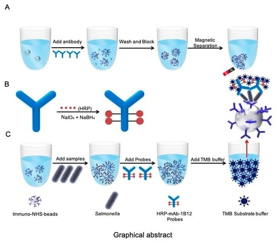
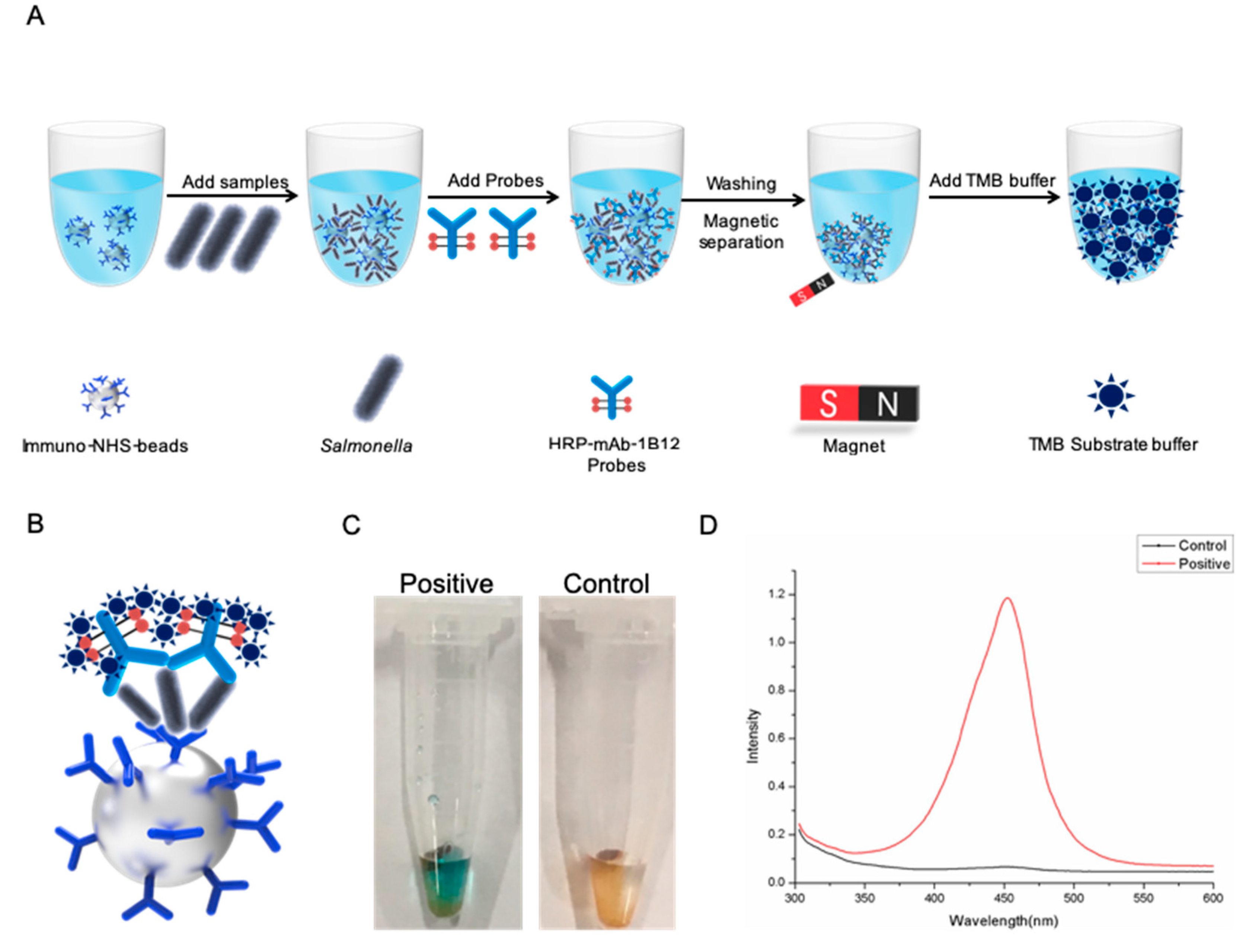
2.1. Antibody-Probe-Based Immuno-N-Hydroxysuccinimide (NHS) bead (AIB) System Design

In this work, we developed an AIB system to rapidly and visually detect Salmonella using INBs and HRP mAb probes (Figure 1A). In the AIB system, mAb 2F1, which binds specifically with Salmonella, is coated on the surface of 25 μm NHS beads to form the INBs. When Salmonella appeared in the reaction, the INBs would capture it. Next, HRP mAb probes are added to the reaction, forming a sandwich structure (Figure 1B). Then, magnetic separation and washing are performed to remove the unbound probes. In the presence of TMB buffer, a positive reaction will be blue (Figure 1C), and the optical density (Figure 1D) will increase significantly.

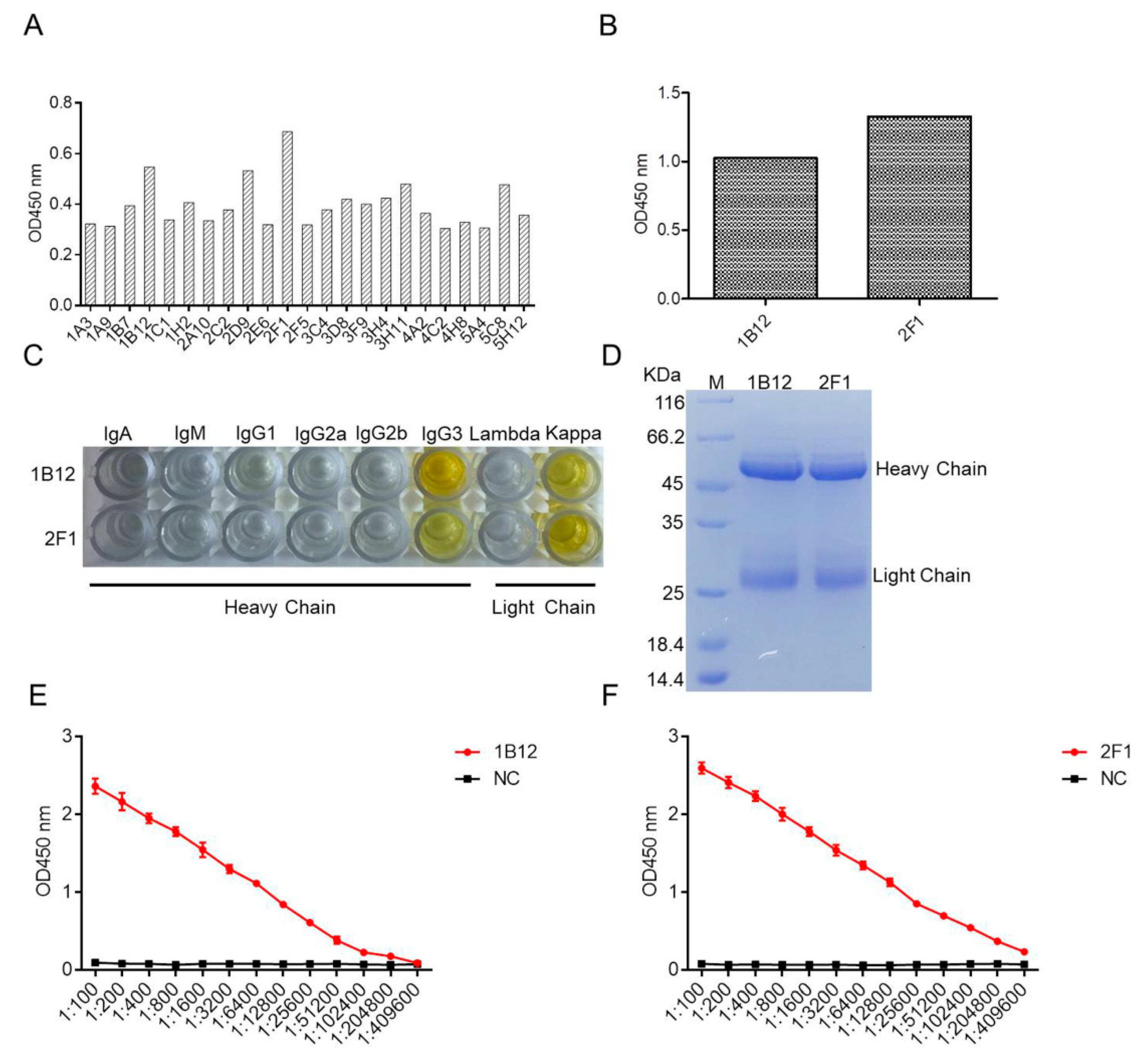
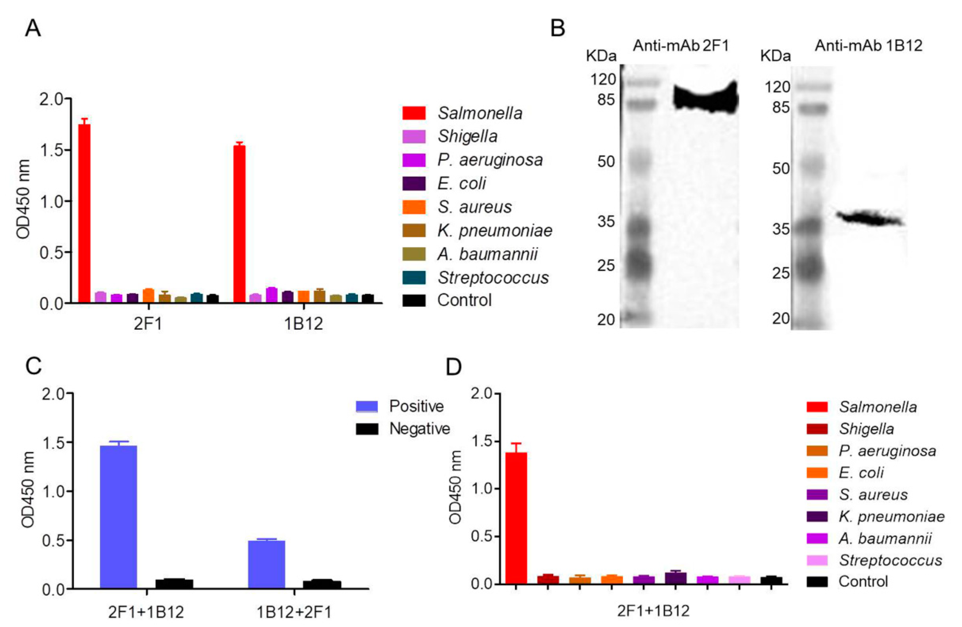
2.2. Generation of Specific mAbs Against Salmonella

Two stable positive hybridomas were screened through three subcloning cycles from twenty-three originally positive wells, designated as 2F1 and 1B12 (Figure 2A). Reactivity of the two mAbs was determined via enzyme-linked immunosorbant assay (ELISA). The results showed that both mAbs reacted with Salmonella (Figure 2B). The immunoglobulin isotypes of 2F1 and 1B12 were determined using a mouse monoclonal antibody isotyping kit. Figure 2C shows 2F1 and 1B12 isotyped as IgG3, and the light chains of the two mAbs belong to the kappa chain. The two mAbs were used to produce ascites. The ascites was purified using protein A-sepharose and tested via sodium dodecyl sulfate-polyacrylamide gel electrophoresis (SDS-PAGE) (Figure 2D). The titers of mAbs 2F1 and 1B12 were evaluated via ELISA, and the titers of both mAbs reached 1:204800 (Figure 2E,F). The KD values of mAb 2F1 and 1B12 were measured as described in our previous published study [22], and were calculated as KD = 3.677 ± 0.33 nM for mAb 2F1 and KD = 1.126 ± 0.15 nM for mAb 1B12.

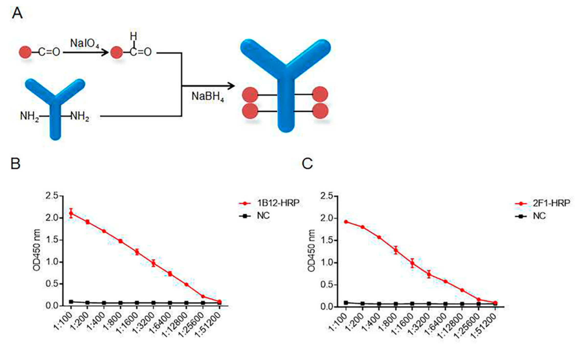
2.3. Synthesis of the HRP mAb Probes

The purified mAbs 2F1 and 1B12 were dialyzed in phosphate-buffered saline (PBS) to remove the Tris-HCl and glycine. The purified mAbs and HRP were then coupled by an aldehyde–amino bridge under NaIO4 and NaBH4 (Figure 3A). The ratio of conjugation was calculated as 1 mg HRP/2.5 mg antibody. After conjugation, we acquired about 25 mg mAb probe containing 10 mg HRP. Next, the activities and titers of the two HRP mAb probes were determined by ELISA (Figure 3B,C).

2.4. Characterization of the Paired Antibodies

The reactivity and specificity of mAb 1B12 and 2F1 were evaluated via ELISA and Western blot. Figure 4A shows that both mAb 1B12 and 2F1 specifically recognized Salmonella and did not cross-react with similar microbial strains. The Western blot results showed that both mAbs recognized the different proteins on the surface of Salmonella (Figure 4B). The optimal mAbs used to establish the AIB system were screened based on double sandwich ELISA (DAS-ELISA). HRP-labeled mAbs (1B12 and 2F1) and unlabeled mAbs (1B12 and 2F1) were constructed in each group for the DAS-ELISA, which showed that the group composed of mAb 2F1 and 1B12 was more effective than the other groups (Figure 4C). In addition, we evaluated the specificity of the mAb 2F1 and 1B12 combination. Figure 4D shows that the group of composed of 1B12 and 2F1 displayed high specificity and did not recognize the control strains (E. coli, S. aureus, K. pneumoniae, Shigella, A. baumannii, P. aeruginosa, and Streptococcus). Figure 4C,D show that the mAb 2F1 and 1B12 group was the optimal combination for detection of Salmonella.

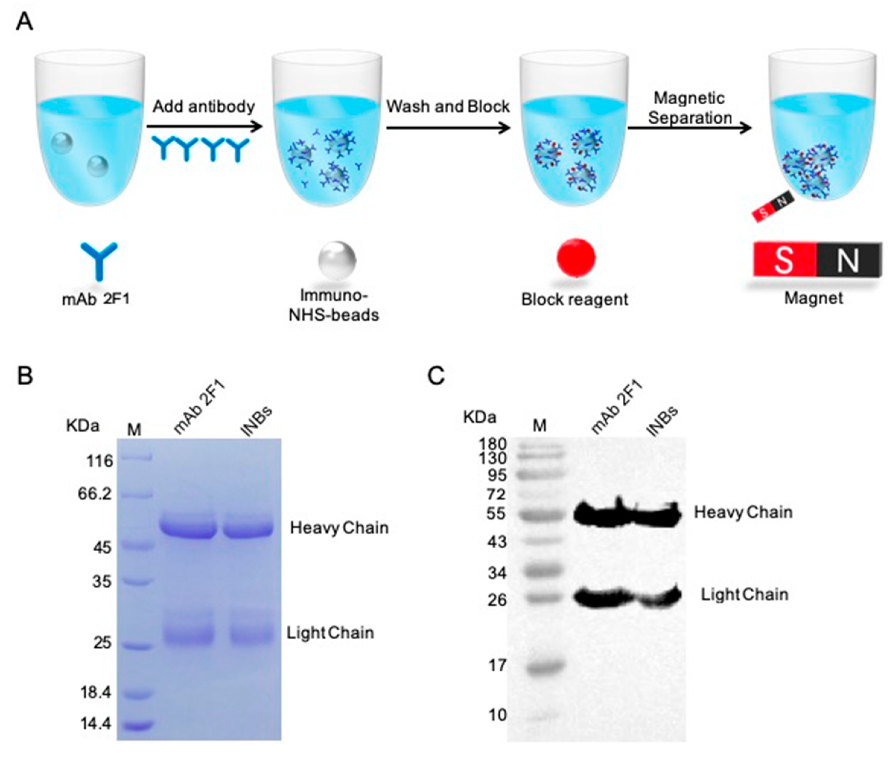
2.5. INB Preparation and Characterization

The INBs were generated by conjugating NHS modified magnetic beads (NHS beads) with mAb 2F1 via covalent coupling (Figure 5A). The prepared INBs were evaluated via SDS-PAGE and Western blot. The results demonstrated that mAb 2F1 had conjugated on the surface of the NHS beads (Figure 5B,C).

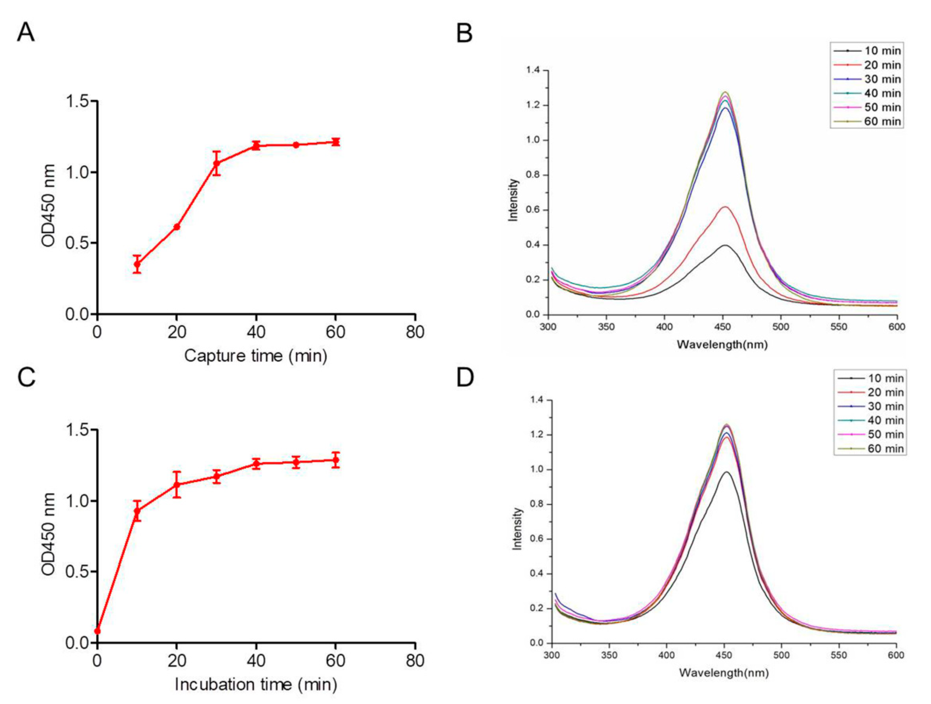
2.6. AIB System Optimization

In the AIB system, we optimized each preparation step to achieve the best detection effect. Using gradient dilutions and plate counts, the average binding efficiency of the INBs was 90%. The optimum INB capture period was confirmed using various incubation times from 10 to 60 min. Figure 6A,B show that the INBs completely captured the Salmonella within 30 min. No significant differences were observed as the incubation time increased. The optimal incubation period, during which the mAb 1B12 probes formed sandwich products, was evaluated over 10 to 60 min (Figure 6C). The optical density from the formed sandwich products was determined via microplate reader. The sandwich products quickly formed within 20 min, demonstrating the high affinity between the mAb 1B12 probes and Salmonella (Figure 6D).

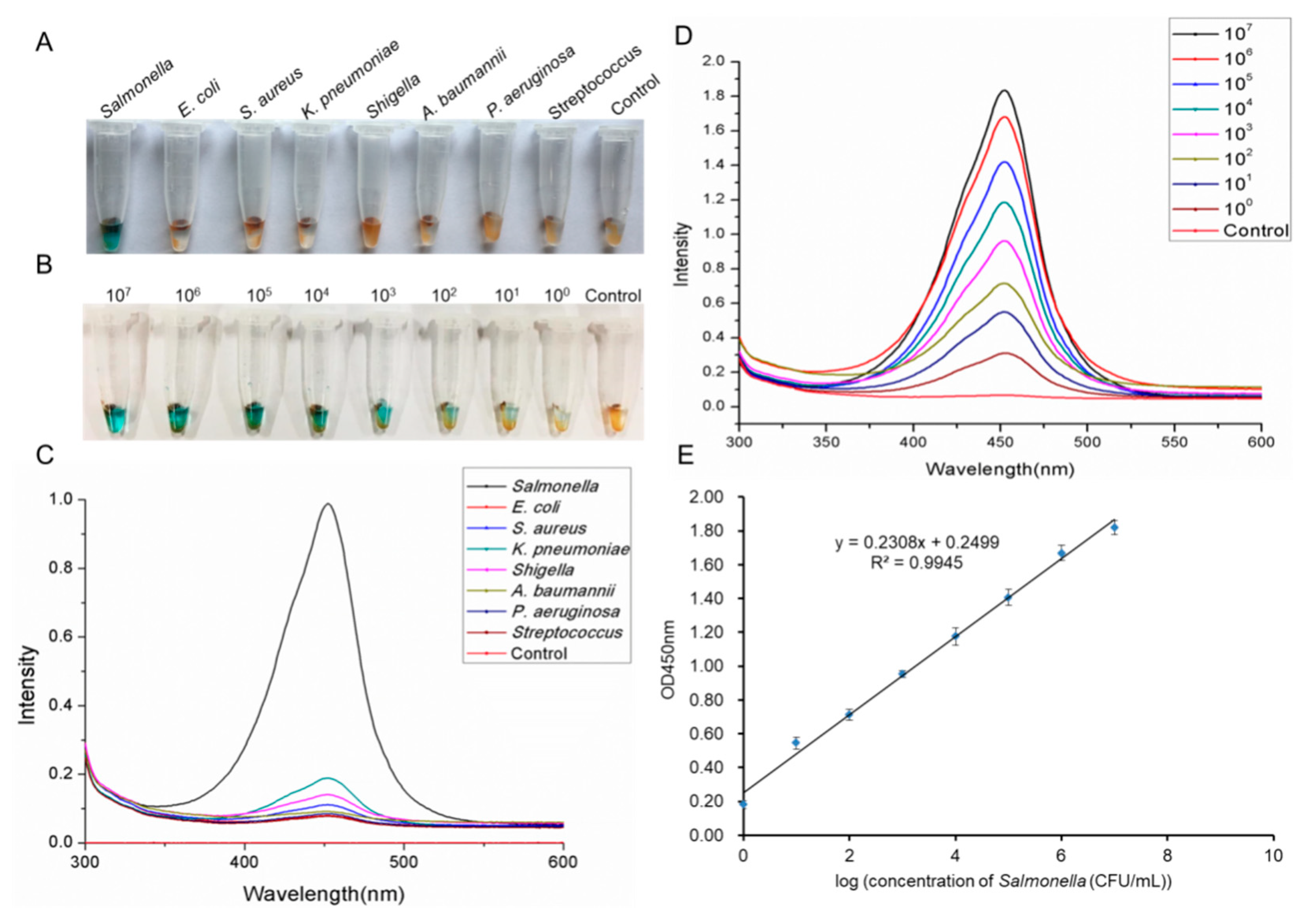
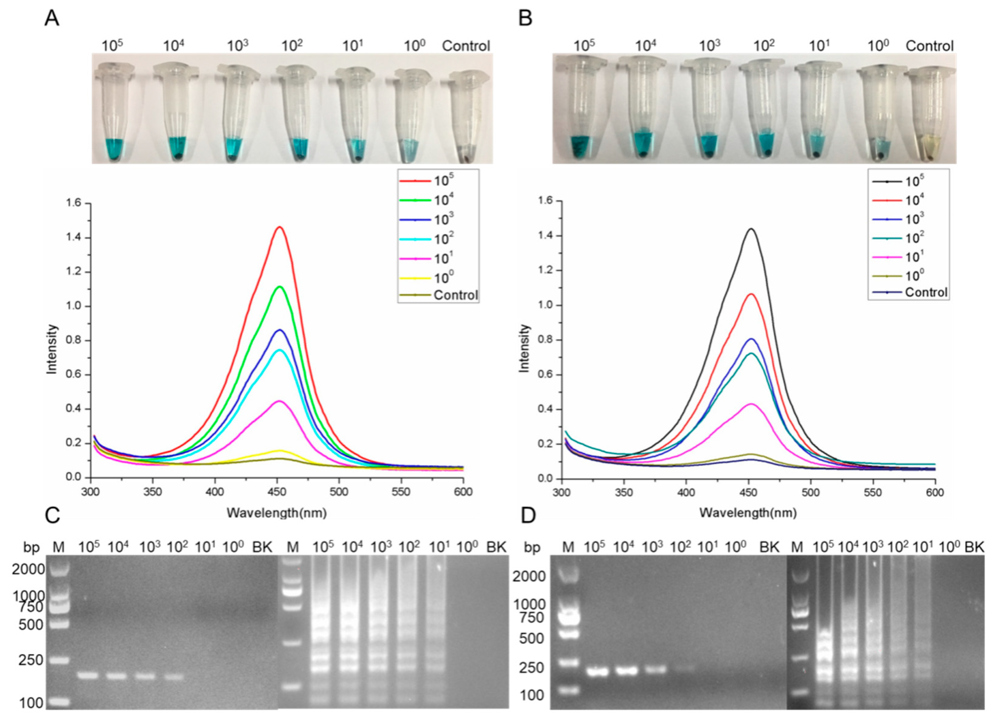
2.7. Assessment of Salmonella Detection Using the AIB System

We initially tested the AIB system specificity using seven similar microbial strains and found that the AIB system was highly specific for detecting Salmonella. The blue color (Figure 7A) and strong optical density (Figure 7C) were observed only in the presence of Salmonella. The controls showed no significant changes. The AIB system sensitivity was tested with different Salmonella concentrations ranging from 9 × 10⁷ to 9 × 100 colony-forming units (CFU) mL−1. Figure 7B,D show the blue color and optical density at different Salmonella concentrations, as recorded by the AIB system. We then set up a plotted linear curve using the different Salmonella concentrations. Figure 7E shows a good linear relationship (R2 = 0.9945).

2.8. Salmonella Detection by the AIB System in Artificially Contaminated Samples

To evaluate the AIB system performance, we used milk and egg samples contaminated with different concentrations of Salmonella ranging from 105 to 100 CFU mL−1. Figure 8A,B show the changes in blue color and strong optical density in the milk and egg samples contaminated with different Salmonella concentrations. These results confirmed that the new AIB system can rapidly and accurately detect Salmonella, even in complex samples such as milk and eggs. Besides, the prepared milk and egg samples were used for the extraction of the genome and then applied for PCR and LAMP assays using the reported primers [23]. Figure 8C,D show that the sensitivity values of PCR and LAMP were 102 CFU and 101 CFU, respectively, in both milk and egg samples.

3. Discussion

Salmonella is an important human pathogen worldwide, infecting humans and various other animals. Improper cooking and processing of animal-derived foods (e.g., raw milk, meat, and eggs) are the main mechanisms by which Salmonella infects humans [24]. Salmonellosis, one of the most important zoonoses, which mainly causes severe foodborne gastroenteritis and bacterial diarrhea, is a huge public health problem [25]. Foods such as meat, milk, and eggs are essential for human survival but are often contaminated with Salmonella [24]. Studies have reported that cases of Salmonella poisoning have occurred in powdered infant formula [26], raw milk [27], eggs [28], and meat [13,29]; thus, the food industry must prioritize developing innovative methods for detecting Salmonella.
Conventional culture-based methods are considered the gold standard for detecting Salmonella in various samples. However, these are labor-intensive and time-consuming, usually taking 2 to 3 days [30]. Thus, these methods are unsuitable for rapid detection. Recently, rapid detection methods have been developed to detect Salmonella that target the nucleic acid, including PCR [13], real-time PCR [31], immunocaptured-PCR (IC-PCR) [32], and LAMP [8,33]. The disadvantages of PCR and IC-PCR detection methods are that they require instruments, professional personnel, bacterial enrichment, and genome or plasmid extraction [15,34]. LAMP is a novel amplification approach developed by Notomi et al. [35], which is rapid and highly specific, and has been applied for various pathogens, including parasites [36,37], fungi [38], bacteria [20], and viruses [39,40]. The biggest disadvantage of LAMP is that it produces aerosol during detection, leading to many false positives.
Conversely, immunodiagnostic approaches are more rapid, sensitive, and stable than nucleic acid assays, especially for samples such as milk [41], whole blood [42], and saliva [43]. Many novel immunosensors have been developed, which combine immunology with magnetic nanoparticles [22], platinum nanoparticles [44], or Pt nanomotors [45]. Instrument-free and mobile diagnostic technologies could transform the current foodborne pathogen detection systems, particularly in resource-limited settings.
In this study, we developed an instrument-free, sensitive immunosensor to effectively, rapidly, and sensitively detect Salmonella based on a pair of mAbs recognizing different antigenic determinants on the surface of Salmonella (Figure 4B) and sensitive probes (Figure 3). The AIB system reported in this manuscript was developed using a mAb pair, NHS beads, and HRP. In this system, mAb 1B12 was conjugated with HRP, forming a sandwich structure (Figure 3). The mAb 2F1 was coated on the surface of the NHS beads used to enrich Salmonella (Figure 5). We demonstrated the feasibility and practicability of the AIB system for detecting Salmonella using INBs and HRP probes. The sensitivity of the AIB system was 9 CFU mL−1 and higher than that of PCR (105 CFU) [13], real-time PCR coupled with immunomagnetic separation or centrifugation (2 × 10⁴ CFU) [42], LAMP (1.3 to 28 CFU) [43], conventional culture-based methods, and antibody or aptamer-based assay (101 to 103 CFU) (Table 1). However, the nucleic-acid based detection methods still take several hours to enrich the Salmonella, followed by extraction of the plasmid or genome using a commercial kit [13,31,46]. Besides, special equipment, including the thermal cycling instrument, an electrophoresis apparatus, and a gel imaging system, are essential for PCR [13] and real-time PCR [31,32] methods. This special equipment is expensive and requires professional technical assistance, which largely limits their applications, especially for areas with poor resources. LAMP is a simple detection method that does not require special equipment (unlike PCR or real-time PCR), but it cannot enrich the target substances from the samples, and a commercial kit is also needed to extract the plasmid or genome [37,38]. Moreover, for some special samples, such as sputum, blood, and feces, it is very difficult to accurately and quickly enrich the target substances, and much time and materials are needed during the process [47]. Additionally, the false positive results caused by LAMP can be deadly. Therefore, the above drawbacks make LAMP inappropriate for fast, sensitive on-site detection. For antibody or aptamer-based ELISA assay, the sensitivity of the related ELISA assay ranged from 101 to 103 CFU for artificially samples, but the process takes several hours. In addition, similar INB methods, ELISA assay lacks the ability to rapidly enrich targets from the environment, especially for special samples such as milk, whole blood, and saliva, meaning antibody or aptamer-based ELISA assays are insufficiently sensitive, which largely limits their applications. Besides, professional experimental skills are essential, as well as a microplate reader, which is expensive and not available anywhere, particularly in resource-poor areas.
Compared with culture-based techniques and biochemical assays, PCR, real-time PCR, and normal LAMP, and antibody or aptamer-based ELISA assay, the AIB assay only needs a portable magnetic frame and TMB buffer. It does not require a long pre-enrichment step followed by genome or plasmid extraction, any special equipment, or professional skills. The AIB system developed here has two advantages over normal detection methods. The first one is that the surfaces of the NHS beads are coated with abundant mAb 2F1 that specifically recognize Salmonella (Figure 4A), which means the prepared INBs can efficiently and accurately enrich the Salmonella from different biological samples within 30 min, as shown in Figure 6A,B. Compared with the carboxylic modified magnetic beads used in our previous study and in other research articles [22], the NHS beads are coupled with antibodies with high efficiency and short timeframes, without needing EDC or glutaraldehyde for activation. The other advantage is that the mAb probe, having high affinity, can rapidly form a sandwich structure within 20 min, as shown in Figure 6C,D. Additionally, the HRP coupled with mAb 1B12 is efficiently catalyzed by TMB buffer, showing a strong blue color (Figure 1C). The optical density (Figure 1D) changed within 10 min. Accordingly, the INBs showing rapid and efficient enrichment together with the sensitive mAb probe allow the AIB system to rapidly and sensitively detect Salmonella within 50 min. Figure 7 shows that the AIB system has good sensitivity and specificity based on the INBs and HRP mAb probes. We evaluated the practical application of the AIB system using artificially contaminated samples, including water (Figure 7B,D), milk (Figure 8A), and eggs (Figure 8B). The results indicated that the AIB system enabled accurate and rapid screening of Salmonella and could potentially be used in the food industry and hospitals. The prepared samples were also identified by PCR and LAMP to evaluate the accuracy of the AIB assay. In Figure 8C,D, we can clearly see that the minimum detection limits for PCR and LAMP were 9 × 10² CFU and 9 × 101 CFU, respectively, in both milk and eggs samples. The sensitivity of the PCR and LAMP methods was lower than the AIB assay for the same samples. Having high sensitivity and specificity, the AIB system can rapidly and accurately detect Salmonella without requiring special equipment or professional skills, making the AIB system more applicable in various environments.

4. Materials and Methods

4.1. Materials

Table 2 shows the eight bacterial strains used in this study. The SP2/0 cells were stored in our lab. Freund’s incomplete adjuvant (FIA), Freund’s complete adjuvant (FCA), hypoxanthine–aminopterin–thymidine (HAT), hypoxanthine–thymidine (HT), and PEG-2000 were purchased from Sigma (St. Louis, MO, USA). The fetal bovine serum (FBS) and Roswell Park Memorial Institute (RPMI) 1640 medium were purchased from Gibco (Grand Island, NY, USA). NHS beads and the MAg25K/NHS kit were purchased from Enriching Biotechnology (Suzhou, China). Protein A-sepharose was purchased from GE Healthcare (Chicago, IL, USA). The mouse monoclonal antibody isotyping kit was purchased from Southern Biotech (Birmingham, AL, USA). Horseradish peroxidase (HRP) and goat anti-mouse IgG (H+L) HRP were purchased from GenScript (Nanjing, China).

4.2. Production of mAbs Against Salmonella

The antigens were prepared using 108 CFU of Salmonella dissolved in PBS buffer and inactivated for 30 min at 80 °C [20]. The prepared antigens were used to immunize BALB/c mice, with 107 CFU being mixed with FIA for the first immunization and 107 CFU mixed with FCA for the second and third immunizations. Three days after the boosted immunization, spleen cells were collected and fused with SP2/0 cells via PEG at 37 °C [79]. The fused cells were maintained in HAT medium for 7 days. One week later, the fused cells were cultured in HT medium until the first round of screening. The positive hybridomas in each plate were screened by ELISA.

4.3. Purification of Ascites

After three subcloning cycles, we successfully obtained two positive hybridoma cell lines that stably secreted antibodies against Salmonella. Two hybridomas were constructed to prepare the ascites, which were purified using protein A-sepharose. Briefly, all ascites were filtered using a 0.2-micron filter, and the pH value of the ascites was adjusted to 8.0 using 1.0 M Tris-Cl (pH 8.0). Next, the prepared ascites were incubated with protein A-sepharose for 30 min at 25 °C. After incubation, the ascites were collected, and the column was washed with 100 mM Tris-Cl (pH 8.0) and 10 mM Tris-Cl (pH 8.0), respectively. Lastly, the column was washed with 50 mM glycine (pH 3.0), and the purified immunoglobulins from each antibody were determined via bicinchoninic acid assay and SDS-PAGE.

4.4. ELISA and Western Blot Assay

For the ELISA, each well was coated with 106 CFU of Salmonella and incubated for 2 h at 37 °C. All wells were then washed with PBS-T and blocked with 5% BSA. Next, mAb 1B12 and 2F1 (1:1000) were added to each well and incubated for 1 h at 37 °C. All wells were washed with PBS-T, then the goat anti-mouse IgG (H+L) HRP (1:5000) was added and incubated for 1 h at 37 °C. One hour later, the supernatant was removed and washed with PBS-T. Lastly, soluble TMB substrate solution (TIANGEN, Beijing, China) was added to detect the immunoreaction.
For the Western blot assay, 106 CFU of Salmonella was transferred to a nitrocellulose (NC) membrane, which was blocked with 5% BSA for 1 h at 37 °C. Next, mAb 1B12 and 2F1 were added (1:1000) and incubated for 2 h at 37 °C. Two hours later, the antibodies were collected and the NC membranes were washed with PBS-T. Goat anti-mouse IgG (H+ L) HRP (1:5000) was then added and incubated for 1 h at 37 °C. Lastly, the NC membranes were washed, and the Western blot kit (BioBest, Anhui, China) was used to detect the immunoreaction.

4.5. HRP mAb Probe Preparation and Characterization

The HRP mAb probes were synthesized using mAbs coupled with HRP. All probes were prepared as described in our previous study [22]. Briefly, 10 mg of HRP was dissolved in 1 mL of 0.1 M NaHCO3 and then oxidized for 2 h with 1 mL of 10 mM NaIO4. Then, the HRP liquid was mixed with 1.5 mL of 0.1 M Na2CO3, 25 mg of 1B12, 2F1, and reacted in a 5 mL tube containing 0.6 g of Sephadex G25 for 3 h at 25 °C. Three hours later, the liquid was collected and terminated with 0.225 mL of 0.132 M NaBH4 for 0.5 h, followed by 0.675 mL of 0.132 M NaBH4 for 1 h at 25 °C. The process was performed in the dark. Lastly, the prepared HRP mAb probes were stored in PBS buffer at 4 °C. The activities and titers of the HRP mAb probes were determined using ELISA. Briefly, two HRP mAb probes at different dilutions (from 1:100 to 1:51200) were added to each well, which had been coated with Salmonella and blocked with BSA, and incubated for 1 h at 37 °C. Next, the supernatant was removed from each well and washed with PBS-T. The immune responses were then detected using soluble TMB substrate solution.

4.6. INB Preparation and Characterization

The INBs were prepared using NHS beads conjugated with mAb 2F1 using a MAg25K/NHS kit. First, the NHS beads were mixed evenly, and 1 mL of NHS bead solution (10%, v/v) was added to a 2 mL tube. The supernatant was removed by magnetic separation, and the NHS beads were washed twice with 2 mL of absolute ethanol. Second, 1.2 mg of mAb 2F1 dissolved in a coupling buffer was added and mixed with NHS beads for 2 h at room temperature. Next, 1 mL of blocking buffer was added to block the beads for 2 h, while being rotated and mixed at room temperature. After blocking, the supernatant was removed, and 1 mL of wash buffer was added to wash the beads five times. Lastly, the supernatant was removed, and the INBs were dissolved in PBS buffer (pH 7.4) for further use. The prepared INBs were evaluated via SDS-PAGE, Western blot, and the AIB system.

4.7. Performance of the AIB System

The sensitivity of the AIB system was evaluated using serially diluted Salmonella, with concentrations ranging from 107 to 100 CFU mL−1. One microliter of each Salmonella concentration was tested using the AIB system following the testing protocol. The specificity of the developed AIB system was confirmed using Salmonella and similar microbial strains (E. coli, S. aureus, K. pneumoniae, Shigella, A. baumannii, P. aeruginosa, and Streptococcus).

4.8. System Evaluation using Contaminated Samples

To evaluate the AIB system, we used artificial milk and egg samples containing Salmonella concentrations ranging from 105 to 100 CFU mL−1. Each sample was tested using the AIB system according to the testing protocol.

5. Conclusions

In this study, we developed a novel immunosensor based on magnetic NHS beads and antibody probes. The novel AIB system enabled visual, rapid, and sensitive Salmonella detection, without requiring specialized equipment or skills. This system can potentially be widely used to diagnose infectious diseases caused by Salmonella spp.

Author Contributions

L.Z. and X.D. designed and drafted the work. L.Z., X.D., Z.C., C.C., N.G., Y.S. (Yihao Song), Y.S. (Yuzhu Song), Q.H., and X.X. performed the experiments, analyzed the data, and interpreted the results. J.Z. and H.L. designed the work and revised it critically.

Funding

This research was funded by National Natural Science Foundation of China (grant number 81860625), Applied Basic Research Projects of Yunnan Province (grant number 2018FB128), and Yunnan Province Biomedical Science and Technology Plan Project (grant number 2019ZF004).

Conflicts of Interest

The authors declare no conflict of interest.

Abbreviations

AIBantibody-probe-based immuno-N-hydroxysuccinimide beads
DAS-ELISAdouble sandwich ELISA
ELISAenzyme-linked immunosorbant assay
FBSfatal bovine serum
FCAFreund’s complete adjuvant
FIAFreund’s incomplete adjuvant
IC-PCRimmunocaptured-PCR
INBsimmuno-NHS beads
LAMPloop-mediated isothermal amplification assay
mAbmonoclonal antibody
NHS beadsN-hydroxysuccinimide (NHS) beads
PCRpolymerase chain reaction
HAThypoxanthine–aminopterin–thymidine
HRPhorse radish peroxidase
HThypoxanthine–thymidine
SDS-PAGEsodium dodecyl sulfate-polyacrylamide gel electrophoresis
TMB3, 3′, 5, 5′-tetramethylbenzidine

References

  1. Alves, J.; Niguma, N.H.; de Oliveira, T.C.R.M. Detection of Salmonella spp. in Eight Complex Food Matrices Using Polymerase Chain Reaction Assay. Int. J. Mol. Sci. 2015, 35, 453–457. [Google Scholar] [CrossRef]
  2. Newell, D.G.; Koopmans, M.; Verhoef, L.; Duizer, E.; Aidara-Kane, A.; Sprong, H.; Opsteegh, M.; Langelaar, M.; Threfall, J.; Scheutz, F.; et al. Food-borne diseases - the challenges of 20 years ago still persist while new ones continue to emerge. Int. J. Food Microblol. 2010, 139, 3–15. [Google Scholar] [CrossRef] [PubMed]
  3. Salzman, N.H.; Ghosh, D.; Huttner, K.M.; Paterson, Y.; Bevins, C.L. Protection against enteric salmonellosis in transgenic mice expressing a human intestinal defensin. Nature 2003, 422, 522–526. [Google Scholar] [CrossRef] [PubMed]
  4. Zhang, S.; Kingsley, R.A.; Santos, R.L.; Andrews-Polymenis, H.; Raffatellu, M.; Figueiredo, J.; Nunes, J.; Tsolis, R.M.; Adams, L.G.; Baumler, A.J. Molecular pathogenesis of Salmonella enterica serotype typhimurium-induced diarrhea. Infect. Immu. 2003, 71, 1–12. [Google Scholar] [CrossRef] [PubMed]
  5. Switt, A.I.; Soyer, Y.; Warnick, L.D.; Wiedmann, M. Emergence, distribution, and molecular and phenotypic characteristics of Salmonella enterica serotype 4, 5, and 12:1. Foodborne Pathog. Dis. 2009, 6, 407–415. [Google Scholar] [CrossRef] [PubMed]
  6. Abdoel, T.H.; Pastoor, R.; Smits, H.L.; Hatta, M. Laboratory evaluation of a simple and rapid latex agglutination assay for the serodiagnosis of typhoid fever. Trans. R. Soc. Trop. Med. Hyg. 2007, 101, 1032–1038. [Google Scholar] [CrossRef] [PubMed]
  7. Fraser, A.; Paul, M.; Goldberg, E.; Acosta, C.J.; Leibovici, L. Typhoid fever vaccines: Systematic review and meta-analysis of randomised controlled trials. Vaccine 2007, 25, 7848–7857. [Google Scholar] [CrossRef]
  8. Liu, H.B.; Zang, Y.X.; Du, X.J.; Li, P.; Wang, S. Development of an isothermal amplification-based assay for the rapid visual detection of Salmonella bacteria. J. Dairy Sci. 2017, 100, 7016–7025. [Google Scholar] [CrossRef] [Green Version]
  9. Shao, Y.; Zhu, S.; Jin, C.; Chen, F. Development of multiplex loop-mediated isothermal amplification-RFLP (mLAMP-RFLP) to detect Salmonella spp. and Shigella spp. in milk. Int. J. Food Microbiol. 2011, 148, 75–79. [Google Scholar] [CrossRef]
  10. Majowicz, S.E.; Musto, J.; Scallan, E.; Angulo, F.J.; Kirk, M.; O’Brien, S.J.; Jones, T.F.; Fazil, A.; Hoekstra, R.M. The global burden of nontyphoidal Salmonella gastroenteritis. Clin. Inf. Dis. 2010, 50, 882–889. [Google Scholar] [CrossRef]
  11. Kuang, H.; Cui, G.; Chen, X.; Yin, H.; Yong, Q.; Xu, L.; Peng, C.; Wang, L.; Xu, C. A one-step homogeneous sandwich immunosensor for Salmonella detection based on magnetic nanoparticles (MNPs) and quantum Dots (QDs). Int. J. Mol. Sci. 2013, 14, 8603–8610. [Google Scholar] [CrossRef] [PubMed]
  12. Khueankhancharoen, J.; Thipayarat, A.; Saranak, J. Optimized microscale detection of amino acid decarboxylase for rapid screening of Salmonella in the selective enrichment step. Food Control. 2016, 69, 352–367. [Google Scholar] [CrossRef]
  13. Chin, W.H.; Sun, Y.; Hogberg, J.; Quyen, T.L.; Engelsmann, P.; Wolff, A.; Bang, D.D. Direct PCR-A rapid method for multiplexed detection of different serotypes of Salmonella in enriched pork meat samples. Mol. Cell. Probes. 2017, 32, 24–32. [Google Scholar] [CrossRef] [PubMed]
  14. Domesle, K.J.; Yang, Q.; Hammack, T.S.; Ge, B. Validation of a Salmonella loop-mediated isothermal amplification assay in animal food. Int. J. Food Microbiol. 2018, 264, 63–76. [Google Scholar] [CrossRef] [PubMed]
  15. Hapuarachchi, C.T.; Jeffery, K.J.M.; Bowler, I. Stool PCR may not be a substitute for enrichment culture for the detection of salmonella. J. Med. Microbiol. 2019, 68, 395–397. [Google Scholar] [CrossRef]
  16. Han, Z.; Shu, J.; Jiang, Q.; Cui, H. Coreactant-Free and Label-Free Eletrochemiluminescence Immunosensor for Copeptin Based on Luminescent Immuno-Gold Nanoassemblies. Anal. Chem. 2018, 90, 6064–6070. [Google Scholar] [CrossRef] [PubMed]
  17. Tominaga, T. Rapid detection of Klebsiella pneumoniae, Klebsiella oxytoca, Raoultella ornithinolytica and other related bacteria in food by lateral-flow test strip immunoassays. J. Microbiol. Methods 2018, 147, 43–49. [Google Scholar] [CrossRef]
  18. Cheng, S.; Sun, J.; Yang, J.; Lv, J.; Wu, F.; Lin, Y.; Liao, L.; Ye, Y.; Cao, C.; Fang, L.; et al. A new immunoassay of serum antibodies against Peste des petits ruminants virus using quantum dots and a lateral-flow test strip. Anal. Bioanal. Chem. 2017, 409, 133–141. [Google Scholar] [CrossRef]
  19. Liu, B.; Si, J.; Zhao, F.; Wang, Q.; Wang, Y.; Li, J.; Li, C.; Li, T. Rapid detection of cow milk adulteration/contamination in goat milk by a lateral flow colloidal gold immunoassay strip. J. Dairy Res. 2019, 86, 94–97. [Google Scholar] [CrossRef]
  20. Zhang, L.; Wei, Q.; Han, Q.; Chen, Q.; Tai, W.; Zhang, J.; Song, Y.; Xia, X. Detection of Shigella in Milk and Clinical Samples by Magnetic Immunocaptured-Loop-Mediated Isothermal Amplification Assay. Front. Microbiol. 2018, 9, 94. [Google Scholar] [CrossRef] [Green Version]
  21. Zhang, L.; Shi, Y.; Chen, C.; Han, Q.; Zhang, M.; Yi, H.; Song, Y.; Xia, X.; Zhang, J. Visual and Rapid Detection of Klebsiella pneumoniae by Magnetic Immunocapture-Loop-Mediated Isothermal Amplification Assay. Jundishapur. J. Microbiol. 2019, 12, e90016. [Google Scholar] [CrossRef]
  22. Zhang, L.; Shi, Y.; Chen, C.; Han, Q.; Chen, Q.; Xia, X.; Song, Y.; Zhang, J. Rapid, Visual Detection of Klebsiella pneumoniae Using Magnetic Nanoparticles and an Horseradish Peroxidase-Probe Based Immunosensor. J. Biomed. Nanotechnol. 2019, 15, 1061–1071. [Google Scholar] [CrossRef] [PubMed]
  23. Yi, W.; Yan, W.; Lijuan, L.; Dongxin, L.; Xia, L.; Yanmei, X.; Shoukui, H.; Lina, N.; Jianguo, X.; Changyun, Y. Rapid and Sensitive Detection of Shigella spp. and Salmonella spp. by Multiple Endonuclease Restriction Real-Time Loop-Mediated Isothermal Amplification Technique. Front. Microbiol. 2015, 6, 428–437. [Google Scholar] [CrossRef]
  24. Moussa, I.M.; Gassem, M.A.; Al-Doss, A.A.; Sadik, W.A.M.; Mawgood, A.A.L. Using molecular techniques for rapid detection of Salmonella serovars in frozen chicken and chicken products collected from Riyadh, Saudi Arabia. Afr. J. Biotechnol. 2010, 9, 612–619. [Google Scholar] [CrossRef] [Green Version]
  25. Karp, B.E.; Campbell, D.; Chen, J.C.; Folster, J.P.; Friedman, C.R. Plasmid-mediated quinolone resistance in human non-typhoidal Salmonella infections: An emerging public health problem in the United States. Zoonoses Public Health 2018, 65, 838–849. [Google Scholar] [CrossRef] [PubMed]
  26. Cahill, S.M.; Wachsmuth, I.K.; Costarrica Mde, L.; Ben Embarek, P.K. Powdered infant formula as a source of Salmonella infection in infants. Clin. Infect. Dis. 2008, 46, 268–273. [Google Scholar] [CrossRef]
  27. Amagliani, G.; Petruzzelli, A.; Carloni, E.; Tonucci, F.; Foglini, M.; Micci, E.; Ricci, M.; Di Lullo, S.; Rotundo, L.; Brandi, G. Presence of Escherichia coli O157, Salmonella spp., and Listeria monocytogenes in Raw Ovine Milk Destined for Cheese Production and Evaluation of the Equivalence Between the Analytical Methods Applied. Foodborne Pathog. Dis. 2016, 13, 626–632. [Google Scholar] [CrossRef]
  28. Kemal, J.; Sibhat, B.; Menkir, S.; Beyene, D. Prevalence, assessment, and antimicrobial resistance patterns of Salmonella from raw chicken eggs in Haramaya, Ethiopia. J. Infect. Dev. Ctries. 2016, 10, 1230–1235. [Google Scholar] [CrossRef] [Green Version]
  29. Nhung, N.T.; Van, N.T.B.; Cuong, N.V.; Duong, T.T.Q.; Nhat, T.T.; Hang, T.T.T.; Nhi, N.T.H.; Kiet, B.T.; Hien, V.B.; Ngoc, P.T.; et al. Antimicrobial residues and resistance against critically important antimicrobials in non-typhoidal Salmonella from meat sold at wet markets and supermarkets in Vietnam. Int. J. Food Microbiol. 2018, 266, 301–309. [Google Scholar] [CrossRef]
  30. Soria, M.C.; Soria, M.A.; Bueno, D.J.; Terzolo, H.R. Comparison of 3 culture methods and PCR assays for Salmonella gallinarum and Salmonella pullorum detection in poultry feed. Poult. Sci. 2013, 92, 1505–1515. [Google Scholar] [CrossRef]
  31. Yang, Y.G.; Song, M.K.; Park, S.J.; Kim, S.W. Direct detection of Shigella flexneri and Salmonella typhimurium in human feces by real-time PCR. J. Microbiol. Biotechnol. 2007, 17, 1616–1621. [Google Scholar] [PubMed]
  32. Warren, B.R.; Yuk, H.G.; Schneider, K.R. Detection of salmonella by flow-through immunocapture real-time PCR in selected foods within 8 hours. J. Food. Prot. 2007, 70, 1002–1006. [Google Scholar] [CrossRef] [PubMed]
  33. Zhao, Y.; Jiang, X.; Qu, Y.; Pan, R.; Pang, X.; Jiang, Y.; Man, C. Salmonella detection in powdered dairy products using a novel molecular tool. J. Dairy. Sci. 2017, 100, 3480–3496. [Google Scholar] [CrossRef] [PubMed] [Green Version]
  34. Quigley, L.; O’Sullivan, O.; Beresford, T.P.; Paul Ross, R.; Fitzgerald, G.F.; Cotter, P.D. A comparison of methods used to extract bacterial DNA from raw milk and raw milk cheese. J. Appl. Microbiol. 2012, 113, 96–105. [Google Scholar] [CrossRef] [PubMed]
  35. Notomi, T.; Okayama, H.; Masubuchi, H.; Yonekawa, T.; Watanabe, K.; Amino, N.; Hase, T. Loop-mediated isothermal amplification of DNA. Nucleic. Acids. Res. 2000, 28, E63. [Google Scholar] [CrossRef] [PubMed]
  36. Mori, Y.; Notomi, T. Loop-mediated isothermal amplification (LAMP): A rapid, accurate, and cost-effective diagnostic method for infectious diseases. J. Infect. Chemother. 2009, 15, 62–69. [Google Scholar] [CrossRef] [PubMed]
  37. Vaagt, F.; Haase, I.; Fischer, M. Loop-mediated isothermal amplification (LAMP)-based method for rapid mushroom species identification. J. Agric. Food. Chem. 2013, 61, 1833–1840. [Google Scholar] [CrossRef] [PubMed]
  38. Nakayama, T.; Yamazaki, T.; Yo, A.; Tone, K.; Mahdi Alshahni, M.; Fujisaki, R.; Makimura, K. Detection of Fungi from an Indoor Environment using Loop-mediated Isothermal Amplification (LAMP) Method. Biocontrol. Sci. 2017, 22, 97–104. [Google Scholar] [CrossRef] [PubMed] [Green Version]
  39. Chotiwan, N.; Brewster, C.D.; Magalhaes, T.; Weger-Lucarelli, J.; Duggal, N.K.; Ruckert, C.; Nguyen, C.; Garcia Luna, S.M.; Fauver, J.R. Rapid and specific detection of Asian- and African-lineage Zika viruses. Sci. Transl. Med. 2017, 9, e0538. [Google Scholar] [CrossRef]
  40. Zhang, L.; Chen, C.; Dai, L.; Zhang, L.; Xu, K.; Song, Y.; Xia, X.; Han, Q.; Chen, Q.; Zhang, J. Efficient Capture and Detection of Zika Virion by Polyclonal Antibody Against Prokaryotic Recombinant Envelope Protein. Jundishapur. J. Microbiol. 2018, 11, e68858. [Google Scholar] [CrossRef]
  41. Chughtai, M.I.; Maqbool, U.; Iqbal, M.; Shah, M.S.; Fodey, T. Development of in-house ELISA for detection of chloramphenicol in bovine milk with subsequent confirmatory analysis by LC-MS/MS. J. Environ. Sci. Health B 2017, 52, 871–879. [Google Scholar] [CrossRef] [PubMed]
  42. Islam, K.; Sayeed, M.A.; Hossen, E.; Khanam, F.; Charles, R.C.; Andrews, J.; Ryan, E.T.; Qadri, F. Comparison of the Performance of the TPTest, Tubex, Typhidot and Widal Immunodiagnostic Assays and Blood Cultures in Detecting Patients with Typhoid Fever in Bangladesh, Including Using a Bayesian Latent Class Modeling Approach. PLoS Negl. Trop. Dis. 2016, 10, e0004558. [Google Scholar] [CrossRef] [PubMed]
  43. Bosqui, L.R.; Goncalves, A.L.R.; Goncalves-Pires, M.R.F.; Pavanelli, W.R.; Conchon-Costa, I.; Costa-Cruz, J.M.; Costa, I.N. Immune complex detection in saliva samples: An innovative proposal for the diagnosis of human strongyloidiasis. Parasitology 2018, 145, 1090–1094. [Google Scholar] [CrossRef] [PubMed]
  44. Draz, M.S.; Venkataramani, M.; Lakshminarayanan, H.; Saygili, E.; Moazeni, M.; Vasan, A.; Li, Y.; Sun, X.; Hua, S.; Yu, X.G.; et al. Nanoparticle-enhanced electrical detection of Zika virus on paper microchips. Nanoscale 2018, 10, 11841–11849. [Google Scholar] [CrossRef] [PubMed]
  45. Draz, M.S.; Lakshminaraasimulu, N.K.; Krishnakumar, S.; Battalapalli, D.; Vasan, A.; Kanakasabapathy, M.K.; Sreeram, A.; Kallakuri, S.; Thirumalaraju, P.; Li, Y.; et al. Motion-Based Immunological Detection of Zika Virus Using Pt-Nanomotors and a Cellphone. ACS. Nano 2018, 12, 5709–5718. [Google Scholar] [CrossRef] [PubMed]
  46. Zheng, Q.; Miks-Krajnik, M.; Yang, Y.; Lee, S.M.; Lee, S.C.; Yuk, H.G. Evaluation of real-time PCR coupled with immunomagnetic separation or centrifugation for the detection of healthy and sanitizer-injured Salmonella spp. on mung bean sprouts. Int. J. Food Microbiol. 2016, 222, 48–55. [Google Scholar] [CrossRef]
  47. Wolff, D.; Gerritzen, A. Overnight Enrichment is Essential for Reliable Salmonella PCR Testing from Fecal Samples. Clin. Lab. 2017, 63, 603–606. [Google Scholar] [CrossRef]
  48. Saeidabadi, M.S.; Nili, H.; Dadras, H.; Sharifiyazdi, H.; Connolly, J.; Valcanis, M.; Raidal, S.; Ghorashi, S.A. Evaluation of PCR and high-resolution melt curve analysis for differentiation of Salmonella isolates. Avian. Pathol. 2017, 46, 319–331. [Google Scholar] [CrossRef] [Green Version]
  49. Chin, W.H.; Sun, Y.; Hogberg, J.; Hung, T.Q.; Wolff, A.; Bang, D.D. Solid-phase PCR for rapid multiplex detection of Salmonella spp. at the subspecies level, with amplification efficiency comparable to conventional PCR. Anal. Bioanal. Chem. 2017, 409, 2715–2726. [Google Scholar] [CrossRef]
  50. Yang, Q.; Domesle, K.J.; Wang, F.; Ge, B. Rapid detection of Salmonella in food and feed by coupling loop-mediated isothermal amplification with bioluminescent assay in real-time. BMC Microbiol. 2016, 16, 112. [Google Scholar] [CrossRef]
  51. Abdullah, J.; Saffie, N.; Sjasri, F.A.; Husin, A.; Abdul-Rahman, Z.; Ismail, A.; Aziah, I.; Mohamed, M. Rapid detection of Salmonella Typhi by loop-mediated isothermal amplification (LAMP) method. Braz. J. Microbiol. 2014, 45, 1385–1391. [Google Scholar] [CrossRef] [PubMed] [Green Version]
  52. Yang, Q.; Wang, F.; Prinyawiwatkul, W.; Ge, B. Robustness of Salmonella loop-mediated isothermal amplification assays for food applications. J. Appl. Microbiol. 2014, 116, 81–88. [Google Scholar] [CrossRef] [PubMed]
  53. Tatavarthy, A.; Ali, L.; Gill, V.; Hu, L.; Deng, X.; Adachi, Y.; Rand, H.; Hammack, T.; Zhang, G. Evaluation of Three Real-Time PCR Methods for Detection of Salmonella from Cloves. J. Food Prot. 2017, 80, 982–989. [Google Scholar] [CrossRef] [PubMed]
  54. Chen, Z.; Biswas, S.; Aminabadi, P.; Stackhouse, J.W.; Jay-Russell, M.T.; Pandey, P.K. Prevalence of Escherichia coli O157 and Salmonella spp. in solid bovine manure in California using real-time quantitative PCR. Lett. Appl. Microbiol. 2019, 69, 23–29. [Google Scholar] [CrossRef] [PubMed]
  55. Tang, X.J.; Yang, Z.; Chen, X.B.; Tian, W.F.; Tu, C.N.; Wang, H.B. Verification and large scale clinical evaluation of a national standard protocol for Salmonella spp./Shigella spp. screening using real-time PCR combined with guided culture. J. Microbiol. Methods 2018, 145, 14–19. [Google Scholar] [CrossRef] [PubMed]
  56. Rohonczy, K.; Zoller, L.; Hermann, Z.; Fodor, A.; Mraz, B.; Tabajdi-Pinter, V. Comparison of an automated ELFA and two different real-time PCR techniques for Salmonella detection in poultry samples. Acta Microbiol. Immunol. Hung. 2014, 61, 261–272. [Google Scholar] [CrossRef] [PubMed]
  57. Goodman, L.B.; McDonough, P.L.; Anderson, R.R.; Franklin-Guild, R.J.; Ryan, J.R.; Perkins, G.A.; Thachil, A.J.; Glaser, A.L.; Thompson, B.S. Detection of Salmonella spp. in veterinary samples by combining selective enrichment and real-time PCR. J. Vet. Diagn. Invest. 2017, 29, 844–851. [Google Scholar] [CrossRef] [Green Version]
  58. Volpe, G.; Delibato, E.; Fabiani, L.; Pucci, E.; Piermarini, S.; D’Angelo, A.; Capuano, F.; De Medici, D.; Palleschi, G. Development and evaluation of an ELIME assay to reveal the presence of Salmonella in irrigation water: Comparison with Real-Time PCR and the Standard Culture Method. Talanta 2016, 149, 202–210. [Google Scholar] [CrossRef]
  59. Almeida, C.; Cerqueira, L.; Azevedo, N.F.; Vieira, M.J. Detection of Salmonella enterica serovar Enteritidis using real time PCR, immunocapture assay, PNA FISH and standard culture methods in different types of food samples. Int. J. Food Microbiol. 2013, 161, 16–22. [Google Scholar] [CrossRef] [Green Version]
  60. Ma, Z.; Yang, X.; Fang, Y.; Tong, Z.; Lin, H.; Fan, H. Detection of Salmonella Infection in Chickens by an Indirect Enzyme-Linked Immunosorbent Assay Based on Presence of PagC Antibodies in Sera. Foodborne Pathog. Dis. 2018, 15, 109–113. [Google Scholar] [CrossRef]
  61. Aribam, S.D.; Ogawa, Y.; Matsui, H.; Hirota, J.; Okamura, M.; Akiba, M.; Shimoji, Y.; Eguchi, M. Monoclonal antibody-based competitive enzyme-linked immunosorbent assay to detect antibodies to O:4 Salmonella in the sera of livestock and poultry. J. Microbiol. Methods 2015, 108, 1–3. [Google Scholar] [CrossRef] [PubMed]
  62. Brooks, B.W.; Lutze-Wallace, C.L.; Devenish, J.; Elmufti, M.; Burke, T. Development of an antigen-capture monoclonal antibody-based enzyme-linked immunosorbent assay and comparison with culture for detection of Salmonella enterica serovar Enteritidis in poultry hatchery environmental samples. J. Vet. Diagn. Invest. 2012, 24, 509–515. [Google Scholar] [CrossRef] [PubMed]
  63. Wilkins, W.; Waldner, C.; Rajic, A.; McFall, M.; Chow, E.; Muckle, A. Comparison of bacterial culture, polymerase chain reaction, and a mix-enzyme-linked immunosorbent assay for the detection of Salmonella status in grow-to-finish pigs in western Canada with a Bayesian approach. Can. J. Vet. Res. 2011, 75, 308–311. [Google Scholar] [PubMed]
  64. Nyman, A.K.; Agren, E.C.; Bergstrom, K.; Wahlstrom, H. Evaluation of the specificity of three enzyme-linked immunosorbent assays for detection of antibodies against Salmonella in bovine bulk milk. Acta Vet. Scand. 2013, 55, 5. [Google Scholar] [CrossRef] [PubMed]
  65. Li, Q.; Zhu, Y.; Yin, K.; Xu, L.; Yin, C.; Li, Y.; Ren, J.; Yuan, Y.; Jiao, X. Purification of recombinant IpaJ to develop an indirect ELISA-based method for detecting Salmonella enterica serovar Pullorum infections in chickens. BMC. Vet. Res. 2019, 15, 3. [Google Scholar] [CrossRef] [PubMed]
  66. Hu, J.; Huang, R.; Wang, Y.; Wei, X.; Wang, Z.; Geng, Y.; Jing, J.; Gao, H.; Sun, X.; Dong, C.; et al. Development of duplex PCR-ELISA for simultaneous detection of Salmonella spp. and Escherichia coli O157: H7 in food. J. Microbiol. Methods 2018, 154, 127–133. [Google Scholar] [CrossRef] [PubMed]
  67. Perelle, S.; Dilasser, F.; Malorny, B.; Grout, J.; Hoorfar, J.; Fach, P. Comparison of PCR-ELISA and LightCycler real-time PCR assays for detecting Salmonella spp. in milk and meat samples. Mol. Cell. Probes. 2004, 18, 409–420. [Google Scholar] [CrossRef] [PubMed]
  68. Metzger-Boddien, C.; Bostel, A.; Kehle, J. AnDiaTec Salmonella sp. PCR-ELISA for analysis of food samples. J. Food Prot. 2004, 67, 1585–1590. [Google Scholar] [CrossRef] [PubMed]
  69. Gillespie, B.E.; Mathew, A.G.; Draughon, F.A.; Jayarao, B.M.; Oliveri, S.P. Detection of Salmonella enterica somatic groups C1 and E1 by PCR-enzyme-linked immunosorbent assay. J. Food Prot. 2003, 66, 2367–2370. [Google Scholar] [CrossRef] [PubMed]
  70. Ravan, H.; Yazdanparast, R. Development and evaluation of a loop-mediated isothermal amplification method in conjunction with an enzyme-linked immunosorbent assay for specific detection of Salmonella serogroup D. Anal. Chim. Acta 2012, 733, 64–70. [Google Scholar] [CrossRef]
  71. Chinnappan, R.; AlAmer, S.; Eissa, S.; Rahamn, A.A.; Abu Salah, K.M.; Zourob, M. Fluorometric graphene oxide-based detection of Salmonella enteritis using a truncated DNA aptamer. Mikrochim. Acta 2017, 185, 61. [Google Scholar] [CrossRef] [PubMed]
  72. Bayrac, C.; Eyidogan, F.; Avni Oktem, H. DNA aptamer-based colorimetric detection platform for Salmonella Enteritidis. Biosens. Bioelectron. 2017, 98, 22–28. [Google Scholar] [CrossRef] [PubMed]
  73. Ma, X.; Jiang, Y.; Jia, F.; Yu, Y.; Chen, J.; Wang, Z. An aptamer-based electrochemical biosensor for the detection of Salmonella. J. Microbiol. Methods 2014, 98, 94–98. [Google Scholar] [CrossRef] [PubMed]
  74. Shin, W.R.; Sekhon, S.S.; Kim, S.G.; Rhee, S.J.; Yang, G.N.; Won, K.; Rhee, S.K.; Ryu, H.; Kim, K.; Min, J.; et al. Aptamer-Based Pathogen Monitoring for Salmonella enterica ser. Typhimurium. J. Biomed. Nanotechnol. 2018, 14, 1992–2002. [Google Scholar] [CrossRef] [PubMed]
  75. Wang, L.; Wang, R.; Chen, F.; Jiang, T.; Wang, H.; Slavik, M.; Wei, H.; Li, Y. QCM-based aptamer selection and detection of Salmonella typhimurium. Food Chem. 2017, 221, 776–782. [Google Scholar] [CrossRef] [PubMed]
  76. Dai, G.; Li, Z.; Luo, F.; Ai, S.; Chen, B.; Wang, Q. Electrochemical determination of Salmonella typhimurium by using aptamer-loaded gold nanoparticles and a composite prepared from a metal-organic framework (type UiO-67) and graphene. Mikrochim. Acta 2019, 186, 620. [Google Scholar] [CrossRef] [PubMed]
  77. Yeom, J.H.; Lee, B.; Kim, D.; Lee, J.K.; Kim, S.; Bae, J.; Park, Y.; Lee, K. Gold nanoparticle-DNA aptamer conjugate-assisted delivery of antimicrobial peptide effectively eliminates intracellular Salmonella enterica serovar Typhimurium. Biomaterials 2016, 104, 43–51. [Google Scholar] [CrossRef]
  78. Xu, X.; Ma, X.; Wang, H.; Wang, Z. Aptamer based SERS detection of Salmonella typhimurium using DNA-assembled gold nanodimers. Mikrochim. Acta 2018, 185, 325. [Google Scholar] [CrossRef]
  79. Zhang, L.; Du, X.; Chen, C.; Chen, Z.; Zhang, L.; Han, Q.; Xia, X.; Song, Y.; Zhang, J. Development and Characterization of Double-Antibody Sandwich ELISA for Detection of Zika Virus Infection. Viruses 2018, 10, 634. [Google Scholar] [CrossRef]
Figure 1. Schematic illustration of the AIB system for Salmonella detection. (A) Salmonella was captured by immuno-NHS beads (INBs), then labeled with specific HRP mAb 1B12 probes. The probe complexes changed color after addition of the TMB substrate solution. (B) Sandwich structure formed in the AIB system. (C) Images of the positive and control reactions in the AIB system. (D) Optical density of the positive and control reactions were recorded in the AIB system.
Ijms 20 04645 g001
Figure 2. Generation and characterization of the mAbs against Salmonella. (A) OD values for each well after cell fusion were determined by ELISA. (B) OD values of 1B12 and 2F1 after three subcloning cycles were determined by ELISA. (C) Immunoglobulin isotypes of mAb 1B12 and 2F1. (D) Purified mAbs 1B12 and 2F1 were confirmed by SDS-PAGE. Titers of mAb 1B12 (E) and 2F1 (F).
Ijms 20 04645 g002
Figure 3. The mAb probe preparation and characterization. (A) Protocol for the mAb probe preparation. Titers of the HRP mAb 1B12 (B) and 2F1 (C) probes.
Ijms 20 04645 g003
Figure 4. Establishment of the double sandwich ELISA (DAS-ELISA). (A) Specificity of mAbs 1B12 and 2F1 were determined by ELISA. (B) Western blot assay of the target proteins recognized by mAb 1B12 and 2F1. (C) Combinations tested with the DAS-ELISA. (D) Specificity of the 2F1+1B12 group based on the DAS-ELISA.
Ijms 20 04645 g004
Figure 5. INB preparation and characterization. (A) Schematic of the INB preparation. (B) SDS-PAGE analysis of the INBs. (C) Western blot analysis of the INBs using goat anti-mouse IgG (H+L) HRP.
Ijms 20 04645 g005
Figure 6. AIB system optimization. OD value (A) and optical density (B) of the time periods in which the INBs captured Salmonella. OD value (C) and optical density (D) of the time periods in which the mAb 1B12 probes formed the sandwich structure.
Ijms 20 04645 g006
Figure 7. Evaluation of the AIB system. Specificity assay of the AIB system for Salmonella detection: (A) images and (C) optical density of Salmonella and non-target strains in the AIB system. Sensitivity assay of the AIB system for Salmonella detection: (B) change in color and (D) optical density due to different concentrations of Salmonella (from 107 CFU mL−1 to 100 CFU mL−1). (E) Plotted linear curve of the AIB system with Salmonella ranging from 107 to 100 CFU mL−1.
Ijms 20 04645 g007
Figure 8. Salmonella detection in milk and egg samples using the AIB system. Salmonella detection in milk and egg samples with Salmonella concentrations from 105 to 100 CFU mL−1. The blue color and images of the optical density recorded in the milk (A) and egg (B) samples. The PCR and LAMP assays applied for the detection of Salmonella in milk samples (C) and egg samples (D). Abbreviations: BK = blank control.
Ijms 20 04645 g008
Table 1. Comparison between AIB, PCR, LAMP, real-time PCR, IC-PCR, ELISA, PCR, or LAMP-ELISA, and DNA aptamer assay.
ResultsMethods
AIBPCR [13,15,47,48,49]LAMP [9,14,50,51,52]Real-Time PCR [31,32,46,53,54,55,56]IC-PCR [46,57,58,59]ELISA [60,61,62,63,64,65]PCR [66,67,68,69], LAMP-ELISA [70]DNA Aptamer Assay [71,72,73,74,75,76,77,78]
Sensitivity9 CFU for artificial sample102 to105 CFU for artificial sample1.3 to 28 CFU for artificial sample102 to 104 CFU for artificial sample102 to 103 CFU for artificial sample102 to 103 CFU for artificial sample101 to 103 CFU for artificial sample101 to 103 CFU for artificial sample
Need times50 min14 h3 h1 h to 8 h1.5 h8 h to 23 h8 h to 23 h3 h to 23 h
EquipmentMagnet, TMB bufferBacterial enrichment,
genomic extraction kit,
PCR equipment and related reagents,
DNeasy
Bacterial enrichment, genomic extraction kit, LAMP equipment and related reagentsBacterial enrichment, genomic extraction kit, real-time PCR equipment and related reagents Magnet, genomic extraction kit, real-time PCR equipment and related reagents96-well plates, antibodies, PBS-T, TMB bufferPCR or LAMP equipment and related reagents,
96-well plates, antibodies, PBS-T, TMB buffer
Aptamers, PBS-T, PBS, centrifuge, TMB buffer
Table 2. Bacterial strains used in this study.
BacterialBacterial Strains Source
SalmonellaIsolate from monkey
SalmonellaATCC13076
ShigellaIsolate from monkey
A. baumanniiIsolate from clinical samples
P. aeruginosaIsolate from secretion substance
E. coliATCC25922
K. pneumoniaeIsolate from clinical samples
S. aureusATCC29213
StreptococcusIsolate from clinical samples

