Article

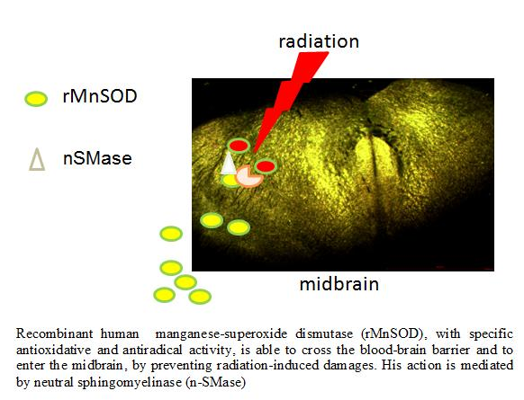
Neutral Sphingomyelinase Modulation in the Protective/Preventive Role of rMnSOD from Radiation-Induced Damage in the Brain

1
Department of Pharmaceutical Sciences, University of Perugia, 06126 Perugia, Italy
2
Molecular Biology and Viral Oncology Unit, Istituto Nazionale Tumori IRCCS “Fondazione G. Pascale”, 80131 Napoli, Italy
3
Laboratory of Radiation Biology, Joint Institute for Nuclear Research, 141980 Dubna, Russia
4
Division of Pathological Anatomy and Histology, Department of Experimental Medicine, School of Medicine and Surgery, University of Perugia, 06126 Perugia, Italy
5
Laedhexa Biotechnologies Inc., San Francisco, CA QB3@953, USA
6
Dipartimento di Area Medica, University of Udine, 33100 Udine, Italy
*
Author to whom correspondence should be addressed.
Int. J. Mol. Sci. 2019, 20(21), 5431; https://0-doi-org.brum.beds.ac.uk/10.3390/ijms20215431
Submission received: 9 October 2019 / Revised: 29 October 2019 / Accepted: 29 October 2019 / Published: 31 October 2019
(This article belongs to the Special Issue Emerging Role of Lipids in Metabolism and Disease)

Abstract

:
Studies on the relationship between reactive oxygen species (ROS)/manganese superoxide dismutase (MnSOD) and sphingomyelinase (SMase) are controversial. It has been demonstrated that SMase increases the intracellular ROS level and induces gene expression for MnSOD protein. On the other hand, some authors showed that ROS modulate the activation of SMase. The human recombinant manganese superoxide dismutase (rMnSOD) exerting a radioprotective effect on normal cells, qualifies as a possible pharmaceutical tool to prevent and/or cure damages derived from accidental exposure to ionizing radiation. This study aimed to identify neutral SMase (nSMase) as novel molecule connecting rMnSOD to its radiation protective effects. We used a new, and to this date, unique, experimental model to assess the effect of both radiation and rMnSOD in the brain of mice, within a collaborative project among Italian research groups and the Joint Institute for Nuclear Research, Dubna (Russia). Mice were exposed to a set of minor γ radiation and neutrons and a spectrum of neutrons, simulating the radiation levels to which cosmonauts will be exposed during deep-space, long-term missions. Groups of mice were treated or not-treated (controls) with daily subcutaneous injections of rMnSOD during a period of 10 days. An additional group of mice was also pretreated with rMnSOD for three days before irradiation, as a model for preventive measures. We demonstrate that rMnSOD significantly protects the midbrain cells from radiation-induced damage, inducing a strong upregulation of nSMase gene and protein expression. Pretreatment with rMnSOD before irradiation protects the brain with a value of very high nSMase activity, indicating that high levels of activity might be sufficient to exert the rMnSOD preventive role. In conclusion, the protective effect of rMnSOD from radiation-induced brain damage may require nSMase enzyme.

Graphical Abstract

1. Introduction

Evidence for the involvement of sphingomyelin (SM) in radiation-induced apoptosis relies on studies focused on specific enzymes as sphingomyelinase (SMase) [1,2,3]. SMase cleaves SM, generating ceramide and choline phosphate. The ceramide pathway, in turn, is responsible for the generation of various lipid mediators for cell signaling. Studies on the relationship between reactive oxygen species (ROS) and SMase are controversial. It has been demonstrated that SMase increases the intracellular ROS level in several experimental models [4,5]. Accordingly, ceramides increase ROS level [6,7]. Neutral SMase (nSMase)-protein kinase Cζ(PKCζ)-NADPH oxidase is essential for ROS production [8]. On the other hand, ceramides derived from SMase activity are involved in ROS production [9] and the activation of ceramide-p47phox-ROS signaling cascade is essential for apoptosis [10]. Moreover, both SMase and ceramide induce MnSOD gene expression [11]. Although numerous studies indicate a stimulatory effect of SMase and ceramide in ROS production, some authors suggest the positive role of ROS in the ceramide generation [12]. In line with this, previous published results showed that p53-induced ROS modulate the activation of nSMase [13]. In addition, by inhibiting ROS production nSMase stimulation and ceramide generation are suppressed [14].
The human recombinant manganese superoxide dismutase (rMnSOD) has specific antioxidant and anti-free radical activity as the native superoxide dismutase (SOD) [15]. MnSOD enzyme has been proposed to be useful in the prevention and treatment of damage caused by physical agents, such as ionizing radiations [16]. More rMnSOD has been identified as a possible pharmaceutical tool to prevent and/or cure the accidental damage derived from exposure to ionizing radiation [15]. Thus, rMnSOD has the invaluable advantage, over the native enzyme, of being able to easily enter into the cells and tissues thanks to the persistence in the recombinant mature protein of its leader peptide. Consequently, treatment side effects should be significantly reduced in comparison with traditional treatments [17,18].
To address a comprehensive analysis of nSMase involvement in the mechanism of rMnSOD protection or prevention from radiation damage in the brain, we performed experiments within a collaborative project among Italian research groups and the Joint Institute for Nuclear Research, Dubna (Russia). Mice, exposed to a set of minor γ radiation and neutrons and a spectrum of neutrons, simulating the radiation levels to which cosmonauts are exposed during deep-space long-term missions, were injected with rMnSOD either at the time (protection) or before (prevention) irradiation. The study aimed to identify a possible target molecule of the rMnSOD administered in order to reduce radiation-induced damage. Thus, the nSMase, known to be involved in the production of ROS and in the response to MnSOS, has been studied in our experimental model.

2. Results

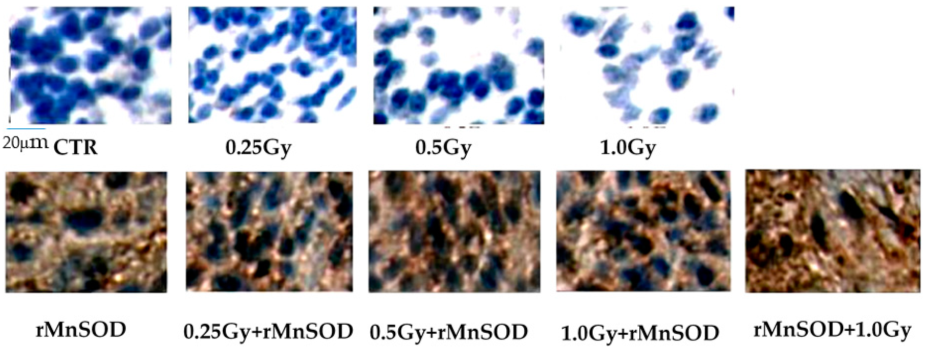
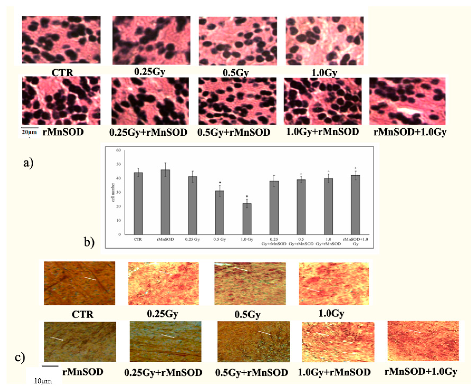
2.1. Protective and Preventive Effect of rMnSOD on Radiation-Induced Structural Changes in Midbrain Tissue

Before evaluating the effect of rMnSOD in mice brain, we confirmed its capacity to freely diffuse through the blood-brain barrier, locating itself within brain tissues. This was accomplished by immunohistochemical analysis (Figure 1).
We also found that the brains of not-irradiated control mice, with or without rMnSOD treatment, had comparable cell numbers. Only medium and high radiation doses induced a loss of the cells with progressive increase of intercellular spaces (Figure 2a). As expected, in these conditions we observed a robust protective effect with rMnSOD (Figure 2b). We thus investigated the possibility that rMnSOD might play a protective role within the neurofilament structure. To date, 200–220 kDa heavy neurofilament (NF200) is considered the specific marker of large myelinated A-βfiber neurons [19]. The analysis of NF200by immunohistochemistry showed that the irradiation caused an accumulation of the labeling in rounded areas with loss of the characteristic length and thickness of neurofilaments (Figure 2c). Such effects were not evident in mice treated or pretreated with rMnSOD; in those samples, normal heavy neurofilaments were present (Figure 2c). Morphological evaluation is however only qualitative, due to the loss of the normal neuritic structure.

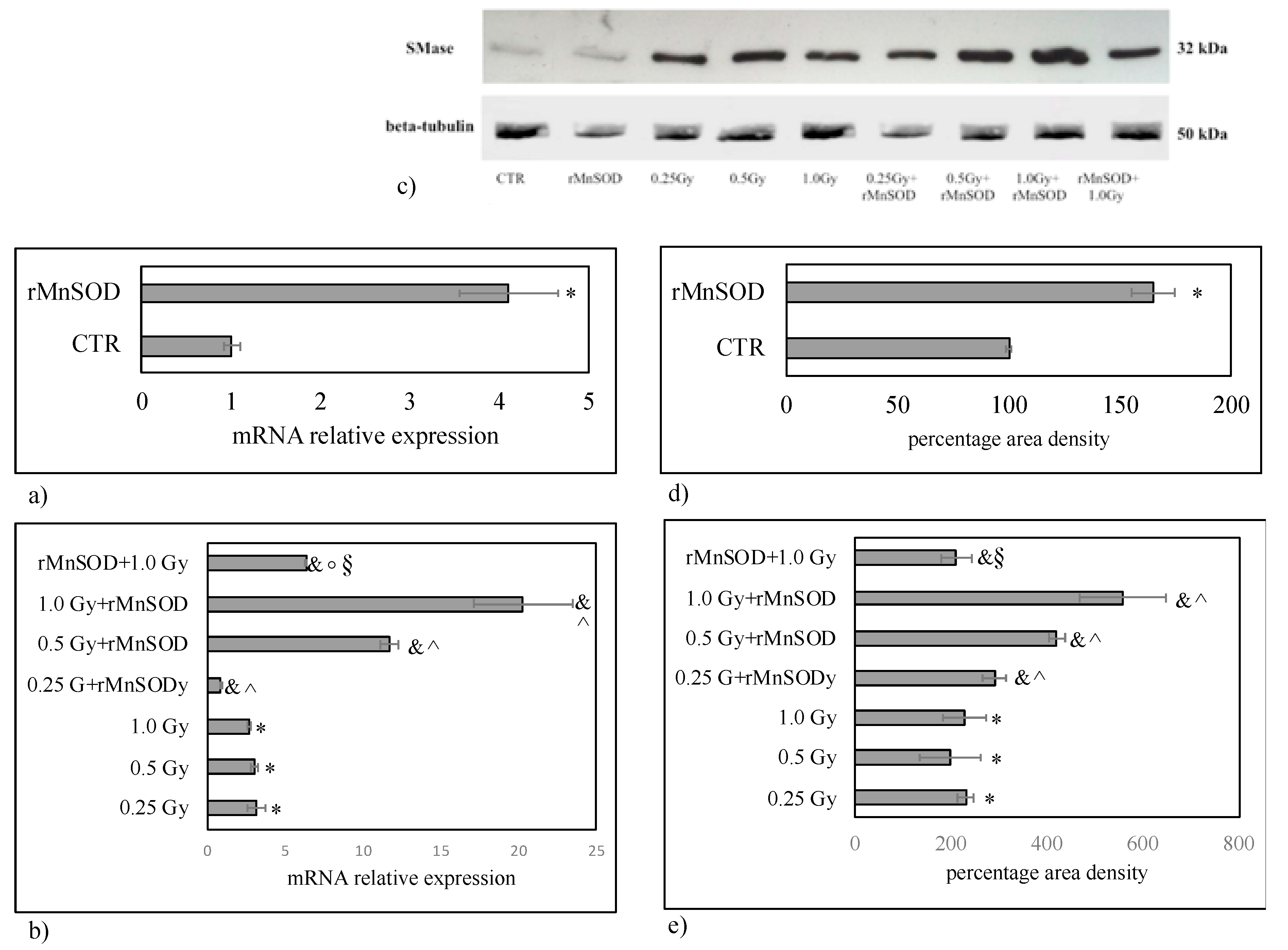
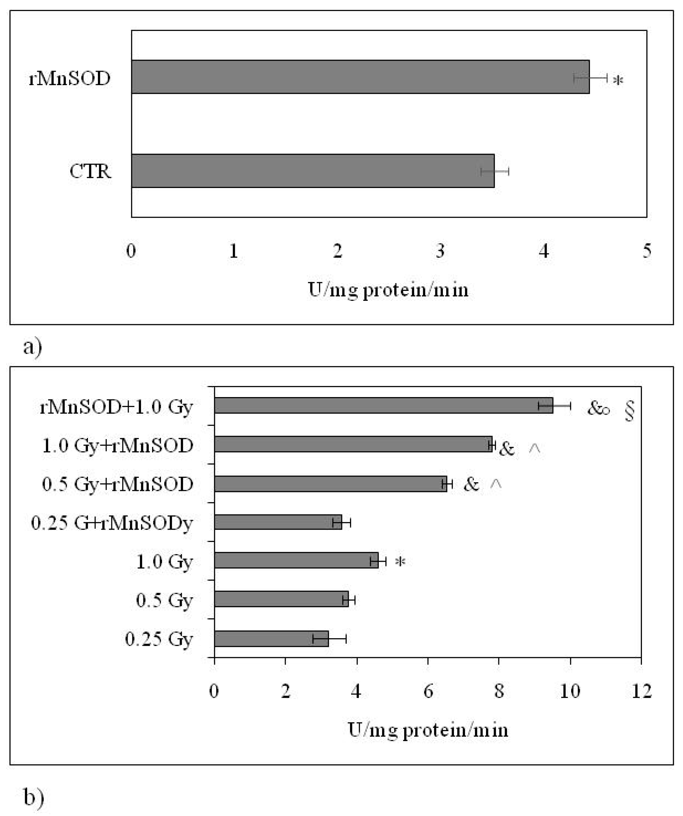
2.2. nSMase Is Required for the Protective and Preventive Effect of rMnSOD

We have previously reported that space radiation stimulated cellular and nuclear SMase in mice thyroids after their long stay in the International Space Station [20]. Our results indicated that radiation increased nSMase gene expression (Figure 3b) in comparison with control samples (Figure 3a). rMnSOD alone had a similar effect (Figure 3a). In comparison with rMnSOD alone considered as control, the nSMase gene expression strongly increased with medium- and high radiation exposure (Figure 3b). The effect was attenuated by rMnSOD pre-treatment (Figure 3b). Treatment and pretreatment with rMnSOD were responsible for the nSMase gene expression increase in irradiated samples (Figure 3b compared withFigure 3a). To analyze the extent of the nSMase response to radiation and rMnSOD treatment, we studied the protein expression. We found that the content of nSMase was low in the control sample and increased with irradiation (Figure 3c). Thus, the presence of rMnSOD resulted in overexpression of nSMase protein, responding to radiation in a dose-dependent mode. The densitometry analysis, performed with Scion Image program by using the corresponding beta-tubulin as control, showed that the enzyme increases about 100–130% over controls with radiation (Figure 3e) and 65% with rMnSOD alone (Figure 3e). By using rMnSOD alone as control, the enzyme increase in response to radiation in a dose-dependent mode in the presence of rMnSOD, was confirmed (Figure 3e).
To investigate the biological role of nSMase, we also assayed the enzyme activity. Only high radiation was able to further stimulate the nSMase activity (Figure 4b) with value similar to those obtained with rMnSOD alone (Figure 4a). The treatment with rMnSOD in irradiated mice strongly increased the nSMase activity in radiation dose-dependent mode (Figure 4b). Surprisingly, the pretreatment of mice with rMnSOD for 3 days before exposure to high radiation doses increased the enzyme activity even further (Figure 4b).

3. Discussion

The aim of this study was to elucidate the rMnSOD-dependent nSMase changes and their role in the protective-preventive effect from ionizing radiation in the brain. Mice were exposed to a set of minor γ radiation and neutrons and a spectrum of neutrons, simulating the radiation levels to which cosmonauts will be exposed during deep-space, long-term missions. At the moment we do not know whether γ radiation only was unable to induce nSMase activation. We show that rMnSOD particles are capable to cross the blood-brain barrier and localize in the midbrain. Here, rMnSOD has the ability to limit radiation-induced damage, such as the loss of neuron number and the alteration of neurofilaments. The effect of radiation on brain damage has previously been reported by other authors [21,22]. Although many studies elucidated the radioprotective role of rMnSOD through antioxidant mechanisms in mitochondria [15,16,17,18], no data exist about its effect on nSMase. We show that rMnSOD stimulates nSMase gene and protein expression and enzymatic activity. The study was articulated with the aim of analyzing the response of nSMase in samples in which rMnSOD has a protective effect (non-pretreated samples) and in samples in which rMnSOD has a preventive effect (pretreated samples). In samples for protective effect study, rMnSOD increases strongly nSMase gene and protein expression in irradiated samples in a dose-dependent manner. Small variations of nSMase enzyme activities are obtained only with high radiation doses. This observation is relevant, as nSMase plays a crucial role in the regulation of ROS generation and in the maintenance of homeostasis between proapoptotic and prosurvival signals [1,4]. In line with this, we show that highest radiation levels induce significant nSMase responses only in the presence of rMnSOD. Although the present results indicate that nSMase may be required for the action of rMnSOD, we cannot prove the direct action of rMnSOD on this enzyme. To our knowledge, this is the first study indicating SMase as a potential effector of rMnSOD during its protection action. In samples for preventive effect study, nSMase gene and protein expression is reduced in comparison with the samples in which the protective effect of rMnSOD is studied but the enzyme activity is much higher. At this moment we can only speculate why nSMase levels are reduced with pretreatment, a possible explanation being that an extended treatment time could stimulate different metabolic patterns, leading to inhibition of nSMase gene expression. Merging already published data with our findings, it is possible to suggest that in brain tissues exposed to radiation, significant amounts of ROS are generated, stimulating nSMase production to significantly increase MnSOD levels. This may in turn stimulate nSMase. At this point, ROS levels may be reduced, with the end result of limiting the damage. In the above described series of events, nSMase may play predominant roles.

4. Materials and Methods

4.1. Chemicals

The rMnSOD radioprotective protein, discovered and obtained in the recombinant form by A.Mancini, was provided by the Molecular Biology and Viral Oncology Unit, Department of Experimental Oncology, Istituto Nazionale Tumori Fondazione G. Pascale—IRCCS, Naples, Italy [15]. Sterile 2 μg aliquots of rMnSOD in 0.5 mL of sterile saline phosphate buffer (PBS) were prepared and stored at −80 °C. Anti-nSMase, and anti-βtubulin were from Abcam (Cambridge, UK). Horseradish peroxidase-conjugated goat anti-rabbit secondary antibodies were from Santa Cruz. Anti-rMnSOD was from InBios International (Washington, WA, USA). TaqMan SNP Genotyping Assay and Reverse Transcription kit were purchased from Applied Biosystems (Foster City, CA, USA). RNAqueous®-4PCR kit was from Ambion Inc. (Austin, TX, USA). SDS-PAGE molecular weight standards were purchased from Bio-Rad Laboratories (Hercules, CA, USA). Chemiluminescence kits were purchased fromAmersham (Rainham, Essex, UK).

4.2. Experimental Design and Animal Care

Animals: Fifty-fourfemale ICR mice weighting approximately 25–30 g were obtained from the Laboratory Animal Nursery of the Russian Academy of Sciences (Pushchino, Russia). After transportation, the animals were adapted to the vivarium at the Joint Institute for Nuclear Research (JINR) in Dubna during a period of 10 days. The animals were divided into 9 cages, 6 mice each, receiving the standard briquetted fodder and water ad libitum. All procedures were performed according to the Russian Guidelines for the Care and Use of Experimental Animals and Bioethics Instructions (Order of the USSR Ministry of Health No. 755 12.08.1987) accepted in the vivarium of the Institute of Biomedical Problems, part of the above-mentioned JINR.
Animal treatments: Mice in groups number 1,3,4 and 5 were treated with daily subcutaneous injections of sterile PBS solution for 7 days from the day of irradiation. Animals in groups 2,6,7,8 and 9 were treated with daily subcutaneous injections of rMnSOD in sterile PBS. In particular, mice in groups 2,6,7 and 8 received 7 injections of rMnSOD while animals in group 9 received a total of 10 injections, being pretreated with rMnSOD for 3 additional days prior to irradiation. All animals were irradiated at the JINR, being exposed to a set of minor γ radiation and neutrons of a Phasatron with high Relative Biological Effectiveness (RBE) and a spectrum of neutrons, to simulate space flight exposure. Animals in groups 3&6, 4&7 and 5&8 were exposed to doses of 0.25, 0.50 and 1.00 Gy (respectively). Mice in groups 1 (mock-treated with PBS) and 2 (rMnSOD-treated) were not exposed to radiation and considered a biological control. At the end of the experiment, all mice were beheaded and brains immediately frozen.

4.3. Morphological and Immunohistochemistry Analysis

Three brains from each group were fixed in 4% neutral phosphate-buffered formaldehyde solution for 24 h and dropped with specific orientation in paraffin. Morphological and immunohistochemical analyses were performed as previously reported [19].

4.4. Reverse Transcription Quantitative PCR (RTqPCR)

Total RNA was extracted from mice brain using RNAqueous-4PCR kit (Ambion Inc., Austin, TX, USA), as previously reported [23]. Before cDNA synthesis, the integrity of RNA was evaluated by electrophoresis in TAE 1.2% agarose gel prepared in our lab. cDNA was synthesized using 1 μg total RNA for all samples by High-Capacity cDNA Reverse Transcription kit (Applied Biosystems, Foster City, CA, USA) under the following conditions: 50 °C for 2 min, 95 °C for 10 min, 95 °C for 15 s and 60 °C for 1 min, for a total of 40 cycles [23]. RTqPCR was performed using TaqMan®Gene Expression Master Mix and 7500 RT-PCR instrument (Applied Biosystems), SM phosphodiesterase 4 (SMPD4, Hs04187047_g1) genes. mRNA expression levels were then normalized to the glyceraldehyde-3-phosphate dehydrogenase (GAPDH, Hs99999905_m1) housekeeping gene (Thermo Fisher Scientific, Austin, MA, USA). mRNA relative expression levels were calculated as 2−ΔΔCt, comparing the results of the treated samples with those of the untreated ones [23].

4.5. Protein Concentration and Western Blotting

Protein concentrations were determined according to the Bradford method, as previously reported [23]. Forty-μgproteins were submitted to 12% SDS (sodium dodecyl sulfate) polyacrylamide gel electrophoresis at 200 V for 60 min [23]. Briefly, proteins were transferred onto 0.45 μm cellulose nitrate strips membrane (Sartorius Stedim Biotech S.A., Aubagne, France) in transfer buffer for 1 h at 100 V at 4 °C. Membranes were blocked with 5% (w/v) non-fat dry milk in PBS, pH 7.5 for 1 h at room temperature. The blot was incubated overnight at 4 °C with specific antibodies (1:1000) and then treated with horseradish peroxidase-conjugated goat anti-rabbit secondary antibodies (1:5000). Super Signal West Pico Chemiluminescent Substrate (Thermo Fisher Scientific) was used to detect chemiluminescent (ECL) HRP substrate. The apparent molecular weight of proteins was calculated referringto the migration rate of molecular size standards. The area density of the bands was evaluated by densitometry scanning and analyzing themwith Scion Image.

4.6. nSMaseActivity Assay

nSMase activity was assayed according to Ceccarini et al. [24]. Brain homogenates were suspended in 0.1% NP-40 detergent in PBS, sonicated for 30 s once at 20 watts, then kept on ice for 30 min and centrifuged at 16,000× g for 10 min. Supernatants were used for nSMase assay. Sixty μg/10μL proteins were incubated with 10 μL HMU-PC substrate for 10 min at 37 °C. The reaction was stopped by adding 200 μL stop buffer [21]. The fluorescence of 6-hexadecanoyl-4-methylumbelliferone (HMU) was measured with FLUOstar Optima fluorimeter (BMG Labtech, Ortenberg, Germany), using the filter set of 4-methylumbelliferone (MU), 360 nm excitation, and 460 nm emission. The fluorimeter was calibrated with MU in stop buffer.

4.7. Statistical Analysis

Data were expressed as means ± SD and their significance was checked by ANOVA test. Significance: (a) * p < 0.05 irradiated samples versus not-irradiated control sample (CTR); (b) & p < 0.05 irradiated and rMnSOD-treated samples versus rMnSOD-treated sample (rMnSOD); (c) ^ p < 0.05 irradiated and rMnSOD-treated samples versus respective irradiated samples; (d) ° p < 0.05 rMnSOD-pretreated and 1.00 Gy irradiated sample versus 1.00 Gy irradiated sample; (e) § p < 0.05 rMnSOD-pretreated and 1.00 Gy irradiated sample versus 1.0 Gy irradiated and rMnSOD-treated sample.

Author Contributions

The conception and design of the study (F.S.A.-I., E.A., A.B., A.M.), experiments (F.S.A.-I., A.B., O.B., A.B., S.C., M.R.C., I.N., I.F., F.F.P., A.I., E.K.) acquisition of data (M.C., C.C.), analysis of data (S.C., F.C., G.T., I.F.), interpretation of data (F.C.), drafting the article or revising it critically for important intellectual content (E.A., T.B., A.M.), founding acquisition (F.C.), final approval of the version to be submitted (E.A., F.S.A.-I.).

Funding

This study was supported by the grant from theItalian Space Agency “Project RASC, number of contract: 2015-008-R.0”.

Conflicts of Interest

The authors declare no conflict of interest.

References

  1. Haimovitz-Friedman, A.; Kan, C.C.; Ehleiter, D.; Persaud, R.S.; McLoughlin, M.; Fuks, Z.; Kolesnick, R.N. Ionizingradiationacts on cellular membranes to generate ceramide and initiate apoptosis. J. Exp. Med. 1994, 180, 525–535. [Google Scholar] [CrossRef] [PubMed]
  2. Chmura, S.J.; Mauceri, H.J.; Advani, S.; Heimann, R.; Beckett, M.A.; Nodzenski, E.; Quintans, J.; Kufe, D.W.; Weichselbaum, R.R. Decreasing the apoptotic threshold of tumor cells through protein kinase C inhibition and sphingomyelinaseactivation increases tumor killing by ionizingradiation. Cancer Res. 1997, 57, 4340–4347. [Google Scholar] [PubMed]
  3. Albi, E.; Cataldi, S.; Lazzarini, A.; Codini, M.; Beccari, T.; Ambesi-Impiombato, F.S.; Curcio, F. Radiation and thyroid cancer. Int. J. Mol. Sci. 2017, 18, E911. [Google Scholar] [CrossRef] [PubMed]
  4. Won, J.S.; Singh, I. Sphingolipid signaling and redox regulation. Free Radic Biol. Med. 2016, 40, 1875–1888. [Google Scholar]
  5. Kitatani, K.; Akiba, S.; Sato, T. Ceramide-induced enhancement of secretory phospholipase A2 expression via generation of reactive oxygen species in tumor necrosis factor-alpha-stimulated mesangial cells. Cell Signal. 2004, 16, 967–974. [Google Scholar] [CrossRef] [PubMed]
  6. Li, H.; Junk, P.; Huwiler, A.; Burkhardt, C.; Wallerath, T.; Pfeilschifter, J.; Förstermann, U. Dual effect of ceramide on human endothelial cells: Induction of oxidative stress and transcriptional upregulation of endothelial nitric oxide synthase. Circulation 2002, 106, 2250–2256. [Google Scholar] [CrossRef]
  7. Li, X.; Becker, K.A.; Zhang, Y. Ceramide in redox signaling and cardiovascular diseases. Cell. Physiol. Biochem. 2010, 26, 41–48. [Google Scholar] [CrossRef]
  8. Frazziano, G.; Moreno, L.; Moral-Sanz, J.; Menendez, C.; Escolano, L.; Gonzalez, C.; Villamor, E.; Alvarez-Sala, J.L.; Cogolludo, A.L.; Perez-Vizcaino, F. Neutral sphingomyelinase, NADPH oxidase and reactive oxygen species. Role in acute hypoxic pulmonary vasoconstriction. J. Cell. Physiol. 2011, 226, 2633–2640. [Google Scholar] [CrossRef]
  9. JazvinšćakJembrek, M.; Hof, P.R.; Šimić, G. Ceramides in Alzheimer’s disease: Key mediators of neuronal apoptosis induced by oxidative stress and Aβ accumulation. Oxid. Med. Cell. Longev. 2015, 346783. [Google Scholar]
  10. Chen, Y.Y.; Hsu, M.J.; Sheu, J.R.; Lee, L.W.; Hsieh, C.Y. Andrographolide, a Novel NF-κB Inhibitor, Induces Vascular Smooth Muscle Cell Apoptosis via a Ceramide-p47phox-ROSSignaling Cascade. Evid. Based. Complement. Alternat. Med. 2013, 2013. [Google Scholar] [CrossRef]
  11. Pahan, K.; Dobashi, K.; Ghosh, B.; Singh, I. Induction of the manganese superoxide dismutase gene by sphingomyelinase and ceramide. J. Neurochem. 1999, 73, 513–520. [Google Scholar] [CrossRef] [PubMed]
  12. He, X.; Schuchman, E.H. Ceramide and ischemia/reperfusion injury. J. Lipids. 2018, 2018. [Google Scholar] [CrossRef] [PubMed]
  13. Dumitru, C.A.; Zhang, Y.; Li, X.; Gulbins, E. Ceramide: A novel player in reactive oxygen species-induced signaling? Antioxid. Redox. Signal. 2007, 9, 1535–1540. [Google Scholar] [CrossRef] [PubMed]
  14. Bezombes, C.; de Thonel, A.; Apostolou, A.; Louat, T.; Jaffrézou, J.P.; Laurent, G.; Quillet-Mary, A. Overexpression of protein kinase Czeta confers protection against antileukemic drugs by inhibiting the redox-dependentsphingomyelinaseactivation. Mol. Pharmacol. 2002, 62, 1446–1455. [Google Scholar] [CrossRef]
  15. Borrelli, A.; Schiattarella, A.; Mancini, R.; Morrica, B.; Cerciello, V.; Mormile, M.; D’Alesio, V.; Bottalico, L.; Morelli, F.; D’Armiento, M.; et al. A recombinant MnSOD is radioprotective for normal cells and radiosensitizing for tumor cells. Free Radic. Biol. Med. 2009, 46, 110–116. [Google Scholar] [CrossRef]
  16. Epperly, M.W.; Gretton, J.E.; Sikora, C.A.; Jefferson, M.; Bernarding, M.; Nie, S.; Greenberger, J.S. Mitochondrial localization of superoxide dismutase is required for decreasing radiation cellular damage. Radiat. Res. 2003, 160, 568–578. [Google Scholar] [CrossRef]
  17. Borrelli, A.; Schiattarella, A.; Bonelli, P.; Tuccillo, F.M.; Buonaguro, F.M.; Mancini, A. The functional role of MnSOD as a biomarker of human diseases and therapeutic potential of a new isoform of a human recombinant MnSOD. Biomed. Res. Int. 2014, 2014. [Google Scholar] [CrossRef]
  18. Pica, A.; Di Santi, A.; D’Angelo, V.; Iannotta, A.; Ramaglia, M.; Di Martino, M.; Pollio, M.L.; Schiattarella, A.; Borrelli, A.; Mancini, A.; et al. Effect ofrMnSODon survival signaling in pediatric high risk T-cell acute lymphoblastic leukaemia. J. Cell Physiol. 2015, 230, 1086–1093. [Google Scholar] [CrossRef]
  19. Cataldi, S.; Codini, M.; Hunot, S.; Légeron, F.P.; Ferri, I.; Siccu, P.; Sidoni, A.; Ambesi-Impiombato, F.S.; Beccari, T.; Curcio, F.; et al. e-Cadherin in 1-Methyl-4-phenyl-1,2,3,6-tetrahydropyridine-Induced Parkinson Disease. Mediators Inflamm. 2016, 2016. [Google Scholar] [CrossRef]
  20. Albi, E.; Ambesi-Impiombato, S.; Villani, M.; DePol, I.; Spelat, R.; Lazzarini, R.; Perrella, G. Thyroid cell growth: Sphingomyelin metabolism as non-invasive marker for cell damage acquired during spaceflight. Astrobiology 2018, 10, 811–820. [Google Scholar] [CrossRef]
  21. Le, O.; Palacio, L.; Bernier, G.; Batinic-Haberle, I.; Hickson, G.; Beauséjour, C. INK4a/ARF Expression Impairs Neurogenesis in theBrainof Irradiated Mice. Stem. Cell Rep. 2018, 10, 1721–1733. [Google Scholar] [CrossRef] [PubMed]
  22. Constanzo, J.; Dumont, M.; Lebel, R.; Tremblay, L.; Whittingstall, K.; Masson-Côté, L.; Geha, S.; Sarret, P.; Lepage, M.; Paquette, B.; et al. Diffusion MRI monitoring of specific structures in the irradiated ratbrain. Magn. Reson. Med. 2018, 80, 1614–1625. [Google Scholar] [CrossRef] [PubMed]
  23. Albi, E.; Cataldi, S.; Ferri, I.; Sidoni, A.; Traina, G.; Fettucciari, K.; Ambesi-Impiombato, F.S.; Lazzarini, A.; Curcio, F.; Ceccarini, M.R.; et al. VDR independent induction of acid-sphingomyelinase by 1,23(OH)2 D3 in gastric cancer cells: Impact on apoptosis and cell morphology. Biochimie 2018, 146, 35–42. [Google Scholar] [CrossRef] [PubMed]
  24. Ceccarini, M.R.; Codini, M.; Cataldi, S.; Vannini, S.; Lazzarini, A.; Floridi, A.; Moretti, M.; Villarini, M.; Fioretti, B.; Beccari, T.; et al. Acid sphingomyelinaseas target of LyciumChinense: Promising new action for cell health. Lipids. Health. Dis. 2016, 15, 183. [Google Scholar] [CrossRef] [PubMed]
Figure 1. Localization of rMnSOD in brain tissue. Immunohistochemical analysis was performed by using specific antibody. The immunostaining was evident only in brain samples from rMnSOD-treated mice.
Ijms 20 05431 g001
Figure 2. Midbrain nuclei and heavy neurofilament 200 kDa (NF200). (a) Hematoxylin-eosin-stained midbrain sections from normal mice exposed to 0.25, 0.5 and 1.0Gy radiation doses, treated in the presence or absence of rMnSOD. rMnSOD + 1.0Gy: mice pretreated with rMnSOD and exposed to 1.0 Gy, then treated with rMnSOD for 3 days. Shown images are representative of similar findings in the nuclear regions of 3 midbrains from each group of mice (40× magnification); (b) cell numbers were counted as described in Results. * p< 0.05 irradiated samples vs. not-irradiated control samples (CRT), ^ p < 0.05 irradiated and rMnSOD-treated samples vs. corresponding irradiated samples, ° p < 0.05 rMnSOD-pretreated and 1.0 Gy irradiated sample vs. 1.0 Gy irradiated sample; (c) NF200 immunohistochemical staining. Normal mice were exposed to 0.25, 0.5 and 1.0 Gy radiation doses, with or without rMnSOD administration. rMnSOD + 1.0Gy: mice pretreated with rMnSOD for 3 days before 1.0 Gy radiation. Images are representative of similar images showing heavy neurofilaments in 3 midbrains from each group of mice (20× magnification). Arrows indicate normal neurofilaments (CTR), accumulation of labeling in round areas with reduction of length and thickness in neurofilaments (irradiated samples), and the presence of normal neurofilaments in rMnSOD-treated samples, thus demonstrating its radioprotective effect.
Ijms 20 05431 g002
Figure 3. Effect of rMnSOD on nSMase gene and protein expression. Samples from mice treated with 0.25, 0.5 and 1.0 Gy radiation doses, in the presence or absence of rMnSOD. rMnSOD + 1.0 Gy: mice pretreated with rMnSOD, exposed to a 1.0 Gy radiation dose, then treated with rMnSOD for 3 days. Left panels (a,b): RTqPCR analyses. GAPDH was used as housekeeping control gene. Data are expressed as mean ± SD of mRNA expression (folds increase). Top panel (c): immunoblotting analysis of nSMase; tubulin was used as control. Right panels (d,e): immunoblotting densitometric analysis, normalized with beta-tubulin. Data are expressed as the mean ± SD of 3 independent experiments, each carried out in triplicate. Significance: (a,d) not-irradiated rMnSOD-treated samples compared to not-irradiated and not-rMnSOD-treated samples (CRT) (* p < 0.05); (b,e): irradiated not-rMnSOD-treated samples and irradiated rMnSOD-treated and pretreated samples. Irradiated samples compared to CTR samples (* p < 0.05), irradiated and rMnSOD-treated and pretreated samples vs. rMnSOD-treated samples (& p < 0.05), irradiated and rMnSOD-treated samples vs. their correspondent irradiated samples (^ p < 0.05), rMnSOD-pretreated and 1.0 Gy irradiated sample vs.1.0 Gy irradiated sample (° p < 0.05), rMnSOD-pretreated and 1.0 Gy irradiated sample vs.1.0 Gy irradiated and rMnSOD-treated sample (§ p < 0.05).
Ijms 20 05431 g003
Figure 4. Activity of nSMase. Significance: (a) not-irradiated rMnSOD-treated samples compared to not-irradiated and not-rMnSOD-treated samples (CRT) (* p < 0.05); (b) irradiated not-rMnSOD-treated samples and irradiated rMnSOD-treated and pretreated samples. Significance: irradiated samples compared to CTR samples (* p < 0.05), irradiated and rMnSOD-treated and pretreated samples vs. rMnSOD-treated samples (& p < 0.05), irradiated and rMnSOD-treated samples vs. their correspondent irradiated samples (^ p < 0.05), rMnSOD pretreated and 1.0 Gy irradiated sample vs. 1.0 Gy irradiated sample (° p < 0.05), rMnSOD pretreated and 1.0 Gy irradiated sample vs. 1.0 Gy irradiated and rMnSOD-treated sample (§ p < 0.05).
Ijms 20 05431 g004

