Review

Mycobacterium abscessus, an Emerging and Worrisome Pathogen among Cystic Fibrosis Patients

by
Giulia Degiacomi
1,†,
José Camilla Sammartino
1,2,†,
Laurent Roberto Chiarelli
1,
Olga Riabova
3,
Vadim Makarov
3 and
Maria Rosalia Pasca
1,*
1
Department of Biology and Biotechnology “Lazzaro Spallanzani”, University of Pavia, 27100 Pavia, Italy
2
IUSS—University School for Advanced Studies, 27100 Pavia, Italy
3
Bach Institute of Biochemistry, Research Center of Biotechnology of the Russian Academy of Sciences, 119071 Moscow, Russia
*
Author to whom correspondence should be addressed.
These authors contributed equally to this work.
Int. J. Mol. Sci. 2019, 20(23), 5868; https://0-doi-org.brum.beds.ac.uk/10.3390/ijms20235868
Submission received: 7 October 2019 / Revised: 20 November 2019 / Accepted: 20 November 2019 / Published: 22 November 2019
(This article belongs to the Special Issue Therapeutic Approaches for Cystic Fibrosis)

Abstract

:
Nontuberculous mycobacteria (NTM) have recently emerged as important pathogens among cystic fibrosis (CF) patients worldwide. Mycobacterium abscessus is becoming the most worrisome NTM in this cohort of patients and recent findings clarified why this pathogen is so prone to this disease. M. abscessus drug therapy takes up to 2 years and its failure causes an accelerated lung function decline. The M. abscessus colonization of lung alveoli begins with smooth strains producing glycopeptidolipids and biofilm, whilst in the invasive infection, “rough” mutants are responsible for the production of trehalose dimycolate, and consequently, cording formation. Human-to-human M. abscessus transmission was demonstrated among geographically separated CF patients by whole-genome sequencing of clinical isolates worldwide. Using a M. abscessus infected CF zebrafish model, it was demonstrated that CFTR (cystic fibrosis transmembrane conductance regulator) dysfunction seems to have a specific role in the immune control of M. abscessus infections only. This pathogen is also intrinsically resistant to many drugs, thanks to its physiology and to the acquisition of new mechanisms of drug resistance. Few new compounds or drug formulations active against M. abscessus are present in preclinical and clinical development, but recently alternative strategies have been investigated, such as phage therapy and the use of β-lactamase inhibitors.

1. Introduction

Cystic fibrosis (CF) is an autosomal recessive disease characterized by the involvement of respiratory, gastrointestinal, and male reproductive tracts, even if most of the morbidity and mortality arises from CF lung disease [1,2]. In particular, thick airway secretions impair the mucociliary clearance, which increases bacterial colonization and infection [3].
In this context, nontuberculous mycobacteria (NTM) have recently emerged as important pathogens in CF lung disease worldwide [2,3,4]. Over the last two decades, the incidence of NTM infections among CF patients has raised from 3.3% to 22.6%, increasing morbidity and mortality associated with these pathogens [4,5,6]. At the same time, for other CF pathogens, such as Pseudomonas aeruginosa and Burkholderia cepacia, the incidence has significantly decreased [7,8]. Additionally, the NTM incidence is underestimated because of misdiagnosis of NTM infections such as tuberculosis (TB) is common in developing countries; moreover, data from several countries are missing [7,8].
The most commonly identified NTM species in CF individuals are the slow growing Mycobacterium avium complex (MAC) and the rapidly growing Mycobacterium abscessus complex (MABSC) (95% of CF cases) [2,3,4]. MABSC is more common in European CF populations and its incidence is globally increasing; moreover, it is frequently found in younger CF patients (including children) and in those with more severe lung disease [4,9,10]. MABSC includes the following M. abscessus subspecies: Mycobacterium abscessus subsp. abscessus (Mab), Mycobacterium abscessus subsp. bolletii (M. bolletii), and Mycobacterium abscessus subsp. massiliense (M. massiliense). The MAC predominantly consists of Mycobacterium avium subsp. avium (Mav) and Mycobacterium avium subsp. intracellulare [2,3,4,5,6].
Among NTM subspecies, Mab is becoming the most prominent and worrisome pathogen in hospitals and CF centers around the world [4,7,10,11]. It is the major NTM causing respiratory infections worldwide (up to 80%), most often in immunocompromised patients, such as those with CF and HIV-positive status, and in patients with chronic obstructive pulmonary disease (COPD) and bronchiectasis [4,7,10,11,12,13,14,15,16].
Mab drug therapy takes up to 2 years (see below), with only about 30% of patients experiencing successful treatment outcomes [11,17]. Mab treatment is also challenging, since failed eradication leads to an accelerated lung function decline. Mab-infected CF patients are even excluded from lung transplant lists in some countries. Consequently, in this cohort of patients, preventing Mab infection is essential. Room cleaning protocols have recently been changed and Mab-infected CF patients are now hospitalized in specialized CF centers, where there are 15 independent air changes per hour in the rooms to remove potentially infectious aerosol, thus preventing transmission [12].
Mab’s success in becoming an emerging CF pathogen is due to several reasons, including:
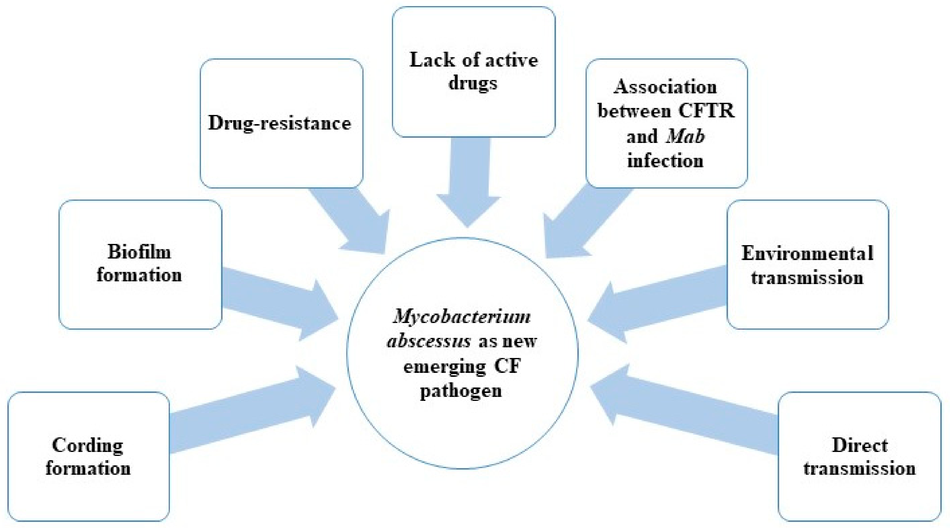
  • Possible direct person-to-person transmission;
  • Biofilm and drug resistance;
  • Association between CFTR (cystic fibrosis transmembrane conductance regulator) mutations and formation of granuloma in the presence of Mab infection;
  • Lack of active drugs (in particular with bactericidal activity) (Figure 1).
In this review, we will focus on analyzing all of these aspects in order to find a possible “Achilles’ heel” to fight this emerging pathogen.

2. Possible M. abscessus Direct Transmission among CF Patients

Mab, similar to other NTM subspecies, is ubiquitous in the environment, such as in soil and drinking water, and remains viable even after water treatment. Therefore, this pathogen can survive in environments near to human populations, particularly in human water sources, including hospital and domestic water supplies [16]. Mab and other NTM subspecies are also commonly found in urban water plumbing and water systems, sometimes in symbiosis with Amoebae [18,19,20,21,22,23,24,25,26,27]. Moreover, Mab has been isolated from fish [28,29,30,31,32,33] and animals [34,35,36,37,38,39,40], who could also represent reservoirs for human infections. This makes exposure common and disinfection difficult, which is very problematic in healthcare settings [7,15,16,19,23].
However, in sporadic and epidemic Mab infections, the pathogen is almost never isolated from the closest environment [7].
Until recently, it was believed that among CF patients, the majority of Mab infections were acquired by individuals through exposure to soil, household dust, or water, potentially via fomites and aerosols [41]. The mode of Mab transmission is still under investigation, and only recently was human-to-human Mab transmission demonstrated using whole-genome sequencing (WGS) [11,14,42].
In fact, Bryant et al. (2016) [14], using WGS of prominent worldwide Mab clinical isolates, showed that the majority of infections were acquired through direct transmission, potentially via fomites and aerosols. In particular, they generated WGS of 1080 Mab clinical isolates from 517 patients, obtained from CF centers from Europe, the United States, and Australia. They also identified that 74% of isolates were clustered in three dominant circulating clones: Mab clusters 1 and 2 and M. massiliense cluster 1. These 3 clusters were present in all CF centers, indicating transcontinental spreading of these strains by a possible human-to-human transmission within the global CF patient community. The clustered strains presented less than 20 single-nucleotide polymorphisms (SNPs), indicating a high level of human-to-human transmission among geographically separated CF patients [14]. Interestingly, these clustered Mab isolates were associated with bad clinical outcomes and presented increased virulence in vivo, thus representing an urgent international challenge [14].
According to the previous study, Yan et al. (2019) performed WGS of Mab isolates from 22 CF patients [43]. WGS identified a cluster of three CF patients infected by Mab isolates that differed by < 7 SNPs, suggesting a possible direct transmission among them. Several hospital attendances were found in common for these 3 patients, even if they were hospitalized in separate single rooms and there were no known social links between them [43]. The genomes of these Mab isolates are very similar to those previously described, confirming the presence of global circulating Mab clones in CF centers [14].
An additional study evaluated the transmission of Mab isolates in 4 Italian CF centers using the WGS of clinical isolates [44]. They found 7 possible person-to-person transmissions (SNP difference cut-off of < 30); only three CF patients were hospitalized in the same CF center at the same time [44]. Moreover, one of the Mab clusters identified in this study is the same as cluster 1 detected by Bryant and collaborators [14,44], again highlighting the presence of circulating virulent Mab strains worldwide in CF centers.
These last studies [14,43,44] show how it is possible to monitor human-to-human Mab transmission by WGS approach only, and to ascertain if different patients, even if geographically separated, share the same strain. Mab and M. massiliense belong to the same complex (MABSC) [2]; at this point, it is important to underline that the possibility of direct transmission for M. massiliense subspecies was also shown by genomic approach [14]. How human-to-human direct Mab transmission occurs is still under investigation.
Moreover, the human-to-human transmission helped Mab evolution from an environmental bacterium to a transmissible human pathogen. This evolution also affected the strategies used in CF centers to contain Mab infections. Before this last discovery, effective sterilizing techniques and other hygiene practices were performed to reduce the risk of environmental Mab transmission. Now, the Mab infection control recommendations include general infection control measures (e.g., hand washing) and advise the segregation of infected CF patients from the other ones in order to avoid direct transmission [12].

3. Pathogenesis of M. abscessus

The most common Mab infection sites are the respiratory tract, the skin, and soft tissue [4].
It is well known that predisposing factors for Mab pulmonary infections are chronic bronchiectasis and CF disease, as in these conditions the pathogen can firstly develop a biofilm, colonizing the host, and later progress into an invasive disease [45,46,47]. The progress of Mab infection in the CF lung is still under investigation [47], but a recent study showed that Mab aggregates form a biofilm around lung alveoli [48]. It is noteworthy that mycobacteria growing in biofilms are more tolerant to antibiotics, contributing to their drug resistance [49].
Moreover, the Mab cell wall contributes largely to its drug resistance and to its pathogenicity, thanks to the large presence of complex lipids, among which are five major glycopeptidolipids (GPLs) that differ in location or number of acetyl and sugar moieties [49,50,51,52,53].
Because of its peculiar cell wall, Mab shows smooth (S) or rough (R) colony morphologies, associated with distinct in vitro and in vivo features. In particular, the wild-type S strains produce abundant GPLs and minimal trehalose dimycolate (TDM), whilst R mutants have genetic lesions in the GPL loci, producing little or no GPLs and higher levels of TDM [49,50,53,54]. TDM, contributing also to the Mycobacterium tuberculosis virulence, was found on the surface of Mab R strains [53,54]. In fact, TDM is responsible for the cording phenotype, a key factor in increasing Mab virulence, causing invasive infection [53]. The lack of cording in the S variant may be due to the presence of GLPs masking other cell surface molecules (e.g., TDM) that activate innate immunity, causing inhibition of the macrophage apoptotic response, reduced production of radical oxygen species (ROS), and limitation of the spread of Mab among macrophages [49,50,51,52,53,54,55]. Furthermore, the high presence of GLPs in S strains is responsible for the formation of a robust biofilm during the infection [48,49].
The gpl locus is highly conserved in Mycobacterium smegmatis, Mab, and Mav [50]. This locus contains mmpS4, mmpL4a, and mmpL4b genes, which encode membrane proteins essential for the GPL biosynthesis and transport across the plasma membrane [50,56,57]. The transition from high-GPL (S strains) to low-GPL producers (R strains) is linked to mutations in genes involved in GPL biosynthesis and transport, similar to in the crucial Tyr/Asp couples in MmpL4a/MmpL4b. In fact, it was shown that point mutations in MmpL4a at Tyr842 or MmpL4b at Tyr854 caused loss of GPL production, suggesting that no functional redundancy exists between MmpL4a and MmpL4b [56]; moreover, the disruption of mmpL4b in Mab S strain inhibits GPL production, causing an R morphotype [58]. Then, the Mab S-to-R transition leads to the following events:
-
Arrest of lipid transport;
-
Production of serpentine cords;
-
Growth as extracellular cords, allowing escape from the innate immune defenses;
-
Induction of a strong humoral response that contributes to acute and severe infections [51,53,54,55,58].
The high virulence of Mab R mutants was confirmed in in vivo infection models, such as with zebrafish, where the transition to the R morphotype is characterized by an increased virulence because of the cording formation, which is responsible for invasive infection and larval death [55,56,59,60]. Moreover, it was found in a zebrafish model that the inactivation of MAB_4780, encoding a dehydratase required for cording formation, strongly affects Mab R pathogenicity; this attenuation causes both cord deficiency and intracellular growth impairment [60]. Furthermore, the Mab R strains induce more aggressive and invasive pulmonary disease, particularly in CF patients; in fact, these mutants are more frequently isolated after a long persistent infection and are associated with increased lung function decline [48,53,61,62,63,64].
Consequently, the possibility of transition between S and R morphotypes is very relevant in Mab pulmonary infection in CF patients [59,61,62]. These patients are particularly susceptible to colonization by biofilm-forming bacteria because of their altered lung physiology. In these conditions, Mab S strains expressing GPL may be favored [65]. A rough cord-forming variant could emerge from CF patients chronically colonized with an S strain, giving a more aggressive, invasive pulmonary infection [60,61,62,63].
The presence of the two morphotypes in Mab is also fundamental to its escape from the innate immune system. In particular, the innate immune response to pulmonary pathogens is mediated by the expression of TLR2 and then of IL-8 and human b-defensin 2 (HbD2) from the respiratory epithelial cells. It was shown that only Mab R strain stimulates the expression of IL-8 and HbD2, while Mab S variant is able to “mask’’ the bioactive cell wall lipids with GPLs. Because S variants are predominant during the first phases of infection, Mab is then able to avoid the innate immune system [51].
Another Mab peculiarity is that although it is a rapid grower, it persists in lungs associated with granulomatous lesions, a landmark of M. tuberculosis infection. Recently, Dubois et al. (2018) showed that an mmpL8 deletion mutant presented a decreased intracellular viability in a zebrafish model and a diminished propensity to induce granuloma formation [66]; moreover, this mutant had impaired adhesion to macrophages. In fact, MmpL8 is also required for the production of a glycolipid (glycosyl diacylated nonadecyl diol alcohol (GDND)), which is derived from a combination of oleic and stearic acids. In mmpL8 knock-out (KO) mutant, the reduced GDND production could be the cause of the modified interaction between bacteria and macrophages, resulting in a decreased virulence [66].
Finally, Laencina and collaborators (2018) [67] demonstrated that genes belonging to Mab ESX-4 locus are essential for its intracellular survival inside amoebae and macrophages. Interestingly, a Mab mutant with a deletion in eccB4 gene, coding for a structural key ESX component, was attenuated. In fact, it was less efficient at blocking phagosomal acidification and failed to damage phagosomes. The authors speculated that because Mab lacks ESX-1, ESX-4 could be a surrogate of M. tuberculosis ESX-1 [67].

CFTR Mutations Specialize M. abscessus as CF Pathogen

Recently, steps were taken to decipher why CF patients are so predisposed to Mab infections. Using zebrafish model, Bernut and collaborators (2016) showed that both macrophages and neutrophils are required to control Mab infection; moreover, impaired TNF signaling produced aberrant granulomas, and subsequent larval death [68].
In 2019, the same research group demonstrated that CFTR participates in neutrophil chemotaxis to the infected Mab sites, stimulating the oxidative host defenses [69]. In fact, in a zebrafish model, Mab infection was characterized by the recruitment of the bacilli by macrophages; in particular, the activation of macrophages resulted in neutrophil chemotaxis leading to granuloma formation, and ROS production by NOX2, which led to intracellular Mab death. In these conditions, the granuloma sequestered Mab, containing the infection [69]. Otherwise, Mab-infected, CFTR-depleted zebrafish were rapidly infected; in this case, the CFTR dysfunction reduced both macrophage bactericidal activity and neutrophil recruitment to form the protective granulomas [69]. Interestingly, these findings are only observed with Mab and not with other mycobacteria; consequently, CFTR seems to have a specific role in the immune control of only Mab infections [68,69].
These findings clarified the increasing emergence of Mab as a CF pathogen. Therefore, Mab’s intrinsic drug resistance often results in long therapies and poor clinical outcomes in CF patients [17,70,71], accelerating lung function decline at a greater rate than other bacteria, including Pseudomonas aeruginosa and Burkholderia cepacia [8,63,65].
Mab is often called the “incurable nightmare” because the cure rate among CF patients with Mab pulmonary infection is only 25%–58% [8,64,65]. CF patients with Mab infection are more likely to require transplant or to die despite adequate treatment [65,71,72]. However, CF patients with pre-transplant Mab infection could develop post-transplant invasive Mab disease. In selected CF patients, surgical resection of infected lung tissue could be beneficial. Consequently, progressive Mab disease, despite antibiotic therapy, is considered as a contraindication for lung transplantation by several CF centers, and is associated with treatment failure and increased mortality [65,70,71,72].
Recently, the U.S. Food and Drug Administration (FDA) approved Trikafta (elexacaftor/tezacaftor/ivacaftor) for the treatment of CF in people aged 12 years and older who have at least one F508del mutation in the CFTR gene. The approval of Trikafta was supported by positive results of two global Phase 3 studies [73,74].
If the association among CFTR mutation and Mab infection is validated, indirectly, Trikafta could also protect CF patients from this pathogen. For this reason, it is mandatory to investigate this topic further, in particular for CF patients.

4. Current Therapy against M. abscessus Infections

Unfortunately, antitubercular drug use is limited in the management of Mab infections, since this bacterium possesses extremely high intrinsic and acquired antibiotic resistance, making its eradication more difficult. Mab drug treatment in CF is even more challenging because thick mucus secretions cause an increased renal drug clearance and a possible decreased gastrointestinal absorption [17,71].
In 2016, the recommendations for NTM management in CF were published [17,71]. The treatment duration was 1 year following culture conversion (the time of conversion starts from the date of the first of three consecutive negative cultures) [17,71]. Mab treatment consists of an intensive phase of therapy followed by a continuation phase. The intensive phase should include daily oral macrolide treatment (preferably azithromycin) in conjunction with 3–12 weeks of intravenous (IV) amikacin and one or more of the following antibiotics: IV tigecycline, imipenem, or cefoxitin. The duration of the intensive phase of therapy depends on the type of infection and the tolerability of the treatment [71]. The continuation phase includes the following drugs: inhaled amikacin and a quotidian oral macrolide (preferably azithromycin), in addition to 2–3 oral antibiotics (to be chosen from minocycline, clofazimine, moxifloxacin, and linezolid) [71] (Figure 2).
Moreover, in agreement with the current American Thoracic Society guidelines, in order to prevent the emergence of macrolide resistance, clarithromycin and azithromycin must be prescribed in combination with other drugs [17,71]. Lastly, the interactions with other chronic medications could affect the tolerance and efficacy of the antibiotic therapy [2].

Mechanisms of Resistance to Drugs Used in Therapy

As mentioned above, Mab is intrinsically resistant to many drugs, including several antitubercular drugs, because of its physiology. At the same time, this pathogen acquired new mechanisms of drug resistance through genomic mutations. In fact, the long drug treatment contributed to the spreading of drug-resistant strains caused by development of mutations either in the target or in other related genes [8,11,75,76]. In this way, drug efficacy is seriously compromised. Several factors contribute to its intrinsic and acquired drug resistance, such as target gene mutations, drug efflux, an impermeable cell wall, and antibiotic-modifying or -inactivating enzymes [11,75,76]. Moreover, particularly among CF patients, respiratory habitats where Mab is very close to other pathogens (for example, P. aeruginosa) could represent reservoirs for transfer of novel drug resistance or virulence genes [75].
The most important and common mechanisms of acquired resistance to drugs used in Mab therapy are reported below (Table 1).
The macrolides, which are used in Mab therapy, bind in the peptide exit tunnel of the ribosome, preventing the growth of the peptide chain and consequently inhibiting protein synthesis [88]. In fact, in several Mab isolates, high levels of macrolide resistance are linked to mutations in the peptidyltransferase-binding region of the 23S rRNA gene [77,88]. Critical mutations in nucleotides 2058 and 2059, which are involved in the binding of macrolides to ribosomes, are a frequent cause of constitutive macrolide resistance in Mab [78]. In fact, the acquisition of 23S rRNA gene mutations during therapy with macrolides has been reported for several NTM species, in particular for Mab [77,89].
Mab erm(41) gene encodes a methyl-transferase that modifies the clarithromicin ribosomal binding site, causing resistance [77]. In particular, the inactivating enzyme Erm(41) methylates A2058 in the peptidyltransferase region of the 23S rRNA (the drug target), preventing the binding with macrolides [8,77,88]. The T28C polymorphism in erm(41) sequence is responsible for the inducible macrolide resistance in Mab [78,79]. The long duration of Mab macrolide therapy could be the major cause of the spreading of constitutively macrolide-resistant isolates [77,78,88,89]. This mechanism of drug resistance is not present in M. massiliense, since it harbors a deletion in the erm(41) gene [77,89].
The mechanism of resistance to aminoglycosides in Mab is mainly based on the modification of the 30S subunit of the ribosome (the drug target); in fact, the 16S rRNA (rrs) and rpsL genes are mutated in the 90% of cases of aminoglycoside resistance. In particular, mutations at position 1408 of the rrs gene in Mab clinical isolates are associated with aminoglycoside resistance [8,80]. In addition, other Mab mutations related to high level of aminoglycoside resistance were isolated in vitro in the rrs gene at position 1406, 1409, and 1491 [81].
Furthermore, the enzymatic drug modification could be the cause of Mab aminoglycoside resistance. The Mab 20-Nacetyltransferase (AAC(2′)), encoded by MAB_4395 gene, is able to acetylate several aminoglycosides. In fact, a Mab strain harboring a deletion of MAB_4395 is more sensitive to aminoglycosides (4–64 fold reduction in the Minimum Inhibitory Concentration (MIC)) [75]. Moreover, another N-acetyltransferase, named Eis2 (coded by MAB_4532c), is able to modify aminoglycosides, conferring resistance in Mab strains [75].
Cefoxitin (cephalosporin) and imipenem (carbapenem) are the only two β-lactams used in therapy against Mab. Regrettably, this pathogen produces a strong, constitutive class A β-lactamase (Bla_Mab, encoded by MAB_2875) responsible for β-lactam resistance [82]. Imipenem and cefoxitin are hydrolyzed at a very slow rate by Bla_Mab, contributing to their clinical activity.
Tetracycline affects bacterial protein synthesis by binding the 30S ribosomal subunit and interfering with the delivery of aminoacylated tRNA to the A-site. Mab resistance to tetracycline is conferred by the tetracycline inactivating monooxygenase MabTetX (coded by MAB_1496c) [83]. Sublethal concentrations of tetracycline confer a strong induction of MabTetX; a strain with a deletion in MAB_1496c is highly sensitive to this drug. However, tigecycline, a glycylcycline tetracycline, is a poor substrate of MabTetX and is not able to induce the expression of MabTetX [83].
Clofazimine and bedaquiline are two drugs used for TB treatment; recently, clofazimine was introduced in Mab therapy, whilst bedaquiline is under preclinical evaluation. Both drugs have a common mechanism of resistance in both M. tuberculosis and Mab [84,90], consisting of mutations in the gene coding for the repressor of the efflux pump MmpS5-MmpL5 (Rv0678 in M. tuberculosis and MAB_2299c in Mab) [84,90].
Mutations in gyrA, which codes for the fluoroquinolone target DNA gyrase, have been related to resistance in Mab [85,86]. However, in a recent study, in which 105 MAC or MABC clinical isolates were analyzed, including 72 resistant to moxifloxacin, no clear correlation was found between mutations in gyrA and gyrB genes and fluoroquinolone resistance, indicating that other new mechanisms of resistance should be involved [86].
Linezolid, belonging to the oxazolidinone class, is active against Mab growth. The main mechanism of resistance involves mutations in 23S rRNA (coded by rrl- MAB_r5052), the drug target, and in ribosomal proteins (L3, L4, and L22) [87]. Currently, the contribution of some efflux pumps to linezolid resistance is being also investigated [87].
The WhiB7 regulator, encoded by MAB_3508c, is a multi-drug inducible transcriptional regulator that activates the expression of genes, conferring aminoglycoside and macrolide resistance (eis2 and erm(41), respectively) in Mab [8,79]. WhiB7 is strongly induced when exposed to antibiotics that target the ribosome, such as erythromycin, clarithromycin, amikacin, tetracycline, and spectinomycin. The strong induction of WhiB7 confers amikacin and clarithromycin resistance; deletion of MAB_3508c gene renders Mab more susceptible to amikacin and clarithromycin [8,79].

5. New Drugs and New Treatments in Preclinical and Clinical Trials
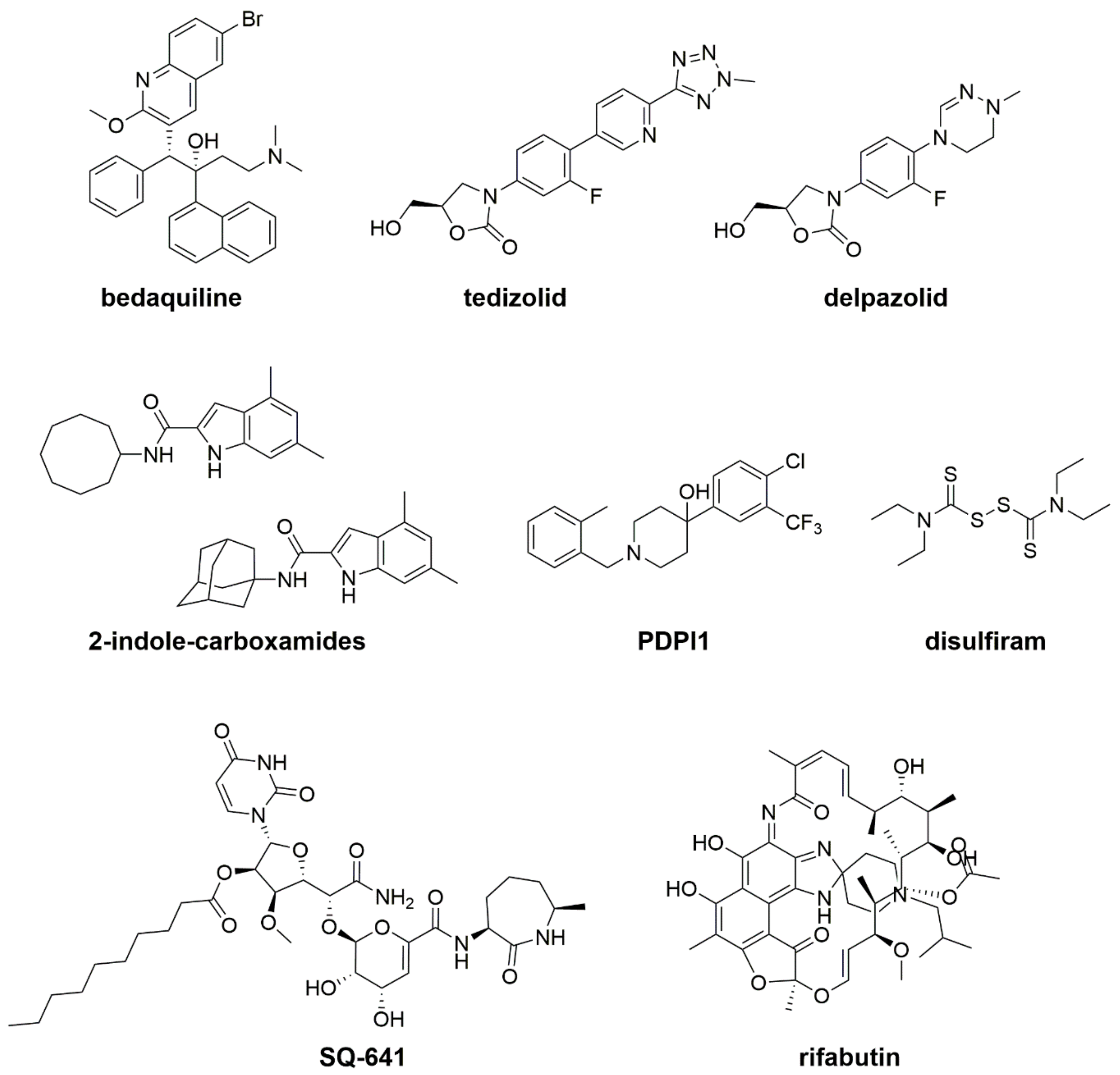
As reported above, the acquired mechanisms of resistance to drugs used in therapy limit the efficacy of the current Mab treatment; for this reason, new active compounds with a novel mechanism of action are needed.
Choo et al. (2014) [91] demonstrated that the Mab genome shares considerable sequence similarity with that of M. tuberculosis. Consequently, the already generated data in TB drug discovery could also be used to identify new compounds active against Mab and other NTM [91]. Unfortunately, only a few antitubercular drugs, such as bedaquiline, are active against Mab growth, and are currently under evaluation for their use in therapy.
The Mab drug discovery is considered very challenging because of the typical lack of bactericidal activity, both in the currently recommended Mab treatment and in new tested compounds; it could be the reason for the poor therapeutic success in Mab infection [92]. For example, among the drugs currently used in Mab treatment, tigecycline and imipenem are bacteriostatic, while clarithromycin presents only a weak bactericidal activity at high concentrations [92]. The antitubercular bedaquiline is active against Mab growth, but it is also bacteriostatic (see below) [8]. The bacteriostatic activity could depend on the presence of several chromosomally encoded drug-modifying enzymes, the typical mycobacterial cell wall, or the presence of GLPs.
The new drugs that are active against Mab should, therefore, be bactericidal in order to be more effective. Moreover, new drug combinations, as well as repurposed compounds, are being tested against this pathogen.
Despite some active compounds being in preclinical development, only a few compounds are present in clinical trials (Table 2). It is noteworthy that several clinical trials include CF patients, highlighting the emerging role of Mab in this cohort of patients. Some drugs active against Mab growth, which are currently both in preclinical and in clinical trials, are described below (Table 2; Figure 3).

5.1. Bedaquiline

Bedaquiline (BED), an antitubercular drug belonging to diarylquinoline class, was approved by the U.S. Food and Drug Administration (FDA) in 2012 for the treatment of patients affected by pulmonary MDR- and XDR-TB only [118,119,120]. BED targets the mycobacterial ATP synthase (subunit c encoded by atpE) [120]. Another mechanism of resistance is linked to mutations in the M. tuberculosis Rv0678 gene, which codes for the transcriptional repressor of the MmpS5-MmpL5 efflux system, causing cross-resistance between clofazimine and BED [90]. BED is also active against MAC and MABSC species, and its possible use is under evaluation because it lacks bactericidal activity, as previously described [93]. Moreover, Mab shares the same mechanism of BED resistance as M. tuberculosis; in fact, Mab_4383/Mab_4382 and Mab_4384 in Mab are homologous to MmpS5/MmpL5 and Rv0678 in M. tuberculosis [84,90]. Several BED-resistant Mab strains harbored mutations in the Mab_4384 gene, and consequently an over-expression of MmpS5-MmpL5 efflux pump as a mechanism of resistance [84,94]. Recently, it was shown that verapamil, an efflux inhibitor, improved the BED activity against Mab clinical isolates, both in vitro and ex vivo [95].
Conflicting results were found when BED was tested in vivo, possibly because of its bacteriostatic activity; in a mouse model the BED treatment seemed to reduce the bacterial load [96], while in another murine model BED did not present any activity [97]. Interestingly, BED was highly effective in a zebrafish model of Mab infection; in particular, a short BED treatment was able to protect the Mab-infected larvae [98].
Overall, more studies are needed in order to understand if BED could be used against Mab infections, overtaking the problem related to its bacteriostatic activity and its mechanism of resistance. Different approaches could be performed, such as the use of BED in combination with an efflux inhibitor or the synthesis of new more effective BED derivatives.

5.2. New Oxazolidinone Derivatives: Tedizolid and Delpazolid

Linezolid, belonging to the oxazolidinone class, is administrated in NTM treatment, including Mab infections, but its clinical use is often related to adverse events. Recently, two other oxazolidinones were found to be more active against NTM than linezolid: tedizolid and delpazolid [99,100].
Currently, the most promising anti-Mab oxazolidinone is delpazolid, which is in phase II trials for TB treatment; it is active against Mab both in vitro and in vivo, likely without adverse effects [100].

5.3. MmpL3 Inhibitors: Indole-2-Carboxamides and PIPD1

Recently, MmpL3 inhibitors were found to be very effective against both M. tuberculosis and Mab [101,102,103,104,121].
Two lead compounds belonging to indole-2-carboxamides showed a potent bactericidal activity against Mab in vitro (MIC = 0.125 µg/mL) and ex vivo. Moreover, they were also active against a panel of Mab clinical isolates. It was shown that their cellular target is the mycolic acid transporter MmpL3. In particular, the indole-2-carboxamides strongly inhibited the transport of trehalose monomycolate, causing the loss of trehalose dimycolate production and abrogating mycolylation of arabinogalactan [101]. Moreover, these compounds showed minimal in vitro cytotoxicity and good selectivity indices [102]. Finally, in 2019 Pandya and collaborators tested the two lead compounds in vivo using a Mab- infected mouse model. Oral administration of the MmpL3 inhibitors showed a statistically significant reduction in bacterial load in the lungs and spleens of Mab-infected mice [103]. This last study confirms that indole-2-carboxamides are very promising anti-Mab compounds.
Dupont and collaborators (2016) performed a screening of a library of 177 anti-TB compounds against Mab growth. A new piperidinol-based molecule, PIPD1, was identified and characterized as having a potent bactericidal activity against Mab. Thanks to the isolation and characterization of some resistant Mab mutants, MmpL3 was identified as a cellular target [104]. Moreover, the treatment with PIPD1 in the Mab-infected zebrafish model decreased bacterial load and improved survival of the infected embryos [8].
MmpL3 represents the only new Mab drug target; moreover, the bactericidal activity of MmpL3 inhibitors indicates this target as one of the most druggable ones for this pathogen.

5.4. Capuramycin SQ641

Capuramycins are a novel class of nucleoside antibiotics targeting translocase-1, which is essential for peptidoglycan synthesis. SQ641 is the most active capuramycin against Mab growth (MIC = 0.25–1 µg/mL) and it is bactericidal. Furthermore, SQ641 showed synergy with rifabutin and streptomycin against Mab [105].

5.5. Repurposing and Repositioning Drugs: Rifabutin, Disulfiram, and β-Lactams

One of the strategies of discovery for new antimicrobial drugs is to reposition or to repurpose existing antibiotics, thus reducing the cost and time of their clinical development.
Following this aim, Aziz et al. (2017) identified rifabutin as a hit against Mab growth, starting from a screening of 2700 FDA-approved drugs [106]. Moreover, this drug is bactericidal against Mab and also effective ex vivo, even if other rifamycins are not active against this pathogen. Rifabutin has synergistic activity in several drug combinations, such as with amikacin, cefoxitin, linezolid, clarithromycin (in triple combination with tigecycline), and azithromycin [107].
Disulfiram is a drug used in the treatment of alcoholism. It was demonstrated that it has an antimicrobial activity against several microorganisms, and clinical trials for its use in treatment of a spectrum of diseases, such as HIV infection, are in progress. Surprisingly, disulfiram is active in vitro and ex vivo against Mab, showing time- and concentration-dependent killing, similar to amikacin [108]. It also synergized with moxifloxacin, ciprofloxacin, vancomycin, teicoplanin, and amikacin. In a murine model infected with Mycobacterium fortuitum, disulfiram significantly reduced bacterial counts in kidneys; consequently, it could also be active in vivo against Mab [108].
Currently, in the guidelines for treatment of Mab infections, only the two β-lactams cefoxitin and imipenem are included [109]. BlaMab is a very active β-lactamase encoded by a chromosomal gene [82], and it is responsible for the poor efficacy of β-lactams against Mab, as previously indicated. Interestingly, avibactam is a β-lactamase inhibitor, approved by the FDA in 2014, able to efficiently inhibit the β-lactamase BlaMab [110]. Recently, 206 paired combinations of antibiotics (β-lactams, β-lactamase inhibitors, rifamycins) were tested for in vitro synergy against Mab growth. Only 24 combinations exhibited synergy. Of these, 13 combinations included two β-lactams; 5 a β-lactam and avibactam; 6 included a β-lactam and a rifamycin. In the 5 combinations with avibactam, the MICs of three β-lactams (cefuroxime, imipenem, and biapenem) were reduced to below therapeutic breakpoints [111].
Pandey and collaborators (2019) discovered that the combinations of ceftazidime with either ceftaroline or imipenem were synergistic and had clinically relevant activities against clinical MABC isolates. Interestingly, these last β-lactams combinations were also active against THP-1 human macrophages infected with three different Mab clinical isolates [112].
Furthermore, the evaluation of these repositioning drugs (rifabutin and disulfiram, combinations of β-lactams) for their use in therapy against Mab is an interesting field of research and could complement the lack of new compounds against this pathogen. Even if more studies are needed, the recent findings of the inhibitory effect of avibactam against Mab β-lactamase support this research field.

5.6. Tigecycline

Tigecycline is a glycylcycline tetracycline in phase II of clinical development [83,113]. In this clinical trial, data were collected from 52 patients (58.3% with CF). Interestingly, in >60% of patients with Mab infections, including those with CF, an improvement was found when tigecycline was added to a multidrug treatment for ≥1 month [113,114].

5.7. Inhaled Formulation Nitric Oxide

An inhaled formulation of Nitric oxide (NO) is in phase II of clinical development for the treatment of Mab and the other NTMs [113].
NO is physiologically formed from L-arginine by NO synthase, and it plays an essential role in a variety of biological processes in the lung, including host defense against pathogens. Unfortunately, the airways of CF patients are NO-deficient, contributing to impaired lung function. Consequently, the increase of airway NO level is related to an improvement in lung function.
For this reason, NO is considered to be very promising against NTM infections. Bentur et al. (2019) evaluated the efficacy, safety, and tolerability of the intermittent inhaled NO in 9 CF patients with refractory Mab lung infection through a prospective, open labeled, multi-center, pilot study [115]. The treatment did not result in a Mab culture conversion (defined as 3 consecutive monthly negative culture of sputum samples), but caused a reduction in airway bacterial load. The main limitation of this study was the small number of CF-treated patients; additional clinical studies, for example with the use of larger cohorts of CF patients as well as an increase in the duration of NO therapy, are needed to understand the potential of this treatment [115].

5.8. Liposomal Amikacin for Inhalation

Liposomal amikacin for inhalation (LAI) is an inhaled drug in phase II of clinical development [113,116]. It is a novel formulation of amikacin, characterized by reduced toxicity and consequently an improved effectiveness in patients with refractory Mab lung disease. Caimmi and collaborators (2018) prescribed LAI to 5 CF patients with Mab infection at the Montpellier CF Center. Interestingly, 3 patients completed the treatment and did not have any respiratory exacerbation, showing negative cultures for Mab in their sputum [117].
Currently, another study is in progress, including 30 CF patients and the efficacy, safety, and tolerability of once-daily dosing of LAI 590 mg for 12 months [116].

5.9. Inhaled Molgramostim

Inhaled molgramostim is a formulation of granulocyte macrophage-colony-stimulating factor. It is a protein naturally occurring in the human immune system that plays an important role in activating the immune system to kill bacteria such as NTM. A phase 2 study is currently underway to test the effectiveness of inhaled mogramostim against NTM (including Mab) in adults with CF [113,116].

6. Conclusions and Future Perspectives

Mab is becoming one of the most frightening CF pathogens, as previously underlined. Clinical isolate sequencing has demonstrated that human-to-human Mab transmission is possible, further threatening CF patient health and contributing to its spread [14,43,44]. Mab virulence among CF patients is related to three important factors: (1) the transition from the S variant, important for colonization, to the R strain, fundamental for cell invasion [53]; (2) the GLPs of S strains, which are able to masque TDM and other lipids responsible for the activation of the innate immune system [51]; (3) the role of CFTR mutations in promoting Mab infections, as CFTR seems to have a specific role in the immune control of only this pathogen [69].
A key challenge for Mab infection treatment is to develop new strategies for its eradication, since, current therapy is poorly effective and no new drugs are on the horizon.
Alternative strategies, such as phage therapy, have already been investigated. Recently, after bilateral lung transplantation, a 15-year-old girl with CF and a disseminated Mab infection was successfully treated with a phage therapy [122,123]. This was the first therapeutic use of phages for a human mycobacterial infection, which was also well tolerated and without adverse reactions. This amazing result opens the possibility to use phage therapy against Mab infections in CF patients, even if accurate clinical trials are needed.
The finding of the β-lactamase inhibitor avibactam that effectively blocked Bla_Mab provides a new chance for the use of old antibiotics, such as β-lactams. Le Run and collaborators (2019) showed that the addition of avibactam improved the activity of the imipenem–tedizolid combination [124]. Interestingly, Lefebvre et al. (2017) demonstrated that the inhibition of Bla_Mab by avibactam improved the efficacy of imipenem against Mab in vitro, in macrophages and in zebrafish models [125], indicating that it should be clinically evaluated. It is noteworthy that the aztreonam–avibactam and ceftazidime–avibactam combinations are in phase I of clinical development for Gram-negative infections [113].
Among the possible new approaches, an interesting anti-virulence strategy could be exploited for inhibiting MAB_4780, the dehydratase required for cording formation [62], which is essential for Mab pathogenicity. This alternative approach to standard chemotherapy may represent an attractive way to attenuate cording, and consequently to control invasive and acute Mab infections, even among CF patients.
Another possible method for fighting drug-resistant Mab infections could be the design of Whib7 inhibitors that could be used in combination with the current therapy [79]. The inhibition of the activation of genes conferring aminoglycoside and macrolide resistance (eis2 and erm(41), respectively) in Mab could improve the efficacy of the current Mab treatment.
As described before, inhaled formulation or drugs or NO are also very promising in the fight against Mab infections. A further evaluation of efficacy and safety is needed prior to effective clinical use.
The emergence of Mab as a CF pathogen has left patients unprepared in the fight against this pathogen, which is intrinsically resistant to several classes of antibiotics already in use. Overall, a better understanding of Mab pathogenicity and more efforts in drug development pipeline could help to pave the way for next-generation antimicrobials that are effective in Mab treatment, in order to save more infected people.

Author Contributions

All the authors reviewed the literature and wrote the manuscript.

Funding

This research was funded by the Italian Cystic Fibrosis Foundation, grant number FFC#19/2018 (adopted by Delegazione FFC di Brindisi Torre, Delegazione FFC di Ascoli Piceno, Delegazione FFC di Novara). This research was also supported by the Italian Ministry of Education, Universities, and Research (MIUR): Dipartimenti di Eccellenza Program (2018–2022)—Department of Biology and Biotechnology “L. Spallanzani”, University of Pavia (to G.D., J.C.S., L.R.C., and M.R.P.).

Acknowledgments

This review is dedicated to Ludovica, a little girl affected by cystic fibrosis with the hope of a future with an effective therapy.

Conflicts of Interest

The authors declare no conflict of interest.

References

  1. Pranke, I.; Golec, A.; Hinzpeter, A.; Edelman, A.; Sermet-Gaudelus, I. Emerging Therapeutic Approaches for Cystic Fibrosis. From Gene Editing to Personalized Medicine. Front. Pharmacol. 2019, 10, 121. [Google Scholar] [CrossRef] [PubMed]
  2. Skolnik, K.; Kirkpatrick, G.; Quon, B.S. Nontuberculous Mycobacteria in Cystic Fibrosis. Curr. Treat. Options Infect. Dis. 2016, 8, 259–274. [Google Scholar] [CrossRef] [PubMed]
  3. Viviani, L.; Harrison, M.J.; Zolin, A.; Haworth, C.S.; Floto, R.A. Epidemiology of nontuberculous mycobacteria (NTM) amongst individuals with cystic fibrosis (CF). J. Cyst. Fibros. 2016, 15, 619–623. [Google Scholar] [CrossRef] [PubMed]
  4. Martiniano, S.L.; Nick, J.A.; Daley, C.L. Nontuberculous Mycobacterial Infections in Cystic Fibrosis. Thorac. Surg. Clin. 2019, 29, 95–108. [Google Scholar] [CrossRef] [PubMed]
  5. Daniel-Wayman, S.; Abate, G.; Barber, D.L.; Bermudez, L.E.; Coler, R.N.; Cynamon, M.H.; Daley, C.L.; Davidson, R.M.; Dick, T.; Floto, R.A.; et al. Advancing Translational Science for Pulmonary Nontuberculous Mycobacterial Infections. A Road Map for Research. Am. J. Respir. Crit. Care Med. 2019, 199, 947–951. [Google Scholar] [CrossRef]
  6. Salsgiver, E.L.; Fink, A.K.; Knapp, E.A.; LiPuma, J.J.; Olivier, K.N.; Marshall, B.C.; Saiman, L. Changing Epidemiology of the Respiratory Bacteriology of Patients with Cystic Fibrosis. Chest 2016, 149, 390–400. [Google Scholar] [CrossRef]
  7. Mougari, F.; Guglielmetti, L.; Raskine, L.; Sermet-Gaudelus, I.; Veziris, N.; Cambau, E. Infections caused by Mycobacterium abscessus: Epidemiology, diagnostic tools and treatment. Expert Rev. Anti-Infect. Ther. 2016, 14, 1139–1154. [Google Scholar] [CrossRef]
  8. Wu, M.L.; Aziz, D.B.; Dartois, V.; Dick, T. NTM drug discovery: Status, gaps and the way forward. Drug Discov. Today 2018, 23, 1502–1519. [Google Scholar] [CrossRef]
  9. Qvist, T.; Pressler, T.; Høiby, N.; Katzenstein, T.L. Shifting paradigms of nontuberculous mycobacteria in cystic fibrosis. Respir. Res. 2014, 15, 41. [Google Scholar] [CrossRef]
  10. Andrew, E.C.; Connell, T.; Robinson, P.; Curtis, N.; Massie, J.; Robertson, C.; Harrison, J.; Shanthikumar, S.; Bryant, P.A.; Starr, M.; et al. Pulmonary Mycobacterium abscessus complex in children with cystic fibrosis: A practical management guideline. J. Paediatr. Child Health 2019, 55, 502–511. [Google Scholar] [CrossRef]
  11. van Dorn, A. Multidrug-resistant Mycobacterium abscessus threatens patients with cystic fibrosis. Lancet Respir. Med. 2017, 5, 15. [Google Scholar] [CrossRef]
  12. Stephenson, D.; Perry, A.; Appleby, M.R.; Lee, D.; Davison, J.; Johnston, A.; Jones, A.L.; Nelson, A.; Bourke, S.J.; Thomas, M.F.; et al. An evaluation of methods for the isolation of nontuberculous mycobacteria from patients with cystic fibrosis, bronchiectasis and patients assessed for lung transplantation. BMC Pulm. Med. 2019, 19, 19. [Google Scholar] [CrossRef] [PubMed]
  13. Davidson, R.M. A Closer Look at the Genomic Variation of Geographically Diverse Mycobacterium abscessus Clones That Cause Human Infection and Disease. Front. Microbiol. 2018, 9, 2988. [Google Scholar] [CrossRef] [PubMed]
  14. Bryant, J.M.; Grogono, D.M.; Rodriguez-Rincon, D.; Everall, I.; Brown, K.P.; Moreno, P.; Verma, D.; Hill, E.; Drijkoningen, J.; Gilligan, P.; et al. Emergence and spread of a human-transmissible multidrug-resistant nontuberculous mycobacterium. Science 2016, 354, 751–757. [Google Scholar] [CrossRef]
  15. Lopeman, R.C.; Harrison, J.; Desai, M.; Cox, J.A.G. Mycobacterium abscessus: Environmental Bacterium Turned Clinical Nightmare. Microorganisms 2019, 7, 90. [Google Scholar] [CrossRef]
  16. Thomson, R.; Tolson, C.; Sidjabat, H.; Huygens, F.; Hargreaves, M. Mycobacterium abscessus isolated from municipal water—A potential source of human infection. BMC Infect. Dis. 2013, 13, 241. [Google Scholar] [CrossRef]
  17. Floto, R.A.; Olivier, K.N.; Saiman, L.; Daley, C.L.; Herrmann, J.L.; Nick, J.A.; Noone, P.G.; Bilton, D.; Corris, P.; Gibson, R.L.; et al. US Cystic Fibrosis Foundation and European Cystic Fibrosis Society consensus recommendations for the management of non-tuberculous mycobacteria in individuals with cystic fibrosis. Thorax 2016, 71 (Suppl. 1), i1–i22. [Google Scholar] [CrossRef]
  18. Sood, G.; Parrish, N. Outbreaks of nontuberculous mycobacteria. Curr. Opin. Infect. Dis. 2017, 30, 404–409. [Google Scholar] [CrossRef]
  19. King, D.N.; Donohue, M.J.; Vesper, S.J.; Villegas, E.N.; Ware, M.W.; Vogel, M.E.; Furlong, E.F.; Kolpin, D.W.; Glassmeyer, S.T.; Pfaller, S. Microbial pathogens in source and treated waters from drinking water treatment plants in the United States and implications for human health. Sci. Total Environ. 2016, 562, 987–995. [Google Scholar] [CrossRef]
  20. Thomson, R.; Tolson, C.; Carter, R.; Coulter, C.; Huygens, F.; Hargreaves, M. Isolation of nontuberculous mycobacteria (NTM) from household water and shower aerosols in patients with pulmonary disease caused by NTM. J. Clin. Microbiol. 2013, 51, 3006–3011. [Google Scholar] [CrossRef]
  21. Torvinen, E.; Suomalainen, S.; Paulin, L.; Kusnetsov, J. Mycobacteria in Finnish cooling tower waters. Apmis 2014, 122, 353–358. [Google Scholar] [CrossRef] [PubMed]
  22. Alqumber, M.A. Prevalence of mycobacteria in water reservoirs of Albaha, Saudi Arabia. Saudi Med. J. 2014, 35, 466–471. [Google Scholar] [PubMed]
  23. Williams, M.M.; Chen, T.H.; Keane, T.; Toney, N.; Toney, S.; Armbruster, C.R.; Butler, W.R.; Arduino, M.J. Point-of-use membrane filtration and hyperchlorination to prevent patient exposure to rapidly growing mycobacteria in the potable water supply of a skilled nursing facility. Infect. Control Hosp. Epidemiol. 2011, 32, 837–844. [Google Scholar] [CrossRef] [PubMed]
  24. Honda, J.R.; Hasan, N.A.; Davidson, R.M.; Williams, M.D.; Epperson, L.E.; Reynolds, P.R.; Smith, T.; Iakhiaeva, E.; Bankowski, M.J.; Wallace, R.J., Jr.; et al. Environmental Nontuberculous Mycobacteria in the Hawaiian Islands. PLoS Negl. Trop. Dis. 2016, 10, e0005068. [Google Scholar] [CrossRef] [PubMed]
  25. Liu, R.; Yu, Z.; Zhang, H.; Yang, M.; Shi, B.; Liu, X. Diversity of bacteria and mycobacteria in biofilms of two urban drinking water distribution systems. Can. J. Microbiol. 2012, 58, 261–270. [Google Scholar] [CrossRef] [PubMed]
  26. Delafont, V.; Mougari, F.; Cambau, E.; Joyeux, M.; Bouchon, D.; Héchard, Y.; Moulin, L. First evidence of amoebae-mycobacteria association in drinking water network. Environ. Sci. Technol. 2014, 48, 11872–11882. [Google Scholar] [CrossRef]
  27. Donohue, M.J.; Mistry, J.H.; Donohue, J.M.; O’Connell, K.; King, D.; Byran, J.; Covert, T.; Pfaller, S. Increased Frequency of Nontuberculous Mycobacteria Detection at Potable Water Taps within the United States. Environ. Sci. Technol. 2015, 49, 6127–6133. [Google Scholar] [CrossRef]
  28. Puk, K.; Banach, T.; Wawrzyniak, A.; Adaszek, Ł.; Ziętek, J.; Winiarczyk, S.; Guz, L. Detection of Mycobacterium marinum, M. peregrinum, M. fortuitum and M. abscessus in aquarium fish. J. Fish Dis. 2018, 41, 153–156. [Google Scholar] [CrossRef]
  29. Schets, F.M.; van den Berg, H.H.; de Zwaan, R.; van Soolingen, D.; de Roda Husman, A.M. The microbiological quality of water in fish spas with Garra rufa fish, the Netherlands, October to November 2012. Euro Surveill. 2015, 20, 2–8. [Google Scholar] [CrossRef]
  30. Varello, K.; Prearo, M.; Serracca, L.; Meloni, D.; Rossini, I.; Righetti, M.; Pezzolato, M.; Fioravanti, M.L.; Ercolini, C.; Bozzetta, E. Granulomatous lesions in a wild mullet population from the eastern Ligurian Sea (Italy): Mycobacteriosis vs. pseudotuberculosis. J. Fish Dis. 2014, 37, 553–558. [Google Scholar] [CrossRef]
  31. Favaro, L.; Scanzio, T.; Varello, K.; Caffara, M.; Righetti, M.; Bozzetta, E.; Prearo, M. Mixed mycobacterial infection in an adult koi carp Cyprinus carpio L. J. Fish Dis. 2014, 37, 753–755. [Google Scholar] [CrossRef] [PubMed]
  32. Zanoni, R.G.; Florio, D.; Fioravanti, M.L.; Rossi, M.; Prearo, M. Occurrence of Mycobacterium spp. in ornamental fish in Italy. J. Fish Dis. 2008, 31, 433–441. [Google Scholar] [PubMed]
  33. Chang, T.C.; Hsieh, C.Y.; Chang, C.D.; Shen, Y.L.; Huang, K.C.; Tu, C.; Chen, L.C.; Wu, Z.B.; Tsai, S.S. Pathological and molecular studies on mycobacteriosis of milkfish Chanos chanos in Taiwan. Dis. Aquat. Org. 2006, 72, 147–151. [Google Scholar] [CrossRef] [PubMed]
  34. Reisfeld, L.; Ikuta, C.Y.; Ippolito, L.; Silvatti, B.; Ferreira Neto, J.S.; Catão-Dias, J.L.; Rosas, F.C.W.; Neto, J.A.; da Silva, V.M.F. Cutaneous mycobacteriosis in a captive Amazonian manatee Trichechus inunguis. Dis. Aquat. Org. 2018, 127, 231–236. [Google Scholar] [CrossRef]
  35. Katale, B.Z.; Mbugi, E.V.; Botha, L.; Keyyu, J.D.; Kendall, S.; Dockrell, H.M.; Michel, A.L.; Kazwala, R.R.; Rweyemamu, M.M.; van Helden, P.; et al. Species diversity of non-tuberculous mycobacteria isolated from humans, livestock and wildlife in the Serengeti ecosystem, Tanzania. BMC Infect. Dis. 2014, 14, 616. [Google Scholar] [CrossRef] [Green Version]
  36. Clayton, L.A.; Stamper, M.A.; Whitaker, B.R.; Hadfield, C.A.; Simons, B.; Mankowski, J.L. Mycobacterium abscessus pneumonia in an Atlantic bottlenose dolphin (Tursiops truncatus). J. Zoo Wildl. Med. 2012, 43, 961–965. [Google Scholar] [CrossRef] [Green Version]
  37. Jassies-van der Lee, A.; Houwers, D.J.; Meertens, N.; van der Zanden, A.G.; Willemse, T. Localised pyogranulomatous dermatitis due to Mycobacterium abscessus in a cat: A case report. Vet. J. 2009, 179, 304–306. [Google Scholar] [CrossRef]
  38. Lunn, J.A.; Martin, P.; Zaki, S.; Malik, R. Pneumonia due to Mycobacterium abscessus in two domestic ferrets (Mustelo putorius furo). Aust. Vet. J. 2005, 83, 542–546. [Google Scholar] [CrossRef]
  39. Karlson, A.G.; Seibold, H.R.; Wolf, R.H. Mycobacterium abscessus infection in an owl monkey (Aotus trivirgatua). Pathol. Vet. 1970, 7, 448–454. [Google Scholar]
  40. Jang, S.S.; Hirsh, D.C. Rapidly growing members of the genus Mycobacterium affecting dogs and cats. J. Am. Anim. Hosp. Assoc. 2002, 38, 217–220. [Google Scholar] [CrossRef]
  41. Falkinham, J.O., 3rd. Nontuberculous mycobacteria from household plumbing of patients with nontuberculous mycobacteria disease. Emerg. Infect. Dis. 2011, 17, 419–424. [Google Scholar] [CrossRef] [PubMed]
  42. Bryant, J.M.; Grogono, D.M.; Greaves, D.; Foweraker, J.; Roddick, I.; Inns, T.; Reacher, M.; Haworth, C.S.; Curran, M.D.; Harris, S.R.; et al. Whole-genome sequencing to identify transmission of Mycobacterium abscessus between patients with cystic fibrosis: A retrospective cohort study. Lancet 2013, 381, 1551–1560. [Google Scholar] [CrossRef] [Green Version]
  43. Yan, J.; Kevat, A.; Martinez, E.; Teese, N.; Johnson, K.; Ranganathan, S.; Harrison, J.; Massie, J.; Daley, A. Investigating transmission of Mycobacterium abscessus amongst children in an Australian cystic fibrosis centre. J. Cyst. Fibros. 2019, in press. [Google Scholar] [CrossRef]
  44. Tortoli, E.; Kohl, T.A.; Trovato, A.; Baldan, R.; Campana, S.; Cariani, L.; Colombo, C.; Costa, D.; Cristadoro, S.; Di Serio, M.C.; et al. Mycobacterium abscessus in patients with cystic fibrosis: Low impact of inter-human transmission in Italy. Eur. Respir. J. 2017, 50, 1602525. [Google Scholar] [CrossRef] [PubMed] [Green Version]
  45. Esteban, J.; García-Coca, M. Mycobacterium Biofilms. Front. Microbiol. 2018, 8, 2651. [Google Scholar] [CrossRef] [PubMed] [Green Version]
  46. Benwill, J.L.; Wallace, R.J., Jr. Mycobacterium abscessus: Challenges in diagnosis and treatment. Curr. Opin. Infect. Dis. 2014, 27, 506–510. [Google Scholar] [CrossRef]
  47. Qvist, T.; Eickhardt, S.; Kragh, K.N.; Andersen, C.B.; Iversen, M.; Høiby, N.; Bjarnsholt, T. Chronic pulmonary disease with Mycobacterium abscessus complex is a biofilm infection. Eur. Respir. J. 2015, 46, 1823–1826. [Google Scholar] [CrossRef] [Green Version]
  48. Fennelly, K.P.; Ojano-Dirain, C.; Yang, Q.; Liu, L.; Lu, L.; Progulske-Fox, A.; Wang, G.P.; Antonelli, P.; Schultz, G. Biofilm Formation by Mycobacterium abscessus in a Lung Cavity. Am. J. Respir. Crit. Care Med. 2016, 193, 692–693. [Google Scholar] [CrossRef]
  49. Hunt-Serracin, A.C.; Parks, B.J.; Boll, J.; Boutte, C. Biofilm-associated Mycobacterium abscessus cells have altered antibiotic tolerance and surface glycolipids in Artificial Cystic Fibrosis Sputum Media. Antimicrob. Agents Chemother. 2019, 63, e02488-18. [Google Scholar] [CrossRef] [Green Version]
  50. Gutiérrez, A.V.; Viljoen, A.; Ghigo, E.; Herrmann, J.L.; Kremer, L. Glycopeptidolipids, a Double-Edged Sword of the Mycobacterium abscessus Complex. Front. Microbiol. 2018, 9, 1145. [Google Scholar] [CrossRef]
  51. Davidson, L.B.; Nessar, R.; Kempaiah, P.; Perkins, D.J.; Byrd, T.F. Mycobacterium abscessus glycopeptidolipid prevents respiratory epithelial TLR2 signaling as measured by HβD2 gene expression and IL-8 release. PLoS ONE 2011, 6, e29148. [Google Scholar] [CrossRef] [PubMed] [Green Version]
  52. Schorey, J.S.; Sweet, L. The mycobacterial glycopeptidolipids: Structure, function, and their role in pathogenesis. Glycobiology 2008, 18, 832–841. [Google Scholar] [CrossRef] [PubMed] [Green Version]
  53. Howard, S.T.; Rhoades, E.; Recht, J.; Pang, X.; Alsup, A.; Kolter, R.; Lyons, C.R.; Byrd, T.F. Spontaneous reversion of Mycobacterium abscessus from a smooth to a rough morphotype is associated with reduced expression of glycopeptidolipid and reacquisition of an invasive phenotype. Microbiology 2006, 152, 1581–1590. [Google Scholar] [CrossRef] [PubMed] [Green Version]
  54. Rüger, K.; Hampel, A.; Billig, S.; Rücker, N.; Suerbaum, S.; Bange, F.C. Characterization of rough and smooth morphotypes of Mycobacterium abscessus isolates from clinical specimens. J. Clin. Microbiol. 2014, 52, 244–250. [Google Scholar] [CrossRef] [Green Version]
  55. Bernut, A.; Herrmann, J.L.; Kissa, K.; Dubremetz, J.F.; Gaillard, J.L.; Lutfalla, G.; Kremer, L. Mycobacterium abscessus cording prevents phagocytosis and promotes abscess formation. Proc. Natl. Acad. Sci. USA 2014, 111, E943–E952. [Google Scholar] [CrossRef] [Green Version]
  56. Bernut, A.; Viljoen, A.; Dupont, C.; Sapriel, G.; Blaise, M.; Bouchier, C.; Brosch, R.; de Chastellier, C.; Herrmann, J.L.; Kremer, L. Insights into the smooth-to-rough transitioning in Mycobacterium bolletii unravels a functional Tyr residue conserved in all mycobacterial MmpL family members. Mol. Microbiol. 2016, 99, 866–883. [Google Scholar] [CrossRef] [Green Version]
  57. Medjahed, H.; Reyrat, J.M. Construction of Mycobacterium abscessus defined glycopeptidolipid mutants: Comparison of genetic tools. Appl. Environ. Microbiol. 2009, 75, 1331–1338. [Google Scholar] [CrossRef] [Green Version]
  58. Nessar, R.; Reyrat, J.M.; Davidson, L.B.; Byrd, T.F. Deletion of the mmpL4b gene in the Mycobacterium abscessus glycopeptidolipid biosynthetic pathway results in loss of surface colonization capability, but enhanced ability to replicate in human macrophages and stimulate their innate immune response. Microbiology 2011, 157, 1187–1195. [Google Scholar] [CrossRef] [Green Version]
  59. Bernut, A.; Herrmann, J.L.; Ordway, D.; Kremer, L. The diverse cellular and animal models to decipher the physiopathological traits of Mycobacterium abscessus infection. Front. Cell Infect. Microbiol. 2017, 7, 100. [Google Scholar] [CrossRef] [Green Version]
  60. Halloum, I.; Carrère-Kremer, S.; Blaise, M.; Viljoen, A.; Bernut, A.; Le Moigne, V.; Vilchèze, C.; Guérardel, Y.; Lutfalla, G.; Herrmann, J.L.; et al. Deletion of a dehydratase important for intracellular growth and cording renders rough Mycobacterium abscessus avirulent. Proc. Natl. Acad. Sci. USA 2016, 113, E4228–E4237. [Google Scholar] [CrossRef] [Green Version]
  61. Koh, W.J.; Jeong, B.H.; Kim, S.Y.; Jeon, K.; Park, K.U.; Jhun, B.W.; Lee, H.; Park, H.Y.; Kim, D.H.; Huh, H.J.; et al. Mycobacterial Characteristics and Treatment Outcomes in Mycobacterium abscessus Lung Disease. Clin. Infect. Dis. 2017, 64, 309–316. [Google Scholar] [CrossRef] [PubMed]
  62. Esther, C.R., Jr.; Esserman, D.A.; Gilligan, P.; Kerr, A.; Noone, P.G. Chronic Mycobacterium abscessus infection and lung function decline in cystic fibrosis. J. Cyst. Fibros. 2010, 9, 117–123. [Google Scholar] [CrossRef] [PubMed] [Green Version]
  63. Sanguinetti, M.; Ardito, F.; Fiscarelli, E.; La Sorda, M.; D’Argenio, P.; Ricciotti, G.; Fadda, G. Fatal pulmonary infection due to multidrug-resistant Mycobacterium abscessus in a patient with cystic fibrosis. J. Clin. Microbiol. 2001, 39, 816–819. [Google Scholar] [CrossRef] [PubMed] [Green Version]
  64. Jönsson, B.E.; Gilljam, M.; Lindblad, A.; Ridell, M.; Wold, A.E.; Welinder Olsson, C. Molecular epidemiology of Mycobacterium abscessus, with focus on cystic fibrosis. J. Clin. Microbiol. 2007, 45, 1497–1504. [Google Scholar] [CrossRef] [Green Version]
  65. Qvist, T.; Taylor-Robinson, D.; Waldmann, E.; Olesen, H.V.; Hansen, C.R.; Mathiesen, I.H.; Høiby, N.; Katzenstein, T.L.; Smyth, R.L.; Diggle, P.J.; et al. Comparing the harmful effects of nontuberculous mycobacteria and Gram negative bacteria on lung function in patients with cystic fibrosis. J. Cyst. Fibros. 2016, 15, 380–385. [Google Scholar] [CrossRef] [Green Version]
  66. Dubois, V.; Viljoen, A.; Laencina, L.; Le Moigne, V.; Bernut, A.; Dubar, F.; Blaise, M.; Gaillard, J.L.; Guérardel, Y.; Kremer, L.; et al. MmpL8(MAB) controls Mycobacterium abscessus virulence and production of a previously unknown glycolipid family. Proc. Natl. Acad. Sci. USA 2018, 115, E10147–E10156. [Google Scholar] [CrossRef] [Green Version]
  67. Laencina, L.; Dubois, V.; Le Moigne, V.; Viljoen, A.; Majlessi, L.; Pritchard, J.; Bernut, A.; Piel, L.; Roux, A.L.; Gaillard, J.L.; et al. Identification of genes required for Mycobacterium abscessus growth in vivo with a prominent role of the ESX-4 locus. Proc. Natl. Acad. Sci. USA 2018, 115, E1002–E1011. [Google Scholar] [CrossRef] [Green Version]
  68. Bernut, A.; Nguyen-Chi, M.; Halloum, I.; Herrmann, J.L.; Lutfalla, G.; Kremer, L. Mycobacterium abscessus-Induced Granuloma Formation Is Strictly Dependent on TNF Signaling and Neutrophil Trafficking. PLoS Pathog. 2016, 12, e1005986. [Google Scholar] [CrossRef]
  69. Bernut, A.; Dupont, C.; Ogryzko, N.V.; Neyret, A.; Herrmann, J.L.; Floto, R.A.; Renshaw, S.A.; Kremer, L. CFTR Protects against Mycobacterium abscessus Infection by Fine-Tuning Host Oxidative Defenses. Cell Rep. 2019, 26, 1828–1840.e4. [Google Scholar] [CrossRef] [Green Version]
  70. Leung, J.M.; Olivier, K.N. Nontuberculous mycobacteria: The changing epidemiology and treatment challenges in cystic fibrosis. Curr. Opin. Pulm. Med. 2013, 19, 662–669. [Google Scholar] [CrossRef]
  71. Floto, R.A.; Olivier, K.N.; Saiman, L.; Daley, C.L.; Herrmann, J.L.; Nick, J.A.; Noone, P.G.; Bilton, D.; Corris, P.; Gibson, R.L.; et al. US Cystic Fibrosis Foundation and European Cystic Fibrosis Society consensus recommendations for the management of non-tuberculous mycobacteria in individuals with cystic fibrosis: Executive summary. Thorax 2016, 71, 88–90. [Google Scholar] [CrossRef] [PubMed] [Green Version]
  72. Lobo, L.J.; Chang, L.C.; Esther, C.R., Jr.; Gilligan, P.H.; Tulu, Z.; Noone, P.G. Lung transplant outcomes in cystic fibrosis patients with pre-operative Mycobacterium abscessus respiratory infections. Clin. Transplant. 2013, 27, 523–529. [Google Scholar] [CrossRef] [PubMed]
  73. Heijerman, H.G.M.; McKone, E.F.; Downey, D.G.; Van Braeckel, E.; Rowe, S.M.; Tullis, E.; Mall, M.A.; Welter, J.J.; Ramsey, B.W.; McKee, C.M.; et al. Efficacy and safety of the elexacaftor plus tezacaftor plus ivacaftor combination regimen in people with cystic fibrosis homozygous for the F508del mutation: A double-blind, randomised, phase 3 trial. Lancet 2019, in press. [Google Scholar] [CrossRef]
  74. Middleton, P.G.; Mall, M.A.; Dřevínek, P.; Lands, L.C.; McKone, E.F.; Polineni, D.; Ramsey, B.W.; Taylor-Cousar, J.L.; Tullis, E.; Vermeulen, F.; et al. Elexacaftor-Tezacaftor-Ivacaftor for Cystic Fibrosis with a Single Phe508del Allele. N. Engl. J. Med. 2019, 381, 1809–1819. [Google Scholar] [CrossRef] [PubMed]
  75. Luthra, S.; Rominski, A.; Sander, P. The Role of Antibiotic-Target-Modifying and Antibiotic-Modifying Enzymes in Mycobacterium abscessus Drug Resistance. Front. Microbiol. 2018, 9, 2179. [Google Scholar] [CrossRef]
  76. Brown-Elliott, B.A.; Nash, K.A.; Wallace, R.J., Jr. Antimicrobial susceptibility testing, drug resistance mechanisms, and therapy of infections with nontuberculous mycobacteria. Clin. Microbiol. Rev. 2012, 25, 545–582. [Google Scholar] [CrossRef] [Green Version]
  77. Nash, K.A.; Brown-Elliott, B.A.; Wallace, R.J., Jr. A novel gene, erm(41), confers inducible macrolide resistance to clinical isolates of Mycobacterium abscessus but is absent from Mycobacterium chelonae. Antimicrob. Agents Chemother. 2009, 53, 1367–1376. [Google Scholar] [CrossRef] [Green Version]
  78. Pfister, P.; Jenni, S.; Poehlsgaard, J.; Thomas, A.; Douthwaite, S.; Ban, N.; Böttger, E.C. The structural basis of macrolide-ribosome binding assessed using mutagenesis of 23S rRNA positions 2058 and 2059. J. Mol. Biol. 2004, 342, 1569–1581. [Google Scholar] [CrossRef]
  79. Hurst-Hess, K.; Rudra, P.; Ghosh, P. Mycobacterium abscessus WhiB7 Regulates a Species-Specific Repertoire of Genes to Confer Extreme Antibiotic Resistance. Antimicrob. Agents Chemother. 2017, 61, e01347-17. [Google Scholar] [CrossRef] [Green Version]
  80. Prammananan, T.; Sander, P.; Brown, B.A.; Frischkorn, K.; Onyi, G.O.; Zhang, Y.; Böttger, E.C.; Wallace, R.J., Jr. A single 16S ribosomal RNA substitution is responsible for resistance to amikacin and other 2-deoxystreptamine aminoglycosides in Mycobacterium abscessus and Mycobacterium chelonae. J. Infect. Dis. 1998, 177, 1573–1581. [Google Scholar] [CrossRef] [Green Version]
  81. Nessar, R.; Reyrat, J.M.; Murray, A.; Gicquel, B. Genetic analysis of new 16S rRNA mutations conferring aminoglycoside resistance in Mycobacterium abscessus. J. Antimicrob. Chemother. 2011, 66, 1719–1724. [Google Scholar] [CrossRef] [PubMed]
  82. Soroka, D.; Dubée, V.; Soulier-Escrihuela, O.; Cuinet, G.; Hugonnet, J.E.; Gutmann, L.; Mainardi, J.L.; Arthur, M. Characterization of broad-spectrum Mycobacterium abscessus class A β-lactamase. J. Antimicrob. Chemother. 2014, 69, 691–696. [Google Scholar] [CrossRef] [PubMed]
  83. Rudra, P.; Hurst-Hess, K.; Lappierre, P.; Ghosh, P. High Levels of Intrinsic Tetracycline Resistance in Mycobacterium abscessus Are Conferred by a Tetracycline-Modifying Monooxygenase. Antimicrob. Agents Chemother. 2018, 62, e00119-18. [Google Scholar] [CrossRef] [PubMed] [Green Version]
  84. Richard, M.; Gutiérrez, A.V.; Viljoen, A.; Rodriguez-Rincon, D.; Roquet-Baneres, F.; Blaise, M.; Everall, I.; Parkhill, J.; Floto, R.A.; Kremer, L. Mutations in the MAB_2299c TetR Regulator Confer Cross-Resistance to Clofazimine and Bedaquiline in Mycobacterium abscessus. Antimicrob. Agents Chemother. 2018, 63, e01316-18. [Google Scholar] [CrossRef] [Green Version]
  85. Guillemin, I.; Jarlier, V.; Cambau, E. Correlation between quinolone susceptibility patterns and sequences in the A and B subunits of DNA gyrase in mycobacteria. Antimicrob. Agents Chemother. 1998, 42, 2084–2088. [Google Scholar] [CrossRef] [Green Version]
  86. Kim, S.Y.; Jhun, B.W.; Moon, S.M.; Shin, S.H.; Jeon, K.; Kwon, O.J.; Yoo, I.Y.; Huh, H.J.; Ki, C.S.; Lee, N.Y.; et al. Mutations in gyrA and gyrB in Moxifloxacin-Resistant Mycobacterium avium Complex and Mycobacterium abscessus Complex Clinical Isolates. Antimicrob. Agents Chemother. 2018, 62, e00527-18. [Google Scholar] [CrossRef] [Green Version]
  87. Ye, M.; Xu, L.; Zou, Y.; Li, B.; Guo, Q.; Zhang, Y.; Zhan, M.; Xu, B.; Yu, F.; Zhang, Z.; et al. Molecular Analysis of Linezolid-Resistant Clinical Isolates of Mycobacterium abscessus. Antimicrob. Agents Chemother. 2019, 63, e01842-18. [Google Scholar] [CrossRef] [Green Version]
  88. Hansen, J.L.; Ippolito, J.A.; Ban, N.; Nissen, P.; Moore, P.B.; Steitz, T.A. The structures of four macrolide antibiotics bound to the large ribosomal subunit. Mol. Cell 2002, 10, 117–128. [Google Scholar] [CrossRef]
  89. Nessar, R.; Cambau, E.; Reyrat, J.M.; Murray, A.; Gicquel, B. Mycobacterium abscessus: A new antibiotic nightmare. J. Antimicrob. Chemother. 2012, 67, 810–818. [Google Scholar] [CrossRef] [Green Version]
  90. Hartkoorn, R.C.; Uplekar, S.; Cole, S.T. Cross-resistance between clofazimine and bedaquiline through upregulation of MmpL5 in Mycobacterium tuberculosis. Antimicrob. Agents Chemother. 2014, 58, 2979–2981. [Google Scholar] [CrossRef] [Green Version]
  91. Choo, S.W.; Wee, W.Y.; Ngeow, Y.F.; Mitchell, W.; Tan, J.L.; Wong, G.J.; Zhao, Y.; Xiao, J. Genomic reconnaissance of clinical isolates of emerging human pathogen Mycobacterium abscessus reveals high evolutionary potential. Sci. Rep. 2014, 4, 4061. [Google Scholar] [CrossRef] [PubMed]
  92. Maurer, F.P.; Bruderer, V.L.; Ritter, C.; Castelberg, C.; Bloemberg, G.V.; Böttger, E.C. Lack of antimicrobial bactericidal activity in Mycobacterium abscessus. Antimicrob. Agents Chemother. 2014, 58, 3828–3836. [Google Scholar] [CrossRef] [PubMed] [Green Version]
  93. Philley, J.V.; Wallace, R.J., Jr.; Benwill, J.L.; Taskar, V.; Brown-Elliott, B.A.; Thakkar, F.; Aksamit, T.R.; Griffith, D.E. Preliminary Results of Bedaquiline as Salvage Therapy for Patients with Nontuberculous Mycobacterial Lung Disease. Chest 2015, 148, 499–506. [Google Scholar] [CrossRef] [PubMed] [Green Version]
  94. Li, B.; Ye, M.; Guo, Q.; Zhang, Z.; Yang, S.; Ma, W.; Yu, F.; Chu, H. Determination of MIC Distribution and Mechanisms of Decreased Susceptibility to Bedaquiline among Clinical Isolates of Mycobacterium abscessus. Antimicrob. Agents Chemother. 2018, 62, e00175-18. [Google Scholar] [CrossRef] [Green Version]
  95. Viljoen, A.; Raynaud, C.; Johansen, M.D.; Roquet-Banères, F.; Herrmann, J.L.; Daher, W.; Kremer, L. Improved activity of bedaquiline by verapamil against Mycobacterium abscessus in vitro and in macrophages. Antimicrob. Agents Chemother. 2019, 63, e00705-19. [Google Scholar] [CrossRef] [Green Version]
  96. Obregón-Henao, A.; Arnett, K.A.; Henao-Tamayo, M.; Massoudi, L.; Creissen, E.; Andries, K.; Lenaerts, A.J.; Ordway, D.J. Susceptibility of Mycobacterium abscessus to antimycobacterial drugs in preclinical models. Antimicrob. Agents Chemother. 2015, 59, 6904–6912. [Google Scholar] [CrossRef] [Green Version]
  97. Lerat, I.; Cambau, E.; Roth Dit Bettoni, R.; Gaillard, J.L.; Jarlier, V.; Truffot, C.; Veziris, N. In Vivo evaluation of antibiotic activity against Mycobacterium abscessus. J. Infect. Dis. 2014, 209, 905–912. [Google Scholar] [CrossRef] [Green Version]
  98. Dupont, C.; Viljoen, A.; Thomas, S.; Roquet-Banères, F.; Herrmann, J.L.; Pethe, K.; Kremer, L. Bedaquiline Inhibits the ATP Synthase in Mycobacterium abscessus and Is Effective in Infected Zebrafish. Antimicrob. Agents Chemother. 2017, 61, e01225-17. [Google Scholar] [CrossRef] [Green Version]
  99. Brown-Elliott, B.A.; Wallace, R.J., Jr. In Vitro Susceptibility Testing of Tedizolid against Nontuberculous Mycobacteria. J. Clin. Microbiol. 2017, 55, 1747–1754. [Google Scholar] [CrossRef] [Green Version]
  100. Kim, T.S.; Choe, J.H.; Kim, Y.J.; Yang, C.S.; Kwon, H.J.; Jeong, J.; Kim, G.; Park, D.E.; Jo, E.K.; Cho, Y.L.; et al. Activity of LCB01-0371, a Novel Oxazolidinone, against Mycobacterium abscessus. Antimicrob. Agents Chemother. 2017, 61, e02752-16. [Google Scholar] [CrossRef] [Green Version]
  101. Kozikowski, A.P.; Onajole, O.K.; Stec, J.; Dupont, C.; Viljoen, A.; Richard, M.; Chaira, T.; Lun, S.; Bishai, W.; Raj, V.S.; et al. Targeting Mycolic Acid Transport by Indole-2-carboxamides for the Treatment of Mycobacterium abscessus Infections. J. Med. Chem. 2017, 60, 5876–5888. [Google Scholar] [CrossRef] [PubMed]
  102. Franz, N.D.; Belardinelli, J.M.; Kaminski, M.A.; Dunn, L.C.; Calado Nogueira de Moura, V.; Blaha, M.A.; Truong, D.D.; Li, W.; Jackson, M.; North, E.J. Design, synthesis and evaluation of indole-2-carboxamides with pan anti-mycobacterial activity. Bioorg. Med. Chem. 2017, 25, 3746–3755. [Google Scholar] [CrossRef] [PubMed]
  103. Pandya, A.N.; Prathipati, P.K.; Hegde, P.; Li, W.; Graham, K.F.; Mandal, S.; Drescher, K.M.; Destache, C.J.; Ordway, D.; Jackson, M.; et al. Indole-2-Carboxamides Are Active against Mycobacterium abscessus in a Mouse Model of Acute Infection. Antimicrob. Agents Chemother. 2019, 63, e02245-18. [Google Scholar] [CrossRef] [PubMed] [Green Version]
  104. Dupont, C.; Viljoen, A.; Dubar, F.; Blaise, M.; Bernut, A.; Pawlik, A.; Bouchier, C.; Brosch, R.; Guérardel, Y.; Lelièvre, J.; et al. A new piperidinol derivative targeting mycolic acid transport in Mycobacterium abscessus. Mol. Microbiol. 2016, 101, 515–529. [Google Scholar] [CrossRef] [PubMed] [Green Version]
  105. Dubuisson, T.; Bogatcheva, E.; Krishnan, M.Y.; Collins, M.T.; Einck, L.; Nacy, C.A.; Reddy, V.M. In vitro antimicrobial activities of capuramycin analogues against non-tuberculous mycobacteria. J. Antimicrob. Chemother. 2010, 65, 2590–2597. [Google Scholar] [CrossRef]
  106. Aziz, D.B.; Low, J.L.; Wu, M.L.; Gengenbacher, M.; Teo, J.W.P.; Dartois, V.; Dick, T. Rifabutin Is Active against Mycobacterium abscessus Complex. Antimicrob. Agents Chemother. 2017, 61, e00155-17. [Google Scholar] [CrossRef] [Green Version]
  107. Ganapathy, U.S.; Dartois, V.; Dick, T. Repositioning rifamycins for Mycobacterium abscessus lung disease. Expert Opin. Drug Discov. 2019, 14, 867–878. [Google Scholar] [CrossRef] [Green Version]
  108. Das, S.; Garg, T.; Chopra, S.; Dasgupta, A. Repurposing disulfiram to target infections caused by non-tuberculous mycobacteria. J. Antimicrob. Chemother. 2019, 74, 1317–1322. [Google Scholar] [CrossRef]
  109. Story-Roller, E.; Maggioncalda, E.C.; Cohen, K.A.; Lamichhane, G. Mycobacterium abscessus and β-Lactams: Emerging Insights and Potential Opportunities. Front. Microbiol. 2018, 9, 2273. [Google Scholar] [CrossRef]
  110. Dubee, V.; Bernut, A.; Cortes, M.; Lesne, T.; Dorchene, D.; Lefebvre, A.-L.; Hugonnet, J.E.; Gutmann, L.; Mainardi, J.L.; Herrmann, J.L.; et al. β-Lactamase inhibition by avibactam in Mycobacterium abscessus. J. Antimicrob. Chemother. 2015, 70, 1051–1058. [Google Scholar] [CrossRef] [Green Version]
  111. Story-Roller, E.; Maggioncalda, E.C.; Lamichhane, G. Select β-Lactam Combinations Exhibit Synergy against Mycobacterium abscessus In Vitro. Antimicrob. Agents Chemother. 2019, 63, e02613-18. [Google Scholar] [CrossRef] [PubMed]
  112. Pandey, R.; Chen, L.; Manca, C.; Jenkins, S.; Glaser, L.; Vinnard, C.; Stone, G.; Lee, J.; Mathema, B.; Nuermberger, E.L.; et al. Dual β-Lactam Combinations Highly Active against Mycobacterium abscessus Complex In Vitro. MBio 2019, 10, e02895-18. [Google Scholar] [CrossRef] [PubMed] [Green Version]
  113. ClinicalTrials.gov. Available online: https://clinicaltrials.gov/ (accessed on 1 October 2019).
  114. Wallace, R.J., Jr.; Dukart, G.; Brown-Elliott, B.A.; Griffith, D.E.; Scerpella, E.G.; Marshall, B. Clinical experience in 52 patients with tigecycline-containing regimens for salvage treatment of Mycobacterium abscessus and Mycobacterium chelonae infections. J. Antimicrob. Chemother. 2014, 69, 1945–1953. [Google Scholar] [CrossRef] [PubMed]
  115. Bentur, L.; Gur, M.; Ashkenazi, M.; Livnat-Levanon, G.; Mizrahi, M.; Tal, A.; Ghaffari, A.; Geffen, Y.; Aviram, M.; Efrati, O. Pilot study to test inhaled nitric oxide in cystic fibrosis patients with refractory Mycobacterium abscessus lung infection. J. Cyst. Fibros. 2019, in press. [Google Scholar] [CrossRef] [PubMed]
  116. Drug Development Pipeline. Available online: https://www.cff.org/Trials/Pipeline/ (accessed on 1 October 2019).
  117. Caimmi, D.; Martocq, N.; Trioleyre, D.; Guinet, C.; Godreuil, S.; Daniel, T.; Chiron, R. Positive Effect of Liposomal Amikacin for Inhalation on Mycobacterium abcessus in Cystic Fibrosis Patients. Open Forum Infect. Dis. 2018, 5, ofy034. [Google Scholar] [CrossRef]
  118. Diacon, A.H.; Pym, A.; Grobusch, M.P.; de los Rios, J.M.; Gotuzzo, E.; Vasilyeva, I.; Leimane, V.; Andries, K.; Bakare, N.; De Marez, T.; et al. Multidrug-resistant tuberculosis and culture conversion with bedaquiline. N. Engl. J. Med. 2014, 371, 723–732. [Google Scholar] [CrossRef] [Green Version]
  119. Guglielmetti, L.; Jaspard, M.; Le Dû, D.; Lachâtre, M.; Marigot-Outtandy, D.; Bernard, C.; Veziris, N.; Robert, J.; Yazdanpanah, Y.; Caumes, E.; et al. Long-term outcome and safety of prolonged bedaquiline treatment for multidrug-resistant tuberculosis. Eur. Respir. J. 2017, 49, 1601799. [Google Scholar] [CrossRef]
  120. Andries, K.; Verhasselt, P.; Guillemont, J.; Göhlmann, H.W.; Neefs, J.M.; Winkler, H.; Van Gestel, J.; Timmerman, P.; Zhu, M.; Lee, E.; et al. A diarylquinoline drug active on the ATP synthase of Mycobacterium tuberculosis. Science 2005, 307, 223–227. [Google Scholar] [CrossRef]
  121. Saxena, A.K.; Singh, A. Mycobacterial tuberculosis Enzyme Targets and their Inhibitors. Curr. Top. Med. Chem. 2019, 19, 337–355. [Google Scholar] [CrossRef]
  122. Fox, A. Engineered phages stymie drug-resistant infection. Science 2019, 364, 518–519. [Google Scholar] [CrossRef]
  123. Dedrick, R.M.; Guerrero-Bustamante, C.A.; Garlena, R.A.; Russell, D.A.; Ford, K.; Harris, K.; Gilmour, K.C.; Soothill, J.; Jacobs-Sera, D.; Schooley, R.T.; et al. Engineered bacteriophages for treatment of a patient with a disseminated drug-resistant Mycobacterium abscessus. Nat. Med. 2019, 25, 730–733. [Google Scholar] [CrossRef]
  124. Le Run, E.; Arthur, M.; Mainardi, J.L. In Vitro and Intracellular Activity of Imipenem Combined with Tedizolid, Rifabutin, and Avibactam against Mycobacterium abscessus. Antimicrob. Agents Chemother. 2019, 63, e01915-18. [Google Scholar] [CrossRef] [Green Version]
  125. Lefebvre, A.L.; Le Moigne, V.; Bernut, A.; Veckerlé, C.; Compain, F.; Herrmann, J.L.; Kremer, L.; Arthur, M.; Mainardi, J.L. Inhibition of the β-Lactamase Bla(Mab) by Avibactam Improves the In Vitro and In Vivo Efficacy of Imipenem against Mycobacterium abscessus. Antimicrob. Agents Chemother. 2017, 61, e02440-16. [Google Scholar] [CrossRef] [PubMed] [Green Version]
Figure 1. Factors contributing to the spread of M. abscessus (Mab) as an emerging pathogen among cystic fibrosis (CF) patients. CFTR: cystic fibrosis transmembrane conductance regulator.
Ijms 20 05868 g001
Figure 2. Drugs currently used in Mab therapy.
Ijms 20 05868 g002
Figure 3. Anti-Mab drugs in preclinical and clinical studies.
Ijms 20 05868 g003
Table 1. Mechanisms of resistance to current drugs used against Mab infections.
DrugsTargetsMechanism of ResistanceEnzymes/Proteins Related to Mechanism of ResistanceReferences
Macrolides23S rRNAMutations in target geneRrl (MAB_r5052)[77,78]
Modification of targetErm(41) (MAB_2297)[77]
Induction of WhiB7 activatorActivation of erm(41) (MAB_2297)[8,79]
Amynoglicosides30S subunit of ribosomeMutations in target genes16S rRNA (rrs, MAB_r5051)[80,81]
RpsL (MAB_3851c)
Enzymatic drug modificationAAC(2′) (MAB_4395)[75]
Eis2 (MAB_4532c)[75]
Induction of WhiB7 activatorActivation of eis2 (MAB_4532c)[8,79]
β-lactamsPenicillin-binding proteinEnzymatic drug modificationBla_Mab (MAB_2875)[82]
Tetracyclines30S subunit of ribosomeEnzymatic drug modificationMabTetX (MAB_1496c)[83]
Clofazimine Mutations in the repressor → Over-expression of an efflux pumpMAB_2299c[84]
FluoroquinolonesA subunit of DNA gyraseMutations in target geneGyrA (MAB_0019)[85,86]
Other mechanisms?not detected[86]
Linezolid23S rRNAMutations in target geneRrl (MAB_r5052)[87]
Efflux pumps?LmrS and MmpL9?
Table 2. Compounds in preclinical and clinical development against Mab infections.
Drugs Development PhaseTargetMechanism of ResistanceReferences
BedaquilinePreclinical studiesATP synthaseMmpS5-MmpL5 efflux pump[84,93,94,95,96,97,98]
TedizolidPreclinical studies50S ribosome-[99]
DelpazolidPreclinical studies50S ribosome-[100]
Indole-2-carboxamidesPreclinical studiesMmpL3-[101,102,103]
PIPD1Preclinical studiesMmpL3-[104]
SQ641Preclinical studiesTranslocase-1-[105]
RifabutinPreclinical studiesRNA polymerase [106,107]
DisulfiramPreclinical studies--[108]
β-lactams (combinations)Preclinical studiesPenicillin-binding proteinBla_Mab[82,109,110,111,112]
TigecyclinePhase II30S subunit of ribosome-[83,113,114]
Nitric oxidePhase II--[113,115,116]
Liposomal Amikacin for InhalationPhase II23S rRNA-[113,116,117]
Inhaled MolgramostimPhase II--[113,116]

