Review

Cell-Clearing Systems Bridging Repeat Expansion Proteotoxicity and Neuromuscular Junction Alterations in ALS and SBMA

by
Fiona Limanaqi
1,†,
Carla Letizia Busceti
2,†,
Francesca Biagioni
2,
Federica Cantini
1,
Paola Lenzi
1 and
Francesco Fornai
1,2,*
1
Department of Translational Research and New Technologies in Medicine and Surgery, University of Pisa, Via Roma 55, 56126 Pisa, Italy
2
I.R.C.C.S. Neuromed, Via Atinense, 18, 86077 Pozzilli, Italy
*
Author to whom correspondence should be addressed.
These authors contributed equally to this work.
Int. J. Mol. Sci. 2020, 21(11), 4021; https://0-doi-org.brum.beds.ac.uk/10.3390/ijms21114021
Submission received: 8 May 2020 / Revised: 29 May 2020 / Accepted: 3 June 2020 / Published: 4 June 2020
(This article belongs to the Special Issue Proteotoxicity and Neurodegenerative Diseases)

Abstract

:
The coordinated activities of autophagy and the ubiquitin proteasome system (UPS) are key to preventing the aggregation and toxicity of misfold-prone proteins which manifest in a number of neurodegenerative disorders. These include proteins which are encoded by genes containing nucleotide repeat expansions. In the present review we focus on the overlapping role of autophagy and the UPS in repeat expansion proteotoxicity associated with chromosome 9 open reading frame 72 (C9ORF72) and androgen receptor (AR) genes, which are implicated in two motor neuron disorders, amyotrophic lateral sclerosis (ALS) and spinal-bulbar muscular atrophy (SBMA), respectively. At baseline, both C9ORF72 and AR regulate autophagy, while their aberrantly-expanded isoforms may lead to a failure in both autophagy and the UPS, further promoting protein aggregation and toxicity within motor neurons and skeletal muscles. Besides proteotoxicity, autophagy and UPS alterations are also implicated in neuromuscular junction (NMJ) alterations, which occur early in both ALS and SBMA. In fact, autophagy and the UPS intermingle with endocytic/secretory pathways to regulate axonal homeostasis and neurotransmission by interacting with key proteins which operate at the NMJ, such as agrin, acetylcholine receptors (AChRs), and adrenergic beta2 receptors (B2-ARs). Thus, alterations of autophagy and the UPS configure as a common hallmark in both ALS and SBMA disease progression. The findings here discussed may contribute to disclosing overlapping molecular mechanisms which are associated with a failure in cell-clearing systems in ALS and SBMA.

1. Introduction

Alterations in the two major eukaryotic cell-clearing systems, autophagy and the ubiquitin-proteasome system (UPS), are promiscuously implicated in a variety of neurological disorders featuring protein misfolding, aggregation, and toxicity [1,2,3,4]. These disorders include a group in which aggregated proteins are encoded by genes containing nucleotide repeat expansions, such as Huntington disease, different forms of spinocerebellar ataxia (SCA), amyotrophic lateral sclerosis (ALS) and/or frontotemporal dementia (FTD), and X-linked spinal-bulbar muscular atrophy (SBMA, Kennedy’s disease) [5,6,7,8,9,10].
Here we focus on chromosome 9 open reading frame 72 (C9ORF72) and androgen receptor (AR) genes which are similarly affected by repeat expansions, leading to two different kinds of motor neuron disorders, namely ALS and SBMA, respectively [11,12,13,14]. Despite differing in disease frequency and clinical course, ALS and SBMA possess key overlapping features that are associated with dysfunctions of cell-clearing systems, namely protein aggregation due to expanded C9ORF72 or AR within both motor neurons and skeletal muscles, as well as early neuromuscular junction (NMJ) and axonal alterations [10,15,16,17,18,19,20,21]. At baseline, both C9ORF72 and AR regulate autophagy, while their aberrantly-expanded isoforms may lead to a failure in both autophagy and the UPS, further promoting protein aggregation and toxicity within motor neurons and skeletal muscles [10,21,22,23].
Repeat expansions within C9ORF72 and AR are generally considered to produce neurotoxicity through a gain-of-function mechanism consisting of the formation of dipeptide repeat (DPR) proteins and polyglutamine-expanded AR (ARpolyQ) which aggregate in cells [10,18,24]. While being substrates for both the UPS and autophagy, these protein aggregates might in turn alter cell-clearing systems, and mostly the UPS, which is unable to process large and insoluble aggregates due to its narrow catalytic chamber [25]. Remarkably, in both ALS and SBMA, a gain-of function toxicity may concomitantly occur along with a loss of normal C9ORF72 and AR function, respectively [18,19,20,26,27]. This is supposed to exacerbate the failure in cell-clearing systems, and mostly the autophagy pathway, which is physiologically activated by the normal C9ORF72 and AR [22,23,28]. This is magnified in the case of C9ORF72 repeat expansions, which often synergize with additional genetic disease modifiers to produce frank toxicity through alterations of cell-clearing systems [29]. In fact, a plethora of mutated genes which concur with C9ORF72 repeat expansions are known to alter autophagy and the UPS [11,30,31,32].
It is noteworthy that regardless of the causative mechanism, disease symptoms in ALS and SBMA are bound to a failure of neurotransmission which may precede protein aggregation [33,34,35]. In particular, early alterations within the skeletal muscle, NMJ, and sensory/motor axons are emerging as a primum movens in both ALS and SBMA [33,35,36,37,38]. In line with this, both diseases are being redefined as neuromuscular synaptopathies, remarking the importance of proteostasis within the sensory–motor system as a whole [36]. In this scenario, autophagy and the UPS hold a center stage, being intermingled with each other and with endocytic/secretory pathways to regulate axonal homeostasis and neurotransmission beyond the clearance of potentially toxic protein aggregates [39,40,41,42]. As we shall see, a plethora of proteins operating at the NMJ and which are altered in ALS and SBMA, are bound to autophagy and/or UPS activities [43,44,45]. This is the case for agrin, acetylcholine receptors (AChRs), and adrenergic beta2 receptors (B2-ARs), which are implicated in NMJ maintenance [43,44,45,46].
In the present manuscript, after providing an overview on the promiscuous roles of autophagy and the UPS in proteoxicity within motor neurons, axons, and muscle cells in ALS and SBMA, we move to discuss potential molecular mechanisms bridging autophagy and UPS alterations with early NMJ alterations. In particular, we focus on C9ORF72 and specific genes which may concur with C9ORF72 repeat expansions to foster disease progression and proteotoxicity through alterations of cell-clearing systems. This is taken as a paradigm to be compared with repeat expansions similarly affecting AR in SBMA. The findings here discussed may contribute to disclosing overlapping molecular mechanisms in ALS and SBMA.

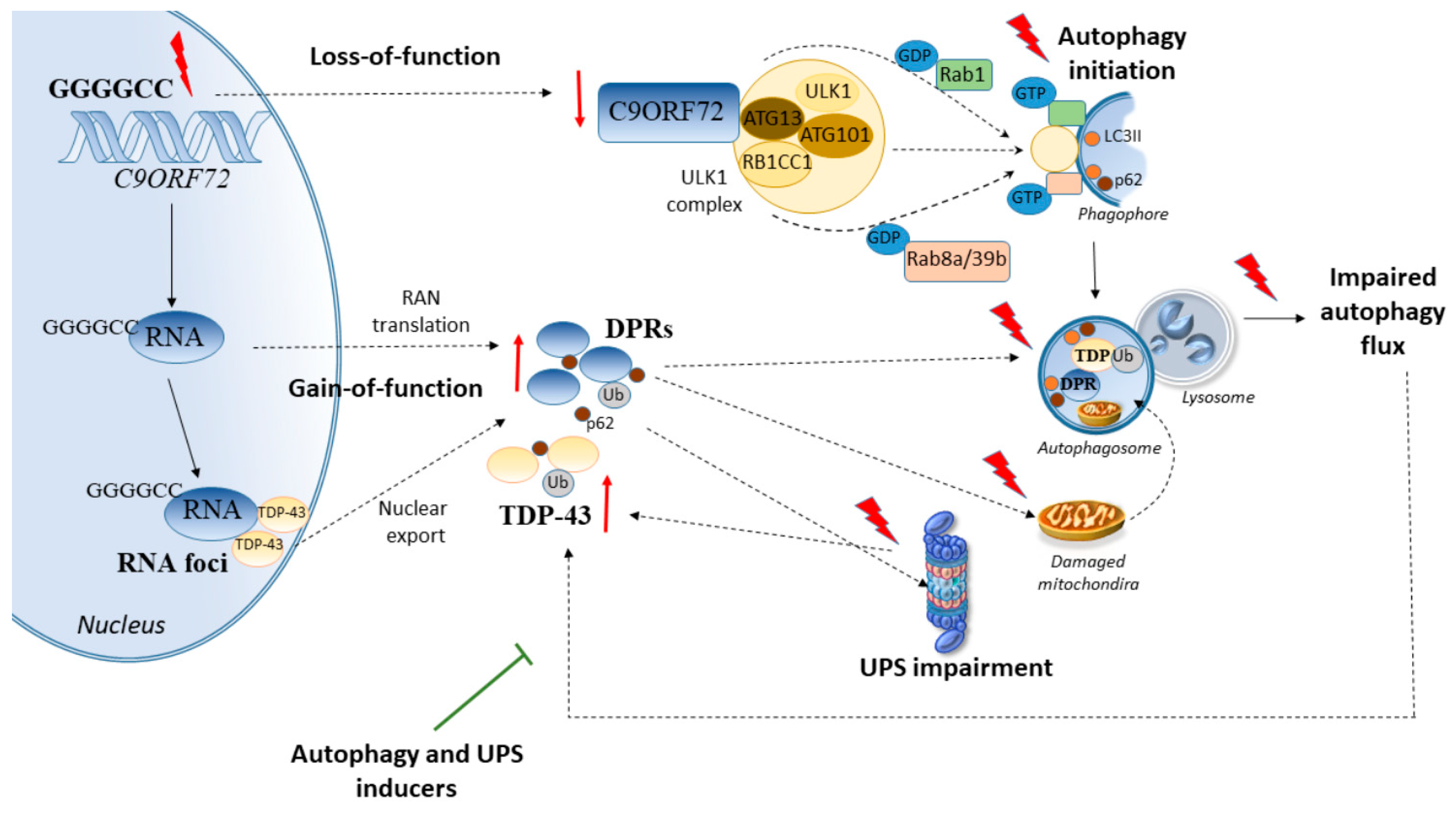
2. Cell-Clearing Systems and C9ORF72 Repeat Expansions in ALS

C9ORF72 repeat expansions are currently the major genetic cause of familial ALS and/or FTD worldwide while occurring with lower frequencies in sporadic ALS and/or FTD as well [11]. In detail, GGGGCC hexanucleotide repeat expansions within the first intron of C9ORF72 lead to C9ORF72 haploinsufficiency and loss-of-function, and/or production of RNA foci and dipeptide repeat (DPR) proteins forming toxic aggregates in motor neurons, with the two mechanisms not being necessarily mutually exclusive [18,20,26]. Besides motor neurons, skeletal muscle cells also experience pathological changes due to the C9ORF72 mutation. In fact, in skeletal myocytes differentiated from induced pluripotent stem cells (iPSCs) of C9ORF72-ALS patients, the occurrence of DPR proteins is accompanied by abnormalities in the expression of mitochondrial genes and a high susceptibility to oxidative stress, as well as increased expression and aggregation of TAR DNA-binding protein 43 (TDP-43) [47]. Supporting these findings, DPR-related pathology is detected in the skeletal muscle of ALS patients with C9ORF72 repeat expansion [48].
Remarkably, both C9ORF72 loss-of-function and production of DPR protein aggregates are bound to alterations of cell-clearing systems. C9ORF72 acts a component of the autophagy initiation complex which is composed of ULK1-RB1CC1-ATG13-ATG101 [49]. Autophagy initiation by C9ORF72 is mediated by a direct interaction between ATG13 and the isoform-specific carboxyl-terminal DENN and dDENN domain of C9ORF72 [49]. Downregulation of C9ORF72 in cell lines and primary neurons impairs autophagy by hampering Rab1a-dependent trafficking of the ULK1 autophagy initiation complex to the phagophore [22]. This in turn, promotes the accumulation of p62-positive puncta similar to what is observed in C9-ALS/FTD patient-derived neurons [22]. In primary hippocampal neurons cultured from C9ORF72-knockout mice, a reduction in dendritic branching and spine density occurs, which is associated with an impairment of autophagy due to reduced ULK1 levels [49]. Thus, besides protein degradation, C9ORF72-dependent promotion of ULK1-mediated autophagy has a key role in neuronal and dendritic morphogenesis.
Recently, a disease mechanism in ALS resulting from reduced C9ORF72 levels which can synergize with DPR-dependent gain of toxicity through autophagy and UPS deficits has been proposed [18,19]. In mice expressing a 450 repeat C9ORF72 transgene that does not encode the C9ORF72 protein, inactivation of the endogenous C9ORF72 alleles exacerbates ALS phenotype and the accumulation of DPR proteins by occluding the autophagy activity [18]. This implies a double-hit pathogenic mechanism, whereby reduced expression of C9ORF72 synergizes with an impairment of DPR clearance fostering their accumulation and toxicity in ALS [20]. This is largely bound to an autophagy impairment, which exacerbates the accumulation of DPR proteins deriving from translation of sense and antisense repeats, eventually promoting neurotoxicity. Conversely, pharmacological autophagy activators prevent these effects [20]. Data from cell lines, primary neurons, transgenic mice, and patient tissue indicate that DPR proteins derived from C9ORF72 repeat expansion, especially poly-glycine-alanine (GA) and poly-proline-alanine (PA), impair the UPS besides autophagy [19,24,50,51,52]. Remarkably, this is associated with TDP-43 cytoplasmic mislocalization, aggregation, and toxicity, which occurs in the absence of TARDBP mutations [19]. In detail, cell-to-cell transmission of DPR proteins inhibits the UPS in neighboring cells, both cell-autonomously and non-cell-autonomously. In turn, UPS inhibition exacerbates the accumulation of TDP-43 which is ubiquitinated specifically within the nuclear localization signal at lysine 95 [19]. Conversely, administration of the UPS activator rolipram completely blocks DRP-dependent mislocalization and aggregation of TDP-43 [19]. In line with this, DPR proteins physically associate with proteasomes to inhibit the degradation of ubiquitinated substrates, while administration of UPS activators occludes the toxic effects of DPR on motor neuron survival (Figure 1) [24]. These findings are in line with neuropathological investigations showing that repeat expansions in C9ORF72 are associated with ubiquitin- and p62-containing cytoplasmic inclusions, being either positive or negative for TDP-43 [11,53,54,55]. However, most of the abovementioned studies did not focus on the potential differences between the effects of UPS vs. autophagy activity and the specific type of DPR, which indeed may vary in aggregation dynamics and toxicity, while occurring within neuronal inclusions other than TDP-43-positive ones.

2.1. C9ORF72 Synergizes with Genetic Disease Modifiers to Alter Cell-Clearing Pathways

A stream of evidence suggests that C9ORF72 depletion might not lead per se to major neuronal toxicity while contributing to ALS pathogenesis by directly interfering with additional genetic disease modifiers [29,56]. In fact, a number of additional mutated genes may co-occur with C9ORF72 repeat expansions, contributing to the pleiotropic clinical and pathological phenotypes observed in ALS, including the ALS–FTD spectrum [11]. These include mutations in superoxide dismutase 1 (SOD1), transcription of RNA activating protein/TAR DNA-binding protein (TARDBP), fused in sarcoma (FUS), optineurin (OPTN), ubiquilin-2 (UBQLN2), progranulin (PGRN), ataxin-2 (ATXN-2), valosin-containing protein (VCP), and dynactin (DCTN1), among others [11,54,57,58,59,60,61,62,63,64]. Remarkably, mutations in these genes are per se bound to alterations of autophagy and the UPS, suggesting a possible mechanism through which they might add on the C9ORF72 expansion-related pathophysiology. The link among cell-clearing systems dysfunctions and mutated proteins such as SOD1, TDP-43, FUS, OPTN, and UBQLN2 has been thoroughly reviewed in the literature [3,30,31,32,65], thus it will be only briefly mentioned here. In detail, misfolded SOD1, TDP-43, FUS are all substrates of the UPS and autophagy, with large and insoluble oligomer species being preferentially degraded by autophagy [66,67,68,69,70,71,72]. In turn, these protein aggregates may impair both the UPS and autophagy, while enhancing the UPS and/or autophagy prevents SOD1, TDP-43, and FUS protein aggregation and toxicity within both motor neurons and muscle cells in various ALS models [66,67,68,69,70,71,72,73,74].
UBQLNs bind the ubiquitin chains which are attached to variety of aggregation-prone proteins, fostering their delivery and degradation by either the UPS or autophagy [75,76,77]. Thus, it is not surprising that mutations in UBQLN2 are associated with an impaired protein degradation by both the UPS and autophagy in ALS [65,78,79].
Finally, OPTN is a multifunctional autophagy receptor which possesses a ubiquitin-binding domain and it plays important roles in vesicle trafficking, maintenance of the Golgi apparatus, and autophagosome maturation. Mutations in OPTN may impair autophagy both through a loss-of function mechanism and through the formation of misfolded and aggregated proteins [80]. In fact, overexpression of wild type OPTN decreases protein inclusions which are induced by mutated OPTN [80]. This occurs in cooperation with the UPS through K63-linked polyubiquitin-mediated autophagy [80], which is in line with recent studies showing that OPTN may be a preferential target of the UPS [81]. This is largely bound to the recently-identified E3 ubiquitin ligase Hrd1, which increases the UPS-dependent degradation and microtubule-dependent aggresome formation of OPTN [81]. Besides these genes, less frequent mutated genes such as ATXN-2, VCP, PGRN, and DCTN1 may concur with C9ORF72 in ALS [60,61,62,63], while potentiating alterations of cell-clearing systems, as discussed below [82,83,84,85] (Figure 2).

2.1.1. ATXN-2

Depletion of C9ORF72 in neurons mildly impairs autophagy by disrupting a molecular complex which acts as a GDP/GTP exchange factor for RAB8 and RAB39 [29,56]. This leads to the accumulation of TDP-43 and p62 aggregates, which intriguingly, is not associated with frank toxicity [29,56]. Instead, dramatic effects on cell survival are documented in neurons bearing nucleotide expansions within both C9ORF72 and ATXN-2, a gene implicated in ALS besides SCA [56]. In line with this, intermediate ATXN-2 repeat lengths are likely to make C9ORF72 expansion carriers more susceptible to the development of motor neuron disease [61]. This is confirmed in animal models, where C9ORF72 haploinsufficiency combined with ATXN-2 intermediate polyglutamine repeats (30Q), markedly exacerbates ALS progression and proteotoxicity, which occurs through a powerful inhibition of the autophagy pathway [29]. When coupled with autoptic findings documenting high levels of pathologic TDP-43 in the motor cortex and spinal cord of ALS patients with C9ORF72 and ATXN-2 expansions [86], these pieces of evidence suggest that autophagy impairment may be a mechanistic link between TDP-43 aggregation and ALS-related repeat expansions.
Remarkably, ATXN-2 mutations are per se bound to an impairment of autophagy, which might explain the more severe alterations which are observed upon a combination with C9ORF72 expansions. In fact, ataxin-2 is an intrinsically-disordered protein which acts as an autophagy inducer through inhibition of the mammalian target of rapamycin complex 1 (mTORC1) signaling [87]. In yeast, ataxin-2 binds to TORC1 specifically during respiratory growth, to inhibit TORC1 through a methionine-rich, low complexity region. This region causes phase separation and forms reversible fibrils while enabling self-association into assemblies that are required for TORC1 inhibition [87]. Mutant ataxin-2 that weakens phase separation in vitro exhibits reduced capacity to inhibit TORC1, causing consistent metabolic disturbances due to autophagy inhibition and mitochondrial dysfunctions [87]. In line with this, a CAG repeat expansion in the ATXN-2 gene leads to mitochondrial dysfunction and autophagy inhibition, which goes along with caspase-8- and caspase-9-mediated apoptosis and production of reactive oxygen species (ROS) in vitro [82]. These events are prevented by administration of either autophagy inducers or compounds promoting oligomer dissolution [82]. This suggests that similarly to DPR proteins arising from C9ORF72 repeat expansions, mutant ATXN-2 may also lead to potentially toxic oligomers which can be targeted by autophagy [82].

2.1.2. VCP

Mutations in the ubiquitously-expressed valosin-containing protein (VCP) gene, which occur in ALS besides inclusion body myopathy (IBM) associated with Paget’s disease of bone and FTD, lead to autophagy alterations and TDP-43-positive, ubiquitinated inclusions within both neurons and muscle cells [83]. Despite not being VCP mutations a major cause of ALS, pathogenic hexanucleotide expansions have been identified in the VCP 5’UTR of C9ORF72-ALS cases [62]. Since VCP is essential to autophagosome maturation both at baseline and during UPS inhibition [88], it is conceivable that a loss of VCP function might synergize with C9ORF72 expansions to occlude autophagy-dependent degradation of ubiquitinated proteins. VCP, in cooperation with the UPS, is also key to promoting autophagy activity upon lysosomal damage. In fact, VCP moves to damaged endosomes and lysosomes where the UPS components UBXD1, PLAA, and YOD1 are concomitantly recruited to foster the removal of p62-shuttled, K63-linked ubiquitinated substrates [89]. This is key to degrading ubiquitinated substrates within damaged endosomes and lysosomes while promoting autophagosome formation. In line with this, by intermingling with endocytic and autophagy pathways, VCP promotes the degradation of TDP-43 and FUS, while TDP-43 and FUS aggregates in turn, impair VCP-dependent protein turnover [90]. As we shall see, reduced expression of VCP also occurs in SBMA models featuring mutant ARpolyQ [10], providing a possible molecular bridge among nucleotide repeat expansions, impaired protein degradation, and neuromuscular disease.

2.1.3. PGRN

A few reports showed that progranulin (PGRN) mutations may concur with C9ORF72 repeat expansions [60] while acting as a disease modifier in ALS through earlier onset and shorter survival [91]. PRGN is a key regulator of autophagy and it is critically involved in motor and sensory axonal alterations which frequently occur in ALS (including C9ORF72 patients and experimental models) and also in SBMA [26,37,92,93,94,95,96,97,98,99]. PGRN overexpression in sensory neurons attenuates neuropathic pain after sciatic nerve injury and accelerates nerve healing [84]. Such an effect is bound to the interactions of PGRN with the autophagy-related proteins ATG12 and ATG4b, as well as lysosomal and endocytic proteins. In line with this, defective autophagy is detected in PGRN-deficient neurons. This is associated with cell death, which is prevented by PGRN overexpression [84]. Likewise, in vivo, nerve injury produces an impairment of autophagy flux in dorsal ganglia sensory neurons and nerves, while PGRN enhances nerve healing and prevents the occurrence of protein aggregates in the injured nerves [84]. In these conditions, inhibition of the autophagy flux by hydroxychloroquine occludes the beneficial effects provided by PGRN, indicating a critical role of autophagy in the mechanisms of action of PGRN in sensory neurons and axons [84]. Similar to what is observed in sensory axons, knockdown of PGRN genes in zebrafish produces alterations in motor axons being characterized by short axonal outgrowth and aberrant branching [100]. Remarkably PGRN overexpression rescues motor axonopathy associated with either PGRN deficiency or TDP-43 aggregation [100], which is likely bound to autophagy activation. This is supported by evidence from zebrafish models, where the expression of C9ORF72-related DPR consistently induces a motor axonopathy which is rescued by the autophagy-and UPS-related protein p62 [93]. Remarkably, trehalose, an mTOR-independent activator of autophagy which exerts beneficial effects in both ALS and SBMA models [10,101,102,103], enhances PGRN expression in both iPSC-derived human neurons carrying a PGRN mutation and in the brains of PGRN haploinsufficient mice [104]. This suggests that autophagy inducers may be potential therapeutics for neurodegenerative diseases featuring peripheral neuropathy.
In line with these findings, autophagy alterations are now emerging as a common mechanism in peripheral neuropathies [105], which frequently occur in ALS and SBMA as well as in specific inherited disorders. Despite the increased recognition that sensory, and mostly proprioceptive neurons and fibers are affected in ALS, to date only mutated SOD1 and TDP-43 has been shown to directly affect sensory neurons and axons [96]. Nonetheless, recent studies unraveled that C9ORF72 expansions, similar to mutated SOD1 and TDP-43, are highly toxic to axons and substantially inhibit axonal mitochondrial and vesicular transport, likely via a combination of gain- and loss-of-function mechanisms [26,106]. Since rescuing autophagy prevents the accumulation of damaged mitochondria associated with early axonal clogging in SOD1(G93A)-ALS models [94,95], it is worth investigating whether an autophagy impairment following C9ORF72 mutations may similarly produce axonal alterations.

2.1.4. DCTN1

Axonal transport defects consistently contribute to axonal alterations and motor neuron degeneration in ALS and also in SBMA [21,94,107,108,109,110]. In fact, mutations in the genes coding for the retrograde motor complex dynein/dynactin occur in both ALS, ALS/FTD, and SBMA patients and animal models [63,64,108,109,110]. Recent studies showed that motor neuron disease-linked mutations in dynactin (DCTN1) may lead to both DCTN1 dysfunction and DCTN1 protein aggregation [85,111]. In line with a deleterious role of dynactin loss-of-function, the depletion of DCTN1 in mice produces a consistent loss of spinal cord motor neurons along with NMJ disintegration and muscle atrophy, which is associated with accumulating autophagosomes and lysosomes witnessing for vacuole transport defects [111]. On the other hand, consistent with a potentially toxic role of DCTN1 mutations, overexpression of transcription factor EB (TFEB) promotes the autophagy-dependent clearance of mutant (G59S) DCTN1 aggregates while preventing cytotoxicity [85]. At baseline, the UPS is the primary degradation system for both wild type (WT) and mutated (G59S) DCTN1, while autophagy is recruited to clear mutated DCTN1 protein aggregates when the UPS is inhibited [85].
Similar compensatory, yet promiscuous mechanisms between autophagy and the UPS are reported in models of motor neuron disease following dynein alterations. In detail, dynein-mediated retrograde transport is key to shuttle misfolded or aggregated proteins toward the perinuclear region of the cells, where they are either degraded by autophagy or stored into the aggresome [21]. Inhibition of dynein-mediated retrograde transport is known to occlude the targeting of misfolded species to autophagy. However, in cell models of expanded polyGP-C9ORF72, the UPS is recruited as a compensatory response to prevent protein aggregation following inhibition of dynein-mediated retrograde transport [21]. The same effects are observed in cells expressing mutant SOD1 and TDP-43. In detail, UPS recruitment and the clearance of polyGP proteins, SOD1 and TDP-43 is associated with an increase in heat shock protein family A (Hsp70) member 8 (HSPA8) cochaperone Bcl-2-associated athanogene 1 (BAG1) [21]. The latter reroutes protein cargoes to the UPS in a dynein-independent manner when autophagy-dependent protein degradation is impaired [21].

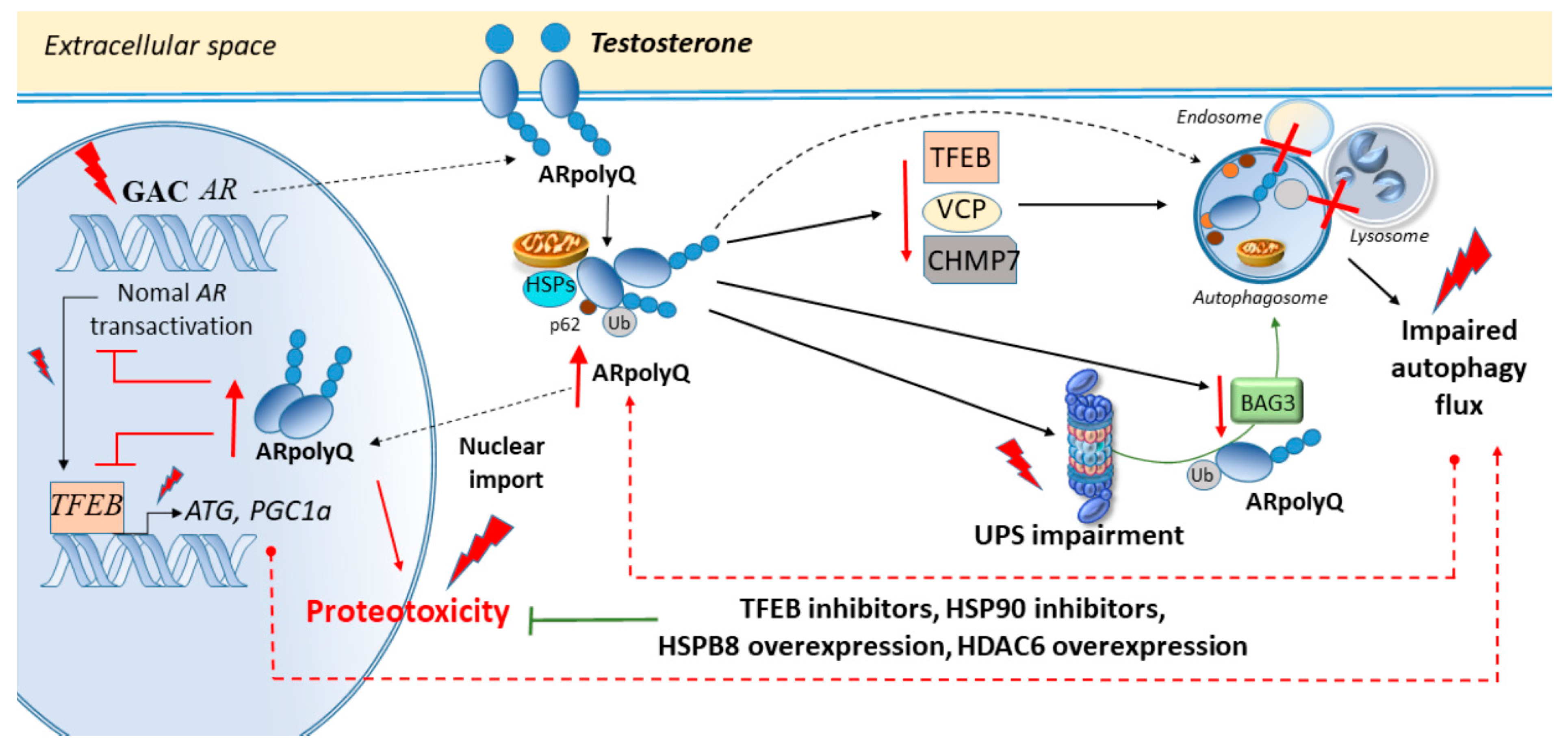
3. Cell-Clearing Systems and AR Nucleotide Repeat Expansions in SBMA

Analogously to what occurs in C9ORF72-ALS, tandem repeats in exon 1 of the AR gene in SBMA lead to an abnormal CAG expansion which produces a long polyglutamine tract (polyQ) in the AR protein [12]. The mutant AR (ARpolyQ) misfolds, and upon activation by the AR ligand testosterone, it forms cytoplasmic and toxic nuclear aggregates through a gain-of function mechanism. Despite neurotoxicity being largely associated with the formation of nuclear aggregates, nuclear localization of ARpolyQ is necessary though not sufficient for toxicity [112]. Remarkably, improving ARpolyQ cytoplasmic clearance contributes to decreasing ARpolyQ nuclear accumulation, which indicates that the occlusion of autophagy-dependent cytoplasmic ARpolyQ enhances the toxicity of nuclear ARpolyQ [112]. In fact, while normal AR promotes the activation of the autophagy inducer TFEB, ARpolyQ impairs TFEB-dependent autophagy flux in motor neuron-like cells [23]. The combination of the TFEB-related autophagy inducer trehalose and the antiandrogen bicalutamide which slows down AR nuclear translocation, reduces insoluble ARpolyQ within motor neurons with a higher efficiency compared with single treatments [113]. Such a combination allows an increased recognition of misfolded species by the autophagy pathway prior to their migration into the nucleus while clearing insoluble AR species which bear a very long polyQ (Q112) tract [113]. Again, in neuronal cells, the heat shock protein 90 (HSP90) inhibitor 17-(allylamino)-17-demethoxygeldanamycin (17-AAG) exerts beneficial action in SBMA [114], and this occurs through an autophagy-dependent clearance of ARpolyQ [115]. Intriguingly, 17-AAG is unable to counteract SOD1 and TDP-43 aggregation, suggesting a quite specific role for HSP90 in AR aggregation [115]. The chaperone HSPB8 also facilitates the autophagy-mediated removal of ARpolyQ aggregating species [116]. Despite not influencing p62 and LC3 levels, it does prevent p62 bodies formation while restoring autophagy flux. Trehalose, which counteracts ARpolyQ through autophagy activation, also induces HSPB8 expression indicating the key role of HSPB8-related autophagy as a potential target against ARpolyQ toxicity [116].
Besides autophagy, rescuing the UPS may be key to counteracting ARpolyQ-protein degradation, as well as mitochondrial alterations and axonal transport defects in SBMA models [117]. In fact, ARpolyQ aggregates sequester mitochondria and stain positively for heat shock proteins such as HSP90 and HSP70 as well as UPS subunits, suggesting a breakdown in UPS processing [9,118]. Ubiquitin is also detected in aggregated ARpolyQ nuclear species whose proteolysis eventually requires UPS activity [119]. Recent studies showed that HSPA8 and its cochaperone BAG1 are key to rerouting ARpolyQ towards the UPS when dynein-mediated retrograde targeting of misfolded proteins to the autophagy pathway is impaired [21]. Again, in a mouse model of SBMA featuring neuronal ARpolyQ obtained through the Tet-On system, cepharanthine phytochemical from Stephania cepharantha decreases ARpolyQ levels both in the cytoplasm and nucleus [120]. In detail, while the UPS appears to be mostly implicated in WT AR degradation, autophagy induction following cepharanthine administration enhances the clearance of cytoplasmic ARpolyQ while providing neuroprotection [120].
In transgenic Drosophila, ARpolyQ produces ligand-dependent degeneration of specific neurons resulting in a rough eye phenotype, which is associated with an impairment of both the UPS and autophagy [121]. Inducing autophagy through overexpression of histone deacetylase 6 (HDAC6) accelerates the turnover of the ARpolyQ while lowering steady-state levels of monomeric and aggregated ARpolyQ in these fly models [121]. Treatment with the TOR inhibitor rapamycin reproduces these effects [121], suggesting a synergistic activity between autophagy and the UPS. This is in line with recent studies linking mTOR inhibition to a simultaneous activation of autophagy and the UPS [4,122,123].

Autophagy and the UPS in SBMA Muscle and Axons

Beyond motor neurons within the spinal cord and brainstem, AR mutations also affect dorsal root ganglia neurons and skeletal muscle cells, leading to sensory dysfunctions and atrophy of bulbar, facial, and limb muscles, with CAG repeat size differentially correlating with motor- and sensory-dominant phenotypes [10,124,125,126,127]. Recent studies suggest that SBMA first manifests in skeletal muscle, prior to any motor neuron degeneration which only occurs in late-stage disease [127]. Although the polyQ expansion is known to impart a toxic gain-of-function effect upon the mutant AR protein, evidence has been provided showing that SBMA pathogenesis may concomitantly involve an AR gain-of-function toxicity and loss of normal AR function, reminiscent of what reported for C9ORF72 repeat expansions [27]. In fact, androgens acting through the AR are important for muscle development suggesting that both loss of normal AR functions and gain of novel harmful functions can contribute to neurodegeneration and muscular atrophy [27,117]. This was addressed by crossing transgenic mice models harboring 100 AR glutamines (AR100) with AR-null mice (testicular feminization; Tfm) [27]. Absence of the endogenous AR protein in AR100Tfm mice has profound effects upon neuromuscular and endocrine features, leading to neurodegeneration and severe androgen insensitivity compared with AR100 littermates. Remarkably, AR transactivation diminishes competitively in a polyQ length-dependent way. Reduction in size and number of androgen-sensitive motor neurons in the spinal cord of AR100Tfm mice underscores the importance of normal AR action for neuronal survival and muscle function [27]. This is key since the transcriptional activity of the normal AR receptor is bound the autophagy pathway. In fact, the core autophagy genes ATG4B, ATG4D, ULK1, and ULK2, as well as TFEB, a master regulator of autophagosome–lysosomal biogenesis and function, are all transcriptional targets of AR [23,28]. This suggests that ARpolyQ may impair cell-clearing systems both directly, by forming large aggregates which engulf autophagy compartments and the UPS, and also indirectly, by competitively blocking AR transactivation and subsequent autophagy induction. Intriguingly, similarly to ARpolyQ which blocks AR transactivation in a length-dependent way, AR transactivation is repressed in a dose-dependent manner by glycogen synthase kinase 3beta (GSK3b) [128], a well-known autophagy modulator [73,129,130,131]. Remarkably, the suppression of AR transactivation by GSK3b is abolished by the GSK3b inhibitor and autophagy inducer lithium [128].
The effects of GSK3b and lithium are poorly investigated in the context of SBMA. However, lithium exerts beneficial effects in both ALS patients and experimental models, where it rescues affected motor neurons, axons, and skeletal muscles through autophagy induction and restoration of mitochondrial homeostasis [73,95,131,132,133,134,135,136,137]. Remarkably, in human muscle cultures, GSK3b activity is also enhanced by UPS inhibition, while treatment with lithium rescues UPS activity while preventing protein aggregation [138]. These findings add to the vast body of evidence linking lithium-induced inhibition of GSK3b activity with autophagy induction, showing that besides autophagy, GSK3b inhibition may empower the UPS.
This is key since RNA-sequencing studies recently identified the UPS as one of the major pathways being disrupted in the muscle of ARpolyQ-knockin mice [139]. In fact, numerous UPS genes are downregulated in AR113Q-expressing muscle, encoding approximately 30% of UPS subunits and 20% of E2 ubiquitin-conjugating enzymes. These changes are age, hormone, and glutamine length-dependent [139]. Furthermore, the reduction in the expression of UPS genes and catalytic activity is associated with decreased levels of the UPS transcription factor NRF1 and its activator DDI2. In fact, the downregulation of NRF1 or ADRM1 Drosophila orthologues reproduces ARpolyQ accumulation and toxicity. These data indicate that AR113Q muscle develops progressive UPS dysfunction promoting the accumulation and toxicity of ARpolyQ protein in SBMA [139]. Conversely, enhancing UPS-dependent clearance of AR through administration of insulin-like growth factor 1 (IGF-1) reduces AR aggregation [140]. This is documented in vitro as well as in SBMA transgenic mice overexpressing a muscle-specific isoform of IGF-1, which leads to Akt-dependent AR phosphorylation and UPS-dependent AR clearance. This is associated with a reversal of behavioral and histopathological abnormalities, and reduction of both muscle and spinal cord pathology [140].
Supporting a role for early impairment of the UPS in SBMA muscle, an increased BAG3:BAG1 ratio along with autophagy markers is detected in the muscle of AR113Q mice, suggesting a preferential routing of misfolded proteins to the autophagy pathway [141]. Recent studies in stabilized skeletal myoblasts show that ARpolyQ forms testosterone-inducible insoluble aggregates which are processed by both the UPS and autophagy. Intriguingly, while the UPS clears both WT AR and ARpolyQ, autophagy clears mostly ARpolyQ and it is early activated by ARpolyQ itself [10]. Nonetheless, ARpolyQ, even in the absence of testosterone, reduces the expression of two autophagy-related proteins BAG3 and VCP, eventually impairing autophagy response in ARpolyQ s-myoblasts [10]. Overexpression of BAG3 ameliorates ARpolyQ clearance, while the treatment with trehalose induces complete ARpolyQ degradation, suggesting that ARpolyQ may eventually impair autophagy besides the UPS in muscle cells [10].
Similar to what was observed for dynein/dynactin-related alterations in ALS, mutations in DNCT1 in a transgenic mouse model of SBMA lead to late-onset, slowly progressive muscle weakness along with deficits in axonal caliber and NMJ integrity, indicating a distal degeneration of motor neurons [142]. Remarkably, this is associated with accumulation of enlarged lysosomes and lipofuscin granules, witnessing for an impaired fusion with autophagosomes [142]. Similar defects in autophagy flux are observed within the motor neurons and hindlimb muscle of SBMA mice models, as well as in SBMA mice embryonic motor neurons and in a human cell model of motor neuron precursor cells derived from reprogrammed patient fibroblasts [16]. In detail, ARpolyQ expansion results in early transcriptional downregulation of the charged multivesicular body protein 7 (CHMP7), which leads to impaired autophagy flux and alterations in the endosome–lysosome pathways. This is intriguing since CHMP7 is part of the endosomal sorting complexes required for transport (ESCRT)-III complex that also includes CHMP2, which is mutated in ALS [143]. These proteins sort ubiquitinated proteins from endosomes to the lysosome through formation of multivesicular bodies while delivering autophagosomes to lysosomes [16]. Besides leading to lysosomes accumulation and impaired autophagy flux, ARpolyQ-induced CHMP7 downregulation is also bound to gene pathways which are associated with mitochondrial clearance and axonal branching as well as NMJ formation and maintenance [16]. Among these genes, an enrichment in the GSK3b signaling pathway, which is known to promote autophagy and UPS inhibition, was identified in SBMA motor neurons along with a concomitant downregulation in pathways being involved in NMJ development [16]. Thus, autophagy and UPS alterations may play a key role in the development of SBMA by affecting early NMJ integrity while promoting protein aggregation and toxicity (Figure 3). In light of these considerations, potential mechanisms through which autophagy and the UPS may affect NMJ integrity will be discussed in the following section.

4. Potential Mechanisms Linking Cell-Clearing Systems and Neuromuscular Junction Alterations in ALS and SBMA

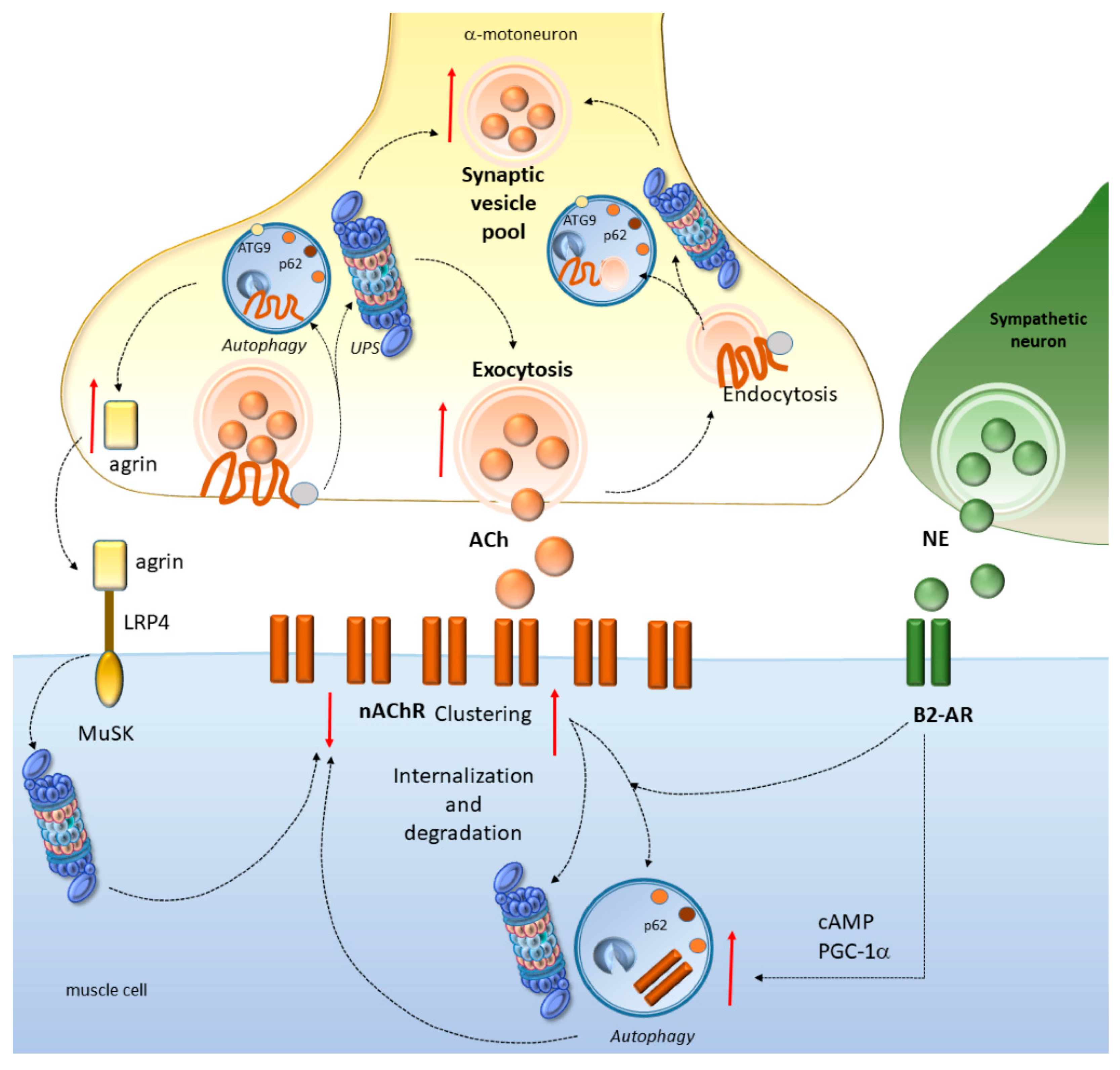
4.1. Autophagy and the UPS Regulate NMJ Development and Function

In Drosophila, autophagy promotes NMJ growth by reducing the levels of a mutated E3 ubiquitin ligase (highwire), suggesting that autophagy may compensate for UPS dysfunction during NMJ development [144]. Autophagy is also key for presynaptic assembly and for axon outgrowth dynamics. In fact, as shown in Caenorhabditis elegans, autophagosome biogenesis occurs in the axon near synapses, and this is largely bound to the presence of the integral membrane autophagy protein ATG9 [145]. In turn, alterations of autophagy in either motor neurons or skeletal muscles promote early NMJ disruption and axon degeneration [146]. This is documented in murine models bearing a transmembrane protein 184B (TMEM184b) mutation, which leads to early sensory–motor alterations that are associated with accumulation of stagnant, rimmed autophagy vacuoles and inclusions [146]. Remarkably, these are reminiscent of those caused by mutations in the autophagy-regulating VCP [146,147]. Again, the ultrastructural terminals of mutant TMEM184b mice feature alterations which resemble those occurring in models of neuro-axonal dystrophy caused by PLA2G6 phospholipase mutations [146,148,149]. Finally, dystrophic presynaptic swellings which are consistently found in TMEM184b mutants are reminiscent of those occurring in mice bearing a spontaneous mutation in the deubiquitinating protease USP14 [146,150]. This is associated with an abnormal accumulation of UPS substrates in axon terminals [150]. These findings strengthen the evidence that autophagy and the UPS are critically involved in sensory–motor terminal structure and function. This may be relevant for early axon degeneration which occurs in neuromuscular disorders including ALS and SBMA.

4.2. Autophagy and the UPS Regulate Neurotransmission at the NMJ

Besides clearing potentially toxic protein aggregates to maintain synaptic and axonal proteostasis, autophagy and the UPS play a key role in neurotransmitter release [39,40,41,42,151,152,153,154,155]. At the Drosophila NMJ, the presynaptically-enriched chaperone Hsc70-4, which is known to form a multimeric complex with HSPB8/BAG3, promotes autophagy to modulate neurotransmitter release through the turnover of specific synaptic proteins such as Unc-13, EndophilinA, WASp, and Comt/NSF [40]. Loss of autophagy slows down neurotransmission, while potentiating autophagy increases neurotransmission through the formation of a larger, readily releasable synaptic vesicle pool. Such a process is modulated by Sgt, a cochaperone of Hsc70-4, which is able to switch the activity of Hsc70-4 from autophagy-promoting toward a protein-folding activity. Thus, Hsc70-4 controls rejuvenation of the synaptic protein pool in a dual way, either by refolding proteins together with Sgt, or by targeting them for autophagy-dependent degradation [40]. Similar findings are reported in mice featuring UPS dysfunctions due to USP14 mutations [42]. This is associated with a reduction in the size of the readily-releasable vesicle pool within the NMJ, which cannot keep pace with physiological rates of transmitter release [42]. Likewise, in mice carrying a spontaneous mutation in the E3 ubiquitin ligase HERC1, a reduction of the motor end-plate area is detected along with inefficient neuromuscular activity and impaired evoked neurotransmitter release at the NMJ [155]. Similarly to autophagy, the UPS at the Drosophila NMJ controls synaptic vesicle priming and neurotransmitter release probability through the turnover of UNC-13 presynaptic protein [156], which is altered in neurological disorders including ALS [131,137]. Therefore, autophagy and UPS-dependent targeting of synaptic proteins is key to orchestrating neurotransmitter release and the size of synaptic vesicles pools at the NMJ.

4.3. Autophagy and the UPS Regulate nAChR Turnover at the NMJ

At vertebrate motor endplates, the conversion of nerve impulses into muscle contraction is initiated by binding of acetylcholine to its nicotinic receptor (nAChR) at the postsynapse [44]. Efficiency and safety of this process largely depend on proper localization of the receptors, which in turn, depends on autophagy- and UPS-dependent turnover. In fact, by intermingling with endocytic trafficking pathways, autophagy and the UPS orchestrate the delivery and clustering of nAChR to the postsynaptic membrane, as well as its endocytic retrieval, leading to either recycling or degradation of nAChR [44,157,158]. In detail, autophagy regulates the turnover of the endocytosed nAChR in cooperation with the E3 ubiquitin ligase, TRIM63, and SQSTM1/p62 [157]. Inhibition of the UPS in cell lines leads to a marked upregulation nAChR, which is reproduced, though at a lesser extent, by the administration of autophagy inhibitors [159]. In detail, nAChR ubiquitination and UPS-dependent degradation modulates its distribution between specialized intracellular compartments and the plasma membrane. This effect is achieved by controlling the stability of the alpha3 subunit and, consequently, the number of receptors at the cell surface [160]. Mice with defective autophagy due to skeletal muscle-specific loss of ATG7, display alterations of nAChR turnover and of endosome trafficking, which goes along with fragmentation of NMJs, and early synaptic dysfunction including partial denervation [15]. In line with this, nAChR expression is increased under muscle wasting conditions such as immobilization and denervation, highlighting the key role of nAChR turnover by autophagy and the UPS in these conditions [157,159].
Muscle denervation in ALS has a deep impact on AChR composition and distribution, and missense variants in nAChR genes are detected in sALS patients [161]. In detail, mutations within alpha3 and alpha4 nAChRs subunits lead to altered receptor trafficking associated with reduced receptor desensitization and sustained intracellular Ca(2+) concentration compared with WT-nAChR [161]. Alterations of nAChR are also reported in SBMA. Recently, a genome-wide transcriptome analysis in SBMA-derived iPSCs differentiated into spinal motor neurons revealed the involvement of synapse alterations associated with aberrant AChR clustering and NMJ formation [38]. This goes along with the upregulation of synaptic proteins which are substrates of autophagy and the UPS, such as synaptotagmin and synaptophysin [38]. Remarkably, nAChR mRNA levels are also upregulated in SBMA mice models featuring muscle AR toxicity [35,162]. This is associated with slowed synaptic potentials and reduced size of the readily releasable synaptic vesicle pool and probability of release [35], which are known to be orchestrated by the UPS and autophagy.
Autophagy and the UPS are also bound to key proteins belonging to a complex-signaling cascade which is required for synapse formation at the mammalian NMJ [41,43]. This is the case of agrin, a motor neuron-derived proteoglycan that stabilizes the junction, and muscle-specific receptor tyrosine kinase (MuSK), a key organizer of post-synaptic components. At the level of muscle fibers, agrin normally inhibits the dispersal of AChRs while its alterations lead to endplate fragmentation [163]. In mice featuring an mTOR-dependent genetic suppression of autophagy, a downregulation of agrin occurs, which is associated with marked NMJ malformations, aberrantly distributed nAChRs, and varicose presynaptic nerve terminals in the muscle [43]. Remarkably, administration of the mTOR inhibitor torin2 reverses both autophagy and agrin downregulation in muscle and nerves while preventing NMJ alterations. Thus, autophagy failure is bound to an aberrant distribution of AChRs and NMJ malformations associated with agrin signaling alterations. Again, the UPS component PDZRN3, a PDZ domain containing the Ring ubiquitin ligase, acts as a MuSK-binding partner. PDZRN3 is concentrated at postsynapses and it promotes MuSK ubiquitination while suppressing the agrin-induced AChRs clustering. The lack of Ring domain abolishes such an effect, suggesting that the UPS is important for AChRs clustering and NMJ development [164].

4.4. Autophagy Converging with the Sympathetic Innervation of NMJs

Besides the tuning of autophagy and of the agrin pathway, the beneficial effects of the sympathetic innervation of NMJs are being widely investigated in the treatment of muscle wasting disorders, including ALS and SBMA [45]. In detail, sympathetic neurons make close contacts with NMJs and they form a network involving blood vessels, motor neurons, and muscle fibers, which is crucial for synapse maintenance and function [165]. Direct stimulation of sympathetic neurons leads to activation of muscle postsynaptic β2-adrenoreceptor (B2-AR) and subsequent cAMP production, which is also key to control the abundance and distribution of AChRs in NMJs [165]. These events are molecularly associated with the import of the transcriptional coactivator peroxisome proliferator-activated receptor γ-coactivator 1α (PPARGC1A, or PGC-1α) into myonuclei [165]. Remarkably, PPARGC1A is known to upregulate autophagy through a SQSTM1-dependent mechanism [166]. In turn, the UPS mediates PPARGC1A nuclear degradation and governs its cellular localization, half-life, and potential biological actions [167]. These findings provide a potential mechanistic link between sympathetic innervation and cell-clearing systems in NMJ homeostasis. This is further supported by studies showing that trehalose, which has beneficial effects in both ALS and SBMA, induces autophagy in a TFEB-dependent manner through upregulation of PPARGC1A as well as well-known autophagy-related genes such as BECN1, LC3, ATG10, ATG12, and SQSTM1/p62 [101]. This is key since norepinephrine was shown to activate autophagy through B2-ARs [46,168,169], even though contradictory results have been provided documenting an activation of the mTOR pathway following chronic B2-AR administration, which is supposed to inhibit autophagy [170]. However, the fact that PPARGC1A activation similarly occurs following B2-AR stimulation and TFEB activation suggests that B2-ARs likely induce autophagy through mTOR-independent mechanisms, which calls for confirmatory studies. Altogether, these findings suggest that cell-clearing systems are key to grant NMJ homeostasis at baseline (Figure 4), while their alterations might promote NMJ derangements which occur early in ALS and SBMA.

5. Conclusions and Future Directions

The findings here discussed suggest that boosting autophagy/UPS activity might counteract early NMJ alterations while facilitating the removal of potentially-toxic protein inclusions, including DPR and ARpolyQ which arise from C9ORF72 and AR repeat expansions, respectively. Obviously, besides ALS and SBMA, this may apply to a very broad range of neurodegenerative proteoinopathies, including those featuring poly-Q protein aggregation such as HD and SCA, where mechanisms underlying autophagy–UPS crosstalk would similarly deserve to be dealt with. Another issue that deserves further attention is the potentially-different role of autophagy vs. the UPS in DPR degradation. In fact, DPR proteins possess peculiar structures and dynamics compared with classical misfolded proteins and their toxicity may also differ for each specific DPR [171,172]. Albeit being less frequent than poly-GP and poly-GA DRP, arginine-containing DPR poly-GR and poly-PR are generally associated with greatest toxicity [172]. Again, DPR inclusions generally sequester SQSTM1/p62, though the preferred pathway of degradation is not uniform for the five DPR proteins [171]. Of all five DPR, only the polyGP seems to be efficiently removed via the UPS, while the others are apparently to be mainly degraded via autophagy. An exception is represented by polyPR, which is not significantly affected by autophagy inhibition through 3-MA administration or HSPB8 depletion [171]. This is likely due to the fact that polyPR inclusions are mostly detectable in the nucleus, where they cannot be cleared by autophagy. Remarkably, HSPB8 overexpression significantly and robustly counteracts the accumulation of insoluble species of all five DPR proteins, which suggests that the action of this chaperone may take place before polyPR nuclear import and aggregation [171]. How HSPB8 recognizes and facilitates clearance of DPR proteins remains to be determined. A peculiar feature of arginine-containing DPR proteins is their potential for post-translational modification by arginine methyl-transferases, which produces methylarginine DPR [172]. Supporting a possible contribution of methylarginine post-translational modification to poly-GR toxicity, an association between dimethylarginine-poly-GR and neurodegeneration was documented in C9-ALS and/or FTD patients [172]. A role for arginine methylation has also been documented in the pathogenesis of experimental FUS-related ALS [173]. Intriguingly, posttranslational arginine methylation is key for modulating degradation efficiency in selective autophagy by regulating the association of the cargo-receptor complex with the scaffold protein [174]. This calls for further studies investigating the potential relationship between alterations in autophagy/UPS and methyl-transferases in the mechanisms of proteotoxicity.

Author Contributions

Writing—original draft preparation, F.L. and F.F.; writing—review, editing, and art-work F.L., C.L.B.; F.B., F.C., P.F. and P.L.; conceptualization, F.L., C.L.B., P.L. and F.F.; supervision F.F. All authors have read and agreed to the published version of the manuscript.

Funding

This research was funded by Ministero della Salute (Ricerca Corrente 2020).

Conflicts of Interest

The authors declare no conflict of interest.

Abbreviations

UPSubiquitin proteasome system
C9ORF72chromosome 9 open reading frame 72
ARandrogen receptor
ALSamyotrophic lateral sclerosis
SMBAspinal bulbar muscular atrophy
FTDfrontotemporal dementia
NMJneuromuscular junction
ARpolyQpolyglutamine-expanded AR
DPRdipeptide repeat
AChRsacetylcholine receptors
B2-ARsadrenergic beta2 receptors
TDP-43TAR DNA-binding protein 43
SOD1superoxide dismutase 1
TARDBPtranscription of RNA activating protein/TAR DNA-binding protein
FUSfused in sarcoma
OPTNoptineurin
UBQLN2ubiquilin-2
PGRNprogranulin
ATXN-2ataxin-2
VCPvalosin-containing protein
DCTN1dynactin
mTORC1mammalian target of rapamycin complex 1
SQSTM1/p62sequestosome 1/p62
HSPA8heat shock protein family A (Hsp70) member 8
Hsc70heat shock cognate 71 kDa protein
BAG1/3Bcl-2-associated athanogene 1/3
TFEBtranscription factor EB
GSK3bglycogen synthase kinase 3beta
CHMP2/7charged multivesicular body protein 2/7
ESCRT-IIIendosomal sorting complexes required for transport III
TMEM184btransmembrane protein 184B
MuSKmuscle-specific receptor tyrosine kinase
PPARGC1A or PGC-1αperoxisome proliferator-activated receptor γ-coactivator 1α

References

  1. Madeo, F.; Eisenberg, T.; Kroemer, G. Autophagy for the avoidance of neurodegeneration. Genes Dev. 2009, 23, 2253–2259. [Google Scholar] [CrossRef] [PubMed] [Green Version]
  2. Ciechanover, A.; Kwon, Y.T. Degradation of misfolded proteins in neurodegenerative diseases: Therapeutic targets and strategies. Exp. Mol. Med. 2015, 47, e147. [Google Scholar] [CrossRef] [PubMed] [Green Version]
  3. Ryskalin, L.; Busceti, C.L.; Limanaqi, F.; Biagioni, F.; Gambardella, S.; Fornai, F. A Focus on the Beneficial Effects of Alpha Synuclein and a Re-Appraisal of Synucleinopathies. Curr. Protein Pept. Sci. 2018, 19, 598–611. [Google Scholar] [CrossRef] [PubMed]
  4. Limanaqi, F.; Biagioni, F.; Gambardella, S.; Familiari, P.; Frati, A.; Fornai, F. Promiscuous Roles of Autophagy and Proteasome in Neurodegenerative Proteinopathies. Int. J. Mol. Sci. 2020, 21, 3028. [Google Scholar] [CrossRef]
  5. Jimenez-Sanchez, M.; Thomson, F.; Zavodszky, E.; Rubinsztein, D.C. Autophagy and polyglutamine diseases. Prog. Neurobiol. 2012, 97, 67–82. [Google Scholar] [CrossRef] [Green Version]
  6. Pearce, M.M.P.; Kopito, R.R. Prion-Like Characteristics of Polyglutamine-Containing Proteins. Cold Spring Harb. Perspect. Med. 2018, 8, a024257. [Google Scholar] [CrossRef] [Green Version]
  7. Babić Leko, M.; Župunski, V.; Kirincich, J.; Smilović, D.; Hortobágyi, T.; Hof, P.R.; Šimić, G. Molecular Mechanisms of Neurodegeneration Related to C9orf72 Hexanucleotide Repeat Expansion. Behav. Neurol. 2019, 2019, 2909168. [Google Scholar] [CrossRef] [Green Version]
  8. Davies, J.E.; Sarkar, S.; Rubinsztein, D.C. The ubiquitin proteasome system in Huntington’s disease and the spinocerebellar ataxias. BMC Biochem. 2007, 8 (Suppl. 1), S2. [Google Scholar] [CrossRef] [Green Version]
  9. Abel, A.; Walcott, J.; Woods, J.; Duda, J.; Merry, D.E. Expression of expanded repeat androgen receptor produces neurologic disease in transgenic mice. Hum. Mol. Genet. 2001, 10, 107–116. [Google Scholar] [CrossRef] [Green Version]
  10. Cicardi, M.E.; Cristofani, R.; Crippa, V.; Ferrari, V.; Tedesco, B.; Casarotto, E.; Chierichetti, M.; Galbiati, M.; Piccolella, M.; Messi, E.; et al. Autophagic and Proteasomal Mediated Removal of Mutant Androgen Receptor in Muscle Models of Spinal and Bulbar Muscular Atrophy. Front. Endocrinol. (Lausanne) 2019, 10, 569. [Google Scholar] [CrossRef]
  11. Van Blitterswijk, M.; DeJesus-Hernandez, M.; Rademakers, R. How do C9ORF72 repeat expansions cause amyotrophic lateral sclerosis and frontotemporal dementia: Can we learn from other noncoding repeat expansion disorders? Curr. Opin. Neurol. 2012, 25, 689–700. [Google Scholar] [CrossRef] [PubMed] [Green Version]
  12. La Spada, A.R.; Wilson, E.M.; Lubahn, D.B.; Harding, A.E.; Fischbeck, K.H. Androgen receptor gene mutations in X-linked spinal and bulbar muscular atrophy. Nature 1991, 352, 77–79. [Google Scholar] [CrossRef] [PubMed]
  13. Parboosingh, J.S.; Figlewicz, D.A.; Krizus, A.; Meininger, V.; Azad, N.A.; Newman, D.S.; Rouleau, G.A. Spinobulbar muscular atrophy can mimic ALS: The importance of genetic testing in male patients with atypical ALS. Neurology 1997, 49, 568–572. [Google Scholar] [CrossRef] [PubMed]
  14. Pradat, P.F. SBMA: A rare disease but a classic ALS mimic syndrome. Presse Med. 2014, 43, 580–586. [Google Scholar] [CrossRef] [PubMed]
  15. Carnio, S.; LoVerso, F.; Baraibar, M.A.; Longa, E.; Khan, M.M.; Maffei, M.; Reischl, M.; Canepari, M.; Loefler, S.; Kern, H.; et al. Autophagy impairment in muscle induces neuromuscular junction degeneration and precocious aging. Cell Rep. 2014, 8, 1509–1521. [Google Scholar] [CrossRef] [PubMed]
  16. Malik, B.; Devine, H.; Patani, R.; La Spada, A.R.; Hanna, M.G.; Greensmith, L. Gene expression analysis reveals early dysregulation of disease pathways and links Chmp7 to pathogenesis of spinal and bulbar muscular atrophy. Sci. Rep. 2019, 9, 3539. [Google Scholar] [CrossRef] [Green Version]
  17. Rudnick, N.D.; Griffey, C.J.; Guarnieri, P.; Gerbino, V.; Wang, X.; Piersaint, J.A.; Tapia, J.C.; Rich, M.M.; Maniatis, T. Distinct roles for motor neuron autophagy early and late in the SOD1G93A mouse model of ALS. Proc. Natl. Acad. Sci. USA 2017, 114, E8294–E8303. [Google Scholar] [CrossRef] [Green Version]
  18. Zhu, Q.; Jiang, J.; Gendron, T.F.; McAlonis-Downes, M.; Jiang, L.; Taylor, A.; Diaz Garcia, S.; Ghosh Dastidar, S.; Rodriguez, M.J.; King, P.; et al. Reduced C9ORF72 function exacerbates gain of toxicity from ALS/FTD-causing repeat expansion in C9orf72. Nat. Neurosci. 2020. [Google Scholar] [CrossRef]
  19. Khosravi, B.; LaClair, K.D.; Riemenschneider, H.; Zhou, Q.; Frottin, F.; Mareljic, N.; Czuppa, M.; Farny, D.; Hartmann, H.; Michaelsen, M.; et al. Cell-to-cell transmission of C9orf72 poly-(Gly-Ala) triggers key features of ALS/FTD. EMBO J. 2020, 39, e102811. [Google Scholar] [CrossRef]
  20. Boivin, M.; Pfister, V.; Gaucherot, A.; Ruffenach, F.; Negroni, L.; Sellier, C.; Charlet-Berguerand, N. Reduced autophagy upon C9ORF72 loss synergizes with dipeptide repeat protein toxicity in G4C2 repeat expansion disorders. EMBO J. 2020, 39, e100574. [Google Scholar] [CrossRef]
  21. Cristofani, R.; Crippa, V.; Rusmini, P.; Cicardi, M.E.; Meroni, M.; Licata, N.V.; Sala, G.; Giorgetti, E.; Grunseich, C.; Galbiati, M.; et al. Inhibition of retrograde transport modulates misfolded protein accumulation and clearance in motoneuron diseases. Autophagy 2017, 13, 1280–1303. [Google Scholar] [CrossRef] [PubMed]
  22. Webster, C.P.; Smith, E.F.; Bauer, C.S.; Moller, A.; Hautbergue, G.M.; Ferraiuolo, L.; Myszczynska, M.A.; Higginbottom, A.; Walsh, M.J.; Whitworth, A.J.; et al. The C9orf72 protein interacts with Rab1a and the ULK1 complex to regulate initiation of autophagy. EMBO J. 2016, 35, 1656–1676. [Google Scholar] [CrossRef] [PubMed]
  23. Cortes, C.J.; Miranda, H.C.; Frankowski, H.; Batlevi, Y.; Young, J.E.; Le, A.; Ivanov, N.; Sopher, B.L.; Carromeu, C.; Muotri, A.R.; et al. Polyglutamine-expanded androgen receptor interferes with TFEB to elicit autophagy defects in SBMA. Nat. Neurosci. 2014, 17, 1180–1189. [Google Scholar] [CrossRef] [PubMed] [Green Version]
  24. Gupta, R.; Lan, M.; Mojsilovic-Petrovic, J.; Choi, W.H.; Safren, N.; Barmada, S.; Lee, M.J.; Kalb, R. The Proline/Arginine Dipeptide from Hexanucleotide Repeat Expanded C9ORF72 Inhibits the Proteasome. eNeuro 2017, 4. [Google Scholar] [CrossRef] [PubMed] [Green Version]
  25. Thibaudeau, T.A.; Anderson, R.T.; Smith, D.M. A common mechanism of proteasome impairment by neurodegenerative disease-associated oligomers. Nat. Commun. 2018, 9, 1097. [Google Scholar] [CrossRef] [PubMed]
  26. Abo-Rady, M.; Kalmbach, N.; Pal, A.; Schludi, C.; Janosch, A.; Richter, T.; Freitag, P.; Bickle, M.; Kahlert, A.K.; Petri, S.; et al. Knocking out C9ORF72 Exacerbates Axonal Trafficking Defects Associated with Hexanucleotide Repeat Expansion and Reduces Levels of Heat Shock Proteins. Stem Cell Rep. 2020, 14, 390–405. [Google Scholar] [CrossRef] [Green Version]
  27. Thomas, P.S., Jr.; Fraley, G.S.; Damian, V.; Woodke, L.B.; Zapata, F.; Sopher, B.L.; Plymate, S.R.; La Spada, A.R. Loss of endogenous androgen receptor protein accelerates motor neuron degeneration and accentuates androgen insensitivity in a mouse model of X-linked spinal and bulbar muscular atrophy. Hum. Mol. Genet. 2006, 15, 2225–2238. [Google Scholar] [CrossRef] [Green Version]
  28. Blessing, A.M.; Rajapakshe, K.; Reddy Bollu, L.; Shi, Y.; White, M.A.; Pham, A.H.; Lin, C.; Jonsson, P.; Cortes, C.J.; Cheung, E.; et al. Transcriptional regulation of core autophagy and lysosomal genes by the androgen receptor promotes prostate cancer progression. Autophagy 2017, 13, 506–521. [Google Scholar] [CrossRef]
  29. Ciura, S.; Sellier, C.; Campanari, M.L.; Charlet-Berguerand, N.; Kabashi, E. The most prevalent genetic cause of ALS-FTD, C9orf72 synergizes the toxicity of ATXN2 intermediate polyglutamine repeats through the autophagy pathway. Autophagy 2016, 12, 1406–1408. [Google Scholar] [CrossRef] [Green Version]
  30. Pasquali, L.; Ruffoli, R.; Fulceri, F.; Pietracupa, S.; Siciliano, G.; Paparelli, A.; Fornai, F. The role of autophagy: What can be learned from the genetic forms of amyotrophic lateral sclerosis. CNS Neurol. Disord. Drug Targets 2010, 9, 268–278. [Google Scholar] [CrossRef]
  31. Ferrucci, M.; Fulceri, F.; Toti, L.; Soldani, P.; Siciliano, G.; Paparelli, A.; Fornai, F. Protein clearing pathways in ALS. Arch. Ital. Biol. 2011, 149, 121–149. [Google Scholar] [CrossRef] [PubMed]
  32. Maurel, C.; Dangoumau, A.; Marouillat, S.; Brulard, C.; Chami, A.; Hergesheimer, R.; Corcia, P.; Blasco, H.; Andres, C.R.; Vourc’h, P. Causative Genes in Amyotrophic Lateral Sclerosis and Protein Degradation Pathways: A Link to Neurodegeneration. Mol. Neurobiol. 2018, 55, 6480–6499. [Google Scholar] [CrossRef] [PubMed]
  33. Manzano, R.; Sorarú, G.; Grunseich, C.; Fratta, P.; Zuccaro, E.; Pennuto, M.; Rinaldi, C. Beyond motor neurons: Expanding the clinical spectrum in Kennedy’s disease. J. Neurol. Neurosurg. Psychiatry 2018, 89, 808–812. [Google Scholar] [CrossRef] [PubMed]
  34. Starr, A.; Sattler, R. Synaptic dysfunction and altered excitability in C9ORF72 ALS/FTD. Brain Res. 2018, 1693, 98–108. [Google Scholar] [CrossRef] [PubMed]
  35. Xu, Y.; Halievski, K.; Henley, C.; Atchison, W.D.; Katsuno, M.; Adachi, H.; Sobue, G.; Breedlove, S.M.; Jordan, C.L. Defects in Neuromuscular Transmission May Underlie Motor Dysfunction in Spinal and Bulbar Muscular Atrophy. J. Neurosci. 2016, 36, 5094–5106. [Google Scholar] [CrossRef] [Green Version]
  36. Dupuis, L.; Echaniz-Laguna, A. Skeletal muscle in motor neuron diseases: Therapeutic target and delivery route for potential treatments. Curr. Drug Targets 2010, 11, 1250–1261. [Google Scholar] [CrossRef]
  37. Clark, J.A.; Southam, K.A.; Blizzard, C.A.; King, A.E.; Dickson, T.C. Axonal degeneration, distal collateral branching and neuromuscular junction architecture alterations occur prior to symptom onset in the SOD1(G93A) mouse model of amyotrophic lateral sclerosis. J. Chem. Neuroanat. 2016, 76, 35–47. [Google Scholar] [CrossRef]
  38. Onodera, K.; Shimojo, D.; Ishihara, Y.; Yano, M.; Miya, F.; Banno, H.; Kuzumaki, N.; Ito, T.; Okada, R.; de Araújo Herculano, B.; et al. Unveiling synapse pathology in spinal bulbar muscular atrophy by genome-wide transcriptome analysis of purified motor neurons derived from disease specific iPSCs. Mol. Brain 2020, 13, 18. [Google Scholar] [CrossRef] [Green Version]
  39. Shen, D.N.; Zhang, L.H.; Wei, E.Q.; Yang, Y. Autophagy in synaptic development, function, and pathology. Neurosci. Bull. 2015, 31, 416–426. [Google Scholar] [CrossRef] [Green Version]
  40. Uytterhoeven, V.; Lauwers, E.; Maes, I.; Miskiewicz, K.; Melo, M.N.; Swerts, J.; Kuenen, S.; Wittocx, R.; Corthout, N.; Marrink, S.J.; et al. Hsc70-4 Deforms Membranes to Promote Synaptic Protein Turnover by Endosomal Microautophagy. Neuron 2015, 88, 735–748. [Google Scholar] [CrossRef] [Green Version]
  41. Ding, M.; Shen, K. The role of the ubiquitin proteasome system in synapse remodeling and neurodegenerative diseases. Bioessays 2008, 30, 1075–1083. [Google Scholar] [CrossRef] [PubMed] [Green Version]
  42. Bhattacharyya, B.J.; Wilson, S.M.; Jung, H.; Miller, R.J. Altered neurotransmitter release machinery in mice deficient for the deubiquitinating enzyme Usp14. Am. J. Physiol. Cell. Physiol. 2012, 302, C698–C708. [Google Scholar] [CrossRef] [PubMed] [Green Version]
  43. Zhang, S.J.; Li, X.X.; Yu, Y.; Chiu, A.P.; Lo, L.H.; To, J.C.; Rowlands, D.K.; Keng, V.W. Schwann cell-specific PTEN and EGFR dysfunctions affect neuromuscular junction development by impairing Agrin signaling and autophagy. Biochem. Biophys. Res. Commun. 2019, 515, 50–56. [Google Scholar] [CrossRef] [PubMed]
  44. Rudolf, R.; Straka, T. Nicotinic acetylcholine receptor at vertebrate motor endplates: Endocytosis, recycling, and degradation. Neurosci. Lett. 2019, 711, 134434. [Google Scholar] [CrossRef]
  45. Rudolf, R.; Deschenes, M.R.; Sandri, M. Neuromuscular junction degeneration in muscle wasting. Curr. Opin. Clin. Nutr. Metab. Care 2016, 19, 177–181. [Google Scholar] [CrossRef] [Green Version]
  46. Aránguiz-Urroz, P.; Canales, J.; Copaja, M.; Troncoso, R.; Vicencio, J.M.; Carrillo, C.; Lara, H.; Lavandero, S.; Díaz-Araya, G. Beta(2)-adrenergic receptor regulates cardiac fibroblast autophagy and collagen degradation. Biochim. Biophys. Acta 2011, 1812, 23–31. [Google Scholar] [CrossRef] [Green Version]
  47. Lynch, E.; Semrad, T.; Belsito, V.S.; FitzGibbons, C.; Reilly, M.; Hayakawa, K.; Suzuki, M. C9ORF72-related cellular pathology in skeletal myocytes derived from ALS-patient induced pluripotent stem cells. Dis. Model Mech. 2019, 12, dmm039552. [Google Scholar] [CrossRef] [Green Version]
  48. Cykowski, M.D.; Powell, S.Z.; Appel, J.W.; Arumanayagam, A.S.; Rivera, A.L.; Appel, S.H. Phosphorylated TDP-43 (pTDP-43) aggregates in the axial skeletal muscle of patients with sporadic and familial amyotrophic lateral sclerosis. Acta Neuropathol. Commun. 2018, 6, 28. [Google Scholar] [CrossRef] [Green Version]
  49. Ho, W.Y.; Tai, Y.K.; Chang, J.C.; Liang, J.; Tyan, S.H.; Chen, S.; Guan, J.L.; Zhou, H.; Shen, H.M.; Koo, E.; et al. The ALS-FTD-linked gene product, C9orf72, regulates neuronal morphogenesis via autophagy. Autophagy 2019, 15, 827–842. [Google Scholar] [CrossRef]
  50. Guo, Q.; Lehmer, C.; Martínez-Sánchez, A.; Rudack, T.; Beck, F.; Hartmann, H.; Pérez-Berlanga, M.; Frottin, F.; Hipp, M.S.; Hartl, F.U.; et al. In Situ Structure of Neuronal C9orf72 Poly-GA Aggregates Reveals Proteasome Recruitment. Cell 2018, 172, 696–705.e12. [Google Scholar] [CrossRef] [Green Version]
  51. Yamakawa, M.; Ito, D.; Honda, T.; Kubo, K.; Noda, M.; Nakajima, K.; Suzuki, N. Characterization of the dipeptide repeat protein in the molecular pathogenesis of c9FTD/ALS. Hum. Mol. Genet. 2015, 24, 1630–1645. [Google Scholar] [CrossRef] [PubMed] [Green Version]
  52. Zhang, Y.J.; Gendron, T.F.; Grima, J.C.; Sasaguri, H.; Jansen-West, K.; Xu, Y.F.; Katzman, R.B.; Gass, J.; Murray, M.E.; Shinohara, M.; et al. C9ORF72 poly(GA) aggregates sequester and impair HR23 andnucleocytoplasmic transport proteins. Nat. Neurosci. 2016, 19, 668–677. [Google Scholar] [CrossRef] [PubMed]
  53. Boeve, B.F.; Boylan, K.B.; Graff-Radford, N.R.; DeJesus-Hernandez, M.; Knopman, D.S.; Pedraza, O.; Vemuri, P.; Jones, D.; Lowe, V.; Murray, M.E.; et al. Characterization of frontotemporal dementia and/or amyotrophic lateral sclerosis associated with the GGGGCC repeat expansion in C9ORF72. Brain 2012, 135, 765–783. [Google Scholar] [CrossRef] [PubMed]
  54. Cooper-Knock, J.; Hewitt, C.; Highley, J.R.; Brockington, A.; Milano, A.; Man, S.; Martindale, J.; Hartley, J.; Walsh, T.; Gelsthorpe, C.; et al. Clinico-pathological features in amyotrophic lateral sclerosis with expansions in C9ORF72. Brain 2012, 135, 751–764. [Google Scholar] [CrossRef]
  55. Al-Sarraj, S.; King, A.; Troakes, C.; Smith, B.; Maekawa, S.; Bodi, I.; Rogelj, B.; Al-Chalabi, A.; Hortobágyi, T.; Shaw, C.E. p62 positive, TDP-43 negative, neuronal cytoplasmic and intranuclear inclusions in the cerebellum and hippocampus define the pathology of C9orf72-linked FTLD and MND/ALS. Acta Neuropathol. 2011, 122, 691–702. [Google Scholar] [CrossRef]
  56. Sellier, C.; Campanari, M.L.; Julie Corbier, C.; Gaucherot, A.; Kolb-Cheynel, I.; Oulad-Abdelghani, M.; Ruffenach, F.; Page, A.; Ciura, S.; Kabashi, E.; et al. Loss of C9ORF72 impairs autophagy and synergizes with polyQ Ataxin-2 to induce motor neuron dysfunction and cell death. EMBO J. 2016, 35, 1276–1297. [Google Scholar] [CrossRef]
  57. van Blitterswijk, M.; van Es, M.A.; Hennekam, E.A.; Dooijes, D.; van Rheenen, W.; Medic, J.; Bourque, P.R.; Schelhaas, H.J.; van der Kooi, A.J.; de Visser, M.; et al. Evidence for an oligogenic basis of amyotrophic lateral sclerosis. Hum. Mol. Genet. 2012, 21, 3776–3784. [Google Scholar] [CrossRef] [Green Version]
  58. Ratti, A.; Corrado, L.; Castellotti, B.; Del Bo, R.; Fogh, I.; Cereda, C.; Tiloca, C.; D’Ascenzo, C.; Bagarotti, A.; Pensato, V.; et al. C9ORF72 repeat expansion in a large Italian ALS cohort: Evidence of a founder effect. Neurobiol. Aging 2012, 33, e7–e14. [Google Scholar] [CrossRef]
  59. Millecamps, S.; Boillée, S.; Le Ber, I.; Seilhean, D.; Teyssou, E.; Giraudeau, M.; Moigneu, C.; Vandenberghe, N.; Danel-Brunaud, V.; Corcia, P.; et al. Phenotype difference between ALS patients with expanded repeats in C9ORF72 and patients with mutations in other ALS-related genes. J. Med. Genet. 2012, 49, 258–263. [Google Scholar] [CrossRef]
  60. Ferrari, R.; Mok, K.; Moreno, J.H.; Cosentino, S.; Goldman, J.; Pietrini, P.; Mayeux, R.; Tierney, M.C.; Kapogiannis, D.; Jicha, G.A.; et al. Screening for C9ORF72 repeat expansion in FTLD. Neurobiol. Aging 2012, 33, e1–e11. [Google Scholar] [CrossRef] [Green Version]
  61. Van Blitterswijk, M.; Mullen, B.; Heckman, M.G.; Baker, M.C.; DeJesus-Hernandez, M.; Brown, P.H.; Murray, M.E.; Hsiung, G.Y.; Stewart, H.; Karydas, A.M.; et al. Ataxin-2 as potential disease modifier in C9ORF72 expansion carriers. Neurobiol. Aging 2014, 35, e13–e17. [Google Scholar] [CrossRef] [PubMed] [Green Version]
  62. Kwok, C.T.; Wang, H.Y.; Morris, A.G.; Smith, B.; Shaw, C.; de Belleroche, J. VCP mutations are not a major cause of familial amyotrophic lateral sclerosis in the UK. J. Neurol. Sci. 2015, 349, 209–213. [Google Scholar] [CrossRef] [PubMed]
  63. Münch, C.; Rosenbohm, A.; Sperfeld, A.D.; Uttner, I.; Reske, S.; Krause, B.J.; Sedlmeier, R.; Meyer, T.; Hanemann, C.O.; Stumm, G.; et al. Heterozygous R1101K mutation of the DCTN1 gene in a family with ALS and FTD. Ann. Neurol. 2005, 58, 777–780. [Google Scholar] [CrossRef] [PubMed]
  64. Münch, C.; Sedlmeier, R.; Meyer, T.; Homberg, V.; Sperfeld, A.D.; Kurt, A.; Prudlo, J.; Peraus, G.; Hanemann, C.O.; Stumm, G.; et al. Point mutations of the p150 subunit of dynactin (DCTN1) gene in ALS. Neurology 2004, 63, 724–726. [Google Scholar] [CrossRef]
  65. Renaud, L.; Picher-Martel, V.; Codron, P.; Julien, J.P. Key role of UBQLN2 in pathogenesis of amyotrophic lateral sclerosis and frontotemporal dementia. Acta Neuropathol. Commun. 2019, 7, 103. [Google Scholar] [CrossRef]
  66. Crippa, V.; Sau, D.; Rusmini, P.; Boncoraglio, A.; Onesto, E.; Bolzoni, E.; Galbiati, M.; Fontana, E.; Marino, M.; Carra, S.; et al. The small heat shock protein B8 (HspB8) promotes autophagic removal of misfolded proteins involved in amyotrophic lateral sclerosis (ALS). Hum. Mol. Genet. 2010, 19, 3440–3456. [Google Scholar] [CrossRef] [Green Version]
  67. Crippa, V.; Galbiati, M.; Boncoraglio, A.; Rusmini, P.; Onesto, E.; Giorgetti, E.; Cristofani, R.; Zito, A.; Poletti, A. Motoneuronal and muscle-selective removal of ALS-related misfolded proteins. Biochem. Soc. Trans. 2013, 41, 1598–1604. [Google Scholar] [CrossRef]
  68. Wang, X.; Fan, H.; Ying, Z.; Li, B.; Wang, H.; Wang, G. Degradation of TDP-43 and its pathogenic form by autophagy and the ubiquitin-proteasome system. Neurosci. Lett. 2010, 469, 112–116. [Google Scholar] [CrossRef]
  69. Scotter, E.L.; Vance, C.; Nishimura, A.L.; Lee, Y.B.; Chen, H.J.; Urwin, H.; Sardone, V.; Mitchell, J.C.; Rogelj, B.; Rubinsztein, D.C.; et al. Differential roles of the ubiquitin proteasome system and autophagy in the clearance of soluble and aggregated TDP-43 species. J. Cell Sci. 2014, 127, 1263–1278. [Google Scholar] [CrossRef] [Green Version]
  70. Cicardi, M.E.; Cristofani, R.; Rusmini, P.; Meroni, M.; Ferrari, V.; Vezzoli, G.; Tedesco, B.; Piccolella, M.; Messi, E.; Galbiati, M.; et al. Tdp-25 Routing to Autophagy and Proteasome Ameliorates its Aggregation in Amyotrophic Lateral Sclerosis Target Cells. Sci. Rep. 2018, 8, 12390. [Google Scholar] [CrossRef]
  71. Watabe, K.; Akiyama, K.; Kawakami, E.; Ishii, T.; Endo, K.; Yanagisawa, H.; Sango, K.; Tsukamoto, M. Adenoviral expression of TDP-43 and FUS genes and shRNAs for protein degradation pathways in rodent motoneurons in vitro and in vivo. Neuropathology 2014, 34, 83–98. [Google Scholar] [CrossRef] [PubMed]
  72. Marrone, L.; Drexler, H.C.A.; Wang, J.; Tripathi, P.; Distler, T.; Heisterkamp, P.; Anderson, E.N.; Kour, S.; Moraiti, A.; Maharana, S.; et al. FUS pathology in ALS is linked to alterations in multiple ALS-associated proteins and rescued by drugs stimulating autophagy. Acta Neuropathol. 2019, 138, 67–84. [Google Scholar] [CrossRef] [PubMed] [Green Version]
  73. Fornai, F.; Longone, P.; Cafaro, L.; Kastsiuchenka, O.; Ferrucci, M.; Manca, M.L.; Lazzeri, G.; Spalloni, A.; Bellio, N.; Lenzi, P.; et al. Lithium delays progression of amyotrophic lateral sclerosis. Proc. Natl. Acad. Sci. USA 2008, 105, 2052–2057. [Google Scholar] [CrossRef] [PubMed] [Green Version]
  74. Zhu, M.; Zhang, S.; Tian, X.; Wu, C. Mask mitigates MAPT- and FUS-induced degeneration by enhancing autophagy through lysosomal acidification. Autophagy 2017, 13, 1924–1938. [Google Scholar] [CrossRef] [Green Version]
  75. Harman, C.A.; Monteiro, M.J. The specificity of ubiquitin binding to ubiquilin-1 is regulated by sequences besides its UBA domain. Biochim. Biophys. Acta Gen. Subj. 2019, 1863, 1568–1574. [Google Scholar] [CrossRef] [PubMed]
  76. Hjerpe, R.; Bett, J.S.; Keuss, M.J.; Solovyova, A.; McWilliams, T.G.; Johnson, C.; Sahu, I.; Varghese, J.; Wood, N.; Wightman, M.; et al. UBQLN2 Mediates Autophagy-Independent Protein Aggregate Clearance by the Proteasome. Cell 2016, 166, 935–949. [Google Scholar] [CrossRef] [Green Version]
  77. Şentürk, M.; Lin, G.; Zuo, Z.; Mao, D.; Watson, E.; Mikos, A.G.; Bellen, H.J. Ubiquilins regulate autophagic flux through mTOR signalling and lysosomal acidification. Nat. Cell Biol. 2019, 21, 384–396. [Google Scholar] [CrossRef]
  78. Kim, S.H.; Stiles, S.G.; Feichtmeier, J.M.; Ramesh, N.; Zhan, L.; Scalf, M.A.; Smith, L.M.; Pandey, U.B.; Tibbetts, R.S. Mutation-dependent aggregation and toxicity in a Drosophila model for UBQLN2-associated ALS. Hum. Mol. Genet. 2018, 27, 322–337. [Google Scholar] [CrossRef] [Green Version]
  79. Osaka, M.; Ito, D.; Suzuki, N. Disturbance of proteasomal and autophagic protein degradation pathways by amyotrophic lateral sclerosis-linked mutations in ubiquilin 2. Biochem. Biophys. Res. Commun. 2016, 472, 324–331. [Google Scholar] [CrossRef]
  80. Shen, W.C.; Li, H.Y.; Chen, G.C.; Chern, Y.; Tu, P.H. Mutations in the ubiquitin-binding domain of OPTN/optineurin interfere with autophagy-mediated degradation of misfolded proteins by a dominant-negative mechanism. Autophagy 2015, 11, 685–700. [Google Scholar] [CrossRef] [Green Version]
  81. Mao, J.; Xia, Q.; Liu, C.; Ying, Z.; Wang, H.; Wang, G. A critical role of Hrd1 in the regulation of optineurin degradation and aggresome formation. Hum. Mol. Genet. 2017, 26, 1877–1889. [Google Scholar] [CrossRef] [PubMed] [Green Version]
  82. Wardman, J.H.; Henriksen, E.E.; Marthaler, A.G.; Nielsen, J.E.; Nielsen, T.T. Enhancement of Autophagy and Solubilization of Ataxin-2 Alleviate Apoptosis in Spinocerebellar Ataxia Type 2 Patient Cells. Cerebellum 2020, 19, 165–181. [Google Scholar] [CrossRef] [PubMed]
  83. Nalbandian, A.; Donkervoort, S.; Dec, E.; Badadani, M.; Katheria, V.; Rana, P.; Nguyen, C.; Mukherjee, J.; Caiozzo, V.; Martin, B.; et al. The multiple faces of valosin-containing protein-associated diseases: Inclusion body myopathy with Paget’s disease of bone, frontotemporal dementia, and amyotrophic lateral sclerosis. J. Mol. Neurosci. 2011, 45, 522–531. [Google Scholar] [CrossRef] [PubMed]
  84. Altmann, C.; Hardt, S.; Fischer, C.; Heidler, J.; Lim, H.Y.; Häussler, A.; Albuquerque, B.; Zimmer, B.; Möser, C.; Behrends, C.; et al. Progranulin overexpression in sensory neurons attenuates neuropathic pain in mice: Role of autophagy. Neurobiol. Dis. 2016, 96, 294–311. [Google Scholar] [CrossRef] [PubMed]
  85. Wang, N.; Ma, Q.; Peng, P.; Yu, Y.; Xu, S.; Wang, G.; Ying, Z.; Wang, H. Autophagy and Ubiquitin-Proteasome System Coordinate to Regulate the Protein Quality Control of Neurodegenerative Disease-Associated DCTN1. Neurotox. Res. 2020, 37, 48–57. [Google Scholar] [CrossRef]
  86. Yang, Y.; Halliday, G.M.; Kiernan, M.C.; Tan, R.H. TDP-43 levels in the brain tissue of ALS cases with and without C9ORF72 or ATXN2 gene expansions. Neurology 2019, 93, e1748–e1755. [Google Scholar] [CrossRef]
  87. Yang, Y.S.; Kato, M.; Wu, X.; Litsios, A.; Sutter, B.M.; Wang, Y.; Hsu, C.H.; Wood, N.E.; Lemoff, A.; Mirzaei, H.; et al. Yeast Ataxin-2 Forms an Intracellular Condensate Required for the Inhibition of TORC1 Signaling during Respiratory Growth. Cell 2019, 177, 697–710.e17. [Google Scholar] [CrossRef] [Green Version]
  88. Tresse, E.; Salomons, F.A.; Vesa, J.; Bott, L.C.; Kimonis, V.; Yao, T.P.; Dantuma, N.P.; Taylor, J.P. VCP/p97 is essential for maturation of ubiquitin-containing autophagosomes and this function is impaired by mutations that cause IBMPFD. Autophagy 2010, 6, 217–227. [Google Scholar] [CrossRef] [Green Version]
  89. Papadopoulos, C.; Kirchner, P.; Bug, M.; Grum, D.; Koerver, L.; Schulze, N.; Poehler, R.; Dressler, A.; Fengler, S.; Arhzaouy, K.; et al. VCP/p97 cooperates with YOD1, UBXD1 and PLAA to drive clearance of ruptured lysosomes by autophagy. EMBO J. 2017, 36, 135–150. [Google Scholar] [CrossRef] [Green Version]
  90. Liu, G.; Byrd, A.; Warner, A.N.; Pei, F.; Basha, E.; Buchanan, A.; Buchan, J.R. Cdc48/VCP and Endocytosis Regulate TDP-43 and FUS Toxicity and Turnover. Mol. Cell. Biol. 2020, 40, e00256-19. [Google Scholar] [CrossRef]
  91. Sleegers, K.; Brouwers, N.; Maurer-Stroh, S.; van Es, M.A.; Van Damme, P.; van Vught, P.W.; van der Zee, J.; Serneels, S.; De Pooter, T.; Van den Broeck, M.; et al. Progranulin genetic variability contributes to amyotrophic lateral sclerosis. Neurology 2008, 71, 253–259. [Google Scholar] [CrossRef] [PubMed]
  92. Pegat, A.; Bouhour, F.; Mouzat, K.; Vial, C.; Pegat, B.; Leblanc, P.; Broussolle, E.; Millecamps, S.; Lumbroso, S.; Bernard, E. Electrophysiological Characterization of C9ORF72-Associated Amyotrophic Lateral Sclerosis: A Retrospective Study. Eur. Neurol. 2019, 82, 106–112. [Google Scholar] [CrossRef] [PubMed]
  93. Swinnen, B.; Bento-Abreu, A.; Gendron, T.F.; Boeynaems, S.; Bogaert, E.; Nuyts, R.; Timmers, M.; Scheveneels, W.; Hersmus, N.; Wang, J.; et al. A zebrafish model for C9orf72 ALS reveals RNA toxicity as a pathogenic mechanism. Acta Neuropathol. 2018, 135, 427–443. [Google Scholar] [CrossRef] [PubMed] [Green Version]
  94. Xie, Y.; Zhou, B.; Lin, M.Y.; Wang, S.; Foust, K.D.; Sheng, Z.H. Endolysosomal Deficits Augment Mitochondria Pathology in Spinal Motor Neurons of Asymptomatic fALS Mice. Neuron 2015, 87, 355–370. [Google Scholar] [CrossRef] [Green Version]
  95. Natale, G.; Lenzi, P.; Lazzeri, G.; Falleni, A.; Biagioni, F.; Ryskalin, L.; Fornai, F. Compartment-dependent mitochondrial alterations in experimental ALS, the effects of mitophagy and mitochondriogenesis. Front. Cell. Neurosci. 2015, 9, 434. [Google Scholar] [CrossRef] [Green Version]
  96. Vaughan, S.K.; Kemp, Z.; Hatzipetros, T.; Vieira, F.; Valdez, G. Degeneration of proprioceptive sensory nerve endings in mice harboring amyotrophic lateral sclerosis-causing mutations. J. Comp. Neurol. 2015, 523, 2477–2494. [Google Scholar] [CrossRef] [Green Version]
  97. Limanaqi, F.; Gambardella, S.; Lazzeri, G.; Ferrucci, M.; Ruggieri, S.; Fornai, F. Revisiting the gamma loop in ALS. Arch. Ital. Biol. 2017, 155, 118–130. [Google Scholar] [CrossRef]
  98. Antonini, G.; Gragnani, F.; Romaniello, A.; Pennisi, E.M.; Morino, S.; Ceschin, V.; Santoro, L.; Cruccu, G. Sensory involvement in spinal-bulbar muscular atrophy (Kennedy’s disease). Muscle Nerve 2000, 23, 252–258. [Google Scholar] [CrossRef]
  99. Meng, L.; Liu, J.; Liu, X.; Wang, Z.; Yuan, Y.; Zhang, W. Pathological features of muscles and peripheral nerves of Kennedy’s disease: A report of 12 cases. Zhonghua Yi Xue Za Zhi 2015, 95, 1681–1685. [Google Scholar]
  100. Laird, A.S.; Van Hoecke, A.; De Muynck, L.; Timmers, M.; Van den Bosch, L.; Van Damme, P.; Robberecht, W. Progranulin is neurotrophic in vivo and protects against a mutant TDP-43 induced axonopathy. PLoS ONE 2010, 5, e13368. [Google Scholar] [CrossRef] [Green Version]
  101. Rusmini, P.; Cortese, K.; Crippa, V.; Cristofani, R.; Cicardi, M.E.; Ferrari, V.; Vezzoli, G.; Tedesco, B.; Meroni, M.; Messi, E.; et al. Trehalose induces autophagy via lysosomal-mediated TFEB activation in models of motoneuron degeneration. Autophagy 2019, 15, 631–651. [Google Scholar] [CrossRef] [PubMed]
  102. Castillo, K.; Nassif, M.; Valenzuela, V.; Rojas, F.; Matus, S.; Mercado, G.; Court, F.A.; van Zundert, B.; Hetz, C. Trehalose delays the progression of amyotrophic lateral sclerosis by enhancing autophagy in motoneurons. Autophagy 2013, 9, 1308–1320. [Google Scholar] [CrossRef] [PubMed] [Green Version]
  103. Zhang, X.; Chen, S.; Song, L.; Tang, Y.; Shen, Y.; Jia, L.; Le, W. MTOR-independent, autophagic enhancer trehalose prolongs motor neuron survival and ameliorates the autophagic flux defect in a mouse model of amyotrophic lateral sclerosis. Autophagy 2014, 10, 588–602. [Google Scholar] [CrossRef] [PubMed] [Green Version]
  104. Holler, C.J.; Taylor, G.; McEachin, Z.T.; Deng, Q.; Watkins, W.J.; Hudson, K.; Easley, C.A.; Hu, W.T.; Hales, C.M.; Rossoll, W.; et al. Trehalose upregulates progranulin expression in human and mouse models of GRN haploinsufficiency: A novel therapeutic lead to treat frontotemporal dementia. Mol. Neurodegener. 2016, 11, 46. [Google Scholar] [CrossRef] [PubMed] [Green Version]
  105. Haidar, M.; Timmerman, V. Autophagy as an emerging common pathomechanism in inherited peripheral neuropathies. Front. Mol. Neurosci. 2017, 10, 143. [Google Scholar] [CrossRef] [PubMed] [Green Version]
  106. Baldwin, K.R.; Godena, V.K.; Hewitt, V.L.; Whitworth, A.J. Axonal transport defects are a common phenotype in Drosophila models of ALS. Hum. Mol. Genet. 2016, 25, 2378–2392. [Google Scholar] [CrossRef] [Green Version]
  107. Ikenaka, K.; Katsuno, M.; Kawai, K.; Ishigaki, S.; Tanaka, F.; Sobue, G. Disruption of axonal transport in motor neuron diseases. Int. J. Mol. Sci. 2012, 13, 1225–1238. [Google Scholar] [CrossRef]
  108. Ström, A.L.; Gal, J.; Shi, P.; Kasarskis, E.J.; Hayward, L.J.; Zhu, H. Retrograde axonal transport and motor neuron disease. J. Neurochem. 2008, 106, 495–505. [Google Scholar] [CrossRef]
  109. Hafezparast, M.; Klocke, R.; Ruhrberg, C.; Marquardt, A.; Ahmad-Annuar, A.; Bowen, S.; Lalli, G.; Witherden, A.S.; Hummerich, H.; Nicholson, S.; et al. Mutations in dynein link motor neuron degeneration to defects in retrograde transport. Science 2003, 300, 808–812. [Google Scholar] [CrossRef]
  110. Puls, I.; Oh, S.J.; Sumner, C.J.; Wallace, K.E.; Floeter, M.K.; Mann, E.A.; Kennedy, W.R.; Wendelschafer-Crabb, G.; Vortmeyer, A.; Powers, R.; et al. Distal spinal and bulbar muscular atrophy caused by dynactin mutation. Ann. Neurol. 2005, 57, 687–694. [Google Scholar] [CrossRef]
  111. Yu, J.; Lai, C.; Shim, H.; Xie, C.; Sun, L.; Long, C.X.; Ding, J.; Li, Y.; Cai, H. Genetic ablation of dynactin p150(Glued) in postnatal neurons causes preferential degeneration of spinal motor neurons in aged mice. Mol. Neurodegener. 2018, 13, 10. [Google Scholar] [CrossRef] [PubMed]
  112. Montie, H.L.; Cho, M.S.; Holder, L.; Liu, Y.; Tsvetkov, A.S.; Finkbeiner, S.; Merry, D.E. Cytoplasmic retention of polyglutamine-expanded androgen receptor ameliorates disease via autophagy in a mouse model of spinal and bulbar muscular atrophy. Hum. Mol. Genet. 2009, 18, 1937–1950. [Google Scholar] [CrossRef] [PubMed] [Green Version]
  113. Giorgetti, E.; Rusmini, P.; Crippa, V.; Cristofani, R.; Boncoraglio, A.; Cicardi, M.E.; Galbiati, M.; Poletti, A. Synergic prodegradative activity of Bicalutamide and trehalose on the mutant androgen receptor responsible for spinal and bulbar muscular atrophy. Hum. Mol. Genet. 2015, 24, 64–75. [Google Scholar] [CrossRef] [PubMed] [Green Version]
  114. Waza, M.; Adachi, H.; Katsuno, M.; Minamiyama, M.; Sang, C.; Tanaka, F.; Inukai, A.; Doyu, M.; Sobue, G. 17-AAG, an Hsp90 inhibitor, ameliorates polyglutamine-mediated motor neuron degeneration. Nat. Med. 2005, 11, 1088–1095. [Google Scholar] [CrossRef] [PubMed]
  115. Rusmini, P.; Simonini, F.; Crippa, V.; Bolzoni, E.; Onesto, E.; Cagnin, M.; Sau, D.; Ferri, N.; Poletti, A. 17-AAG increases autophagic removal of mutant androgen receptor in spinal and bulbar muscular atrophy. Neurobiol. Dis. 2011, 41, 83–95. [Google Scholar] [CrossRef] [PubMed]
  116. Rusmini, P.; Crippa, V.; Giorgetti, E.; Boncoraglio, A.; Cristofani, R.; Carra, S.; Poletti, A. Clearance of the mutant androgen receptor in motoneuronal models of spinal and bulbar muscular atrophy. Neurobiol. Aging 2013, 34, 2585–2603. [Google Scholar] [CrossRef] [Green Version]
  117. Beitel, L.K.; Alvarado, C.; Mokhtar, S.; Paliouras, M.; Trifiro, M. Mechanisms mediating spinal and bulbar muscular atrophy: Investigations into polyglutamine-expanded androgen receptor function and dysfunction. Front. Neurol. 2013, 4, 53. [Google Scholar] [CrossRef] [Green Version]
  118. Stenoien, D.L.; Cummings, C.J.; Adams, H.P.; Mancini, M.G.; Patel, K.; DeMartino, G.N.; Marcelli, M.; Weigel, N.L.; Mancini, M.A. Polyglutamine-expanded androgen receptors form aggregates that sequester heat shock proteins, proteasome components and SRC-1, and are suppressed by the HDJ-2 chaperone. Hum. Mol. Genet. 1999, 8, 731–741. [Google Scholar] [CrossRef] [Green Version]
  119. Heine, E.M.; Berger, T.R.; Pluciennik, A.; Orr, C.R.; Zboray, L.; Merry, D.E. Proteasome-mediated proteolysis of the polyglutamine-expanded androgen receptor is a late event in spinal and bulbar muscular atrophy (SBMA) pathogenesis. J. Biol. Chem. 2015, 290, 12572–12584. [Google Scholar] [CrossRef] [Green Version]
  120. Wang, Y.; Su, G.; Huang, Z.; Fan, J.; Wang, Y. Cepharanthine hydrochloride degrades polyglutamine-expanded androgen receptor proteins through an autophagy pathway in neuron cells. Eur. J. Pharmacol. 2019, 861, 172534. [Google Scholar] [CrossRef]
  121. Pandey, U.B.; Nie, Z.; Batlevi, Y.; McCray, B.A.; Ritson, G.P.; Nedelsky, N.B.; Schwartz, S.L.; DiProspero, N.A.; Knight, M.A.; Schuldiner, O.; et al. HDAC6 rescues neurodegeneration and provides an essential link between autophagy and the UPS. Nature 2007, 447, 859–863. [Google Scholar] [CrossRef] [PubMed]
  122. Zhao, J.; Zhai, B.; Gygi, S.P.; Goldberg, A.L. mTOR inhibition activates overall protein degradation by the ubiquitin proteasome system as well as by autophagy. Proc. Natl. Acad. Sci. USA 2015, 112, 15790–15797. [Google Scholar] [CrossRef] [PubMed] [Green Version]
  123. Lazzeri, G.; Biagioni, F.; Fulceri, F.; Busceti, C.L.; Scavuzzo, M.C.; Ippolito, C.; Salvetti, A.; Lenzi, P.; Fornai, F. mTOR Modulates Methamphetamine-Induced Toxicity through Cell Clearing Systems. Oxid. Med. Cell. Longev. 2018, 2018, 6124745. [Google Scholar] [CrossRef] [PubMed] [Green Version]
  124. Suzuki, K.; Katsuno, M.; Banno, H.; Takeuchi, Y.; Atsuta, N.; Ito, M.; Watanabe, H.; Yamashita, F.; Hori, N.; Nakamura, T.; et al. CAG repeat size correlates to electrophysiological motor and sensory phenotypes in SBMA. Brain 2008, 131, 229–239. [Google Scholar] [CrossRef] [Green Version]
  125. Li, M.; Sobue, G.; Doyu, M.; Mukai, E.; Hashizume, Y.; Mitsuma, T. Primary sensory neurons in X-linked recessive bulbospinal neuropathy: Histopathology and androgen receptor gene expression. Muscle Nerve 1995, 18, 301–308. [Google Scholar] [CrossRef]
  126. Lieberman, A.P. Spinal and bulbar muscular atrophy. Handb. Clin. Neurol. 2018, 148, 625–632. [Google Scholar] [CrossRef]
  127. Gray, A.L.; Annan, L.; Dick, J.R.T.; La Spada, A.R.; Hanna, M.G.; Greensmith, L.; Malik, B. Deterioration of muscle force and contractile characteristics are early pathological events in spinal and bulbar muscular atrophy mice. Dis. Model Mech. 2020. [Google Scholar] [CrossRef] [Green Version]
  128. Wang, L.; Lin, H.K.; Hu, Y.C.; Xie, S.; Yang, L.; Chang, C. Suppression of androgen receptor-mediated transactivation and cell growth by the glycogen synthase kinase 3 beta in prostate cells. J. Biol. Chem. 2004, 279, 32444–32452. [Google Scholar] [CrossRef] [Green Version]
  129. Sarkar, S.; Krishna, G.; Imarisio, S.; Saiki, S.; O’Kane, C.J.; Rubinsztein, D.C. A rational mechanism for combination treatment of Huntington’s disease using lithium and rapamycin. Hum. Mol. Genet. 2008, 17, 170–178. [Google Scholar] [CrossRef] [Green Version]
  130. Pasquali, L.; Busceti, C.L.; Fulceri, F.; Paparelli, A.; Fornai, F. Intracellular pathways underlying the effects of lithium. Behav. Pharmacol. 2010, 21, 473–492. [Google Scholar] [CrossRef]
  131. Limanaqi, F.; Biagioni, F.; Ryskalin, L.; Busceti, C.L.; Fornai, F. Molecular Mechanisms Linking ALS/FTD and Psychiatric Disorders, the Potential Effects of Lithium. Front. Cell. Neurosci. 2019, 13, 450. [Google Scholar] [CrossRef] [PubMed]
  132. Shin, J.H.; Cho, S.I.; Lim, H.R.; Lee, J.K.; Lee, Y.A.; Noh, J.S.; Joo, I.S.; Kim, K.W.; Gwag, B.J. Concurrent administration of Neu2000 and lithium produces marked improvement of motor neuron survival, motor function, and mortality in a mouse model ofamyotrophic lateral sclerosis. Mol. Pharmacol. 2007, 71, 965–975. [Google Scholar] [CrossRef] [PubMed]
  133. Feng, H.L.; Leng, Y.; Ma, C.H.; Zhang, J.; Ren, M.; Chuang, D.M. Combined lithium and valproate treatment delays disease onset, reduces neurological deficits and prolongs survival in an amyotrophic lateral sclerosis mouse model. Neuroscience 2008, 155, 567–572. [Google Scholar] [CrossRef] [PubMed] [Green Version]
  134. Calderó, J.; Brunet, N.; Tarabal, O.; Piedrafita, L.; Hereu, M.; Ayala, V.; Esquerda, J.E. Lithium prevents excitotoxic cell death of motoneurons in organotypic slice cultures of spinal cord. Neuroscience 2010, 165, 1353–1369. [Google Scholar] [CrossRef]
  135. Wang, S.Y.; Ren, M.; Jiang, H.Z.; Wang, J.; Jiang, H.Q.; Yin, X.; Qi, Y.; Wang, X.D.; Dong, G.T.; Wang, T.H.; et al. Notch pathway is activated in cell culture and mouse models of mutant SOD1-related familial amyotrophic lateral sclerosis, with suppression of its activation as an additional mechanism of neuroprotection for lithium and valproate. Neuroscience 2015, 301, 276–288. [Google Scholar] [CrossRef]
  136. Boll, M.C.; Bayliss, L.; Vargas-Cañas, S.; Burgos, J.; Montes, S.; Peñaloza-Solano, G.; Rios, C.; Alcaraz-Zubeldia, M. Clinical and biological changes under treatment with lithium carbonate and valproic acid in sporadic amyotrophic lateral sclerosis. J. Neurol. Sci. 2014, 340, 103–108. [Google Scholar] [CrossRef]
  137. Van Eijk, R.P.A.; Jones, A.R.; Sproviero, W.; Shatunov, A.; Shaw, P.J.; Leigh, P.N.; Young, C.A.; Shaw, C.E.; Mora, G.; Mandrioli, J.; et al. Meta-analysis of pharmacogenetic interactions in amyotrophic lateral sclerosis clinical trials. Neurology 2017, 89, 1915–1922. [Google Scholar] [CrossRef] [Green Version]
  138. Terracciano, C.; Nogalska, A.; Engel, W.K.; Askanas, V. In AbetaPP-overexpressing cultured human muscle fibers proteasome inhibition enhances phosphorylation of AbetaPP751 and GSK3beta activation: Effects mitigated by lithium and apparently relevant to sporadic inclusion-body myositis. J. Neurochem. 2010, 112, 389–396. [Google Scholar] [CrossRef] [Green Version]
  139. Nath, S.R.; Yu, Z.; Gipson, T.A.; Marsh, G.B.; Yoshidome, E.; Robins, D.M.; Todi, S.V.; Housman, D.E.; Lieberman, A.P. Androgen receptor polyglutamine expansion drives age-dependent quality control defects and muscle dysfunction. J. Clin. Investig. 2018, 128, 3630–3641. [Google Scholar] [CrossRef] [Green Version]
  140. Palazzolo, I.; Stack, C.; Kong, L.; Musaro, A.; Adachi, H.; Katsuno, M.; Sobue, G.; Taylor, J.P.; Sumner, C.J.; Fischbeck, K.H.; et al. Overexpression of IGF-1 in muscle attenuates disease in a mouse model of spinal and bulbar muscular atrophy. Neuron 2009, 63, 316–328. [Google Scholar] [CrossRef] [Green Version]
  141. Rusmini, P.; Polanco, M.J.; Cristofani, R.; Cicardi, M.E.; Meroni, M.; Galbiati, M.; Piccolella, M.; Messi, E.; Giorgetti, E.; Lieberman, A.P.; et al. Aberrant Autophagic Response in The Muscle of A Knock-in Mouse Model of Spinal and Bulbar Muscular Atrophy. Sci. Rep. 2015, 5, 15174. [Google Scholar] [CrossRef] [PubMed] [Green Version]
  142. Chevalier-Larsen, E.S.; Wallace, K.E.; Pennise, C.R.; Holzbaur, E.L. Lysosomal proliferation and distal degeneration in motor neurons expressing the G59S mutation in the p150Glued subunit of dynactin. Hum. Mol. Genet. 2008, 17, 1946–1955. [Google Scholar] [CrossRef] [PubMed] [Green Version]
  143. Cox, L.E.; Ferraiuolo, L.; Goodall, E.F.; Heath, P.R.; Higginbottom, A.; Mortiboys, H.; Hollinger, H.C.; Hartley, J.A.; Brockington, A.; Burness, C.E.; et al. Mutations in CHMP2B in lower motor neuron predominant amyotrophic lateral sclerosis (ALS). PLoS ONE 2010, 5, e9872. [Google Scholar] [CrossRef] [PubMed]
  144. Shen, W.; Ganetzky, B. Autophagy promotes synapse development in Drosophila. J. Cell Biol. 2009, 187, 71–79. [Google Scholar] [CrossRef] [PubMed] [Green Version]
  145. Stavoe, A.K.; Hill, S.E.; Hall, D.H.; Colón-Ramos, D.A. KIF1A/UNC-104 Transports ATG-9 to Regulate Neurodevelopment and Autophagy at Synapses. Dev. Cell 2016, 38, 171–185. [Google Scholar] [CrossRef] [PubMed] [Green Version]
  146. Bhattacharya, M.R.C.; Geisler, S.M.; Pittman, S.K.; Doan, R.A.; Weihl, C.C.; Milbrandt, J.; DiAntonio, A. TMEM184b Promotes Axon Degeneration and Neuromuscular Junction Maintenance. J. Neurosci. 2016, 36, 4681–4689. [Google Scholar] [CrossRef] [Green Version]
  147. Ju, J.S.; Fuentealba, R.A.; Miller, S.E.; Jackson, E.; Piwnica-Worms, D.; Baloh, R.H.; Weihl, C.C. Valosin-containing protein (VCP) is required for autophagy and is disrupted in VCP disease. J. Cell. Biol. 2009, 187, 875–888. [Google Scholar] [CrossRef]
  148. Malik, I.; Turk, J.; Mancuso, D.J.; Montier, L.; Wohltmann, M.; Wozniak, D.F.; Schmidt, R.E.; Gross, R.W.; Kotzbauer, P.T. Disrupted membrane homeostasis and accumulation of ubiquitinated proteins in a mouse model of infantile neuroaxonal dystrophy caused by PLA2G6 mutations. Am. J. Pathol. 2008, 172, 406–416. [Google Scholar] [CrossRef] [Green Version]
  149. Sumi-Akamaru, H.; Beck, G.; Kato, S.; Mochizuki, H. Neuroaxonal dystrophy in PLA2G6 knockout mice. Neuropathology 2015, 35, 289–302. [Google Scholar] [CrossRef]
  150. Chen, P.C.; Qin, L.N.; Li, X.M.; Walters, B.J.; Wilson, J.A.; Mei, L.; Wilson, S.M. The proteasome-associated deubiquitinating enzyme Usp14 is essential for the maintenance of synaptic ubiquitin levels and the development of neuromuscular junctions. J. Neurosci. 2009, 29, 10909–10919. [Google Scholar] [CrossRef] [Green Version]
  151. Limanaqi, F.; Biagioni, F.; Busceti, C.L.; Ryskalin, L.; Fornai, F. The effects of proteasome on baseline and methamphetamine-dependent dopamine transmission. Neurosci. Biobehav. Rev. 2019, 102, 308–317. [Google Scholar] [CrossRef] [PubMed]
  152. Limanaqi, F.; Biagioni, F.; Busceti, C.L.; Ryskalin, L.; Soldani, P.; Frati, A.; Fornai, F. Cell Clearing Systems Bridging Neuro-Immunity and Synaptic Plasticity. Int. J. Mol. Sci. 2019, 20, 2197. [Google Scholar] [CrossRef] [PubMed] [Green Version]
  153. Limanaqi, F.; Biagioni, F.; Gambardella, S.; Ryskalin, L.; Fornai, F. Interdependency Between Autophagy and Synaptic Vesicle Trafficking: Implications for Dopamine Release. Front. Mol. Neurosci. 2018, 11, 299. [Google Scholar] [CrossRef] [PubMed] [Green Version]
  154. Hernandez, D.; Torres, C.A.; Setlik, W.; Cebrián, C.; Mosharov, E.V.; Tang, G.; Cheng, H.C.; Kholodilov, N.; Yarygina, O.; Burke, R.E.; et al. Regulation of presynaptic neurotransmission by macroautophagy. Neuron 2012, 74, 277–284. [Google Scholar] [CrossRef] [Green Version]
  155. Bachiller, S.; Rybkina, T.; Porras-García, E.; Pérez-Villegas, E.; Tabares, L.; Armengol, J.A.; Carrión, A.M.; Ruiz, R. The HERC1 E3 Ubiquitin Ligase is essential for normal development and for neurotransmission at the mouse neuromuscular junction. Cell. Mol. Life Sci. 2015, 72, 2961–2971. [Google Scholar] [CrossRef]
  156. Aravamudan, B.; Broadie, K. Synaptic Drosophila UNC-13 is regulated by antagonistic G-protein pathways via a proteasome-dependent degradation mechanism. J. Neurobiol. 2003, 54, 417–438. [Google Scholar] [CrossRef]
  157. Khan, M.M.; Strack, S.; Wild, F.; Hanashima, A.; Gasch, A.; Brohm, K.; Reischl, M.; Carnio, S.; Labeit, D.; Sandri, M.; et al. Role of autophagy, SQSTM1, SH3GLB1, and TRIM63 in the turnover of nicotinic acetylcholine receptors. Autophagy 2014, 10, 123–136. [Google Scholar] [CrossRef]
  158. Wild, F.; Khan, M.M.; Straka, T.; Rudolf, R. Progress of endocytic CHRN to autophagic degradation is regulated by RAB5-GTPase and T145 phosphorylation of SH3GLB1 at mouse neuromuscular junctions in vivo. Autophagy 2016, 12, 2300–2310. [Google Scholar] [CrossRef]
  159. Rezvani, K.; Teng, Y.; De Biasi, M. The ubiquitin-proteasome system regulates the stability of neuronal nicotinic acetylcholine receptors. J. Mol. Neurosci. 2010, 40, 177–184. [Google Scholar] [CrossRef] [Green Version]
  160. Rezvani, K.; Teng, Y.; Pan, Y.; Dani, J.A.; Lindstrom, J.; García Gras, E.A.; McIntosh, J.M.; De Biasi, M. UBXD4, a UBX-containing protein, regulates the cell surface number and stability of alpha3-containing nicotinic acetylcholine receptors. J. Neurosci. 2009, 29, 6883–6896. [Google Scholar] [CrossRef] [Green Version]
  161. Sabatelli, M.; Eusebi, F.; Al-Chalabi, A.; Conte, A.; Madia, F.; Luigetti, M.; Mancuso, I.; Limatola, C.; Trettel, F.; Sobrero, F.; et al. Rare missense variants of neuronal nicotinic acetylcholine receptor altering receptor function are associated with sporadic amyotrophic lateral sclerosis. Hum. Mol. Genet. 2009, 18, 3997–4006. [Google Scholar] [CrossRef] [PubMed]
  162. Monks, D.A.; Johansen, J.A.; Mo, K.; Rao, P.; Eagleson, B.; Yu, Z.; Lieberman, A.P.; Breedlove, S.M.; Jordan, C.L. Overexpression of wild-type androgen receptor in muscle recapitulates polyglutamine disease. Proc. Natl. Acad. Sci. USA 2007, 104, 18259–18264. [Google Scholar] [CrossRef] [PubMed] [Green Version]
  163. Poort, J.E.; Rheuben, M.B.; Breedlove, S.M.; Jordan, C.L. Neuromuscular junctions are pathological but not denervated in two mouse models of spinal bulbar muscular atrophy. Hum. Mol. Genet. 2016, 25, 3768–3783. [Google Scholar] [CrossRef] [PubMed] [Green Version]
  164. Lu, Z.; Je, H.S.; Young, P.; Gross, J.; Lu, B.; Feng, G. Regulation of synaptic growth and maturation by a synapse-associated E3 ubiquitin ligase at the neuromuscular junction. J. Cell. Biol. 2007, 177, 1077–1089. [Google Scholar] [CrossRef] [Green Version]
  165. Khan, M.M.; Lustrino, D.; Silveira, W.A.; Wild, F.; Straka, T.; Issop, Y.; O’Connor, E.; Cox, D.; Reischl, M.; Marquardt, T.; et al. Sympathetic innervation controls homeostasis of neuromuscular junctions in health and disease. Proc. Natl. Acad. Sci. USA 2016, 113, 746–750. [Google Scholar] [CrossRef] [Green Version]
  166. Salazar, G.; Cullen, A.; Huang, J.; Zhao, Y.; Serino, A.; Hilenski, L.; Patrushev, N.; Forouzandeh, F.; Hwang, H.S. SQSTM1/p62 and PPARGC1A/PGC-1alpha at the interface of autophagy and vascular senescence. Autophagy 2019. [Google Scholar] [CrossRef]
  167. Trausch-Azar, J.; Leone, T.C.; Kelly, D.P.; Schwartz, A.L. Ubiquitin proteasome-dependent degradation of the transcriptional coactivator PGC-1{alpha} via the N-terminal pathway. J. Biol. Chem. 2010, 285, 40192–40200. [Google Scholar] [CrossRef] [Green Version]
  168. Farah, B.L.; Sinha, R.A.; Wu, Y.; Singh, B.K.; Zhou, J.; Bay, B.H.; Yen, P.M. β-Adrenergic agonist and antagonist regulation of autophagy in HepG2 cells, primary mouse hepatocytes, and mouse liver. PLoS ONE 2014, 9, e98155. [Google Scholar] [CrossRef] [Green Version]
  169. Zhi, X.; Li, B.; Li, Z.; Zhang, J.; Yu, J.; Zhang, L.; Xu, Z. Adrenergic modulation of AMPK-dependent autophagy by chronic stress enhances cell proliferation and survival in gastric cancer. Int. J. Oncol. 2019, 54, 1625–1638. [Google Scholar] [CrossRef] [Green Version]
  170. Joassard, O.R.; Amirouche, A.; Gallot, Y.S.; Desgeorges, M.M.; Castells, J.; Durieux, A.C.; Berthon, P.; Freyssenet, D.G. Regulation of Akt-mTOR, ubiquitin-proteasome and autophagy-lysosome pathways in response to formoterol administration in rat skeletal muscle. Int. J. Biochem. Cell. Biol. 2013, 45, 2444–2455. [Google Scholar] [CrossRef]
  171. Cristofani, R.; Crippa, V.; Vezzoli, G.; Rusmini, P.; Galbiati, M.; Cicardi, M.E.; Meroni, M.; Ferrari, V.; Tedesco, B.; Piccolella, M.; et al. The small heat shock protein B8 (HSPB8) efficiently removes aggregating species of dipeptides produced in C9ORF72-related neurodegenerative diseases. Cell Stress Chaperones 2018, 23, 1–12. [Google Scholar] [CrossRef] [PubMed] [Green Version]
  172. Sakae, N.; Bieniek, K.F.; Zhang, Y.J.; Ross, K.; Gendron, T.F.; Murray, M.E.; Rademakers, R.; Petrucelli, L.; Dickson, D.W. Poly-GR dipeptide repeat polymers correlate with neurodegeneration and Clinicopathological subtypes in C9ORF72-related brain disease. Acta Neuropathol. Commun. 2018, 6, 63. [Google Scholar] [CrossRef] [PubMed]
  173. Scaramuzzino, C.; Monaghan, J.; Milioto, C.; Lanson, N.A., Jr.; Maltare, A.; Aggarwal, T.; Casci, I.; Fackelmayer, F.O.; Pennuto, M.; Pandey, U.B. Protein arginine methyltransferase 1 and 8 interact with FUS to modify its sub-cellular distribution and toxicity in vitro and in vivo. PLoS ONE 2013, 8, e61576. [Google Scholar] [CrossRef] [PubMed] [Green Version]
  174. Li, S.; Yang, P.; Tian, E.; Zhang, H. Arginine methylation modulates autophagic degradation of PGL granules in C. elegans. Mol. Cell 2013, 52, 421–433. [Google Scholar] [CrossRef] [PubMed] [Green Version]
Figure 1. Chromosome 9 open reading frame 72 (C9ORF72) repeat expansions alter autophagy and the ubiquitin proteasome system (UPS) through a combination of loss- and gain-of-function mechanisms. Reduced C9ORF72 levels due to GGGGCC expansions may synergize with dipeptide repeat (DPR)-dependent gain of toxicity through autophagy and UPS deficits. In detail, C9ORF72 loss-of-function impairs autophagy initiation by hampering the recruitment of Rab1, Rab8/39, and ULK1 complex to the phagophore. At the same time, through a gain-of function mechanism, C9ORF72 repeat expansions lead to the formation of DPR proteins through a repeat-associated non-AUG (RAN) translation and/or through formation of RNA foci that sequester proteins such as TAR DNA-binding protein 43 (TDP-43) which are exported from the nucleus to the cytoplasm. This leads to the formation of cytosolic protein aggregates that alter mitochondria and impair the UPS while engulfing autophagy compartments. A failure in autophagy flux along with UPS catalytic activity eventually promotes further DPR protein and TDP-43 aggregation, which can be prevented by autophagy or UPS inducers. Flashlights indicate mutations or alterations.
Ijms 21 04021 g001
Figure 2. Mutated genes synergizing with C9ORF72 repeat expansions to produce proteotoxicity through autophagy and UPS impairment. Ataxin-2 (ATNX-2) mutations may synergize with C9ORF72 repeat expansions to hamper autophagy initiation through occlusion of ATNX-2-mediated mTOR inhibition. Progranulin (PGRN) mutations may hamper autophagosome formation by occluding the recruitment of ATG12. Valosin-containing protein (VCP) mutations may contribute to hampering autophagy flux by occluding the interaction with proteins (UBXD1 and YOD1) which mediate the fusion with lysosomes. Optineurin (OPTN) mutations occlude the targeting of ubiquitinated substrates or aggresomes to the UPS and autophagy pathways. Dinactin (DCNT1) mutations occlude the retrograde transport of ubiquitinated substrates or aggresomes to the autophagy pathway. Superoxide dismutase 1 (SOD1), TDP-43, and FUS mutations contribute to increase the amount of intracellular protein aggregates which impair mitochondrial homeostasis while overwhelming the UPS and autophagy. TDP-43 and FUS may also be sequestered within C9ORF72-induced RNA foci to be subsequently exported from the nucleus to the cytoplasm, thus potentiating the accumulation of aggregated proteins in the cytoplasm. In the context of a UPS impairment, the chaperones heat shock protein family A (Hsp70) member 8 (HSP8)/Bcl-2-associated athanogene 3 (BAG3) may be recruited as a compensatory attempt to route misfolded/aggregated proteins to the autophagy pathway. Nonetheless, a failure in autophagy flux may eventually promote protein aggregation and toxicity, which can be prevented by various autophagy inducers. Flashlights indicate mutations and alterations.
Ijms 21 04021 g002
Figure 3. Androgen receptor (AR) repeat expansions produce proteotoxicity through autophagy and UPS impairment. A CAG expansion in the AR produces a long polyglutamine tract (polyQ) in the AR protein. The mutant AR (ARpolyQ) misfolds, and upon activation by the AR ligand testosterone, it forms cytoplasmic and toxic nuclear aggregates through a gain-of function mechanism. ARpolyQ occludes autophagy by decreasing transcription factor EB (TFEB), VCP, charged multivesicular body protein 7 (CHMP7), and also BAG3, which is activated as a compensatory response following UPS impairment by ARpolyQ. ARpolyQ nuclear accumulation may also lead to a loss of normal AR transactivation while further impairing autophagy. In fact, while normal AR promotes the activation of the autophagy inducer TFEB, ARpolyQ impairs both normal AR transactivation and TFEB-dependent autophagy, eventually promoting the accumulation of cytoplasmic ARpolyQ species which are imported in the nucleus to produce toxicity. Boosting ARpolyQ cytoplasmic clearance through autophagy and UPS inducers contributes to decreasing ARpolyQ nuclear accumulation and toxicity.
Ijms 21 04021 g003
Figure 4. Autophagy and the UPS at the neuromuscular junction. Autophagy and the UPS promote neurotransmitter (acetylcholine, ACh) release and contribute to rejuvenating the synaptic vesicle pool through the turnover of specific proteins that are implicated in synaptic vesicle exocytosis and endocytosis. Autophagy activity is also bound the to the production of agrin, which together with muscle-specific receptor tyrosine kinase (MuSK), a UPS substrate, forms a complex that is key to maintaining the neuromuscular junction (NMJ) and to regulating nAChR at the postsynapse. Here, autophagy and the UPS are key to promote the internalization and degradation of AChR, which is also bound to the activity of adrenergic beta2 receptors (B2-ARs). In fact, B2-ARs contribute to regulating nAChR clustering and NMJ functions, likely through stimulation of autophagy via cAMP-peroxisome proliferator-activated receptor γ-coactivator 1α (PGC-1α).
Ijms 21 04021 g004

