Article

A Dipeptidyl Peptidase-4 Inhibitor Inhibits Foam Cell Formation of Macrophages in Type 1 Diabetes via Suppression of CD36 and ACAT-1 Expression

1
Department of Medicine, Division of Diabetes, Metabolism, and Endocrinology, Showa University School of Medicine, Tokyo 142-8666, Japan
2
Department of Pathophysiology and Therapeutics of Diabetic Vascular Complications, Kurume University School of Medicine, Kurume 830-0011, Japan
3
Diabetes Center, Ebina General Hospital, Ebina 243-0433, Japan
*
Author to whom correspondence should be addressed.
Int. J. Mol. Sci. 2020, 21(13), 4811; https://0-doi-org.brum.beds.ac.uk/10.3390/ijms21134811
Submission received: 11 May 2020 / Revised: 27 June 2020 / Accepted: 6 July 2020 / Published: 7 July 2020

Abstract

:
Dipeptidyl peptidase-4 (DPP-4) inhibitors have been reported to play a protective role against atherosclerosis in both animal models and patients with type 2 diabetes (T2D). However, since T2D is associated with dyslipidemia, hypertension and insulin resistance, part of which are ameliorated by DPP-4 inhibitors, it remains unclear whether DPP-4 inhibitors could have anti-atherosclerotic properties directly by attenuating the harmful effects of hyperglycemia. Therefore, we examined whether a DPP-4 inhibitor, teneligliptin, could suppress oxidized low-density lipoprotein (ox-LDL) uptake, foam cell formation, CD36 and acyl-coenzyme A: cholesterol acyltransferase-1 (ACAT-1) gene expression of macrophages isolated from streptozotocin-induced type 1 diabetes (T1D) mice and T1D patients as well as advanced glycation end product (AGE)-exposed mouse peritoneal macrophages and THP-1 cells. Foam cell formation, CD36 and ACAT-1 gene expression of macrophages derived from T1D mice or patients increased compared with those from non-diabetic controls, all of which were inhibited by 10 nmol/L teneligliptin. AGEs mimicked the effects of T1D; teneligliptin attenuated all the deleterious effects of AGEs in mouse macrophages and THP-1 cells. Our present findings suggest that teneligliptin may inhibit foam cell formation of macrophages in T1D via suppression of CD36 and ACAT-1 gene expression partly by attenuating the harmful effects of AGEs.

1. Introduction

Diabetes is associated with an increased risk of atherosclerotic cardiovascular disease, and half of diabetic patients die from this devastating disorder [1]. Various biochemical pathways are activated under diabetic conditions, thereby being involved in the development and progression of atherosclerosis [2,3]. Among them, advanced glycation end products (AGEs), senescent macromolecule derivatives formed at an accelerated rate under hyperglycemic and oxidative stress conditions, play a crucial role in atherosclerotic cardiovascular disease of patients with type 1 diabetes (T1D) and type 2 diabetes (T2D) [4,5,6]. Indeed, AGEs are localized in macrophage-derived foam cells within the atherosclerotic lesions and associated with endothelial dysfunction and arterial stiffness, being a prognostic marker of future cardiovascular events in T1D and T2D patients [7,8,9,10,11,12].
Accumulation of cholesterol esters and foam cell formation of macrophages are one of the early characteristic features of atherosclerosis [2,3], which are partly dependent on uptake of oxidized low-density lipoprotein (ox-LDL) via scavenger receptor CD36 [13] and esterification of free cholesterol to cholesteryl ester by acyl-coenzyme A: cholesterol acyltransferase-1 (ACAT-1) [13]. Furthermore, the foam cell formation of macrophages is enhanced under diabetic states [14,15,16], which could contribute to the increased risk of macrovascular complications in diabetes [17].
Dipeptidyl peptidase-4 (DPP-4) inhibitors have been known to improve hyperglycemia in T2D patients by stimulating the incretin effects via suppressing the degradation of incretins, such as glucagon-like peptide-1 (GLP-1) and glucose-dependent insulinotropic polypeptide (GIP) [18]. DPP-4 inhibitors are now one of the widely used drugs for the treatment of T2D patients due to its low risk of weight gain and hypoglycemia [18]. Recently, DPP-4 inhibitors have been shown to play a protective role against atherosclerosis in diabetic animal models [17,19] and T2D patients [20,21,22]. Indeed, we have found that foam cell formation and atherosclerotic lesions of diabetic apolipoprotein E-deficient mice were significantly increased compared with non-diabetic counterparts, which were prevented by vildagliptin [17]. Furthermore, an inhibitor of DPP-4, teneligliptin has been shown to significantly attenuate ox-LDL uptake and foam cell formation of macrophages isolated from obese T2D mice and patients partly by suppressing CD36 and ACAT-1 expression [14]. However, since T2D is associated with dyslipidemia, hypertension, and insulin resistance, part of which are ameliorated by DPP-4 inhibitors [18], it remains unclear whether DPP-4 inhibitors could have anti-atherosclerotic properties directly by blocking the harmful effects of hyperglycemia or indirectly by ameliorating these comorbidities. On the other hand, we have previously found that DPP-4 inhibitors suppress atherosclerotic vascular injury in diabetic animals by inhibiting the deleterious effects of AGEs [18,23,24,25,26]. These findings led us to speculate that DPP-4 inhibitors could attenuate atherosclerosis partly by suppressing the harmful effects of AGEs on macrophages. To address the issue, we examined here whether teneligliptin could inhibit ox-LDL uptake, foam cell formation, CD36 and ACAT-1 gene expression of macrophages isolated from streptozotocin-induced T1D mice and T1D patients as well as AGE-exposed mouse peritoneal macrophages and THP-1 cells.

2. Results

2.1. Characteristics and Laboratory Data of Mice and Humans

Laboratory data of wild-type mice and streptozotocin (STZ)-induced T1D mice are presented in Table 1. Compared with the wild-type mice, T1D mice exhibited severe hyperglycemia, low body weight, and few insulin levels with marked elevation of glycated hemoglobin (HbA1c). The area under the curve of blood glucose during oral glucose tolerance test (OGTT) was significantly higher in T1D mice than wild-type mice (Table 1).
Table 2 summarizes the clinical characteristics of five T1D patients and six volunteers. Blood glucose and HbA1c values were significantly higher in T1D patients than controls. Fasting C-peptide and stimulated C-peptide levels were dramatically decreased in T1D patients. The number of patients with simple retinopathy, stage 2–3 diabetic nephropathy, and peripheral artery disease are 1, 2, and 1, respectively. Two T1D patients received statins for dyslipidemia, while 3 anti-hypertensive agents for hypertension.

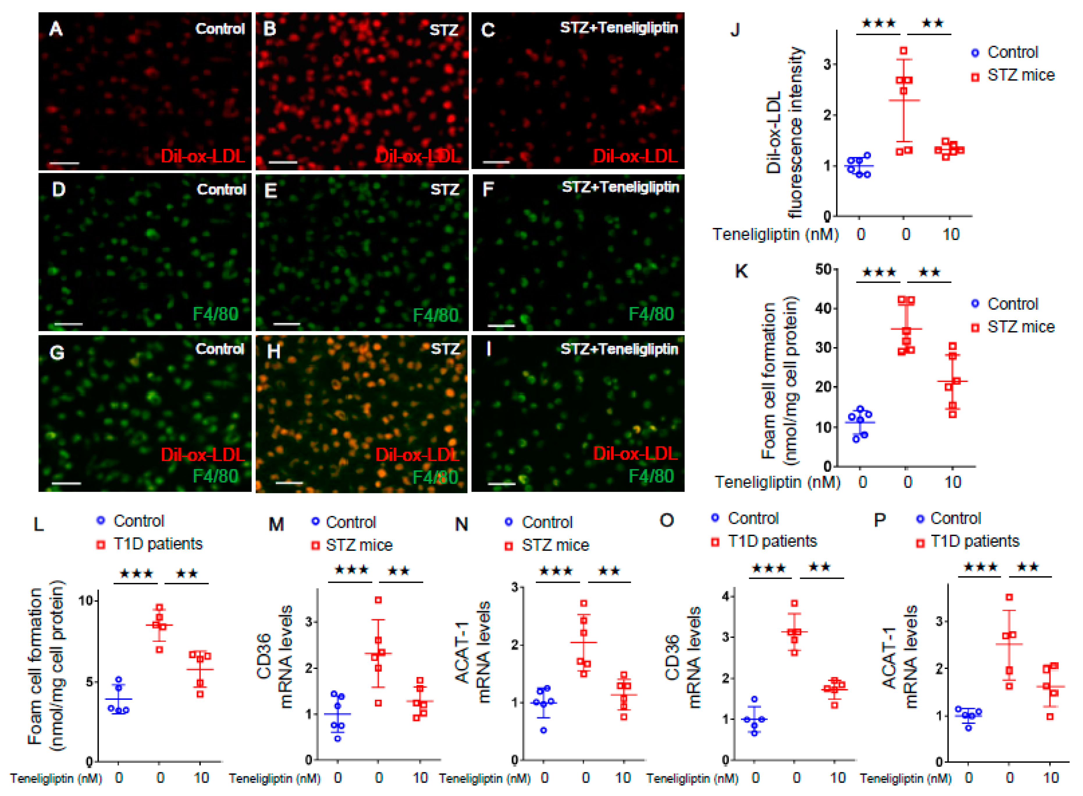
2.2. Teneligliptin Suppressed Foam Cell Formation of Macrophages Isolated from T1D Mice and T1D Patients

We first evaluated the effects of teneligliptin on ox-LDL uptake and foam cell formation of macrophages isolated from T1D mice and wild-type mice. Immunofluorescent staining showed that 1,1'-dioctadecyl-3,3,3',3'-tetramethylindocarbocyanine perchlorate-oxidized-low-density lipoprotein (Dil-ox-LDL)-positive cells were co-stained with F4/80, a marker of macrophages (Figure 1A–I), confirming the uptake of ox-LDL into mouse peritoneal macrophages. As shown in Figure 1J, Dil-ox-LDL uptake into macrophages was significantly increased in T1D mice compared with wild-type mice, which was completely prevented by the treatment with 10 nmol/L teneligliptin. When the foam cell formation of macrophages was evaluated by cholesterol esterification assay using ox-LDL with [3H]oleate, foam cell formations of macrophages isolated from T1D mice and T1D patients were significantly increased in comparison with controls, both of which were attenuated by 10 nmol/L teneligliptin (Figure 1K,L). Furthermore, 10 nmol/L teneligliptin significantly inhibited the up-regulation of CD36 and ACAT-1 gene expression in macrophages derived from T1D mice and T1D patients (Figure 1M–P).

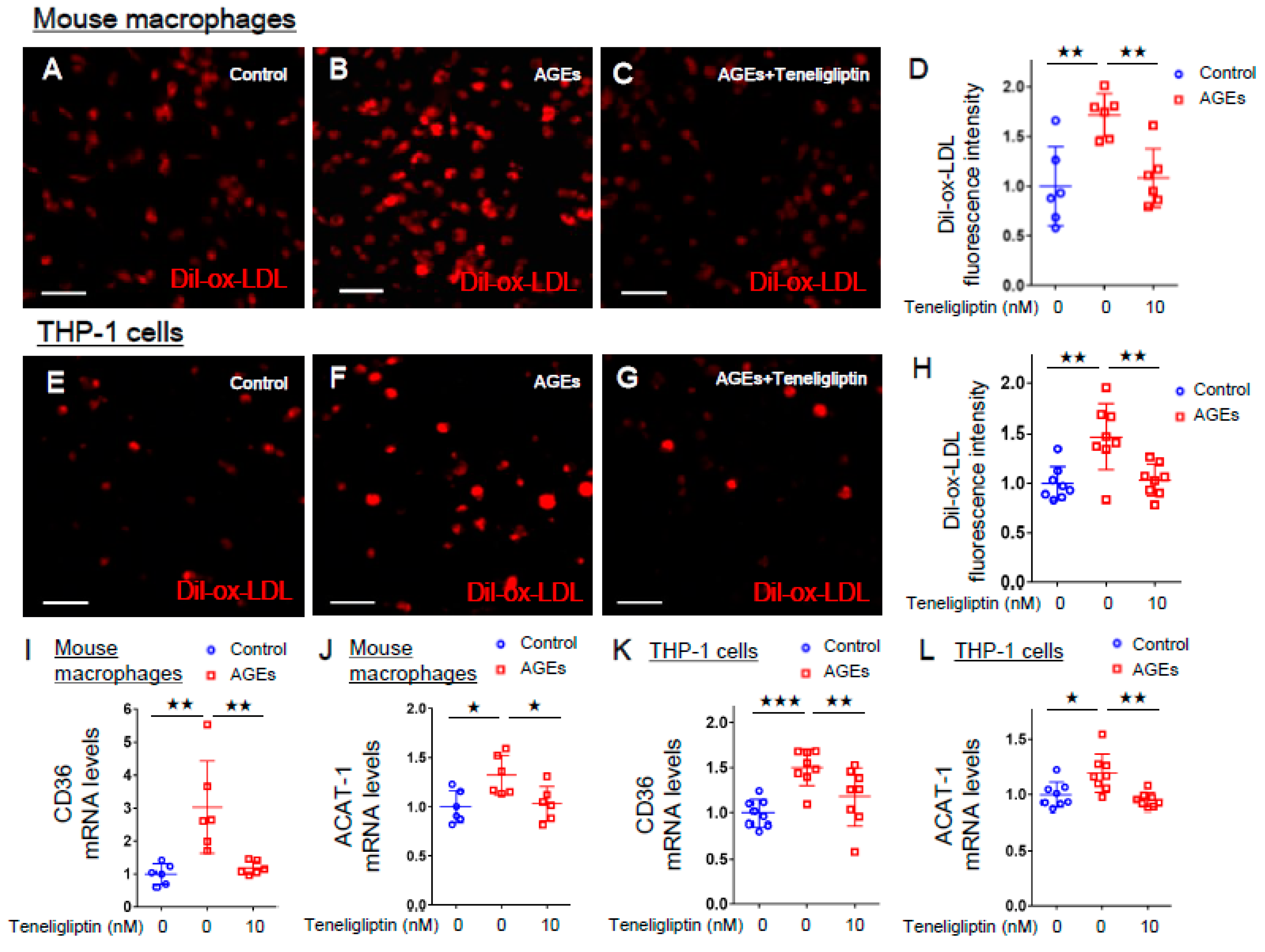
2.3. Teneligliptin Inhibited ox-LDL Uptake in AGE-exposed Mouse Macrophages and Human THP-1 Macrophages

We further evaluated the effects of teneligliptin on ox-LDL uptake in AGE-exposed mouse peritoneal macrophages and human THP-1 cells. As shown in Figure 2A–H, AGE-bovine serum albumin (AGE-BSA) significantly increased ox-LDL uptake into mouse peritoneal macrophages and human THP-1 cells compared to non-glycated control BSA, which were completely prevented by 10 nmol/L teneligliptin. In addition, 10 nmol/L teneligliptin completely suppressed the up-regulation of CD36 and ACAT-1 gene expression in AGE-exposed mouse peritoneal macrophages and human THP-1 cells (Figure 2I–L).

3. Discussion

DPP-4 inhibitors have been shown to play atheroprotective properties in both animal models and patients with T2D [17,19,20,21,22]. Indeed, although large clinical trials with DPP-4 inhibitors did not reduce the risk of major cardiovascular events [27,28,29,30], a couple of clinical papers reported that DPP-4 inhibitors showed favorable effects on carotid intima-media thickness in T2D patients, a surrogate marker of atherosclerosis, and sitagliptin use was independently associated with the lower incidence rates of atherosclerotic cardiovascular disease in subjects with T2D [20,21,22]. DPP-4 inhibitors not only ameliorate hyperglycemia, but also improve dyslipidemia with modest blood pressure lowering effects [31,32,33]. Furthermore, there is an accumulating body of evidence to show that GLP-1 and GIP have pleiotropic effects on diabetic vessels; GLP-1 and GIP could attenuate oxidative stress generation, inflammatory reactions, and foam cell formation of macrophages within the atherosclerotic plaques [34,35,36,37], thus playing a protective role against atherosclerosis. Therefore, it remains unclear whether DPP-4 inhibitors slow the process of atherosclerosis directly via the improvement of hyperglycemia and related AGE formation, indirectly via the amelioration of comorbid risk factors, or through the pleiotropic effects of incretins. To address whether the effects of DPP-4 inhibitor are partly independent of these comorbidities associated with T2D and pleiotropic actions of incretins, we first examined the effects of a DPP-4 inhibitor, teneligliptin on ox-LDL uptake and foam cell formation of macrophages isolated from STZ-induced T1D mice and T1D patients because (1) blood pressure levels and lipid parameters in T1D mice and patients were comparable with those in controls, (2) body weight, triglycerides, and homeostasis model assessment of insulin resistance (HOMA-IR), a marker of insulin resistance were higher in the previously published T2D mice models or patients than controls, (3) there were significant differences of triglycerides and HOMA-IR or fasting C-peptide between T1D and T2D, and (4) DPP-4 inhibitors did not modulate the effects of incretins on macrophages in vitro. In other words, we used here macrophages from T1D model mice and patients to rule out the effects of these comorbidities.
In this study, we found for the first time that teneligliptin at 10 nmol/L significantly blocked ox-LDL uptake and foam cell formation of macrophages isolated from STZ-induced T1D mice or T1D patients. Furthermore, teneligliptin significantly inhibited the up-regulation of CD36 and ACAT-1 mRNA levels in macrophages isolated from T1D mice and T1D patients. CD36 is a major scavenger receptor mediating uptake of ox-LDL into macrophages, whereas ACAT-1 is a rate-limiting enzyme for the esterification of free cholesterol, which could contribute to macrophage foam cell formation [13]. Therefore, our present observations suggest that teneligliptin could inhibit ox-LDL uptake and foam cell formation of macrophages in T1D partly via suppression of CD36 and ACAT-1 expression. The present findings have extended the previous observations showing that DPP-4 inhibitors directly suppressed foam cell formation of macrophages in vitro partly via inhibition of CD36 [14,38]. The peak plasma concentration of teneligliptin after oral administration of clinical dose of 20 mg is about 280 nmol/L, and approximately 20% of the amount of teneligliptin in the blood is a protein-unbound, free form [39]. Therefore, the concentration of teneligliptin (10 nmol/L) with beneficial effects on macrophages observed here may be comparable to the therapeutic level (less than 50 nmon/L) which is achieved in the treatment for patients with T2D. Since ox-LDL uptake and foam cell formation are key components of atherosclerosis [2,3], our present study suggests that teneligliptin may inhibit the progression of atherosclerosis through its direct pleiotropic effects on macrophages.
We have already reported that teneligliptin significantly reduced ox-LDL uptake, foam cell formation, CD36/ACAT-1 mRNA levels of macrophages derived from T2D patients and db/db mice, an animal model of T2D [14]. However, it remains unclear how teneligliptin directly inhibits ox-LDL uptake and foam cell formation of macrophages. In other words, the mechanisms of action of the drug are not fully elucidated. Therefore, we examined the effects of teneligliptin on ox-LDL uptake, CD36 and ACAT-1 gene expression in AGE-exposed mouse peritoneal macrophages and THP-1 cells, a human macrophage cell line because (1) we have previously shown that DPP-4 inhibitors could block the harmful effects of AGE in cultured endothelial cells and renal proximal tubular cells [24,40] and (2) AGE play a central role in atherosclerosis in diabetes [18]. In this study, we found that AGE mimicked the effects of T1D; AGE significantly increased ox-LDL uptake, CD36 and ACAT-1 gene expression in mouse macrophages and THP-1 cells, all of which were prevented by the treatment with 10 nmol/L teneligliptin. Therefore, teneligliptin could suppress the ox-LDL uptake and foam cell formation of macrophages isolated from T1D mice and patients partly via suppression of deleterious effects of AGE on macrophages. AGE and macrophages were co-existed within the atherosclerotic plaques, which were associated with the severity of atherosclerosis [7,8,41]. In addition, circulating and tissue accumulation levels of AGE were associated with the increased risk of cardiovascular disease in both diabetic and non-diabetic patients [5,6,10,11,42,43,44,45,46]. These findings further support the clinical relevance of anti-atherosclerotic effects of teneligliptin in diabetes.
The present study has some potential limitations. First, we examined the atheroprotective role of teneligliptin only focusing on macrophage foam cell formation in vitro. Second, it is probable that any of clinical characteristics of T1D patients, including disease duration, age and drug medications, such as statins and anti-hypertensive agents could have impacted the present findings. Indeed, in this study, age and sex were not completely matched between T1D cases and controls. Since accumulation of AGE has been known to progress in a normal ageing process and under diabetic condition, especially diabetic patients with a long disease history [45], the difference of mean ages of the T1D cases and controls may influence the present results. In addition, there is a sex disparity in cardiovascular event and mortality rates associated with diabetes [47], and therefore the difference of number of male/female in cases/controls may also affect the ox-LDL uptake and foam cell formation of macrophages. However, we could not sub-analyze the data because of small number of patients in this study. Third, in order to avoid the effects of streptozotocin and narrow it down the specific effect of hyperglycemia on macrophages, we investigated the effects of AGE, a marker of cumulative hyperglycemic exposure on ox-LDL uptake, CD36 and ACAT-1 gene expression of macrophages derived from non-diabetic mice. Fourth it would be relevant to see the impact of teneligliptin on ox-LDL, foam cell formation and CD36/ACAT-1 mRNA levels under normal glycemic conditions. Since teneligliptin is an inhibitor of DPP-4, which is approved for the treatment of diabetes, we examined here the effects of teneligliptin on macrophages under diabetic conditions. Fifth, we performed the present experiments at only 1 dose of teneligliptin (10 nmol/L). It would be interesting to examine the dose-dependent effects of teneligliptin on macrophages. Sixth, it would be valuable to demonstrate CD36 and ACAT-1 protein levels by western blotting. However, CD36 and ACAT-1 protein expression levels are functionally correlated with ox-LDL uptake and foam cell formation of macrophages, respectively. The observation suggests that these gene expression levels would be correlated with protein levels. Seventh, it would be interesting to explore other genes, such as interleukin-6 with changing expression levels by teneligliptin through RNA-sequencing. Eighth, although Xu et al. reported that AGE at supraphysiological concentrations (300–600 µg/mL AGE-BSA) increased lipid accumulation in macrophages partly by regulating CD36, scavenger receptor A2, hydroxymethylglutaryl-CoA reductase, ACAT-1, and ATP-binding cassette transporter G1 [48], we provided here a new line of following evidence; concentration of AGE (100 µg/mL AGE-BSA), which is comparable with that of in vivo-diabetic situation [49,50], actually stimulated ox-LDL uptake of macrophages via CD36 and ACAT-1 expression, which is a molecular target for atheroprotective properties of teneligliptin. Ninth, intraperitoneal administration of AGE-BSA has been reported to impair glucose tolerance in mice in association with decrease in acute insulin secretion [51], AGE may augment foam cell formation of macrophages by further deteriorating hyperglycemia. Specifically, the salient findings of our present study are that teneligliptin at a therapeutic level inhibited foam cell formation of macrophages by suppressing the harmful effects of AGE, whose concentration is also comparable with that of diabetic conditions. In any case, further clinical studies should be needed to clarify whether DPP-4 inhibitors could inhibit foam cell formation of macrophages and resultantly reduce the risk of cardiovascular events in patients with diabetes.

4. Materials and Methods

4.1. Animal Experiments

STZ was purchased from Sigma–Aldrich (St. Louis, MO, USA) and a DPP-4 inhibitor teneligliptin was generously gifted by Tanabe Mitsubishi Pharma (Tokyo, Japan). Animal experiments were conducted under strict accordance with the recommendations in the Guide for the Care and Use of Laboratory Animals [52]. The study design was approved by the Animal Care Committee of Showa University (permission number: 07005). All surgeries and sacrifices were performed under general anesthesia using isoflurane and with efforts to minimize the suffering.
A total of 12 male C57BL/6J (wild-type) mice at 7 weeks old were purchased from Sankyo Labo Service (Tokyo, Japan), kept on a standard rodent chow (Labo MR Stock, NOSAN, Yokohama, Kanagawa, Japan) with free access to water, and housed within a specific pathogen-free facility in the Division of Animal Experimentation of Showa University School of Medicine. The rooms were controlled under a 12-h dark/light cycle, 21 °C temperature, and 40–60% humidity. At 8 weeks old, the mice received intraperitoneal injections of saline or STZ (50 mg/kg/day) for 5 consecutive days to create a T1D model mouse, as previously described [17]. All mice showed FBG levels higher than 200 mg/dL, and were used in the present experiments as T1D model mice. All mice did not show any clinical signs, including severe weight loss by more than 20% from the baseline or obvious weakness. At 13 weeks old, blood samples were collected, and peritoneal macrophages were extracted from mice after intraperitoneal injection of thioglycolate broth as previously described [14,17,31,36,37].

4.2. Measurements of Laboratory Parameters in Mice

Blood samples collected after a 12-h fast were used for the analysis of biochemical analysis. Systolic and diastolic blood pressures (SBP and DBP) were measured, and FBG, HbA1c, total cholesterol (Total-C), high-density lipoprotein cholesterol (HDL-C), triglycerides, and insulin levels were measured, and OGTT was performed as previously described [14,17].

4.3. Experiments of Human Macrophages

The study protocol was approved by the Ethics Committee of Showa University School of Medicine (Tokyo, Japan; approval number: 2799). Written informed consent was obtained from all T1D patients and healthy volunteers. The study was designed in compliance with the Declaration of Helsinki.
Five patients with uncontrolled T1D despite multiple daily insulin injections over ≥12 weeks and six controls were enrolled in the present study. Blood samples were collected, and human monocyte-derived macrophages were isolated using anti-CD14 antibody-conjugated magnetic microbeads (Miltenyi Biotec, Auburn, CA, USA) as previously described [14].

4.4. Measurements of Clinical Parameters in Humans

Body mass index (BMI), SBP and DBP were measured and FBG, HbA1c (NGSP), LDL-C, HDL-C, triglycerides, fasting C-peptide and the 6-min value of C-peptide after glucagon (1 mg) stimulation test (stimulated C-peptide) were measured by standard methods as described previously [53].

4.5. Preparation of AGE-BSA

AGE-BSA was prepared as previously described [54]. In brief, BSA (25 mg/mL) was incubated under sterile conditions with 0.1 M glyceraldehyde in 0.2 M NaPO4 buffer (pH 7.4) for 7 days. Control non-glycated BSA was incubated in the same conditions except for the absence of reducing sugars.

4.6. Differentiation of THP-1 Macrophages

A human monocytic cell line, THP-1 cells were maintained in RPMI 1640 medium with 10% fetal bovine serum. The cells were seeded onto 3.5-cm dishes (1.0 × 10⁶ cells/dish) and incubated with phorbol 12-myristate 13-acetate (40 ng/mL; Sigma–Aldrich, St. Louis, MO, USA) for 24 h to differentiate macrophages [55].

4.7. Immunofluorescent Staining of Mouse Macrophages and THP-1 Macrophages

Peritoneal macrophages isolated from wild-type mice and T1D model mice, or THP-1 macrophages were incubated with or without 100 µg/mL AGE-BSA or 100 µg/mL non-glycated BSA for 24 h, and then treated with 10 μg/mL Dil-ox-LDL in the presence or absence of 10 nmol/L teneligliptin for 18 h. After washing, they were mounted in Vectashield mounting medium (H-1500, Vector Laboratories, Burlingame, CA, USA) and were imaged with BZ-X710 microscope/software (Keyence, Osaka, Japan) as previously described [14].

4.8. Cholesterol Esterification Assay in Macrophages Isolated from Mice and Humans

Cholesterol esterification assay was conducted as previously described [14,17,31,36,37]. In brief, peritoneal macrophages isolated from wild-type mice and T1D model mice or human monocyte-derived macrophages from T1D patients and controls were incubated with 10 μg/mL ox-LDL and 0.1 mmol/L [3H]oleate for 18 h in the presence or absence of 10 nmol/L teneligliptin [14]. Cellular lipids were extracted and the radioactivity of cholesterol [3H]oleate was determined by a thin-layer chromatography.

4.9. Gene Expression Levels in Macrophages Isolated from T1D Mice and T1D Patients and in AGE-Exposed Cells

Peritoneal macrophages isolated from wild-type and T1D model mice, monocyte-derived macrophages from T1D patients and controls, or THP-1 macrophages were treated with or without 100 µg/mL AGE-BSA or 100 µg/mL non-glycated BSA for 24 h, and then incubated with or without 10 nmol/L teneligliptin for 18 h [14]. Total RNA was isolated with QIAGEN reagents (Hilden, Germany), and gene expression was analyzed by real-time reverse-transcription polymerase chain reactions using the TaqMan gene expression assay and a sequence detection system (ABI PRISM 7900, Life Technologies, Thermo Fischer Scientific, Pleasanton, CA, USA) as previously described [14]. The pre-designed TaqMan probe sets used in mice were as follows: Cd36, Mm01135198_ml; Acat-1, Mm00507463_ml; glyceraldehyde-3-phosphate dehydrogenase (Gapdh), Mm03302249_g1. Human probes were as follows: Cd36, Hs00169627_ml; Acat-1, Hs01009746_ml; Gapdh, Hs99999905_ml.

4.10. Statistical Analysis

Values were expressed as mean ± standard deviation. Statistical analyses were performed by unpaired t-test to compare two groups and analysis of variance (ANOVA) to compare multiple groups followed by appropriate multiple comparison tests. Categorical variables were compared by chi-square test. All analyses were performed by PRISM 7.0 software (GraphPad, San Diego, CA, USA). The significance level was defined as p < 0.05.

5. Conclusions

The present study suggests that teneligliptin could inhibit foam cell formation of macrophages in T1D via suppression of CD36 and ACAT-1 gene expression partly by attenuating the harmful effects of AGEs.

Author Contributions

M.T. conceptualized and designed the study; investigated, analyzed, interpreted the data; and drafted, revised and finalized the manuscript. H.Y., Y.M., T.S., T.M. investigated, analyzed and interpreted the data. M.H., H.K., N.O., M.O., T.F., T.H. interpreted the data, discussed and contributed to revision of the manuscript. S.-i.Y. conceptualized and designed the study; interpreted the data; and critically revised the manuscript. All authors have read and agree to the published version of manuscript.

Funding

None.

Acknowledgments

The authors would like to thank Hiroko Takeuchi for their technical assistance.

Conflicts of Interest

The authors declare no conflict of interest.

Disclosure

This study is presented in part at the 63rd Annual Meeting of the Japan Diabetes Society, Shiga, Japan, 21–23 May 2020 and at the 56th Annual Meeting of the European Association for the Study of Diabetes, Vienna, Austria, 22–25 September 2020.

Data Availability Statement

All data used to support the findings of this study are available from the corresponding author upon reasonable request.

References

  1. Rao Kondapally Seshasai, S.; Kaptoge, S.; Thompson, A.; Di Angelantonio, E.; Gao, P.; Sarwar, N.; Whincup, P.H.; Mukamal, K.J.; Gillum, R.F.; Holme, I.; et al. Diabetes mellitus, fasting glucose, and risk of cause-specific death. N. Engl. J. Med. 2011, 364, 829–841. [Google Scholar] [PubMed] [Green Version]
  2. Glass, C.K.; Witztum, J.L. Atherosclerosis. the road ahead. Cell 2001, 104, 503–516. [Google Scholar] [CrossRef] [Green Version]
  3. Lusis, A.J. Atherosclerosis. Nature 2000, 407, 233–241. [Google Scholar] [CrossRef] [PubMed]
  4. Goldin, A.; Beckman, J.A.; Schmidt, A.M.; Creager, M.A. Advanced glycation end products: Sparking the development of diabetic vascular injury. Circulation 2006, 114, 597–605. [Google Scholar] [CrossRef] [Green Version]
  5. Kilhovd, B.K.; Juutilainen, A.; Lehto, S.; Rönnemaa, T.; Torjesen, P.A.; Hanssen, K.F.; Laakso, M. Increased serum levels of advanced glycation endproducts predict total, cardiovascular and coronary mortality in women with type 2 diabetes: A population-based 18 year follow-up study. Diabetologia 2007, 50, 1409–1417. [Google Scholar] [CrossRef] [Green Version]
  6. Kilhovd, B.K.; Juutilainen, A.; Lehto, S.; Rönnemaa, T.; Torjesen, P.A.; Hanssen, K.F.; Laakso, M. Increased serum levels of methylglyoxal-derived hydroimidazolone-AGE are associated with increased cardiovascular disease mortality in nondiabetic women. Atherosclerosis 2009, 205, 590–594. [Google Scholar] [CrossRef]
  7. Kume, S.; Takeya, M.; Mori, T.; Araki, N.; Suzuki, H.; Horiuchi, S.; Kodama, T.; Miyauchi, Y.; Takahashi, K. Immunohistochemical and ultrastructural detection of advanced glycation end products in atherosclerotic lesions of human aorta with a novel specific monoclonal antibody. Am. J. Pathol. 1995, 147, 654–667. [Google Scholar]
  8. Tahara, N.; Yamagishi, S.; Takeuchi, M.; Honda, A.; Tahara, A.; Nitta, Y.; Kodama, N.; Mizoguchi, M.; Kaida, H.; Ishibashi, M.; et al. Positive association between serum level of glyceraldehyde-derived advanced glycation end products and vascular inflammation evaluated by [(18)F]fluorodeoxyglucose positron emission tomography. Diabetes Care 2012, 35, 2618–2625. [Google Scholar] [CrossRef] [Green Version]
  9. Kajikawa, M.; Nakashima, A.; Fujimura, N.; Maruhashi, T.; Iwamoto, Y.; Iwamoto, A.; Matsumoto, T.; Oda, N.; Hidaka, T.; Kihara, Y.; et al. Ratio of serum levels of AGEs to soluble form of RAGE is a predictor of endothelial function. Diabetes Care 2015, 38, 119–125. [Google Scholar] [CrossRef] [Green Version]
  10. Nin, J.W.; Jorsal, A.; Ferreira, I.; Schalkwijk, C.G.; Prins, M.H.; Parving, H.H.; Tarnow, L.; Rossing, P.; Stehouwer, C.D. Higher plasma levels of advanced glycation end products are associated with incident cardiovascular disease and all-cause mortality in type 1 diabetes: A 12-year follow-up study. Diabetes Care 2011, 34, 442–447. [Google Scholar] [CrossRef] [Green Version]
  11. Hanssen, N.M.; Beulens, J.W.; van Dieren, S.; Scheijen, J.L.; Daphne van der, A.; Spijkerman, A.M.; van der Schouw, Y.T.; Stehouwer, C.D.; Schalkwijk, C.G. Plasma advanced glycation end products are associated with incident cardiovascular events in individuals with type 2 diabetes: A case-cohort study with a median follow-up of 10 years (EPIC-NL). Diabetes 2015, 64, 257–265. [Google Scholar] [CrossRef] [Green Version]
  12. Tahara, N.; Yamagishi, S.; Matsui, T.; Nishino, Y.; Honda, A.; Tahara, A.; Igata, S.; Fukumoto, Y. Serum levels of pigment epithelium-derived factor (PEDF) are inversely associated with circulating levels of dipeptidyl peptidase-4 (DPP-4) in humans. Int. J. Cardiol. 2015, 184, 14–16. [Google Scholar] [CrossRef] [PubMed]
  13. Allahverdian, S.; Pannu, P.S.; Francis, G.A. Contribution of monocyte-derived macrophages and smooth muscle cells to arterial foam cell formation. Cardiovasc. Res. 2012, 95, 165–172. [Google Scholar] [CrossRef] [PubMed] [Green Version]
  14. Terasaki, M.; Hiromura, M.; Mori, Y.; Kohashi, K.; Kushima, H.; Koshibu, M.; Saito, T.; Yashima, H.; Watanabe, T.; Hirano, T. A dipeptidyl peptidase-4 inhibitor suppresses macrophage foam cell formation in diabetic db/db mice and type 2 diabetes patients. Int. J. Endocrinol. 2018, 2018. [Google Scholar] [CrossRef] [PubMed] [Green Version]
  15. Fukuhara-Takaki, K.; Sakai, M.; Sakamoto, Y.; Takeya, M.; Horiuchi, S. Expression of class A scavenger receptor is enhanced by high glucose in vitro and under diabetic conditions in vivo: One mechanism for an increased rate of atherosclerosis in diabetes. J. Biol. Chem. 2005, 280, 3355–3364. [Google Scholar] [CrossRef] [PubMed] [Green Version]
  16. Li, L.; Sawamura, T.; Renier, G. Glucose enhances human macrophage LOX-1 expression: Role for LOX-1 in glucose-induced macrophage foam cell formation. Circ. Res. 2004, 94, 892–901. [Google Scholar] [CrossRef]
  17. Terasaki, M.; Nagashima, M.; Nohtomi, K.; Kohashi, K.; Tomoyasu, M.; Sinmura, K.; Nogi, Y.; Katayama, Y.; Sato, K.; Itoh, F.; et al. Preventive effect of dipeptidyl peptidase-4 inhibitor on atherosclerosis is mainly attributable to incretin's actions in nondiabetic and diabetic apolipoprotein E-null mice. PLoS ONE 2013, 8, e70933. [Google Scholar] [CrossRef]
  18. Yamagishi, S.; Fukami, K.; Matsui, T. Crosstalk between advanced glycation end products (AGEs)-receptor RAGE axis and dipeptidyl peptidase-4-incretin system in diabetic vascular complications. Cardiovasc. Diabetol. 2015, 14, 2. [Google Scholar] [CrossRef] [Green Version]
  19. Ta, N.N.; Schuyler, C.A.; Li, Y.; Lopes-Virella, M.F.; Huang, Y. DPP-4 (CD26) inhibitor alogliptin inhibits atherosclerosis in diabetic apolipoprotein E-deficient mice. J. Cardiovasc. Pharmacol. 2011, 58, 157–166. [Google Scholar] [CrossRef]
  20. Yang, T.Y.; Liaw, Y.P.; Huang, J.Y.; Chang, H.R.; Chang, K.W.; Ueng, K.C. Association of Sitagliptin with cardiovascular outcome in diabetic patients: A nationwide cohort study. Acta Diabetol. 2016, 53, 461–468. [Google Scholar] [CrossRef]
  21. Mita, T.; Katakami, N.; Shiraiwa, T.; Yoshii, H.; Onuma, T.; Kuribayashi, N.; Osonoi, T.; Kaneto, H.; Kosugi, K.; Umayahara, Y.; et al. Sitagliptin attenuates the progression of carotid intima-media thickening in insulin-treated patients with type 2 diabetes: The Sitagliptin Preventive Study of Intima-Media Thickness Evaluation (SPIKE): A randomized controlled trial. Diabetes Care 2016, 39, 455–464. [Google Scholar] [CrossRef] [PubMed] [Green Version]
  22. Mita, T.; Katakami, N.; Yoshii, H.; Onuma, T.; Kaneto, H.; Osonoi, T.; Shiraiwa, T.; Kosugi, K.; Umayahara, Y.; Yamamoto, T.; et al. Alogliptin, a dipeptidyl peptidase 4 inhibitor, prevents the progression of carotid atherosclerosis in patients with type 2 diabetes: The Study of Preventive Effects of Alogliptin on Diabetic Atherosclerosis (SPEAD-A). Diabetes Care 2016, 39, 139–148. [Google Scholar] [CrossRef] [Green Version]
  23. Matsui, T.; Nishino, Y.; Takeuchi, M.; Yamagishi, S. Vildagliptin blocks vascular injury in thoracic aorta of diabetic rats by suppressing advanced glycation end product-receptor axis. Pharmacol. Res. 2011, 63, 383–388. [Google Scholar] [CrossRef] [PubMed]
  24. Ishibashi, Y.; Matsui, T.; Maeda, S.; Higashimoto, Y.; Yamagishi, S. Advanced glycation end products evoke endothelial cell damage by stimulating soluble dipeptidyl peptidase-4 production and its interaction with mannose 6-phosphate/insulin-like growth factor II receptor. Cardiovasc. Diabetol. 2013, 12, 125. [Google Scholar] [CrossRef] [Green Version]
  25. Matsui, T.; Nakashima, S.; Nishino, Y.; Ojima, A.; Nakamura, N.; Arima, K.; Fukami, K.; Okuda, S.; Yamagishi, S. Dipeptidyl peptidase-4 deficiency protects against experimental diabetic nephropathy partly by blocking the advanced glycation end products-receptor axis. Lab. Invest. 2015, 95, 525–533. [Google Scholar] [CrossRef] [Green Version]
  26. Ishibashi, Y.; Matsui, T.; Takeuchi, M.; Yamagishi, S. Sitagliptin augments protective effects of GLP-1 against advanced glycation end product receptor axis in endothelial cells. Horm. Metab. Res. 2011, 43, 731–734. [Google Scholar] [CrossRef] [PubMed]
  27. White, W.B.; Cannon, C.P.; Heller, S.R.; Nissen, S.E.; Bergenstal, R.M.; Bakris, G.L.; Perez, A.T.; Fleck, P.R.; Mehta, C.R.; Kupfer, S.; et al. Investigators. Alogliptin after acute coronary syndrome in patients with type 2 diabetes. N. Engl. J. Med. 2013, 369, 1327–1335. [Google Scholar] [CrossRef] [Green Version]
  28. Scirica, B.M.; Bhatt, D.L.; Braunwald, E.; Steg, P.G.; Davidson, J.; Hirshberg, B.; Ohman, P.; Frederich, R.; Wiviott, S.D.; Hoffman, E.B.; et al. SAVOR-TIMI 53 steering committee and investigators. saxagliptin and cardiovascular outcomes in patients with type 2 diabetes mellitus. N. Engl. J. Med. 2013, 369, 1317–1326. [Google Scholar] [CrossRef] [PubMed] [Green Version]
  29. Green, J.B.; Bethel, M.A.; Armstrong, P.W.; Buse, J.B.; Engel, S.S.; Garg, J.; Josse, R.; Kaufman, K.D.; Koglin, J.; Korn, S.; et al. TECOS study group. Effect of sitagliptin on cardiovascular outcomes in type 2 diabetes. N. Engl. J. Med. 2015, 373, 232–242. [Google Scholar] [CrossRef] [Green Version]
  30. Rosenstock, J.; Perkovic, V.; Johansen, O.E.; Cooper, M.E.; Kahn, S.E.; Marx, N.; Alexander, J.H.; Pencina, M.; Toto, R.D.; Wanner, C.; et al. CARMELINA investigators. Effect of linagliptin vs. placebo on major cardiovascular events in adults with type 2 diabetes and high cardiovascular and renal risk: The CARMELINA randomized clinical trial. J. Am. Med. Assoc. 2019, 321, 69–79. [Google Scholar] [CrossRef]
  31. Terasaki, M.; Nagashima, M.; Watanabe, T.; Nohtomi, K.; Mori, Y.; Miyazaki, A.; Hirano, T. Effects of PKF275-055, a dipeptidyl peptidase-4 inhibitor, on the development of atherosclerotic lesions in apolipoprotein E-null mice. Metabolism 2012, 61, 974–977. [Google Scholar] [CrossRef] [PubMed]
  32. Nishida, Y.; Takahashi, Y.; Tezuka, K.; Akimoto, H.; Nakayama, T.; Asai, S. Comparative effect of dipeptidyl-peptidase 4 inhibitors on laboratory parameters in patients with diabetes mellitus. BMC Pharmacol. Toxicol. 2020, 21, 28. [Google Scholar] [CrossRef] [PubMed] [Green Version]
  33. Kishimoto, S.; Kinoshita, Y.; Matsumoto, T.; Maruhashi, T.; Kajikawa, M.; Matsui, S.; Hashimoto, H.; Takaeko, Y.; Kihara, Y.; Chayama, K.; et al. Effects of the dipeptidyl peptidase 4 inhibitor alogliptin on blood pressure in hypertensive patients with type 2 diabetes mellitus. Am. J. Hypertens. 2019, 32, 695–702. [Google Scholar] [CrossRef] [PubMed]
  34. Yamagishi, S.; Matsui, T. Pleiotropic effects of glucagon-like peptide-1 (GLP-1)-based therapies on vascular complications in diabetes. Curr. Pharm. Des. 2011, 17, 4379–4385. [Google Scholar] [CrossRef] [PubMed]
  35. Ojima, A.; Matsui, T.; Maeda, S.; Takeuchi, M.; Yamagishi, S. Glucose-dependent insulinotropic polypeptide (GIP) inhibits signaling pathways of advanced glycation end products (AGEs) in endothelial cells via its antioxidative properties. Horm. Metab. Res. 2012, 44, 501–505. [Google Scholar] [CrossRef] [PubMed]
  36. Nagashima, M.; Watanabe, T.; Terasaki, M.; Tomoyasu, M.; Nohtomi, K.; Kim-Kaneyama, J.; Miyazaki, A.; Hirano, T. Native incretins prevent the development of atherosclerotic lesions in apolipoprotein E knockout mice. Diabetologia 2011, 54, 2649–2659. [Google Scholar] [CrossRef] [PubMed] [Green Version]
  37. Nogi, Y.; Nagashima, M.; Terasaki, M.; Nohtomi, K.; Watanabe, T.; Hirano, T. Glucose-dependent insulinotropic polypeptide prevents the progression of macrophage-driven atherosclerosis in diabetic apolipoprotein E-null mice. PLoS ONE 2012, 7, e35683. [Google Scholar] [CrossRef]
  38. Dai, Y.; Wang, X.; Ding, Z.; Dai, D.; Mehta, J.L. DPP-4 inhibitors repress foam cell formation by inhibiting scavenger receptors through protein kinase C pathway. Acta Diabetol. 2014, 51, 471–478. [Google Scholar] [CrossRef]
  39. Halabi, A.; Maatouk, H.; Siegler, K.E.; Faisst, N.; Lufft, V.; Klause, N. Pharmacokinetics of teneligliptin in subjects with renal impairment. Clin. Pharmacol. Drug Dev. 2013, 2, 246–254. [Google Scholar] [CrossRef]
  40. Kaifu, K.; Ueda, S.; Nakamura, N.; Matsui, T.; Yamada-Obara, N.; Ando, R.; Kaida, Y.; Nakata, M.; Matsukuma-Toyonaga, M.; Higashimoto, Y.; et al. Advanced glycation end products evoke inflammatory reactions in proximal tubular cells via autocrine production of dipeptidyl peptidase. Microvasc. Res. 2018, 120, 90–93. [Google Scholar] [CrossRef]
  41. Hanssen, N.M.; Wouters, K.; Huijberts, M.S.; Gijbels, M.J.; Sluimer, J.C.; Scheijen, J.L.; Heeneman, S.; Biessen, E.A.; Daemen, M.J.; Brownlee, M.; et al. Higher levels of advanced glycation endproducts in human carotid atherosclerotic plaques are associated with a rupture-prone phenotype. Eur. Heart J. 2014, 35, 1137–1146. [Google Scholar] [CrossRef] [PubMed] [Green Version]
  42. Semba, R.D.; Bandinelli, S.; Sun, K.; Guralnik, J.M.; Ferrucci, L. Plasma carboxymethyl-lysine, an advanced glycation end product, and all-cause and cardiovascular disease mortality in older community-dwelling adults. J. Am. Geriatr. Soc. 2009, 57, 1874–1880. [Google Scholar] [CrossRef] [PubMed] [Green Version]
  43. Semba, R.D.; Ferrucci, L.; Sun, K.; Beck, J.; Dalal, M.; Varadhan, R.; Walston, J.; Guralnik, J.M.; Fried, L.P. Advanced glycation end products and their circulating receptors predict cardiovascular disease mortality in older community-dwelling women. Aging Clin. Exp. Res. 2009, 21, 182–190. [Google Scholar] [CrossRef] [PubMed] [Green Version]
  44. van Waateringe, R.P.; Fokkens, B.T.; Slagter, S.N.; van der Klauw, M.M.; van Vliet-Ostaptchouk, J.V.; Graaff, R.; Paterson, A.D.; Smit, A.J.; Lutgers, H.L.; Wolffenbuttel, B.H.R. Skin autofluorescence predicts incident type 2 diabetes, cardiovascular disease and mortality in the general population. Diabetologia 2019, 62, 269–280. [Google Scholar] [CrossRef] [Green Version]
  45. Yamagishi, S. Role of Advanced Glycation Endproduct (AGE)-Receptor for Advanced Glycation Endproduct (RAGE) axis in cardiovascular disease and its therapeutic intervention. Circ. J. 2019, 83, 1822–1828. [Google Scholar] [CrossRef] [Green Version]
  46. Sakata, K.; Hayakawa, M.; Yano, Y.; Tamaki, N.; Yokota, N.; Eto, T.; Watanabe, R.; Hirayama, N.; Matsuo, T.; Kuroki, K.; et al. Efficacy of alogliptin, a dipeptidyl peptidase-4 inhibitor, on glucose parameters, the activity of the advanced glycation end product (AGE)-Receptor for AGE (RAGE) axis and albuminuria in Japanese type 2 diabetes. Diabetes Metab. Res. Rev. 2013, 29, 624–630. [Google Scholar] [CrossRef]
  47. Yamagishi, S. Sex disparity in cardiovascular mortality rates associated with diabetes. Diabetes Metab. Res. Rev. 2018, 34, e3059. [Google Scholar] [CrossRef]
  48. Xu, L.; Wang, Y.R.; Li, P.C.; Feng, B. Advanced glycation end products increase lipids accumulation in macrophages through upregulation of receptor of advanced glycation end products: Increasing uptake, esterification and decreasing efflux of cholesterol. Lipids Health Dis. 2016, 15, 161. [Google Scholar] [CrossRef] [Green Version]
  49. Yamagishi, S.; Nakamura, N.; Suematsu, M.; Kaseda, K.; Matsui, T. Advanced glycation end products: A molecular target for vascular complications in diabetes. Mol. Med. 2015, 21, S32–S40. [Google Scholar] [CrossRef]
  50. Takeuchi, M.; Makita, Z.; Yanagisawa, K.; Kameda, K.; Koike, T. Detection of noncarboxymethyllysine and carboxymethyllysine advanced glycation end products (AGE) in serum of diabetic patients. Mol. Med. 1999, 5, 393–405. [Google Scholar] [CrossRef] [Green Version]
  51. Jung, H.; Joo, J.; Jeon, Y.; Lee, J.; In, J.; Kim, D.; Kang, E.; Kim, Y.; Lim, Y.; Kang, J.; et al. Advanced glycation end products downregulate glucokinase in mice. Diabetes Metab. Res. Rev. 2011, 27, 557–563. [Google Scholar] [CrossRef] [PubMed]
  52. Committee for the Update of the Guide for the Care and Use of Laboratory Animals; Institute for Laboratory Animal Research; Division on Earth and Life Studies; National Research Council of the National Academies. Guide for the Care and Use of Laboratory Animals, 8th ed.; National Academies Press: Washington, DC, USA, 2011. [Google Scholar]
  53. Ohara, M.; Hiromura, M.; Nagaike, H.; Kohata, Y.; Fujikawa, T.; Goto, S.; Sato, N.; Kushima, H.; Terasaki, M.; Yamamoto, T.; et al. Relationship between glucose variability evaluated by continuous glucose monitoring and clinical factors, including glucagon-stimulated insulin secretion in patients with type 2 diabetes. Diabetes Res. Clin. Pract. 2019, 158, 107904. [Google Scholar] [CrossRef]
  54. Yamagishi, S.; Nakamura, K.; Matsui, T.; Inagaki, Y.; Takenaka, K.; Jinnouchi, Y.; Yoshida, Y.; Matsuura, T.; Narama, I.; Motomiya, Y.; et al. Pigment epithelium-derived factor inhibits advanced glycation end product-induced retinal vascular hyperpermeability by blocking reactive oxygen speciesmediated vascular endothelial growth factor expression. J. Biol. Chem. 2006, 281, 20213–20220. [Google Scholar] [CrossRef] [PubMed] [Green Version]
  55. Takashiba, S.; Van Dyke, T.E.; Amar, S.; Murayama, Y.; Soskolne, A.W.; Shapira, L. Differentiation of monocytes to macrophages primes cells for lipopolysaccharide stimulation via accumulation of cytoplasmic nuclear factor kappaB. Infect. Immun. 1999, 67, 5573–5578. [Google Scholar] [CrossRef] [PubMed] [Green Version]
Figure 1. Effects of teneligliptin on oxidized low-density lipoprotein (ox-LDL) uptake, foam cell formation, and CD36 and ACAT-1 gene expression in macrophages extracted from type 1 diabetes (T1D) model mice and T1D patients. (A–I) Representative immunofluorescent staining images in the peritoneal macrophages isolated from wild-type mice and T1D model mice. Dil-ox-LDL staining cells were in red, and F4/80 expressing cells were in green. Scale bars represent 50 µm. (J) Fluorescence intensity of Dil-ox-LDL per area. (K) and (L) Foam cell formation was evaluated by the radioactivity of cholesterol [3H] oleate. (M–P) Gene expression levels of CD36 (M,O) and ACAT1 (N,P) in peritoneal macrophages isolated from mice and in monocyte-derived macrophages from humans. (J,M–P) are normalized to the control levels.(A–K,M,N); number = 6 for each group. L, O, and P, number = 6 for control group. number = 5 for each T1D group. *** p < 0.005, ** p < 0.01.
Ijms 21 04811 g001
Figure 2. Effects of teneligliptin on ox-LDL uptake, CD36 and ACAT-1 gene expression in AGE-exposed mouse macrophages and THP-1 cells. (AC,EG) Representative immunofluorescent staining images in mouse peritoneal macrophages (AC) and THP-1 cells (EG). Dil-ox-LDL staining cells were in red. Scale bars represent 50 µm for A-C and 100 µm for E-G. (D,H) Fluorescence intensity of Dil-ox-LDL per area. (I–L) Gene expression levels of CD36 (I,K) and ACAT-1 (J,L) in mouse macrophages and THP-1 cells. (A–C,D,I,J) number = 6 for each group. (E–G,H,K,L) number = 8 for each group. (D,H-L) are normalized to the control levels. *** p < 0.005, ** p < 0.01, * p < 0.05.
Ijms 21 04811 g002
Table 1. Laboratory characteristics of wild-type mice and streptozotocin-induced type 1 diabetes mice at 13 weeks old.
Wild-Type MiceT1D Model Micep-Value
Number66
Final body weight (g)24.3 ± 1.620.3 ± 3.2P < 0.01
Food Intake (g/day)4.5 ± 0.44.8 ± 1.00.459
SBP (mmHg)100 ± 16102 ± 150.840
DBP (mmHg)61 ± 665 ± 70.302
Total-C (mg/dL)74 ± 986 ± 220.592
HDL-C (mg/dL)40 ± 1732 ± 150.378
Triglycerides (mg/dL)60 ± 982 ± 390.205
FBG (mg/dL)90 ± 8164 ± 56p < 0.01
Insulin (ng/mL)0.27 ± 0.080.05 ± 0.02p < 0.001
HbA1c (%)4.3 ± 0.27.9 ± 0.5p < 0.001
OGTT-AUC of glucose(mg/dL x hour)608 ± 361150 ± 365p < 0.005
T1D, type 1 diabetes; SBP, systolic blood pressure; DBP, diastolic blood pressure; Total-C, Total cholesterol; HDL-C, high-density lipoprotein cholesterol; FBG, fasting blood glucose; HbA1c, glycated hemoglobin; OGTT, oral glucose tolerance test; AUC, Area under the curve. Results are presented as mean values ± SD and analyzed with unpaired t-test. p < 0.05 vs. C57BL6/J mice.
Table 2. Clinical parameters of type 1 diabetes patients and controls.
ControlsT1D Patientsp-Value
Number (male/female)6 (5/1)5 (3/2)0.251
Age (years)42 ± 1058 ± 300.25
Duration of diabetes (years)11 ± 10
Body Weight (kg)67 ± 871 ± 200.725
BMI (kg/m2)23.1 ± 1.321.3 ± 3.90.318
SBP (mmHg)114 ± 5114 ± 100.912
DBP (mmHg)71 ± 768 ± 130.662
Total-C (mg/dL)187 ± 7167 ± 240.087
LDL-C (mg/dL)110 ± 3892 ± 280.11
HDL-C (mg/dL)52 ± 1457 ± 200.657
Triglycerides (mg/dL)110 ± 3890 ± 480.465
FBG (mg/dL)93 ± 3233 ± 78p < 0.005
HbA1c (%)5.2 ± 0.47.7 ± 0.4p < 0.001
Fasting C-peptide (ng/mL)N.A.0.16 ± 0.09
Stimulated C-peptide (ng/mL)N.A.0.28 ± 0.25
Retinopathy (NDR/SDR/PPDR/PDR)N.A.(4/1/0/0)
Nephropathy (1/2/3/4/5)N.A.(3/1/1/0/0)
PAD/noneN.A.(1/4)
Total daily insulin dose (Unit)48 ± 36
Lipid-lowering drugs (statins/none)(1/5)(2/3)0.251
Anti-hypertensive drugs(ARBs/ARBs+CCBs/CCBs/none)(0/0/0/6)(1/1/1/2)0.176
T1D, type 1 diabetes; BMI, body mass index; SBP, systolic blood pressure; DBP, diastolic blood pressure; Total-C, total cholesterol; LDL-C, low-density lipoprotein cholesterol; HDL-C, high-density lipoprotein cholesterol; FBG, fasting blood glucose; HbA1c, glycated hemoglobin; NDR, no diabetic retinopathy; SDR, simple diabetic retinopathy; PPDR, pre-proliferative diabetic retinopathy; PDR, proliferative diabetic retinopathy; N.A., not available; −, none; PAD, peripheral artery disease; ARB, angiotensin II receptor blockers; CCBs, calcium channel blockers. Results are presented as mean values ± SD and analyzed with unpaired t-test. Categorical variables were compared by chi-square test. p < 0.05 vs. controls.

