Review

SOX2 and p53 Expression Control Converges in PI3K/AKT Signaling with Versatile Implications for Stemness and Cancer

by
Thorsten Schaefer
1,*,
Rebekah Steiner
2 and
Claudia Lengerke
1,3
1
Stem Cells & Hematopoiesis Laboratory, Department of Biomedicine, University of Basel and University Hospital Basel, CH-4031 Basel, Switzerland
2
Immunology Laboratory, Department of Biomedicine, University of Basel and University Hospital Basel, CH-4031 Basel, Switzerland
3
Internal Medicine II, University Hospital Tübingen, 72076 Tübingen, Germany
*
Author to whom correspondence should be addressed.
Int. J. Mol. Sci. 2020, 21(14), 4902; https://0-doi-org.brum.beds.ac.uk/10.3390/ijms21144902
Submission received: 29 May 2020 / Revised: 3 July 2020 / Accepted: 8 July 2020 / Published: 11 July 2020
(This article belongs to the Special Issue Akt and Its Role in DNA Damage Response (DDR) Pathways)

Abstract

:
Stemness and reprogramming involve transcriptional master regulators that suppress cell differentiation while promoting self-renewal. A distinguished example thereof is SOX2, a high mobility group (HMG)-box transcription factor (TF), whose subcellular localization and turnover regulation in embryonic, induced-pluripotent, and cancer stem cells (ESCs, iPSCs, and CSCs, respectively) is mediated by the PI3K/AKT/SOX2 axis, a stem cell-specific branch of the PI3K/AKT signaling pathway. Further effector functions associated with PI3K/AKT induction include cell cycle progression, cellular (mass) growth, and the suppression of apoptosis. Apoptosis, however, is a central element of DNA damage response (DDR), where it provides a default mechanism for cell clearance when DNA integrity cannot be maintained. A key player in DDR is tumor suppressor p53, which accumulates upon DNA-damage and is counter-balanced by PI3K/AKT enforced turnover. Accordingly, stemness sustaining SOX2 expression and p53-dependent DDR mechanisms show molecular–functional overlap in PI3K/AKT signaling. This constellation proves challenging for stem cells whose genomic integrity is a functional imperative for normative ontogenesis. Unresolved mutations in stem and early progenitor cells may in fact provoke transformation and cancer development. Such mechanisms are also particularly relevant for iPSCs, where genetic changes imposed through somatic cell reprogramming may promote DNA damage. The current review aims to summarize the latest advances in the understanding of PI3K/AKT/SOX2-driven stemness and its intertwined relations to p53-signaling in DDR under conditions of pluripotency, reprogramming, and transformation.

1. An Introduction to SOX2 Biology

SRY homology box 2 (SOX2) is a transcriptional modulator imperative to the induction and maintenance of stem cells [1,2]. A given cell qualifies as a “stem cell” if it meets the following functional criteria: stem cells persist in a largely undifferentiated, often dormant physiological default state unless triggered to (1) self-renew and (2) differentiate supporting tissue (re)generation in ontogenesis, homeostasis, and wound healing. This accounts for a wide diversity in stem cell classes, in which SOX2 is a recurrent molecular hallmark.
In healthy conditions, Sox2 (hereafter SOX2, unless a murine origin is otherwise noted) is expressed in ESCs of the morula and the blastula’s inner cell mass (ICM), from which entire organisms can be derived [3]. Deviant SOX2 expression during this developmental stage induces untimely ESC differentiation and is associated with embryonic lethality in mice [4,5]. In later stages of ontogenesis, SOX2 predominantly co-segregates with stem/progenitor cells of the neuro-ectodermal lineage from which epithelial, neuronal, and eye structures derive [6]. Accordingly, several mutant alleles of SOX2 have been identified by their association with micro-/anophthalmology syndromes in humans [7,8], which correlates with Sox2 expression amongst optical cup and retina progenitors in mouse embryos [9,10]. However, SOX2 is also expressed in adult stem/progenitor cells, where it is associated with tissue homeostasis and wound healing (e.g., in murine skin [11]). This significance is also evident in an inducible Sox2 knock-out model which, upon Sox2 depletion, indicated strong tissue damage and lethality within two weeks [12].
Consistent with the notion that developmental pathways have transforming potential when inadequately or untimely induced, dysregulated SOX2 expression was also reported as a molecular hallmark in human cancer [13,14]. This includes testicular germ cell tumors [15], as well as various carcinomas and gliomas/glioblastomas, that match SOX2’s lineage commitment. In cancer, SOX2 expression frequently coincides with the CSC compartment [16,17,18] from which tumorigenicity, therapy-resistance, and disease relapse are thought to arise [13,19], and moreover with circulating CSC islets as structural correlates of tumor dissemination and metastasis [20,21].
Finally, SOX2 received major attention as a pluripotency inducing transcription factor (TF) in reprogramming technology, where it drives the conversion of terminally differentiated human/murine somatic cells to iPSCs in conjunction with co-factors [2,22]. However, reprogramming can be also imposed by nuclear transfer (i.e., in the absence of ectopic TF expression) [23]. Standardly applied, e.g., in livestock breeding, somatic nuclei carrying a desired phenotypic predisposition can be transferred for reprogramming into de-nucleated oocytes expressing SOX2 [24]. Taken together, various lines of evidence define SOX2 as a critical co-inductor and/or maintenance factor in healthy, diseased, and induced stem cell settings.

2. Molecular–Functional Aspects of SOX2-Imposed Stemness

The SOX/Sox family of TFs comprises 20 individual members in mice and humans, of which SOX is the most studied [25]. These proteins share a near invariant DNA binding element, the high mobility group (HMG) [26], with the transcriptional master regulator of virility, SRY [27]. The term SOX (SRY homology box) indicates this descendance. SOX proteins are subclassified by the relative localization of the HMG within their protein sequence and further DNA motifs shared only amongst individual family members [28]. In the derived hereditary tree, SOX2 clusters in the SOX-B1 subfamily that further comprises SOX1 and SOX3. SOX1 and SOX3 can also support reprograming and can substitute for SOX2 in iPSC induction from mouse embryonic fibroblasts (MEFs), although at considerably lower efficacy rates [29]. More distantly related SOX proteins exert distinct biological functions [30,31] and accordingly, do not support iPSC induction [29]. It is noteworthy that while classical reprogramming protocols standardly involve OCT4, KLF4, cMYC, and SOX2 (so-called OKMS reprogramming) [2,22], more advanced procedures have since been described in which individual reprogramming factors can be omitted [32,33], SOX2 can be specifically replaced by TGF-beta inhibitors [34,35], or even the entire array of pluripotency TFs can be surrogated by chemical stimuli [36,37]. However, none of these protocols reached the broad applicability of the OKMS procedure, suggesting the presence of further cell-specific contributions in such non-canonical settings.
Although SOX2 unquestionably interacts with DNA [26,38,39], a direct causal assignment of individual target genes with distinct functional manifestations remains difficult. Indeed, while the human genome comprises several thousand potential docking sites for SOX2, as predicted by an in silico search in advanced human glioma cells [40], an effective association with no more than 489 protein coding genes and 105 pre-miRNAs was experimentally determined by ChIP-seq [40]. A comparable number of bona-fide target factors (699 significantly modulated genes) was also identified by cDNA microarray in nasopharyngeal cancer cells [41]. This target spectrum is further expanded and diversified by interactions with RNA-based polynucleotides [42,43,44]. Accordingly, SOX2’s contributions to stemness are inherently multifactorial and are probably best described as an expression pattern shift.
Given the plethora of individual factors influenced by SOX2, the modulation of its expression inevitably impinges on various physiological features including proliferation, anti-apoptosis, migration, wound healing, epithelial to mesenchymal transition (EMT), clonogenicity in vitro, and elevated tumorigenicity in experimental models in vivo [13,14]. However, investigations in transformed organisms or transformed cells inherently focus on aberrant forms of SOX2 expression and thus potentially give a distorted reflection of SOX2’s true physiological significance. Indeed, in healthy murine ESCs, a modulation of SOX2 expression (either by depletion or overexpression) associates with untimely differentiation phenomena [4,5]. The primordial significance of SOX2 thus is the maintenance of stemness under tightly balanced conditions, while its growth stimulatory significance is context specific and becomes the predominant functional feature in dysregulated settings such as transformation and cancer.
Interestingly, although normally detected in association with DNA in the nucleus, SOX2 actually shuttles between nuclear and cytosolic compartments by virtue of nuclear localization and nuclear export sequences (NLS, NES, respectively), as documented for both human and mouse-derived cell samples [45,46]. This shuttling activity becomes most evident upon AKT pathway inhibition when SOX2 is retained in the cytosol and (if treatment persists) is successively cleared by proteasomal decay [47,48,49,50]. This implies that cells may adaptively tailor their stem cell status by modulation of SOX2’s subcellular distribution in dependence of AKT activity. Supporting this notion, SOX2 localizes to the nucleus of ESCs (from which the embryo derives as a generative function), while it localizes to the cytosol of trophectoderm cells (that contribute to placenta formation as a vegetative function) in developing mouse embryos [5]. Taken together, SOX2 is a highly promiscuous and versatile transcriptional modulator that imposes expression pattern shifts critical for stemness induction and maintenance, although its significance may not be confined to merely these roles.

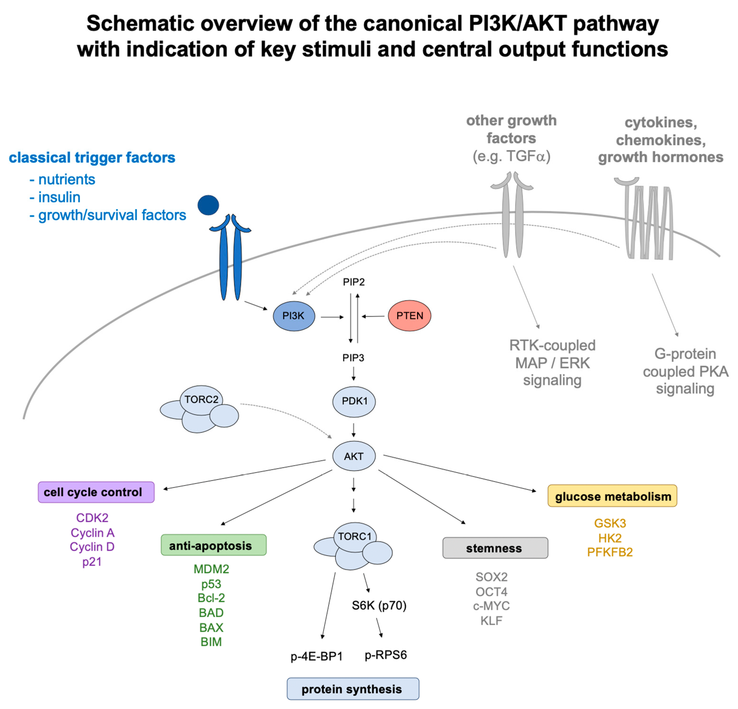
3. The PI3K/AKT/SOX2 Axis in Stemness, Reprogramming, and Cancer

Proliferation and differentiation depend on cell cycle progression and adjusted cell mass growth (i.e., aggravated biosynthetic processes) as underlying molecular principles. While numerous individual factors impinge on these mechanisms, the PI3K/AKT signaling axis resembles a primordial functional module of superordinate significance for mass growth and adjusted proliferation control [51]. In its key components, conserved from unicellular eukaryotic organisms to higher vertebrates and humans, the PI3K/AKT pathway converts nutrient and growth factor sensing on the plasma membrane into downstream effector functions within the cell [52]. Central metabolic stimuli, including glutamate as an indicator of amino acid availability [53] and insulin as a surrogate marker of sugar [54], trigger TORC1-dependent protein synthesis as a fundamentum of mass growth [53,55,56], and cyclin kinase-driven cell cycle progression as a pacemaker of adjusted proliferation [57,58]. For a more comprehensive overview, please refer to Figure 1, which illustrates external trigger factors, key signaling components, and central output relays jointly understood as manifestations of PI3K/AKT signaling.
In dormant stem cells, where growth and proliferation are largely dispensable or even effectively undesired, PI3K/AKT signaling may be broadly attenuated so that somatic stem cells are, on average, smaller (with cell size being an indicator of protein synthesis, see above) and show an overall decreased translation rate when compared to surrounding non-stem cells [59,60]. In stem cells actively involved in tissue regeneration, growth and proliferation become highly desirable functional features instead. For example, in tissue replenishing stem cells, FGF/EGF-triggered PI3K/AKT signaling critically contributes to wound healing [61]. In cancer, where constitutive PI3K/AKT signaling has long been identified as a recurrent driver axis [62,63], a more specific stem cell involvement can be deduced, e.g., from PIK3CA(H1047R) induced trans-conversion of luminal and basal cell layers of the mammary gland in mouse from which composed, multi-lineage carcinomas derive [64]. Accordingly, PI3K/AKT signaling is a critical determinant of stem cells and adaptively tailored to local demand, whereas dysregulated PI3K/AKT signaling is more specifically associated with cancer and stem cell-driven tumorigenesis.
In line with these findings, we and others identified the PI3K/AKT axis as a central mediator of SOX2’s subcellular distribution and turnover regulation in ESCs, iPSCs, and CSCs of mouse and human [65]. A first indicator of such connectivity was the PI3K/AKT cross-reactive inhibitor perifosine, which attenuated PTEN-mutant induced outgrowth of human mammary epithelium-derived stem cells [66]. Subsequent analyses reconfirmed this observation and further dissected the roles of PI3K and AKT as a knock-down of AKT expression alone impaired clonogenicity arising from breast carcinoma stem cells [67]. A molecular–functional relation to SOX2 in this context was first established by studies in mouse, where the Akt-imposed phosphorylation of Sox2 was linked to ESC maintenance [68] and iPSC induction [69]. Further underscored by findings in human cancer cells, PI3K/AKT signaling sustains nuclear entry and DNA modulatory functions of SOX2, whereas in AKT-inhibited cells, SOX2 is retained in the cytosol and is successively cleared by proteasomal turnover [47,48,49,50]. Suggesting a potential applicability of these findings in the fight against cancer, AKT inhibition attenuates clonogenicity effects of human breast cancer-derived cells in vitro [47] and tumorigenicity from nasopharyngeal cancer cells upon xenotransplantation in vivo [48]. Furthermore, while cytostatic drugs standardly applied in the treatment of breast cancer (e.g., cisplatin or paclitaxel) impose a selection advantage for CSCs, AKT inhibition impairs proliferation of CSCs and bulk tumor cells alike [47]. These observations have since been confirmed in various cell types and cancers, as recently summarized in a review on SOX2 secondary modifications in stemness, reprogramming, and cancer [65].

4. DNA Damage Control in Stemness and Reprogramming

The maintenance of genome integrity is a functional prerequisite for stem cells, from which organs or even entire organisms derive and where the repopulating potential is passed on from one generation to the other. A cell’s genome, however, is constantly challenged by environmental risk factors and cell-intrinsic influences that impose damage to DNA [70]. If not stringently repaired, these mutations successively accumulate and increase the risk of transformation [71]. Long-lived cell types (such as quiescent stem cells) or clonal lineages derived from stem cell founders, are particularly affected. However, an array of intertwined molecular relays (collectively referred to as DNA damage response—DDR) has evolved to correct for the various different forms of DNA damage that arise [72,73]. Mutations in these repair factors not only associate with an elevated incidence of cancer [74,75,76], but also with progeria syndromes where there is compelling evidence for stem cell-specific contributions [77,78,79,80].
This interplay of aging and stemness is unquestionably best resolved in the hematopoietic system, which is described here as an archetype of tissue-replenishing stemness. The blood system is qualitatively maintained by stem cells (hematopoietic stem cells, HSCs), but quantitatively replenished by their derived progenitor populations (multi-potent progenitors, MPPs), which still retain full lineage differentiation potential but no longer possess self-renewal capacity, and further, more lineage-restricted species. In many experimental settings, these populations are not strictly discriminated and thus often jointly referred to as hematopoietic stem/progenitor cells (HSPCs) or else, defined as long-term vs. short-term HSCs, respectively. However, only individual HSCs transiently cycle to replenish the progenitor pool, whereas the major part of the murine HSC compartment persists in G0 dormancy [81,82], and thus is not surveyed by cell cycle-checkpoints and their associated DNA integrity control mechanisms. DNA damage accumulating in quiescent murine HSCs is, however, largely resolved as individual HSCs re-enter the cell cycle [83]. Leukemia, by contrast, arises from consecutive mutational hits within divisionally active HSPCs and their derived clonal lineages [84]. Nonetheless, the incidence of neoplastic conversions gradually increases with age and aged HSCs show severe functional impairments over juvenile counterparts with regards to homing, engraftment, lineage reconstitution, and self-renewal capacities, as deduced from blood reconstitution analyses in mice [85]. However, such age-dependent comparisons are inherently confounded by overlapping epigenetic changes and thus not exclusively linked to mutational burden [86]. The specific significance of DDR mechanisms for genome integrity in HSCs is therefore better indicated by mouse models, where distinct DDR mutations functionally associate with impaired hematopoiesis under respective stress settings [87,88,89]. In line with these murine studies, human HSCs are particularly sensitive to irradiation damage, characterized by delayed double-strand break repair (DSBR), by persisting, non-resolved DNA foci, and by aggravated apoptosis over equally irradiated progenitor populations [90]. These data further emphasize that in quiescent HSCs, DNA repair mechanism are overall less active and damaged HSCs are preferentially sacrificed to maintain a pristine stem cell pool.
Specific adaptations in the DNA control mechanisms of mESCs were reported as early as 1993, when their in vitro differentiation was noted to coincide with an increasing risk of UV-imposed DNA damage [91]. Subsequent comparative analyses with littermate somatic cell types confirmed these results and revealed enforced embryonic resilience to further mutagens such as oxidative stress [92]. DDR mechanisms are thus generally enforced in actively cycling mESCs, as also confirmed for human ESCs, where cDNA arrays indicated aggravated expression of DDR genes and comet assays illustrated improved DNA integrity over somatic cells when exposed to various individual mutagens (H202, UV, or psoralen) [93]. Conversely, defects in mismatch repair, nucleotide excision repair, and non-homologous end joining (MMR, NER, and NHEJ, respectively) associated with impaired stem cell functions and accelerated murine stem cell exhaustion [94,95]. However, as already seen for HSCs, a pristine stem cell population may not only be maintained through repair mechanisms, but also by the elimination of aberrant clones. In line with this notion, irradiation and further stress factors have been associated with facilitated apoptosis amongst mESCs as well [96,97]. Accordingly, while ESC may also be further sub-classified into distinct pluripotency states (naïve, primed, and ground state [98]), they are on average privileged over somatic cells and respond to DNA damage in more efficient ways (i.e., by aggravated repair or else, facilitated apoptosis). In total, these differences account for an approximate 100-fold lower mutational burden in mESCs than in isogenic MEFs or adult somatic tissue cells (10E-4 vs. 10E-6, respectively) [99,100].
iPSCs resemble natural stem cells in several physiological characteristics, including DNA maintenance mechanisms such as the reduction of mitochondria to minimize DNA damage arising from reactive oxygen species (ROS) [101]. Although shared pluripotency characteristics identify iPSCs as a functional homologue of ECSs, the expression profiles of these cell types do not perfectly match [102]. This distinction is particularly evident on the mutational level as well. While the mutational burden of healthy ESCs is markedly lower than in their derived somatic populations (see above), the mutational load of human iPSCs exceeds that of their somatic cell origin and only declines gradually thereafter with extended cultivation time [103]. Such reprogramming induced hypermutation was also reported for murine iPSCs, irrespective of whether pluripotency was induced by integrative or non-integrative viral transformation, although the latter is generally considered the “safer” procedure [104]. Accordingly, while the integration of viral particles clearly imposes an additional risk factor, the immanent genetic reorganizations that underlie de-differentiation and pluripotency induction may evoke DNA damage per se [103,104]. This technology-immanent elevated risk of DNA damage provides one potential explanation as to why reprogramming efficacy, despite serious improvement throughout the past years, remains modest overall (i.e., while unfit iPSCs are statistically eliminated through clonal selection, more compatible but nevertheless mutated clones may be eliminated through apoptosis). Accordingly, mutations in cell cycle regulators and apoptosis inductors (such as p21 and p53, respectively) raise reprogramming efficacy and accelerate the induction rates of human/murine iPSCs, but concomitantly increase the risk of genetic aberrations [105,106,107,108,109]. Conversely, mutations acquired though reprogramming often hit tumor suppressor genes or DDR factors [110]. Jointly, these system immanent liabilities remain major obstacles for an otherwise highly desirable applicability of iPSCs in homologous regenerative medicine.
Taken together, the extent and regulation of DDR mechanisms clearly distinguishes stem cells from somatic cells, while individual stem cell populations follow distinct DNA maintenance strategies, as exemplified here for HSCs, ESCs, and iPSCs.

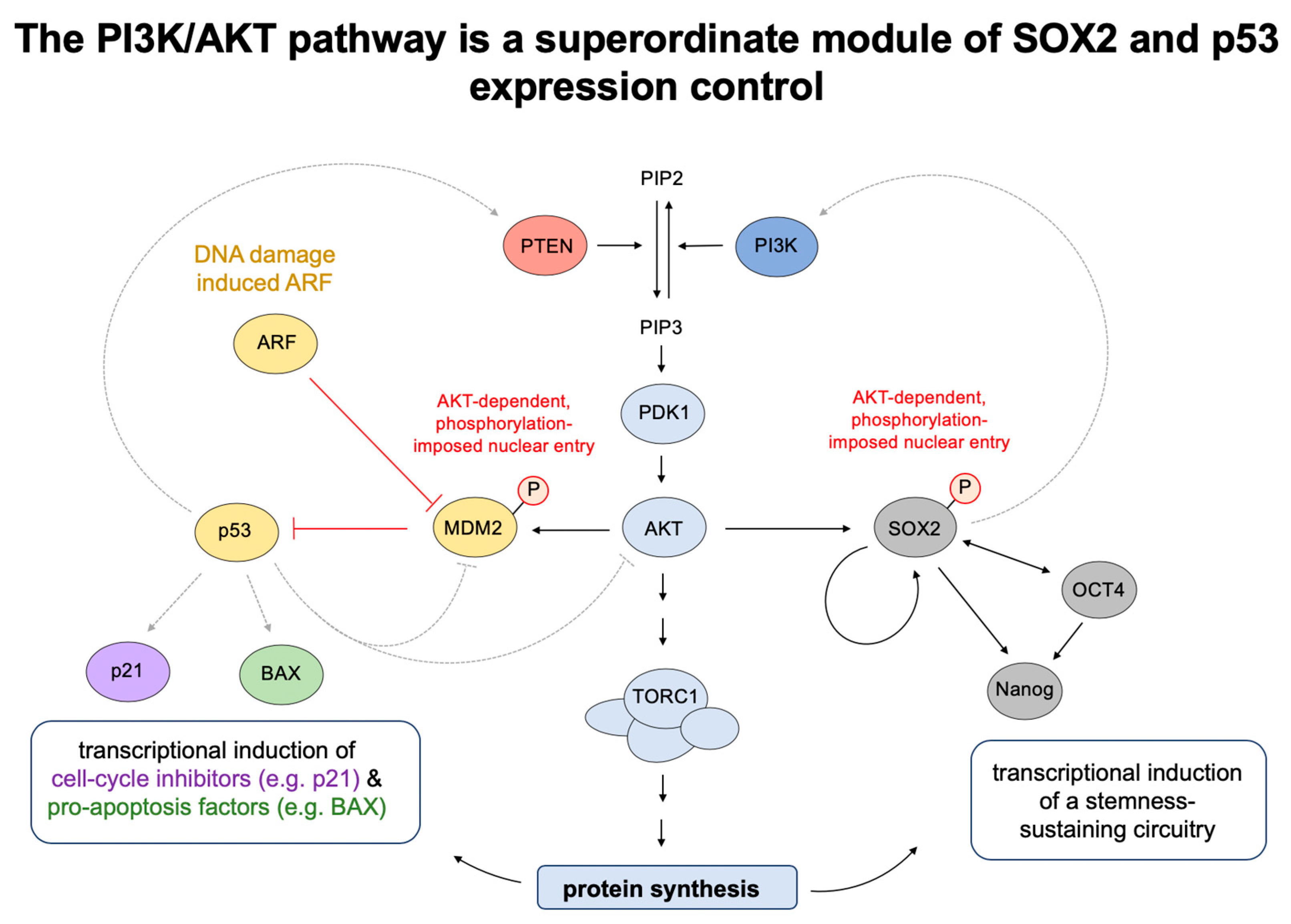
5. AKT/p53 Antagonisms Balance Cell Cycle Progression and DNA Damage Control

The complexity of DDR modules, their dynamics, and the adaptive changes in response to external stimuli, are potentially best illustrated by integrative system biology approaches [111]. While such models incorporate numerous individual elements, they converge on a number of common nodes that define single factorial contributions of superordinate significance. One such relay factor is the tumor suppressor p53 (historically TP53, gene name TP53, hereafter consequently p53), a multi-functional protein that, among other activities, associates with DNA to impose transcription modulatory effects [112,113]. Underscored by phylogenetic conservation, a primordial function of p53 lies in DNA damage-imposed cell cycle arrest (to provide time for repair mechanisms) and subsequent, conditional apoptosis (selectively in those cells, whose DNA integrity cannot be maintained or restored) [114]. This sequence of events correlates with a gradual increase of p53 protein abundance in DNA damaged cells [115] and, furthermore, with overall higher affinities to the DNA recognition elements of cell cycle regulatory, rather than apoptosis inductory genes [116].
p53’s anti-proliferative, pro-apoptotic roles in response to (replication) stress inherently antagonize with growth stimulatory, anti-apoptotic signaling pathways such as the PI3K/AKT axis that triggers the cell cycle in dependence of trophic stimuli and growth factor sensing (see preceding paragraphs). These evidently opposing roles are balanced by intertwined, antagonist regulatory mechanisms [117]. In particular, while AKT-imposed phosphorylation enforces nuclear entry of MDM2, an E3-type ubiquitin ligase that promotes proteasomal degradation of p53 [118], p53 induced caspase cascades enforce degradation of both MDM2 and AKT [119]. This immediate cross-talk is augmented by further accessory relays, such as the AKT-imposed, phosphorylation-dependent inhibition of pro-apoptotic Bad [120] or p53-driven, enforced transcription of PTEN as a natural antagonist of PI3K/AKT signaling [121]. However, p53 expression is largely compromised unless MDM2 becomes inactivated, mostly via stress-imposed phosphorylation ARF [122]. Accordingly, the here outlined antagonism between p53 and AKT activities may only prevail under conditions of effective DNA stress (i.e., when the MDM2-imposed turnover control of p53 is overcome).
By contrast, in the absence of DNA damage, AKT sustains p53 baseline expression via mTORC1-mediated translational control and, in this context, correlates with p53-imposed cellular senescence, not apoptosis [123]. In cancer, where regulatory networks are often distorted and physiological settings perturbed, mutant forms of p53 display gain-of-function phenotypes and accumulate to degrees not effectively seen for wild-type p53 [124,125]. This specifically includes cells with constitutive PI3K/AKT signaling, so that also in transformed settings, AKT cannot be considered a stringent antagonist of p53 expression [126].
Taken together, while AKT counteracts an accumulation of p53 in the absence of DNA damage, it sustains p53 baseline expression via TORC1, whereas analyses in p53mutant conditions indicate further regulatory relays that gain particular significance when p53’s structural integrity and canonical effector functions are distorted [127]. These basic concepts of p53 expression control are illustrated in Figure 2, with further indication of PI3K/AKT/SOX2-imposed stemness as an overlapping molecular module.

6. P53 Mutant-Dependent, Impaired Apoptosis and PI3K/AKT Enforced Growth Signaling Synergize in Cancer

Due to its inherently anti-proliferative, mutation-resolving significance, a functional disruption of p53 signaling ranks amongst the most prevalent mutations in cancer [128]. However, mutations in p53 have comparatively little transforming potential on their own, may not significantly raise tumorigenesis unless triggered by external stressors (UV, IR, cytostatic drugs), or depend on additional driver mutations to boast clonal expansion. By contrast, mutations in proliferation drivers (such as Ras) may initially boast the cell cycle, but the concomitant accumulation of DNA damage rapidly forces cells into senescence thereafter. This has been deduced, for example, from Ras mutant human fibroblast cell lines xenotransplanted into immunocompromised mice [129]. Interestingly, under conditions of constitutive PI3K/AKT signaling (either induced by depletion of PTEN, expression of constitutively active PI3KCA alleles, or mutations in AKT1), cellular senescence can be imposed among human fibroblast cells even without accumulation of DNA damage [123]. These mechanisms involve the enforced TORC1-imposed translation of p53 protein and phosphorylation-based inhibition of its turnover regulator, MDM2 [123]. Accordingly, although PI3K/AKT signaling and p53 control mechanisms mostly antagonize, the disruption of either one axis generally is insufficient to induce cancerous transformation.
Remarkably, although a cooperative distortion of proliferation control and counteracting apoptosis regulation seemingly matches the “hallmarks of cancer” paradigm [130], it is considerably less certain if deviant PI3K/AKT signaling and p53 dysregulation functionally synergize in patient settings or merely co-exist and overlap due to individually high incidence rates. Whereas functional synergism with cooperatively impaired prognosis has been reported for bladder [131,132] and endometrial cancers [133], no evidence for such cooperativity was found in head and neck squamous cell carcinoma [134] or eosophagial cancers [135], despite high coincidence rates. In mammary carcinoma, exon 20 mutations in PIK3CA were described to correlate with TP53 mutations in one study [136], whereas mutant TP53 preferentially associates with triple negative BC, and PI3KCA mutations predominantly associate with ER + /PR+ BC in another [137]. Accordingly, while the functional synergy between dysregulated PI3K/AKT and p53 relays may seem self-explanatory, in clinical practice, such cooperativity remains case-dependent or reaches statistical significance only in certain cancer entities or patient subsets.
This inconsistency may be largely explained by differences between effective p53null settings and functionally impaired, but nevertheless expressed, p53mutant alleles [138]. In fact, while p53 expression is largely compromised by mutations in upstream ARF, the TP53 locus itself is rarely deleted in cancer and nearly all naturally occurring p53 mutants have a spontaneous, somatic origin (except for some rare hereditary forms, e.g., in Li-Fraumeni-syndrome) [127]. Moreover, most cancer patient-derived p53 mutants are missense mutations that predominantly concern a small number of mutational hot spots, with >70 percent of all polymorphisms affecting the protein’s DNA binding domain [139]. Accordingly, while p53 single residue mutants may be impaired in DNA binding (and thus lose their pro-apoptotic, tumor suppressive transcriptional role) [124,125] various mutant forms of p53 are stably expressed and effectively accumulate to degrees not equally seen for the wild-type form of the protein [126,140]. Some mutants even elicit gain-of-function effects, supposedly reflecting perturbations in other DDR relays that selectively manifest in p53mut conditions [138]. Tumorigenicity arising from p53(R172H) mutant mice, for example, repeatedly tested higher than that of p53null littermates [141]. Individual mutations prove particularly detrimental under conditions of enforced DNA stress, such that classical cytostatic drug treatments or reduction therapies (involving paclitaxel, doxorubicin, or γ-irradiation) may paradoxically impose adverse effects, as can be deduced, e.g., from p53 mutant-dependent responses to neoadjuvant therapy in breast cancer [141,142].
These notions may help explain some of the historic inconsistencies that relate to the prognostic significance of p53 in cancer. For example, in mammary carcinoma p53 mutations have been associated with impaired clinical outcome in primary patients [143], whereas the histopathological detection of p53 expression by antibody staining was associated with beneficial outcomes selectively in HER2+ breast cancer [144]. In light of the above notions, the significance of such analyses (and of the cooperativity studies mentioned further above) is strongly compromised unless distinct p53 alleles are described in consideration of individual patient history (i.e., treatment-imposed DNA stress cycles and concomitant stages of clonal selection) and further reference to ARF status.
In selected cancers, p53 and AKT signaling may functionally synergize without underlying mutational distortions. Indeed, while most tumors overexpress telomerase for immortalization, app. 15% of cancers maintain functional telomeres by mechanisms jointly referred to as alternative lengthening of telomeres (ALT) [145]. These involve homologous recombination events that often induce (sub)telomeric lesions as indicated by telomer dysfunction-induced loci (TIFs). TIF scoring indicated inherently higher DNA damage rates in ALT cancer cell lines (VA-13, U2OS, SAOS2, and SKLU-1) than in telomerase-positive, non-ALT cancer cells (MCF7, A549), or healthy-human BJ-fibroblasts. These findings correspond with slightly increased p53 levels in ALT over non-ALT references [146]. Such residual p53 expression may be insufficient to stall the cell cycle and trigger the expression of apoptosis inductors but it has been proposed to transcriptionally activate Rictor and, consequently, TORC2-induced AKT-driven proliferation of ALT tumor cells [146]. Accordingly, the functional significance of wild-type p53 remains case dependent and its expression is often paradoxically maintained in cancer. Furthermore, wild-type p53 has been described to suppress ferroptosis (iron-induced cell death) in cysteine deprived human cancer cell lines of variable tissue origin [147], and to act as a driver of tumorigenesis specifically in hepatocellular carcinoma where it contributes to the attenuation of oxidative phosphorylation [148]. Jointly, these findings indicate that endogenous p53 and AKT activities may not inevitably antagonize, but occasionally also synergize in cancer within certain concentration windows or individual disease settings.
Advanced tumor stages and relapsing cancer(s) are often characterized by treatment-imposed clonal selection effects that favor the development of a resistance mechanism. Specifically in epithelial and neuronal cancers, these effects often coincide with SOX2 expression [16,17,18,19] and aggravated PI3K/AKT signaling as a superordinate module of SOX2 post-transcriptional stabilization [65]. However, while treatment-imposed DNA damage may either induce or favor mutations that help overcome DDR mechanisms (e.g., by disruption of TP53 or ARF), other cancers are paradoxically characterized by aggravated DDR. This involves, for example, IR-resistant forms of hepatocellular carcinoma. In this indication, the appearance of poorly differentiated, stem cell like features coincides with enforced AKT signaling, a joint induction of SOX2 and p53 mRNAs, and enhanced repair mechanisms to resolve IR-imposed DNA lesions [149]. Taken together, the interplay of PI3K/AKT and p53 in cancer presents highly divergent. While constitutive PI3K/AKT signaling and impaired, p53-dependent DDR relays may synergize in various cancers, their endogenous forms have also been described to potentially cooperate in selected disease types, tumor stages, or treatment conditions.

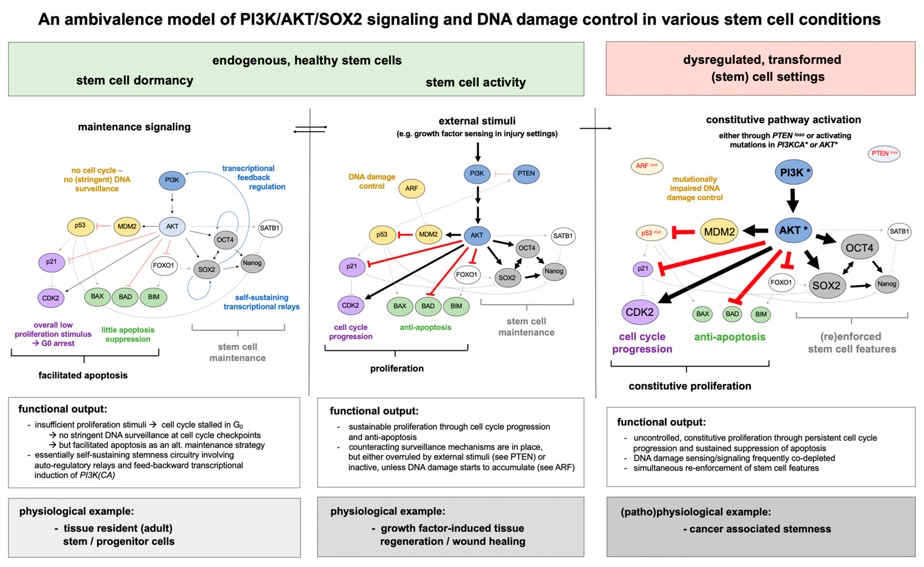
7. PI3K/AKT/SOX2 Signaling and p53-Dependent Apoptosis Regulation in Stem Cells

Modulations in p53 expression are not only of relevance to cancer (stem) cells, but they also impact iPSCs induction. In fact, a depletion of p53 strongly increases reprogramming efficacy by suppression of apoptosis, but at the expense of DNA integrity [150]. Conversely, in other stem cell settings, a depletion of p53 has been associated with improved DNA quality. For example, in HSPCs derived from Fanconi anemia patients (an inherited DNA repair deficiency syndrome characterized by progressive bone marrow failure with juvenile onset), p53 is endogenously hyperactivated due to replication stress and unresolved DNA lesions [151]. Here, the knock-down of p53 improves DNA quality as HSPCs are no longer forced into apoptosis and eliminated, but rather may re-enter the cell cycle and thus benefit from its associated DNA control and repair systems [152]. Accordingly, the functional significance of p53 in stem cell settings remains case-dependent.
These versatile roles of p53 become particularly evident in systemic conditions in vivo, where individual stem cell compartments exert distinct ontological functions at different times. The embryonic lethality of Mdm2−/− knock-out mice, for example, is functionally overcome by Mdm2−/− Tp53−/− co-depletion. The double knock-outs grow into adulthood, but die preterm (at app. month eight) due to tumor burden [153]. Mutant alleles of TP53, such as the p53515C hypomorph, likewise restore viability although Mdm2−/− Tp53515C/515C mice do not reach maturity. These animals succumb to postnatal bone marrow failure, characterized by ROS-induced cell cycle arrest and apoptosis in the hematopoietic compartment [154]. Accordingly, individual stem cell populations are differently challenged by p53, with embryonic lethality linked to dysregulated p53 expression in ESCs, juvenile bone marrow failure arising from p53 mutations in HSPCs, and adult tumorigenesis from p53 deletion in more mature stem/progenitor populations.
In fact, p53 expression may be largely incompatible with the maintenance of stemness. This is evident in the inherently unresolvable molecular antagonism between PI3K/AKT/SOX2-imposed stemness and PI3K/AKT/MDM2-mediated p53 suppression, but also in additional aspects. For example, in DNA-damaged murine ESCs p53 not only associates with response elements that regulate DDR genes, but also with the promoter of Nanog, a transcriptional co-inductor of stemness. Indeed, Nanog functionally cooperates with SOX2 not only in ESCs maintenance [155], but also in germ cell-derived tumors [15], in various carcinomas and gliomas [156], and in reprogramming settings [32]. A DNA damage-imposed, p53-enforced transcriptional suppression of Nanog may thus coincide with an effective loss of stemness, as experimentally validated by an induction of differentiation amongst mESCs [157].
Further critical influences arise from the cyclin-dependent kinase inhibitor, p21. While p21 is well known to arrest the cell cycle in response to DNA damage and p53 activation [158], its knock-down is functionally associated with an early exhaustion of HSCs and neuronal stem cells (NSCs) in mice [159,160]. In NSCs specifically, p21 has been implicated in the attenuation of a Sox2 enhancer element, SRR2 [161], which otherwise sustains Sox2 expression through autocrine self-induction (e.g., imposed by Sox2/Oct4 complexes in mESCs) [162]. Hence, next to the aforementioned feedback regulation of Nanog, the cell-cycle regulator p21 also offers a regulatory relay for stemness control through expression modulation of SOX2. Conversely, SOX2 overexpression in p21 knock-down cells enforces the DNA damage characteristic of replication stress and a corresponding induction of p53 [161]. Accordingly, p21, p53, and SOX2 create a functional triangle that balances cell cycle control, apoptosis induction, and stemness in stem cell populations such as NSCs.
Finally, various further cell cycle regulators and apoptosis factors besides the here featured p21, p53, and MDM2 proteins underly adaptive modulation through PI3K/AKT-imposed phosphorylation (e.g., BAD, BRCA1, CASP9, to name a view) and further downstream mTORC1-associated relay functions, which vastly exceed mere protein expression control [163]. Accordingly, the PI3K/AKT axis resembles a superordinate module of SOX2-imposed stemness and p53-dependent apoptosis control, while various additional contributions impinge on this balance and offer regulatory options for case-dependent fine tuning, as exemplified here for Nanog and p21.

8. Concluding Remarks and Outlook

Research over the past few years characterizes SOX2 as (1) a transcriptional regulator of stemness in early embryogenesis and in ontologically more advanced, mostly neuro-ectodermal stem/progenitor cells, (2) a devastating oncogene in germ line-derived tumors, neuronal cancers and various carcinomas, and (3) a critical co-inductor in the reprogramming of terminally differentiated somatic cells to iPSCs. A unifying molecular module in all these settings has been identified as the PI3K/AKT/SOX2 axis, a stem cell-specific branch from the canonical PI3K/AKT pathway that mediates SOX2 nuclear entry and turnover. The PI3K/AKT/SOX2 axis thus provides an attractive lever to improve reprogramming efficacy and to address some of the safety concerns associated with iPSC induction. At the same time, this axis may offer a molecular mechanism for therapeutic intervention in SOX2+ cancers and, more specifically, against SOX2+ CSCs.
The link to PI3K/AKT signaling and its growth promoting, anti-apoptotic significance seemingly matches the functional requirements of divisionally active stem cells (e.g., in early ontogenesis or wound healing). A constitutive activation of PI3K/AKT signaling is also frequently seen in cancer, where SOX2 expression functionally coincides with the CSC compartment and its implication in treatment resistance, tumor dissemination, and relapse. Here, the PI3K/AKT/SOX2 axis may be augmented by further distortions in DNA control and apoptosis relays (mostly by mutations in either TP53 and/or ARF) which establishes functional synergy in some, but not all settings.
Although advantageous under proliferative conditions, the immanent link to PI3K/AKT is detrimental to stem cell dormancy when cell cycle progression must be suppressed and protein synthesis (as a driving force of differentiation) is attenuated to a basic maintenance level. Similarly, PI3K/AKT-associated anti-apoptosis is a relevant challenge for germ cells and ESCs, where genomic integrity must be maintained despite high proliferative capacity. In iPSCs, the balance between proliferation control and DNA maintenance is severely perturbed. However, whereas transient stem cell conditions and trans-differentiation events are natural phenomena, exogenously enforced somatic cell reprogramming remains an artificial intervention.
Accordingly, the interplay between PI3K/AKT/SOX2-imposed stemness and more canonical PI3K/AKT effects is challenging for stem cells, requiring compromises and individual stem cell compartments to follow distinct strategies. A successive accumulation of DNA damage, for example, may be tolerated in dormant stem cells (such as HSCs) and is only secondarily cleared as individual cells re-enter the cell cycle. Other compartments (e.g., ESCs) follow the opposite strategy and enforce apoptotic mechanisms to warrant stem cell integrity. Overall, the interplay between stem cell induction and maintenance, and DNA control and repair does not converge in a unifying concept, but must be adaptively tailored to distinct stem cell requirements (see Figure 3 for exemplification).
Stemness and DNA control mechanisms also attain particular significance in cancer, as can be deduced from the individually high incidence rates and occasionally synergistic mutational distortion of PTEN/PIK3CA/AKT1 and ARF/TP53 in tumors. Here, PI3K/AKT-sustained SOX2 expression and dysfunctional DDR specifically relate to the development of resistance effects, to CSC dormancy despite clinical remission, and often to unmet medical need particularly in relapsing disease. Accordingly, these dynamics functionally contribute to the major obstacles for a sustained, long-term cure to cancer. The highly diversified examples of stemness-related DNA maintenance strategies summarized in this article argue that these hurdles will unlikely be overcome by comprehensive approaches, while hope for future advances lies within personalized strategies.

Author Contributions

T.S. conceptualized the manuscript and wrote this review jointly with C.L., R.S. and C.L. reviewed and edited the manuscript. C.L. coordinated the study. All authors have read and agreed to the published version of the manuscript.

Funding

This work has been funded by grants from the Swiss Cancer League dedicated to C.L. and T.S. (KFS-4852-08-2019), by the University of Basel Research Funds to T.S. (FO141020_DMS2351), and institutional support from the University of Basel to C.L.

Conflicts of Interest

The authors declare no conflict of interest.

References

  1. Feng, R.; Wen, J. Overview of the roles of Sox2 in stem cell and development. Boil. Chem. 2015, 396, 883–891. [Google Scholar] [CrossRef]
  2. Takahashi, K.; Yamanaka, S. Induction of Pluripotent Stem Cells from Mouse Embryonic and Adult Fibroblast Cultures by Defined Factors. Cell 2006, 126, 663–676. [Google Scholar] [CrossRef] [Green Version]
  3. Masui, S.; Nakatake, Y.; Toyooka, Y.; Shimosato, D.; Yagi, R.; Takahashi, K.; Okochi, H.; Okuda, A.; Matoba, R.; Sharov, A.A.; et al. Pluripotency governed by Sox2 via regulation of Oct3/4 expression in mouse embryonic stem cells. Nat. Cell Biol. 2007, 9, 625–635. [Google Scholar] [CrossRef]
  4. Kopp, J.L.; Ormsbee, B.D.; Desler, M.; Rizzino, A. Small Increases in the Level of Sox2 Trigger the Differentiation of Mouse Embryonic Stem Cells. Stem Cells 2008, 26, 903–911. [Google Scholar] [CrossRef] [PubMed]
  5. Avilion, A.A.; Nicolis, S.K.; Pevny, L.H.; Perez, L.; Vivian, N.; Lovell-Badge, R. Multipotent cell lineages in early mouse development depend on SOX2 function. Genome Res. Dev. 2003, 17, 126–140. [Google Scholar] [CrossRef] [Green Version]
  6. Zhao, S.; Nichols, J.; Smith, A.; Li, M. SoxB transcription factors specify neuroectodermal lineage choice in ES cells. Mol. Cell. Neurosci. 2004, 27, 332–342. [Google Scholar] [CrossRef] [PubMed]
  7. Williamson, K.A.; Hever, A.M.; Rainger, J.; Rogers, R.C.; Magee, A.; Fiedler, Z.; Keng, W.T.; Sharkey, F.H.; McGill, N.; Hill, C.J.; et al. Mutations in SOX2 cause anophthalmia-esophageal-genital (AEG) syndrome. Hum. Mol. Genet. 2006, 15, 1413–1422. [Google Scholar] [CrossRef] [PubMed]
  8. Schneider, A.; Bardakjian, T.; Reis, L.M.; Tyler, R.C.; Semina, E.V. Novel SOX2 mutations and genotype-phenotype correlation in anophthalmia and microphthalmia. Am. J. Med Genet. Part A 2009, 149A, 2706–2715. [Google Scholar] [CrossRef] [PubMed] [Green Version]
  9. Matsushima, D.; Heavner, W.; Pevny, L.H. Combinatorial regulation of optic cup progenitor cell fate by SOX2 and PAX6. Development 2011, 138, 443–454. [Google Scholar] [CrossRef] [Green Version]
  10. Taranova, O.V.; Magness, S.T.; Fagan, B.M.; Wu, Y.; Surzenko, N.; Hutton, S.R.; Pevny, L.H. SOX2 is a dose-dependent regulator of retinal neural progenitor competence. Genome Res. Dev. 2006, 20, 1187–1202. [Google Scholar] [CrossRef] [Green Version]
  11. Fernandes, K.J.L.; McKenzie, I.A.; Mill, P.; Smith, K.; Akhavan, M.; Barnabé-Heider, F.; Biernaskie, J.; Junek, A.; Kobayashi, N.R.; Toma, J.G.; et al. A dermal niche for multipotent adult skin-derived precursor cells. Nat. Cell Biol. 2004, 6, 1082–1093. [Google Scholar] [CrossRef] [PubMed]
  12. Arnold, K.; Sarkar, A.; Yram, M.A.; Polo, J.M.; Bronson, R.; Sengupta, S.; Seandel, M.; Geijsen, N.; Hochedlinger, K. Sox2+ Adult Stem and Progenitor Cells Are Important for Tissue Regeneration and Survival of Mice. Cell Stem Cell 2011, 9, 317–329. [Google Scholar] [CrossRef] [PubMed] [Green Version]
  13. Wuebben, E.L.; Rizzino, A. The dark side of SOX2: Cancer—A comprehensive overview. Oncotarget 2017, 8, 44917–44943. [Google Scholar] [CrossRef] [Green Version]
  14. Grimm, D.; Bauer, J.; Wise, P.; Krüger, M.; Simonsen, U.; Wehland, M.; Infanger, M.; Corydon, T.J. The role of SOX family members in solid tumours and metastasis. Semin. Cancer Boil. 2019. [Google Scholar] [CrossRef] [PubMed]
  15. Gillis, A.J.M.; Stoop, H.; Biermann, K.; Van Gurp, R.J.H.L.M.; Swartzman, E.; Cribbes, S.; Ferlinz, A.; Shannon, M.; Oosterhuis, J.; Looijenga, L.H.J. Expression and interdependencies of pluripotency factors LIN28, OCT3/4, NANOG and SOX2 in human testicular germ cells and tumours of the testis. Int. J. Androl. 2011, 34, e160–e174. [Google Scholar] [CrossRef] [PubMed]
  16. Bareiss, P.M.; Paczulla, A.; Wang, H.; Schairer, R.; Wiehr, S.; Kohlhofer, U.; Rothfuss, O.C.; Fischer, A.; Perner, S.; Staebler, A.; et al. SOX2 Expression Associates with Stem Cell State in Human Ovarian Carcinoma. Cancer Res. 2013, 73, 5544–5555. [Google Scholar] [CrossRef] [PubMed] [Green Version]
  17. Boumahdi, S.; Driessens, G.; Lapouge, G.; Rorive, S.; Nassar, D.; Le Mercier, M.; Delatte, B.; Caauwe, A.; Lenglez, S.; Nkusi, E.; et al. SOX2 controls tumour initiation and cancer stem-cell functions in squamous-cell carcinoma. Nature 2014, 511, 246–250. [Google Scholar] [CrossRef] [PubMed]
  18. Takeda, K.; Mizushima, T.; Yokoyama, Y.; Hirose, H.; Wu, X.; Qian, Y.; Ikehata, K.; Miyoshi, N.; Takahashi, H.; Haraguchi, N.; et al. Sox2 is associated with cancer stem-like properties in colorectal cancer. Sci. Rep. 2018, 8, 17639. [Google Scholar] [CrossRef]
  19. Al Mamun, M.; Mannoor, K.; Cao, J.; Qadri, F.; Song, X. SOX2 in cancer stemness: Tumor malignancy and therapeutic potentials. J. Mol. Cell Boil. 2018, 12, 85–98. [Google Scholar] [CrossRef] [Green Version]
  20. Aceto, N.; Bardia, A.; Miyamoto, D.T.; Donaldson, M.C.; Wittner, B.S.; Spencer, J.A.; Yu, M.; Pely, A.; Engstrom, A.; Zhu, H.; et al. Circulating tumor cell clusters are oligoclonal precursors of breast cancer metastasis. Cell 2014, 158, 1110–1122. [Google Scholar] [CrossRef] [Green Version]
  21. Gkountela, S.; Castro-Giner, F.; Szczerba, B.M.; Vetter, M.; Landin, J.; Scherrer, R.; Krol, I.; Scheidmann, M.C.; Beisel, C.; Stirnimann, C.U.; et al. Circulating Tumor Cell Clustering Shapes DNA Methylation to Enable Metastasis Seeding. Cell 2019, 176, 98–112.e14. [Google Scholar] [CrossRef] [PubMed] [Green Version]
  22. Takahashi, K.; Tanabe, K.; Ohnuki, M.; Narita, M.; Ichisaka, T.; Tomoda, K.; Yamanaka, S. Induction of Pluripotent Stem Cells from Adult Human Fibroblasts by Defined Factors. Cell 2007, 131, 861–872. [Google Scholar] [CrossRef] [PubMed] [Green Version]
  23. Wilmut, I.; Schnieke, A.; McWhir, J.; Kind, A.J.; Campbell, K.H.S. Viable offspring derived from fetal and adult mammalian cells. Nature 1997, 385, 810–813. [Google Scholar] [CrossRef] [PubMed]
  24. Hochedlinger, K.; Jaenisch, R. Nuclear Transplantation, Embryonic Stem Cells, and the Potential for Cell Therapy. N. Engl. J. Med. 2003, 349, 275–286. [Google Scholar] [CrossRef] [Green Version]
  25. Schepers, G.E.; Teasdale, R.D.; Koopman, P. Twenty Pairs of Sox. Dev. Cell 2002, 3, 167–170. [Google Scholar] [CrossRef] [Green Version]
  26. Hou, L.; Srivastava, Y.; Jauch, R. Molecular basis for the genome engagement by Sox proteins. Semin. Cell Dev. Boil. 2017, 63, 2–12. [Google Scholar] [CrossRef] [Green Version]
  27. Bowles, J.; Schepers, G.; Koopman, P. Phylogeny of the SOX Family of Developmental Transcription Factors Based on Sequence and Structural Indicators. Dev. Boil. 2000, 227, 239–255. [Google Scholar] [CrossRef] [Green Version]
  28. Kamachi, Y.; Kondoh, H. Sox proteins: Regulators of cell fate specification and differentiation. Develpoment 2013, 140, 4129–4144. [Google Scholar] [CrossRef] [Green Version]
  29. Nakagawa, M.; Koyanagi, M.; Tanabe, K.; Takahashi, K.; Ichisaka, T.; Aoi, T.; Okita, K.; Mochiduki, Y.; Takizawa, N.; Yamanaka, S. Generation of induced pluripotent stem cells without Myc from mouse and human fibroblasts. Nat. Biotechnol. 2007, 26, 101–106. [Google Scholar] [CrossRef]
  30. Sarkar, A.; Hochedlinger, K. The sox family of transcription factors: Versatile regulators of stem and progenitor cell fate. Cell Stem Cell 2013, 12, 15–30. [Google Scholar] [CrossRef] [Green Version]
  31. Guth, S.I.E.; Wegner, M. Having it both ways: Sox protein function between conservation and innovation. Cell. Mol. Life Sci. 2008, 65, 3000–3018. [Google Scholar] [CrossRef]
  32. Li, Y.; Zhao, H.; Lan, F.; Lee, A.; Chen, L.; Lin, C.; Yao, Y.; Li, L. Generation of Human-Induced Pluripotent Stem Cells from Gut Mesentery-Derived Cells by Ectopic Expression of OCT4/SOX2/NANOG. Cell. Reprogram. 2010, 12, 237–247. [Google Scholar] [CrossRef] [PubMed]
  33. Mao, J.; Zhang, Q.; Ye, X.; Liu, K.; Liu, L. Efficient Induction of Pluripotent Stem Cells from Granulosa Cells byOct4andSox2. Stem Cells Dev. 2014, 23, 779–789. [Google Scholar] [CrossRef] [Green Version]
  34. Maherali, N.; Hochedlinger, K. Tgfβ Signal Inhibition Cooperates in the Induction of iPSCs and Replaces Sox2 and cMyc. Curr. Boil. 2009, 19, 1718–1723. [Google Scholar] [CrossRef] [PubMed] [Green Version]
  35. Ichida, J.K.; Blanchard, J.; Lam, K.; Son, E.Y.; Chung, J.E.; Egli, D.; Loh, K.M.; Carter, A.C.; Di Giorgio, F.P.; Koszka, K.; et al. A Small-Molecule Inhibitor of Tgf-β Signaling Replaces Sox2 in Reprogramming by Inducing Nanog. Cell Stem Cell 2009, 5, 491–503. [Google Scholar] [CrossRef] [PubMed] [Green Version]
  36. Hou, P.; Li, Y.; Zhang, X.; Liu, C.; Guan, J.; Li, H.; Zhao, T.; Ye, J.; Yang, W.; Liu, K.; et al. Pluripotent Stem Cells Induced from Mouse Somatic Cells by Small-Molecule Compounds. Science 2013, 341, 651–654. [Google Scholar] [CrossRef]
  37. Lin, T.; Wu, S. Reprogramming with Small Molecules instead of Exogenous Transcription Factors. Stem Cells Int. 2015, 2015, 794632. [Google Scholar] [CrossRef] [Green Version]
  38. Takayama, Y.; Clore, G.M. Interplay between Minor and Major Groove-binding Transcription Factors Sox2 and Oct1 in Translocation on DNA Studied by Paramagnetic and Diamagnetic NMR*. J. Boil. Chem. 2012, 287, 14349–14363. [Google Scholar] [CrossRef] [Green Version]
  39. Yesudhas, D.; Anwar, M.A.; Panneerselvam, S.; Kim, H.-K.; Choi, S. Evaluation of Sox2 binding affinities for distinct DNA patterns using steered molecular dynamics simulation. FEBS Open Bio 2017, 7, 1750–1767. [Google Scholar] [CrossRef]
  40. Fang, X.; Yoon, J.-G.; Li, L.; Yu, W.; Shao, J.; Hua, D.; Zheng, S.; Hood, L.; Goodlett, D.R.; Foltz, G.; et al. The SOX2 response program in glioblastoma multiforme: An integrated ChIP-seq, expression microarray, and microRNA analysis. BMC Genom. 2011, 12, 11. [Google Scholar] [CrossRef] [Green Version]
  41. Tang, J.; Zhong, G.; Wu, J.; Chen, H.; Jia, Y. SOX2 recruits KLF4 to regulate nasopharyngeal carcinoma proliferation via PI3K/AKT signaling. Oncogenesis 2018, 7, 61. [Google Scholar] [CrossRef] [PubMed]
  42. Ng, S.-Y.; Bogu, G.; Soh, B.S.; Stanton, L.W. The Long Noncoding RNA RMST Interacts with SOX2 to Regulate Neurogenesis. Mol. Cell 2013, 51, 349–359. [Google Scholar] [CrossRef] [PubMed] [Green Version]
  43. Holmes, Z.E.; Hamilton, D.J.; Hwang, T.; Parsonnet, N.V.; Rinn, J.L.; Wuttke, D.S.; Batey, R.T. The Sox2 transcription factor binds RNA. Nat. Commun. 2020, 11, 1805–1812. [Google Scholar] [CrossRef] [PubMed] [Green Version]
  44. Hou, L.; Wei, Y.; Lin, Y.; Wang, X.; Lai, Y.; Yin, M.; Chen, Y.; Guo, X.; Wu, S.; Zhu, Y.; et al. Concurrent binding to DNA and RNA facilitates the pluripotency reprogramming activity of Sox2. Nucleic Acids Res. 2020, 48, 3869–3887. [Google Scholar] [CrossRef]
  45. Li, J.; Pan, G.; Cui, K.; Liu, Y.; Xu, S.; Pei, D. A Dominant-negative Form of Mouse SOX2 Induces Trophectoderm Differentiation and Progressive Polyploidy in Mouse Embryonic Stem Cells. J. Boil. Chem. 2007, 282, 19481–19492. [Google Scholar] [CrossRef] [Green Version]
  46. Baltus, G.A.; Kowalski, M.P.; Zhai, H.; Tutter, A.V.; Quinn, D.; Wall, D.; Kadam, S. Acetylation of Sox2 Induces its Nuclear Export in Embryonic Stem Cells. Stem Cells 2009, 27, 2175–2184. [Google Scholar] [CrossRef]
  47. Schaefer, T.; Wang, H.; Mir, P.; Konantz, M.; Pereboom, T.C.; Paczulla, A.M.; Merz, B.; Fehm, T.; Perner, S.; Rothfuss, O.C.; et al. Molecular and functional interactions between AKT and SOX2 in breast carcinoma. Oncotarget 2015, 6, 43540–43556. [Google Scholar] [CrossRef] [Green Version]
  48. Qin, J.; Ji, J.; Deng, R.; Tang, J.; Yang, F.; Feng, G.-K.; Chen, W.-D.; Wu, X.-Q.; Qian, X.-J.; Ding, K.; et al. DC120, a novel AKT inhibitor, preferentially suppresses nasopharyngeal carcinoma cancer stem-like cells by downregulating Sox2. Oncotarget 2015, 6, 6944–6958. [Google Scholar] [CrossRef]
  49. Wang, Z.; Kang, L.; Zhang, H.; Huang, Y.; Fang, L.; Li, M.; Brown, P.J.; Arrowsmith, C.H.; Li, J.; Wong, J. AKT drives SOX2 overexpression and cancer cell stemness in esophageal cancer by protecting SOX2 from UBR5-mediated degradation. Oncogene 2019, 38, 5250–5264. [Google Scholar] [CrossRef]
  50. Schaefer, T.; Ramadoss, A.; Leu, S.; Tintignac, L.; Tostado, C.; Bink, A.; Schürch, C.; Müller, J.; Schärer, J.; Moffa, G.; et al. Regulation of glioma cell invasion by 3q26 gene products PIK3CA, SOX2 and OPA1. Brain Pathol. 2019, 29, 336–350. [Google Scholar] [CrossRef]
  51. Manning, B.D.; Toker, A. AKT/PKB Signaling: Navigating the Network. Cell 2017, 169, 381–405. [Google Scholar] [CrossRef] [Green Version]
  52. Wullschleger, S.; Loewith, R.; Hall, M.N. TOR Signaling in Growth and Metabolism. Cell 2006, 124, 471–484. [Google Scholar] [CrossRef] [PubMed] [Green Version]
  53. Liao, X.-H.; Majithia, A.; Huang, X.; Kimmel, A.R. Growth control via TOR kinase signaling, an intracellular sensor of amino acid and energy availability, with crosstalk potential to proline metabolism. Amino Acids 2008, 35, 761–770. [Google Scholar] [CrossRef] [PubMed]
  54. Siddle, K. Signalling by insulin and IGF receptors: Supporting acts and new players. J. Mol. Endocrinol. 2011, 47, R1–R10. [Google Scholar] [CrossRef] [PubMed] [Green Version]
  55. Meyuhas, O. Ribosomal Protein S6 Phosphorylation. Int. Rev. Cell Mol. Biol. 2015, 320, 41–73. [Google Scholar] [CrossRef]
  56. Josse, L.; Xie, J.; Proud, C.G.; Smales, C.M. mTORC1 signalling and eIF4E/4E-BP1 translation initiation factor stoichiometry influence recombinant protein productivity from GS-CHOK1 cells. Biochem. J. 2016, 473, 4651–4664. [Google Scholar] [CrossRef] [Green Version]
  57. Heuvel, S.v.d.; Harlow, E. Distinct roles for cyclin-dependent kinases in cell cycle control. Science 1993, 262, 2050–2054. [Google Scholar] [CrossRef]
  58. Chen, Y.; Liu, X.; Wang, H.; Liu, S.; Hu, N.; Li, X. Akt Regulated Phosphorylation of GSK-3β/Cyclin D1, p21 and p27 Contributes to Cell Proliferation Through Cell Cycle Progression from G1 to S/G2M Phase in Low-Dose Arsenite Exposed HaCat Cells. Front. Pharmacol. 2019, 10, 1176. [Google Scholar] [CrossRef] [Green Version]
  59. Signer, R.A.J.; Magee, J.A.; Salic, A.; Morrison, S.J. Haematopoietic stem cells require a highly regulated protein synthesis rate. Nature 2014, 509, 49–54. [Google Scholar] [CrossRef]
  60. Blanco, S.; Bandiera, R.; Popis, M.; Hussain, S.; Lombard, P.; Aleksic, J.; Sajini, A.; Tanna, H.; Cortés-Garrido, R.; Gkatza, N.; et al. Stem cell function and stress response are controlled by protein synthesis. Nature 2016, 534, 335–340. [Google Scholar] [CrossRef] [Green Version]
  61. Dehkordi, A.N.; Babaheydari, F.M.; Chehelgerdi, M.; Dehkordi, S.R. Skin tissue engineering: Wound healing based on stem-cell-based therapeutic strategies. Stem Cell Res. Ther. 2019, 10, 111. [Google Scholar] [CrossRef] [PubMed] [Green Version]
  62. Martini, M.; De Santis, M.C.; Braccini, L.; Gulluni, F.; Hirsch, E. PI3K/AKT signaling pathway and cancer: An updated review. Ann. Med. 2014, 46, 372–383. [Google Scholar] [CrossRef] [PubMed]
  63. Porta, C.; Paglino, C.; Mosca, A. Targeting PI3K/Akt/mTOR Signaling in Cancer. Front. Oncol. 2014, 4, 64. [Google Scholar] [CrossRef] [PubMed] [Green Version]
  64. Koren, S.; Reavie, L.; Couto, J.P.; De Silva, D.; Stadler, M.B.; Roloff, T.; Britschgi, A.; Eichlisberger, T.; Kohler, H.; Aina, O.; et al. PIK3CAH1047R induces multipotency and multi-lineage mammary tumours. Nature 2015, 525, 114–118. [Google Scholar] [CrossRef]
  65. Schaefer, T.; Lengerke, C. SOX2 protein biochemistry in stemness, reprogramming, and cancer: The PI3K/AKT/SOX2 axis and beyond. Oncogene 2019, 39, 278–292. [Google Scholar] [CrossRef] [Green Version]
  66. Korkaya, H.; Paulson, A.; Charafe-Jauffret, E.; Ginestier, C.; Brown, M.; Dutcher, J.; Clouthier, S.G.; Wicha, M.S. Regulation of Mammary Stem/Progenitor Cells by PTEN/Akt/β-Catenin Signaling. PLoS Boil. 2009, 7, e1000121. [Google Scholar] [CrossRef]
  67. Gargini, R.; Cerliani, J.P.; Escoll, M.; Antón, I.M.; Wandosell, F. Cancer Stem Cell-Like Phenotype and Survival Are Coordinately Regulated by Akt/FoxO/Bim Pathway. Stem Cells 2015, 33, 646–660. [Google Scholar] [CrossRef]
  68. Fang, L.; Zhang, L.; Wei, W.; Jin, X.; Wang, P.; Tong, Y.; Li, J.; Du, J.X.; Wong, J. A Methylation-Phosphorylation Switch Determines Sox2 Stability and Function in ESC Maintenance or Differentiation. Mol. Cell 2014, 55, 537–551. [Google Scholar] [CrossRef] [Green Version]
  69. Jeong, C.-H.; Cho, Y.-Y.; Kim, M.-O.; Kim, S.-H.; Cho, E.-J.; Lee, S.-Y.; Jeon, Y.-J.; Lee, K.Y.; Yao, K.; Keum, Y.-S.; et al. Phosphorylation of Sox2 Cooperates in Reprogramming to Pluripotent Stem Cells. Stem Cells 2010, 28, 2141–2150. [Google Scholar] [CrossRef]
  70. Jackson, S.P.; Bartek, J. The DNA-damage response in human biology and disease. Nature 2009, 461, 1071–1078. [Google Scholar] [CrossRef] [Green Version]
  71. Chalmers, Z.R.; Connelly, C.F.; Fabrizio, D.; Gay, L.; Ali, S.M.; Ennis, R.; Schrock, A.; Campbell, B.; Shlien, A.; Chmielecki, J.; et al. Analysis of 100,000 human cancer genomes reveals the landscape of tumor mutational burden. Genome Med. 2017, 9, 34. [Google Scholar] [CrossRef] [PubMed]
  72. Blackford, A.N.; Jackson, S.P. ATM, ATR, and DNA-PK: The Trinity at the Heart of the DNA Damage Response. Mol. Cell 2017, 66, 801–817. [Google Scholar] [CrossRef] [PubMed] [Green Version]
  73. Giglia-Mari, G.; Zotter, A.; Vermeulen, W. DNA Damage Response. Cold Spring Harb. Perspect. Boil. 2010, 3, a000745. [Google Scholar] [CrossRef] [PubMed]
  74. Goldstein, M.; Kastan, M.B. The DNA Damage Response: Implications for Tumor Responses to Radiation and Chemotherapy. Annu. Rev. Med. 2015, 66, 129–143. [Google Scholar] [CrossRef] [Green Version]
  75. Lang, S.H.; Swift, S.L.; White, H.; Misso, K.; Kleijnen, J.; Quek, R.G. A systematic review of the prevalence of DNA damage response gene mutations in prostate cancer. Int. J. Oncol. 2019, 55, 597–616. [Google Scholar] [CrossRef] [Green Version]
  76. Parikh, A.R.; He, Y.; Hong, T.S.; Corcoran, R.B.; Clark, J.W.; Ryan, D.P.; Zou, L.; Ting, D.T.; Catenacci, D.V.; Chao, J.; et al. Analysis of DNA Damage Response Gene Alterations and Tumor Mutational Burden Across 17,486 Tubular Gastrointestinal Carcinomas: Implications for Therapy. Oncologist 2019, 24, 1340–1347. [Google Scholar] [CrossRef] [Green Version]
  77. Park, Y.; Gerson, S.L. DNA Repair Defects in Stem Cell Function and Aging. Annu. Rev. Med. 2005, 56, 495–508. [Google Scholar] [CrossRef]
  78. Tiwari, V.; Wilson, D.M. DNA Damage and Associated DNA Repair Defects in Disease and Premature Aging. Am. J. Hum. Genet. 2019, 105, 237–257. [Google Scholar] [CrossRef] [Green Version]
  79. Hoeijmakers, J. Genome maintenance mechanisms are critical for preventing cancer as well as other aging-associated diseases. Mech. Ageing Dev. 2007, 128, 460–462. [Google Scholar] [CrossRef]
  80. McNeely, T.; Leone, M.; Yanai, H.; Beerman, I. DNA damage in aging, the stem cell perspective. Hum. Genet. 2019, 139, 309–331. [Google Scholar] [CrossRef]
  81. Seita, J.; Weissman, I.L. Hematopoietic stem cell: Self-renewal versus differentiation. Wiley Interdiscip. Rev. Syst. Boil. Med. 2010, 2, 640–653. [Google Scholar] [CrossRef] [PubMed] [Green Version]
  82. Takizawa, H.; Regoes, R.R.; Boddupalli, C.S.; Bonhoeffer, S.; Manz, M.G. Dynamic variation in cycling of hematopoietic stem cells in steady state and inflammation. J. Exp. Med. 2011, 208, 273–284. [Google Scholar] [CrossRef] [PubMed] [Green Version]
  83. Beerman, I.; Seita, J.; Inlay, M.A.; Weissman, I.L.; Rossi, D.J. Quiescent Hematopoietic Stem Cells Accumulate DNA Damage during Aging that Is Repaired upon Entry into Cell Cycle. Cell Stem Cell 2014, 15, 37–50. [Google Scholar] [CrossRef] [Green Version]
  84. Genovese, G.; Kähler, A.K.; Handsaker, R.E.; Lindberg, J.; Rose, S.; Bakhoum, S.F.; Chambert, K.; Mick, E.; Neale, B.M.; Fromer, M.; et al. Clonal hematopoiesis and blood-cancer risk inferred from blood DNA sequence. N. Engl. J. Med. 2014, 371, 2477–2487. [Google Scholar] [CrossRef] [PubMed] [Green Version]
  85. Liang, Y.; Van Zant, G.; Szilvassy, S.J. Effects of aging on the homing and engraftment of murine hematopoietic stem and progenitor cells. Blood 2005, 106, 1479–1487. [Google Scholar] [CrossRef] [PubMed] [Green Version]
  86. Beerman, I.; Bock, C.; Garrison, B.S.; Smith, Z.D.; Gu, H.; Meissner, A.; Rossi, D.J. Proliferation-Dependent Alterations of the DNA Methylation Landscape Underlie Hematopoietic Stem Cell Aging. Cell Stem Cell 2013, 12, 413–425. [Google Scholar] [CrossRef] [Green Version]
  87. Prasher, J.M.; Lalai, A.S.; Heijmans-Antonissen, C.; Ploemacher, R.E.; Hoeijmakers, J.H.J.; Touw, I.P.; Niedernhofer, L.J. Reduced hematopoietic reserves in DNA interstrand crosslink repair-deficient Ercc1−/− mice. EMBO J. 2005, 24, 861–871. [Google Scholar] [CrossRef] [PubMed] [Green Version]
  88. Parmar, K.; Kim, J.; Sykes, S.M.; Shimamura, A.; Stuckert, P.; Zhu, K.; Hamilton, A.; Deloach, M.K.; Kutok, J.L.; Akashi, K.; et al. Hematopoietic Stem Cell Defects in Mice with Deficiency of Fancd2 or Usp1. Stem Cells 2010, 28, 1186–1195. [Google Scholar] [CrossRef] [Green Version]
  89. Cho, J.; Kook, S.H.; Robinson, A.R.; Niedernhofer, L.J.; Lee, B.C. Cell autonomous and nonautonomous mechanisms drive hematopoietic stem/progenitor cell loss in the absence of DNA repair. Stem Cells 2013, 31, 511–525. [Google Scholar] [CrossRef] [Green Version]
  90. Milyavsky, M.; Gan, O.I.; Trottier, M.; Komosa, M.; Tabach, O.; Notta, F.; Lechman, E.; Hermans, K.G.; Eppert, K.; Konovalova, Z.; et al. A Distinctive DNA Damage Response in Human Hematopoietic Stem Cells Reveals an Apoptosis-Independent Role for p53 in Self-Renewal. Cell Stem Cell 2010, 7, 186–197. [Google Scholar] [CrossRef] [Green Version]
  91. Rasko, I.; Georgieva, M.; Farkas, G.; Santha, M.; Coates, J.; Burg, K.; Mitchell, D.L.; Johnson, R.T. New patterns of bulk DNA repair in ultraviolet irradiated mouse embryo carcinoma cells following differentiation. Somat. Cell Mol. Genet. 1993, 19, 245–255. [Google Scholar] [CrossRef] [PubMed]
  92. Saretzki, G.; Leake, A.; Armstrong, L.; Lako, M.; Von Zglinicki, T. Stress Defense in Murine Embryonic Stem Cells Is Superior to That of Various Differentiated Murine Cells. Stem Cells 2004, 22, 962–971. [Google Scholar] [CrossRef] [PubMed]
  93. Maynard, S.; Swistowska, A.M.; Lee, J.W.; Liu, Y.; Liu, S.-T.; Da Cruz, A.B.; Rao, M.; Souza-Pinto, N.; Zeng, X.; Bohr, V.A.; et al. Human Embryonic Stem Cells Have Enhanced Repair of Multiple Forms of DNA Damage. Stem Cells 2008, 26, 2266–2274. [Google Scholar] [CrossRef] [PubMed] [Green Version]
  94. Rossi, D.J.; Bryder, D.; Seita, J.; Nussenzweig, A.; Hoeijmakers, J.; Weissman, I.L. Deficiencies in DNA damage repair limit the function of haematopoietic stem cells with age. Nature 2007, 447, 725–729. [Google Scholar] [CrossRef] [PubMed]
  95. Nijnik, A.; Woodbine, L.; Marchetti, C.; Dawson, S.; Lambe, T.; Liu, C.; Rodrigues, N.P.; Crockford, T.L.; Cabuy, E.; Vindigni, A.; et al. DNA repair is limiting for haematopoietic stem cells during ageing. Nature 2007, 447, 686–690. [Google Scholar] [CrossRef] [PubMed]
  96. Hong, Y.; Stambrook, P.J. Restoration of an absent G1 arrest and protection from apoptosis in embryonic stem cells after ionizing radiation. Proc. Natl. Acad. Sci. USA 2004, 101, 14443–14448. [Google Scholar] [CrossRef] [PubMed] [Green Version]
  97. Roos, W.P.; Christmann, M.; Fraser, S.T.; Kaina, B. Mouse embryonic stem cells are hypersensitive to apoptosis triggered by the DNA damage O6-methylguanine due to high E2F1 regulated mismatch repair. Cell Death Differ. 2007, 14, 1422–1432. [Google Scholar] [CrossRef]
  98. Ghimire, S.; Van Der Jeught, M.; Neupane, J.; Roost, M.S.; Anckaert, J.; Popovic, M.; Van Nieuwerburgh, F.; Mestdagh, P.; Vandesompele, J.; Deforce, D.; et al. Comparative analysis of naive, primed and ground state pluripotency in mouse embryonic stem cells originating from the same genetic background. Sci. Rep. 2018, 8, 5884. [Google Scholar] [CrossRef] [Green Version]
  99. Cervantes, R.B.; Stringer, J.R.; Shao, C.; Tischfield, J.; Stambrook, P.J. Embryonic stem cells and somatic cells differ in mutation frequency and type. Proc. Natl. Acad. Sci. USA 2002, 99, 3586–3590. [Google Scholar] [CrossRef] [Green Version]
  100. Hong, Y.; Cervantes, R.; Tichy, E.; Tischfield, J.; Stambrook, P.J. Protecting genomic integrity in somatic cells and embryonic stem cells. Mutat. Res./Fundam. Mol. Mech. Mutagen. 2007, 614, 48–55. [Google Scholar] [CrossRef]
  101. Armstrong, L.; Tilgner, K.; Saretzki, G.; Atkinson, S.P.; Stojkovic, M.; Moreno, R.; Przyborski, S.; Lako, M. Human Induced Pluripotent Stem Cell Lines Show Stress Defense Mechanisms and Mitochondrial Regulation Similar to Those of Human Embryonic Stem Cells. Stem Cells 2010, 28, 661–673. [Google Scholar] [CrossRef] [PubMed]
  102. Chin, M.H.; Mason, M.J.; Xie, W.; Volinia, S.; Singer, M.; Peterson, C.; Ambartsumyan, G.; Aimiuwu, O.; Richter, L.; Zhang, J.; et al. Induced Pluripotent Stem Cells and Embryonic Stem Cells Are Distinguished by Gene Expression Signatures. Cell Stem Cell 2009, 5, 111–123. [Google Scholar] [CrossRef] [PubMed] [Green Version]
  103. Simara, P.; Tesarova, L.; Rehakova, D.; Matula, P.; Stejskal, S.; Hampl, A.; Koutna, I. DNA double-strand breaks in human induced pluripotent stem cell reprogramming and long-term in vitro culturing. Stem Cell Res. Ther. 2017, 8, 73. [Google Scholar] [CrossRef] [Green Version]
  104. González, F.; Georgieva, D.; Vanoli, F.; Shi, Z.-D.; Stadtfeld, M.; Ludwig, T.; Jasin, M.; Huangfu, D. Homologous recombination DNA repair genes play a critical role in reprogramming to a pluripotent state. Cell Rep. 2013, 3, 651–660. [Google Scholar] [CrossRef] [PubMed] [Green Version]
  105. Hong, H.; Takahashi, K.; Ichisaka, T.; Aoi, T.; Kanagawa, O.; Nakagawa, M.; Okita, K.; Yamanaka, S. Suppression of induced pluripotent stem cell generation by the p53–p21 pathway. Nature 2009, 460, 1132–1135. [Google Scholar] [CrossRef]
  106. Marión, R.M.; Strati, K.; Li, H.; Murga, M.; Blanco, R.; Ortega, S.; Fernandez-Capetillo, O.; Serrano, M.; Blasco, M.A. A p53-mediated DNA damage response limits reprogramming to ensure iPS cell genomic integrity. Nature 2009, 460, 1149–1153. [Google Scholar] [CrossRef] [Green Version]
  107. Utikal, J.S.; Polo, J.M.; Stadtfeld, M.; Maherali, N.; Kulalert, W.; Walsh, R.M.; Khalil, A.; Rheinwald, J.G.; Hochedlinger, K. Immortalization eliminates a roadblock during cellular reprogramming into iPS cells. Nature 2009, 460, 1145–1148. [Google Scholar] [CrossRef]
  108. Digweed, M.; DeMuth, I.; Rothe, S.; Scholz, R.; Jordan, A.; Grötzinger, C.; Schindler, D.; Grompe, M.; Sperling, K. SV40 large T-antigen disturbs the formation of nuclear DNA-repair foci containing MRE11. Oncogene 2002, 21, 4873–4878. [Google Scholar] [CrossRef] [Green Version]
  109. Mali, P.; Ye, Z.; Hommond, H.H.; Yu, X.; Lin, J.; Chen, G.; Zou, J.; Cheng, L. Improved Efficiency and Pace of Generating Induced Pluripotent Stem Cells from Human Adult and Fetal Fibroblasts. Stem Cells 2008, 26, 1998–2005. [Google Scholar] [CrossRef] [Green Version]
  110. Laurent, L.; Ulitsky, I.; Slavin, I.; Tran, H.; Schork, A.; Morey, R.; Lynch, C.; Harness, J.V.; Lee, S.; Barrero, M.J.; et al. Dynamic Changes in the Copy Number of Pluripotency and Cell Proliferation Genes in Human ESCs and iPSCs during Reprogramming and Time in Culture. Cell Stem Cell 2011, 8, 106–118. [Google Scholar] [CrossRef] [Green Version]
  111. Arora, D.; Singh, A. Systems biology approach deciphering the biochemical signaling pathway and pharmacokinetic study of PI3K/mTOR/p53-Mdm2 module involved in neoplastic transformation. Netw. Model. Anal. Health Inform. Bioinform. 2017, 7, 2. [Google Scholar] [CrossRef]
  112. Lane, D.P.; Levine, A. P53 Research: The Past Thirty Years and the Next Thirty Years. Cold Spring Harb. Perspect. Boil. 2010, 2, a000893. [Google Scholar] [CrossRef] [PubMed] [Green Version]
  113. Riley, T.; Sontag, E.; Chen, P.A.; Levine, A. Transcriptional control of human p53-regulated genes. Nat. Rev. Mol. Cell Boil. 2008, 9, 402–412. [Google Scholar] [CrossRef] [PubMed]
  114. Schumacher, B.; Hofmann, K.; Boulton, S.; Gartner, A. The C. elegans homolog of the p53 tumor suppressor is required for DNA damage-induced apoptosis. Curr. Boil. 2001, 11, 1722–1727. [Google Scholar] [CrossRef] [Green Version]
  115. Chen, X.; Ko, L.J.; Jayaraman, L.; Prives, C. p53 levels, functional domains, and DNA damage determine the extent of the apoptotic response of tumor cells. Genome Res. Dev. 1996, 10, 2438–2451. [Google Scholar] [CrossRef] [Green Version]
  116. Weinberg, R.L.; Veprintsev, D.B.; Bycroft, M.; Fersht, A.R. Comparative Binding of p53 to its Promoter and DNA Recognition Elements. J. Mol. Boil. 2005, 348, 589–596. [Google Scholar] [CrossRef]
  117. Wee, K.B.; Surana, U.; Aguda, B.D. Oscillations of the p53-Akt Network: Implications on Cell Survival and Death. PLoS ONE 2009, 4, e4407. [Google Scholar] [CrossRef] [PubMed] [Green Version]
  118. Levav-Cohen, Y.; Goldberg, Z.; Tan, K.H.; Alsheich-Bartok, O.; Zuckerman, V.; Haupt, S.; Haupt, Y. The p53-Mdm2 Loop: A Critical Juncture of Stress Response. Membr. Biog. 2014, 85, 161–186. [Google Scholar] [CrossRef]
  119. Gottlieb, T.M.; Leal, J.F.M.; Seger, R.; Taya, Y.; Oren, M. Cross-talk between Akt, p53 and Mdm2: Possible implications for the regulation of apoptosis. Oncogene 2002, 21, 1299–1303. [Google Scholar] [CrossRef]
  120. Datta, S.R.; Dudek, H.; Tao, X.; Masters, S.; Fu, H.; Gotoh, Y.; Greenberg, M.E. Akt Phosphorylation of BAD Couples Survival Signals to the Cell-Intrinsic Death Machinery. Cell 1997, 91, 231–241. [Google Scholar] [CrossRef] [Green Version]
  121. Stambolic, V.; MacPherson, D.; Sas, D.; Lin, Y.; Snow, B.; Jang, Y.; Benchimol, S.; Mak, T.W. Regulation of PTEN Transcription by p53. Mol. Cell 2001, 8, 317–325. [Google Scholar] [CrossRef]
  122. Pomerantz, J.; Schreiber-Agus, N.; Liégeois, N.J.; Silverman, A.; Alland, L.; Chin, L.; Potes, J.; Chen, K.; Orlow, I.; Lee, H.-W.; et al. The Ink4a Tumor Suppressor Gene Product, p19Arf, Interacts with MDM2 and Neutralizes MDM2’s Inhibition of p53. Cell 1998, 92, 713–723. [Google Scholar] [CrossRef] [Green Version]
  123. Astle, M.V.; Hannan, K.M.; Ng, P.Y.; Lee, R.S.; George, A.J.; Hsu, A.K.; Haupt, Y.; Hannan, R.D.; Pearson, R.B. AKT induces senescence in human cells via mTORC1 and p53 in the absence of DNA damage: Implications for targeting mTOR during malignancy. Oncogene 2011, 31, 1949–1962. [Google Scholar] [CrossRef] [PubMed] [Green Version]
  124. Hanel, W.; Marchenko, N.; Xu, S.; Yu, S.X.; Weng, W.; Moll, U. Two hot spot mutant p53 mouse models display differential gain of function in tumorigenesis. Cell Death Differ. 2013, 20, 898–909. [Google Scholar] [CrossRef] [PubMed] [Green Version]
  125. Muller, P.A.; Caswell, P.T.; Doyle, B.; Iwanicki, M.P.; Tan, E.H.; Karim, S.; Lukashchuk, N.; Gillespie, D.; Ludwig, R.L.; Gosselin, P.; et al. Mutant p53 Drives Invasion by Promoting Integrin Recycling. Cell 2009, 139, 1327–1341. [Google Scholar] [CrossRef]
  126. Abraham, A.G.; O’Neill, E. PI3K/Akt-mediated regulation of p53 in cancer. Biochem. Soc. Trans. 2014, 42, 798–803. [Google Scholar] [CrossRef]
  127. Freed-Pastor, W.; Prives, C. Mutant p53: One name, many proteins. Genes Dev. 2012, 26, 1268–1286. [Google Scholar] [CrossRef] [PubMed] [Green Version]
  128. Kandoth, C.; McLellan, M.D.; Vandin, F.; Ye, K.; Niu, B.; Lu, C.; Xie, M.; Zhang, Q.; McMichael, J.F.; Wyczalkowski, M.; et al. Mutational landscape and significance across 12 major cancer types. Nature 2013, 502, 333–339. [Google Scholar] [CrossRef] [PubMed] [Green Version]
  129. Di Micco, R.; Fumagalli, M.; Cicalese, A.; Piccinin, S.; Gasparini, P.; Luise, C.; Schurra, C.; Garre’, M.; Nuciforo, P.G.; Bensimon, A.; et al. Oncogene-induced senescence is a DNA damage response triggered by DNA hyper-replication. Nature 2006, 444, 638–642. [Google Scholar] [CrossRef] [PubMed]
  130. Hanahan, D.; Weinberg, R.A. Hallmarks of Cancer: The Next Generation. Cell 2011, 144, 646–674. [Google Scholar] [CrossRef] [Green Version]
  131. Ching, C.B.; Hansel, D.E. Expanding therapeutic targets in bladder cancer: The PI3K/Akt/mTOR pathway. Lab. Investig. 2010, 90, 1406–1414. [Google Scholar] [CrossRef] [PubMed] [Green Version]
  132. Cordes, I.; Kluth, M.; Zygis, D.; Rink, M.; Chun, F.K.-H.; Eichelberg, C.; Dahlem, R.; Fisch, M.; Höppner, W.; Wagner, W.; et al. PTENdeletions are related to disease progression and unfavourable prognosis in early bladder cancer. Histopathology 2013, 63, 670–677. [Google Scholar] [CrossRef] [PubMed]
  133. Catasus, L.; Gallardo, A.; Cuatrecasas, M.; Prat, J. Concomitant PI3K–AKT and p53 alterations in endometrial carcinomas are associated with poor prognosis. Mod. Pathol. 2009, 22, 522–529. [Google Scholar] [CrossRef]
  134. Molinolo, A.A.; Hewitt, S.; Amornphimoltham, P.; Keelawat, S.; Rangdaeng, S.; Garcia, A.M.; Raimondi, A.R.; Jufe, R.; Itoiz, M.; Gao, Y.; et al. Dissecting the Akt/Mammalian Target of Rapamycin Signaling Network: Emerging Results from the Head and Neck Cancer Tissue Array Initiative. Clin. Cancer Res. 2007, 13, 4964–4973. [Google Scholar] [CrossRef] [PubMed] [Green Version]
  135. Shigaki, H.; Baba, Y.; Watanabe, M.; Murata, A.; Ishimoto, T.; Iwatsuki, M.; Iwagami, S.; Nosho, K.; Baba, H. PIK3CA Mutation Is Associated with a Favorable Prognosis among Patients with Curatively Resected Esophageal Squamous Cell Carcinoma. Clin. Cancer Res. 2013, 19, 2451–2459. [Google Scholar] [CrossRef] [Green Version]
  136. Mangone, F.R.; Bobrovnitchaia, I.G.; Salaorni, S.; Manuli, E.; Nagai, M.A. PIK3CA exon 20 mutations are associated with poor prognosis in breast cancer patients. Clinics 2012, 67, 1285–1290. [Google Scholar] [CrossRef]
  137. Boyault, S.; Drouet, Y.; Navarro, C.; Bachelot, T.; Lasset, C.; Treilleux, I.; Tabone, E.; Puisieux, A.; Wang, Q. Mutational characterization of individual breast tumors: TP53 and PI3K pathway genes are frequently and distinctively mutated in different subtypes. Breast Cancer Res. Treat. 2011, 132, 29–39. [Google Scholar] [CrossRef]
  138. Brosh, R.; Rotter, V. When mutants gain new powers: News from the mutant p53 field. Nat. Rev. Cancer 2009, 9, 701–713. [Google Scholar] [CrossRef]
  139. Blanden, A.R.; Yu, X.; Loh, S.N.; Levine, A.J.; Carpizo, D.R. Reactivating mutant p53 using small molecules as zinc metallochaperones: Awakening a sleeping giant in cancer. Drug Discov. Today 2015, 20, 1391–1397. [Google Scholar] [CrossRef] [Green Version]
  140. Terzian, T.; Suh, Y.-A.; Iwakuma, T.; Post, S.M.; Neumann, M.; Lang, G.A.; Van Pelt, C.S.; Lozano, G. The inherent instability of mutant p53 is alleviated by Mdm2 or p16INK4a loss. Genome Res. Dev. 2008, 22, 1337–1344. [Google Scholar] [CrossRef] [Green Version]
  141. Suh, Y.-A.; Post, S.M.; Elizondo-Fraire, A.C.; Maccio, D.R.; Jackson, J.G.; El-Naggar, A.K.; Van Pelt, C.; Terzian, T.; Lozano, G.; Maccio, D.R. Multiple stress signals activate mutant p53 in vivo. Cancer Res. 2011, 71, 7168–7175. [Google Scholar] [CrossRef] [PubMed] [Green Version]
  142. Kandioler-Eckersberger, D.; Ludwig, C.; Rudas, M.; Kappel, S.; Janschek, E.; Wenzel, C.; Schlagbauer-Wadl, H.; Mittlböck, M.; Gnant, M.; Steger, G.; et al. TP53 mutation and p53 overexpression for prediction of response to neoadjuvant treatment in breast cancer patients. Clin. Cancer Res. 2000, 6, 50–56. [Google Scholar] [PubMed]
  143. Olivier, M.; Langer, A.; Klaar, S.; Eyfjord, J.; Lidereau, R.; Bieche, I.; Varley, J.; Bignon, Y.; Winqvist, R.; Jukkola-Vuorinen, A.; et al. The clinical value of somatic TP53 gene mutations in 1794 patients with breast cancer. Clin. Cancer Res. 2006, 12, 1157–1167. [Google Scholar] [CrossRef] [Green Version]
  144. Duman, B.B.; Sahin, B.; Acikalin, A.; Ergin, M.; Zorludemir, S. PTEN, Akt, MAPK, p53 and p95 expression to predict trastuzumab resistance in HER2 positive breast cancer. J. Balk. Union Oncol. 2013, 18, 44–50. [Google Scholar]
  145. Pickett, H.A.; Reddel, R.R. Molecular mechanisms of activity and derepression of alternative lengthening of telomeres. Nat. Struct. Mol. Boil. 2015, 22, 875–880. [Google Scholar] [CrossRef]
  146. Ge, Y.; Wu, S.; Zhang, Z.; Li, X.; Li, F.; Yan, S.; Liu, H.; Huang, J.; Zhao, Y. Inhibition of p53 and/or AKT as a new therapeutic approach specifically targeting ALT cancers. Protein Cell 2019, 10, 808–824. [Google Scholar] [CrossRef] [Green Version]
  147. Tarangelo, A.; Magtanong, L.; Bieging-Rolett, K.T.; Li, Y.; Ye, J.; Attardi, L.D.; Dixon, S.J. p53 Suppresses Metabolic Stress-Induced Ferroptosis in Cancer Cells. Cell Rep. 2018, 22, 569–575. [Google Scholar] [CrossRef] [Green Version]
  148. Kim, J.; Yu, L.; Chen, W.; Xu, Y.; Wu, M.; Todorova, D.; Tang, Q.; Feng, B.; Jiang, L.; He, J.; et al. Wild-Type p53 Promotes Cancer Metabolic Switch by Inducing PUMA-Dependent Suppression of Oxidative Phosphorylation. Cancer Cell 2019, 35, 191–203.e8. [Google Scholar] [CrossRef] [Green Version]
  149. Bamodu, O.A.; Chang, H.-L.; Ong, J.-R.; Lee, W.-H.; Yeh, C.-T.; Tsai, J.-T. Elevated PDK1 Expression Drives PI3K/AKT/MTOR Signaling Promotes Radiation-Resistant and Dedifferentiated Phenotype of Hepatocellular Carcinoma. Cells 2020, 9, 746. [Google Scholar] [CrossRef] [Green Version]
  150. Krizhanovsky, V.; Lowe, S.W. The promises and perils of p53. Nature 2009, 460, 1085–1086. [Google Scholar] [CrossRef] [Green Version]
  151. Duxin, J.P.; Walter, J.C. What is the DNA repair defect underlying Fanconi anemia? Curr. Opin. Cell Boil. 2015, 37, 49–60. [Google Scholar] [CrossRef] [PubMed] [Green Version]
  152. Ceccaldi, R.; Parmar, K.; Mouly, E.; Delord, M.; Kim, J.M.; Regairaz, M.; Pla, M.; Vasquez, N.; Zhang, Q.-S.; Pondarre, C.; et al. Bone marrow failure in Fanconi anemia is triggered by an exacerbated p53/p21 DNA damage response that impairs hematopoietic stem and progenitor cells. Cell Stem Cell 2012, 11, 36–49. [Google Scholar] [CrossRef] [Green Version]
  153. McDonnell, T.J.; Luna, R.M.D.O.; Cho, S.; Amelse, L.L.; Chavez-Reyes, A.; Lozano, G. Loss of one but not twomdm2 null alleles alters the tumour spectrum inp53 null mice. J. Pathol. 1999, 188, 322–328. [Google Scholar] [CrossRef]
  154. Abbas, H.A.; Maccio, D.R.; Coskun, S.; Jackson, J.G.; Hazen, A.L.; Sills, T.M.; You, M.J.; Hirschi, K.K.; Lozano, G. Mdm2 Is Required for Survival of Hematopoietic Stem Cells/Progenitors via Dampening of ROS-Induced p53 Activity. Cell Stem Cell 2010, 7, 606–617. [Google Scholar] [CrossRef] [PubMed] [Green Version]
  155. Johansson, H.; Simonsson, S. Core transcription factors, Oct4, Sox2 and Nanog, individually form complexes with nucleophosmin (Npm1) to control embryonic stem (ES) cell fate determination. Aging 2010, 2, 815–822. [Google Scholar] [CrossRef] [PubMed] [Green Version]
  156. Guo, Y.; Liu, S.; Wang, P.; Zhao, S.; Wang, F.; Bing, L.; Zhang, Y.; Ling, E.-A.; Gao, J.; Hao, A. Expression profile of embryonic stem cell-associated genes Oct4, Sox2 and Nanog in human gliomas. Histopathology 2011, 59, 763–775. [Google Scholar] [CrossRef]
  157. Lin, T.; Chao, C.; Saito, S.; Mazur, S.J.; Murphy, M.E.; Appella, E.; Xu, Y. p53 induces differentiation of mouse embryonic stem cells by suppressing Nanog expression. Nat. Cell Biol. 2004, 7, 165–171. [Google Scholar] [CrossRef]
  158. Besson, A.; Dowdy, S.F.; Roberts, J.M. CDK Inhibitors: Cell Cycle Regulators and Beyond. Dev. Cell 2008, 14, 159–169. [Google Scholar] [CrossRef] [Green Version]
  159. Kippin, T.E.; Martens, D.J.; Van Der Kooy, D. p21 loss compromises the relative quiescence of forebrain stem cell proliferation leading to exhaustion of their proliferation capacity. Genome Res. Dev. 2005, 19, 756–767. [Google Scholar] [CrossRef] [Green Version]
  160. Orford, K.W.; Scadden, D.T. Deconstructing stem cell self-renewal: Genetic insights into cell-cycle regulation. Nat. Rev. Genet. 2008, 9, 115–128. [Google Scholar] [CrossRef]
  161. Marqués-Torrejón, M.Á.; Porlan, E.; Banito, A.; Gómez-Ibarlucea, E.; Lopez-Contreras, A.J.; Fernandez-Capetillo, O.; Vidal, A.; Gil, J.; Torres, J.; Fariñas, I. Cyclin-dependent kinase inhibitor p21 controls adult neural stem cell expansion by regulating Sox2 gene expression. Cell Stem Cell 2012, 12, 88–100. [Google Scholar] [CrossRef] [PubMed] [Green Version]
  162. Miyagi, S.; Saito, T.; Mizutani, K.-I.; Masuyama, N.; Gotoh, Y.; Iwama, A.; Nakauchi, H.; Masui, S.; Niwa, H.; Nishimoto, M.; et al. The Sox-2 Regulatory Regions Display Their Activities in Two Distinct Types of Multipotent Stem Cells. Mol. Cell. Boil. 2004, 24, 4207–4220. [Google Scholar] [CrossRef] [PubMed] [Green Version]
  163. Ma, Y.; Vassetzky, Y.; Dokudovskaya, S. mTORC1 pathway in DNA damage response. Biochim. Biophys. Acta (BBA) Mol. Cell. Res. 2018, 1865, 1293–1311. [Google Scholar] [CrossRef] [PubMed]
Figure 1. Schematic overview of the canonical PI3K/AKT pathway with indication of key stimuli and central output functions. The PI3K/AKT signaling pathway is a versatile molecular module that converts external stimuli into downstream effector functions within the cell. Key trigger factors comprise metabolic cues (such as individual amino acids or insulin) as well as classical growth and survival factors (e.g. FGF, IGF, and others) that dock to cognate receptors on the plasma membrane and induce the conversion of phosphatidyl-inositol (4,5,)-bisphosphate (PIP2) into phosphatidyl-inositol (3,4,5,)-trisphosphate (PIP3), which acts as second messenger in the pathway. This conversion is catalyzed by enzymes of the PI3 kinase (PI3K) family, which comprises several distinct subclasses and isoforms expressed in a tissue-selective, yet overlapping manner. PIP3 recruits and activates PDK1 at the plasma membrane, which in turn activates AKT, a Ser/Thr kinase also known as protein kinase B (PKB). AKT serves as a central relay factor in the pathway from which glucose metabolic, anti-apoptotic, cell-cycle stimulatory, and stemness-sustaining activities arise (here illustrated in different color codes, with further indication of selected downstream factors). One downstream scaffold of exceptional significance has been identified in mTORC1, a multi-factorial protein complex that chiefly regulates cellular protein synthesis via the phosphorylation of the translation co-regulator 4E-BP1 and the phosphorylation target of S6-kinase (S6K), the ribosomal small subunit component RPS6. The pathway is counter-balanced by PTEN, a phosphatase implicated in the turnover of PIP3. Further regulatory input is provided through crosstalk with related growth stimulatory modules such as MAP/ERK or PKA-signaling pathways that respond to other growth factors, cytokines, chemokines, or hormones. It is noteworthy that basic PI3K/AKT signaling is also maintained in the effective absence of external stimuli by related cellular relays (e.g., mTORC2) and auto-regulatory feedback mechanisms that warrant the expression of housekeeping factors under conditions of cellular dormancy or senescence. Finally, the PI3K/AKT axis also attains particular relevance in transformation and cancer, where hot spot mutations in the genes encoding for PTEN, PI3K, and AKT enforce constitutive forms of pathway activation with high prevalence.
Ijms 21 04902 g001
Figure 2. The PI3K/AKT pathway is a superordinate module of SOX2 and p53 expression control. The PI3K/AKT pathway (blue) resembles a superordinate regulatory module for the expression control of SOX2 (grey), a stemness inducing transcriptional master regulator (see chapter 3), and the tumor-suppressor p53, a central transcriptional modulator at the intersection of DNA-damage control (yellow), apoptosis induction (green), and cell-cycle progression (purple). Note an overt degree of molecular symmetry between these output functions, which shows in (1) AKT-enforced nuclear entry of key components (i.e., MDM2 and SOX2), (2) corresponding transcriptional adaptations that modulate the expression of respective downstream effectors, (3) a mutual dependence on PI3K/AKT-regulated protein synthesis to translationally impose these expression changes, and finally (4) near-symmetric feedback regulatory relays to balance and adjust these output functions. These interrelations are of particular significance for stem cells, whose genome integrity is a functional imperative. In cancer, though, the indicated circuitry is frequently distorted by constitutively activating mutations in PI3K/AKT signaling and/or functional disruptions within DNA response elements that rank amongst the most prevalent transforming aberrations.
Ijms 21 04902 g002
Figure 3. An ambivalence model of PI3K/AKT/SOX2 signaling and DNA damage control in various stem cell conditions. The PI3K/AKT signaling pathway is a versatile molecular module that branches into intertwined downstream relays, each of them linked to cognate cell biological outputs such as cell proliferation, anti-apoptosis, and stem cell maintenance. For comprehensiveness, each relay is represented by a select number of key constituents and critical intermediary nodes. In particular, the pluripotency factors SOX2, OCT4, and Nanog are illustrated as mediators of stemness (grey), BAX, BAD, and BIM as examples of apoptosis regulators (green), p21 and CDK2 for cell cycle control (purple), and ARF, MDM2, and p53 as an intermediate relay linked to DNA damage control (yellow). The interplay of these modules is adaptively tailored to physiological requirements and thus varies between individual stem cell conditions. Stem cell dormancy (left) is primarily characterized by self-sustained maintenance signaling and an overall cell cycle arrest that compromises stringent DNA surveillance but facilitates apoptosis as an alternative maintenance strategy for a pristine stem cell pool. Proliferative stem cell settings (center), are characterized by synergist functional effects between PI3K/AKT sustained stemness, cell cycle progression, and anti-apoptosis, that are balanced by counteracting relays (i.e., DNA damage control or pathway inactivation by PTEN). In dysregulated stem cell conditions, e.g., upon transformation (right), the PI3K/AKT axis is often mutationally distorted so that growth and stemness are constitutively enforced. These effects are further exacerbated in cancers where DDR components (mostly p53 and/or ARF) are co-mutated. Here, proliferation is largely uncoupled from counterbalancing control mechanisms.
Ijms 21 04902 g003

