Article

ATG4 Mediated Psm ES4326/AvrRpt2-Induced Autophagy Dependent on Salicylic Acid in Arabidopsis Thaliana

1
MOE Key Laboratory of Laser Life Science & Institute of Laser Life Science, College of Biophotonics, South China Normal University, Guangzhou 510631, China
2
Guangdong Provincial Key Laboratory of Laser Life Science, College of Biophotonics, South China Normal University, Guangzhou 510631, China
*
Author to whom correspondence should be addressed.
Co-first authors.
Int. J. Mol. Sci. 2020, 21(14), 5147; https://0-doi-org.brum.beds.ac.uk/10.3390/ijms21145147
Submission received: 10 May 2020 / Revised: 12 July 2020 / Accepted: 15 July 2020 / Published: 21 July 2020
(This article belongs to the Special Issue New Insight into Signaling and Autophagy in Plants)

Abstract

:
Psm ES4326/AvrRpt2 (AvrRpt2) was widely used as the reaction system of hypersensitive response (HR) in Arabidopsis. The study showed that in npr1 (GFP-ATG8a), AvrRpt2 was more effective at inducing the production of autophagosome and autophagy flux than that in GFP-ATG8a. The mRNA expression of ATG1, ATG6 and ATG8a were more in npr1 during the early HR. Based on transcriptome data analysis, enhanced disease susceptibility 1 (EDS1) was up-regulated in wild-type (WT) but was not induced in atg4a4b (ATG4 deletion mutant) during AvrRpt2 infection. Compared with WT, atg4a4b had higher expression of salicylic acid glucosyltransferase 1 (SGT1) and isochorismate synthase 1 (ICS1); but less salicylic acid (SA) in normal condition and the same level of free SA during AvrRpt2 infection. These results suggested that the consumption of free SA should be occurred in atg4a4b. AvrRpt2 may trigger the activation of Toll/Interleukin-1 receptor (TIR)-nucleotide binding site (NB)-leucine rich repeat (LRR)—TIR-NB-LRR—to induce autophagy via EDS1, which was inhibited by nonexpressor of PR genes 1 (NPR1). Moreover, high expression of NPR3 in atg4a4b may accelerate the degradation of NPR1 during AvrRpt2 infection.

1. Introduction

Autophagy is a highly conserved intracellular degradation and recycling procession, which exists in yeast, plants and mammals. It controls cellular homeostasis, stress adaptation, and programmed cell death (PCD) in eukaryotes [1]. Autophagy acts as a key regulator of plant innate immunity and contributes with both pro-death and pro-survival functions to antimicrobial defenses [2]. ATG knockout mutants display impaired autophagy activity and fail to regulate hypersensitive response (HR)-PCD that initiated by Psm ES4326/AvrRpt2 (AvrRpt2) infection [3], which are recognized by Arabidopsis R proteins resistant to Pseudomonas syringae 2 (RPS2) [4]. The recognition by R proteins triggers signaling events of effector-triggered immunity (ETI), which is generally associated with immune responses [5]. Genetic screening identifies the signaling components of R protein mediated HR: The signals are generated by the binding of Toll/Interleukin-1 receptor (TIR) and leucine rich repeat (LRR) (TIR- nucleotide binding site (NB)-LRR) R protein. Then TIR-NB-LRR promoted enhanced disease susceptibility 1 (EDS1), which induced autophagy by resistant to P. syringae 4 (RPS4) under AvrRps4 infection; non-race specific disease resistance (NDR1) is required for a different R proteins called coiled-coil (CC)-NB-LRR (CC-NB-LRR), which was mediated by RPS2 under AvrRpt2 infection, and triggers HR independent of autophagy [6]. EDS1 was found to be a key mediator of autophagosome maturation, and without it, ATG4 would no longer be activated due to regulation from the ATG12–ATG5 complex, which has been described in previous work [7]. In this study, we explored autophagy induced by AvrRpt2 during the initial of HR and the new functions of ATG4, related to EDS1.
Salicylic acid (SA) plays an important role in plant immunity against biotic and abiotic stress [8]. Previous work has suggested that plants synthesize SA from phenylalanine ammonia lyase (PAL) and isochorismate synthase 1 (ICS1) [9]. Once synthesized, SA undergoes a number of biologically relevant chemical modifications including glucosylation, methylation, and amino acid (AA) conjugation. Most modifications render SA inactive, while at the same time they allow fine-tuning of its accumulation, function and/or mobility [10]. Abiotic (e.g., UV-C) and biotic (e.g., P. syringae) stresses significantly induce the formation of free SA and SA glucose conjugates in Arabidopsis. Consistent with this induced response, SA glucosyltransferase (SGT) is induced by SA and appropriate biotic and abiotic stress, which catalyzes the conversion of SA into a conjugated form [11].
The components of the SA mediated immunity pathway are the nonexpressor of pathogenesis-related genes (NPR) proteins that include NPR1–NPR4 four close isoforms. NPR1 functions as a transcriptional activator, whereas NPR3 and NPR4 are transcriptional repressors. They all work independently and harmoniously to regulate the expression of downstream genes [12]. NPR1 is central to the activation of SA defense-related genes, such as PR genes [13], which is used as molecular markers for generating plant resistance responses [14]. Previous work suggested that plant autophagy operated a negative feedback loop modulating SA signaling to negatively regulate senescence and immunity-related PCD [15]. NPR3 and NPR4 function as adaptors of the Cullin 3 ubiquitin E3 ligase to mediate NPR1 degradation in an SA-regulated manner. NPR3 mediates NPR1 breakdown via 26S proteasome only in the presence of SA, while NPR4 does that only in its absence [16]. The roles of NPRs under AvrRpt2 infection still need further study.
Here, we report that EDS1 is involved in the AvrRpt2-induced autophagy and that ATG4 inhibits the consumption of free SA and alleviates the degradation of NPR1, providing a new insight into the plant autophagy.

2. Results

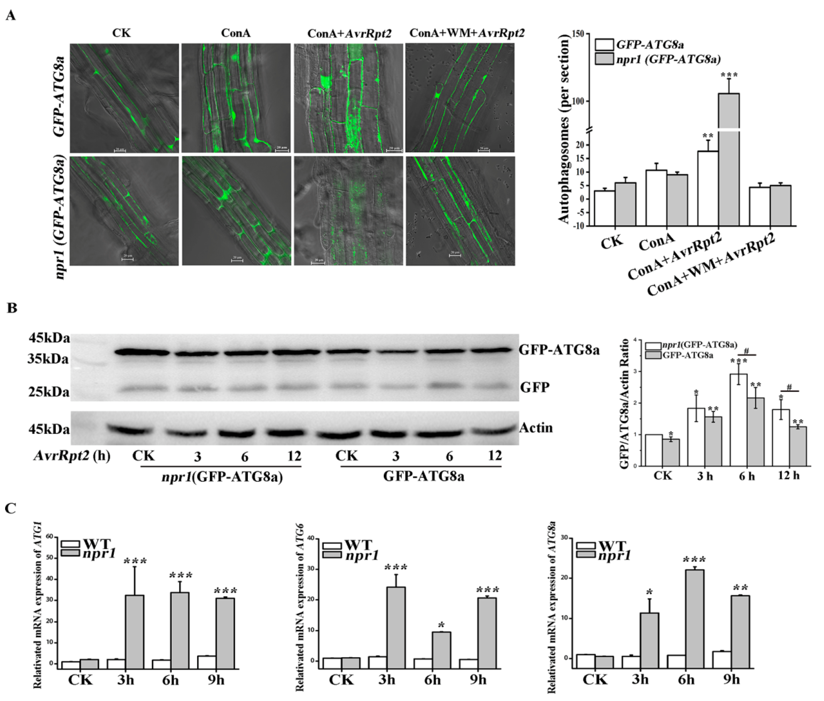
2.1. NPR1 Inhibited AvrRpt2-Induced Autophagy

First, we checked the autophagic vesicle formation directly in the GFP-ATG8a and npr1 (GFP- ATG8a) with concanamycin A (ConA) and wortmannin (WM) by confocal microscopy under AvrRpt2 infection (Figure 1A). Autophagic bodies were accumulated in the central vacuole of GFP-ATG8a cells upon AvrRpt2 + ConA infection for 3 h. These puncta were more evident in equally treated npr1 (GFP-ATG8a) cells. Then we used 8.95 μM WM to block autophagy effectively in Arabidopsis [17]. As Figure 1A showed, WM blocked the formation of autophagic bodies in GFP-ATG8a and npr1 (GFP-ATG8a) under AvrRpt2 infection as most of the fluorescence remaining diffuse within the cytosol (Figure 1A). This result suggested that the absence of NPR1 affect the formation of autophagosomes. Western blot technology was used to assess the release of autophagy flux (free GFP) and detect the degradation of GFP-ATG8a (Figure 1B). ATG8 proteins are lipidated with phosphatidylethanolamine (PE) to initiate autophagosome formation in autophagy process, and the outer membrane of the autophagosome subsequently fuses with the vacuolar membrane to transport the contents of the autophagic bodies into the vacuole, where GFP-ATG8a degraded to release a free, relatively stable GFP. Therefore, the levels of free GFP reflect the rate of autophagy [18]. The result showed that the level of free GFP in GFP-ATG8a and npr1 (GFP-ATG8a) increased with the time of AvrRpt2 infection and both reached the maximum at 6 h, but decreased significantly at 12 h. The level of free GFP in each npr1 (GFP-ATG8a) group was higher than that in wild-type (WT) group (Figure 1B). These results showed that AvrRpt2 induced the production of autophagosome and NPR1 inhibited the autophagy flux.
The core machinery of autophagy could be broken down into three functional units: ATG1-ATG13 comprising the kinase complex, an upstream regulator that initiates autophagosome formation; The ATG9 and ATG6/vps30 complexes are involved in vacuolar protein sorting and boosting phagophore expansion; Ubiquitin like conjugation systems (ATG5-ATG12 complex and ATG8-PE complex) are essential for autophagosome formation [19]. The expression of ATG1, ATG6 and ATG8a in WT and npr1 were examined by qRT-PCR (Figure 1C). Compared with WT, the result showed that the expression of ATG1 increased at 3 h and then no longer changed in npr1 with AvrRpt2 treated for 9 h; the expression of ATG6 in npr1 increased at 3 h, decreased rapidly at 6 h, then gradually increased again at 9 h; the expression of ATG8a gradually increased until 6 h, then decreased at 9 h in npr1. In summary, these results suggested that NPR1 inhibits the mRNA expression of ATG1, ATG6 and ATG8a during AvrRpt2 infections.

2.2. EDS1 Was Up-Regulated Under AvrRpt2 Infection

ATG4 is a requisite factor in the ATG8 conjugation system. To investigate the autophagy induced by AvrRpt2 and new function of ATG4, we performed transcriptome sequencing and analyzed on BMKCloud (www.biocloud.net) in WT and atg4a4b (ATG4 deletion mutant) under AvrRpt2 infection for 12 h. The number R2, which represent that means the square of the Pearson coefficient, was larger than 0.85 for both the tested samples (Figure 2A). It demonstrated the experiment’s reliability and its usefulness in revealing differences in gene expression between samples. In total, 3518 and 2344 expressing genes were identified in the WT and atg4a4b under AvrRpt2 infection, respectively. Among those, 977 genes were specific to the atg4a4b, whereas 2151 genes were specifically expressing in the control WT as demonstrated in the Venn diagram (Figure 2B). Kyoto Encyclopedia of Genes and Genomes (KEGG) pathway was used to reveal the differences in metabolic pathways. Each point in the Figure 2C represented a KEGG pathway. The path names were shown on the left axis. The abscissa was the enrichment factor, which represented the ratio of the proportion of genes differentially annotated to the pathway to the proportion of genes annotated to the pathway. The larger the enrichment factor, the more reliable the significance of the enrichment of differential genes in this pathway. According to the result, it had showed significant differences in the signal pathways of interaction between plants and pathogens, and plant hormone signal transduction when WT and atg4a4b infected with AvrRpt2. Then, the partly KEGG pathway was further analyzed. When WT was infected with AvrRpt2, its downstream factors salicylic acid glucosyltransferase 1 (SGT1) (Log2FC 1.6187) and heat shock protein 90 (HSP90) (Log2FC 1.0307) that associated with plant resistance showed red, which meant the expression was up-regulated. Surprisingly, the downstream factor EDS1 (Log2FC 1.2281) in response to AvrRps4 that triggered TIR-NB-LRR to induce autophagy also showed red. However, in atg4a4b, all the responding genes had no change under AvrRpt2 infection (Figure 2D). In order to strengthen the conclusion, the mRNA expression of EDS1 was checked in WT and atg4a4b (Figure 2E). The result showed that the expression of EDS1 increased in WT but remained unchanged in atg4a4b under AvrRpt2 infection for 12 h. The above results showed that EDS1 played a key role to autophagy induced by AvrRpt2, and ATG4 maybe had new functions of inhibiting the expression of SGT1, HSP90 and EDS1.

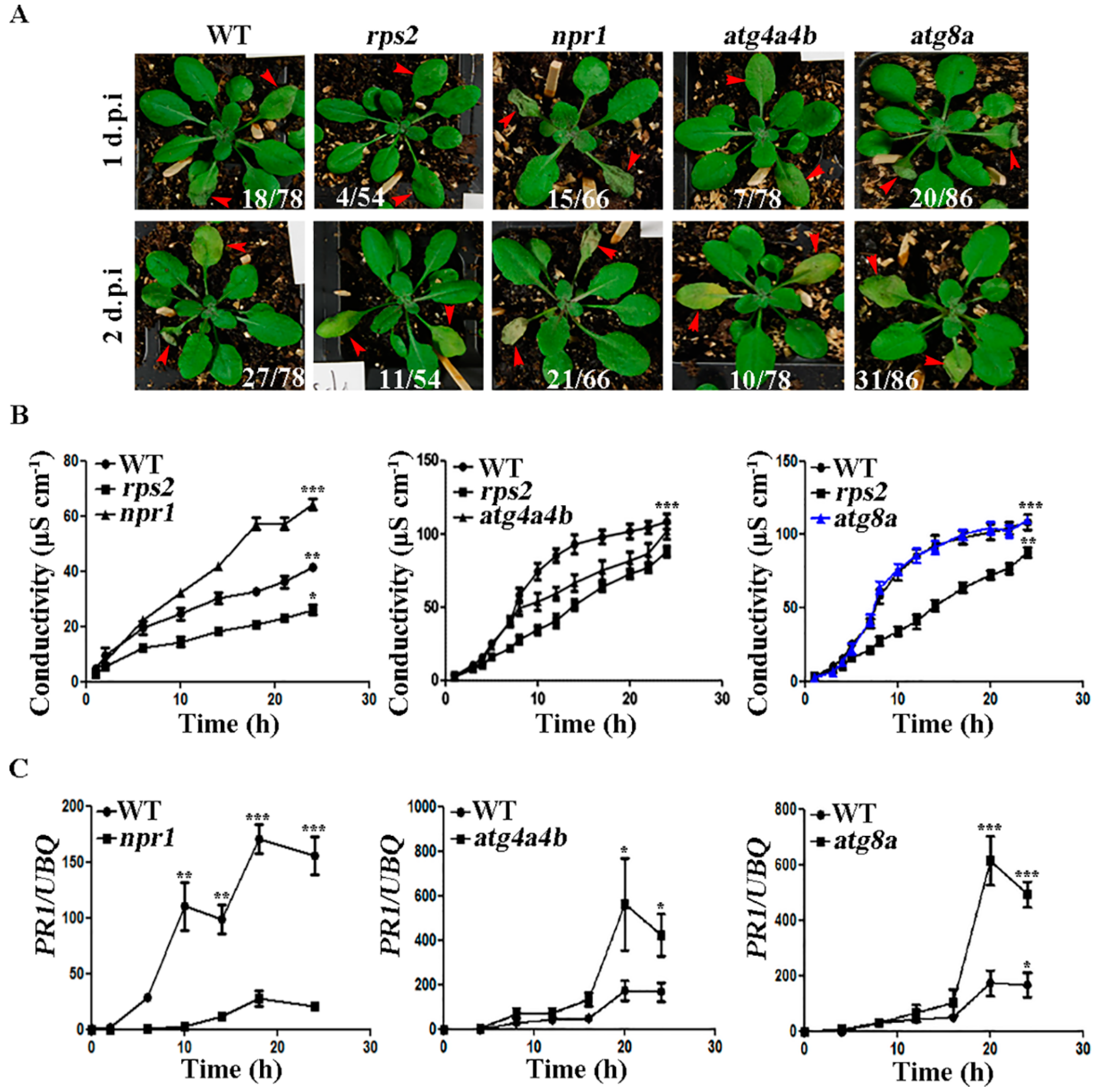
2.3. ATG4 Inhibited the Occurrence of HR during AvrRpt2 Infection

To further study the function of ATG4 in AvrRpt2-induced autophagy, we did phenotypic experiments for intuitive exploration (Figure 3A). rps2 was used as a negative control, which fails to recognize the AvrRpt2 effector [20]. At 1 d.p.i of AvrRpt2 infection, rps2 and atg4a4b had no obvious plaques, while the leaves of npr1 and atg8a (ATG8a deletion mutant) shrunk. At 2 d.p.i of AvrRpt2 infection, the leaves of rps2 and atg4a4b were only yellowed, while the leaves of npr1 and atg8a were especially degraded. Numbers are ratios of leaves with HR phenotype from by repeating the experiment independently (Supplementary Data Figure S1). WT served as a positive control and rps2 served as a negative control in ion leakage assay. The results showed that npr1 had significantly higher ion concentration than WT and rps2; and atg4a4b had lower ion concentration than WT, but still higher than rps2; the trend of ion concentration in atg8a was consistent with WT (Figure 3B). Next, the expression of pathogenesis-related 1 (PR1) in npr1, atg4a4b and atg8a were examined (Figure 3C). The results showed that mRNA expression levels of PR1 in atg4a4b and atg8a were higher in comparison to WT, indicating that ATG4 and ATG8a inhibited PR1 mRNA expression. The above results indicated that ATG4 inhibited the occurrence of HR during AvrRpt2 infection.

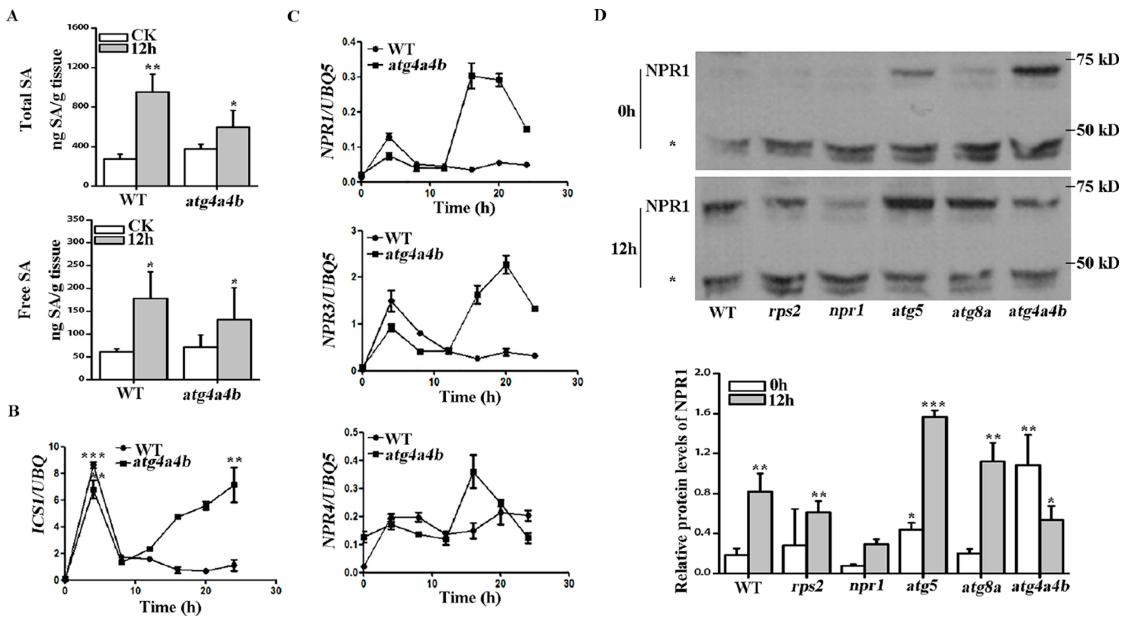
2.4. ATG4 Inhibited SA Consumption during AvrRpt2 Induced Autophagy-Dependent HR
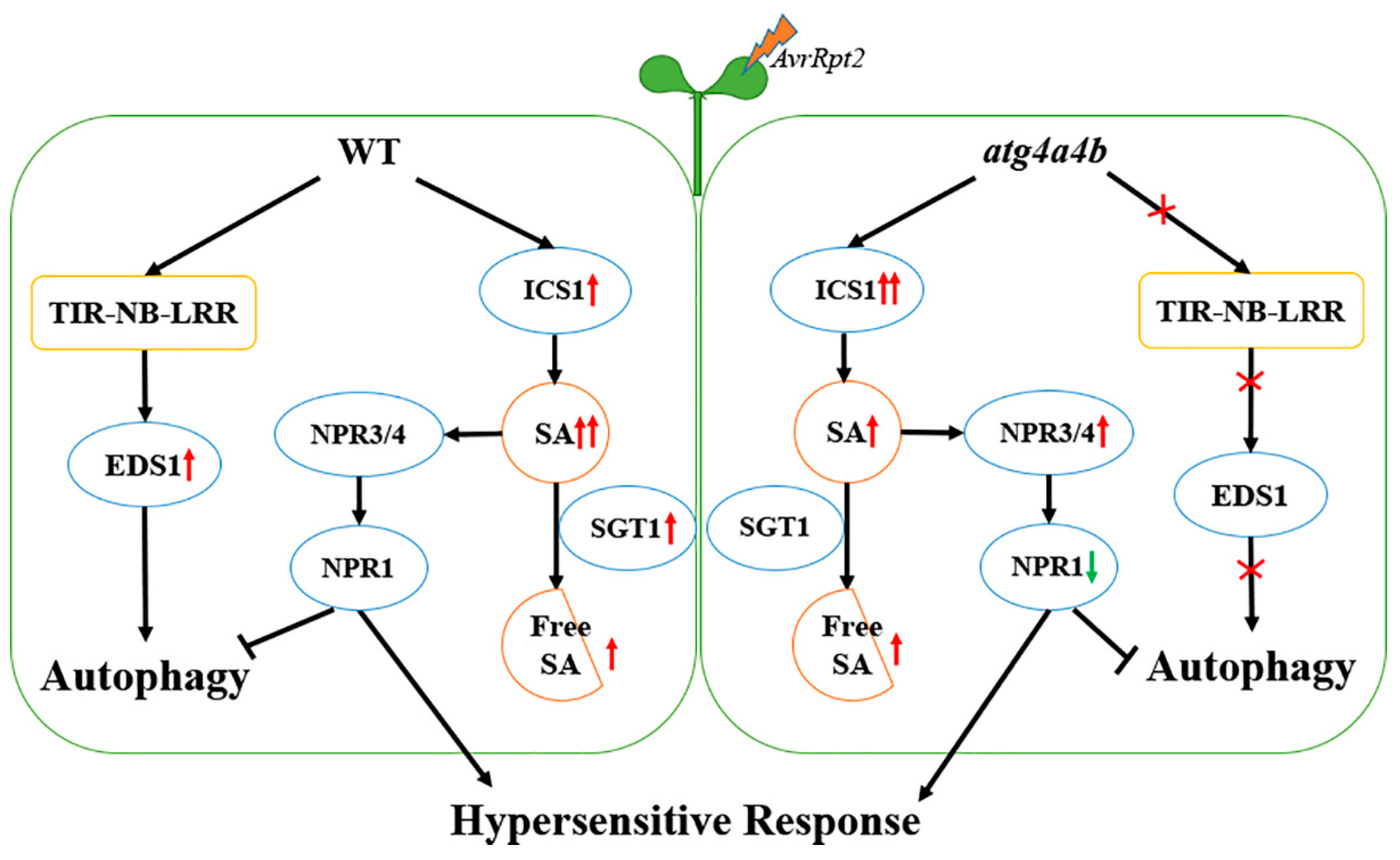
We originally reported that SA-associated EDS1 and SGT1 expression was significantly up-regulated under AvrRpt2 infection (Figure 2D). Firstly, contents of total SA and free SA in WT and atg4a4b with AvrRpt2 infection for 12 h were tested. The results showed that the absence of ATG4 did not cause significant changes in SA content. After AvrRpt2 infection, the total SA and free SA content in WT and atg4a4b were increased and the total SA of WT increased significantly more than that in atg4a4b (Figure 4A). ICS1 was the key enzyme for SA synthesis and these results prompted us to check the expression of ICS1 to further study. The results in Figure 4B showed that the expression of ICS1 had an increasing expression in WT and atg4a4b at the early stages of infection, which was more obviously in WT. After reaching the highest expression, both of them decline until 8 h. Then, WT declined slowly and finally reached stability, while atg4a4b continued to increase until it reached the highest expression level. NPR1, NPR3 and NPR4 are receptors for SA [12] and their gene expression was tested in WT and atg4a4b. The results of Figure 4C showed that the background expression level of NPR4 in atg4a4b was higher than that in WT. The expression of NPR1, NPR3 and NPR4 in WT and atg4a4b increased, and then declined gradually with AvrRpt2 infection time at the initial stage of infection. At 12 h, there was no difference in the expression of NPRs in WT and atg4a4b. NPR3 showed the highest expression and NPR4 was higher than NPR1. After 12 h, the expression of NPR1, NPR3 and NPR4 in atg4a4b rose again with the increase of infection time, then decline after reaching the highest expression level; while the expression of NPR1 and NPR3 in WT did not increase with the infection time prolonging, the expression of NPR4 continued to increase with the infection time. In addition, NPR1 protein expression was tested by using NPR1 antibody. The NPR1 level was determined on the basis of the ratio of the NPR1 band intensity to that of the non-specific band (asterisk) [16]. We unexpectedly found that NPR1 protein was highly expressed in atg4a4b. With 12 h of AvrRpt2 infection, the NPR1 protein expression level increased significantly in WT, rps2, atg5 and atg8a, while decreased significantly in atg4a4b (Figure 4D).
The mRNA expression of ICS1 in atg4a4b increased, while total SA content decreased and free SA content unchanged when compared with WT at 12 h of AvrRpt2 infection (Figure 4A,B), suggesting that ATG4 inhibited free SA consumption when responded to pathogen infection. Collectively, these results suggested that ATG4 inhibited the consumption of free SA, which promoted the interaction between NPR3 and NPR4 to accelerate the degradation of NPR1 during AvrRpt2 induced autophagy-dependent HR.

3. Discussion

Plants have evolved a multilayer immune system to recognize and respond to invading pathogens. The first layer includes pattern recognition receptors (PRRs) that detects conserved pathogen-associated molecular patterns (PAMPs) and initiates plant immune response by the name of PAMP-triggered immunity (PTI) [21]. Another layer uses resistance (R) proteins to directly or indirectly identify effectors of pathogen called ETI, then it triggers defense response rapidly that often includes a localized PCD reaction known as HR [22]. The most prevalent type of plant R proteins belongs to the NB-LRR class that can be further separated into TIR-NB-LRR associate with EDS1 and CC-NB-LRR R proteins [6].
AvrRpt2 recognized by the CC-NB-LRR type R protein RPS2 and was considered to trigger hypersensitive cell death that was strictly NDR1-dependent but autophagy-independent [6,23]. For the first time, we found that AvrRpt2 induced the production of autophagosomes and autophagy flux, which was inhibited by NPR1 (Figure 1A,B). In addition, NPR1 inhibited the mRNA expression of ATG1, ATG6 and ATG8a in the early stages of HR (Figure 1C). These results indicated that the production of autophagosomes was induced by AvrRpt2 and was suppressed by NPR1. The mRNA expression of other ATG genes in WT, such as the mRNA expression of ATG4a and ATG4b decreased and then rise; the mRNA expression of ATG5 and ATG12a decreased with the infection time of AvrRpt2 (data not shown); these are very interesting. Previous work showed that AvrRpt2-induced the expression of several primary jasmonic acid (JA)-responsive genes and JA is a positive regulator of RPS2-mediated ETI [24]. We speculated that AvrRpt2-induced autophagy maybe involve in other key hormone signals (for example JA) besides SA signal, which needed further study.
ATG4 is the only protease among dozens of ATG proteins. It also serves as a requisite factor in the ATG8 conjugation system: one of the unique mechanisms in autophagy [25,26]. Then, we further explored the mechanism with transcriptome analyses in WT and atg4a4b. We found that the expression of EDS1, SGT1 and HSP90 was up-regulated in WT, while there was no change in atg4a4b (Figure 2B,C). Similar result was showed in the mRNA expression of EDS1 with qRT-PCR (Figure 2E), suggesting that atg4a4b led to a concomitant EDS1 blockage. Therefore, the up-regulation of EDS1 in WT was conducted by the R protein TIR-NB-LRR, which leaded to the occurrence of autophagy during AvrRpt2 infection.
Moreover, SGT1 and HSP90 as chaperone proteins are required for plant disease resistance and involved in SA metabolism [27,28,29]. Glucosylation inactivates SA and allows vacuolar storage of relatively large quantities of SA due to reduced toxicity. SGT1 catalyzes the conversion of free SA to salicylic acid 2-O-β-D-glucose (SAG) [11]. The results in Figure 2B,C showed that SGT1 increased in WT, but remained unchanged in atg4a4b. The results in Figure 4b showed that the expression of ICS1 in atg4a4b was higher than that in WT, indicating that the free SA in atg4a4b should be more than that in WT. Actually, total SA content in atg4a4b was lower than that in WT, while the free SA didn’t change between WT and atg4a4b after AvrRpt2 infected for 12 h, we speculated that free SA in atg4a4b was consumed. In the early stage of HR induced by AvrRpt2, a large amount of SA was consumed in atg4a4b, resulting in a low concentration of free SA, which promoted the interaction of NPR3 and NPR1 [16]. The high mRNA expression of NPR3 was detected in Figure 4C, leading the actual expression of NPR1 decreased after 12h infection.
Therefore, these results showed that AvrRpt2 induced autophagy also via EDS1 pathway. In the early stage of HR caused by AvrRpt2, ATG4 may suppress NPR3 synthesis via inhibiting the consumption of free SA and promote the expression of NPR1 (Figure 5). In the future, we will continue to explore the interaction between ATG4 and NPRs.

4. Materials and Methods

4.1. Plant Materials and Chemical Treatment

Seeds were cultivated in soil culture in growth cabinets at 22 °C (day) and 18 °C (night), with a 16 h light period (120 μmol m−2 s−1) and 82% relative humidity for 2–4 weeks. The Arabidopsis thaliana mutants (in ecotype Col-0), rps2, ATG8a-lacking mutant (atg8a) and NPR1-lacking mutant (npr1) were provided by Dr. Xinnian Dong (Duke University, NC, USA). Transgenic GFP-ATG8a (Col-0 background) was donated by Dr. Kohki Yoshimoto of the Plant Science Center of Japan. npr1 (GFP-ATG8a) was produced by crossing npr1 and GFP-ATG8a. The atg4a4b was produced by crossing atg4a (SALK_085300) and atg4b (SALK_056994). atg8a (SALK_045344) was obtained from the Arabidopsis Biological Resource Center. SA was purchased from Sigma-Aldrich, China (S5922-100G; 239763-5GM-M, Shanghai, China).

4.2. Pathogen Growth and Inoculation

The bacterial used in this study was Psm ES4326/AvrRpt2 and was grown at 28 °C in King’s B medium containing 50 mg/L streptomycin and 10 mg/L tetracycline. Overnight log-phase cultures were collected by centrifugation, washed with 10 mM MgCl2, and then diluted to a final optical density of 0.02 at 600 nm (OD600).

4.3. SA Measurement

Following the previous procedure [30,31], 4-weeks-old plants leaves were used to measure the SA levels. High-performance liquid chromatography (HPLC) with fluorescence detectors (HPLC, Shimadzu LC-6A, Japan) was used to analyze total extracted SA and free SA, at a 294 nm excitation wavelength and a 426 nm emission wavelength.

4.4. Total RNA Extraction and Quantitative Reverse Transcription-PCR (qRT-PCR)

Total RNAs were extracted from Arabidopsis leaves at indicated times after the treatment of TRI reagent according to the manufacturer’s instruction (Invitrogen, Carlsbad, CA, USA). The first-strand complementary DNA was synthesized from total RNA using a Reverse-iT first-strand synthesis kit (Perfect Real Time, RR047Q, TaKaRa, Dalian, China). qPCR was performed using the ChamQ SYBR qPCR Master Mix (Low ROX Premixed, Q331-02/03, Vazyme Biotech Co., Ltd., Nanjing, China) on ABI Life QuantStudio 6. The thermal cycles of qPCR were initial denaturation at 95 °C for 30 s, followed by 40 cycles by 40 cycles at 95 °C for 5 s and 60 °C for 34 s. Ubiquitin 5 and AtACTIN2 were used as internal control. The primers used are listed in Supplementary Data Table S1.

4.5. Protein Extraction

Amounts of 0.4 g leaves were ground in liquid nitrogen and the powder was resuspended in 1 mL ice-cold extraction buffer comprised of 50 mM Tris-HCl (pH 7.5), 150 mM NaCl, 5 mM EDTA, 0.2% (w/v) Triton X-100, 0.2% (w/v) Nonidet P-40 and 1% (w/v) phenylmethanesulfonyl fluoride (PMSF). To extract NPR1, add 40 µM MG115, 1% β-ME, 500× protease inhibitor cocktail (PMSF not included) and 5000× phosphatase inhibitor cocktail to the protein extraction buffer. The extracts were centrifuged and the protein concentration of the supernatant was determined with a Bio-Rad protein assay.

4.6. Western Blotting

Total proteins were extracted from leaves at the indicated time points after different treatments with the protocol of Karppinen [32]. The antibodies used for western blotting included actin (Engibody Biotechnology, AT0004, WB: 1:3000, Dover, DE 19901, USA) and GFP (JL-8 Monoclonal Antibody, A-6455, WB: 1:5000, Fisher, Invitrogen, Waltham, MA, USA). Detection was performed using a LI-COR Odyssey Infrared Imaging System (LI-COR, Inc., Lincoln, NE, USA). NPR1 protein expression was tested by using NPR1 antibody. The NPR1 level was determined on the basis of the ratio of the NPR1 band intensity to that of the non-specific band (asterisk) [16].

4.7. Confocal Microscopy

GFP-ATG8a and npr1 (GFP-ATG8a) seedlings (7 days old) were treated with MgCl2 or 1 μm concanamycin A (ConA) (Invitrogen, Waltham, MA, USA) or ConA + Psm ES4326/AvrRpt2 or ConA + 8.95 μm wortmannin (WM) (19545-26-7, MCE, NJ, USA) + Psm ES4326/AvrRpt2 for 3 h. Root epidermal cells below the cotyledon were imaged using a Zeiss LSM880 confocal laser scanning microscope. GFP fluorescence was excited by a 488 nm argon laser and detected at 505–550 nm by a photomultiplier detector. At least 10 sets of images were obtained for quantification analysis. GFP was counted per section according to 10 sets of images field of vision.

4.8. Ion Leakage

Ion leakage assay was performed as previously described [33]. The leaves of 4-week-old WT, rps2, npr1, atg4a4b and atg8a plants were infiltrated with Psm ES4326/AvrRpt2, and 6 leaf discs (8 mm diameter) were removed rapidly following infection and washed in 50 mL ddH2O (twice). After 10 min, we removed the wash water and replaced it with 15 mL of ddH2O. Ion leakage was then measured over time.

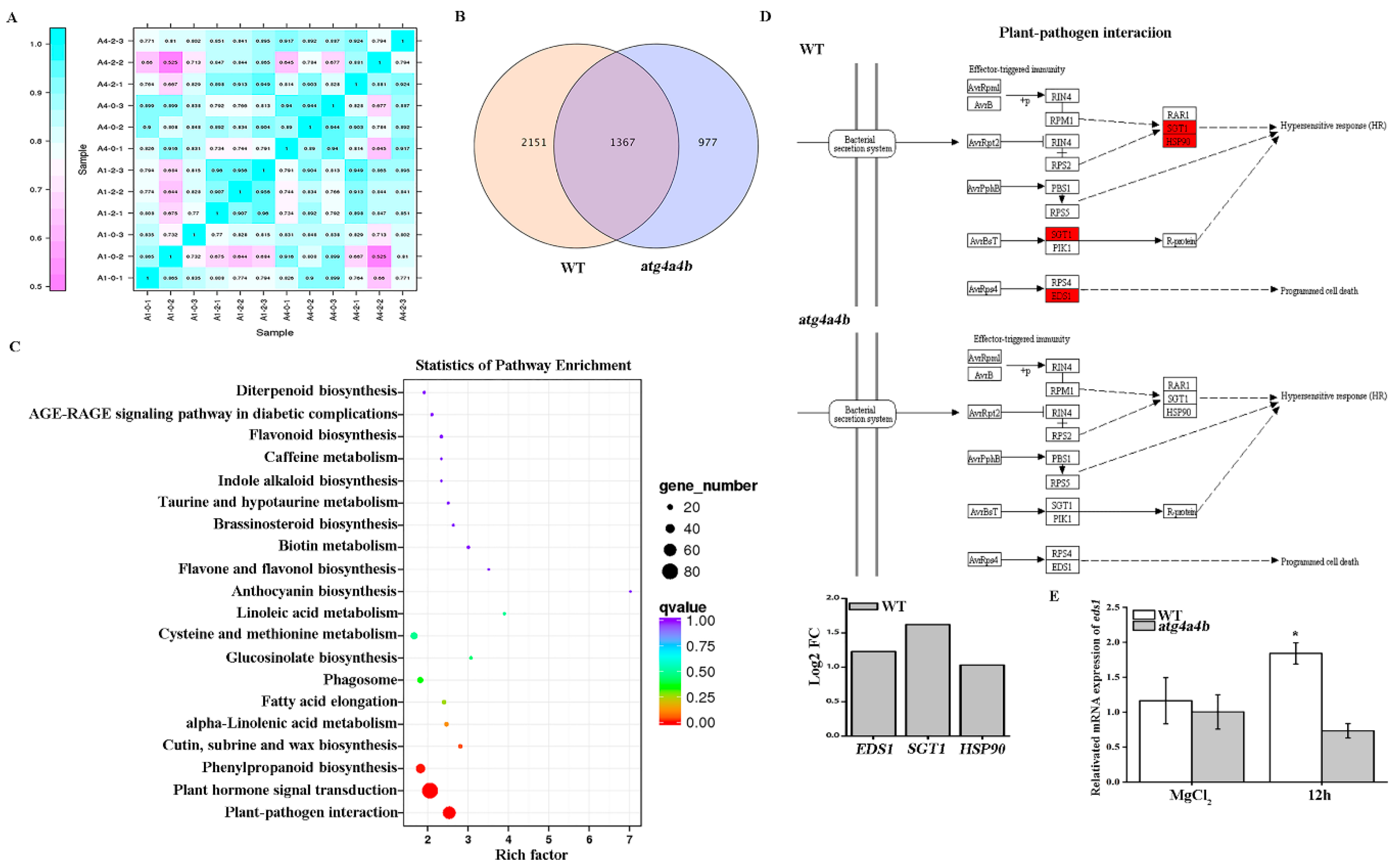
4.9. Transcriptome Analysis

WT and atg4a4b were infected by Psm ES4326/AvrRpt2 for 12 h, and then their samples were collected by taking 8–10 real leaves from three different culture pots as three biological replicates. Total RNA was extracted by TRIzol Reagent (Invitrogen, Thermo Fisher Scientific, Shanghai, China). RNA quality was determined using Agilent 2100 Bioanalyzer (Agilent Technologies Canada Inc., Mississauga, ON, Canada). RNA libraries were constructed from 2 lg of total RNA and subjected to deep sequencing at an Illumina Hiseq 2500 platform (BioMarker Technologies Illumina, Inc., Shanghai, China).

Supplementary Materials

Supplementary materials can be found at https://0-www-mdpi-com.brum.beds.ac.uk/1422-0067/21/14/5147/s1. Figure S1: phenotypes of WT, rps2, npr1, atg4a4b and atg8a after Psm ES4326/avrRpt2 infiltration for 1 or 2 days. Table S1: Primers for several genes.

Author Contributions

W.G. and W.C. designed the research. W.G., B.L. and W.C. performed the research. W.G., B.Z. and W.C. analyzed the data and prepared figures. W.G. wrote the manuscript in consultation with W.C., W.G. and B.Z. review & editing. W.C. contributed reagents/materials/analysis tools. All authors have read and agreed to the published version of the manuscript.

Funding

This research was funded by the National Natural Science Foundation of China (Grants 31570256) China Normal University, and a grant from the science and technology project of Guangzhou (Grant No. 201805010002).

Acknowledgments

We are grateful to Xinnian Dong (Duke University, USA), Alan M. Jones (University of North Carolina, Chapel Hill, USA), Hengming Ke (University of North Carolina, Chapel Hill, USA) for their help and input, allowing us to complete the experiments in this paper. We thank Zheng Qing Fu, Shunping Yan, and Abdelaty Saleh in Xinnian Dong’s laboratory for their help and input. We thank Kohki Yoshimoto (RIKEN, Plant Science Center, Japan) and Richard D. Vierstra (University of Wisconsin, USA) for kindly providing transgenic GFP-ATG8a. We thank Chengqian Zhou master of Boston University College of Engineering for English editing.

Conflicts of Interest

The authors declare no conflict of interest.

Abbreviations

NPR1nonexpressor of PR genes 1
EDS1enhanced disease susceptibility 1
SGT1salicylic acid glucosyltransferase 1
SAGsalicylic acid 2-O-β-D-glucose
ICS1isochorismate synthase 1
NDR1non-race specific disease resistance
PALphenylalanine ammonia lyase
PRpathogenesis-related
SAsalicylic acid
JAjasmonic acid
TIR-NB-LRRToll/Interleukin-1 receptor (TIR)-nucleotide binding site (NB)-leucine rich repeat (LRR)
CC-NB-LRRcoiled-coil (CC)-nucleotide binding site (NB)-leucine rich repeat (LRR)
PCDprogrammed cell death
HRhypersensitive response
PAMPpathogen-associated molecular patterns
ETIeffector-triggered immunity
PTIPAMP-triggered immunity
PRS2resistant to P. syringae 2
PRRspattern recognition receptors
KEGGKyoto encyclopedia of genes and genomes
WMwortmannin
ConAconcanamycin A

References

  1. Mizushima, N.; Komatsu, M. Autophagy: Renovation of Cells and Tissues. Cell 2011, 147, 728–741. [Google Scholar] [CrossRef] [Green Version]
  2. Hofius, D.; Lie, L.; Hafren, A.; Coll, N.S. Autophagy as an emerging arena for plant-pathogen interactions. Curr. Opin. Plant Biol. 2017, 38, 117–123. [Google Scholar] [CrossRef]
  3. Il Kwon, S.; Park, O.K. Autophagy in Plants. J. Plant Biol. 2008, 51, 313–320. [Google Scholar] [CrossRef]
  4. Bisgrove, S.R.; Simonich, M.T.; Smith, N.M.; Sattler, A.; Innes, R.W. A disease resistance gene in Arabidopsis with specificity for two different pathogen avirulence genes. Plant. Cell 1994, 6, 927–933. [Google Scholar] [CrossRef] [Green Version]
  5. Hatsugai, N.; Igarashi, D.; Mase, K.; Lu, Y.; Tsuda, Y.; Chakravarthy, S.; Wei, H.L.; Foley, J.W.; Collmer, A.; Glazebrook, J.; et al. A plant effector-triggered immunity signaling sector is inhibited by pattern-triggered immunity. Embo J. 2017, 36, 2758–2769. [Google Scholar] [CrossRef] [PubMed]
  6. Hofius, D.; Schultz-Larsen, T.; Joensen, J.; Tsitsigiannis, D.I.; Petersen, N.H.T.; Mattsson, O.; Jorgensen, L.B.; Jones, J.D.G.; Mundy, J.; Petersen, M. Autophagic Components Contribute to Hypersensitive Cell Death in Arabidopsis. Cell 2009, 137, 773–783. [Google Scholar] [CrossRef] [PubMed] [Green Version]
  7. Gao, Y.Y.; Wang, X.D.; Ma, C.; Chen, W.L. EDS1-mediated activation of autophagy regulates Pst DC3000 (AvrRps4)-induced programmed cell death in Arabidopsis. Acta Physiol. Plant. 2016, 38. [Google Scholar] [CrossRef]
  8. Betsuyaku, S.; Katou, S.; Takebayashi, Y.; Sakakibara, H.; Nomura, N.; Fukuda, H. Salicylic Acid and Jasmonic Acid Pathways are Activated in Spatially Different Domains Around the Infection Site During Effector-Triggered Immunity in Arabidopsis thaliana. Plant Cell Physiol. 2018, 59, 8–16. [Google Scholar] [CrossRef]
  9. Wildermuth, M.C.; Dewdney, J.; Wu, G.; Ausubel, F.M. Isochorismate synthase is required to synthesize salicylic acid for plant defence. Nature 2001, 414, 562–565. [Google Scholar] [CrossRef]
  10. Sendon, P.M.; Seo, H.S.; Song, J.T. Salicylic Acid Signaling: Biosynthesis, Metabolism, and Crosstalk with Jasmonic Acid. J. Korean Soc. Appl. Biol. Chem. 2011, 54, 501–506. [Google Scholar] [CrossRef]
  11. Song, J.T.; Koo, Y.J.; Seo, H.S.; Kim, M.C.; Choi, Y.D.; Kim, J.H. Overexpression of AtSGT1, an Arabidopsis salicylic acid glucosyltransferase, leads to increased susceptibility to Pseudomonas syringae. Phytochemistry 2008, 69, 1128–1134. [Google Scholar] [CrossRef] [PubMed]
  12. Tan, S.T.; Abas, M.; Verstraeten, I.; Glanc, M.S.; Molnar, G.; Hajny, J.; Lasak, P.; Petrik, I.; Russinova, E.; Petrasek, J.; et al. Salicylic Acid Targets Protein Phosphatase 2A to Attenuate Growth in Plants. Curr. Biol. 2020, 30, 381–395. [Google Scholar] [CrossRef] [PubMed] [Green Version]
  13. Fu, Z.Q.; Dong, X.N. Systemic Acquired Resistance: Turning Local Infection into Global Defense. Annu. Rev. Plant Biol. 2013, 64, 839–863. [Google Scholar] [CrossRef] [PubMed] [Green Version]
  14. Li, W.W.; Zhao, D.; Dong, J.W.; Kong, X.L.; Zhang, Q.; Li, T.T.; Meng, Y.L.; Shan, W.X. AtRTP5 negatively regulates plant resistance to Phytophthora pathogens by modulating the biosynthesis of endogenous jasmonic acid and salicylic acid. Mol. Plant Pathol. 2019. [Google Scholar] [CrossRef] [PubMed] [Green Version]
  15. Yoshimoto, K.; Jikumaru, Y.; Kamiya, Y.; Kusano, M.; Consonni, C.; Panstruga, R.; Ohsumi, Y.; Shirasu, K. Autophagy Negatively Regulates Cell Death by Controlling NPR1-Dependent Salicylic Acid Signaling during Senescence and the Innate Immune Response in Arabidopsis. Plant Cell 2009, 21, 2914–2927. [Google Scholar] [CrossRef] [Green Version]
  16. Fu, Z.Q.; Yan, S.; Saleh, A.; Wang, W.; Ruble, J.; Oka, N.; Mohan, R.; Spoel, S.H.; Tada, Y.; Zheng, N.; et al. NPR3 and NPR4 are receptors for the immune signal salicylic acid in plants. Nature 2012, 486, 228–232. [Google Scholar] [CrossRef] [Green Version]
  17. Zhuang, X.H.; Wang, H.; Lam, S.K.; Gao, C.J.; Wang, X.F.; Cai, Y.; Jiang, L.W. A BAR-Domain Protein SH3P2, Which Binds to Phosphatidylinositol 3-Phosphate and ATG8, Regulates Autophagosome Formation in Arabidopsis. Plant Cell 2013, 25, 4596–4615. [Google Scholar] [CrossRef] [Green Version]
  18. Huang, L.; Yu, L.J.; Zhang, X.; Fan, B.; Wang, F.Z.; Dai, Y.S.; Qi, H.; Zhou, Y.; Xie, L.J.; Xiao, S. Autophagy regulates glucose-mediated root meristem activity by modulating ROS production in Arabidopsis. Autophagy 2019, 15, 407–422. [Google Scholar] [CrossRef] [Green Version]
  19. Wang, W.Y.; Xu, M.Y.; Wang, G.P.; Galili, G. Autophagy: An Important Biological Process That Protects Plants from Stressful Environments. Front. Plant Sci. 2017, 7. [Google Scholar] [CrossRef] [Green Version]
  20. Yoo, H.; Greene, G.H.; Yuan, M.; Xu, G.Y.; Burton, D.; Liu, L.J.; Marques, J.; Dong, X.N. Translational Regulation of Metabolic Dynamics during Effector-Triggered Immunity. Mol. Plant 2020, 13, 88–98. [Google Scholar] [CrossRef] [Green Version]
  21. Hartmann, M.; Zeier, J. N-hydroxypipecolic acid and salicylic acid: A metabolic duo for systemic acquired resistance. Curr. Opin. Plant Biol. 2019, 50, 44–57. [Google Scholar] [CrossRef] [PubMed]
  22. Romera, F.J.; Garcia, M.J.; Lucena, C.; Martinez-Medina, A.; Aparicio, M.A.; Ramos, J.; Alcantara, E.; Angulo, M.; Perez-Vicente, R. Induced Systemic Resistance (ISR) and Fe Deficiency Responses in Dicot Plants. Front. Plant Sci. 2019, 10. [Google Scholar] [CrossRef] [PubMed]
  23. Mackey, D.; Belkhadir, Y.; Alonso, J.M.; Ecker, J.R.; Dangl, J.L. Arabidopsis RIN4 is a target of the type III virulence effector AvrRpt2 and modulates RPS2-mediated resistance. Cell 2003, 112, 379–389. [Google Scholar] [CrossRef] [Green Version]
  24. Liu, L.J.; Sonbol, F.M.; Huot, B.; Gu, Y.N.; Withers, J.; Mwimba, M.; Yao, J.; He, S.Y.; Dong, X.N. Salicylic acid receptors activate jasmonic acid signalling through a non-canonical pathway to promote effector-triggered immunity. Nat. Commun. 2016, 7. [Google Scholar] [CrossRef] [Green Version]
  25. Kirisako, T.; Ichimura, Y.; Okada, H.; Kabeya, Y.; Mizushima, N.; Yoshimori, T.; Ohsumi, M.; Takao, T.; Noda, T.; Ohsumi, Y. The reversible modification regulates the membrane-binding state of Apg8/Aut7 essential for autophagy and the cytoplasm to vacuole targeting pathway. J. Cell Biol. 2000, 151, 263–276. [Google Scholar] [CrossRef] [PubMed]
  26. Ichimura, Y.; Kirisako, T.; Takao, T.; Satomi, Y.; Shimonishi, Y.; Ishihara, N.; Mizushima, N.; Tanida, I.; Kominami, E.; Ohsumi, M.; et al. A ubiquitin-like system mediates protein lipidation. Nature 2000, 408, 488–492. [Google Scholar] [CrossRef]
  27. Wang, K.; Mysore, K.S. SGT1 is required for plant disease resistance and symptom development during disease susceptibility. Phytopathology 2006, 96, S120. [Google Scholar]
  28. Qian, L.C.; Zhao, J.P.; Du, Y.M.; Zhao, X.J.; Han, M.; Liu, Y.L. Hsp90 Interacts With Tm-2(2) and Is Essential for Tm-2(2)-Mediated Resistance to Tobacco mosaic virus. Front. Plant Sci. 2018, 9. [Google Scholar] [CrossRef] [Green Version]
  29. Oh, S.K.; Kwon, S.Y.; Choi, D. Rpi-blb2-Mediated Hypersensitive Cell Death Caused by Phytophthora infestans AVRblb2 Requires SGT1, but not EDS1, NDR1, Salicylic Acid-, Jasmonic Acid-, or Ethylene-Mediated Signaling. Plant. Pathol. J. 2014, 30, 254–260. [Google Scholar] [CrossRef]
  30. Wang, X.D.; Gao, Y.Y.; Yan, Q.Q.; Chen, W.L. Salicylic acid promotes autophagy via NPR3 and NPR4 in Arabidopsis senescence and innate immune response. Acta Physiol. Plant. 2016, 38. [Google Scholar] [CrossRef]
  31. Lv, F.; Zhou, J.; Zeng, L.; Xing, D. beta-cyclocitral upregulates salicylic acid signalling to enhance excess light acclimation in Arabidopsis. J. Exp. Bot. 2015, 66, 4719–4732. [Google Scholar] [CrossRef] [PubMed] [Green Version]
  32. Karppinen, K.; Taulavuori, E.; Hohtola, A. Optimization of protein extraction from Hypericum perforatum tissues and immunoblotting detection of Hyp-1 at different stages of leaf development. Mol. Biotechnol. 2010, 46, 219–226. [Google Scholar] [CrossRef] [PubMed]
  33. Dong, J.J.; Chen, W.L. The Role of Autophagy in Chloroplast Degradation and Chlorophagy in Immune Defenses during Pst DC3000 (AvrRps4) Infection. PLoS ONE 2013, 8, e0073091. [Google Scholar] [CrossRef] [PubMed] [Green Version]
Figure 1. Nonexpressor of PR genes 1 (NPR1) inhibited Psm ES4326/AvrRpt2 (AvrRpt2)-induced autophagy. (A) Autophagosomes formation. GFP-ATG8a and npr1 (GFP-ATG8a) treated with four groups: MgCl2; concanamycin A (ConA); ConA + AvrRpt2 and ConA + wortmannin (WM) + AvrRpt2 and then examined by confocal microscopy. Scale bars, 20 µm. Numbers of puncta per section in the root cells of GFP-ATG8a or npr1 (GFP-ATG8a) seedlings in the left. n = 10 sections from three independent experiments per genotype. (B) Western Blot to detect autophagy flow in GFP-ATG8a and npr1 (GFP-ATG8a) when plants treated with AvrRpt2 at 3 h, 6 h and 12 h and quantitative analyses of GFP/GFP-ATG8a/Actin ratio. Each data is three independent replicates. Each value is the mean ± SD of three replicates. Statistically significant differences between treatments (# p < 0.05, * p < 0.05, ** p < 0.01 and *** p < 0.001). (C) Quantitative RT-PCR data showed the expression of ATG1, ATG6 and ATG8a in wild-type (WT) and npr1 after AvrRpt2 infiltration for 3 h, 6 h and 9 h. The CK group was treated with MgCl2 as control. Each data is three independent replicates. Each value is the mean ± SD of three replicates. Statistically significant differences between treatments (* p < 0.05, ** p < 0.01 and *** p < 0.001).
Ijms 21 05147 g001
Figure 2. Enhanced disease susceptibility 1 (EDS1) was up-regulated under AvrRpt2 infection. (A) Correlation between RNA-Seq samples. A 1-0-1/2/3 represent three replicates of WT, A 1-2-1/2/3 represent three replicates of WT infected with AvrRpt2, A 4-0-1/2/3 represent three replicates of atg4a4b and A 4-2-1/2/3 represent three replicates of atg4a4b infected with AvrRpt2, heat maps of the correlation coefficient between samples, the number represent R2 that means the square of the Pearson coefficient. (B) Venn diagram of expressed genes in WT and atg4a4b when infected with AvrRpt2. FPKM > 1 is the expression threshold. (C) Transcriptome analysis of Kyoto Encyclopedia of Genes and Genomes (KEGG) enrichment in WT and atg4a4b when infected with AvrRpt2 for 12 h. (D) Transcriptome analysis of KEGG pathway of plants and pathogens interaction. The red color means that the expression was up-regulated. (E) The mRNA expression of EDS1 in WT and atg4a4b. Each data is three independent replicates. Each value is the mean ± SD of three replicates. Statistically significant differences between treatments (* p < 0.05).
Ijms 21 05147 g002
Figure 3. ATG4 inhibited the occurrence of hypersensitive response (HR) during AvrRpt2 infection. (A) Phenotypes of WT, rps2, npr1, atg4a4b and atg8a after AvrRpt2 infiltration for 1 or 2 days. Numbers are ratios of leaves with HR phenotype. (B) Ion leakage assay in WT, rps2, npr1, atg4a4b and atg8a when infected with AvrRpt2. (C) Quantitative RT-PCR data showed the expression of pathogenesis-related (PR1) in WT, npr1, atg4a4b and atg8a when infected with AvrRpt2. Each value is the mean ± SD of three replicates. Statistically significant differences between treatments (* p < 0.05, ** p < 0.01 and *** p < 0.001).
Ijms 21 05147 g003
Figure 4. ATG4 inhibited salicylic acid (SA) consumption during AvrRpt2 induced autophagy-dependent HR. (A) Total SA and free SA contents in WT and atg4a4b under AvrRpt2 infection for 12 h. (B) Quantitative RT-PCR data showed the expression of isochorismate synthase 1 (ICS1) in WT and atg4a4b when infected with AvrRpt2. (C) Quantitative RT-PCR data showed the expression of NPR1, NPR3 and NPR4 in WT and atg4a4b when infected with AvrRpt2. (D) Western Blot to detect NPR1 in WT, rps2, npr1, atg5, atg8a and atg4a4b when plants treated with AvrRpt2 for 12 h and quantitative analyses of the results of NPR1 by statistical methods. The NPR1 level was determined on the basis of the ratio of the NPR1 band intensity to that of the non-specific band (asterisk). Each value is the mean ± SD of three replicates. Statistically significant differences between treatments (* p < 0.05, ** p < 0.01 and *** p < 0.001).
Ijms 21 05147 g004
Figure 5. Working model. AvrRpt2 induced autophagy-dependent hypersensitive response via EDS1. ATG4 may suppress NPR3 synthesis via inhibiting the consumption of free SA and promote the expression of NPR1. The red arrow means up-regulated and the green arrow means down-regulated. Two arrows indicate high degree.
Ijms 21 05147 g005

